method for automatic recognition of ... for automatic recognition of neoplastic lymphoid cells using...
TRANSCRIPT
METHOD FOR AUTOMATIC RECOGNITION OF
NEOPLASTIC LYMPHOID CELLS USING
PERIPHERAL BLOOD CELL IMAGES
Santiago Alférez
Anna Merino
José Rodellar

Morphology
Immunophenotype
Cytogenetic
Molecular Studies
PROGNOSIS TREATMENT
DIAGNOSIS
CLINICAL
INFORMATION
SAMPLE
DIAGNOSIS IN HEMATOLOGY

Is it possible to automatically detect blood atypical lymphoid cells through their morphological characteristics and other quantitative parameters ?
SUBJECTIVITY -
SKILLED EXPERIENCE
AUTOMATIC SYSTEMS DO NOT CLASSIFY
ATYPICAL LYMPHOID CELLS
CLINICAL DIAGNOSIS OF 80% OF HEMATOLOGICAL DISEASES IS MADE BY MORPHOLOGICAL STUDIES OF PERIPHERAL BLOOD
Design of a new methodology for the automatic classification of atypical lymphoid cells by using digital images from peripheral blood
OBJECTIVE
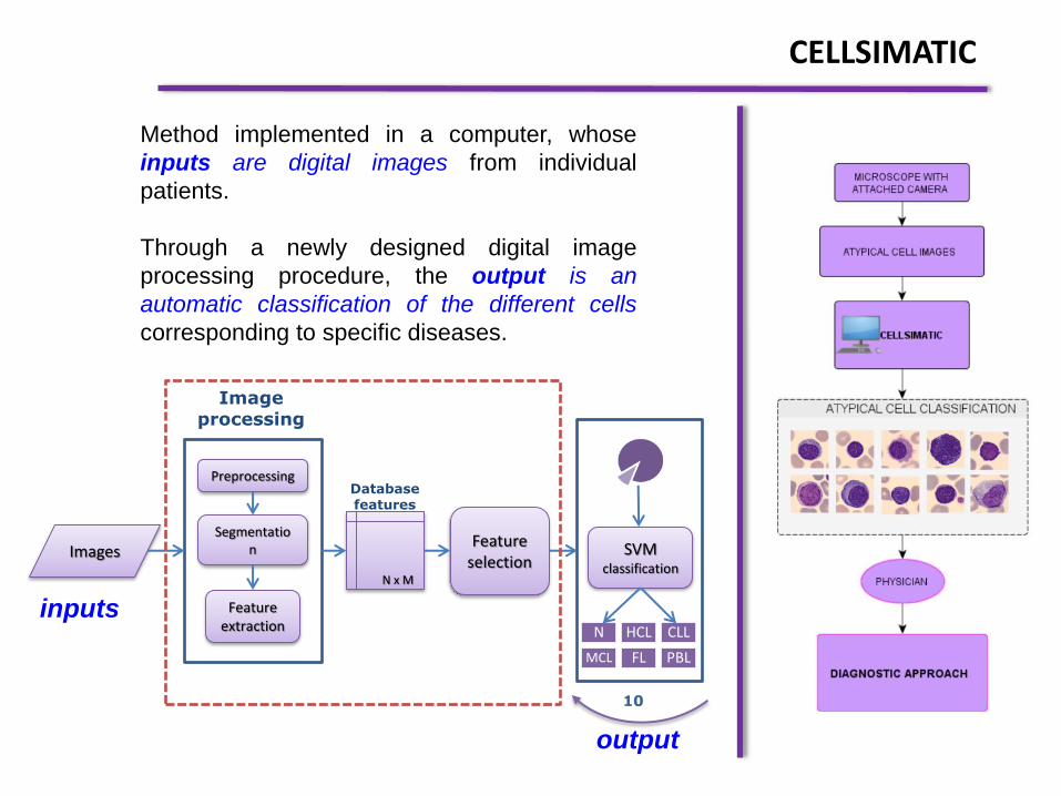
Method implemented in a computer, whose
inputs are digital images from individual
patients.
Through a newly designed digital image
processing procedure, the output is an
automatic classification of the different cells
corresponding to specific diseases.
Images
Preprocessing
Segmentation
Feature extraction
1834 x 1429
Information theory feature
selection: CMIM
SVM
classification
N HCL
CLL M
CL FL
PBL
Image processing
Database features
10
CELLSIMATIC
inputs
output
Images
Preprocessing
Segmentation
Feature extraction
N x M
Feature selection
SVM
classification
N HCL CLL
MCL FL PBL
Image processing
Database features
10

Normal lymphocytes (NL)
Chronic lymphocytic leukemia (CLL)
B Prolymphocytes (B-PLL)
Hairy cell leukemia (HCL)
Follicular lymphoma
Mantle cell lymphoma (MCL)
Reactive lymphocytes
TARGETS
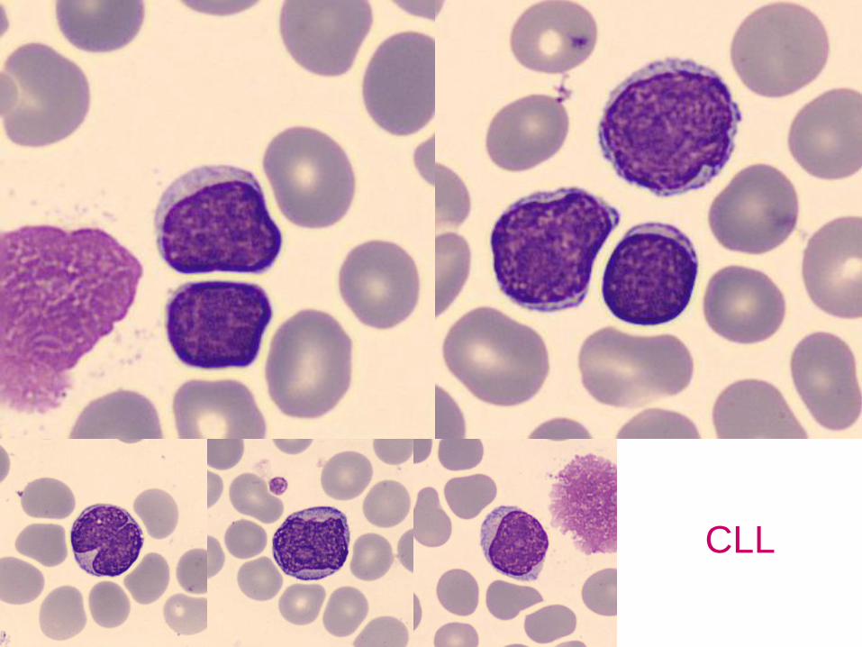
CLL

HCL
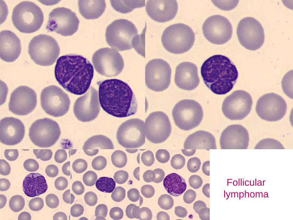
Follicular
lymphoma

MCL

B
Prolymphocytes

Reactive
lymphocytes
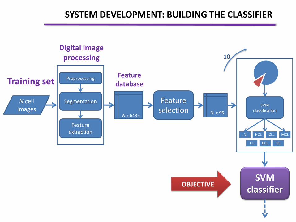
N cell images
Preprocessing
Segmentation
Feature extraction
N x 6435
Feature
selection
Digital image processing
Feature database
N x 95
SVM classification
N HCL CLL MCL
FL BPL RL
10
SVM classifier
Training set
SYSTEM DEVELOPMENT: BUILDING THE CLASSIFIER
OBJECTIVE

Results of completed segmentation for different cells
Cytoplasm, nucleus and external region are automatically identified.
SEGMENTATION

Size
N/C ratio
Nuclear contour
Chromatin texture
Mature Condensed Inmature
Lax nucleolus
Cytoplasm
High basophilia
Low basophilia
Vacuoles
MORPHOLOGIC FEATURES OF WBC’s

Reactive lymphocytes (RL)
3617 Cells 70 patients
Training set
SYSTEM DEVELOPMENT
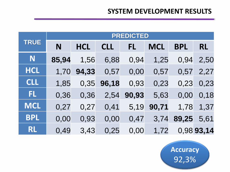
Accuracy
92,3%
TRUE PREDICTED
N HCL CLL FL MCL BPL RL
N 85,94 1,56 6,88 0,94 1,25 0,94 2,50
HCL 1,70 94,33 0,57 0,00 0,57 0,57 2,27
CLL 1,85 0,35 96,18 0,93 0,23 0,23 0,23
FL 0,36 0,36 2,54 90,93 5,63 0,00 0,18
MCL 0,27 0,27 0,41 5,19 90,71 1,78 1,37
BPL 0,00 0,93 0,00 0,47 3,74 89,25 5,61
RL 0,49 3,43 0,25 0,00 1,72 0,98 93,14
SYSTEM DEVELOPMENT RESULTS


PRECISION, SENSITIVITY, SPECIFICITY
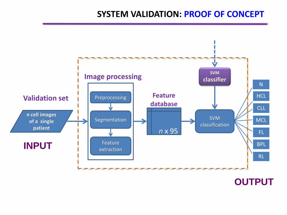
n cell images of a single
patient
n x 95
Feature database
Validation set
SVM classification
N
HCL
CLL
MCL
FL
BPL
RL
Preprocessing
Segmentation
Feature extraction
SVM
classifier
SYSTEM VALIDATION: PROOF OF CONCEPT
Image processing
INPUT
OUTPUT
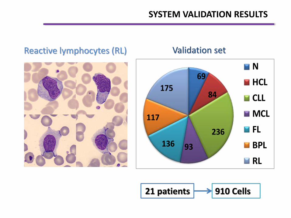
69
84
236
93136
117
175
N
HCL
CLL
MCL
FL
BPL
RL
910 Cells 21 patients
Validation set
SYSTEM VALIDATION RESULTS
Reactive lymphocytes (RL)

Accuracy
85,2%
TRUE PREDICTED
N HCL LLC LF LCM PLB LR
N 92,75 0,00 2,90 1,45 1,45 0,00 1,45
HCL 1,19 98,81 0,00 0,00 0,00 0,00 0,00
LLC 2,97 1,27 80,51 10,17 3,81 0,85 0,42
LF 0,00 0,00 2,21 80,15 17,65 0,00 0,00
LCM 2,15 0,00 1,08 31,18 64,52 1,08 0,00
PLB 3,42 0,00 0,00 0,00 11,11 83,76 1,71
LR 0,57 1,14 0,00 0,00 0,00 0,57 97,71
SYSTEM VALIDATION RESULTS
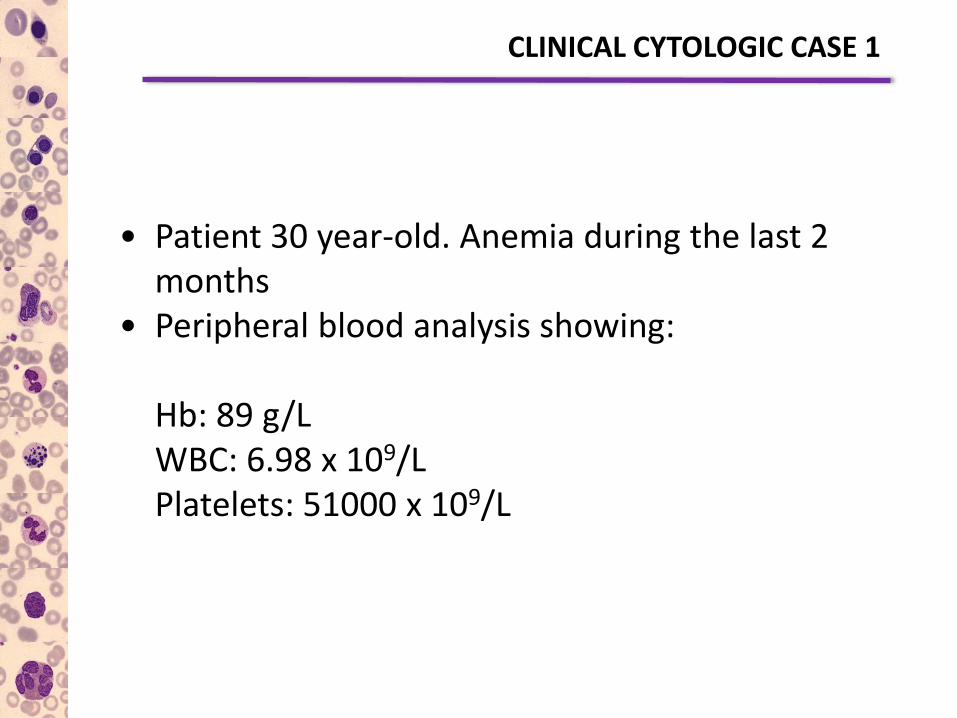
• Patient 30 year-old. Anemia during the last 2 months
• Peripheral blood analysis showing: Hb: 89 g/L WBC: 6.98 x 10⁹/L Platelets: 51000 x 109/L
CLINICAL CYTOLOGIC CASE 1

CellsiMatic: 100% HCL, 0% N, 0% RL, 0% LLC, 0% LCM, 0% FL, 0% PL
CellaVision Images

• Patient 73 year-old in which a finding of lymphocytosis is referred to the Hospital. No symptoms. Small cervical lymphadenopathy are detected.
Hb: 99 g/L WBC: 19.37 x 10⁹/L Platelets: 153000 x 109/L
CLINICAL CYTOLOGIC CASE 2

CellsiMatic: 0% HCL, 0% N, 0% RL, 0% LLC, 4.1% LCM, 95.9% FL, 0% PL
CellaVision Images

• Patient 79 year-old in which a finding of leukocytosis and lymphocytosis is referred to the Hospital. No symptoms.
Hb: 133 g/L WBC: 53.0 x 10⁹/L Platelets: 163 x 109/L
CLINICAL CYTOLOGIC CASE 3
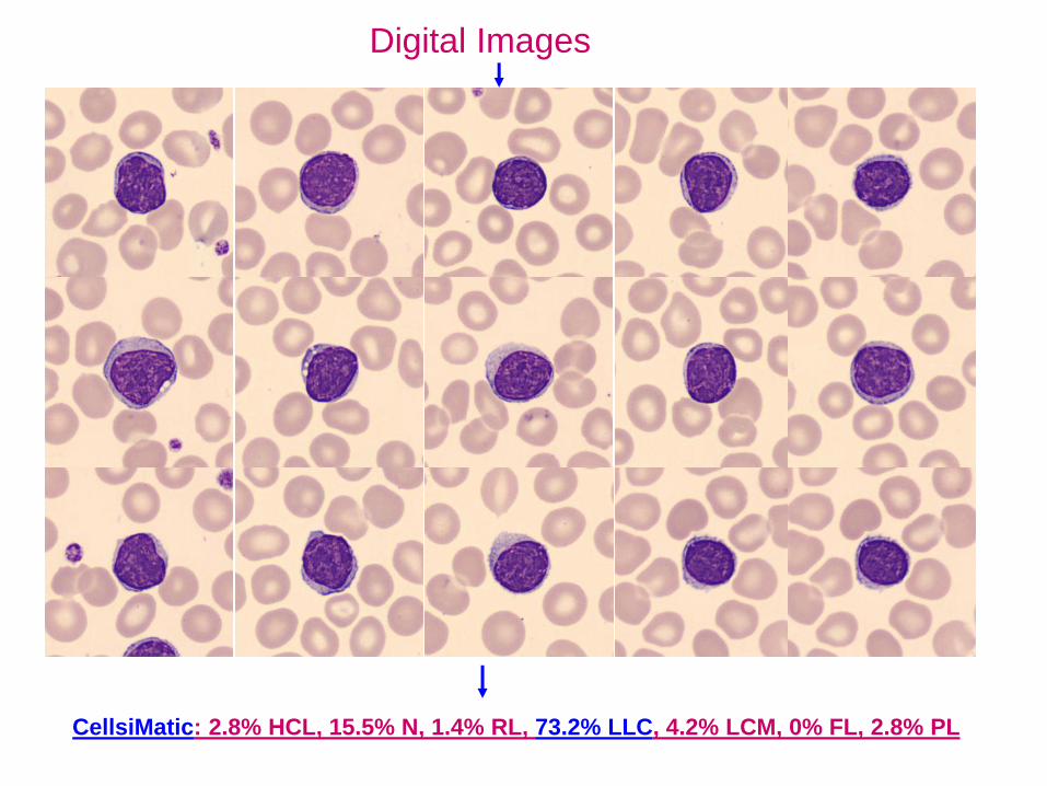
CellsiMatic: 2.8% HCL, 15.5% N, 1.4% RL, 73.2% LLC, 4.2% LCM, 0% FL, 2.8% PL
Digital Images
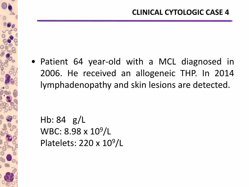
• Patient 64 year-old with a MCL diagnosed in 2006. He received an allogeneic THP. In 2014 lymphadenopathy and skin lesions are detected.
Hb: 84 g/L WBC: 8.98 x 10⁹/L Platelets: 220 x 109/L
CLINICAL CYTOLOGIC CASE 4

CellsiMatic: 0% HCL, 0% N, 0% RL, 0% LLC, 62.5% LCM, 36.3% FL, 1.3% PL
Digital Images

• Patient 20 year-old visited in the emergency Service because of high fever.
Hb: 149 g/L WBC: 17.13 x 10⁹/L Platelets: 99 x 109/L
CLINICAL CYTOLOGIC CASE 5

CellsiMatic: 0% HCL, 0% N, 100% RL, 0% LLC, 0% LCM, 0% FL, 0% PL
Digital Images

1. Our strategy includes a robust segmentation method, a complete feature extraction and a successful classification procedure.
2. It is important to remark the high number (7) of groups of lymphoid cells involved in the classification.
3. The contribution of this work combining medical, engineering and mathematical backgrounds is the development of a complete method that could allow the design of a practical diagnosis support tool in the future.
CONCLUSIONS

“Cytology is like to see
the stars and through
them to see the origin of
the universe”