methods of chromosomal study, postnatal and prenatal cytogenetic diagnosis rndr z.polívková...
TRANSCRIPT
Methods of chromosomal Methods of chromosomal study, postnatal and prenatal study, postnatal and prenatal
cytogenetic diagnosiscytogenetic diagnosis
RNDr Z.PolívkováRNDr Z.Polívková
Lecture No 437 - Lecture No 437 - course: Heredity
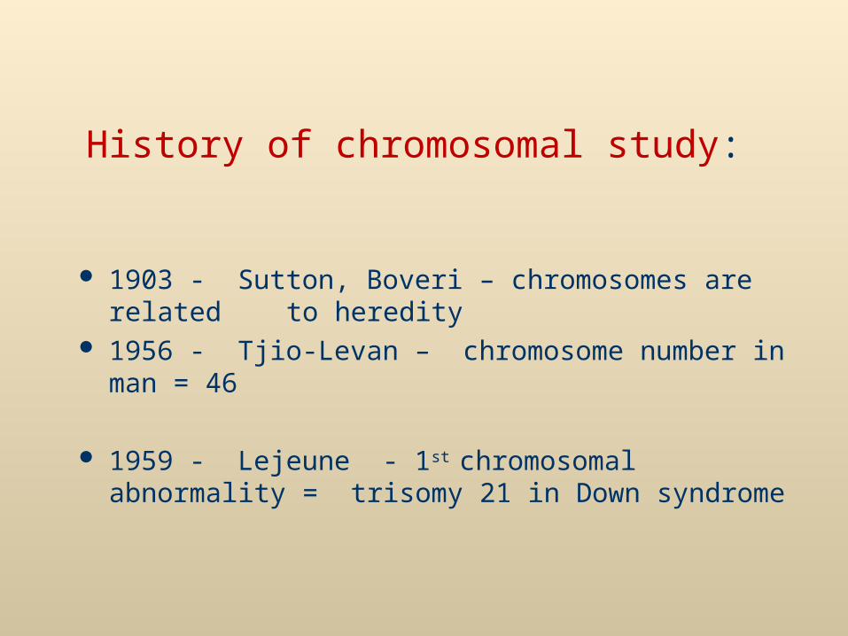
History of chromosomal study:
1903 - Sutton, Boveri – chromosomes are related to heredity
1956 - Tjio-Levan – chromosome number in man = 46
1959 - Lejeune - 1st chromosomal abnormality =
trisomy 21 in Down syndrome
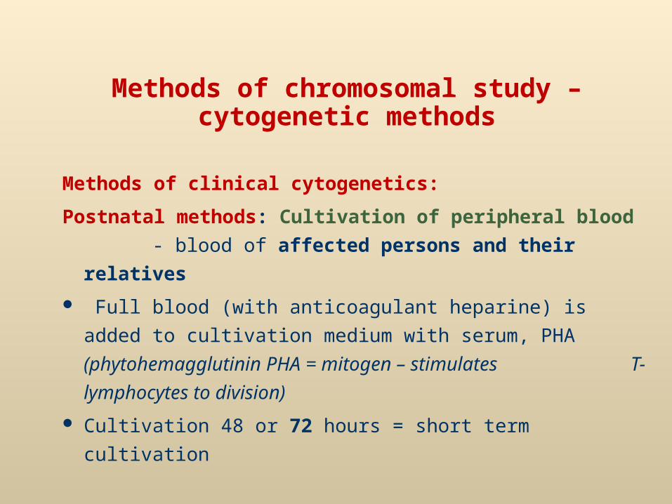
Methods of chromosomal study – cytogenetic methods
Methods of clinical cytogenetics:
Postnatal methods: Cultivation of peripheral blood
- blood of affected persons and their relatives
Full blood (with anticoagulant heparine) is added to
cultivation medium with serum, PHA
(phytohemagglutinin PHA = mitogen – stimulates
T-lymphocytes to division)
Cultivation 48 or 72 hours = short term cultivation
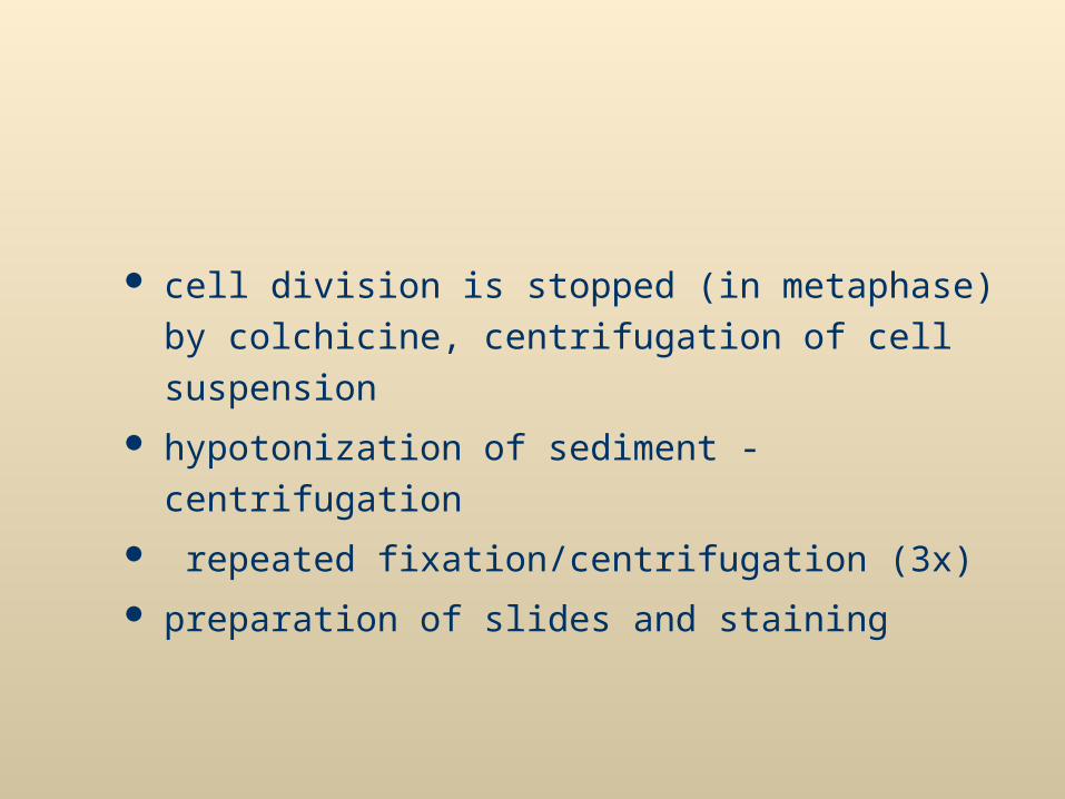
cell division is stopped (in metaphase) by colchicine,
centrifugation of cell suspension
hypotonization of sediment - centrifugation
repeated fixation/centrifugation (3x)
preparation of slides and staining
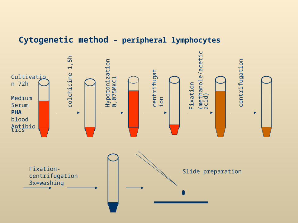
Cytogenetic method – peripheral lymphocytes
MediumSerumPHAbloodAntibiotics
Cultivation 72h
colc
hici
ne 1
,5h
Hyp
oto
niza
tion
0,07
5MK
Cl
cent
rifug
atio
n
cent
rifug
atio
n
Fix
atio
n
(met
hano
le/a
cetic
aci
d)
Fixation-centrifugation 3x=washing
Slide preparation
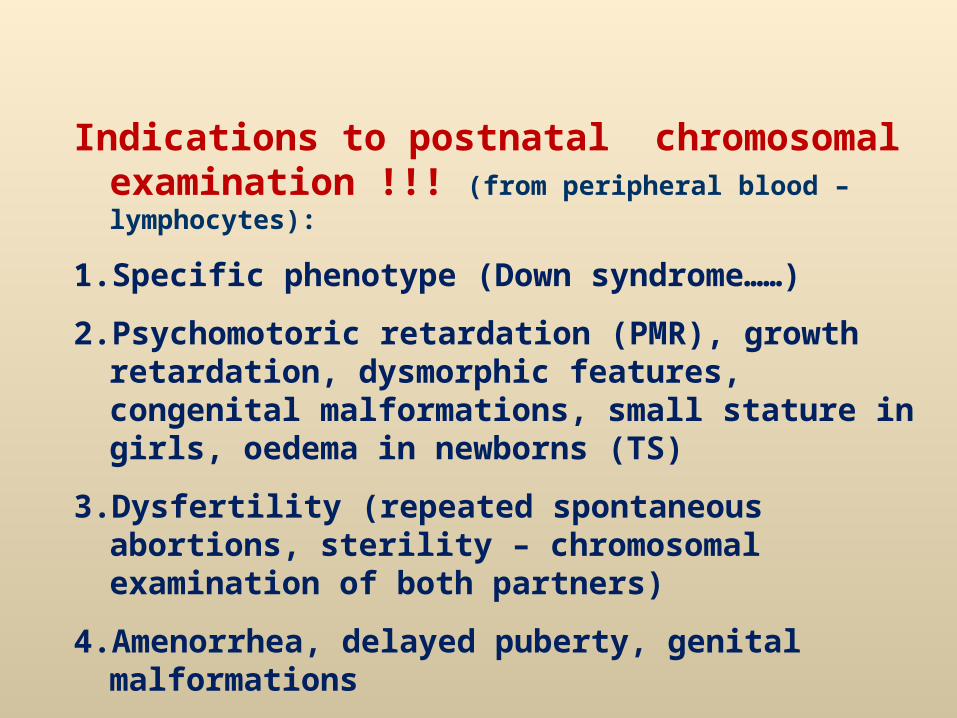
Indications to postnatal chromosomal examination !!! (from peripheral blood – lymphocytes):
1. Specific phenotype (Down syndrome……)
2. Psychomotoric retardation (PMR), growth retardation, dysmorphic features, congenital malformations, small stature in girls, oedema in newborns (TS)
3. Dysfertility (repeated spontaneous abortions, sterility – chromosomal examination of both partners)
4. Amenorrhea, delayed puberty, genital malformations
5. Relatives of patient with chromosomal aberration
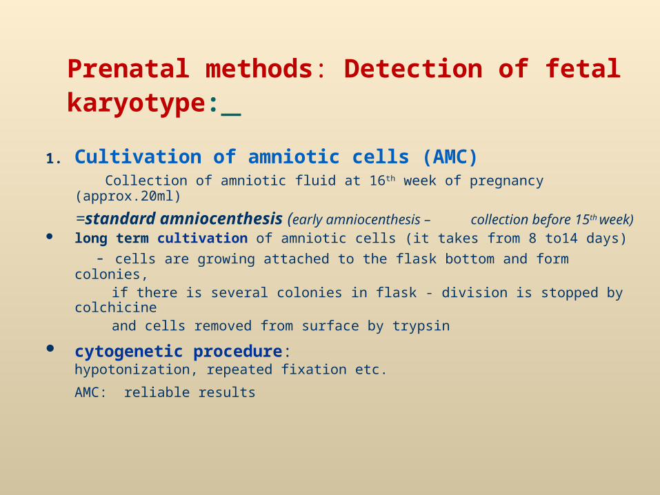
Prenatal methods: Detection of fetal karyotype:
1. Cultivation of amniotic cells (AMC) Collection of amniotic fluid at 16th week of pregnancy (approx.20ml)
=standard amniocenthesis (early amniocenthesis – collection before 15th week)
long term cultivation of amniotic cells (it takes from 8 to14 days)
- cells are growing attached to the flask bottom and form colonies, if there is several colonies in flask - division is stopped by colchicine and cells removed from surface by trypsin
cytogenetic procedure: hypotonization, repeated fixation etc.
AMC: reliable results
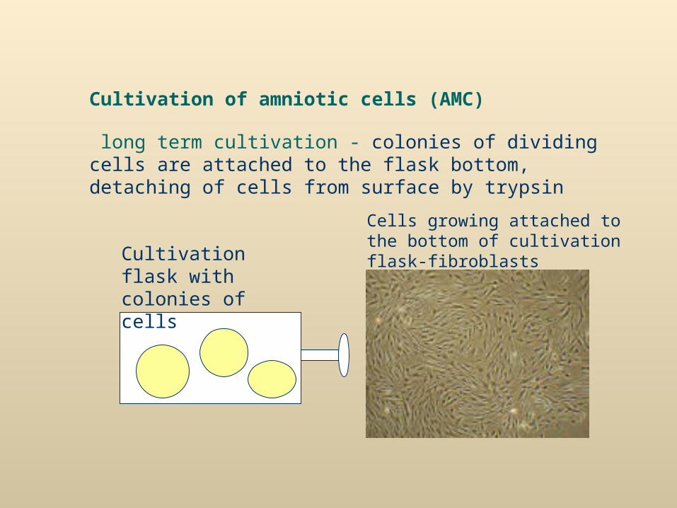
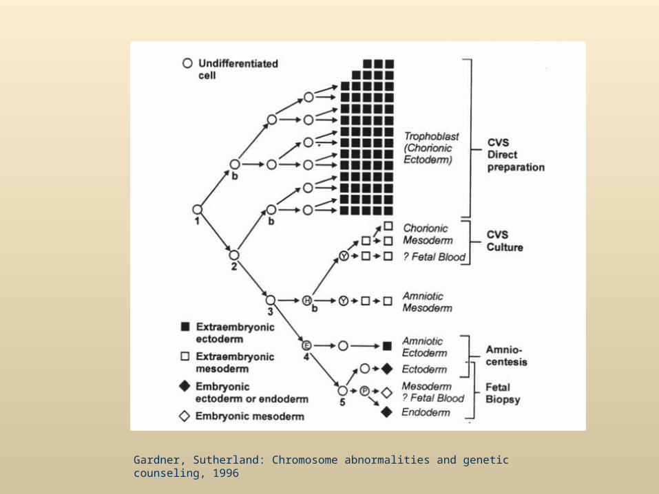
Cultivation of amniotic cells (AMC)
long term cultivation - colonies of dividing cells are attached to the flask bottom, detaching of cells from surface by trypsin
Cells growing attached to the bottom of cultivation flask-fibroblasts
Cultivation flask with colonies of cells
Chorionic villi examination (CVS)
Collection of CVS at 10th week of pregnancy Direct method or long term cultivation Snipping cleaned tissue, detaching of cells by trypsin,
setting cells for cultivation
Chorionic villi = extraembryonal tissue – risk of karyotype discrepancy !!!
It is better to combine both direct and cultivation methods,
or verify pathologic result (if only direct method is used) by different method (fetal blood) or by detection of abnormality by ultrasonography

Gardner, Sutherland: Chromosome abnormalities and genetic counseling, 1996
Gardner, Sutherland: Chromosome abnormalities and genetic counseling, 1996
3. Cultivation of fetal blood
Similar procedure as cultivation of peripheral blood Blood sample is collected from the loop of umbilical cord Short term cultivation (48 h) and cytogenetic procedure
Used to verify some vague result of previous examination, in case of abnormal finding of CVS examination (esp. if only direct method is used), or in case of late detection of abnormality on ultrasound
Indication of prenatal cytogenetic examination!!!From cells of amniotic fluid, chorionic villi, fetal blood
1. Increased maternal age (≥ 35 years)
2. Patological values of biochemical markersScreening in the 2nd trimester: „triple test“ = 3 biochemical markers:
AFP = α-fetoprotein βhCG = choriogonadotropine uE3 = estriol
Screening in the 1st trimester: markers – PAPP-A (pregnancy associated plasma protein A), free -hCG
Combined screening in the 1st trimester - PAPP-A, free -hCG + ultrasonography (nuchal translucency, nasal
bone)
performed at 10th-13th week
Integrated test: biochemical markers of the 1st trimester + ultrasonography markers + biochem.markers of the 2nd trimester (at 15th-17 th week)highest effectivity, lowest degree of false positivity
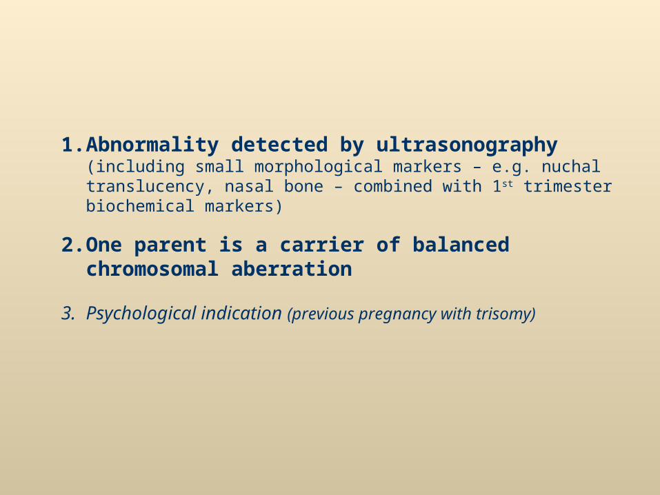
1. Abnormality detected by ultrasonography (including small morphological markers – e.g. nuchal translucency, nasal bone – combined with 1st trimester biochemical markers)
2. One parent is a carrier of balanced chromosomal aberration
3. Psychological indication (previous pregnancy with trisomy)
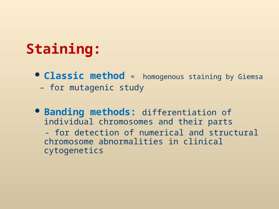
Staining:
Classic method = homogenous staining by Giemsa
– for mutagenic study
Banding methods: differentiation of individual chromosomes and their parts
- for detection of numerical and structural chromosome abnormalities in clinical cytogenetics
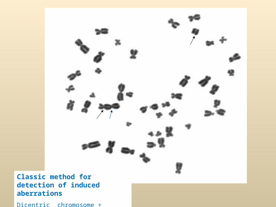
Classic method for detection of induced aberrations
Dicentric chromosome + difragment
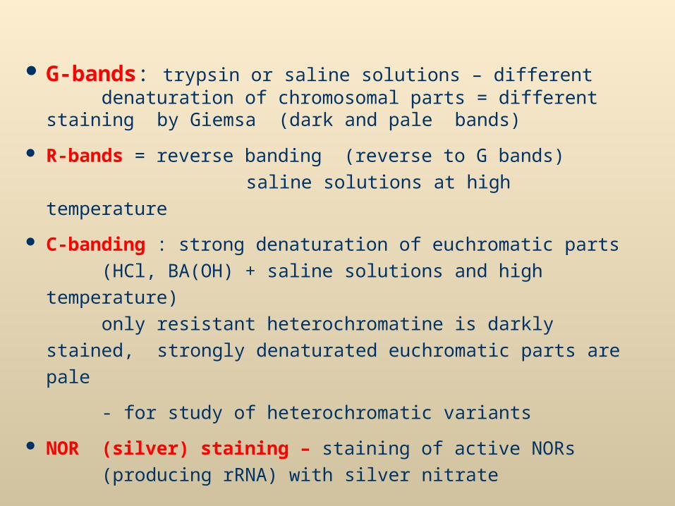
G-bands: trypsin or saline solutions – different denaturation of chromosomal parts = different staining
by Giemsa (dark and pale bands)
R-bands = reverse banding (reverse to G bands)
saline solutions at high temperature
C-banding : strong denaturation of euchromatic parts (HCl,
BA(OH) + saline solutions and high temperature)
only resistant heterochromatine is darkly stained,
strongly denaturated euchromatic parts are pale
- for study of heterochromatic variants
NOR (silver) staining – staining of active NORs
(producing rRNA) with silver nitrate
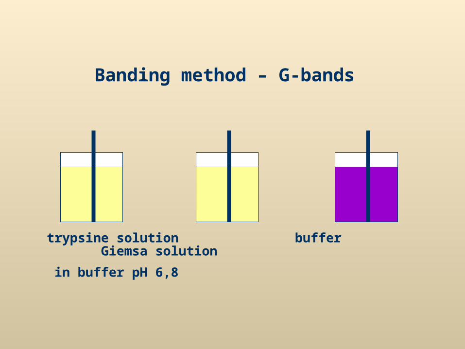
Banding method – G-bands
trypsine solution buffer Giemsa solution
in buffer pH 6,8
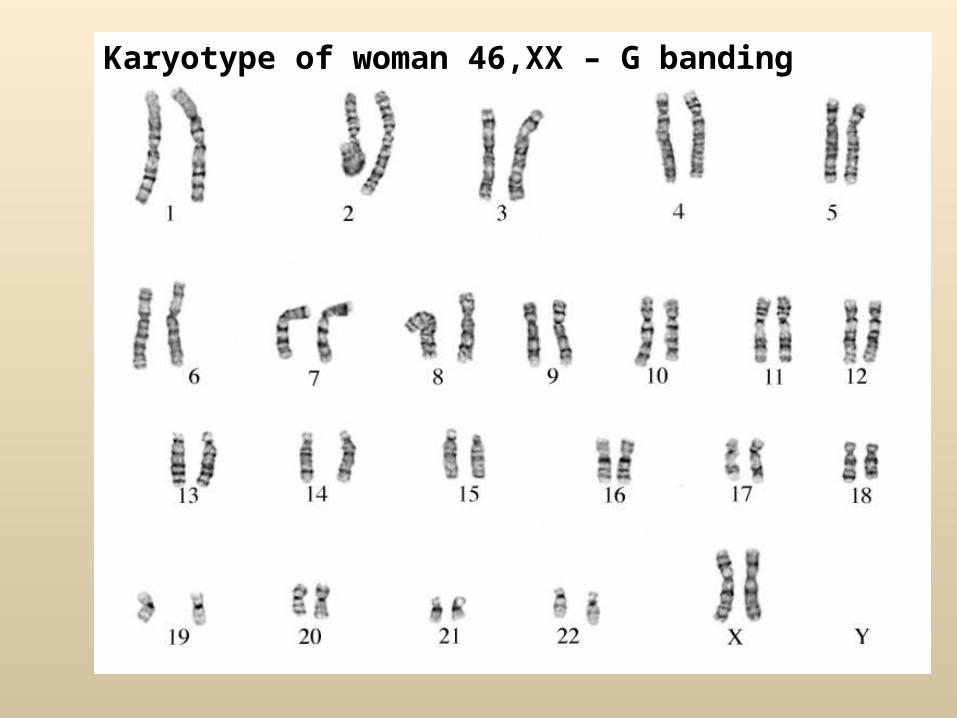
Karyotype of woman 46,XX – G banding
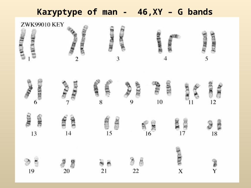
Karyptype of man - 46,XY – G bands
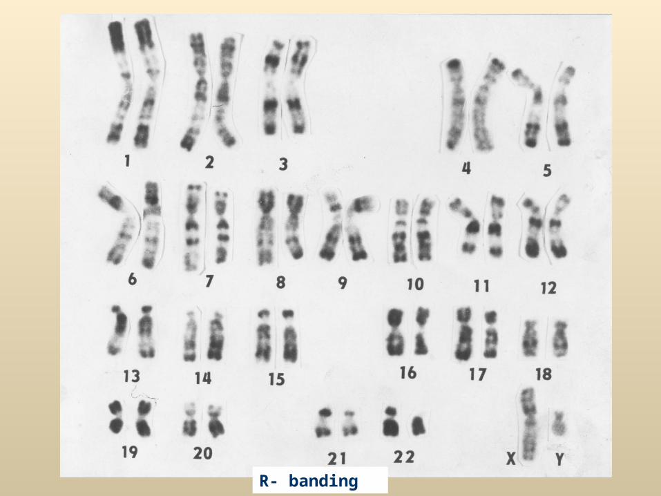
R- banding
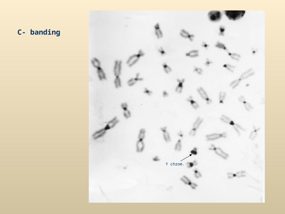
C- banding
Y chrom.
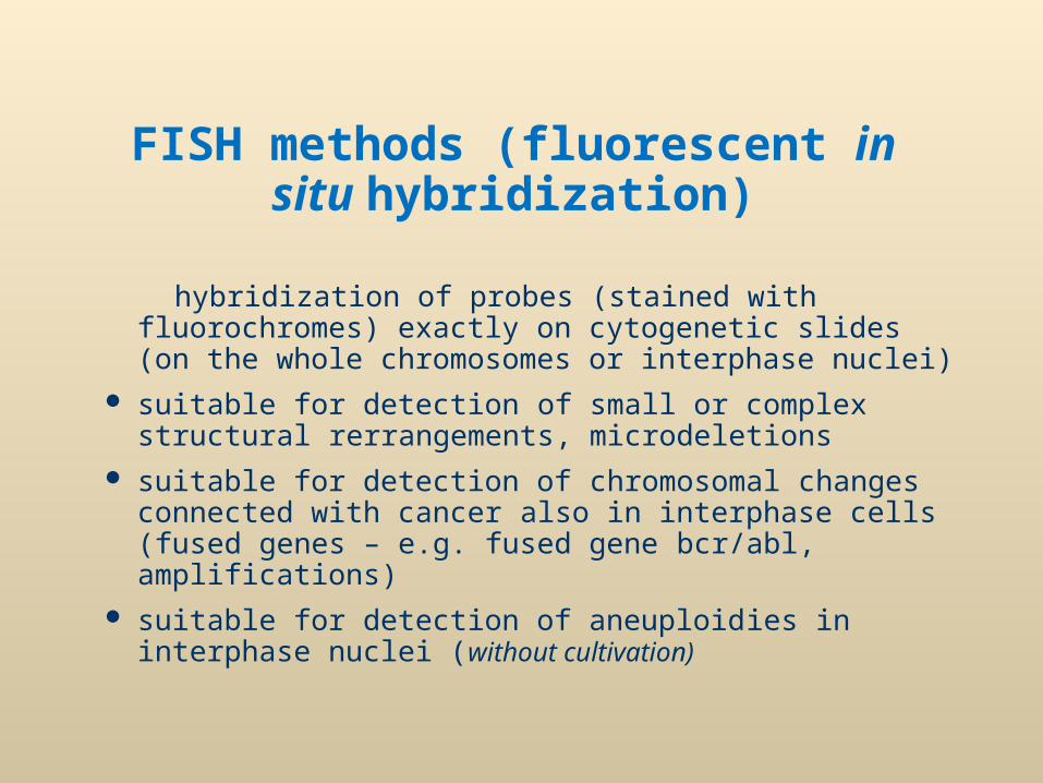
FISH methods (fluorescent in situ hybridization)
hybridization of probes (stained with fluorochromes) exactly on cytogenetic slides (on the whole chromosomes or interphase nuclei)
suitable for detection of small or complex structural rerrangements, microdeletions
suitable for detection of chromosomal changes connected with cancer also in interphase cells (fused genes – e.g. fused gene bcr/abl, amplifications)
suitable for detection of aneuploidies in interphase nuclei (without cultivation)
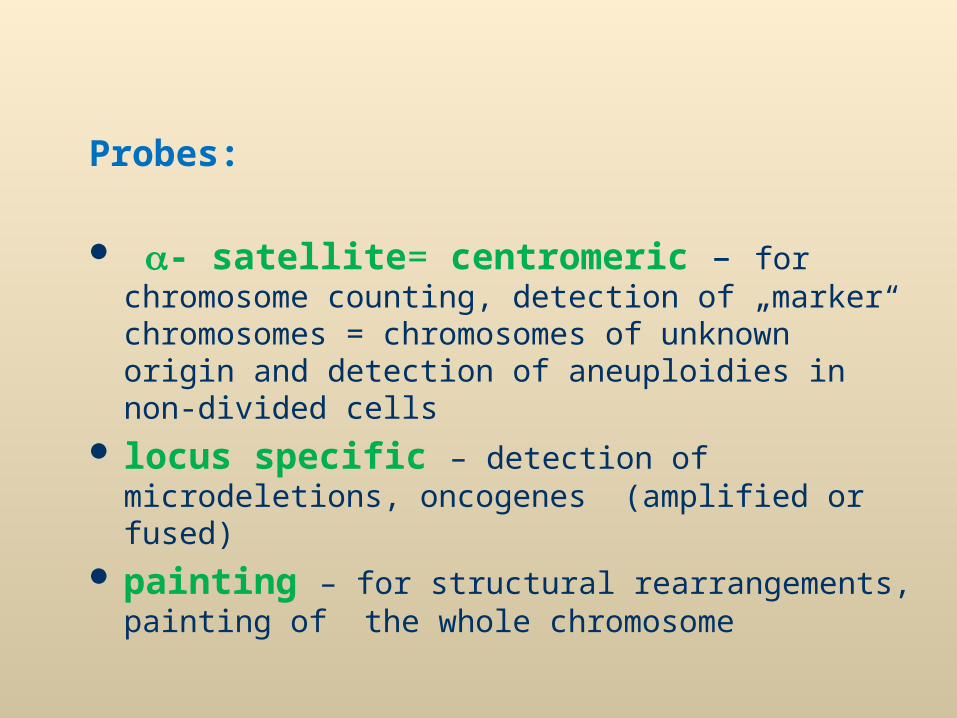
Probes:
- satellite= centromeric – for chromosome counting, detection of „marker“ chromosomes = chromosomes of unknown origin and detection of aneuploidies in non-divided cells
locus specific – detection of microdeletions, oncogenes (amplified or fused)
painting – for structural rearrangements, painting of the whole chromosome
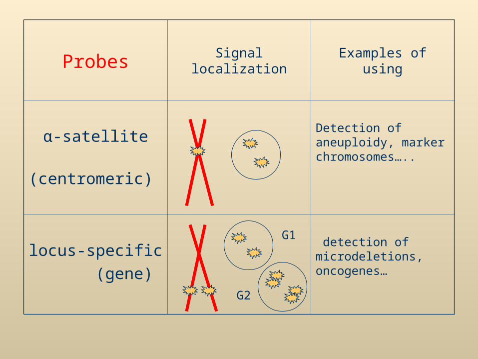
Probes Signal localization Examples of using
α-satellite (centromeric)
Detection of aneuploidy, marker chromosomes…..
locus-specific (gene)
detection of microdeletions, oncogenes…
G1
G2

Principle of FISH methods Chromosome or gene is marked with probe =Probe = short sequence of DNA, labelled with fluorescent dye, complementary to
specific region on chromosome (gene, group of genes, chromosomal part or whole chromosome)
DNA Probe denaturation of probe and hybridization of DNA DNA examined with labelled probe
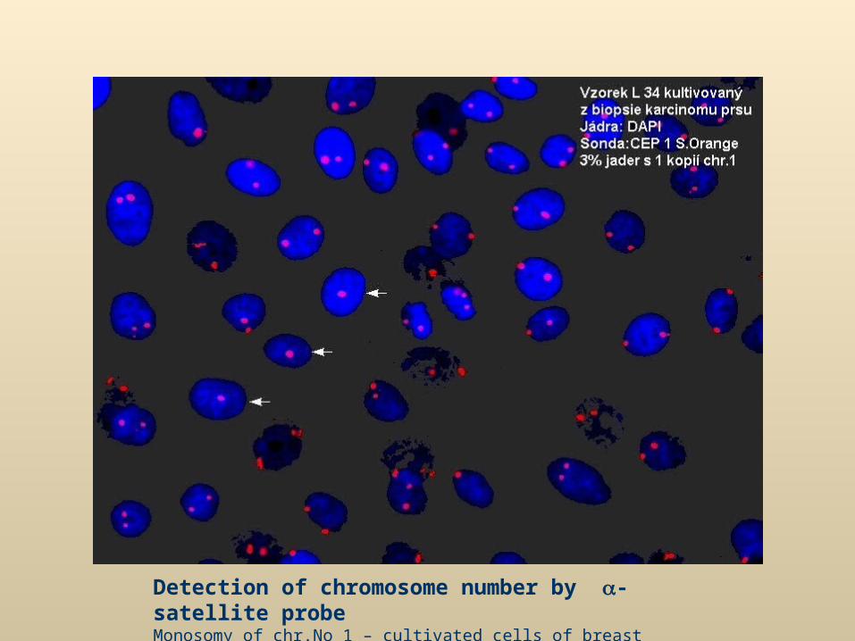
Detection of chromosome number by -satellite probeMonosomy of chr.No 1 – cultivated cells of breast carcinoma
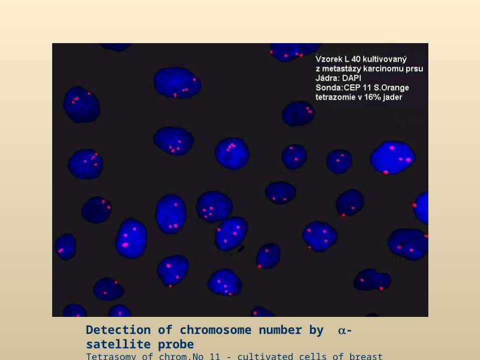
Detection of chromosome number by -satellite probeTetrasomy of chrom.No 11 - cultivated cells of breast carcinoma
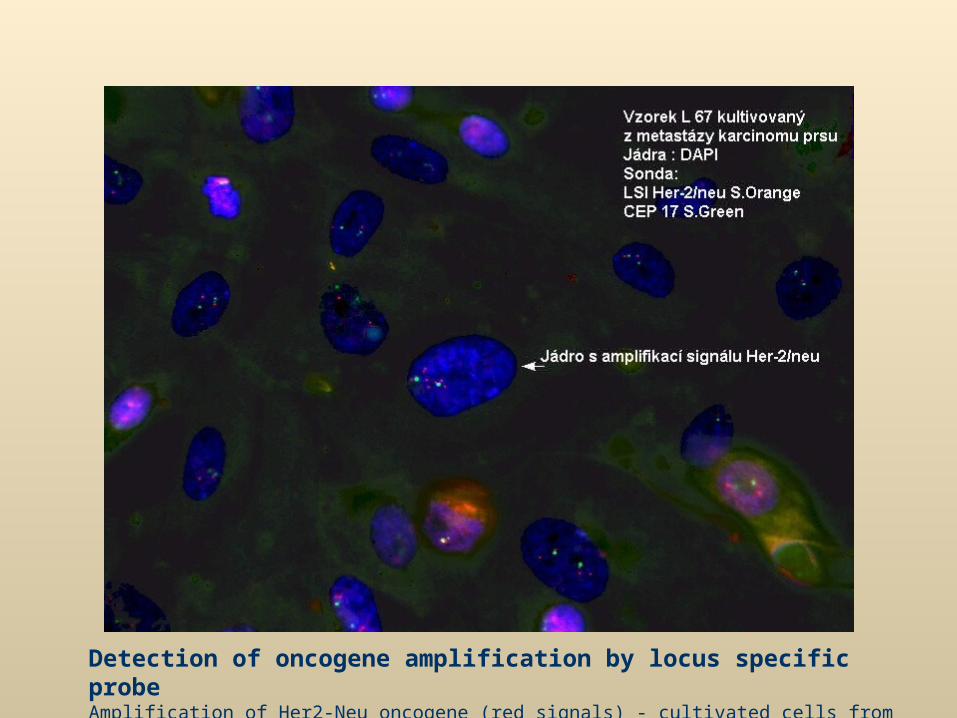
Detection of oncogene amplification by locus specific probeAmplification of Her2-Neu oncogene (red signals) - cultivated cells from metastase of breast carcinoma
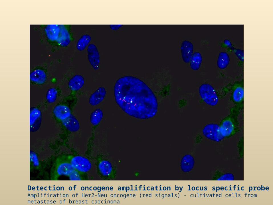
Detection of oncogene amplification by locus specific probeAmplification of Her2-Neu oncogene (red signals) - cultivated cells from metastase of breast carcinoma
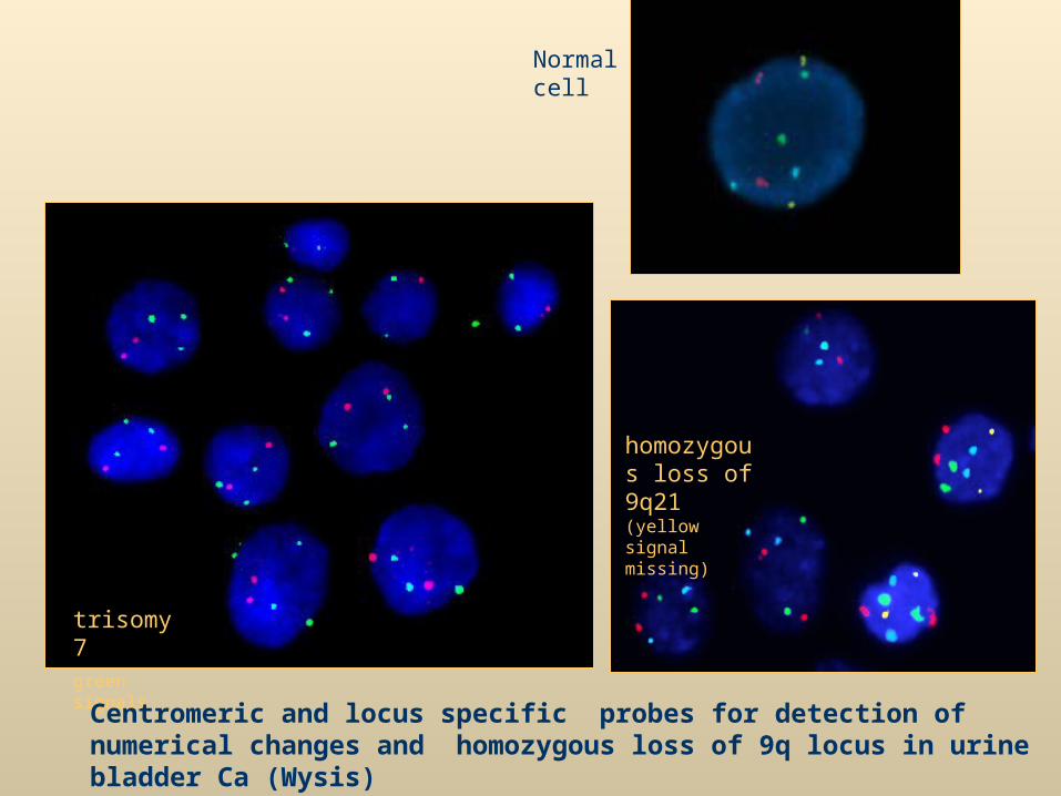
Normal cell
trisomy 7green signals
homozygous loss of 9q21 (yellow signal missing)
Centromeric and locus specific probes for detection of numerical changes and homozygous loss of 9q locus in urine bladder Ca (Wysis)
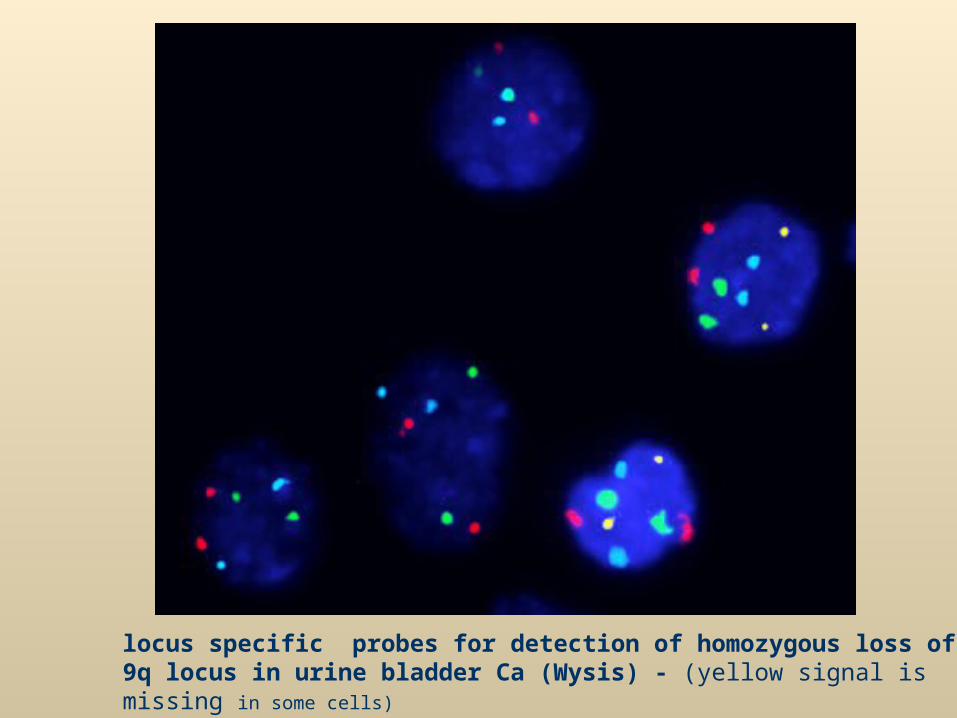
locus specific probes for detection of homozygous loss of 9q locus in urine bladder Ca (Wysis) - (yellow signal is missing in some cells)
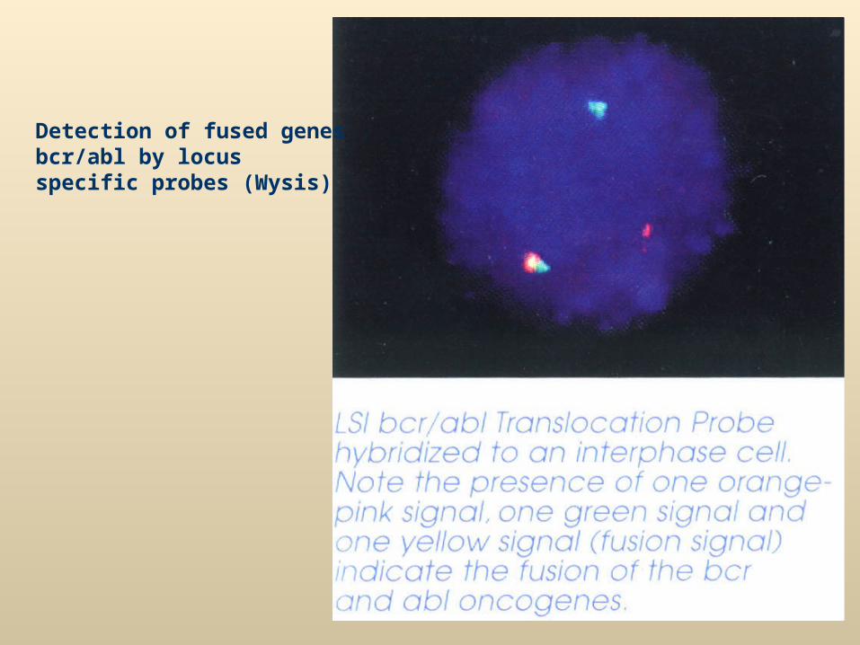
Detection of fused genes bcr/abl by locus specific probes (Wysis)
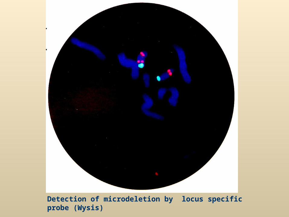
Detection of microdeletion by locus specific probe (Wysis)
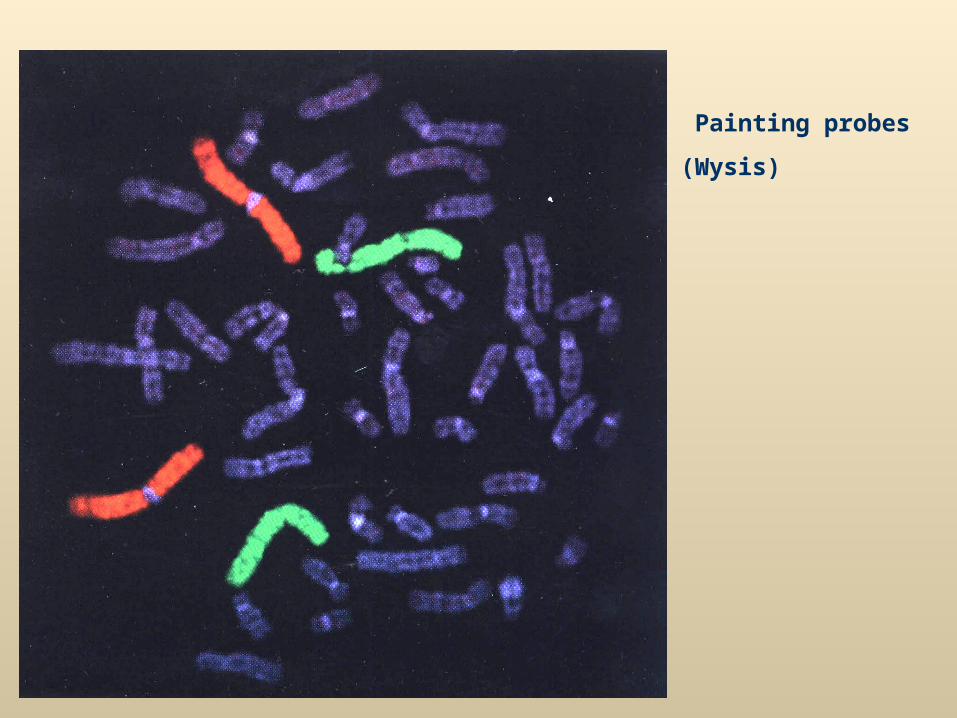
Painting probes
(Wysis)
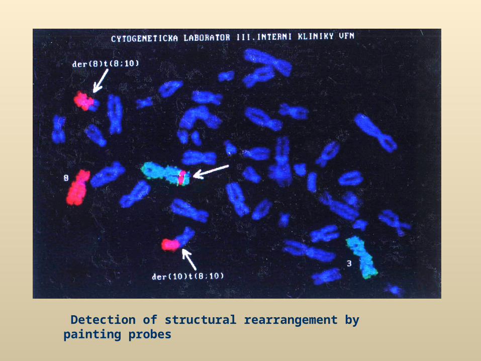
Detection of structural rearrangement by painting probes
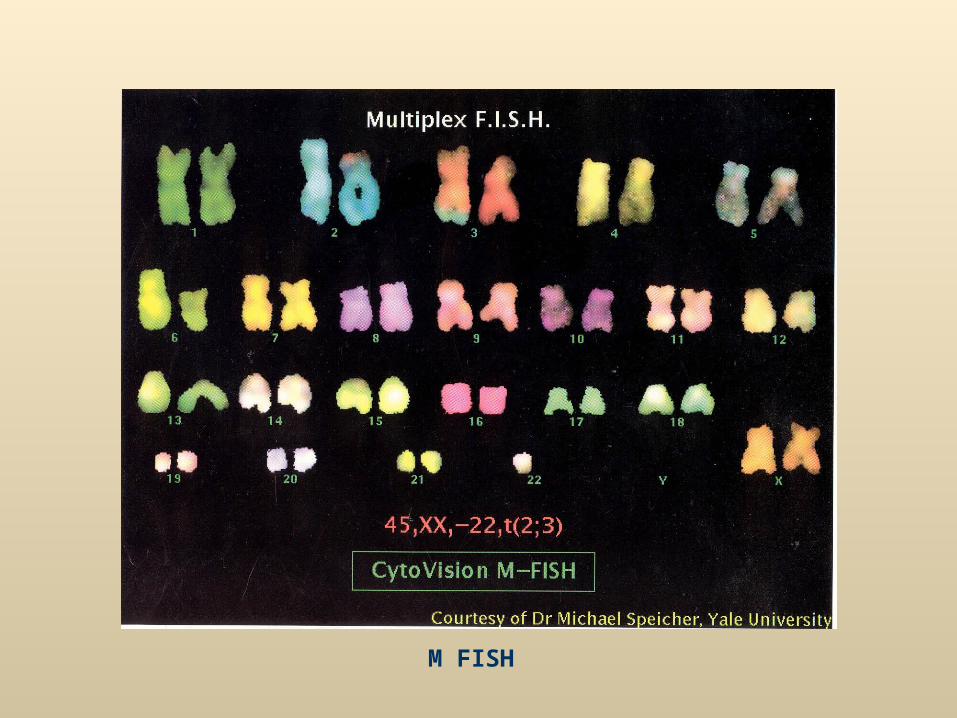
M FISH
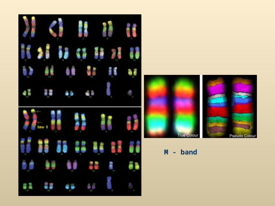
M - band
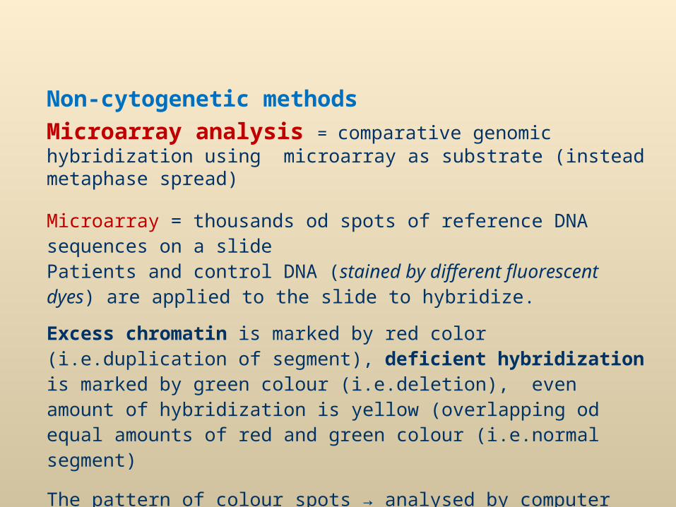
Non-cytogenetic methods
Microarray analysis = comparative genomic hybridization using microarray as substrate (instead metaphase spread)
Microarray = thousands od spots of reference DNA sequences on a slidePatients and control DNA (stained by different fluorescent dyes) are applied to the slide to hybridize.
Excess chromatin is marked by red color (i.e.duplication of segment), deficient hybridization is marked by green colour (i.e.deletion), even amount of hybridization is yellow (overlapping od equal amounts of red and green colour (i.e.normal segment)
The pattern of colour spots → analysed by computer
Method can detecet only unbalanced rearrangements
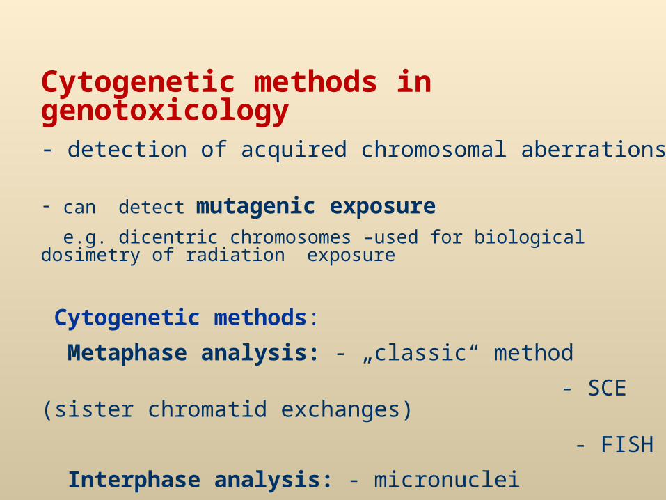
Cytogenetic methods in genotoxicology- detection of acquired chromosomal aberrations
- can detect mutagenic exposure e.g. dicentric chromosomes –used for biological dosimetry of radiation exposure
Cytogenetic methods:
Metaphase analysis: - „classic“ method
- SCE (sister chromatid exchanges)
- FISH
Interphase analysis: - micronuclei
- FISH
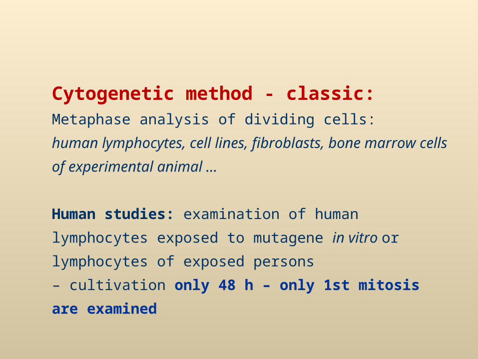
Cytogenetic method - classic:Metaphase analysis of dividing cells:
human lymphocytes, cell lines, fibroblasts, bone marrow cells
of experimental animal …
Human studies: examination of human lymphocytes
exposed to mutagene in vitro or lymphocytes of exposed
persons
– cultivation only 48 h – only 1st mitosis are examined
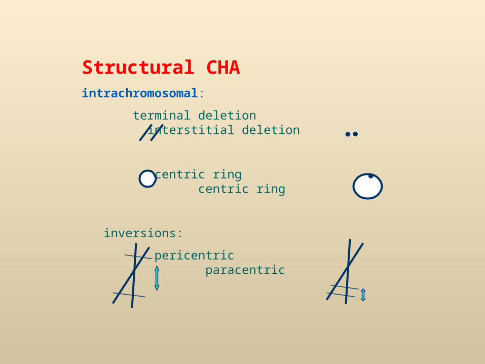
Structural CHAintrachromosomal:
terminal deletion interstitial deletion
acentric ring centric ring
inversions:
pericentric paracentric
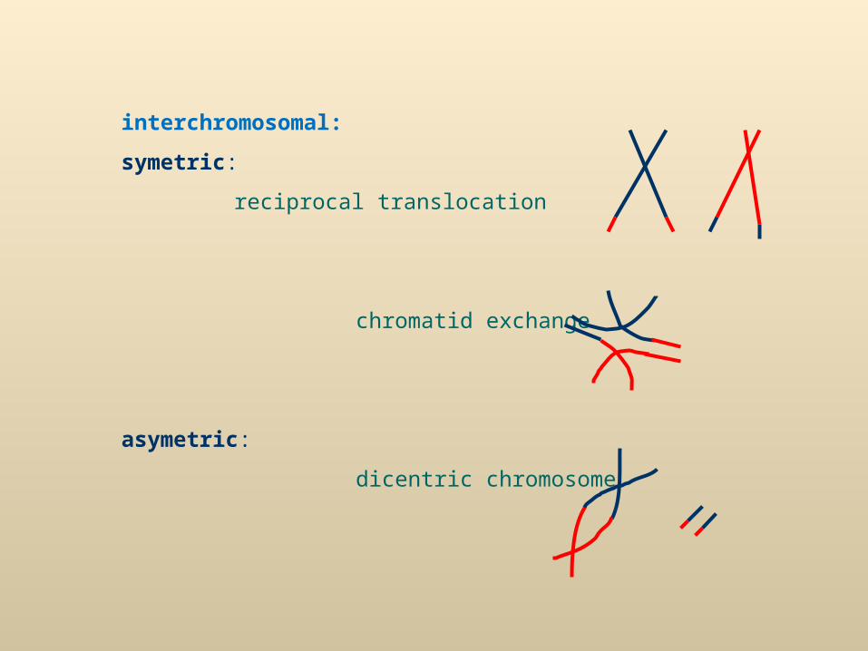
interchromosomal:
symetric:
reciprocal translocation
chromatid exchange
asymetric:
dicentric chromosome
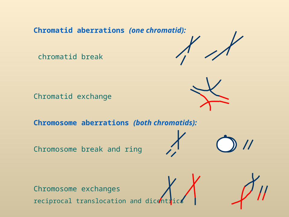
Chromatid aberrations (one chromatid):
chromatid break
Chromatid exchange
Chromosome aberrations (both chromatids)::
Chromosome break and ring
Chromosome exchanges
reciprocal translocation and dicentrics
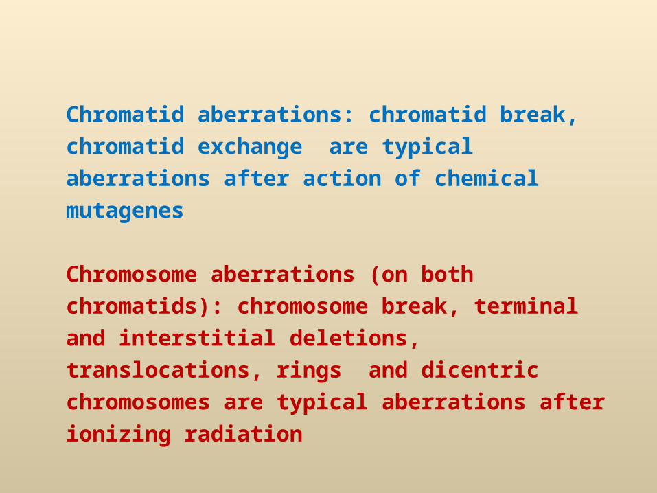
Chromatid aberrations: chromatid break, chromatid
exchange are typical aberrations after action of
chemical mutagenes
Chromosome aberrations (on both chromatids):
chromosome break, terminal and interstitial
deletions, translocations, rings and dicentric
chromosomes are typical aberrations after ionizing
radiation
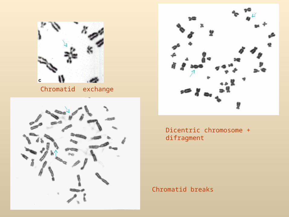
Dicentric chromosome + difragment
Chromatid breaks
Chromatid exchange
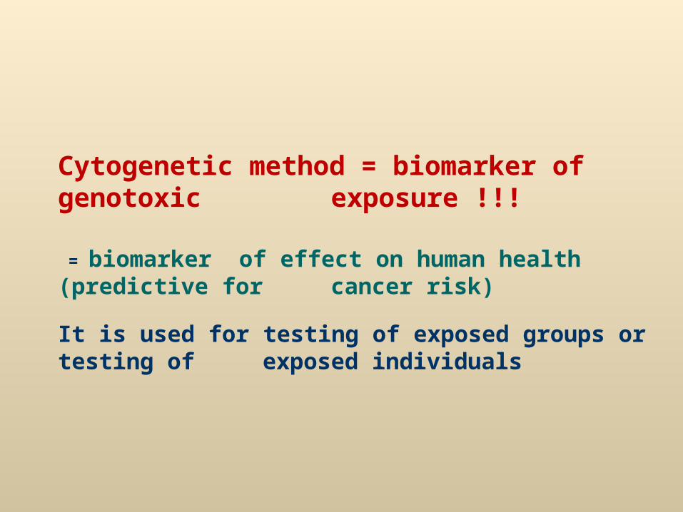
Cytogenetic method = biomarker of genotoxic exposure !!!
= biomarker of effect on human health (predictive for cancer risk)
It is used for testing of exposed groups or testing of exposed individuals
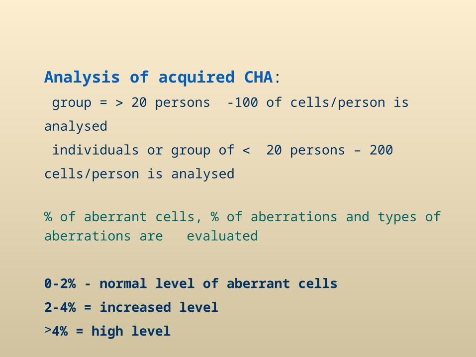
Analysis of acquired CHA:
group = 20 persons -100 of cells/person is analysed
individuals or group of 20 persons – 200 cells/person is analysed
% of aberrant cells, % of aberrations and types of aberrations are
evaluated
0-2% - normal level of aberrant cells
2-4% = increased level
4% = high level
1.
2.
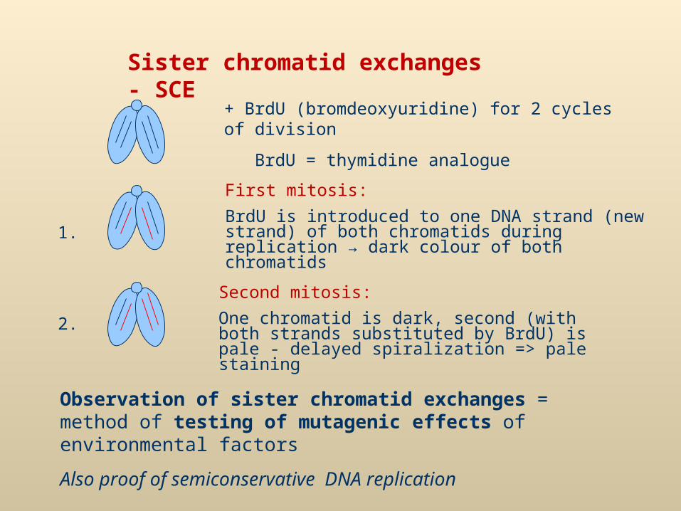
+ BrdU (bromdeoxyuridine) for 2 cycles of division
BrdU = thymidine analogue
First mitosis:
BrdU is introduced to one DNA strand (new strand) of both chromatids during replication → dark colour of both chromatids
Second mitosis:
One chromatid is dark, second (with both strands substituted by BrdU) is pale - delayed spiralization => pale staining
Observation of sister chromatid exchanges = method of testing of mutagenic effects of environmental factors
Also proof of semiconservative DNA replication
Sister chromatid exchanges - SCE
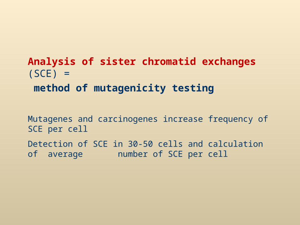
Analysis of sister chromatid exchanges (SCE) =
method of mutagenicity testing
Mutagenes and carcinogenes increase frequency of SCE per cell
Detection of SCE in 30-50 cells and calculation of average number of SCE per cell
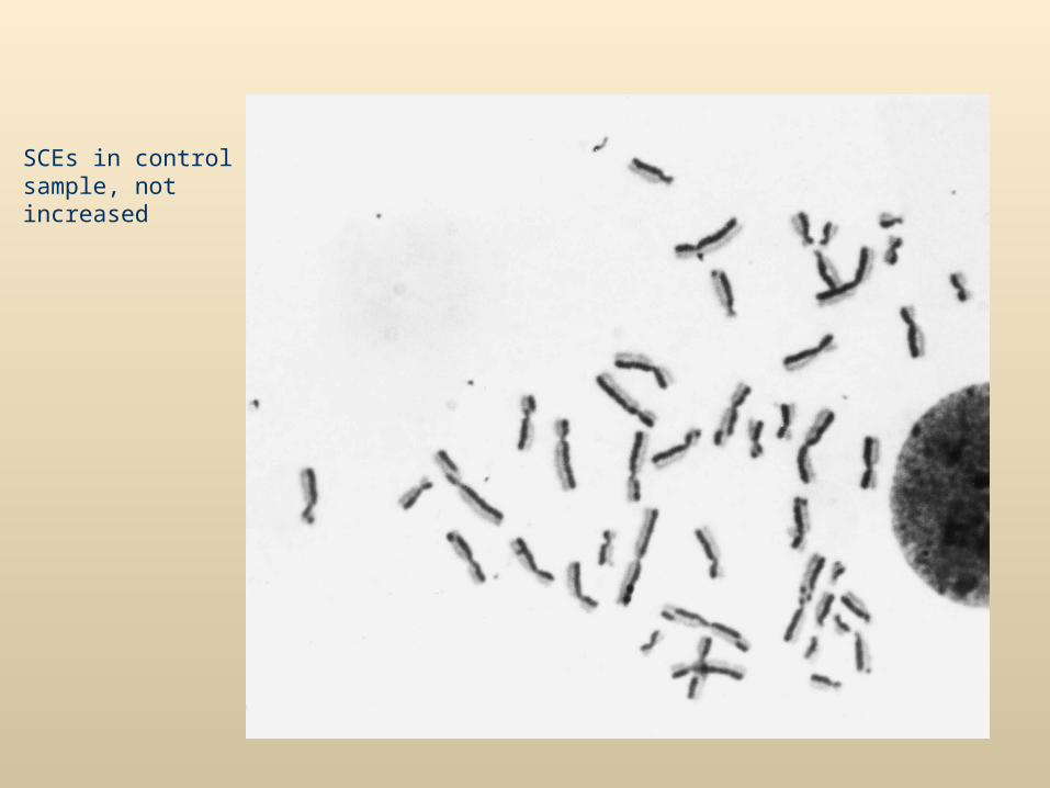
SCEs in control sample, not increased
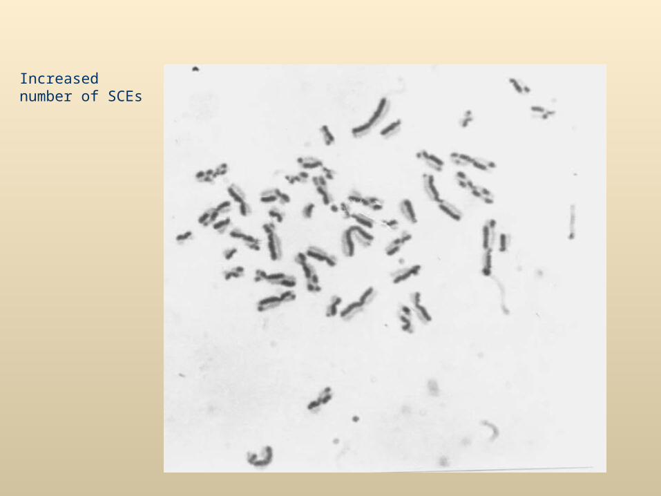
Increased number of SCEs
FISH in genotoxicology
Painting probes
analysis of translocations and other rearrangements –
2 painting probes are used
Exchanges of two painted chromosomes with other chromosomes are
scored and result is corrected for the whole genom good agreement
of FISH with banding methods
Significance of FISH in genotoxicology:
- new knowledge about frequency and mechanisms of CHA
- FISH = quick method, easy scoring, many cells can be scored
- Detection of translocations – used for biological (retrospective)
dosimetry of radiation exposure – in case of long interval
between radiation accident and examination (translocations
are stable aberrations)
Dicentrics are unstable and suitable for biodosimetry
in short term after radiation
Biodosimetry – frequency of dicentrics or translocations increases with radiation dose

Interphase analysis of CHA
Micronucleus test (MN)Micronucleus = chromosomal fragment (mutagenic origin)
= whole chromosome - lost by anaphase lag
Analysis of human lymphocytes - analysed cell must go throug cell division
Method of cytokinesis block by cytochalasine (CB) binuclear cells –
micronucleus = small body stained darkly as nucleus
or detection of MN in bone marrow cell of experimental animals
Positive correlation of MN numbers with age, more MN in women (probably part of MN is formed by chromosome X - lost in older women -1,4xmore MN in women)
Automatic analysis: flow cytometry
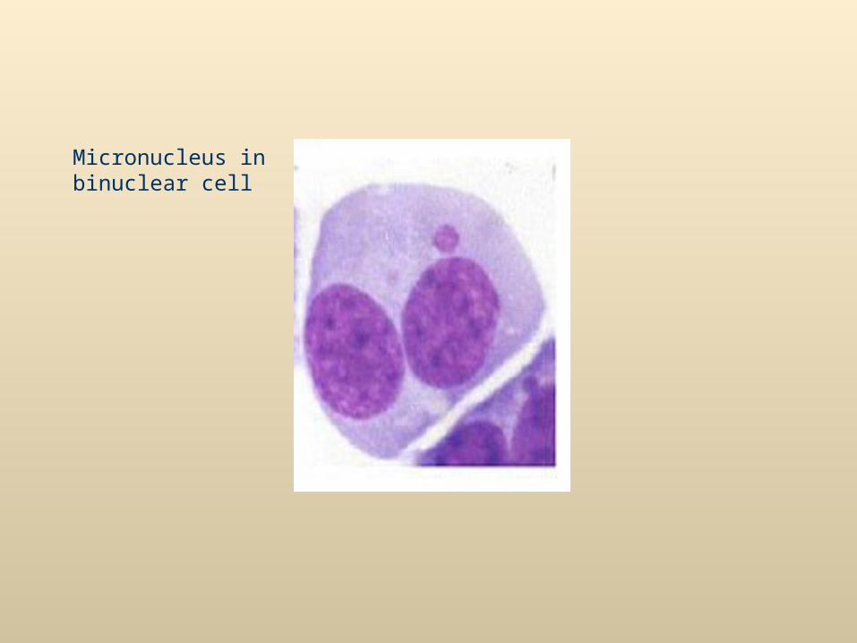
Micronucleus in binuclear cell
http://dl1.cuni.cz/course/view.php?id=324 presentation
http://dl1.cuni.cz/course/view.php?id=324 supplementary text to cytogenetics
Thompson &Thompson: Genetics in medicine, 7th ed.
Chapter 5: Principles of clinical cytogenetics: Introduction to cytogenetics
Chapter 15: Prenatal diagnosis
+ informations from presentation