methohexital-lnduced change s in spectra l powe r of
TRANSCRIPT
Brain Topography, Volume 10, Number 1, 1997 41
Methohexital-lnduced Changes in Spectral Power ofNeuromagnetic Signals: Beta Augmentation is SmallerOver the Hemisphere Containing the EpileptogenicFocus
Christian Wienbruch ,̂ Carsten Eulitz ,̂ Klaus Lehnertz~, Anke Brockhaus~, Christian E. Elger ~,Thomas Elbert̂ , and Manfried Hoke*
Summary: Previous research has suggested that methohexital, a short-term barbiturate, alters activity in the primary epileptogenic area. It can beassumed that drug-induced activation of the epileptogenic focus provides a rapid and safe method to obtain a sufficient amount of informationrelevant for the lateralization and localisation of the primary epileptogenic area. This study shows that methohexital changes spectral power in thebeta band derived from magnetoencephalographic (MEG) signals over the hemisphere ipsilateral to the primary epileptogenic area. This effect wasdemonstrated for 10/13 of the investigated patients suffering from unilateral temporal lobe epilepsy (TLE). The side and location of the primaryepileptogenic area of these patients (5 left TLE, 8 right TLE) was determined invasively during presurgical evaluation. During a 1-2 minute intervalafter intravenous bolus injection of 100 mg methohexital a clear lateralization effect in the beta band was observed, which differed marginally betweenfronto-central, fronto-temporal and temporo-parietal brain regions. In addition, bilateral spectral power changes were obtained in the theta, alphaand gamma bands that differed between brain regions. Analyses of simultaneously recorded scalp electroencephalographic (EEG) data revealedeffects consistent with those of the MEG analysis. The reduced enhancement of beta band spectral power of MEG recordings provides a potentialapplication for the non-invasive lateralization of the primary epileptogenic area.
Introduction
Effects of anaesthetics on electroencephalographic(EEG) activity are well known. Barbiturates modify theEEG in frequency and amplitude. These drug-inducedchanges depend on dose (Brazier and Finesinger 1945;Essig and Fraser 1958; Schwartz et al. 1971) and are more
*Institute for Experimental Audiology, University of Munster, Ger-many.
^Department of Psychology, University of Konstanz, Germany.~Clinic of Epileptology, University of Bonn, Germany.Accepted for publication: July 11, 1997.This research was supported by grants from the Deutsche For-
schungsgemeinschaft (Klinische Forschergruppe Biomagnetismus &Biosignalanalyse, Ho 847/6) and is part of a PhD thesis. The authorswish to thank A. Kowalik MD, A. Baborie MD, M. Mollmann MD, N.Mertes MD, L. Spinne, G. Westermeier, C. Goeters MD, M. Reuss MD,W. Frebel MD, U. Ruta MD, T. Jakob, and S. Lorenz for their technicalassistance and W. Burr, PhD for his useful comments on the study.
Correspondence and reprint requests should be addressed toChristian Wienbruch, Department of Psychology, University of Kon-stanz, Fach D27, D-78457, Konstanz, Germany.
Fax: +49-7531-977492Email: [email protected] © 1997 Human Sciences Press, Inc.
pronounced or even limited to certain scalp regions (San-nita et al. 1990). Some of these effects can be used tolocalise brain lesions as well as epileptogenic areas (forreview see Bauer 1982; Kaplan and Lesser 1990; Modicaet al. 1990). Small doses of barbiturates result in anincrease of fast activity in the beta band known to belarger over normal brain areas as compared to areas withcerebral lesions (Pampiglione 1952; Kennedy and Hill1958). In addition, various authors described qualita-tively a loss of methohexital-induced beta band activityin the electrocorticogramm (ECoG) over the epilepto-genic area (Pampiglione 1952; Lieb et al. 1989; Hufnagelet al. 1992).
Because recordings of neuromagnetic fields havesome advantages as compared to recordings of associ-ated electric potentials, we expect the non-invasive mag-netoencephalography (MEG) to provide additionalinformation about the origin of methohexital inducedchanges in its spectral power characteristics. We there-fore investigated the value of methohexital induced ac-tivity for the presurgical lateralization of the primaryepileptogenic area using MEG during a short-time narco-sis in patients suffering from temporal lobe epilepsy
Key words: MEG; EEG; Methohexital; Beta band activity; Temporal lobe epilepsy; Spectral analysis.
42 Wienbruch et al.
Methods
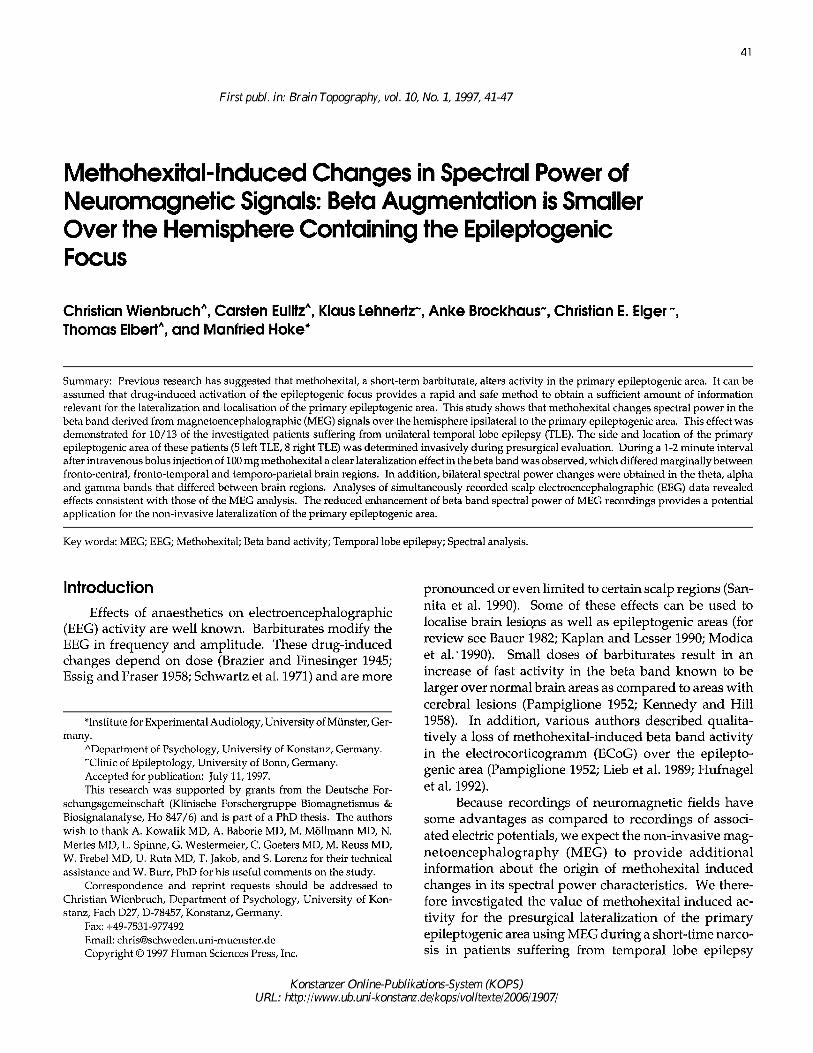
Figure 1. A: Schematic view of MEG recording locations.Spectral power was averaged across data from fronto-central (black circles), fronto-temporal (open circles) andtemporo-parietal (light grey circles) sites. B: Schematicview of EEG electrode positions according to the interna-tional 10-20 system, Spectral power was averaged acrossdata from fronto-central (F3, F7, C3 / F4, F8, C4) andtemporal recording sites (T1, T3, T5 / T2, T4, T6). C: Typicalexamples of MEG and EEG beta band activity (patientR03) before (upper) and after (lower) i.v. injection of 100mg methohexital. Data measured over the hemisphereipsilateral to the primary epileptogenic focus are shownon the left side. During narcosis beta band activity is moreenhanced over the hemisphere contralateral to the pri-mary epileptogenic area (right). The selected MEG traceswere recorded from fronto-central (fc), temporo-parietal(tp) and fronto-temporal (ft) brain regions, EEG traces (T3,T4, F3, F4) from electrode positions according to the inter-national 10-20 system. Data were digitally bandpass fil-tered to remove activity from other frequency bandsusing a 13-24 Hz recursive 8th order Butterworth-filter.
(TLE). The aim of this study was to quantify anaesthesia-induced changes in the spectral power characteristics ofsimultaneously recorded MEG and EEG signals and toanalyse the topographical aspects of these changes withrespect to the side of the primary epileptogenic area.
Patient characteristics
Thirteen patients (age: 34 ± 7 years; range: 23 - 43years) suffering from pharmacoresistant unilateral tem-poral lobe epilepsy were investigated. All patients wereinformed about the procedure before the examinationand declared their informed consent. Before performingthis study, all patients underwent invasive presurgicalevaluation (see Bonn protocol of presurgical evaluation;Engel 1993) using electrocorticographic and intrahippo-campal stereo-EEG recordings for the localisation anddelineation of the primary epileptogenic area. As con-firmed by the post-operative outcome a left temporalseizure origin was found in 5 patients and a right tempo-ral seizure origin in 8 patients. Selective amygdalo-hip-pocampectomy (AHE) was performed in 8 patients, 2/3anterior temporal lobe resection and AHE in 4 patientsand a resection of a ganglioglioma in one patient. Aftersurgery all but two patients are completely free of sei-zures for at least 6 month. The present examination wasperformed while the patients' anticonvulsant medication(Carbamazepine-mono-therapy - 11 patients, Phenytoin-Clobazam-therapy - 1 patient and Carbamazepine-Vi-gabatrin-Clobazam-therapy - 1 patient) laid withintherapeutical range.
Measurement techniques and procedure
Neuromagnetic data were recorded using a 37-chan-nel neuromagnetometer (Magnes™; Biomagnetic Tech-nologies, Inc.; consisting of first-order axial gradiometerswith 5 cm baseline and a coil diameter of 2 cm; pick-upcoils were arranged in an array of concentric circles witha diameter of 14.4 cm). Measurements were carried outunder video control in a magnetically shielded room. Thesensor array was positioned first either over the patients'left (4) or right (9) supra-temporal cortex and centeredabout 1.5 cm superior to T3 or T4 electrode position of theinternational 10-20 system (figure la). The head positionrelative to sensor pickup coils was measured by a sensorposition indicator. EEG data were simultaneously re-corded (SYN-AMPS™; Neuro Scan, Inc.) from temporal,frontal, central and parietal electrode positions accordingto the international 10-20 system with the nose used asreference (figure 1b). To remove a possible influence ofthe reference electrode the corresponding average refer-ence EEG signal was calculated. Continuous data wererecorded in 10 minute blocks using a sampling rate of297.6 Hz and a passband of 0.03 to 100 Hz.
Patients were lying on their side with their head andbody fixed by a vacuum cushion. They were instructedto avoid eye blinks and head movements. After a 2 minbaseline recording a dose of 100 mg methohexital was

Methohexital-lnduced Spectral Power Changes in MEG/EEG 43
applied intravenously (i.v.) in a bolus injection accord-ing to the conditions used during the invasive presurgi-cal evaluation (Hufnagel et al. 1992). After a break of 41min mean (range: 22 - 89 min) exactly the same proce-dure was repeated on the opposite hemisphere, Thisbreak is sufficient for the recovery of the patient withrespect to consciousness and recovery of MEG and EEGback to the baseline due to methohexital's short half-lifetime of approximately 2 min. Anaesthetic monitoringand standby was provided, and precautions were takento counteract anaesthetic complications. During themeasurements an anaesthetist was within the magneti-cally shielded room.
Data Analysis
Off-line the data were digitally bandpass filteredusing a 4-48 Hz recursive Butterworth-filter (2nd order)to remove low-frequency artefacts (like patient move-ments due to hiccup etc.) as well as frequency compo-nents above the gamma-band especially the 50 Hz noise.The spectral power was estimated for each magnetic andelectric channel. Data sets of 300 seconds length starting20 seconds before injection of methohexital were ana-lysed using an overlapping segmentation technique.The length of the Parzen-windowed segments was 512points which corresponds to 1.720 s. According to Presset al. (1988) the segments were overlapped by one halfof their length to obtain the smallest spectral varianceper data point. Power spectra of these segments wereaveraged within 19.785 seconds time windows. Thus,one mean spectrum across a 19.785 seconds windowbefore and 14 windows after the administration ofmethohexital were calculated (the 19.785 seconds pre-medication baseline proved representative for the wholebaseline in all cases). Due to different distances betweenMEG sensors and the patient's head, spectra were nor-malised by dividing each mean spectrum by the meanspectrum of the time before the injection of methohexitalseparately for each channel.
The spectral power in the theta, alpha, beta andgamma band represents the integrated power within theborder frequencies (theta: 4.07-7.55 Hz, alpha: 8.13-12.79Hz, beta: 13.37-23.83 Hz and gamma: 24.41-47.66 Hz). Tofurther reduce the data, spectral band power was aver-aged across neighbouring channels separately for eachhemisphere. MEG channels were collapsed over fronto-central, temporo-parietal and fronto-temporal brain re-gions (figure la), EEG-channels over fronto-central andtemporal recording sites (figure 1b).
The resulting normalised (i.e., with respect to thebaseline) spectral power values (termed NSP) of eachfrequency band were statistically tested for differencesbetween hemispheres and brain regions as dependent
variables. For MEG data we used two-way univariateanalyses of variance (ANOVA) for a 3 brain-region(fronto-central vs. fronto-temporal vs. temporo-parietal)x 2 side-of-focus (ipsilateral vs. contralateral side) design.For the statistical analyses of EEG data the factor brain-regions had only two steps (fronto-central vs. temporal).Wherever appropriate, a Greenhouse-Geisser correctionof the degrees of freedom was applied.
Results
General observations
No adverse effects during or after anaesthesia werenoticed by either the patients or the investigators. Patientsrecovered quickly and had no clinical excitatory effects orsigns like vomiting and nausea. No seizure was elicited.MEG and EEG raw data of one representative patient fortime segments before and during the methohexital activa-tion recorded over the ipsilateral and contralateral hemi-sphere are shown in figure 1c. In contrast to the baselinerecording, a high amplitude beta band activity was ob-tained after administration of methohexital.
Spatial aspects of methohexital-induced changesin MEG/EEG spectral band power
In order to exclude a possible influence of repeatedanaesthesia, spectral band power values of the baselinewere statistically tested for differences. Wilcoxonmatched pairs test and Student's t-test yielded no signifi-cant differences between baselines recordings.
The dynamics of changes in the theta, alpha, betaand gamma band NSP over hemispheres in the courseof the methohexital activation are illustrated in figure 2.A well pronounced difference of both MEG and EEGbeta band spectral power between the hemispheres ipsi-and contralateral to the epileptogenic focus was ob-served two minutes after the injection of methohexitalfor a period of 1-2 minutes. While these changes in-cluded activity in the alpha band of the EEG recordings,they were especially obvious in the gamma band activityof the MEG recordings.
In order to evaluate the influence of the factors brain-region and side-of-focus statistical analyses were basedon changes of each band NSP obtained during the thirdminute after methohexital injection. In contrast to EEGrecordings ANOVA showed the factor brain-region tosignificantly affect NSP in the theta, alpha and gammaband of the MEG recordings (theta: F(2/24)=4.55, p<0.03;alpha: F(2/24)=14.60, p<0.0001); gamma: F(2/24)=6.22,p<0.008). The influence of the side of the primary epilep-togenic area, however, was not evident in these fre-quency bands of both MEG and EEG recordings.
44 Wienbruch et al.
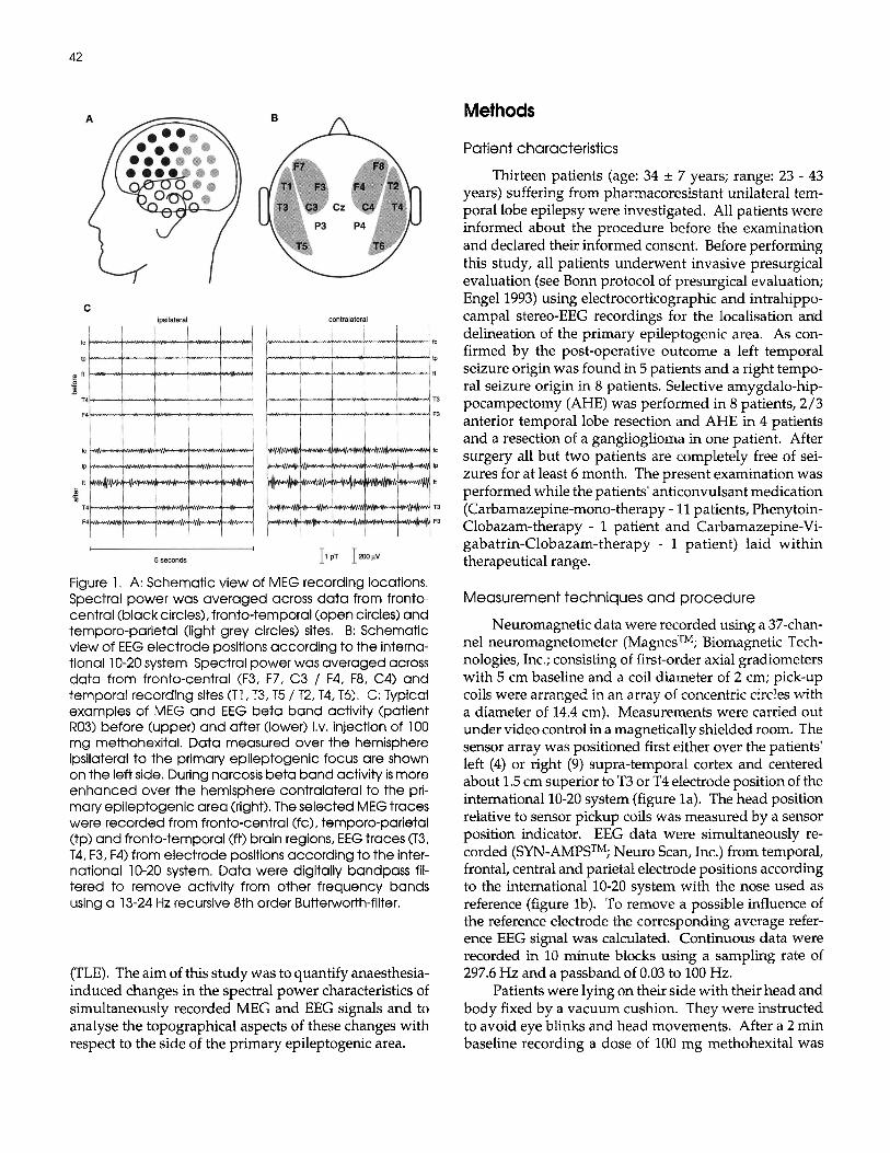
Figure 2. Changes of theta, alpha, beta and gamma band spectral power in the course of the methohexital activationfor MEG (upper) and EEG (lower) recordings, averaged across all subjects. The standard deviation is shown for thecontralateral side (dotted lines) only. Note that the standard deviations of EEG spectral power in the alpha, beta andgamma band are much larger than for the corresponding MEG spectral band power.
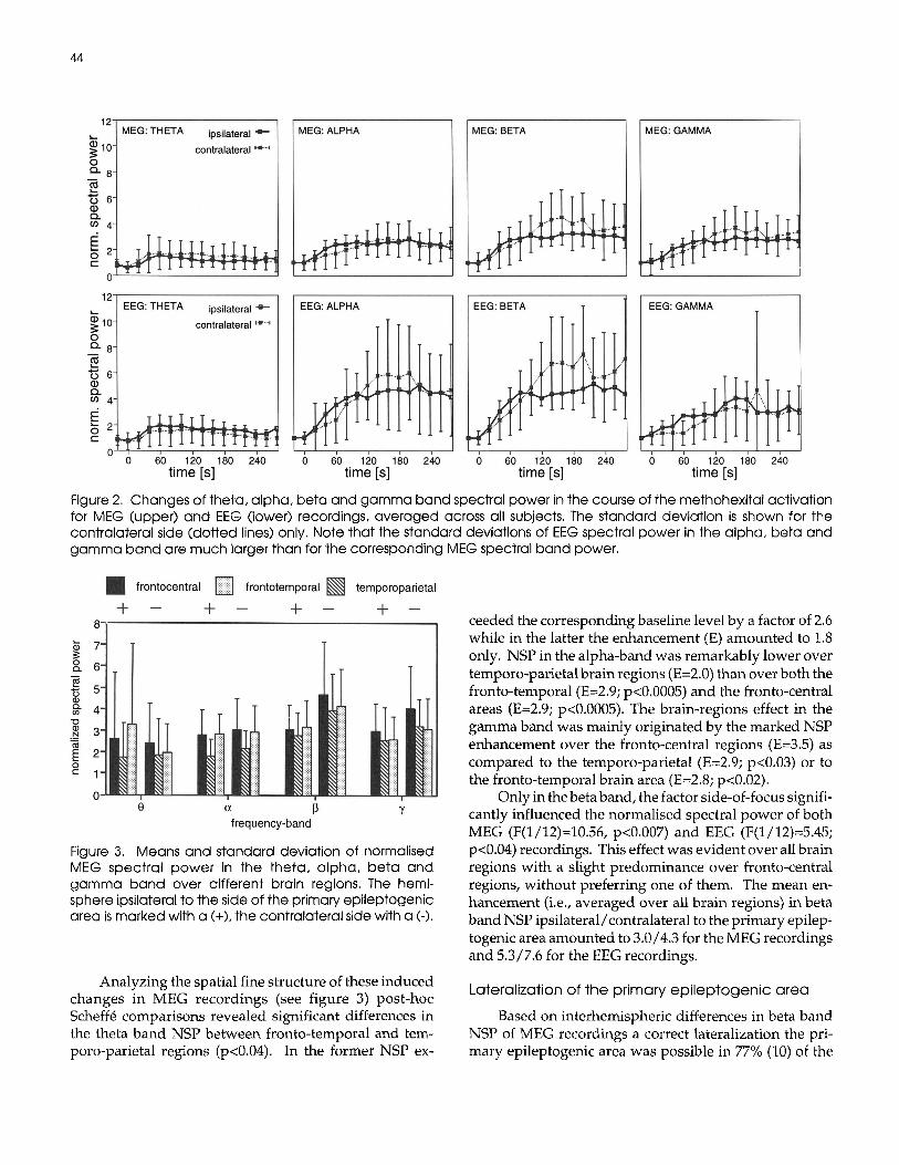
Figure 3. Means and standard deviation of normalisedMEG spectral power in the theta, alpha, beta andgamma band over different brain regions. The hemi-sphere ipsilateral to the side of the primary epileptogenicarea is marked with a (+), the contralateral side with a (-).
Analyzing the spatial fine structure of these inducedchanges in MEG recordings (see figure 3) post-hocScheffe comparisons revealed significant differences inthe theta band NSP between fronto-temporal and tem-poro-parietal regions (p<0.04). In the former NSP ex-
ceeded the corresponding baseline level by a factor of 2.6while in the latter the enhancement (E) amounted to 1.8only. NSP in the alpha-band was remarkably lower overtemporo-parietal brain regions (E=2.0) than over both thefronto-temporal (E=2.9; p<0.0005) and the fronto-centralareas (E=2.9; p<0.0005). The brain-regions effect in thegamma band was mainly originated by the marked NSPenhancement over the fronto-central regions (E=3.5) ascompared to the temporo-parietal (E=2.9; p<0.03) or tothe fronto-temporal brain area (E=2.8; p<0.02).
Only in the beta band, the factor side-of-focus signifi-cantly influenced the normalised spectral power of bothMEG (F(l/12)=10.56, p<0.007) and EEG (F(l/12)=5.45;p<0.04) recordings. This effect was evident over all brainregions with a slight predominance over fronto-centralregions, without preferring one of them. The mean en-hancement (i.e., averaged over all brain regions) in betaband NSP ipsilateral/contralateral to the primary epilep-togenic area amounted to 3.0/4.3 for the MEG recordingsand 5.3/7.6 for the EEG recordings.
Lateralization of the primary epileptogenic area
Based on interhemispheric differences in beta bandNSP of MEG recordings a correct lateralization the pri-mary epileptogenic area was possible in 77% (10) of the

Methohexital-lnduced Spectral Power Changes in MEG/EEG 45
Table 1. Lateralization of the primary epileptogenic focus using the reduced enhancement of the normalised beta bandspectral power as a measure in comparison to the result of the presurgical evaluation. Abbreviations: RIC = ratio of thebeta band power ipsi- to contralateral.
Pat.
R01
R03
R04
R05
L06
R07
LOS
R09
L12
L13
L14
R16
R17
MEG Lateralization
correct
correct
correct
false positive
correct
correct
correct
false positive
correct
correct
correct
false positive
correct
EEG Lateralization
correct
correct
correct
false positive
false positive
correct
correct
correct
correct
false positive
correct
false positive
correct
RIC MEG
0.71
0.56
0.53
1.08
0.76
0.79
0.64
1.22
0.63
0.63
0.64
1.69
0.44
RIC EEG
0.51
0.69
0,56
1.36
1,35
0.63
0.76
0.57
0.25
1,21
0,32
2.21
0.35
investiagted patients (see table I), while false positivedecisions would have been made in 23% (3). EEG record-ings yielded a correct lateralization in 69% (9) and falsepositive decisions in 31% (4) of the cases.
Discussion
The aim of the presurgical evaluation of patientswith pharmacoresistant epilepsies is the correct localisa-tion and exact delineation of the primary epileptogenicarea. In order to avoid the invasive and time-consumingprocedure of recording seizures via intracranial elec-trodes reliable non-invasive recording techniques are re-quired. However, ictal symptoms render non-invasiverecording techniques as the MEG non-feasible in themajority of the cases. To overcome these problems, theevaluation of drug-induced activities provides an alter-native method for the rapid and safe localisation of theprimary epileptogenic area. Up to now, research hasfocussed on drug-induced activities using EEG andECoG recordings (Fuster et al. 1948, Pampiglione 1952;Kennedy and Hill 1958; Brazier 1969; Wilder 1969,1971;Harris and Paul 1969; Celesia 1972; Wyler et al. 1987;Aasly et al. 1989; Hufnagel et al. 1992). Thus, the aim ofthe present study was to evaluate possible advantages ofMEG compared to EEG/ECoG recordings. We analyzeddrug-induced activities because of the follwing advan-tages compared to interictal or ictal activities: due tospecific interactions of the short-acting barbituratemethohexital with the epileptogenic focus it can be ex-
pected that a sufficient amount of relevant data can beobtained within short acquisition times. Furthermore,movement artefacts that represent a major source of errorin MEG recordings are minimised or even suppressedduring the short-time narcosis.
Apart from an induction of epileptiform activity likespikes and spike-burst-suppression patterns (see Brock-haus et al. 1997 for a MEG based localization of thesepatterns), a loss of barbiturate-induced beta activity overthe side of the primary epileptogenic area has been de-scribed qualitatively and quantitatively in a number ofstudies based on EEG or electrocorticographic (ECoG)recordings (Lieb et al. 1974; Duffy et al. 1984; Lieb et al.1986; Hufnagel et al. 1992; Dasheiff and Kofke 1993;Brockhaus et al. 1995). Our results, based on a spectralanalysis of simultaneous MEG and EEG recordings con-firm these findings. A less pronounced induction of beta-band activity ipsilateral to the primary epileptogenic areawas clearly expressed during a 1-2 minute interval start-ing approximately two minutes after the administrationof methohexital and could be established in 10 (MEG-based lateralization) or 9 (EEG-based lateralization) ofthe 13 investigated patients.
The loss of methohexital-induced beta activity overthe side of the primary epileptogenic area has also beenreported by Hufnagel et al. (1992), who described thephenomenon qualitatively in ECoG recordings, meas-ured under comparable experimental conditions. Al-though they concluded that a reduction of drug-inducedfast activity over the epileptogenic area may be an addi-

46 Wienbruch et al.
tional aid in the localisation of cerebral lesions and func-tional epileptogenic foci, a beta reduction could only beobserved in 48% of the investigated patients. The mostlikely reason might probably be due to the fact thatfrontal electrode strips were implanted only when indi-cated by previous investigations. However, a drug-in-duced enhancement of fast beta activity is morepronounced at frontal, fronto-lateral and temporo-lateralaspects of the brain but is rarely recordable using tem-poro-basal strip electrodes.
Regardless of the better signal-to-noise ratio of ECoGrecordings compared to EEG recordings, both methodsseem to have common disadvantages for the registrationof the spatio-temporal dynamics of barbiturate interac-tions with the epileptogenic focus. Obviously, MEGshows some advantages for the description of drug-in-duced focal activities. Besides the independence of areference electrode, MEG has the advantage of the bodybeing transparent to low frequency magnetic fields up tothe kHz range. Thus, the MEG is not attenuated bysurrounding tissue as holds true for the EEG. Further-more, it can be expected that a blurring of the recordedmagnetic activity due to secondary sources produced byconcentric inhomogeneities of the conductivity of thebody is much less pronounced as for the electric potential.This is consistent with our finding of an additional barbi-turate-induced gamma-band activity in the MEG com-pared to a more pronounced alpha-band activity in EEGrecordings. These effects may reflect differential sensi-tivities of the recording techniques to brain activity indifferent frequency bands.
The mechanism of drug-induced beta reduction overthe epileptogenic area is still unclear. However, our re-sults indicate that the loss of methohexital-induced en-hancement of beta band spectral power in MEG and EEGmight contribute to the non-invasive lateralization of theprimary epileptogenic area in patients suffering frompharmacoresistant temporal lobe epilepsy. This holdstrue particularly in those cases not exhibiting an increaseof drug-induced spike activity or spike-burst-suppres-sion (SBS) patterns in the MEG or EEG recordings, thelatter pattern being of high significance for the lateraliza-tion and localisation of epileptogenicity to the ictogenictemporal lobe based on invasive ECoG recordings viasubdural strip electrodes (Hufnagel et al. 1992).
Although the number of patients investigated so faris still too small in order to determine the value of thepresented method for non-invasive presurgical evalu-ation, we would have correctly lateralized the primaryepileptogenic area in 77% of the cases applying the calcu-lated threshold of MEG beta band power only. Thisillustrates the potential significance of this method forpresurgical lateralization of the primary epileptogenicarea based on non-invasive MEG recordings.
In conclusion, our findings of a reduced enhance-ment of beta band spectral power in MEG recordingsipsilateral to the side of the primary epileptogenic areaconfirm previous studies using EEG recordings. In orderto determine the value of this method for the non-inva-sive presurgical evaluation, additional investigations ona larger number of cases, especially with single or multi-ple epileptogenic foci in brain regions other than tempo-ral lobes are required. Future investigations should alsomake use of the now available whole head MEG systems,which will allow simultaneous measurements from bothhemispheres.
ReferencesAasly, J., Silfvenius, H. and Zetterlund, B. Barbiturate effects
on EEG abnormality in complex partial epilepsy. J Neurol,1989,236:15-20.
Bauer, G. EEG, drug effects, and central nervous system poi-soning. In E. Niedermeyer and F. H. L. da Silva (Eds),Electroencephalography, Urban and Schwarzenberg, Bal-timore, 1982: 479-489.
Brazier, M. A.B. and Finesinger, J.E. Action of barbiturates in thecerebral cortex. Arch Neurol Psychiat, 1945, 53: 51-58.
Brazier, M. Prenarcotic doses of barbiturates as an aid in local-izing diseased brain tissue. Anesthesiology, 1969, 31: 78-83.
Brockhaus, A., Hufnagel, A., Nadstawek, J., Ebeling, B.J., vanRoost, D. and Elger, C.E. Activation of epileptogenic fociby thiopental in electrocorticographic recordings with sub-dural strip electrodes and intrahippocampal depth elec-trodes. J Epilepsy, 1995,8:153-163.
Brockhaus, A., Lehnertz, K., Wienbruch, C., Kowalik, A.,Burr, W., Elbert, T., Hoke, M. and Elger, C.E. Possibilitiesand limitations of magnetic source imaging of metho-hexital-induced epileptiform patterns in temporal lobeepilepsy patients. Electroenceph clin Neurophysiol, 1997,102: 423-436.
Celesia, G.G. and Paulsen, R.E, Electroencephalographic acti-vation with sleep and metho-hexital. Arch Neurol, 1972,27:361-363.
Dasheiff, R. and Kofke, W. Evaluation of the thiopental test inepilepsy surgery patients. Epilepsy Res, 1993,15:253-238.
Duffy, F., Jensen, F., Erba, G., Burchfiel, J. and Lombroso, C.Extraction of clinical information from electroencepha-lographic background activity: The combined use of brainelectrical activity mapping and intravenous sodium thio-pental. Ann Neurol, 1984,15: 22-30.
Engel, J. Jr. (Ed.) Surgical Treatment of the Epilepsies. RavenPress, New York, 1993: 740-742.
Essig, C.F. and Fraser, H.F. Electroencephalographic changesin man during use and withdrawal of barbiturates in mod-erate dosages. Electroenceph clin Neurophysiol, 1958,10:649-656.
Fuster, B., Gibbs, E. and Gibbs, F. Pentothal sleep as an aid tothe diagnosis and localization of seizure discharges of thepsychomotor type. Dis Nerv System, 1948, 7:199-202.

Methohexital-lnduced Spectral Power Changes in MEG/EEG 47
Harris, R. and Paul, R. The use of methohexitone in electrocor-ticography. Electroenceph clin Neurophysiol, 1969, 27:333-334.
Hufnagel, A., Burr, W., Elger, C.E., Nadstawek, J. and Hefner,G. Localization of the epileptic focus during methohexital-induced anesthesia. Epilepsia, 1992,33: 271-284.
Kaplan, P.W. and Lesser, R.P. Prolonged extracranial and in-tracranial in-patient monitoring. In J.A. Wada and R. J.Ellingson (Eds.), Handbook of Electroencephalographyand Clinical Neurophysiology, Elsevier, 1990,4:121-154.
Kennedy, W. A. and Hill, D. The surgical prognostic significanceof the electroencephalo-graphic prediction of Ammon'shorn sclerosis in epileptics. J Neurol Neurosurg Psychiat,1958, 21: 24-30.
Lieb, J.P., Sclabassi, R., Crandal, P. and Buchness, R. Compari-son of the action of diazepam and phenobarbital usingEEG-derived power spectra obtained from temporal lobeepileptics. Neuropharmacology, 1974,13: 769-783.
Lieb, J.P., Sperling, M., Mendius, R., Skomer, C. and Engel, J. Jr.Visual versus computer evaluation of thiopental inducedEEG changes in temporal lobe epilepsy. Electroenceph clinNeurophysiol, 1986,63: 395-407.
Lieb, J.P., Babb, T.L. and Engel, J. Jr. Quantitative comparisonof cell loss and thiopental-induced EEG changes in humanepileptic hippocampus. Epilepsia, 1989,30:147-156.
Modica, P.A., Tempelhoff, R. and White, P.F. Pro- and anticon-vulsant effects of anesthetics (part ii). Anesth Analg, 1990,70: 433-444.
Pampiglione, G. Induced fast activity in the EEG as an aid inthe location of cerebral lesions. Electroenceph clin Neuro-physiol, 1952,4: 79-82.
Press, W.H., Flannery, B.P., Teukolsky, S. A and Vetterling, W.T.Numerical Recipes in C. Cambridge University Press,Cambridge, 1988: 437-447.
Sannita, W.G., Balbi, A., Giacchino, F. and Rosadini, G. Quan-titative EEG effects and drug plasma concentration of phe-nobarbital, 50 and 100 mg single-dose oral administrationto healthy volunteers: evidence of early CNS bioavailabil-ity. Neuropsychobiology, 1990,23: 205-212.
Schwartz, J., Feldstein, S., Fink, M., Shapiro, D.M. and M, T.M.Evidence for a characteristic EEG frequency response tothiopental. Electroenceph clin Neurophysiol, 1971,31:149-153.
Wilder, B.J. Activation of epileptic foci in psychomotor epi-lepsy. Epilepsia, 1969,10: 418.
Wilder, B.J. Electroencephalogram activation in medically in-tractable epileptic patients. Arch Neurol, 1971,25:415-426.
Wyler, A.R., Richey, E.T., Atkinson, R.A. and Herman, B.P.Methohexital activation of epileptogenic foci during acuteelectrocorticography. Epilepsia, 1987, 28: 490-494.