michael o. navarro 1,2,*, emily e. bockmon 3 1,4 1
TRANSCRIPT

Water 2014, 6, 2233-2254; doi:10.3390/ w6082233
water ISSN 2073-4441
www.mdpi.com/journal/water
Article
Environmental pH, O2 and Capsular Effects on the Geochemical Composition of Statoliths of Embryonic Squid Doryteuthis opalescens
Michael O. Navarro 1,2,*, Emily E. Bockmon 3, Christina A. Frieder 1,4, Jennifer P. Gonzalez 1
and Lisa A. Levin 1,2
1 Integrative Oceanography Division, Scripps Institution of Oceanography,
University of California San Diego, 9500 Gilman Drive, La Jolla, CA 92093-0218, USA;
E-Mails: [email protected] (J.P.G.); [email protected] (L.A.L.) 2 Center for Marine Biodiversity and Conservation, Scripps Institution of Oceanography,
University of California San Diego, 9500 Gilman Drive, La Jolla, CA 92093-0218, USA 3 Marine Physical Laboratory, Scripps Institution of Oceanography, University of California San Diego,
9500 Gilman Drive, La Jolla, CA 92093-0244, USA; E-Mail: [email protected] (E.E.B.) 4 Department of Biological Sciences, University of Southern California, 3616 Trousdale Parkway,
Los Angeles, CA 92089-0371, USA; E-Mail: [email protected] (C.A.F.)
* Author to whom correspondence should be addressed; E-Mail: [email protected];
Tel.: +1-949-842-6519.
Received: 19 February 2014; in revised form: 16 July 2014 / Accepted: 18 July 2014 /
Published: 30 July 2014
Abstract: Spawning market squid lay embryo capsules on the seafloor of the continental
shelf of the California Current System (CCS), where ocean acidification, deoxygenation
and intensified upwelling lower the pH and [O2]. Squid statolith geochemistry
has been shown to reflect the squid’s environment (e.g., seawater temperature and elemental
concentration). We used real-world environmental levels of pH and [O2] observed on
squid-embryo beds to test in the laboratory whether or not squid statolith geochemistry
reflects environmental pH and [O2]. We asked whether pH and [O2] levels might affect the
incorporation of element ratios (B:Ca, Mg:Ca, Sr:Ca, Ba:Ca, Pb:Ca, U:Ca) into squid
embryonic statoliths as (1) individual elements and/or (2) multivariate elemental signatures,
and consider future applications as proxies for pH and [O2] exposure. Embryo exposure to
high and low pH and [O2] alone and together during development over four weeks only
moderately affected elemental concentrations of the statoliths, and uranium was an
OPEN ACCESS

Water 2014, 6 2234
important element driving these differences. Uranium:Ca was eight-times higher in
statoliths exposed to low pHT (7.57–7.58) and low [O2] (79–82 µmol·kg−1) than those
exposed to higher ambient pHT (7.92–7.94) and [O2] (241–243 µmol·kg−1). In a separate
experiment, exposure to low pHT (7.55–7.56) or low [O2] (83–86 µmol·kg−1) yielded
elevated U:Ca and Sr:Ca in the low [O2] treatment only. We found capsular effects on
multiple elements in statoliths of all treatments. The multivariate elemental signatures of
embryonic statoliths were distinct among capsules, but did not reflect environmental
factors (pH and/or [O2]). We show that statoliths of squid embryos developing inside
capsules have the potential to reflect environmental pH and [O2], but that these “signals”
are generated in concert with the physiological effects of the capsules and embryos themselves.
Keywords: market squid; statolith; geochemistry; deoxygenation; acidification; intensified
upwelling; climate change; uranium
1. Introduction
Considerable environmental variation in upwelling ecosystems regularly exposes coastal fishery
species to varying levels of pH, and [O2] through space and time [1–7]. Environmental fluctuations in
seawater properties, including low levels of pH and [O2], can cause sublethal and lethal effects in
loliginids (nearshore squid; [8]). Within squid habitats, low pH and [O2] seawater can be tightly
associated with upwelling events [2]. Average pH and [O2] conditions can be further decreased in
southern California during La Niña years as the thermocline shoals [1]. Early life stages of non-calcifying
metazoans exposed to high levels of pCO2 and associated low pH (e.g., acidification; [9–11]) are
affected in several ways, including altered developmental, physiological and behavioral processes [12–15].
For cuttlefish and squid embryos, acidified environmental conditions generate additive effects, increasing
an already acidified perivitelline fluid that baths embryos within the egg (cuttlefish) [16,17] and chorion
(squid) [18,19]. Thus, environmental hypercapnia could even be more pronounced in early life stages.
Further, some mollusks [20,21] and fish [22,23] are negatively affected when exposed to low levels of
oxygen in their early life stages.
Environmental [O2] is decreasing more quickly along the coast of the Southern California Bight
than in the offshore pelagic realm [24,25]. Ecological theory suggests that organisms will respond with
species-specific shifts in size frequency and biogeographic range [8,26]. Many of the knowledge gaps
regarding population level effects of [O2] and pH/pCO2 exist because of inadequate tools for
assessment, and geochemical proxies have not yet been utilized for squid.
The near-shore squid, Doryteuthis opalescens, is particularly sensitive to environmental change
associated with the El Niño Southern Oscillation (ENSO; [27–29]), yet basic knowledge of the market
squid population dynamics is lacking. This includes assessment of their population connectivity [30],
as well as knowledge of critical ecological mechanisms controlling their population size [31].
Fishery boom and bust cycles often correlate with environmental change, such as that associated with
ENSO [27–29]. Numerous hypotheses for annual catch fluctuation have been presented, but are
difficult to test in the field because of sampling method biases [28,29,32]. The developmental,

Water 2014, 6 2235
physiological, behavioral and ecological mechanisms that lead to such drastic changes in annual catch
have yet to be described, and the magnitude of risk for the population associated with environmental
change is unknown. The embryonic life stage may be especially susceptible to low [O2] and pH/pCO2
exposure during La Niña events, because unlike other stages, embryos are site attached to the seafloor
and are completely reliant upon a fixed energy reservoir (i.e., yolk). Without the ability to move,
embryos must tolerate exposure to low [O2] and pH/pCO2 levels using their fixed energy supply.
One of the most promising tools available to investigators to understand environmental effects at the
population level is through the use of statoliths [30,33,34] that are developed during this time.
Statoliths, made of ~95% CaCO3 in the aragonite crystal form [35], are used by squid as part of
their equilibrium and motion sensory organs (statocysts; [30,32,33,36]). Paired statoliths develop in
each market squid embryo during the last two-thirds of the embryogenesis period (Figure 1; [37]),
then remain embedded within the statocyst as it grows during each following life stage. After death,
the statoliths can sometimes even be preserved in the fossil record [33,38,39]. Statolith aragonite
crystal grows with a daily banding pattern, and growth is heavily influenced by the
environment [33,34,38,40]. However, the squid statolith is not in direct contact with the environment,
but rather with endolymph fluid within the statocyst [41].
Environmental chemical effects (e.g., [O2] and pH) on embryos can be integrated with capsular
effects. The structures of the capsule can connect the external chemical environment to the embryo via the
capsular membranes, interstitial jelly and the chorion membrane (hereafter, these effects are collectively
referred to as “capsular effects”). The pH/pCO2 and [O2] of the perivitelline fluid that surrounds the
embryo are impacted additively by the environment and by the physiological processes of the embryo
itself [17–19,42–47]. Elemental incorporation within statoliths can be influenced by the
environment [30,33,34,48], but physiological process impacts on statolith geochemistry, including
processes within the statocyst [41], embryo [18,19,45,46], as well as within outer-embryo
structures [49,50], are not well understood. Further, the squid embryonic metabolic rate affects statolith
formation [51,52]. The cephalopod-embryo metabolic rate greatly increases at the end of
development [42,45,46] and is variable among embryos within the capsule [53]. The molluscan
metabolic rate is highly influenced by temperature [45,46,54] and environmental oxygen [20,55,56];
cephalopods can be influenced by environmental pH/pCO2 levels at the embryonic stages [45–47,52],
but are tolerant at older stages [57,58]. As the statolith grows, the volume of the statolith increases
exponentially. Thus, as a potential environmental recorder, the geochemistry of embryonic statoliths is
weighted towards the end of benthic development. Glycoproteins, Sr2+, Ca2+, Mg2+ and HCO3−
influence the biomineralization process of the squid [38]. Sr2+ is required for the initiation of statolith
development [59], and Ca2+, Mg2+ and glycoproteins are important for continued statolith growth [38].
Clear geochemical proxies for exposure to low pH have been established for foraminifera
shells [60–62], coral skeletons [63] and mollusk shells [64] using either δ11boron or uranium:calcium
ratios. In addition, δ18O has been explored as a proxy for O2 in statoliths of Illex illecebrosus [35].
Here, we explore the element:calcium composition of market squid (Doryteuthis opalescens) statoliths
as a potential proxy of pH/pCO2 and [O2] exposure using levels of pH/pCO2 and [O2] that reflect the
highs and lows observed within embryo beds in southern California. This investigation is the first
that we are aware of to test for a proxy of pH exposure using squid statoliths.

Water 2014, 6 2236
Any information about these squid could help to fill large knowledge gaps concerning scenarios of
rapid climate change. Low pH/high pCO2 and low [O2] (hereafter referred to as “low pHOx”) can
cause species-specific negative effects in isolation or in tandem, and the magnitude of each of these
effects is likely to be habitat specific [65,66]. We conducted experiments to investigate whether
statolith geochemistry can reveal squid exposure to low pHOx, low [O2] only and/or low pH/high
pCO2 only (hereafter, referred to as “low pH”) during benthic encapsulated stages. We hypothesize
that: (1) conditions associated with upwelled seawater observed in the D. opalescens spawning habitat
influence the geochemical composition within embryonic carbonate structures in a manner useful as a
proxy and (2) environmental exposure effects can be separated from capsular effects. More
specifically, we assessed whether: (A) encapsulated embryos exposed to low pHOx yield distinct
individual elemental levels in squid statoliths; (B) multi-elemental signatures can classify statolith
exposures independent of individual elemental ratios; and (C) exposure to low pHOx levels yields
elemental signatures different from individual effects of low [O2] or low pH. A goal is to form the
ability to assess the exposure of early developmental recruits collected from the field in the absence of
seawater pH and [O2] measurements.
2. Materials and Methods
Treatments were held constant over the entire embryogenesis period to allow for chronic exposure.
Encapsulated squid embryos were reared under controlled conditions for the majority of embryogenesis.
Experiment 1 (November, 2011) compared the statolith geochemical response between embryos
exposed to high pH and [O2] (high pHOx) and low pH and [O2] (low pHOx) treatments, and
Experiment 2 (March, 2012) compared the statolith geochemical response between embryos exposed
to low [O2] and high pH (low [O2]) and low pH and high [O2] (low pH) treatments. Levels were based
on field measurements of water conditions made from water depths of 35–88 m water depth, ~6 km off
of Del Mar, USA [1] (see System Overview and Experimental Treatments Section). Unlike near-surface
waters, seawater in the lower to mid-shelf depths that are regularly utilized by squid [32] experiences
reduced environmental variability (i.e., environmental conditions are more stable). In the Southern
California Bight, the magnitude of environmental variability (e.g., range of pH and [O2]) decreases with
depth. For example, the range of [O2] decreased by 37% and the range of pH by 39% from a 7 to 17-m
depth [3]. The variability [O2] and pH continues to decrease with depth [1] near areas where
squid-embryo capsules were collected (< 10 km). For embryo beds at 80–90 m on the shelf, pH and
[O2] conditions can be near constant over month-long periods. A novel laboratory approach using the
Multiple Stressor Experimental Aquarium at Scripps (MSEAS; [67]) was used to control pH and [O2]
levels. Two experiments were conducted. Each included four tanks: two treatments with two replicate
tanks each. For each experiment, capsules were randomized among treatments, aquaria and position
within each aquarium.
2.1. Collection of Squid Embryos and Seawater Data
For Experiment 1, newly laid squid capsules (encapsulated embryos) were collected by hand from
La Jolla Bay, San Diego, USA (32.86° N, 117.27° W), and from capsules laid at Scripps Institution of
Oceanography (squid caught from Del Mar, USA; 32.96° N, 117.28° W). For Experiment 2, newly

Water 2014, 6 2237
laid capsules were taken from La Jolla Bay, USA (32.87° N, 117.25° W). All capsules were exposed to
treatments for 24 days or more, allowing at least one week of exposure to treatment conditions prior to
the initiation of statolith development (statoliths are completely grown under controlled conditions).
Environmental data were collected continuously from a 30-m depth, 0.5 m above the seafloor, using a
site-attached instrument (SeapHOx: described by Frieder et al. [3]) that measured conductivity,
temperature, pressure, pHT and [O2] from 23 June 2012 to 4 July 4 2013 (location: 32.86° N, 117.27° W).
2.2. System Overview and Experimental Treatments
Embryo capsules of D. opalescens in Experiment 1 were cultured under treatments of constant low
(7.55, 90 µmol·kg−1) and high levels (7.9, 240 µmol·kg−1) of pHT and dissolved oxygen (Table 1,
Figure 1). Several step-wise changes in the system level setting were conducted during the course of
each experiment to maintain stable environmental conditions [67], and thus, data were not normally
distributed. Treatment conditions were distinct for each experiment with respect to pH, Ωaragonite and
[O2], but not for temperature and alkalinity (Wilcoxon test; Table 1). Capsules were cultured in 55-L
aquaria that had been acid-rinsed in 1 N HCl and then rinsed five times with ultrapure H2O (resistivity
>18.0 MΩ·cm). Treatments were implemented using MSEAS, a manipulated flow-through aquarium
design [67]. Seawater was supplied with seawater pumped from a 5 ± 1.5 m depth off of the Scripps Pier.
Table 1. Average ±1 standard deviation for temperature (°C), alkalinity (µmol·kg−1), pHT,
and [O2] (µmol·kg−1) in tanks during Experiment 1 and Experiment 2. Treatments were
distinct for pHT, Ωaragonite and [O2], but not temperature and alkalinity (Wilcoxon test).
Treatment (Tank) Temp (°C) Alkalinity
(µmol·kg−1)
pHT (in-situ)
Total Scale ΩAragonite
[O2]
(µmol·kg−1)
Experiment 1
High pHOx (1) 11.3 ± 0.4 2215.5 ± 4.8 7.938 ± 0.053 1.62 ± 0.17 241.3 ± 12.3
High pHOx (2) 11.1 ± 0.4 2214.2 ± 6.6 7.916 ± 0.062 1.54 ± 0.21 242.6 ± 13.1
Low pHOx (1) 11.4 ± 0.8 2214.8 ± 6.3 7.578 ± 0.067 0.76 ± 0.12 82.1 ± 15.8
Low pHOx (2) 11.2 ± 0.9 2215.4 ± 5.8 7.567 ± 0.065 0.74 ± 0.12 78.6 ± 21.5
Treatment Effect
(df = 1, N = 36)
χ2 = 0.02,
p = 0.876
χ2 = 0.01,
p = 0.921
χ2 = 109.35
p < 0.0001 χ2 = 109.35
p < 0.0001 χ2 = 90.76,
p < 0.0001 Experiment 2
Low [O2] (1) 11.2 ± 0.5 2239.1 ± 5.5 7.923 ± 0.035 1.58 ± 0.10 86.4 ± 8.3
Low [O2] (2) 11.6 ± 0.5 2241.8 ± 4.5 7.908 ± 0.072 1.57 ± 0.21 83.0 ± 12.9
Low pH (1) 11.3 ± 0.5 2241.1 ± 5.8 7.559 ± 0.029 0.73 ± 0.04 241.1 ± 9.1
Low pH (2) 11.6 ± 0.6 2244.2 ± 7.1 7.552 ± 0.026 0.73 ± 0.04 241.7 ± 7.6
Treatment Effect
(df = 1, N = 32)
χ2 = 0.05,
p = 0.819
χ2 = 3.14,
p = 0.077
χ2 = 93.74,
p < 0.0001 χ2 = 93.74,
p < 0.0001 χ2 = 72.74,
p < 0.0001
Embryos used for both experiments were collected from the field pre-organogenesis,
Stages 11–12 [37,68,69]. For Experiment 1, 24 capsules were collected from the field just after being
laid and were allowed to acclimate for 3 days at 11 °C. An additional 16 capsules laid in captivity at
Scripps Institution of Oceanography were added to the experiment 7 days later. Ten capsules
(6 field, 4 aquaria) were randomly assigned and placed into a position in each of four aquaria and were

Water 2014, 6 2238
evenly distributed among sources. Twenty capsules were placed into each aquarium. Densities in
Experiments 1 (~100 capsules·m−2) and 2 (density ~200 capsules·m−2) are within the viable range
found in the field, where they are reported to range from 1 to 47,720 capsules·m-2 [32,70].
Figure 1. Multiple Stressor Experimental Aquarium at Scripps (MSEAS) environmental
data. From top to bottom, the graphs depict the (a) pHT, (b) saturation state (Ωaragonite),
(c) [O2] (µmol·kg−1) and (d) temperature (°C) of the seawater within each tank of
(A) Experiment 1 and (B) Experiment 2. Purple = Low pHOx; Black = High pHOx;
Blue = Low [O2]; Red = Low pH. The graphic is modified from Bockmon et al. (2013) [67].
Blue shading = the estimated period prior to statocyst development (prior to the formation of
the statolith).
(A) (B)
Embryos were cultured to embryonic-developmental Stages 28 or 29 (near-hatch paralarvae) [68] to
reduce ontogenetic effects [71] and were collected only within the central portion of the capsule in
order to reduce capsule-position effects [72] (Figure 2). These stages were indicated by the
pigmentation of the ink sac (this occurs earlier in D. opalescens than in D. pealeii), the complete
covering of the eyes by the cornea, but not a prominent Hoyle’s Organ [37,68,69]. Cultures were
maintained at constant temperature (11.3 °C ± 0.3 °C, SD), salinity (33.4 ± 0.2) and light levels using
15 W LED lights on a 12:12 h light:dark cycle to reduce these types of environmental effects on statolith
development [21,40,72]. Salinity, temperature and seawater flow rate were constant among treatments.
Statolith development takes place at the start of organogenesis [37], and our experiments exposed
embryos to treatments one week or more prior to the statolith formation. The low [O2] and pH
treatments developed more slowly (5–7 d) compared to the other treatments; thus, samples from the 3
treatments (Experiment 1: low pHOx and Experiment 2: low pH, low [O2]) were gathered at two times,
once to match the high pHOx treatment exposure duration and once again 5–7 d later to allow embryos

Water 2014, 6 2239
exposed to low [O2] and/or pH to develop to near-hatch Stages 28–29. The levels of [O2], pH, ΩAragonite
and temperature were held constant for each treatment (Figure 1).
Further, to verify whether or not seawater trace-metal concentration varied among treatments,
100-mL seawater samples were taken weekly using clean-lab protocols to minimize any possibility of
contamination [73]. Seawater samples were filtered, acidified with 50 µL of 12 N optima HCl and
stored in darkness at room temperature (21 °C ± 3 °C) until they were analyzed at Arizona State
University for magnesium24(Mg), calcium48(Ca), strontium88(Sr), barium138(Ba) and uranium28(U).
Boron (B) was estimated using discrete salinity values taken daily [74].
Figure 2. (a) Two squid-embryo capsules. Capsules are directly exposed to
the environment, and each contains between 100 and 300 embryos. Ruler units are in cm;
(b) Subsection of the capsule with the capsular membranes removed. Gelatinous material
fills the interstitial space between the chorions and the capsular membranes; tick marks at
the bottom of the image demarcate mm; (c) Chorion filled with perivitelline fluid and
containing a squid embryo; (d) Squid embryo: statocysts are circled in red; (e) Statocysts
are filled with endolymph fluid, and each contains a single statolith; (f) Embryonic
statolith, made of aragonite.
2.3. Extraction and Mounting of Statoliths for Elemental Analyses
Statolith extraction and mounting procedures followed clean lab protocols [73]. Statoliths were
removed for analysis when embryos were developed to Stages 28–29 [68]. Statoliths were removed by
dissection, and then chemical digestion of soft parts (encapsulation and embryo proteins) was carried
out by placing embryos on a slide with digesting solution (10 µL of ultrapure water and 5 mL of
ultrapure 15% H2O2 buffered with 0.05 N NaOH in ultrapure H2O) for 10–20 min, depending on the
amount of soft tissue. Digestion solution was removed by pipetting with a clean tip, and statoliths were
Water 2014, 6 2240
rinsed three times in ultrapure H2O. All remaining H2O was removed by evaporation overnight
underneath a hood within a Class 100 clean room. One statolith from each embryo was extracted, and
10–20 embryos were dissected from the center position of each capsule (total = 10–20 statoliths from
each capsule). Statoliths were mounted on double-sided tape (ScotchTM), attached to a slide and
prepared for laser ablation. The chemical composition of mounting tape was determined by laser
ablation-inductively coupled plasma-mass spectrometry (LA-ICP-MS) [75]; all elemental counts from
the tape were at least three orders of magnitude lower than counts found in statoliths. Statolith lengths
ranged from 65 to 130 µm.
2.4. LA-ICP-MS Instrument Settings and Methods for Elemental Analyses of Statoliths
Statoliths were analyzed by LA-ICP-MS at low resolution for an elemental menu consisting of boron11
(B), magnesium24 (Mg), calcium48 (Ca), manganese55 (Mn), copper63 (Cu), zinc66 (Zn), strontium88 (Sr),
barium138 (Ba), lead208 (Pb) and uranium238 (U). Ablated material from haphazardly selected statoliths
from each slide was introduced via a New Wave UP-213 UV laser, frequency-quadrupled to a 213-nm
wavelength with a nominal beam width of 40 µm in the spot beam setting, into a Finnigan Element 2
sector field ICP-MS using a micro-flow nebulizer at 20 µL·min−1. The laser was set to the spot beam
mode at 40% power and a frequency of 20 Hz. Consistent plasma conditions were maintained using
1% HNO3 during analysis of standards, on instrument blanks and on laser-ablated samples [75].
Instrument sensitivity was monitored measuring indium (In) and was approximately 1 × 10⁶ counts·s−1 for
1 ppb. All slides were assigned a number and then analyzed in a random order. The laser-ablation
process completely vaporized each statolith.
To standardize the mass variation of statolith samples, the concentrations of elements in the
statoliths are reported as ratios with respect to Ca, the dominant elemental constituent of the
statolith [76]. Aragonite is a solid, acellular, metabolically inert, crystalline structure [76] that is ~95%
CaCO3 [35] and differs from other calcified structures found in cephalopods that are porous, cellular
and metabolically active, such as cuttlebone, which have been shown to become less porous
(hypercalcification or increased CaCO3 density) in response to hypercapnia [47,77]. Further, if
hypercalcification or hypocalcification is induced by a treatment, we would expect all element:calcium
ratios to be significantly increased (hypocalcification) or decreased (hypercalcification). Therefore, we
would predict that this method is sensitive to changes in calcium concentration. Element:Ca of the
sample was determined by using matrix-matched solution standards of known element:Ca and a
mass-bias correction [78].
Several steps were taken to ensure accurate element:Ca of the samples (i.e., the error in
measurements using the ICP-MS and laser ablation unit). Throughout the analyses, the ICP-MS tested
solution-based measurements, which remained within 5% of each element’s known values. Using a
solution standard containing Ca, Mg, Sr, Ba, Ce, Pb, U, Mn and Zn (Spex Certified primary
standard solutions), values of the ICP-MS measurements compared to known values were as follows:
Mg:Ca (mmol:mol) = 1.24%, Sr:Ca (mmol:mol) = 0.37%, Ba:Ca (µmol:mol) = 2.77%,
Pb:Ca (µmol:mol) = 3.09%, U:Ca (µmol:mol) = 0.40%, (N = 20)). The accuracy of the laser ablation
method was estimated by using glass standard number 612 from the National Institute of Standards and
Technology, Gaithersburg, USA (NIST612). The repeatability (relative standard deviation, % rsd) of

Water 2014, 6 2241
the method was determined using the results of the laser-ablated NIST612 standard reference material,
except for Sr:Ca, which was determined using the otolith material: B:Ca (mmol:mol) = 0.02%,
Sr:Ca (mmol:mol) = 0.61%, Ba:Ca (µmol:mol) = 0.18%, Pb:Ca (µmol:mol) = 11.13%,
U:Ca (µmol·mol−1mol) = 4.60% (N = 12). Accuracy for B:Ca was calculated using NIST612, because
carbonate and solution-based standards were unavailable, leaving the potential for matrix effects.
Detection limits were defined as the intensities (counts per second) of elements present in the
instrument blank plus three times the standard deviation [79]. The average intensity of each element
above the detection limit was as follows (measured as multiples of detection limit): Mg > 1770,
Sr > 1,280, Ba > 440, U > 220, Pb and Cu > 50, Zn > 40, Mn > 1.2 and B > 0.8. The percentage of
samples where each element was above the detection limit was as follows: Mg 97%, Sr 99%,
Ba 100%, U 97%, Pb 98% and B 40%. For the B data, we only included those samples that were above
detection limits and eliminated all others from further analysis.
2.5. Statistical Analyses of Seawater and Statolith Elemental Composition
Seawater samples were taken weekly from each tank within each treatment. For statolith samples,
tanks were considered replicates for statistical testing of the hypotheses. The average number of
embryos (i.e., statoliths) analyzed per capsule was 5.4 ± 0.5 (standard error). All data were first
examined for variance homogeneity and tested for normality by means of residual analysis prior to
using ANOVA models. All seawater elemental data were normal, and statolith elemental data were
normal for B:Ca, Sr:Ca and Ba:Ca (no transformations needed). Statolith Mg:Ca, Pb:Ca and U:Ca data
were right-skewed, with U:Ca being the most intensely skewed of the data sets. Mg:Ca and Pb:Ca data
were square-root transformed, whereas U:Ca data were cube-root transformed; data were normally
distributed after transformation. Experiments 1 and 2 were tested separately. Treatment effects
were tested using a one-way mixed model ANOVAs nesting the tank (fixed) and capsular (random)
factors [80]. In Experiment 1, the effects of treatment on the elemental concentration (element:Ca)
of statoliths were tested between groups with high pHOx to those of low pHOx. In Experiment 2, the
effects for treatment onto the elemental concentration (element:Ca) of statoliths were tested between
groups with low [O2] and low pH levels.
Multivariate analysis of similarity (ANOSIM, Euclidean Distance, N = 9999 permutations) was
used to test the hypothesis that the treatment influences the multi-elemental composition of the
statoliths. Principle component analysis (PCA) was used to investigate elemental signatures among
groups using a correlation matrix (i.e., data were not transformed). Statistical analyses were conducted
using JMP (Version Pro 11) statistical software (SAS Institute, Cary, NC, USA).
3. Results and Discussion
Seawater element concentrations were not different among treatments in Experiment 1 or Experiment 2
(Appendix Table A1). Further, chronic exposure to undersaturated seawater (Ωaragonite < 1; Table 1) did not
prevent statolith formation. Six elements measured within the statoliths had concentrations detectable
at levels sufficient for analyses: B, Mg, Sr, Ba, Pb and U. This is the first report of B being detectable
within statoliths of the cephalopod taxon and the first time U has been reported as detectable within
statoliths of D. opalescens. Further, several patterns emerged through elemental analyses.

Water 2014, 6 2242
3.1. Elemental Variations among Treatment Groups
In Experiment 1, U:Ca was significantly higher (eight times) within the statoliths from the low
pHOx treatment (F1,3 = 16.86, p = 0.0005) than the high pHOx treatment (Figure 3). The only tank
effect observed was also for U:Ca in Experiment 1 (F2,3 = 5.42, p = 0.0130), but this was driven by
capsular effects (F3,13 = 2.65, p = 0.0060) and not by seawater effects (F1,4 = 0.0302, p = 0.86; Appendix
Table A1). Statolith U:Ca averages among capsules in the low pHOx treatment varied over an order of
magnitude (capsule values = 0.357, 0.343, 0.248, 0.157, 0.021 µmol/mol). No other individual
elements significantly varied between treatments in Experiment 1, and we did not collect any evidence
to support a change in calcium concentration indicative of either hypercalcification [47,77] or
hypocalcification. Experiment 2 was conducted to reveal whether or not low pH only or low [O2]
treatments induced a similar or distinct response. In this experiment, the low [O2] only treatment
elicited a distinct response of U:Ca relative to embryos exposed to low pH only (F1,6 = 5.91,
p = 0.0225; Figure 3). The endolymph fluid within the statocyst is highly regulated in squid [41] with
similarities to the highly-regulated sacculus found in fish [81,82]. Low environmental [O2] may impair
the regulation of internal pH by embryonic squid, due to the reduced aerobic metabolic rate. This
can lead to insufficient ATP production necessary to fuel active mechanisms for pH regulation
and calcification.
Figure 3. Element:Ca for each treatment analyzed. (A) Experiment 1: Low pHOx results in
statoliths with high levels of U:Ca (F1, 3 = 16.86, p = 0.0005); (B) Experiment 2:
Low [O2] results in a higher U:Ca concentration within statoliths in comparison to low pH
(F1, 6 = 5.91, p = 0.0225) and higher Sr:Ca concentrations (F1, 6 = 6.47, p = 0.0174);
(C) Field values are from a single capsule that developed in the field. Treatments were
tested using one-way ANOVA. Number within column = Number of capsules analyzed.
* Significant. Bar = ±1 standard error from the mean.

Water 2014, 6 2243
Interestingly, statoliths from the low-[O2] treatment had a higher Sr:Ca ratio in comparison to the
low-pH treatment (F1,6 = 6.136, p = 0.021; Figure 3). Environmental strontium is critical for statolith
formation [59]. Strontium has been associated with temperature effects and salinity effects, but this is
the first report of a strontium effect associated with [O2] or pH. No other elements exhibited
treatment effects.
The capsular effects (integrated effects of outer-embryo structures, capsular and chorion membranes and
embryonic processes) were significant for all element:calcium ratios for each experiment (Table 2).
Table 2. The statolith elemental:calcium composition among capsules within treatments
was tested using a one-way mixed model ANOVA (nested within treatment and tank
factors). Significance = bold.
Element:Ca Experiment 1 Experiment 2
B:Ca F3, 4 = 6.14, p < 0.0001 F6, 10 = 17.43, p < 0.0001 Mg:Ca F3, 14 = 20.15, p < 0.0001 F6, 25 = 45.07, p < 0.0001 Sr:Ca F3, 14 = 13.27, p < 0.0001 F6, 25 = 41.74, p < 0.0001 Ba:Ca F3, 14 = 2.82, p = 0.0026 F6, 25 = 51.12, p < 0.0001 Pb:Ca F3, 14 = 31.05, p < 0.0001 F6, 25 = 39.92, p < 0.0001 U:Ca F3, 13 = 2.65, p = 0.0060 F6, 25 = 103.28, p < 0.0001
The capsular effects are significant for several reasons. First, these findings support the importance of
the direct and/or indirect maternal influence on embryonic statolith geochemistry. Evidence for the
direct maternal transfer of two essential elements (75Se, 65Zn) and one non-essential element (110 mAg)
has been reported for a cuttlefish [83]. Other studies have found evidence indicating a direct role of
maternal transfer to embryonic statoliths [73,84]. Indirect maternal influence could be caused by a
variation in the quality of the capsular and chorion membrane. Although this issue has not been
explicitly studied, other investigators have found significant differences of Co uptake among capsules
(one capsule of three capsules) of the squid, Loligo vulgaris [49]. Second, our findings show that the
embryonic-statolith geochemistry is distinct for many elements among capsules, but not distinct for
most elements among environmental treatments. These differences may be attributable to differences
among capsular units. Each unit may have differences in: (1) elemental uptake in their capsular or
chorion membranes [49,50]; (2) utilization of embryonic-epidermal ionocytes [17–19]; (3) embryonic
metabolism [45,46] and the effect total embryo metabolism per capsule has on the diffusion of
environmental [O2] and pCO2 [20,55,56]; and (4) the rate of active transport of the statocyst
membrane [41]; or (5) any combination these factors. Although there are many reports that support
environmental “recording” within statolith geochemistry [30,33,34,59], our data suggest that statolith
geochemistry records both the environment and capsular effects within each embryo.
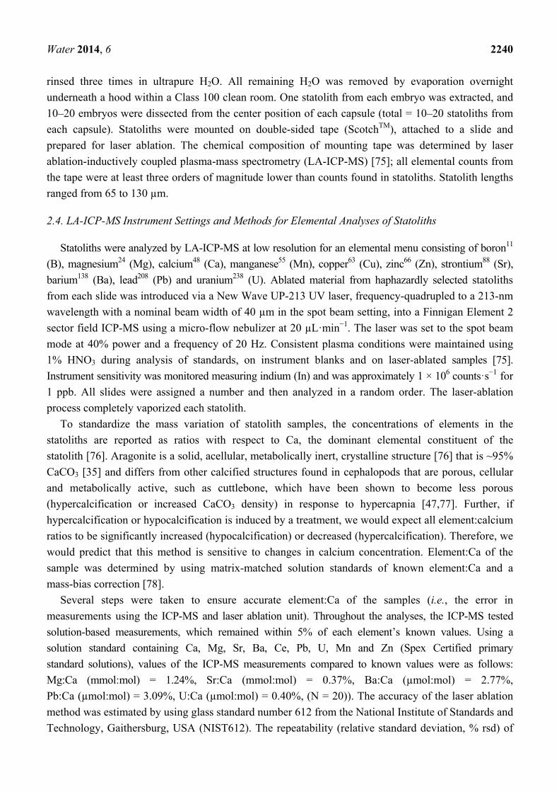
3.2. Multivariate Analyses
To test whether the elemental composition of statoliths varied among treatments, multivariate
analyses were conducted for each experiment using analysis of similarities (ANOSIM; one-way,
Euclidean-distance matrix, N = 9999 permutations) and principle component analysis (PCA). The

Water 2014, 6 2244
elemental composition between treatments was not distinguished in either Experiments 1 or 2 (ANOSIM:
Experiment 1, R = 0.022, p = 0.369; Experiment 2, R = 0.007, p = 0.359).
These multivariate results presented emphasize a limited relationship between statolith elemental
chemistry and the environmental pH and [O2]. ANOSIM analyses were used to compare statolith
chemistry among capsules within treatment (total of four tests; Table 3). Again, the capsules exhibited
a significant effect on statolith chemistry within each treatment group, with the exception of the low
pHOx treatment (although no effect was detected, this may be caused by a lack of statistical power
associated with a small sample size). Elements driving capsular differences were not similar between
the experiments (Table 4, Figure 4). These data indicate that any statolith-geochemical record would
be an integrated signal between the environmental pH and [O2] and physiological processes within
outer embryonic structures.
Table 3. Statolith elemental composition differences among capsules within each treatment
were tested using a one-way analysis of similarities (Euclidean-distance matrix; elemental
menu = B:Ca, Mg:Ca, Sr:Ca, Ba:Ca, U:Ca; N = 9999 permutations for all treatments, except
low pHOx (N = 3)).
Groups R Statistic p-Value
High pHOx 0.890 0.010 Low pHOx 1.000 0.333 Low [O2] 0.920 0.010 Low pH 0.892 0.010
Table 4. Elements driving compositional differences among treatments (correlation matrix;
elemental menu = B:Ca, Mg:Ca, Sr:Ca, Ba:Ca, Pb:Ca, U:Ca). Eigenvalues and the
percentage of variance explained are listed beneath each principle component (PC).
Bold lettering = significant.
Experiment 1
Element
PC 1
(2.43, 40.5%)
PC 2
(1.79, 30.0%)
PC 3
(0.76, 12.7%)
PC 4
(0.64, 10.7%)
PC 5
(0.20, 3.3%)
B:Ca (mmol:mol) 0.00280 0.62922 -0.43943 0.38658 0.47883
Mg:Ca (mmol:mol) 0.55641 −0.14115 0.51101 0.40241 −0.47422
Sr:Ca (mmol:mol) 0.55046 0.27674 0.18102 0.05333 −0.18725
Ba:Ca (µmol:mol) −0.38689 −0.35043 0.37102 0.78068 −0.04598
Pb:Ca (µmol:mol) −0.06976 0.50151 0.80384 0.24169 −0.15971
U:Ca (µmol:mol) 0.48254 −0.36505 0.36425 0.13378 0.69485
Experiment 2
Element
PC 1
(2.33, 38.8%)
PC 2
(1.37, 22.7%)
PC 3
(1.06, 17.7%)
PC 4
(0.57, 9.6%)
PC 5
(0.37, 6.2%)
B:Ca (mmol:mol) −0.35216 0.13043 0.74688 0.08813 0.21714
Mg:Ca (mmol:mol) 0.44385 0.17315 0.59349 −0.07149 0.06847
Sr:Ca (mmol:mol) 0.52043 0.25779 −0.21675 0.15301 0.71352
Ba:Ca (µmol:mol) −0.24548 0.64466 −0.12698 0.66566 −0.15275
Pb:Ca (µmol:mol) −0.24588 0.62852 −0.13803 −0.71931 0.08053
U:Ca (µmol:mol) 0.53611 0.27552 0.08822 −0.05656 −0.63972
Water 2014, 6 2245
Figure 4. Principal component analyses. Experiment 1: (A) Score plots among treatment groups; (B) Loading plot (correlation matrix, elemental menu = B:Ca, Mg:Ca, Sr:Ca, Ba:Ca, U:Ca). Purple = low pHOx; Black = high pHOx. Experiment 2: (C) Score plots among treatment groups; (D) Loading plot (correlation matrix, elemental menu = B:Ca, Mg:Ca, Sr:Ca, Ba:Ca, U:Ca). Blue = low [O2]; Red = low pH.
3.3. Statoliths as an Indicator of Environmental Response
The experiments conducted here provide the first evidence that embryonic statolith geochemistry can be affected by environmental [O2] and pH at levels that occur at natal sites. B:Ca and U:Ca were investigated as pH proxies; however, they did not exhibit the direct relationship with environmental pH that has been found with foraminifera shells [60–62], coral skeletons [63] and mollusk shells [64]. These B:Ca results suggest that, when environmental pHT is low (7.55–7.56) and [O2] is high

Water 2014, 6 2246
(241–242 µmol·kg−1), squid embryos can regulate the endolymph pH within the statocyst, where the
statolith crystal grows. Constant pH was found in a study of the endolymph fluid of squid
statocysts [41], and pH has been shown to be highly regulated in the endolymph fluid of saccules of
fish (the squid statocyst analog) [81,82]. Lower taxonomical groups have a more direct relationship
with seawater, with less integration of physiological processes [76]. Seawater with low pHT has higher
levels of bioavailable U, and in foraminifera, the incorporation of U into biogenic CaCO3 increases
with decreasing [CO32−] ([61,62,85]). Because we did not find evidence of U enrichment within
statoliths in the low-pH treatment, we presume that [CO32−] is also regulated within endolymph fluid.
These results suggest that both pH and [CO32−] are highly regulated within the squid embryos and
the statocyst, as has been found with other squid [41], and these regulation processes are unaffected by
an environmental pHT level of 7.55. One option is that high pCO2/low pH in seawater does not
significantly affect the blood chemistry of squid embryos [86], due to ion-regulating epithelia regulating
internal pH [17–19] (Figure 2). However, it is also possible that squid embryos exposed to high
pCO2/low pH can utilize energy derived from yolk reserves to compensate for putative alterations in
their internal pH. Further testing is needed to determine if there is a threshold below which squid are
not able to regulate their pH. Moreover, it is essential to know if the D. opalescens embryo is internally
acidified during exposure to realistic, high pCO2/low pH conditions and the compensatory mechanisms
that are involved.
Development of an environmental [O2] proxy is still in its infancy, and more research is needed to
test for different mechanisms. However, U:Ca and Sr:Ca were enriched in squid statoliths grown in low
[O2] treatments (Figure 3). Environmental strontium is critical in the formation of the statolith [59]. Sr:Ca
and Ba:Ca are widely reported to have a strong, often negative, relationship with temperature, although
for Sr:Ca, the relationship can be more complex [87]. U:Ca was recently reported to have a positive
relationship with temperature [88]. Since all tanks were kept within 1 °C of one another (Figure 1) and
did not reach the temperature differentials reported to generate Sr signals for a congener squid species
(>2 °C, [48]) and one gastropod (= 4 °C, [73]), our results are not likely related to temperature effects.
Curiously, when exposed to low [O2] with low pH (low pHOx), statoliths were not enriched with
Sr. Strontium incorporation into squid statoliths may be inversely related to metabolic rate.
Low environmental pH/high pCO2 and high temperatures can cause metabolic depression in squid
embryos [45,46] and reduced growth [52].
The only element:calcium measured in this study that might be a useful indicator of low pHOx
conditions is U:Ca. We showed that U:Ca is enriched (eight-fold increase) in the statoliths of embryos
exposed to low pHOx relative to those exposed to the high pHOx treatment. We propose that this
enrichment is driven by low [O2] and exacerbated by the interactive effect of low [O2] and pH (low
pHOx). Under low pH stress, low [O2] may impair the regulation of internal pH by embryonic squid,
due to the reduced aerobic metabolic rate. This can lead to insufficient ATP production necessary to
fuel active mechanisms for pH regulation and calcification. However, this indicator of an
environmental response may not be useful as a proxy per se, because it likely tracks a sublethal effect
on the embryo. Specifically, the uranium enrichment reflects the loss of pH regulation in the
endolymph of the statocyst and may represent a threshold rather than a gradient.
The results presented show that the statolith chemistry records integrated the effects of the
environment in concert with physiological processes, here identified as capsular effects (Tables 2 and 4).
Water 2014, 6 2247
These data suggest that statolith-chemical composition has a substantial disconnect from the external
seawater environment (unlike foraminifera and their shells and some corals and their skeletons).
Elemental composition measurements from the capsule jelly, perivitelline fluid [89] and within the
endolymph fluid of the statocyst in addition to environmental measurements would help clarify the
relationship between the environment and statolith geochemistry.
4. Conclusions
For the first time that we are aware, we demonstrated that environmental pH and [O2] affect squid
statolith geochemistry (uranium:calcium) and that statolith geochemistry is strongly affected by factors
associated with the capsule (capsular effects). The only other known study that tested the effects of
environmental pH on squid-embryo statolith geochemistry found that only 65Zn significantly differed
from an elemental suite that included 110mAg, 109Cd, 57Co, 203Hg and 54Mn [49]. Evidence that
environmental tracers in squid statoliths can track seawater pH and [O2] is especially useful, because
uranium has been shown to be promising for understanding squid life history, migrations and
habitat use [88,90,91].
However, we did not find strong evidence that environmental pH and [O2] effects can be resolved
for the use of statolith geochemistry as environmental proxies of pH and [O2]. We did find strong
capsular effects. The mechanism behind the capsular effect on statolith elemental incorporation is
presumably due to a process that similarly affects all embryos of the capsule. Mechanisms include
maternal transfer [53,73,83,84] and capsular and chorion membrane structural differences [49,50]
among capsules. Less likely mechanisms include processes within the embryos [17–19,45,46] that are
expressed similarly among embryos. These capsular effects are the first evidence (statolith chemistry)
of strong physiological differences among the same cohort, and the importance of these differences for
the persistence of the D. opalescens population is not known. Future applications might include the use
of uranium:calcium as a geochemical marker tracking the initiation and duration of sublethal effects.
Acknowledgments
We thank the Birch Aquarium at Scripps Institution of Oceanography for providing critical
experimental space and seawater. We thank Georges Paradis at UC Santa Barbara for his expertise
running the LA-ICP-MS, Andrew G. Dickson and Todd R. Martz for allowing us to use their labs to
run AT, salinity and [O2] samples, and Gwyneth Gordon, Arizona State University, for analyzing our
seawater samples. We acknowledge the value of the Del Mar Mooring data in the experimental design
and thank Uwe Send, SungHyun Nam and Todd Martz for keeping the oxygen and pH data flowing.
We also thank Martin Tresguerres, Serge Belongie, and Guillermo “Lionel Richie” Mendoza for
reviewing and improving this manuscript. We thank those assisting with the field collections (CA
Permit No. 7230), especially Phil Zerofski and Javier Naretto. This work was funded by National
Oceanic and Atmospheric Administration (NOAA) Grant No. NA10OAR4170060, California Sea
Grant College Program Project Nos. R/CC-02 and R/CC-04, through NOAA’s National Sea Grant
College Program, U.S. Department of Commerce, and by the National Science Foundation-Division of
Ocean Science Award Nos. 0903551, 0927445 and 1041062. We thank three anonymous reviewers for
their comments that improved the manuscript. The statements, findings, conclusions and
Water 2014, 6 2248
recommendations are those of the authors and do not necessarily reflect the views of California Sea
Grant, state agencies, NOAA, NSF or the U.S. Department of Commerce.
Author Contributions
Lisa A. Levin and Michael O. Navarro conceived and funded the research. Michael O. Navarro,
Emily E. Bockmon and Lisa A. Levin designed the experiments. Michael O. Navarro, Jennifer P.
Gonzalez, Christina A. Frieder and Emily E. Bockmon performed the experiments. Michael O.
Navarro, Emily E. Bockmon and Christina A. Frieder analyzed the data. Michael O. Navarro wrote the
paper, and Lisa A. Levin, Christina A. Frieder and Emily E. Bockmon edited and proofed the paper.
Appendix
Table A1. Seawater elemental concentrations measured in the laboratory experiments.
Values are the average ± one standard error for samples taken once a week in each
treatment (n = 5). No treatment effects were found for either Experiments 1 or 2.
Treatment (Tank) B (ppm) Mg (ppm) Ca (ppm) Sr (ppm) Ba (ppb) U (ppb)
Experiment 1
Low pHOx (1) 4.45 ± 0.005 1092 ± 11.6 351.0 ± 3.40 5.64 ± 0.055 4.41 ± 0.120 1.98 ± 0.410
Low pHOx (2) 4.45 ± 0.005 1100 ± 14.8 353.2 ± 4.39 5.70 ± 0.084 4.51 ± 0.129 2.03 ± 0.408
High pHOx (1) 4.45 ± 0.003 1113 ± 12.6 357.7 ± 4.16 5.76 ± 0.064 4.65 ± 0.062 2.12 ± 0.428
High pHOx (2) 4.44 ± 0.014 1118 ± 14.7 358.5 ± 5.00 5.78 ± 0.074 4.51 ± 0.153 2.03 ± 0.404
Treatment Effect F1,4 = 1.588,
p = 0.222
F1,4 = 2.039,
p = 0.169
F1,4 = 1.969,
p = 0.176
F1,4 = 2.077,
p = 0.165
F1,4 = 0.957,
p = 0.340
F1,4 = 0.030,
p = 0.864
Experiment 2
Low [O2] (1) 4.48 ± 0.004 1100 ± 14.9 355.6 ± 4.99 5.69 ± 0.077 4.34 ± 0.115 2.11 ± 0.190
Low [O2] (2) 4.48 ± 0.004 1115 ± 15.1 358.6 ± 6.28 5.76 ± 0.089 4.51 ± 0.168 2.20 ± 0.176
Low pH (1) 4.48 ± 0.003 1131 ± 17.8 365.4 ± 6.10 5.87 ± 0.097 4.41 ± 0.067 2.08 ± 0.247
Low pH (2) 4.48 ± 0.004 1108 ± 31.7 359.2 ± 10.45 5.72 ± 0.163 4.34 ± 0.083 1.85 ± 0.122
Treatment Effect F1,4 = 0.038,
p = 0.847
F1,4 = 0.336,
p = 0.571
F1,4 = 0.513,
p = 0.484
F1,4 = 0.607,
p = 0.447
F1,4 = 0.189,
p = 0.669
F1,4 = 1.016,
p = 0.329
Conflicts of Interest
The authors declare no conflict of interest.

Water 2014, 6 2249
References
1. Nam, S.; Kim, H.J.; Send, U. Amplification of hypoxic and acidic events by La Niña conditions on
the continental shelf off California. Geophys. Res. Lett. 2011, 38, doi:10.1029/2011gl049549.
2. Send, U.; Nam, S. Relaxation from upwelling: The effect on dissolved oxygen on the contintental
shelf. J. Geophys. Res. Oceans 2012, 117, doi:10.1029/2011JC007517.
3. Frieder, C.A.; Nam, S.H.; Martz, T.R.; Levin, L.A. High temporal and spatial variability of dissolved
oxygen and pH in a nearshore California kelp forest. Biogeosciences 2012, 9, 3917–3930.
4. Booth, J.A.T.; McPhee-Shaw, E.E.; Chua, P.; Kingsley, E.; Denny, M.; Phillips, R.; Bograd, S.J.;
Zeidberg, L.D.; Gilly, W. Natural intrusions of hypoxic, low pH water into nearshore marine
environments on the California coast. Cont. Shelf Res. 2012, 45, 108–115.
5. Gilly, W.F.; Beman, J.M.; Litvin, S.Y.; Robison, B.H. Oceanographic and biological effects of
shoaling of the oxygen minimum zone. Annu. Rev. Mar. Sci. 2013, 5, 393–420.
6. Booth, J.A.T.; Woodson C.B.; Sutula M.; Micheli F.; Weisberg S.B.; Bograd S.J.; Steele A.;
Schoen J.; Crowder L.B. Patterns and potential drivers of declining oxygen content along the
southern California coast. Limnol. Oceanogr.2014, 59, in press.
7. Bograd, S.J.; Buil, M.P.; DiLirenzo, E.; Castro, C.G.; Schroeder, I.D.; Goericke, R.; Anderson, C.R.;
Benitez-Nelson, C.; Whitney, F.A. Changes in source waters to the Southern California Bight.
Deep Sea Res. II 2014, doi:10.1016/j.dsr2.2014.04.009.
8. Pörtner, H.O.; Langenbuch, M.; Michaelidis, B. Synergistic effects of temperature extremes,
hypoxia, and increases in CO2 on marine animals: From Earth history to global change.
J. Geophys. Res. Oceans 2005, 110, doi:10.1029/2004jc002561.
9. Doney, S.C.; Balch, W.M.; Fabry, V.J.; Feely, R.A. Ocean acidification: A critical emerging
problem for the ocean sciences. Oceanography 2009, 22, 16–25.
10. Doney, S.C.; Fabry, V.J.; Feely, R.A.; Kleypas, J.A. Ocean acidification: The other CO2 problem.
Ann. Rev. Mar. Sci. 2009, 1, 169–192.
11. Gruber, N.; Hauri, C.; Lachkar, Z.; Loher, D.; Frolicher, T.L.; Plattner, G. Rapid progression of
ocean acidification in the California Current System. Science 2012, 337, 220–223.
12. Munday, P.L.; Dixson, D.L.; McCormick, M.I.; Meekan, M.; Ferrari, M.C.O.; Chivers, D.P.
Replenishment of fish populations is threatened by ocean acidification. PNAS 2010, 107,
12930–12934.
13. Munday, P.L.; Hernaman, V.; Dixson, D.L.; Thorrold, S.R. Effect of ocean acidification on
otolith development in larvae of a tropical marine fish. Biogeosciences 2011, 8, 1631–1641.
14. Munday, P.L.; Pratchett, M.S.; Dixson, D.L.; Donelson, J.M.; Endo, G.G.K.; Reynolds, A.D.;
Knuckey, R. Elevated CO2 affects the behavior of an ecologically and economically important
reef fish. Mar. Biol. 2013, 160, 2137–2144.
15. Hamilton, T.J.; Holcombe, A.; Tresguerres, M. CO2-induced ocean acidification increases anxiety
in rockfish via alteration of GABAA receptor functioning. Proc. R. Soc. B 2014, 281, 20132509.
16. Melzner, F.; Gutowska, M.A.; Langenbuch, M.; Dupont, S.; Lucassen, M.; Thorndyke, M.C.;
Bleich, M.; Pörtner, H.O. Physiological basis for high CO2 tolerance in marine ectothermic
animals: Pre-adaptation through lifestyle and ontogeny? Biogeosciences 2009, 6, 2313–2331.

Water 2014, 6 2250
17. Hu, M.Y.; Tseng, Y.; Stumpp, M.; Gutowska, M.A.; Kiko, R.; Lucassen, M.; Melzner, F.
Elevated seawater pCO2 differentially affects brachial acid-base transporters over the course of
development in the cephalopod Sepia officinalis. Am. J. Physiol. Regul. Integr. Comp. Physiol.
2011, 300, R1100–R1114.
18. Hu, M.Y.; Tseng, Y.; Lin, L.; Chen, P.; Charmantier-Daures, M.; Hwang, P.; Melzner, F.
New insights into ion regulation of cephalopod molluscs: A role of epidermal ionocytes in
acid-base regulation during embryogenesis. Am. J. Physiol. Regul. Integr. Comp. Physiol. 2011,
301, R1700–R1709.
19. Hu, M.Y.; Lee, J.; Lin, L.; Shih, T.; Stumpp, M.; Lee, M.; Hwang, P.; Tseng, Y. Development in
a naturally acidified environment: Na+/H+-exchanger 3-based proton secretion leads to CO2
tolerance in cephalopod embryos. Front. Zool. 2013, 10, 1–16.
20. Strathmann, R.R.; Chaffee, C. Constraints on egg masses. II. Effect of spacing, size, and number
of eggs on ventilation of masses of embryos in jelly, adherent groups, or thin-walled capsules.
J. Exp. Mar. Biol. Ecol. 1984, 84, 85–93.
21. Zeidberg, L.D.; Isaac, G.; Widmer, C.L.; Neumeister, H.; Gilly, W.F. Egg capsule hatch rate and
incubation duration of the California market squid, Doryteuthis (=Loligo) opalescens: Insights
from laboratory manipulations. Mar. Ecol. 2011, 32, 468–479.
22. Giorgi, A.E.;Congleton, J.L. Effects of current velocity on development and survival of lingcod,
Ophiodon elongatus, embryos. Environ. Biol. Fishes 1984, 10, 15–27.
23. Messieh, S.N.; Rosenthal, H. Mass mortality of herring eggs on spawning beds on and near
Fisherman’s Bank, Gulf of St. Lawrence. Can. Aquat. Living Resour. 1989, 2, 1–8.
24. Bograd, S.J.; Castro, C.G.; DiLorenzo, E.; Palacios, D.M.; Bailey, H.; Gilly, W.; Chavez, F.P.
Oxygen declines and the shoaling of the hypoxic boundary in the California Current.
Geophys. Res. Lett. 2008, 35, doi:10.1029/2008GL034185.
25. Checkley, D.M.; Barth, J.A. Patterns and processes in the California Current System.
Prog. Oceanogr. 2009, 83, 49–64.
26. Pauly, D. Gasping fish and panting squids: Oxygen, temperature and the growth of
water-breathing animals. In Excellence in Ecology; Kline, O., Ed.; International Ecology Institute:
Oldendorf/Luhe, Germany, 2010.
27. Vojkovich, M. The California fishery for market squid (Loligo opalescens). CalCOFI Rep. 1998,
39, 55–60.
28. Reiss, C.S.; Maxwell, M.R.; Hunter, J.R. Investigating environmental effects on population
dynamics of Loligo opalescens in the Southern California Bight. CalCOFI Rep. 2004, 45, 87–97.
29. Koslow, J.A.; Allen, C. The influence of the ocean environment on the abundance of market squid,
Doryteuthis (Loligo) opalescens, paralarvae in the Southern California Bight. CalCOFI Rep. 2011,
52, 205–213.
30. Warner, R.R.; Hamilton, S.L.; Sheehy, M.S.; Zeidberg, L.D.; Brady, B.C.; Caselle, J.E.
Geographic variation in natal and early larval trace-elemental signatures in the statoliths of the
market squid Doryteuthis (formerly Loligo) opalescens. Mar. Ecol. Prog. Ser. 2009, 379, 109–121.
31. Dorval, E.; Crone, P.R.; McDaniel, J.D. Variability of egg escapement, fishing mortality
and spawning population in the market squid fishery in the California Current Ecosystem.
Mar. Freshw. Res. 2013, 64, 80–90.

Water 2014, 6 2251
32. Zeidberg, L.D.; Butler, J.L.; Ramon, D.; Cossio, A.; Stierhoff, K.L.; Henry, A. Estimation of
spawning habitats of market squid (Doryteuthis opalescens) from field surveys of eggs off central
and southern California. Mar. Ecol. 2011, 33, 326–336.
33. Arkhipkin, A.I. Statoliths as “black boxes” (life recorders) in squid. Mar. Freshw. Res. 2005, 56,
573–583.
34. Thorrold, S.R.; Zacherl, D.C.; Levin, L.A. Population connectivity and larval dispersal using
geochemical signatures in calcified structures. Oceanography 2007, 20, 80–89.
35. Radtke, R.L. Chemical and structural characteristics of statoliths from the short-finned squid
Illex illecebrosus. Mar. Biol. 1983, 76, 47–54.
36. Arkhipkin, A.I.; Bizikov, V.A. Role of the statolith in functioning of the acceleration receptor
system in squids and sepioids. J. Zool. Lond. 2000, 250, 31–55.
37. Fields, W.G. The structure, development, food relations, reproduction, and life history of the
squid, Loligo opalescens, Berry. CA Fish. Bull. 1965, 131, 1–105.
38. Bettencourt V.; Guerra A. Growth increments and biomineralization process in cephalopod
statoliths. J. Exp. Mar. Biol. Ecol. 2000, 248, 191–205.
39. Clarke, M.R. The cephalopod statolith—An introduction to its form. J. Mar. Biol. Ass. U.K. 1978,
58, 701–712.
40. Villanueva R.; Moltschaniwskyj, N.A.; Bozzano, A. Abiotic influences on embryo growth:
Statoliths as experimental tools in the squid early life history. Rev. Fish. Biol. Fish. 2007, 17,
101–110.
41. Morris, C.C. Statocyst fluid composition and its effects on calcium carbonate precipitation in the squid
Alloteuthis subulata (Lamarck, 1798): Towards a model for biomineralization. Bull. Mar. Sci. 1991,
49, 379–388.
42. Cronin, E.R.; Seymour, R.S. Respiration of the eggs of the giant cuttlefish Sepia apama. Mar.
Biol. 2000, 136, 863–870.
43. Gutowska, M.A.; Melzner, F. Abiotic conditions in cephalopod (Sepia officinalis) eggs:
Embryonic development at low pH and high pCO2. Mar. Biol. 2009, 156, 515–519.
44. Strobel, A.; Hu, M.Y.A.; Gutowska, M.A.; Lieb, B.; Lucassen, M.; Melzner, F.; Pörtner, H.O.;
Mark, F.C. Influence of temperature, hypercapnia, and development on the relative expression
of different hemocyanin isoforms in the common cuttlefish Sepia officinalis. J. Exp. Zool. A Ecol.
Genet. Physiol. 2012, 317, 511–523.
45. Rosa, R.; Pimentel, M.S.; Boavida-Portugal, J.; Teixeira, T.; Trubenbach, K.; Diniz, M. Ocean
warming enhances malformations, premature hatching, metabolic suppression, and oxidative
stress in the early life stages of a keystone squid. PLOS ONE 2012, 7, e38282.
46. Rosa, R.; Trubenbach, K.; Pimentel, M.S.; Boavida-Portugal, J.; Faleiro, F.; Baptista, M.;
Dionisio, G.; Calado, R.; Pörtner, H.O.; Repolho, T. Differential impact of ocean acidification on
winter and summer progeny of a coastal squid (Loligo vulgaris). J. Exp. Biol. 2014, 217, 518–525.
47. Dorey, N.; Melzner, F.; Martin, S.; Oberhansli, F.; Teyssie, J.; Bustamante, P.; Gattuso, J.;
Lacoue-Labarthe, T. Ocean acidification and temperature rise: Effects on calcification during
early development of the cuttlefish Sepia officinalis. Mar. Biol., 2013, 160, 2007–2022.
Water 2014, 6 2252
48. Arkhipkin, A.I.; Campana, S.E.; FitzGerald, J.; Thorrold, S.R. Spatial and temporal variation in
elemental signatures of statoliths from the Patagonian longfin squid (Loligo gahi). Can. J. Fish.
Aquat. Sci. 2004, 61, 1212–1224.
49. Lacoue-Labarthe, T.; Reveillac, E.; Oberhansli, F.; Teyssie, J.L.; Jeffree, R.; Gattuso, J.P.
Effects of ocean acidification on trace element accumulation in the early-life stages of squid
Loligo vulgaris. Aquat. Toxicol. 2011, 105, 166–176.
50. Lacoue-Labarthe, T.; Villanueva, R.; Rouleau, C.; Oberhansli, F.; Teyssie, J.; Jeffree, R.;
Bustamante, P. Radioisotopes demonstrate the contrasting bioaccumulation capacities of heavy
metals in embryonic stages of cephalopod species. PLOS ONE 2011, 6, e27653.
51. Villanueva, R. Differential increment-deposition rate in embryonic statoliths of the loliginid squid
Loligo vulgaris. Mar. Biol. 2000, 137, 161–168.
52. Kaplan, M.B.; Mooney, T.A.; McCorkle, D.C.; Cohen, A.L. Adverse effects of ocean acidification on
early development of squid (Doryteuthis pealeii). PLoS ONE 2013, 8, e63714.
53. Steer, M.A.; Moltschaniwskyj, N.A.; Gowland, F.C. Temporal variability in embryonic
development and mortality in the southern calamary Sepioteuthis australis: A field assessment.
Mar. Ecol. Prog. Ser. 2002, 243, 143–150.
54. O’Dor, R.K.; Wells, M.J. Energy and nutrient flow. In Cephalopod Life Cycles. Academic Press:
London, UK, 1987.
55. Strathmann, R.R.; Strathmann, M.F. Oxygen supply and limits on aggregation of embryos.
J. Mar. Biol. Ass. U.K. 1995, 75, 413–428.
56. Cohen, C.S.; Strathmann, R.R. Embryos at the edge of tolerance: Effects of environment and
structure of egg masses on supply of oxygen to embryos. Biol. Bull. 1996, 190, 8–15.
57. Gutwoska, M.A.; Pörtner, H.O.; Melzner, F. Growth and calcification in the cephalopod Sepia
officianlis under elevated seawater pCO2. Mar. Ecol. Prog. Ser. 2008, 373,303–309.
58. Gutowska, M.A.; Melzner, F.; Langenbuch, M.; Bock, C.; Claireaux, G.; Pörtner, H.O. Acid-base
regulatory ability of the cephalopod (Sepia officinalis) in response to environmental hypercapnia.
J. Comp. Physiol. B. 2010, 180, 323–335.
59. Hanlon, R.T.; Bidwell, J.P.; Tait, R. Strontium is required for statolith development and thus
normal swimming behaviour of hatchling cephalopods. J. Exp. Biol. 1989, 141, 187–195.
60. Spivack, A.J.; You, C.; Smith, H.J. Foraminiferal boron isotope ratios as a proxy for surface
ocean pH over the past 21 Myr. Nature 1993, 363, 149–151.
61. Raitzsch, M.; Kuhnert, H.; Hathorne, E.C.; Groeneveld, J.; Bickert, T. U/Ca in benthic
foraminifers: A proxy for the deep-sea carbonate saturation. Geochem. Geophys. Geosyst. 2011,
12, Q06019.
62. Keul, N.; Langer, G.; Nehrke, G.; Jan de Nooijer, L.; Nehrke, G.; Reichart, G.; Bijma, J.
Incorporation of uranium in benthic formaniferal calcite reflects seawater carbonate ion
concentration. Geochem. Geophys. Geosyst. 2013, 14, 102–111.
63. Inoue, M.; Suwa, R.; Suzuki, A.; Sakai, K.; Kawhata, H. Effects of seawater pH on growth and
skeletal U/Ca ratios of Acropora digitifera coral polyps. Geophys. Res. Lett. 2011, 38,
doi:10.1029/2011GL047786.
64. Frieder, C.A.; Gonzalez, J.P.; Levin, L.A. Uranium in larval shells as a barometer of molluscan
ocean acidification exposure. Environ. Sci. Technol. 2014, 48, 6401–6408.

Water 2014, 6 2253
65. Mayol, E.; Ruiz-Halpern, S.; Duarte, C.M.; Castilla, J.C.; Pelegri, J.L. Coupled CO2 and
O2-driven compromises to marine life in summer along the Chilean sector of the Humboldt
Current System. Biogeosciences 2012, 9, 1183–1194.
66. Melzner, F.T.; Thomsen, J.; Koeve, W.; Oschlies, A.; Gutowska, M.A.; Bange, H.W.;
Hansen, H.P.; Körtzinger, A. Future ocean acidification will be amplified by hypoxia in coastal
habitats. Mar. Biol. 2013, 160, 1875–1888.
67. Bockmon, E.E.; Frieder, C.A.; Navarro, M.O.; White-Kershek, L.A.; Dickson, A.G. Technical
note: Controlled experimental aquarium system for multi-stressor investigation of carbonate
chemistry, oxygen saturation, and temperature. Biogeosciences 2013, 10, 5967–5975.
68. Arnold, J.M. Normal embryonic stages of the squid Loligo pealii (Lesueur). Biol. Bull. 1965, 128,
24–32.
69. Segawa, S.; Yang, W.T.; Marthy, H.J.; Hanlon, R.T. Illustrated embyonic stages of the eastern
Atlantic squid, Loligo forbesi. Veliger 1988, 30, 230–243.
70. Foote, K.G.; Hanlon, R.T.; Iampietro, P.J.; Kvitek, R.G. Acoustic detection and quantification of
benthic egg beds of the squid Loligo opalescens in Monterey Bay, California. J. Acoust. Soc. Am.
2006, 119, 844–856.
71. Yatsu, A.; Mochioka, N.; Morishita, K.; Toh, H. Strontium/Calcium ratios in statoliths of the neon
flying squid, Ommastrephes bartrami (Cephalopoda), in the North Pacific Ocean. Mar. Biol.
1998, 131, 275–282.
72. Villanueva, R.; Arkhipkin, A.; Jereb, P.; Lefkaditou, E.; Lipinski, M.R.; Perales-Raya, C.;
Riba, J.; Rocha, F. Embryonic life of the loliginid squid Loligo vulgaris: Comparison between
statoliths of Atlantic and Mediterranean populations. Mar. Ecol. Prog. Ser. 2003, 253, 197–208.
73. Lloyd, D.C.; Zacherl, D.C.; Walker, S.; Paradis, G.; Sheehy, M.; Warner, R.R. Egg source,
temperature and culture seawater affect elemental signatures in Kelletia kelletii larval statoliths.
Mar. Ecol. Prog. Ser. 2008, 353, 115–130.
74. Lee, K.; Kim, T.; Byrne, R.H.; Millero, F.J.; Feely, R.A.; Lui, Y. The universal ratio of boron to
chlorinity for the North Pacific and North Atlantic oceans. Geochim. Cosmochim. Acta 2010, 74,
1801–1811.
75. Zacherl, D.C.; Paradis, G.; Lea, D. Barium and strontium uptake into larval protoconchs and
statoliths of the marine neogastropod Kelletia kelletii. Geochim. Comoschim. Acta 2003, 67,
4091–4099.
76. Campana, S.E. Chemistry and composition of fish otoliths: Pathways, mechanisms and
applications. Mar. Ecol. Prog. Ser. 1999, 188, 263–297.
77. Gutowska, M.A.; Melzner, F.; Pörtner, H.O.; Meier, S. Cuttlebone calcification increases during
exposure to elevated seawater pCO2 in the cephalopod Sepia officinalis. Mar. Biol. 2010, 157,
1653–1663.
78. Rosenthal, Y.; Field, M.P.; Sherrell, R.M. Precise determination of element/Calcium ratios in
calcareous samples using sector field inductively coupled plasma mass spectrometry. Anal. Chem.
1999, 71, 3248–3253.
79. Swearer, S.E.; Forrester, G.E.; Steele, M.A.; Brooks, A.J.; Lea, D.W. Spatio-temporal and
interspecific variation in otolith trace-elemental signatures in a temperate estuarine fish assemblage.
Estuar. Coast. Shelf Sci. 2003, 56, 1111–1123.

Water 2014, 6 2254
80. Bennington, C.C.; Thayne, W.V. Use and misuse of mixed model analysis of variance in
ecological studies. Ecology 1994, 75, 717–722.
81. Payan, P.; Kossmann, H.; Watrin, A.; Mayer-Gostan, N.; Boeuf, G. Ionic composition of
endolymph in teleosts: Origin and importance of endolymph alkalinity. J. Exp. Biol. 1997, 200,
1905–1912.
82. Shiao, J.; Lin, L.; Horng, J.; Hwang, P.; Kaneko, T. How can teleostean inner ear hair cells
maintain the proper association with the accreting otolith? J. Comp. Neurol. 2005, 488, 331–341.
83. Lacoue-Labarthe, T.; Warnau, M.; Oberhansli, F.; Teyssie, J.; Jeffree, R.; Bustamante, P.
First experiments on the maternal transfer of metals in the cuttlefish Sepia officinalis. Mar. Pollut. Bull.
2008, 57, 826–831.
84. Thorrold, S.R.; Jones, G.P.; Planes, S.; Hare, J.A. Transgenerational marking of embryonic otoliths in
marine fishes using barium stable isotopes. Can. J. Fish. Aquat. Sci. 2006, 63, 1193–1197.
85. Russell, A.D.; Hönisch, B.; Spero, H.J.; Lea, D.W. Effects of seawater carbonate ion
concentration and temperature on shell U, Mg, and Sr in cultured planktonic foraminifera.
Geochim. Cosmochim. Acta 2004, 68, 4347–4361.
86. Redfield, A.C.; Goodkind, R. The significance of the Bohr effect in the respiration and
asphysixiation of the squid, Loligo pealei. J. Exp. Biol. 1929, 6, 340–349.
87. Bath, G.E.; Thorrold, S.R.; Jones, C.M.; Campana, S.E.; McLaren, J.W.; Lam, J.W.H. Strontium
and barium uptake in aragonitic otoliths of marine fish, Geochim. Cosmochim. Acta 2000, 64,
1705–1714.
88. Liu, B.; Chen, X.; Chen, Y.; Tian, S. Geographic variation in statolith trace elements of the
Humboldt squid, Dosidicus gigas, in high seas of Eastern Pacific Ocean. Mar. Biol. 2013, 160,
2853–2862.
89. De Leersnyder, M.; Lemaire, J. Sur la composition minerale du liquid periembryonnaire de
L’oeuf de Sepia officinalis L. J. Cah. Biol. Mar. 1972, 8, 429–431.
90. Zumholz, K.; Klugel, A.; Hansteen, T.; Piatkowski, U. Statolith microchemistry traces the
environmental history of the boreoatlantic armhook squid, Gonatus. fabricii. Mar. Ecol. Prog. Ser.
2007, 333, 195–204.
91. Arbuckle, N.S.M.; Wormuth, J.H. Trace elemental patterns in Humboldt squid statoliths from
three geographic regions. Hydrobiologia 2014, 725, 115–123.
© 2014 by the authors; licensee MDPI, Basel, Switzerland. This article is an open access article
distributed under the terms and conditions of the Creative Commons Attribution license
(http://creativecommons.org/licenses/by/3.0/).