microbial production of silver nanoparticles … 4, issue 4...microbial production of silver...
TRANSCRIPT

IOSR Journal of Biotechnology and Biochemistry (IOSR-JBB)
ISSN: 2455-264X, Volume 4, Issue 4 (Jul. – Aug. 2018), PP 26-38
www.iosrjournals.org
DOI: 10.9790/264X-0404022638 www.iosrjournals.org 26 | Page
Microbial Production of Silver Nanoparticles (Agnps) By Some
Bacterial Isolates
Amar Kumar1, Baidyanath Kumar
2, Ashok Ghosh
1, Mini Tiwari
1, Muddassir
Reyaz3
1. Department of EWM, A. N. College (Patliputra University), Patna- 800013
2. Visiting Professor, Department of Biotechnology, Patna Science College (Patna University), Patna- 800005
3. Department of Biotechnology, College of Commerce, Arts and Science (Patliputra University), Patna-
800020
Corresponding Author: Dr. Baidyanath Kumar
Abstract The five bacterial strains viz. Brevibacillus centrosporus DSM8445T, Brevibacillus invocatus NCIMB13772T,
Brevibacillus choshinensis DSM8552T, Brevibacillus panacihumi DCY35 and Brevibacillus levickii
LKG22481were screened for production of AgNPs using their extracellular (cell free supernatant) of bacterial
cultures. The primary sight for AgNPs formation was change of colour of reaction mixture from pale yellow to
dark brown.
In the present investigation it is evident that isolated bacterial strain Brevibacillus invocatus NCIMB13772T
was the most efficient bacterial strain in production of AgNPs (0.996) followed by isolated bacterial strain
Brevibacillus centrosporus DSM8445T (0.960), Brevibacillus panacihumi DCY35 (0.920), Brevibacillus levickii
LMG22481 (0.382) and Brevibacillus chosinensis DSM8552T (Table- 2). The primary conformation of synthesis
of nanoparticles in the medium was characterized by the changes in color from yellowish white to brown , the
knowledge about the reduction of silver ions and formation of silver nanoparticles were still not clear, but it is
believed that protein molecules and enzyme, the nitrate reductase act as good regulating agent in silver
nanoparticles synthesis. The biosynthesis of AgNPs by these five bacterial strains has also been confirmed by
FTIR, XRD and SEM techniques.
Key Words: Biogenic nanoparticles; Biological nanoparticles, Cell free supernatant, Brevibacillus strains,
Silver nitrate, UV- Vis Spectroscopy, XRD, SEM, FTIR
----------------------------------------------------------------------------------------------------------------------------- ----------
Date of Submission: 22-08-2018 Date Of Acceptance: 04-09-2018
----------------------------------------------------------------------------------------------------------------------------- ----------
I. Introduction The term “nano” is derived from a Greek word “nanos” which means dwarf and thus denotes a
measurement on the scale of one-billionth (109) of a meter in size
1, 2 (Narayanan and Sakthivel, 2010; Thakkar et
al. 2010). Nanoparticles are defined as particulate dispersions of solid particles with at least one dimension at a
size range of 10-1000 nm2, 3
(Thakkar et al. 2010; Mohanray and Chen, 2006). The most important feature of
nanoparticles is their surface area to volume aspect ratio, allowing them to interact with other particles easier1, 2
(Narayanan and Sakthivel, 2010; Thakkar et al. 2010).
In order to survive in environments containing high levels of metals, living organisms have adapted by
evolving mechanisms to cope with them. These mechanisms may involve altering the chemical nature of the
toxic metal so that it no longer causes toxicity, resulting in the formation of nanoparticles of the metal
concerned. Thus nanoparticle formation is the “by-product” of a resistance mechanism against a specific metal,
and this can be used as an alternative way of producing them. Nanoparticles have unique thermal, optical,
physical, chemical, magnetic and electrical properties compared to their bulk material counterparts4, 5
(Husseiny
et al. 2007; Duran et al. 2007). These features can be exploited for next generation biosensors, electronics,
catalysts and antimicrobials1,5
(Narayanan and Sakthivel, 2010; Duran et al. 2007). Metallic nanoparticles are
most important in present day science and widely studied group of materials, showing great diversity and many
different uses.
There are important links between the way nanoparticles are synthesised and their potential uses. Silver
nanoparticles (AgNPs) have been shown in numerous studies to display antibacterial properties5, 6, 7, 8
(Duran et
al. 2007; Guzman et al. 2012; Guzman et al. et al. 2009; Krishnaraj et al. 2010). For instance nanoparticles such
as silver and gold have been shown to be effective in inhibiting growth of both Gram-positive and Gram-
negative bacteria7, 9
(Guzman et al. 2009; Lima et al. 2013). With the rise in antibiotic resistance in recent years
and the development of fewer new antibiotics, research has begun to focus on these antibacterial nanoparticles
Microbial Production Of Silver Nanoparticles (Agnps) By Some Bacterial Isolates
DOI: 10.9790/264X-0404022638 www.iosrjournals.org 27 | Page
as potential new medical tools. Silver nanoparticles have also been used as optical sensors for the formation of
small molecule adsorbates10
(Mc Farland and Van Duyne, 2003). Whereas catalysts based on Pt nanoparticles
have been shown to exhibit high activity for the electrooxidation of formic acid11
(Waszczuk et al. 2002). The
most common methods for preparing all of these nanoparticles are wet-chemical techniques, which are generally
low-cost and high-volume. However, the need for toxic solvents and the contamination from chemicals used in
nanoparticle production limit their potential use in biomedical applications12
(Li et al. 2011). Therefore a
“green”, non-toxic way of synthesizing metallic nanoparticles is needed in order to allow them to be used in a
wider range of industries. This could potentially be achieved by using biological methods.
Many bacteria, fungi and plants have the ability to synthesise metallic nanoparticles and all have their
own advantages and disadvantages13, 14, 15
(Suresh et al. 2004; Bhainsa and D’ Souza, 2006; Song and Kim,
2009). Intracellular or extracellular synthesis, growth temperature, synthesis time, ease of extraction and
percentage synthesised versus percentage removed from sample ratio, all play an important role in biological
nanoparticle production. The right biological method for production of nanoparticles can depend upon a number
of variables. Most importantly, the type of metal nanoparticle under investigation is of vital consideration, as in
general organisms have developed resistance against a small number of metals, potentially limiting the choice of
organism. However, synthetic biology; a nascent field of science, is starting to address these issues in order to
create more generalized chassis, able to synthesise more than one type of metallic nanoparticle using the same
organism16
(Edmundson et al. 2014).
“Natural” biogenic metallic nanoparticle synthesis can be split into two categories. The first is
bioreduction, in which metal ions are chemically reduced into more stable forms biologically. Many organisms
have the ability to utilize dissimilatory metal reduction, in which the reduction of a metal ion is coupled with the
oxidation of an enzyme17
(Deplanche et al. 2010). This results in stable and inert metallic nanoparticles that can
then be safely removed from a contaminated sample. The second category is biosorption. This involves the
binding of metal ions from an aqueous or soil sample onto the organism itself, such as on the cell wall, and does
not require the input of energy. Certain bacteria, fungi and plants express peptides or have a modified cell wall
which binds to metal ions, and these are able to form stable complexes in the form of nanoparticles18
(Yong et
al. 2002).
Metallic nanoparticles are becoming increasingly important due to their potential application in many
fields. The development of an environmentally friendly and inexpensive way of synthesizing them is therefore
crucial. There are numerous organisms possessing the ability to synthesise nanoparticles and which therefore
have the potential to be exploited and modified to optimise them to fulfil this purpose.
Several bacteria are known to produce metal nanoparticles by either reduction process or by both
biosorption and reduction process both. Thermomonospora sp, Rhodococcus sp, rhodopseudomonas capsulate,
Pseudomonas aeruginosa, and Delfia acidovorans are known to produce gold nanoparticles (AuNPs)19, 20, 21, 1, 22
(Kasthuri et al. 2008; Park et al. 2011; He et al. 2007; Narayanan and Sakthivel, 2010; Johnston et al. 2013);
Eschericia coli to lead and platinum nanoparticles, (PbNP and PtNP)17
(Deplanche et al. 2010); Shewanella sp
to selenium SeNP23
(Raveendran et al. 2003) and Arsenic AsNPs24
(Laudenslager et al. 2008) nanoparticles;
Desulfovibrio desulfuricansto to lead ( PbNP) nanoparticles25
(Cai et al. 2009); Bacillus sphaericus JG- A12 to
uranium (UNsP), copper (CuNPs) , lead (PbNPs), aluminium (AlNPs) and cadmium (CdNPs)26
(Das et al.
2014). Similarly, the silver nanoparticles (AgNPs) are produced by a large number of bacteria viz. Bacillus
licheniformis23
(Raveendran et al. 2003), Bacillus sp27
(Lloyd, 1998), Klebsiella pneumonia, Escherichia coli,
Enterobacter cloacae,28
(Kalimuthu et al. 2008), Lactobacillus sp, Enterococcus faecium and Lactobacillus
gravieae29
(Shahverdi et al. 2007).
Microbial source to produce the silver nanoparticles shows the great interest towards the precipitation
of nanoparticles due to its metabolic activity. Extracellular synthesis of nanoparticles using cell filtrate could be
beneficial over intracellular synthesis4 (Husseiny et al. 2007).
In the present investigation the cell-free culture supernatants (extract) of five bacterial isolates viz.
Brevibacillus centrosporus DSM8445T, Brevibacillus invocatus NCIMB13772T, Brevibacillus choshinensis
DSM8552T, Brevibacillus panacihumi DCY35 and Brevibacillus levickii LKG22481 have been used to
synthesize silver nanoparticles (AgNPs).
II. Materials And Methods Bacteria were isolated from Gangetic alluvial soil of Patna by serial dilution and pour plate techniques
using Nutrient Agar medium and the strains were identified by IMTECH, Chandigarh (India) following 16S-
rRNA (ribotyping). The bacterial strains identified were Brevibacillus agri, B. choshinensis DSM8552T, B.
brevis, B. formosus, B. centrosporus DSM8445T, B. panacihumi DCY35, B. invocatus NCIMB13772T, B.
borstelensisMTCC10644, B. bortelensis, B. thermoruber, B. levickii LMG22481, Bacillus acidicola 105- 2,
Paenibacillus sp strain 324, P. popilliae ATCC14706T, P. dendritiformis MTCC and Fontibacillus aquaticus.
Of these strains five bacterial isolates viz. Brevibacillus centrosporus DSM8445T, Brevibacillus invocatus

Microbial Production Of Silver Nanoparticles (Agnps) By Some Bacterial Isolates
DOI: 10.9790/264X-0404022638 www.iosrjournals.org 28 | Page
NCIMB13772T, Brevibacillus choshinensis DSM8552T, Brevibacillus panacihumi DCY35 and Brevibacillus
levickii LKG22481 were used for the production of Silver nanoparticles (AgNPs).
In order to screen the most efficient bacterial isolated strain (s) for the synthesis of AgNPs, all the five
bacterial strains were inoculated separately in 50 ml Nutrient broth medium (peptone 5gm, Beef extract 3gm,
NaCl 5gm per litre of distilled water) in 250 ml Erlenmeyer flask . The flasks were incubated at 370C for 48
hours in shaking incubator (150 rpm).
After incubation, cultures supernatants were obtained by centrifugation at 8000 rpm for 10 min. The
final concentration of 1mM AgNO3 in deionized water was added separately into 2ml of cell free supernatant in
clean, sterile test tube. Three replicates were used for each strain. The cell free supernatant without addition of
AgNO3 used as control. The bio-reduction of silver ions was monitored by visual colour change and
spectrophotometrically by UV/Vis Spectrometer with wavelength ranging from 200 to 800 nm for reaction
mixture. The culture was then centrifuged at 10000 rpm for 10 minutes to recover the synthesized nanoparticles
in the aliquot.
Characterization of Silver Nanoparticles
The silver nanoparticles produced by bacterial strains were characterized on the basis of following parameters:
1. Observed value:
The synthesis of silver nanoparticles was observed by colour change. The colour of the mixed solution changed
to deep orange after 24 hours of agitated incubation with silver nitrate which further blackened in 72 hours. The
black colour the culture culture confirmed the reduction of silver salt to silver nanoparticles.
2. UV- Vis Spectral analysis:
The microbial production of silver nanoparticles was confirmed by sampling the culture filtrate at different time
intervals and the absorption was scanned by UV- Vis Spectrophotometer at wavelength of 300 – 800nm on
Cole- Parmer Spectrophotometer.
3. FTIR analysis:
The dried samples were grinded with Potassium Bromide and the made pellets which were used for Fourier
Transform Infra Red measurements. The spectrum was recorded in the range of 4000- 400/cm using Spectrum
Two- FTIR Spectrophotometer.
4. XRD analysis:
The X- Ray Diffraction pattern was measured by powder diffractometer and the Co- K alpha radiation in the
range of 20- 80 degree at a scan rate of 0.02 degree per minute with the time constant of 2 second.
5. SEM analysis:
The morphology and size of the silver nanoparticles were investigated by Scanning Electron Microscopic
images using Phillips instrument.
III. Results And Discussion The five bacterial strains viz. Brevibacillus centrosporus DSM8445T, Brevibacillus invocatus
NCIMB13772T, Brevibacillus choshinensis DSM8552T, Brevibacillus panacihumi DCY35 and Brevibacillus
levickii LKG22481were screened for production of AgNPs using their extracellular (cell free supernatant) of
bacterial cultures. The primary sight for AgNPs formation was change of colour of reaction mixture from pale
yellow to dark brown. This change in colour could be noticed by nacked eye. As the colour intensity increased,
the accumulation of AgNPs increased (deep brown colour). The five extracellular culture supernatants and 1.0
mM AgNO3 mixture (formed colour) were scanned by spectrophotometer (200-800nm) by measuring the
maximum absorbances, as indicated in Tables (1 and 3). The most efficient bacterial isolate Brevibacillus
invocatus NCIMB13772T scane has been presented in Figure-1.
Table-1: Scanning for the maximum absorbance of dark brown colour formed by extracellular
supernatants of five different bacterial strains Bacterial stains Wavelenght (nm)
Brevibacillus centrosporus SM8445T 421
Brevibacillus invocatus NCIMB13772T 436
Brevibacillus choshinensis DSM8552T 396
Brevibacillus panacihumi DCY35 415
Brevibacillus levickii LMG22481 365

Microbial Production Of Silver Nanoparticles (Agnps) By Some Bacterial Isolates
DOI: 10.9790/264X-0404022638 www.iosrjournals.org 29 | Page
Table- 2: The Absorbance of colour formed by five different bacterial strains at 420 nm Bacterial stains Absorbance at 420 nm
Brevibacillus centrosporus SM8445T 0.960
Brevibacillus invocatus NCIMB13772T 0.996
Brevibacillus choshinensis DSM8552T 0.365
Brevibacillus panacihumi DCY35 0.920
Brevibacillus levickii LMG22481 0.382
Figure- 1: Scan for the most efficient bacterial isolate Brevibacillus invocatus NCIMB13772T
From the results it is evident that isolated bacterial strain Brevibacillus invocatus NCIMB13772T
was the most efficient bacterial strain in production of AgNPs (0.996) followed by isolated bacterial strain
Brevibacillus centrosporus DSM8445T (0.960), Brevibacillus panacihumi DCY35 (0.920), Brevibacillus
levickii LMG22481 (0.382) and Brevibacillus chosinensis DSM8552T (Table- 2). The primary conformation of
synthesis of nanoparticles in the medium was characterized by the changes in color from yellowish white to
brown, the knowledge about the reduction of silver ions and formation of silver nanoparticles were still not
clear, but it is believed that protein molecules and enzyme, the nitrate reductase act as good regulating agent in
silver nanoparticles synthesis30
(Natarajan et al. 2010). As cleared by31
(Kuber et al. 2006) biological method of
synthesis of silver nanoparticles exhibit strong absorption of electromagnetic waves in the visible range due to
their optical resonant property, called Surface Plasmon Resonance (SPR).
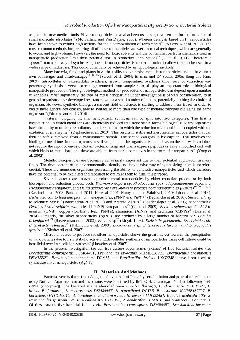
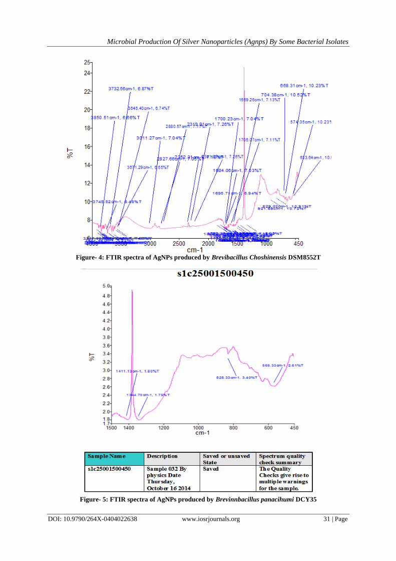
Mechanism of formation of AgNPs by FTIR
The formation of AgNPs by five different bacterial strains has been depicted by FTIR spectra (Figure-
2 to 6). The FTIR measurements were performed to identify the potential biomolecules in the in the bacterial
cell free extracts responsible for the reduction Silver ions (Ag+) to AgNPs. The peak was centred at 1345/cm
which indicated the presence of Nitrate in the residual solution. The band at 3626/cm corresponds to O – H
stretch Carboxylic acids. The stretch at 1651/cm corresponds to N – H bond primary amines. The peak at
1410/cm corresponds to C – N stretch of Aromatic amine group and the bands observed at 631/cm corresponds
to C – N stretching esters.

Microbial Production Of Silver Nanoparticles (Agnps) By Some Bacterial Isolates
DOI: 10.9790/264X-0404022638 www.iosrjournals.org 30 | Page
Figure- 2: FTIR Spectra of AgNPs produced by Brevibacillus centrosporus DSM8445T
Figure- 3: FTIR Spectra of AgNPs produced by Brevibacillus invocatus NCIMB13772T
Microbial Production Of Silver Nanoparticles (Agnps) By Some Bacterial Isolates
DOI: 10.9790/264X-0404022638 www.iosrjournals.org 31 | Page
Figure- 4: FTIR spectra of AgNPs produced by Brevibacillus Choshinensis DSM8552T
Figure- 5: FTIR spectra of AgNPs produced by Brevinnbacillus panacihumi DCY35

Microbial Production Of Silver Nanoparticles (Agnps) By Some Bacterial Isolates
DOI: 10.9790/264X-0404022638 www.iosrjournals.org 32 | Page
Figure- 6: FTIR Spectra of AgNPs produced by Brevibacillus levickii LMG22481
Confirmation of AgNPs production by XRD
In the present investigation the production of AgNPs by five bacterial isolates has been confirmed by
XRD technique (Figure- 7 to 11; Table-3). The XRD patterns were obtained by measuring the angles at which
X- ray beam is diffracted by the crystalline phases in the specimen. The XRD pattern of AgNPs produced by
five bacterial strains showed four prominent peaks at 320 (2 theta), 460 (2 theta), 570 (2 theta), 760 (2 theta)
which indicated the presence of (111), (200), (220), and (311) sets of lattice planes and accordingly could be
indexed as Fee structures of AgNPs. Hence, from the XRD results it is evident that AgNPs formed by all the
five bacterial strains using their extracts were essentially crystalline in nature (Table- 3; Figure- 7 to 11).
Fig : 3 Six Data parameters of Prominent Peak
Measurement profile

Microbial Production Of Silver Nanoparticles (Agnps) By Some Bacterial Isolates
DOI: 10.9790/264X-0404022638 www.iosrjournals.org 33 | Page
Figure- 7: XRD pattern of AgNPs of Brevibacillus centrosporous DSM8445T
Figure- 8: XRD pattern of AgNPs of Brevibacillus invocatus NCIMB13772T
Figure- 9: XRD pattern of AgNPs of Brevibacillus choshinensis DSM8552T

Microbial Production Of Silver Nanoparticles (Agnps) By Some Bacterial Isolates
DOI: 10.9790/264X-0404022638 www.iosrjournals.org 34 | Page
Figure- 10: XRD pattern of AgNPs of Brevibacillus panacihumi DCY35
Figure- 11: XRD pattern of AgNPs of Brevibacillus levickii LMG22481
In the present investigation the size and shape of AgNPs synthesized by present five bacterial strains
were also analyzed using Scanning Electron Microscopic (SEM) images (Figure- 12 to 16). The SEM images
confirmed that the AgNPs produced by all the five bacterial strains were cubical in shape with diameter ranging
from 5 nm to 15 nm.

Microbial Production Of Silver Nanoparticles (Agnps) By Some Bacterial Isolates
DOI: 10.9790/264X-0404022638 www.iosrjournals.org 35 | Page
Figure- 12: SEM of AgNPs produced by Brevibacillus centrosporus DSM8445T
Figure- 13: SEM of AgNPs produced by Brevibacillus invocatus NCIMB13772T
Figure- 14: SEM of AgNPs produced by Brevibacillus choshinensis DSM8552T

Microbial Production Of Silver Nanoparticles (Agnps) By Some Bacterial Isolates
DOI: 10.9790/264X-0404022638 www.iosrjournals.org 36 | Page
Figure- 15: SEM of AgNPs produced by Brevibacillus panacihumi DCY35
Figure- 16: SEM of AgNPs produced by Brevibacillus levickii LMG22481
SPR is highly influenced by shape and size of the nanoparticles. Likewise, the microorganisms has the
metal-microbe interaction to produce inorganic metal ions, and have several applications in biotechnological
fields, includes bioremediation, biomineralization, bioleaching and microbial corrosion32
(Link et al. 2003).
The IR spectrum results showed the amide linkage of the protein has the stronger ability to bind silver
so that the protein could most possibly to form a coat covering around AgNPs and it stabilize the aqueous
medium. This evidence suggests that the biomolecules present in the cell free supernatant of B. flexus could
possibly perform the function for the formation of stable AgNPs33
(Priyadarshinia et al. 2013).
It was well known that the protein can bind to AgNPs through free amide groups. High negative f-
potential of AgNPs due to capping with negatively charged proteins may be the reason for stabilization of
AgNPs34
(Sarkar et al. 2006). In a study of silver nanoparticle synthesis, it was observed that most of particles
clearly attached on the surface of the cytoplasmic membrane35
(Priyabatra et al. 2001).
Many bacterial species were tested to synthesise AgNPs Sintubin et al. (2009)36
viz. Lactobacillus spp.,
Pediococcus pentosaceus, Enterococcus faecium and Lactococcus garvieae and a two-step process of AgNP
formation was proposed. First, the Ag ions were accumulated at the cell wall via biosorption and then
subsequent reduction of those ions formed the metallic nanoparticles (Sintubin et al. 2009)36
. Sintubin et al.
(2009)36
also suggest that the cell wall may act as a capping agent for the nanoparticles, which keeps them stable
by preventing aggregation and showed that by increasing the pH of the medium, the reduction rate of the

Microbial Production Of Silver Nanoparticles (Agnps) By Some Bacterial Isolates
DOI: 10.9790/264X-0404022638 www.iosrjournals.org 37 | Page
nanoparticles increased. The effect of pH on nanoparticle synthesis was also observed by He et al. (2007)21
. By
varying the pH levels, nanoparticles of differing size and shape were formed (He et al. 2009)37
. They illustrated
that by increasing the pH, AgNPs of around 10-20 nm were formed and by lowering the pH to 4, nanoplate
formation was observed.
The present findings are in agreement with the work of Abo- State and Partila (2015)38
who studied the
production of Silver nanoparticles by Pseudomonas aeruginosa cell free extract. The present findings gain
support from the work of Amar Kumar and Ashok Ghosh (2016)39
who observed a more or less similar
production of AgNPs by cell free extract of Brevibacillus borstelensis MTCC10642.
IV. Conclusions It is clear that metallic nanoparticles have great potential in many different industries. The need for a
process to synthesize such nanoparticles in a reliable and green way is becoming more pressing. Current
chemical and physical methods involve toxic chemicals and high temperatures that are not only dangerous to the
environment but costly too. Numerous groups have focused on alternative ways of synthesizing nanoparticles.
Biological systems have been investigated in an effort to provide a sustainable, resource efficient and cheap
method of synthesis. Many different biological chassis have been studied for their ability to resist the toxic
effects of metal ions whilst producing metallic nanoparticles. Bacteria are relatively cheap and easy to cultivate
and have a high growth rate compared to other biological systems such as fungi or plants. Their ease of
manipulation gives them the advantage over plants and fungi as the chassis of choice for the near-term bio-
production of nanoparticles that require optimized synthesis through genetic engineering.
Whatever the choice of biological chassis may be, whether it is a bacterium, fungus or plant, they all
need to be studied comprehensively in order to gain a clearer understanding of mechanism and to close the
knowledge gap in biological nanoparticle synthesis methods by different organisms. The risks of such
nanoparticles must also be assessed, but here biological synthesis may offer yet another advantage. The rapidly
developing field of synthetic biology aims to create predictable, standardized systems and with such new
technologies directed towards the production of metallic nanoparticles, biogenic nanoparticle samples are likely
to become more homogenous and more reproducible, therefore the environmental and health risks posed will be
more easily and more reliably assessed.
The field of biological production of metallic nanoparticles is relatively new and underexplored.
However, it shows great potential in the biotechnology sector. There are many aspects of these biological
methods to be discovered, and later manipulated, as the technology emerges.
Acknowledgement The authors are thankful to Dr. Baidyanath Kumar, Visiting Professor, Dept. of Biotechnology, Patna
Science College, Patna for providing necessary suggestion for the preparation of this manuscript. Authors are
also thankful to Dr. Abha sharan, H. O. D Dept. of Physics, Magadh Mahila College (Patna University) for
support in FTIR analysis. Authors are also thankful to Dr. Amrendra Narayan, Dept. of Physics, Patna Science
College, Patna for his help in XRD analysis, and to Mr. Raju Kumar, INESC Microsystems and
Nanotechnologies, Lisbon, Portugal for SEM analysis. The first author is also thankful to UGC, New Delhi for
financial support through Rajiv Gandhi National Fellowship.
References [1]. Narayanan KB, Sakthivel N (2010) Biological synthesis of metal nanoparticles by microbes. Adv Colloid Interface Sci 156: 1-13. [2]. Thakkar KN, Mhatre SS, Parikh RY (2010) Biological synthesis of metallic nanoparticles. Nanomedicine 6: 257-262.
[3]. Mohanray VJ, Chen Y (2006) Nanoparticles – A Review. Tropical Journal of Pharmaceutical Research 5: 561-573.
[4]. Husseiny M. I, M. A. El-Aziz, Y. Badr , and M. A. Mahmoud (2007): Spectrochimca Acta. Part A: Molecular and Biomolecular
Spectroscopy, 67, 1003, 1006.
[5]. Durán N, Marcato PD, De Souza DIH, Alves OL, Esposito E (2007) Antibacterial Effect of Silver Nanoparticles Produced by
Fungal Process on Textile Fabrics and Their Effluent Treatment. Journal of Biomedical Nanotechnology 3: 203- 208. [6]. Guzman M, Dille J, Godet S (2012) Synthesis and antibacterial activity of silver nanoparticles against gram-positive and gram-
negative bacteria. Nanomedicine: Nanotechnology, Biology and Medicine 8: 37-45.
[7]. Guzmán M, Jean GD, Stephan G (2009) Synthesis of silver nanoparticles by chemical reduction method and their antibacterial activity. International Journal of Chemical and Biomolecular Engineering 2: 104-111.
[8]. Krishnaraj C, Jagan EG, Rajasekar S, Selvakumar P, Kalaichelvan PT, et al. (2010) Synthesis of silver nanoparticles using
Acalypha indica leaf extracts and its antibacterial activity against water borne pathogens. Colloids Surf B Biointerfaces 76: 50-56. [9]. Lima E, Guerra R, Lara V, Guzmán A (2013) Gold nanoparticles as efficient antimicrobial agents for Escherichia coli and
Salmonella typhi. Chem Cent J 7: 11.
[10]. Mc Farland AD, Van Duyne RP (2003) Single Silver Nanoparticles as Real-Time Optical Sensors with Zeptomole Sensitivity. Nano Letters 3: 1057-1062.
[11]. Waszczuk P, Barnard T, Rice MC, Masel RI, Wieckowsky A (2002) A nanoparticle catalyst with superior activity for
electrooxidation of formic acid. Electrochemistry Communications 4: 599-603. [12]. Li X, Xu H, Chen ZS, Chen G (2011) Biosynthesis of Nanoparticles by Microorganisms and Their Applications. Journal of
Nanomaterials 2011: 1-16.

Microbial Production Of Silver Nanoparticles (Agnps) By Some Bacterial Isolates
DOI: 10.9790/264X-0404022638 www.iosrjournals.org 38 | Page
[13]. Suresh K, Prabagaran SR, Sengupta S, Shivaji S (2004) Bacillus indicus sp.nov., an arsenic-resistant bacterium isolated from an
aquifer in West Bengal, India. Int J Syst Evol Microbiol 54: 1369-1375.
[14]. Bhainsa KC, D’Souza SF (2006) Extracellular biosynthesis of silver nanoparticles using the fungus Aspergillus fumigatus. Colloids Surf B Biointerfaces 47: 160-164.
[15]. Song JY, Kim BS (2009) Rapid biological synthesis of silver nanoparticles using plant leaf extracts. Bioprocess Biosyst Eng 32: 79-
84. [16]. Edmundson MC, Capeness M, Horsfall L (2014) Exploring the potential of metallic nanoparticles within synthetic biology. N
Biotechnol 31: 572-578.
[17]. Deplanche K, Caldelari I, Mikheenko IP, Sargent F, Macaskie LE (2010) Involvement of hydrogenases in the formation of highly catalytic Pd(0) nanoparticles by bioreduction of Pd(II) using Escherichia coli mutant strains. Microbiology 156: 2630-2640.
[18]. Yong P, Rowson AN, Farr JPG, Harris IR, Mcaskie LE (2002) Bioaccumulation of palladium by Desulfovibrio desulfuricans.
Journal of Chemical Technology and Biotechnology 55: 593-601. [19]. Kasthuri J, Kathiravan K, Rajendiran N (2008) Phyllanthin-assisted biosynthesis of silver and gold nanoparticles: a novel biological
approach. Journal of Nanoparticle Research 11: 1075-1085.
[20]. Park Y, Hong YN, Weyers A, Kim YS, Linhardt RJ (2011) Polysaccharides and phytochemicals: a natural reservoir for the green synthesis of gold and silver nanoparticles. IET Nanobiotechnol 5: 69-78.
[21]. He, C., L. Morawska, and L. Taplin 2007, 'Particle emission characteristics of office printers' Environmental Science and
Technology, vol. 41(7), pp. 6039-6045. [22]. Johnston CW, Wyatt MA, Li X, Ibrahim A, Shuster J, et al. (2013) Gold biomineralization by a metallophore from a gold-
associated microbe. Nat Chem Biol 9: 241-243.
[23]. Raveendran P, Fu J, Wallen SL (2003) Completely “green” synthesis and stabilization of metal nanoparticles. J Am Chem Soc 125: 13940-13941.
[24]. Laudenslager MJ, Schiffman JD, Schauer CL (2008) Carboxymethyl chitosan as a matrix material for platinum, gold, and silver
nanoparticles. Biomacromolecules 9: 2682-2685. [25]. Cai J, Kimura S, Wada M, Kuga S (2009) Nanoporous cellulose as metal nanoparticles support. Biomacromolecules 10: 87-94.
[26]. Das V, Thomas R, Varghese R, Soniya EV, Mathew J, et al. (2014) Extracellular synthesis of silver nanoparticles by the Bacillus
strain CS 11 isolated from industrialized area.3 Biotech 4: 121-126. [27]. Lloyd JR, Yong P, Macaskie LE (1998) Enzymatic recovery of elemental palladium by using sulfate-reducing bacteria Appl
Environ Microbiol 64: 4607- 4609.
[28]. Kalimuthu K, Suresh Babu R, Venkataraman D, Bilal M, Gurunathan S (2008) Biosynthesis of silver nanocrystals by Bacillus licheniformis. Colloids Surf B Biointerfaces 65: 150-153.
[29]. Shahverdi AR, Minaeian S, Shahverdi HR, Jamalifar H, Nohi AA (2007) Rapid synthesis of silver nanoparticles using culture
supernatants of Enterobacteria: A novel biological approach. Process Biochemistry 42: 919-923 [30]. ]Natarajan K, S. V. Selvaraj, R. Murty (2010): Microbial production of silver Nanoparticles. Digest Journal of Nanomaterials and
Biostructures 5, 1, 135 – 140,
[31]. Kuber C. B , S. F.: D’Souza (2006): Colloids Surf B, 47, 160–164. [32]. Link S , M. A. El-Sayed (2003): Annual Review of Physical Chemistry, 54, 331.
[33]. Priyadarshinia S, V.Gopinatha, M. N. Priyadharsshinia, M. D. Alib, P. Velusamya (2013) : Synthesis of anisotropic silver
nanoparticles using novel strain, Bacillus flexus and its biomedical application Colloids and Surfaces B: Biointerfaces, 102, 232–
237,
[34]. Sarkar S, A. D. Jana, S. K. Samanta, G. Mostafa Polyhedron., 26, 441- 449, (2007). Science’’, Trends. Biotechnol, 19, 15–20, (2001), Sensitivity of plasmon response to size, shape, and metal composition, J. Phys.Chem. B, 110, 192-200, (2006).
[35]. Priyabrata M, A.Ahmad , M . Deendayal, S. Satyajyoti , R. S. Sudhakar , I. K. Mohammad , P .Renu , P. V. Ajaykumar , A.
Mansoor , K. Rajiv , S. Murali : Fungusmediated synthesis of silver nanoparticles and their immobilization in the mycelial matrix: a novel biological approach to nanoparticle synthesis. Nano. Lett, 1, 515–519, (2001).
[36]. Sintubin L, De Windt W, Dick J, Mast J, van der Ha D, et al. (2009) Lactic acid bacteria as reducing and capping agent for the fast
and efficient production of silver nanoparticles. Appl Microbiol Biotechnol 84: 741-749. [37]. He YX, Gui L, Liu YZ, Du Y, Zhou Y, Li P, Zhou CZ (2009) : Crystal structure of Saccharomyces cerevisiae glutamine synthetase
Gln1 suggests a nanotube-like supramolecular assembly.Proteins 76(1):249-54
[38]. Abo- State, M. A. M and A. M. Partila (2015): Microbial production of silver nanoparticles by Pseudomonas aeruginosa cell free extract, J. Eco. Hael. Env, 3(3), 91- 98
[39]. Amar Kumar and Ashok Ghosh (2016): International Journal of Biotechnology and Biochemistry, 12(2), 95- 102
Dr. Baidyanath Kumar"Microbial Production of Silver Nanoparticles (Agnps) By Some
Bacterial Isolates " IOSR Journal of Biotechnology and Biochemistry (IOSR-JBB) 4.4 (2018):
26-38.