microfluidic biosensor for β -amyloid(1-42) detection using cyclic...
TRANSCRIPT

Delivered by Publishing Technology to Sung Kyun Kwan UniversityIP 11514515516 On Tue 25 Nov 2014 054931
Copyright American Scientific Publishers
RESEARCH
ARTIC
LE
Copyright copy 2011 American Scientific PublishersAll rights reservedPrinted in the United States of America
Journal ofNanoscience and Nanotechnology
Vol 11 5657ndash5662 2011
Microfluidic Biosensor for -Amyloid(1-42)Detection Using Cyclic Voltammetry
Kamrul Islam1 You-Cheol Jang1 Rohit Chand1 Sandeep Kumar Jha1Hyun Ho Lee2 and Yong-Sang Kim13lowast
1Department of Nano Science and Engineering Myongji University Gyeonggi 449-728 Republic of Korea2Department of Chemical Engineering Myongji University Gyeonggi 449-728 Republic of Korea3Department of Electrical Engineering Myongji University Gyeonggi 449-728 Republic of Korea
Numerous studies have identified -amyloid(1-42) protein (A42) in the cerebrospinal fluid as apotential biomarker of Alzheimerrsquos disease It is of particular interest to establish the diagnosisbefore reaching the stage of clinical severity The current methods for studying amyloid detectionhowever is often time-consuming expensive and labor intensive making the analytical processvery slow Thus a critical need exists for an analytical system that would enable the rapid inves-tigation of amyloid formation with a very small amount of amyloidogenic peptides and reagentsIn our present work we report a simple microfluidic biosensor to analyze very small quantities ofA42 peptide on gold surface that were modified with Au nano-particles onto the thiol groups ofself-assembled 16-hexandithiol cross-linkers The vital advantage of this method includes retain-ing the bioactivity and environment similar to nature for protein immobilization while using minimalamounts of reagents and highly sensitive detection
Keywords Microfluidic Biosensor -Amyloid(1-42) Cyclic Voltammetry
1 INTRODUCTION
Alzheimerrsquos disease (AD) is the most common form ofdementia among the elderly A major pathological hall-mark of AD is the formation of senile plaques consistingof -amyloid (A) aggregates in the brain tissues12 Themajor component of the amyloid plaques exists in two pre-dominant forms the 40-residue A(1ndash40) and 42-residueA(1ndash42) These peptides differ in the absence or pres-ence of two extra C-terminal residues and are producedfrom the processing of a larger amyloid precursor pro-tein (APP)3 resulting from sequential cleavage by pro-teases named - and -secretases4ndash6 Biochemical studiesalso suggest that the longer 42-residue A(1ndash42) is morepathogenic than the shorter 40-residue A(1ndash40)7 due toits greater in vitro tendency to aggregate and precipitateas amyloid8 In fact the senile plaques probably appearmany years prior to the detection of the cognitive symp-toms of AD910 As a consequence it might be possiblewithin the aging population that people show signs ofAD neuropathology but still have no detectable cognitivesymptoms
lowastAuthor to whom correspondence should be addressed
Despite the significance of amyloid formation littleresearch progress had been made due to the limited num-ber of effective analytical methods and probably for thelack of any method to analyze the physiology of amy-loid aggregation A few attempts have been made inpast for the development of new screening assay methodsfor -amyloid which included capillary electrophoresis11
mass spectrometry12 enzyme-linked immunosorbent assay(ELISA)13 surface plasmon resonance spectrometry14 andcell-based assay15 yet these assay systems could not fullysatisfy the required criteria for detection of amyloid plaqueformation Moreover most of these methods were time-consuming expensive and labor intensive lasting frommonths to years making the analytical process very slowThus it is critically important to develop an analyt-
ical system that can detect A(1ndash42) amyloid concen-tration in its aggregate forming step which would be amore suitable candidate to predict Alzheimerrsquos diseaseAlthough it is difficult to devise an analytical systemfor in-vivo operation in-vitro microreactor coupled withsuitable detection system can easily analyze the proteinmolecules Such microfluidic reactors can be miniatur-ized in the form of microchips using soft-lithographictechniques1617 The microchip should consume very small
J Nanosci Nanotechnol 2011 Vol 11 No 7 1533-48802011115657006 doi101166jnn20114498 5657
Delivered by Publishing Technology to Sung Kyun Kwan UniversityIP 11514515516 On Tue 25 Nov 2014 054931
Copyright American Scientific Publishers
RESEARCH
ARTIC
LE
Microfluidic Biosensor for -Amyloid(1-42) Detection Using Cyclic Voltammetry Islam et al
amount of amyloidogenic peptides can expedite the reac-tion and detection and would require less manual laborcompared with existing procedures followed in analysis ofsuch proteinsIn this regard we describe herein a scalable microre-
actor device architecture with integrated electrochemicalsensors as an analytical system for monitoring of in-vitroA(1ndash42) plaque formation The crucial step of proposedmethod is the suitable immobilization of A(1ndash42) pep-tide on the electrode surface It is known that adsorp-tion of proteins onto bulk metal surfaces leads to theirdenaturation and loss of their bioactivity18 The addi-tional modification of gold electrodes with colloidal goldnanoparticles is one of the best solutions to overcomeabove problems It has been recently reported that proteinsadsorbed onto colloidal Au retain their bioactivity19ndash21 Thegold-nano particles have attracted a lot scientific attentiondue to their not only biocompatibility but also for theother properties such as large surface area and efficientelectron conducting features22ndash24 In the work presentedthe gold electrodes were modified with Au nano-particlesby self-assembly while using 16-hexanedithiol as cross-linkers The self-assembly of colloidal gold provides anenvironment similar to nature for protein immobilizationwhile retaining their bioactivity Therefore A(1ndash42) pep-tide molecules were allowed to adsorb directly on thesurface of colloidal Au Subsequent electrochemical mea-surements were performed using cyclic voltammetry (CV)for monitoring of each step of gold electrode modifica-tion with A(1ndash42) peptide The redox behavior of a solu-tion of K3[Fe(CN)6]K4[Fe(CN)6] was studied in presenceof hexanedithiol colloidal nanoparticles and with aggre-gated A(1ndash42) peptide Presence of these bulk materialsover working electrode resulted in increase of double layercapacitance and hence reduced electrochemical behaviorof test solution This was used as the basis for determina-tion of A(1ndash42) peptide concentration
2 EXPERIMENTAL DETAILS
21 Chemicals
16-Hexanedithiol 20 nm colloidal gold K3[Fe(CN)6]and K4[Fe(CN)6] were purchased from Sigma-Aldrich(St Louis Mo) Human -amyloid(1ndash42) peptide wasobtained from rPeptide Co (Athens GA) All other sol-vents and chemicals were of analytical grade All solutionswere prepared and diluted using double distilled deionized(DDI) water
22 Design and Fabrication of the Microchip
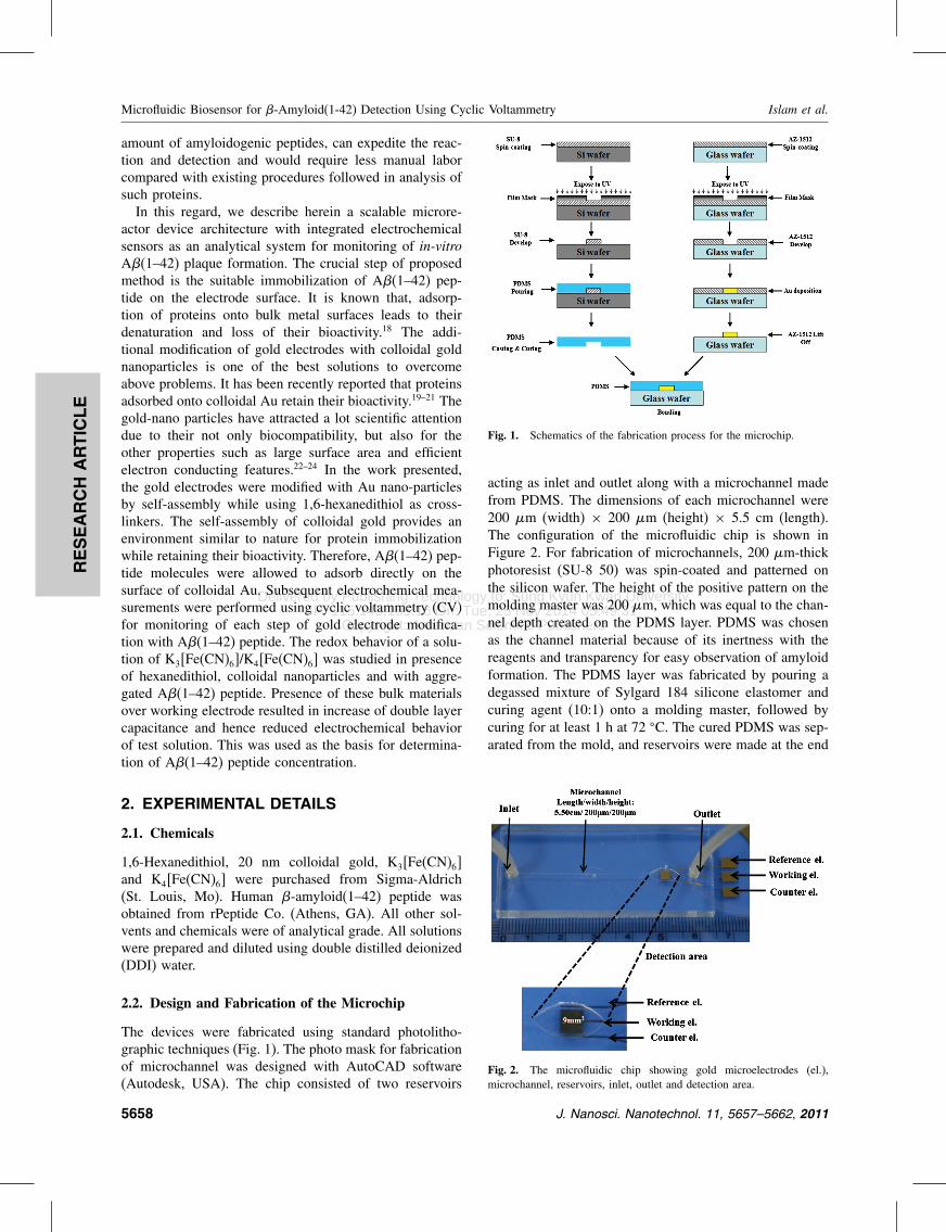
The devices were fabricated using standard photolitho-graphic techniques (Fig 1) The photo mask for fabricationof microchannel was designed with AutoCAD software(Autodesk USA) The chip consisted of two reservoirs
Fig 1 Schematics of the fabrication process for the microchip
acting as inlet and outlet along with a microchannel madefrom PDMS The dimensions of each microchannel were200 m (width) times 200 m (height) times 55 cm (length)The configuration of the microfluidic chip is shown inFigure 2 For fabrication of microchannels 200 m-thickphotoresist (SU-8 50) was spin-coated and patterned onthe silicon wafer The height of the positive pattern on themolding master was 200 m which was equal to the chan-nel depth created on the PDMS layer PDMS was chosenas the channel material because of its inertness with thereagents and transparency for easy observation of amyloidformation The PDMS layer was fabricated by pouring adegassed mixture of Sylgard 184 silicone elastomer andcuring agent (101) onto a molding master followed bycuring for at least 1 h at 72 C The cured PDMS was sep-arated from the mold and reservoirs were made at the end
Fig 2 The microfluidic chip showing gold microelectrodes (el)microchannel reservoirs inlet outlet and detection area
5658 J Nanosci Nanotechnol 11 5657ndash5662 2011
Delivered by Publishing Technology to Sung Kyun Kwan UniversityIP 11514515516 On Tue 25 Nov 2014 054931
Copyright American Scientific Publishers
RESEARCH
ARTIC
LE
Islam et al Microfluidic Biosensor for -Amyloid(1-42) Detection Using Cyclic Voltammetry
of each channel using a 3-mm circular punch At the sametime gold electrodes were fabricated on a glass substrateusing standard photolithographic methodsThe three electrodes namely working reference and
counter electrodes were fabricated by thermal evaporationat the detection area The size of the working electrode inthe detection area was 3 mm in length as well as widthOther two electrodes were fabricated adjacent to the work-ing electrode having a gap of 250 mBonding of PDMS layer on glass substrate con-
taining the electrodes was performed with UV-Ozonecleaner to get improved bond strength25 When neededthe working electrode on the glass slides was modi-fied with 16-hexanedithiol solution before bonding soas to avoid modification of other electrodes with thissolution
23 Surface Modification of the Electrode
The surface modification of gold electrode is shown inFigure 3 Clean working electrode was modified with10 mM 16-hexanedithiol solution in ethanol by placing3 l of liquid over it and incubating at room temperatureunder humid condition in a closed petri plate for 3 h It waswashed with ethanol and the substrate was bonded withPDMS The working electrode was then modified with20 nm colloidal gold solution for 18 h under stationarycondition In subsequent steps microfluidic condition wasmaintained in the microchannel using a precision pump(KD Scientific USA) at 5 lmin flow rate and electrodeswere cleaned repeatedly with DDI water and ethanol usingsame condition As a stock 500 M A(1ndash42) peptidedissolved in 002 NH4OH were prepared and kept at4 C The test sample was diluted with A assay buffer(20 mM phosphate buffer pH 72 containing 100 mMNaCl) from stock solution just before treatment to avoidthe A aggregation A 100 L of the above solution wasinjected in the microchannel for deposition on the surface
Fig 3 Model of surface modification on gold electrode andashb self-assembly of 16 hexanedithiol as cross-linkers c 16-hexanditiolAu-colloid modification d after deposition of A(1ndash42) peptide
of electrodes modified by colloidal Au Next the elec-trodes were kept overnight in darkness at 4 C under sta-tionary condition After deposition of A(1ndash42) peptidethe electrodes were washed and stored at 4 C in phos-phate buffer (01 M KCl 01 M KH2PO4 pH 72) untilused for sensor measurements
24 Electrochemical Measurements
All electrochemical measurements including cyclicvoltammetry (CV) were performed on the microflu-idic chip using a CH Instruments (CHI model 800B)electrochemical analyzer under stationary liquid condi-tion At first measurement was performed with bareelectrode on the device and latter upon each step ofelectrode modifications Cyclic voltammetry (CV) wasperformed in 01 M Na2HPO4ndashNaH2PO4 buffer contain-ing 10 mM K3[Fe(CN)6]K4[Fe(CN)6] and potential wascycled between +02 V to minus06 V with a scan rate of005 Vsminus1
3 RESULTS AND DISCUSSION
31 Microfluidic Chip Operation
The study of amyloidogenic peptide aggregation remainsmost important factor in successful diagnosis ofAlzheimer as non-aggregated peptide is avirulent Yetonly scant literature is available until date that studiedamyloidogenic peptide aggregation Meier et al studiedon-chip peptide aggregation but rather focused on A(1ndash40) peptide26 whereas A(1ndash42) peptide is a more promis-ing candidate for diagnosis of Alzheimer7 The groupalso did not attempt to measure the concentration of thepeptide Therefore in the present study we focused onaggregation as well as detection of A(1ndash42) peptide byemploying indirect electrochemical method in a microre-actor In this perspective we fabricated a microfluidicchip (Fig 2) in addition to electrochemical detector on aglass substrate The volume of the detection chamber was27 mm3 and the fluidic control of the analyte in and outof this chamber was achieved through capillary motionand precision pump This kind of microfluidic fabricationis advantageous in several aspects First it allows hex-anedithiol gold nanoparticle and the amyloid peptides tobe immobilized efficiently on the gold electrode and pre-vent from contamination and thus supporting high qualitylow-background electrochemical sensing Second a three-electrode set-up was integrated onto the chip which isparticularly useful compared to the commercially avail-able cells in cases where only limited sample volumes areavailable And last the key microfluidic components in thechip are simple in design inexpensive and easy to fabri-cate The use of microfluidic components with low powerconsumption along with the employment of the electro-chemical detection suggests that the proposed device canbe operated as a handheld device in future
J Nanosci Nanotechnol 11 5657ndash5662 2011 5659
Delivered by Publishing Technology to Sung Kyun Kwan UniversityIP 11514515516 On Tue 25 Nov 2014 054931
Copyright American Scientific Publishers
RESEARCH
ARTIC
LE
Microfluidic Biosensor for -Amyloid(1-42) Detection Using Cyclic Voltammetry Islam et al
32 Amyloid (1ndash42) Peptide Immobilization onColloidal Gold Modified Electrodes
In order to detect A(1ndash42) peptide in the microfluidicchip the first concern was to immobilize the amyloid pep-tide on the chip Earlier studies showed that it is hardto retain the bioactivity of proteins if it is absorbed ontobulk metal surface18 Instead if it is adsorbed onto col-loidal Au then their bioactivity is well retained19ndash21 Inaddition the colloidal Au nanoparticle provides larger sur-face area than the bulk metal surface2728 Hence theproposed method incorporated a hexanedithiol linker tobridge between the Au electrode surface and that ofthe colloidal Au nanoparticles For the electrochemicaldetection K3[Fe(CN)6]K4[Fe(CN)6] solution was usedassuming that with each modification of the electrode byhexanedithiol colloidal nanoparticle and peptides therewill be changes in peak currents for this widely usedredox active solution The K3[Fe(CN)6]K4[Fe(CN)6] solu-tion will give its original peak on the metal surface fol-lowed by reduced peak for hexanedithiol as it will actas an insulator on the surface Again when the colloidalnanoparticle will be immobilized the peak current willreturn to its origin and after the immobilization of thepeptide the peak-to-peak separation will be enhanced dueto electrode passivationAs expected K3[Fe(CN)6]K4[Fe(CN)6] showed
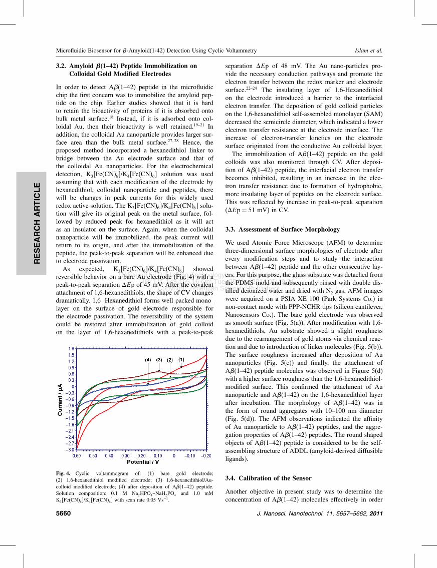
reversible behavior on a bare Au electrode (Fig 4) with apeak-to-peak separation Ep of 45 mV After the covalentattachment of 16-hexanedithiols the shape of CV changesdramatically 16- Hexanedithiol forms well-packed mono-layer on the surface of gold electrode responsible forthe electrode passivation The reversibility of the systemcould be restored after immobilization of gold colloidon the layer of 16-hexanedithiols with a peak-to-peak
Fig 4 Cyclic voltammogram of (1) bare gold electrode(2) 16-hexanedithiol modified electrode (3) 16-hexanedithiolAu-colloid modified electrode (4) after deposition of A(1ndash42) peptideSolution composition 01 M Na2HPO4ndashNaH2PO4 and 10 mMK3[Fe(CN)6]K4[Fe(CN)6] with scan rate 005 Vsminus1
separation Ep of 48 mV The Au nano-particles pro-vide the necessary conduction pathways and promote theelectron transfer between the redox marker and electrodesurface22ndash24 The insulating layer of 16-Hexanedithiolon the electrode introduced a barrier to the interfacialelectron transfer The deposition of gold colloid particleson the 16-hexanedithiol self-assembled monolayer (SAM)decreased the semicircle diameter which indicated a lowerelectron transfer resistance at the electrode interface Theincrease of electron-transfer kinetics on the electrodesurface originated from the conductive Au colloidal layerThe immobilization of A(1ndash42) peptide on the gold
colloids was also monitored through CV After deposi-tion of A(1ndash42) peptide the interfacial electron transferbecomes inhibited resulting in an increase in the elec-tron transfer resistance due to formation of hydrophobicmore insulating layer of peptides on the electrode surfaceThis was reflected by increase in peak-to-peak separation(Ep= 51 mV) in CV
33 Assessment of Surface Morphology
We used Atomic Force Microscope (AFM) to determinethree-dimensional surface morphologies of electrode afterevery modification steps and to study the interactionbetween A(1ndash42) peptide and the other consecutive lay-ers For this purpose the glass substrate was detached fromthe PDMS mold and subsequently rinsed with double dis-tilled deionized water and dried with N2 gas AFM imageswere acquired on a PSIA XE 100 (Park Systems Co) innon-contact mode with PPP-NCHR tips (silicon cantileverNanosensors Co) The bare gold electrode was observedas smooth surface (Fig 5(a)) After modification with 16-hexanedithiols Au substrate showed a slight roughnessdue to the rearrangement of gold atoms via chemical reac-tion and due to introduction of linker molecules (Fig 5(b))The surface roughness increased after deposition of Aunanoparticles (Fig 5(c)) and finally the attachment ofA(1ndash42) peptide molecules was observed in Figure 5(d)with a higher surface roughness than the 16-hexanedithiol-modified surface This confirmed the attachment of Aunanoparticle and A(1ndash42) on the 16-hexanedithiol layerafter incubation The morphology of A(1ndash42) was inthe form of round aggregates with 10ndash100 nm diameter(Fig 5(d)) The AFM observations indicated the affinityof Au nanoparticle to A(1ndash42) peptides and the aggre-gation properties of A(1ndash42) peptides The round shapedobjects of A(1ndash42) peptide is considered to be the self-assembling structure of ADDL (amyloid-derived diffusibleligands)
34 Calibration of the Sensor
Another objective in present study was to determine theconcentration of A(1ndash42) molecules effectively in order
5660 J Nanosci Nanotechnol 11 5657ndash5662 2011
Delivered by Publishing Technology to Sung Kyun Kwan UniversityIP 11514515516 On Tue 25 Nov 2014 054931
Copyright American Scientific Publishers
RESEARCH
ARTIC
LE
Islam et al Microfluidic Biosensor for -Amyloid(1-42) Detection Using Cyclic Voltammetry
Fig 5 AFM images of the bare gold substrate (a) after the immobilization of 16-hexanedithiol (b) after modified by gold nanoparticle on 16-hexanedithiol linker (c) and finally after the deposition of -amyloid(1ndash42) protein
to predict the onset of Alzheimer disease For this pur-pose different concentrations (10 M 50 M 100 M200 M 300 M 350 M 400 M) of A(1ndash42) pep-tide were applied on the electrode and the CV signals wererecorded Figure 6 shows the effect of different concentra-tions of A(1ndash42) on cyclic voltammetric response on thechip As the concentration of A(1ndash42) is increased from10 M to 400 M the peak current decreased correspond-ingly There is a good sigmoidal relationship between thepeak current and the concentration A(1ndash42) in the entire
Fig 6 Changes in the peak current for the different concentrations of-amyloid(1ndash42) protein
0 100 200 300 4005
6
7
8
9
10
Sens
or r
espo
nse
(Ι μ
A)
Concentration of β amyloid μM
Fig 7 Sensor calibration curve sensor output versus -amyloidconcentrations
concentration range studied in this case (Fig 7) Each datain Figure 7 was an average value of three measurementsThe linear detection range existed between 100 M to300 M of A(1ndash42) These experimental results wereindicative of a successful working model for detectionof amyloidogenic peptides on the fabricated microfluidicchip
4 CONCLUSIONS
We have developed an electrochemical biosensor for thedetection of A(1ndash42) peptide in a microfluidic chip
J Nanosci Nanotechnol 11 5657ndash5662 2011 5661
Delivered by Publishing Technology to Sung Kyun Kwan UniversityIP 11514515516 On Tue 25 Nov 2014 054931
Copyright American Scientific Publishers
RESEARCH
ARTIC
LE
Microfluidic Biosensor for -Amyloid(1-42) Detection Using Cyclic Voltammetry Islam et al
architecture The process used cyclic voltammetric studyof redox behavior of K3[Fe(CN)6]K4[Fe(CN)6] on col-loidal gold nanoparticle bound through 16- hexanedithiollinkers on gold working electrode The modification ofgold electrode with Au nanoparticles provided a suit-able environment for stable immobilization of A(1ndash42)keeping its bioactivity in microreactor environmentAggregation of A(1ndash42) peptide in increasing order ofconcentration resulted in passivation of working electrodeand hence decreased the sensor output current The pro-posed detection method could be a suitable diagnosticcandidate for Alzheimer if an additional layer of mon-oclonal antibody against A(1ndash42) peptide be used toenhance selectivity of detection This method shall behelpful in analyzing other important biomarker peptidesof Alzheimerrsquos disease such as Total tau and Phospho-tauand can be effectively useful in detection of other pro-tein molecules that are otherwise difficult to detect usingconventional techniques due to loss of bioactivity
Acknowledgments This word was supported by Grantno (ROA-2006-000-10274-0) from National ResearchLaboratory Program of the Korea Science and EngineeringFoundation
References and Notes
1 R M Murphy Annu Rev Biomed Eng 4 155 (2002)2 D K Lahiri M R Farlow N H Greig and K Sambamurti Drug
Dev Res 56 267 (2002)3 J Hardy Trends Neurosci 20 154 (1997)4 S G Younkin J Physiol - Paris 92 289 (1998)5 D J Selkoe Physiol Rev 81 741 (2001a)6 D J Selkoe Neuron 32 177 (2001b)7 S G Younkin Ann Neurol 37 287 (1995)
8 C J Barrow and M G Zagorski Science 253 179 (1991)9 H Braak and E Braak Neurobiology Aging 18 351 (1997)10 D R Thal U Rub M Orantoes and H Braak Neurology 58 1791
(2002)11 S Sabella M Quaglia C Lanni M Racchi S Govoni
G Caccialanza A Calligaro V Bellotti and E De Lorenzi Elec-trophoresis 25 3186 (2004)
12 X Cheng and R B van Breemen Anal Chem 77 7012 (2005)13 P Inbar M R Bautista and S A Takayama J Yang Anal Chem
80 3502 (2008)14 J Ryu H-A Joung M-G Kim and C B Park Anal Chem
80 2400 (2008)15 B L Apostol A Kazantsev S Raffioni K Illes J Pallos L Bodai
N Slepko J E Bear F B Gertler S Hersch D E HousmaJ L Marsh and L M Thompson Proc Natl Acad Sci 100 5950(2003)
16 G-S Joo S K Jha and Y-S Kim Curr Appl Phys 9 e222(2009)
17 S K Jha G-S Ra G-S Joo and Y-S Kim Curr Appl Phys9 e301 (2009)
18 M Wang L Wang G Wang X Ji Y Bai T Li S Gong andJ Li Biosens Bioelectron 19 575 (2004)
19 Y Liu F Yin Y Long Z Zhang and S Yao J Colloid Interf Sci258 75 (2003)
20 S Liu and H Ju Biosens Bioelectron 19 177 (2003)21 C-X Li K-Q Deng G-L Shen and R-Q Yu Anal Sci 20 1277
(2004)22 M Lu X H Li B Z Yu and H L Li J Colloid Interf Sci
248 376 (2002)23 M Yang and Z Zhang Electrochim Acta 49 5089 (2004)24 Ch-Z Li Y Liu and J H T Luong Anal Chem 77 478
(2005)25 J H Kim C J Kang and Y S Kim Microelectron Eng 71 119
(2004a)26 M Meier J Kenney-Darling S H Choi E M Norstrom S S
Sisodia and R F Ismagilov Angew Chem Int Ed 48 1487 (2009)27 S Singha H Datta and A K Dasgupta J Nanosci Nanotechnol
10 826 (2010)28 R R Bhattacharjee A K Das D Haldar S Si A Banerjee and
T K Mandal J Nanosci Nanotechnol 5 1141 (2005)
Received 24 June 2010 Accepted 27 January 2011
5662 J Nanosci Nanotechnol 11 5657ndash5662 2011
Delivered by Publishing Technology to Sung Kyun Kwan UniversityIP 11514515516 On Tue 25 Nov 2014 054931
Copyright American Scientific Publishers
RESEARCH
ARTIC
LE
Microfluidic Biosensor for -Amyloid(1-42) Detection Using Cyclic Voltammetry Islam et al
amount of amyloidogenic peptides can expedite the reac-tion and detection and would require less manual laborcompared with existing procedures followed in analysis ofsuch proteinsIn this regard we describe herein a scalable microre-
actor device architecture with integrated electrochemicalsensors as an analytical system for monitoring of in-vitroA(1ndash42) plaque formation The crucial step of proposedmethod is the suitable immobilization of A(1ndash42) pep-tide on the electrode surface It is known that adsorp-tion of proteins onto bulk metal surfaces leads to theirdenaturation and loss of their bioactivity18 The addi-tional modification of gold electrodes with colloidal goldnanoparticles is one of the best solutions to overcomeabove problems It has been recently reported that proteinsadsorbed onto colloidal Au retain their bioactivity19ndash21 Thegold-nano particles have attracted a lot scientific attentiondue to their not only biocompatibility but also for theother properties such as large surface area and efficientelectron conducting features22ndash24 In the work presentedthe gold electrodes were modified with Au nano-particlesby self-assembly while using 16-hexanedithiol as cross-linkers The self-assembly of colloidal gold provides anenvironment similar to nature for protein immobilizationwhile retaining their bioactivity Therefore A(1ndash42) pep-tide molecules were allowed to adsorb directly on thesurface of colloidal Au Subsequent electrochemical mea-surements were performed using cyclic voltammetry (CV)for monitoring of each step of gold electrode modifica-tion with A(1ndash42) peptide The redox behavior of a solu-tion of K3[Fe(CN)6]K4[Fe(CN)6] was studied in presenceof hexanedithiol colloidal nanoparticles and with aggre-gated A(1ndash42) peptide Presence of these bulk materialsover working electrode resulted in increase of double layercapacitance and hence reduced electrochemical behaviorof test solution This was used as the basis for determina-tion of A(1ndash42) peptide concentration
2 EXPERIMENTAL DETAILS
21 Chemicals
16-Hexanedithiol 20 nm colloidal gold K3[Fe(CN)6]and K4[Fe(CN)6] were purchased from Sigma-Aldrich(St Louis Mo) Human -amyloid(1ndash42) peptide wasobtained from rPeptide Co (Athens GA) All other sol-vents and chemicals were of analytical grade All solutionswere prepared and diluted using double distilled deionized(DDI) water
22 Design and Fabrication of the Microchip
The devices were fabricated using standard photolitho-graphic techniques (Fig 1) The photo mask for fabricationof microchannel was designed with AutoCAD software(Autodesk USA) The chip consisted of two reservoirs
Fig 1 Schematics of the fabrication process for the microchip
acting as inlet and outlet along with a microchannel madefrom PDMS The dimensions of each microchannel were200 m (width) times 200 m (height) times 55 cm (length)The configuration of the microfluidic chip is shown inFigure 2 For fabrication of microchannels 200 m-thickphotoresist (SU-8 50) was spin-coated and patterned onthe silicon wafer The height of the positive pattern on themolding master was 200 m which was equal to the chan-nel depth created on the PDMS layer PDMS was chosenas the channel material because of its inertness with thereagents and transparency for easy observation of amyloidformation The PDMS layer was fabricated by pouring adegassed mixture of Sylgard 184 silicone elastomer andcuring agent (101) onto a molding master followed bycuring for at least 1 h at 72 C The cured PDMS was sep-arated from the mold and reservoirs were made at the end
Fig 2 The microfluidic chip showing gold microelectrodes (el)microchannel reservoirs inlet outlet and detection area
5658 J Nanosci Nanotechnol 11 5657ndash5662 2011
Delivered by Publishing Technology to Sung Kyun Kwan UniversityIP 11514515516 On Tue 25 Nov 2014 054931
Copyright American Scientific Publishers
RESEARCH
ARTIC
LE
Islam et al Microfluidic Biosensor for -Amyloid(1-42) Detection Using Cyclic Voltammetry
of each channel using a 3-mm circular punch At the sametime gold electrodes were fabricated on a glass substrateusing standard photolithographic methodsThe three electrodes namely working reference and
counter electrodes were fabricated by thermal evaporationat the detection area The size of the working electrode inthe detection area was 3 mm in length as well as widthOther two electrodes were fabricated adjacent to the work-ing electrode having a gap of 250 mBonding of PDMS layer on glass substrate con-
taining the electrodes was performed with UV-Ozonecleaner to get improved bond strength25 When neededthe working electrode on the glass slides was modi-fied with 16-hexanedithiol solution before bonding soas to avoid modification of other electrodes with thissolution
23 Surface Modification of the Electrode
The surface modification of gold electrode is shown inFigure 3 Clean working electrode was modified with10 mM 16-hexanedithiol solution in ethanol by placing3 l of liquid over it and incubating at room temperatureunder humid condition in a closed petri plate for 3 h It waswashed with ethanol and the substrate was bonded withPDMS The working electrode was then modified with20 nm colloidal gold solution for 18 h under stationarycondition In subsequent steps microfluidic condition wasmaintained in the microchannel using a precision pump(KD Scientific USA) at 5 lmin flow rate and electrodeswere cleaned repeatedly with DDI water and ethanol usingsame condition As a stock 500 M A(1ndash42) peptidedissolved in 002 NH4OH were prepared and kept at4 C The test sample was diluted with A assay buffer(20 mM phosphate buffer pH 72 containing 100 mMNaCl) from stock solution just before treatment to avoidthe A aggregation A 100 L of the above solution wasinjected in the microchannel for deposition on the surface
Fig 3 Model of surface modification on gold electrode andashb self-assembly of 16 hexanedithiol as cross-linkers c 16-hexanditiolAu-colloid modification d after deposition of A(1ndash42) peptide
of electrodes modified by colloidal Au Next the elec-trodes were kept overnight in darkness at 4 C under sta-tionary condition After deposition of A(1ndash42) peptidethe electrodes were washed and stored at 4 C in phos-phate buffer (01 M KCl 01 M KH2PO4 pH 72) untilused for sensor measurements
24 Electrochemical Measurements
All electrochemical measurements including cyclicvoltammetry (CV) were performed on the microflu-idic chip using a CH Instruments (CHI model 800B)electrochemical analyzer under stationary liquid condi-tion At first measurement was performed with bareelectrode on the device and latter upon each step ofelectrode modifications Cyclic voltammetry (CV) wasperformed in 01 M Na2HPO4ndashNaH2PO4 buffer contain-ing 10 mM K3[Fe(CN)6]K4[Fe(CN)6] and potential wascycled between +02 V to minus06 V with a scan rate of005 Vsminus1
3 RESULTS AND DISCUSSION
31 Microfluidic Chip Operation
The study of amyloidogenic peptide aggregation remainsmost important factor in successful diagnosis ofAlzheimer as non-aggregated peptide is avirulent Yetonly scant literature is available until date that studiedamyloidogenic peptide aggregation Meier et al studiedon-chip peptide aggregation but rather focused on A(1ndash40) peptide26 whereas A(1ndash42) peptide is a more promis-ing candidate for diagnosis of Alzheimer7 The groupalso did not attempt to measure the concentration of thepeptide Therefore in the present study we focused onaggregation as well as detection of A(1ndash42) peptide byemploying indirect electrochemical method in a microre-actor In this perspective we fabricated a microfluidicchip (Fig 2) in addition to electrochemical detector on aglass substrate The volume of the detection chamber was27 mm3 and the fluidic control of the analyte in and outof this chamber was achieved through capillary motionand precision pump This kind of microfluidic fabricationis advantageous in several aspects First it allows hex-anedithiol gold nanoparticle and the amyloid peptides tobe immobilized efficiently on the gold electrode and pre-vent from contamination and thus supporting high qualitylow-background electrochemical sensing Second a three-electrode set-up was integrated onto the chip which isparticularly useful compared to the commercially avail-able cells in cases where only limited sample volumes areavailable And last the key microfluidic components in thechip are simple in design inexpensive and easy to fabri-cate The use of microfluidic components with low powerconsumption along with the employment of the electro-chemical detection suggests that the proposed device canbe operated as a handheld device in future
J Nanosci Nanotechnol 11 5657ndash5662 2011 5659
Delivered by Publishing Technology to Sung Kyun Kwan UniversityIP 11514515516 On Tue 25 Nov 2014 054931
Copyright American Scientific Publishers
RESEARCH
ARTIC
LE
Microfluidic Biosensor for -Amyloid(1-42) Detection Using Cyclic Voltammetry Islam et al
32 Amyloid (1ndash42) Peptide Immobilization onColloidal Gold Modified Electrodes
In order to detect A(1ndash42) peptide in the microfluidicchip the first concern was to immobilize the amyloid pep-tide on the chip Earlier studies showed that it is hardto retain the bioactivity of proteins if it is absorbed ontobulk metal surface18 Instead if it is adsorbed onto col-loidal Au then their bioactivity is well retained19ndash21 Inaddition the colloidal Au nanoparticle provides larger sur-face area than the bulk metal surface2728 Hence theproposed method incorporated a hexanedithiol linker tobridge between the Au electrode surface and that ofthe colloidal Au nanoparticles For the electrochemicaldetection K3[Fe(CN)6]K4[Fe(CN)6] solution was usedassuming that with each modification of the electrode byhexanedithiol colloidal nanoparticle and peptides therewill be changes in peak currents for this widely usedredox active solution The K3[Fe(CN)6]K4[Fe(CN)6] solu-tion will give its original peak on the metal surface fol-lowed by reduced peak for hexanedithiol as it will actas an insulator on the surface Again when the colloidalnanoparticle will be immobilized the peak current willreturn to its origin and after the immobilization of thepeptide the peak-to-peak separation will be enhanced dueto electrode passivationAs expected K3[Fe(CN)6]K4[Fe(CN)6] showed
reversible behavior on a bare Au electrode (Fig 4) with apeak-to-peak separation Ep of 45 mV After the covalentattachment of 16-hexanedithiols the shape of CV changesdramatically 16- Hexanedithiol forms well-packed mono-layer on the surface of gold electrode responsible forthe electrode passivation The reversibility of the systemcould be restored after immobilization of gold colloidon the layer of 16-hexanedithiols with a peak-to-peak
Fig 4 Cyclic voltammogram of (1) bare gold electrode(2) 16-hexanedithiol modified electrode (3) 16-hexanedithiolAu-colloid modified electrode (4) after deposition of A(1ndash42) peptideSolution composition 01 M Na2HPO4ndashNaH2PO4 and 10 mMK3[Fe(CN)6]K4[Fe(CN)6] with scan rate 005 Vsminus1
separation Ep of 48 mV The Au nano-particles pro-vide the necessary conduction pathways and promote theelectron transfer between the redox marker and electrodesurface22ndash24 The insulating layer of 16-Hexanedithiolon the electrode introduced a barrier to the interfacialelectron transfer The deposition of gold colloid particleson the 16-hexanedithiol self-assembled monolayer (SAM)decreased the semicircle diameter which indicated a lowerelectron transfer resistance at the electrode interface Theincrease of electron-transfer kinetics on the electrodesurface originated from the conductive Au colloidal layerThe immobilization of A(1ndash42) peptide on the gold
colloids was also monitored through CV After deposi-tion of A(1ndash42) peptide the interfacial electron transferbecomes inhibited resulting in an increase in the elec-tron transfer resistance due to formation of hydrophobicmore insulating layer of peptides on the electrode surfaceThis was reflected by increase in peak-to-peak separation(Ep= 51 mV) in CV
33 Assessment of Surface Morphology
We used Atomic Force Microscope (AFM) to determinethree-dimensional surface morphologies of electrode afterevery modification steps and to study the interactionbetween A(1ndash42) peptide and the other consecutive lay-ers For this purpose the glass substrate was detached fromthe PDMS mold and subsequently rinsed with double dis-tilled deionized water and dried with N2 gas AFM imageswere acquired on a PSIA XE 100 (Park Systems Co) innon-contact mode with PPP-NCHR tips (silicon cantileverNanosensors Co) The bare gold electrode was observedas smooth surface (Fig 5(a)) After modification with 16-hexanedithiols Au substrate showed a slight roughnessdue to the rearrangement of gold atoms via chemical reac-tion and due to introduction of linker molecules (Fig 5(b))The surface roughness increased after deposition of Aunanoparticles (Fig 5(c)) and finally the attachment ofA(1ndash42) peptide molecules was observed in Figure 5(d)with a higher surface roughness than the 16-hexanedithiol-modified surface This confirmed the attachment of Aunanoparticle and A(1ndash42) on the 16-hexanedithiol layerafter incubation The morphology of A(1ndash42) was inthe form of round aggregates with 10ndash100 nm diameter(Fig 5(d)) The AFM observations indicated the affinityof Au nanoparticle to A(1ndash42) peptides and the aggre-gation properties of A(1ndash42) peptides The round shapedobjects of A(1ndash42) peptide is considered to be the self-assembling structure of ADDL (amyloid-derived diffusibleligands)
34 Calibration of the Sensor
Another objective in present study was to determine theconcentration of A(1ndash42) molecules effectively in order
5660 J Nanosci Nanotechnol 11 5657ndash5662 2011
Delivered by Publishing Technology to Sung Kyun Kwan UniversityIP 11514515516 On Tue 25 Nov 2014 054931
Copyright American Scientific Publishers
RESEARCH
ARTIC
LE
Islam et al Microfluidic Biosensor for -Amyloid(1-42) Detection Using Cyclic Voltammetry
Fig 5 AFM images of the bare gold substrate (a) after the immobilization of 16-hexanedithiol (b) after modified by gold nanoparticle on 16-hexanedithiol linker (c) and finally after the deposition of -amyloid(1ndash42) protein
to predict the onset of Alzheimer disease For this pur-pose different concentrations (10 M 50 M 100 M200 M 300 M 350 M 400 M) of A(1ndash42) pep-tide were applied on the electrode and the CV signals wererecorded Figure 6 shows the effect of different concentra-tions of A(1ndash42) on cyclic voltammetric response on thechip As the concentration of A(1ndash42) is increased from10 M to 400 M the peak current decreased correspond-ingly There is a good sigmoidal relationship between thepeak current and the concentration A(1ndash42) in the entire
Fig 6 Changes in the peak current for the different concentrations of-amyloid(1ndash42) protein
0 100 200 300 4005
6
7
8
9
10
Sens
or r
espo
nse
(Ι μ
A)
Concentration of β amyloid μM
Fig 7 Sensor calibration curve sensor output versus -amyloidconcentrations
concentration range studied in this case (Fig 7) Each datain Figure 7 was an average value of three measurementsThe linear detection range existed between 100 M to300 M of A(1ndash42) These experimental results wereindicative of a successful working model for detectionof amyloidogenic peptides on the fabricated microfluidicchip
4 CONCLUSIONS
We have developed an electrochemical biosensor for thedetection of A(1ndash42) peptide in a microfluidic chip
J Nanosci Nanotechnol 11 5657ndash5662 2011 5661
Delivered by Publishing Technology to Sung Kyun Kwan UniversityIP 11514515516 On Tue 25 Nov 2014 054931
Copyright American Scientific Publishers
RESEARCH
ARTIC
LE
Microfluidic Biosensor for -Amyloid(1-42) Detection Using Cyclic Voltammetry Islam et al
architecture The process used cyclic voltammetric studyof redox behavior of K3[Fe(CN)6]K4[Fe(CN)6] on col-loidal gold nanoparticle bound through 16- hexanedithiollinkers on gold working electrode The modification ofgold electrode with Au nanoparticles provided a suit-able environment for stable immobilization of A(1ndash42)keeping its bioactivity in microreactor environmentAggregation of A(1ndash42) peptide in increasing order ofconcentration resulted in passivation of working electrodeand hence decreased the sensor output current The pro-posed detection method could be a suitable diagnosticcandidate for Alzheimer if an additional layer of mon-oclonal antibody against A(1ndash42) peptide be used toenhance selectivity of detection This method shall behelpful in analyzing other important biomarker peptidesof Alzheimerrsquos disease such as Total tau and Phospho-tauand can be effectively useful in detection of other pro-tein molecules that are otherwise difficult to detect usingconventional techniques due to loss of bioactivity
Acknowledgments This word was supported by Grantno (ROA-2006-000-10274-0) from National ResearchLaboratory Program of the Korea Science and EngineeringFoundation
References and Notes
1 R M Murphy Annu Rev Biomed Eng 4 155 (2002)2 D K Lahiri M R Farlow N H Greig and K Sambamurti Drug
Dev Res 56 267 (2002)3 J Hardy Trends Neurosci 20 154 (1997)4 S G Younkin J Physiol - Paris 92 289 (1998)5 D J Selkoe Physiol Rev 81 741 (2001a)6 D J Selkoe Neuron 32 177 (2001b)7 S G Younkin Ann Neurol 37 287 (1995)
8 C J Barrow and M G Zagorski Science 253 179 (1991)9 H Braak and E Braak Neurobiology Aging 18 351 (1997)10 D R Thal U Rub M Orantoes and H Braak Neurology 58 1791
(2002)11 S Sabella M Quaglia C Lanni M Racchi S Govoni
G Caccialanza A Calligaro V Bellotti and E De Lorenzi Elec-trophoresis 25 3186 (2004)
12 X Cheng and R B van Breemen Anal Chem 77 7012 (2005)13 P Inbar M R Bautista and S A Takayama J Yang Anal Chem
80 3502 (2008)14 J Ryu H-A Joung M-G Kim and C B Park Anal Chem
80 2400 (2008)15 B L Apostol A Kazantsev S Raffioni K Illes J Pallos L Bodai
N Slepko J E Bear F B Gertler S Hersch D E HousmaJ L Marsh and L M Thompson Proc Natl Acad Sci 100 5950(2003)
16 G-S Joo S K Jha and Y-S Kim Curr Appl Phys 9 e222(2009)
17 S K Jha G-S Ra G-S Joo and Y-S Kim Curr Appl Phys9 e301 (2009)
18 M Wang L Wang G Wang X Ji Y Bai T Li S Gong andJ Li Biosens Bioelectron 19 575 (2004)
19 Y Liu F Yin Y Long Z Zhang and S Yao J Colloid Interf Sci258 75 (2003)
20 S Liu and H Ju Biosens Bioelectron 19 177 (2003)21 C-X Li K-Q Deng G-L Shen and R-Q Yu Anal Sci 20 1277
(2004)22 M Lu X H Li B Z Yu and H L Li J Colloid Interf Sci
248 376 (2002)23 M Yang and Z Zhang Electrochim Acta 49 5089 (2004)24 Ch-Z Li Y Liu and J H T Luong Anal Chem 77 478
(2005)25 J H Kim C J Kang and Y S Kim Microelectron Eng 71 119
(2004a)26 M Meier J Kenney-Darling S H Choi E M Norstrom S S
Sisodia and R F Ismagilov Angew Chem Int Ed 48 1487 (2009)27 S Singha H Datta and A K Dasgupta J Nanosci Nanotechnol
10 826 (2010)28 R R Bhattacharjee A K Das D Haldar S Si A Banerjee and
T K Mandal J Nanosci Nanotechnol 5 1141 (2005)
Received 24 June 2010 Accepted 27 January 2011
5662 J Nanosci Nanotechnol 11 5657ndash5662 2011

Delivered by Publishing Technology to Sung Kyun Kwan UniversityIP 11514515516 On Tue 25 Nov 2014 054931
Copyright American Scientific Publishers
RESEARCH
ARTIC
LE
Islam et al Microfluidic Biosensor for -Amyloid(1-42) Detection Using Cyclic Voltammetry
of each channel using a 3-mm circular punch At the sametime gold electrodes were fabricated on a glass substrateusing standard photolithographic methodsThe three electrodes namely working reference and
counter electrodes were fabricated by thermal evaporationat the detection area The size of the working electrode inthe detection area was 3 mm in length as well as widthOther two electrodes were fabricated adjacent to the work-ing electrode having a gap of 250 mBonding of PDMS layer on glass substrate con-
taining the electrodes was performed with UV-Ozonecleaner to get improved bond strength25 When neededthe working electrode on the glass slides was modi-fied with 16-hexanedithiol solution before bonding soas to avoid modification of other electrodes with thissolution
23 Surface Modification of the Electrode
The surface modification of gold electrode is shown inFigure 3 Clean working electrode was modified with10 mM 16-hexanedithiol solution in ethanol by placing3 l of liquid over it and incubating at room temperatureunder humid condition in a closed petri plate for 3 h It waswashed with ethanol and the substrate was bonded withPDMS The working electrode was then modified with20 nm colloidal gold solution for 18 h under stationarycondition In subsequent steps microfluidic condition wasmaintained in the microchannel using a precision pump(KD Scientific USA) at 5 lmin flow rate and electrodeswere cleaned repeatedly with DDI water and ethanol usingsame condition As a stock 500 M A(1ndash42) peptidedissolved in 002 NH4OH were prepared and kept at4 C The test sample was diluted with A assay buffer(20 mM phosphate buffer pH 72 containing 100 mMNaCl) from stock solution just before treatment to avoidthe A aggregation A 100 L of the above solution wasinjected in the microchannel for deposition on the surface
Fig 3 Model of surface modification on gold electrode andashb self-assembly of 16 hexanedithiol as cross-linkers c 16-hexanditiolAu-colloid modification d after deposition of A(1ndash42) peptide
of electrodes modified by colloidal Au Next the elec-trodes were kept overnight in darkness at 4 C under sta-tionary condition After deposition of A(1ndash42) peptidethe electrodes were washed and stored at 4 C in phos-phate buffer (01 M KCl 01 M KH2PO4 pH 72) untilused for sensor measurements
24 Electrochemical Measurements
All electrochemical measurements including cyclicvoltammetry (CV) were performed on the microflu-idic chip using a CH Instruments (CHI model 800B)electrochemical analyzer under stationary liquid condi-tion At first measurement was performed with bareelectrode on the device and latter upon each step ofelectrode modifications Cyclic voltammetry (CV) wasperformed in 01 M Na2HPO4ndashNaH2PO4 buffer contain-ing 10 mM K3[Fe(CN)6]K4[Fe(CN)6] and potential wascycled between +02 V to minus06 V with a scan rate of005 Vsminus1
3 RESULTS AND DISCUSSION
31 Microfluidic Chip Operation
The study of amyloidogenic peptide aggregation remainsmost important factor in successful diagnosis ofAlzheimer as non-aggregated peptide is avirulent Yetonly scant literature is available until date that studiedamyloidogenic peptide aggregation Meier et al studiedon-chip peptide aggregation but rather focused on A(1ndash40) peptide26 whereas A(1ndash42) peptide is a more promis-ing candidate for diagnosis of Alzheimer7 The groupalso did not attempt to measure the concentration of thepeptide Therefore in the present study we focused onaggregation as well as detection of A(1ndash42) peptide byemploying indirect electrochemical method in a microre-actor In this perspective we fabricated a microfluidicchip (Fig 2) in addition to electrochemical detector on aglass substrate The volume of the detection chamber was27 mm3 and the fluidic control of the analyte in and outof this chamber was achieved through capillary motionand precision pump This kind of microfluidic fabricationis advantageous in several aspects First it allows hex-anedithiol gold nanoparticle and the amyloid peptides tobe immobilized efficiently on the gold electrode and pre-vent from contamination and thus supporting high qualitylow-background electrochemical sensing Second a three-electrode set-up was integrated onto the chip which isparticularly useful compared to the commercially avail-able cells in cases where only limited sample volumes areavailable And last the key microfluidic components in thechip are simple in design inexpensive and easy to fabri-cate The use of microfluidic components with low powerconsumption along with the employment of the electro-chemical detection suggests that the proposed device canbe operated as a handheld device in future
J Nanosci Nanotechnol 11 5657ndash5662 2011 5659
Delivered by Publishing Technology to Sung Kyun Kwan UniversityIP 11514515516 On Tue 25 Nov 2014 054931
Copyright American Scientific Publishers
RESEARCH
ARTIC
LE
Microfluidic Biosensor for -Amyloid(1-42) Detection Using Cyclic Voltammetry Islam et al
32 Amyloid (1ndash42) Peptide Immobilization onColloidal Gold Modified Electrodes
In order to detect A(1ndash42) peptide in the microfluidicchip the first concern was to immobilize the amyloid pep-tide on the chip Earlier studies showed that it is hardto retain the bioactivity of proteins if it is absorbed ontobulk metal surface18 Instead if it is adsorbed onto col-loidal Au then their bioactivity is well retained19ndash21 Inaddition the colloidal Au nanoparticle provides larger sur-face area than the bulk metal surface2728 Hence theproposed method incorporated a hexanedithiol linker tobridge between the Au electrode surface and that ofthe colloidal Au nanoparticles For the electrochemicaldetection K3[Fe(CN)6]K4[Fe(CN)6] solution was usedassuming that with each modification of the electrode byhexanedithiol colloidal nanoparticle and peptides therewill be changes in peak currents for this widely usedredox active solution The K3[Fe(CN)6]K4[Fe(CN)6] solu-tion will give its original peak on the metal surface fol-lowed by reduced peak for hexanedithiol as it will actas an insulator on the surface Again when the colloidalnanoparticle will be immobilized the peak current willreturn to its origin and after the immobilization of thepeptide the peak-to-peak separation will be enhanced dueto electrode passivationAs expected K3[Fe(CN)6]K4[Fe(CN)6] showed
reversible behavior on a bare Au electrode (Fig 4) with apeak-to-peak separation Ep of 45 mV After the covalentattachment of 16-hexanedithiols the shape of CV changesdramatically 16- Hexanedithiol forms well-packed mono-layer on the surface of gold electrode responsible forthe electrode passivation The reversibility of the systemcould be restored after immobilization of gold colloidon the layer of 16-hexanedithiols with a peak-to-peak
Fig 4 Cyclic voltammogram of (1) bare gold electrode(2) 16-hexanedithiol modified electrode (3) 16-hexanedithiolAu-colloid modified electrode (4) after deposition of A(1ndash42) peptideSolution composition 01 M Na2HPO4ndashNaH2PO4 and 10 mMK3[Fe(CN)6]K4[Fe(CN)6] with scan rate 005 Vsminus1
separation Ep of 48 mV The Au nano-particles pro-vide the necessary conduction pathways and promote theelectron transfer between the redox marker and electrodesurface22ndash24 The insulating layer of 16-Hexanedithiolon the electrode introduced a barrier to the interfacialelectron transfer The deposition of gold colloid particleson the 16-hexanedithiol self-assembled monolayer (SAM)decreased the semicircle diameter which indicated a lowerelectron transfer resistance at the electrode interface Theincrease of electron-transfer kinetics on the electrodesurface originated from the conductive Au colloidal layerThe immobilization of A(1ndash42) peptide on the gold
colloids was also monitored through CV After deposi-tion of A(1ndash42) peptide the interfacial electron transferbecomes inhibited resulting in an increase in the elec-tron transfer resistance due to formation of hydrophobicmore insulating layer of peptides on the electrode surfaceThis was reflected by increase in peak-to-peak separation(Ep= 51 mV) in CV
33 Assessment of Surface Morphology
We used Atomic Force Microscope (AFM) to determinethree-dimensional surface morphologies of electrode afterevery modification steps and to study the interactionbetween A(1ndash42) peptide and the other consecutive lay-ers For this purpose the glass substrate was detached fromthe PDMS mold and subsequently rinsed with double dis-tilled deionized water and dried with N2 gas AFM imageswere acquired on a PSIA XE 100 (Park Systems Co) innon-contact mode with PPP-NCHR tips (silicon cantileverNanosensors Co) The bare gold electrode was observedas smooth surface (Fig 5(a)) After modification with 16-hexanedithiols Au substrate showed a slight roughnessdue to the rearrangement of gold atoms via chemical reac-tion and due to introduction of linker molecules (Fig 5(b))The surface roughness increased after deposition of Aunanoparticles (Fig 5(c)) and finally the attachment ofA(1ndash42) peptide molecules was observed in Figure 5(d)with a higher surface roughness than the 16-hexanedithiol-modified surface This confirmed the attachment of Aunanoparticle and A(1ndash42) on the 16-hexanedithiol layerafter incubation The morphology of A(1ndash42) was inthe form of round aggregates with 10ndash100 nm diameter(Fig 5(d)) The AFM observations indicated the affinityof Au nanoparticle to A(1ndash42) peptides and the aggre-gation properties of A(1ndash42) peptides The round shapedobjects of A(1ndash42) peptide is considered to be the self-assembling structure of ADDL (amyloid-derived diffusibleligands)
34 Calibration of the Sensor
Another objective in present study was to determine theconcentration of A(1ndash42) molecules effectively in order
5660 J Nanosci Nanotechnol 11 5657ndash5662 2011
Delivered by Publishing Technology to Sung Kyun Kwan UniversityIP 11514515516 On Tue 25 Nov 2014 054931
Copyright American Scientific Publishers
RESEARCH
ARTIC
LE
Islam et al Microfluidic Biosensor for -Amyloid(1-42) Detection Using Cyclic Voltammetry
Fig 5 AFM images of the bare gold substrate (a) after the immobilization of 16-hexanedithiol (b) after modified by gold nanoparticle on 16-hexanedithiol linker (c) and finally after the deposition of -amyloid(1ndash42) protein
to predict the onset of Alzheimer disease For this pur-pose different concentrations (10 M 50 M 100 M200 M 300 M 350 M 400 M) of A(1ndash42) pep-tide were applied on the electrode and the CV signals wererecorded Figure 6 shows the effect of different concentra-tions of A(1ndash42) on cyclic voltammetric response on thechip As the concentration of A(1ndash42) is increased from10 M to 400 M the peak current decreased correspond-ingly There is a good sigmoidal relationship between thepeak current and the concentration A(1ndash42) in the entire
Fig 6 Changes in the peak current for the different concentrations of-amyloid(1ndash42) protein
0 100 200 300 4005
6
7
8
9
10
Sens
or r
espo
nse
(Ι μ
A)
Concentration of β amyloid μM
Fig 7 Sensor calibration curve sensor output versus -amyloidconcentrations
concentration range studied in this case (Fig 7) Each datain Figure 7 was an average value of three measurementsThe linear detection range existed between 100 M to300 M of A(1ndash42) These experimental results wereindicative of a successful working model for detectionof amyloidogenic peptides on the fabricated microfluidicchip
4 CONCLUSIONS
We have developed an electrochemical biosensor for thedetection of A(1ndash42) peptide in a microfluidic chip
J Nanosci Nanotechnol 11 5657ndash5662 2011 5661
Delivered by Publishing Technology to Sung Kyun Kwan UniversityIP 11514515516 On Tue 25 Nov 2014 054931
Copyright American Scientific Publishers
RESEARCH
ARTIC
LE
Microfluidic Biosensor for -Amyloid(1-42) Detection Using Cyclic Voltammetry Islam et al
architecture The process used cyclic voltammetric studyof redox behavior of K3[Fe(CN)6]K4[Fe(CN)6] on col-loidal gold nanoparticle bound through 16- hexanedithiollinkers on gold working electrode The modification ofgold electrode with Au nanoparticles provided a suit-able environment for stable immobilization of A(1ndash42)keeping its bioactivity in microreactor environmentAggregation of A(1ndash42) peptide in increasing order ofconcentration resulted in passivation of working electrodeand hence decreased the sensor output current The pro-posed detection method could be a suitable diagnosticcandidate for Alzheimer if an additional layer of mon-oclonal antibody against A(1ndash42) peptide be used toenhance selectivity of detection This method shall behelpful in analyzing other important biomarker peptidesof Alzheimerrsquos disease such as Total tau and Phospho-tauand can be effectively useful in detection of other pro-tein molecules that are otherwise difficult to detect usingconventional techniques due to loss of bioactivity
Acknowledgments This word was supported by Grantno (ROA-2006-000-10274-0) from National ResearchLaboratory Program of the Korea Science and EngineeringFoundation
References and Notes
1 R M Murphy Annu Rev Biomed Eng 4 155 (2002)2 D K Lahiri M R Farlow N H Greig and K Sambamurti Drug
Dev Res 56 267 (2002)3 J Hardy Trends Neurosci 20 154 (1997)4 S G Younkin J Physiol - Paris 92 289 (1998)5 D J Selkoe Physiol Rev 81 741 (2001a)6 D J Selkoe Neuron 32 177 (2001b)7 S G Younkin Ann Neurol 37 287 (1995)
8 C J Barrow and M G Zagorski Science 253 179 (1991)9 H Braak and E Braak Neurobiology Aging 18 351 (1997)10 D R Thal U Rub M Orantoes and H Braak Neurology 58 1791
(2002)11 S Sabella M Quaglia C Lanni M Racchi S Govoni
G Caccialanza A Calligaro V Bellotti and E De Lorenzi Elec-trophoresis 25 3186 (2004)
12 X Cheng and R B van Breemen Anal Chem 77 7012 (2005)13 P Inbar M R Bautista and S A Takayama J Yang Anal Chem
80 3502 (2008)14 J Ryu H-A Joung M-G Kim and C B Park Anal Chem
80 2400 (2008)15 B L Apostol A Kazantsev S Raffioni K Illes J Pallos L Bodai
N Slepko J E Bear F B Gertler S Hersch D E HousmaJ L Marsh and L M Thompson Proc Natl Acad Sci 100 5950(2003)
16 G-S Joo S K Jha and Y-S Kim Curr Appl Phys 9 e222(2009)
17 S K Jha G-S Ra G-S Joo and Y-S Kim Curr Appl Phys9 e301 (2009)
18 M Wang L Wang G Wang X Ji Y Bai T Li S Gong andJ Li Biosens Bioelectron 19 575 (2004)
19 Y Liu F Yin Y Long Z Zhang and S Yao J Colloid Interf Sci258 75 (2003)
20 S Liu and H Ju Biosens Bioelectron 19 177 (2003)21 C-X Li K-Q Deng G-L Shen and R-Q Yu Anal Sci 20 1277
(2004)22 M Lu X H Li B Z Yu and H L Li J Colloid Interf Sci
248 376 (2002)23 M Yang and Z Zhang Electrochim Acta 49 5089 (2004)24 Ch-Z Li Y Liu and J H T Luong Anal Chem 77 478
(2005)25 J H Kim C J Kang and Y S Kim Microelectron Eng 71 119
(2004a)26 M Meier J Kenney-Darling S H Choi E M Norstrom S S
Sisodia and R F Ismagilov Angew Chem Int Ed 48 1487 (2009)27 S Singha H Datta and A K Dasgupta J Nanosci Nanotechnol
10 826 (2010)28 R R Bhattacharjee A K Das D Haldar S Si A Banerjee and
T K Mandal J Nanosci Nanotechnol 5 1141 (2005)
Received 24 June 2010 Accepted 27 January 2011
5662 J Nanosci Nanotechnol 11 5657ndash5662 2011
Delivered by Publishing Technology to Sung Kyun Kwan UniversityIP 11514515516 On Tue 25 Nov 2014 054931
Copyright American Scientific Publishers
RESEARCH
ARTIC
LE
Microfluidic Biosensor for -Amyloid(1-42) Detection Using Cyclic Voltammetry Islam et al
32 Amyloid (1ndash42) Peptide Immobilization onColloidal Gold Modified Electrodes
In order to detect A(1ndash42) peptide in the microfluidicchip the first concern was to immobilize the amyloid pep-tide on the chip Earlier studies showed that it is hardto retain the bioactivity of proteins if it is absorbed ontobulk metal surface18 Instead if it is adsorbed onto col-loidal Au then their bioactivity is well retained19ndash21 Inaddition the colloidal Au nanoparticle provides larger sur-face area than the bulk metal surface2728 Hence theproposed method incorporated a hexanedithiol linker tobridge between the Au electrode surface and that ofthe colloidal Au nanoparticles For the electrochemicaldetection K3[Fe(CN)6]K4[Fe(CN)6] solution was usedassuming that with each modification of the electrode byhexanedithiol colloidal nanoparticle and peptides therewill be changes in peak currents for this widely usedredox active solution The K3[Fe(CN)6]K4[Fe(CN)6] solu-tion will give its original peak on the metal surface fol-lowed by reduced peak for hexanedithiol as it will actas an insulator on the surface Again when the colloidalnanoparticle will be immobilized the peak current willreturn to its origin and after the immobilization of thepeptide the peak-to-peak separation will be enhanced dueto electrode passivationAs expected K3[Fe(CN)6]K4[Fe(CN)6] showed
reversible behavior on a bare Au electrode (Fig 4) with apeak-to-peak separation Ep of 45 mV After the covalentattachment of 16-hexanedithiols the shape of CV changesdramatically 16- Hexanedithiol forms well-packed mono-layer on the surface of gold electrode responsible forthe electrode passivation The reversibility of the systemcould be restored after immobilization of gold colloidon the layer of 16-hexanedithiols with a peak-to-peak
Fig 4 Cyclic voltammogram of (1) bare gold electrode(2) 16-hexanedithiol modified electrode (3) 16-hexanedithiolAu-colloid modified electrode (4) after deposition of A(1ndash42) peptideSolution composition 01 M Na2HPO4ndashNaH2PO4 and 10 mMK3[Fe(CN)6]K4[Fe(CN)6] with scan rate 005 Vsminus1
separation Ep of 48 mV The Au nano-particles pro-vide the necessary conduction pathways and promote theelectron transfer between the redox marker and electrodesurface22ndash24 The insulating layer of 16-Hexanedithiolon the electrode introduced a barrier to the interfacialelectron transfer The deposition of gold colloid particleson the 16-hexanedithiol self-assembled monolayer (SAM)decreased the semicircle diameter which indicated a lowerelectron transfer resistance at the electrode interface Theincrease of electron-transfer kinetics on the electrodesurface originated from the conductive Au colloidal layerThe immobilization of A(1ndash42) peptide on the gold
colloids was also monitored through CV After deposi-tion of A(1ndash42) peptide the interfacial electron transferbecomes inhibited resulting in an increase in the elec-tron transfer resistance due to formation of hydrophobicmore insulating layer of peptides on the electrode surfaceThis was reflected by increase in peak-to-peak separation(Ep= 51 mV) in CV
33 Assessment of Surface Morphology
We used Atomic Force Microscope (AFM) to determinethree-dimensional surface morphologies of electrode afterevery modification steps and to study the interactionbetween A(1ndash42) peptide and the other consecutive lay-ers For this purpose the glass substrate was detached fromthe PDMS mold and subsequently rinsed with double dis-tilled deionized water and dried with N2 gas AFM imageswere acquired on a PSIA XE 100 (Park Systems Co) innon-contact mode with PPP-NCHR tips (silicon cantileverNanosensors Co) The bare gold electrode was observedas smooth surface (Fig 5(a)) After modification with 16-hexanedithiols Au substrate showed a slight roughnessdue to the rearrangement of gold atoms via chemical reac-tion and due to introduction of linker molecules (Fig 5(b))The surface roughness increased after deposition of Aunanoparticles (Fig 5(c)) and finally the attachment ofA(1ndash42) peptide molecules was observed in Figure 5(d)with a higher surface roughness than the 16-hexanedithiol-modified surface This confirmed the attachment of Aunanoparticle and A(1ndash42) on the 16-hexanedithiol layerafter incubation The morphology of A(1ndash42) was inthe form of round aggregates with 10ndash100 nm diameter(Fig 5(d)) The AFM observations indicated the affinityof Au nanoparticle to A(1ndash42) peptides and the aggre-gation properties of A(1ndash42) peptides The round shapedobjects of A(1ndash42) peptide is considered to be the self-assembling structure of ADDL (amyloid-derived diffusibleligands)
34 Calibration of the Sensor
Another objective in present study was to determine theconcentration of A(1ndash42) molecules effectively in order
5660 J Nanosci Nanotechnol 11 5657ndash5662 2011
Delivered by Publishing Technology to Sung Kyun Kwan UniversityIP 11514515516 On Tue 25 Nov 2014 054931
Copyright American Scientific Publishers
RESEARCH
ARTIC
LE
Islam et al Microfluidic Biosensor for -Amyloid(1-42) Detection Using Cyclic Voltammetry
Fig 5 AFM images of the bare gold substrate (a) after the immobilization of 16-hexanedithiol (b) after modified by gold nanoparticle on 16-hexanedithiol linker (c) and finally after the deposition of -amyloid(1ndash42) protein
to predict the onset of Alzheimer disease For this pur-pose different concentrations (10 M 50 M 100 M200 M 300 M 350 M 400 M) of A(1ndash42) pep-tide were applied on the electrode and the CV signals wererecorded Figure 6 shows the effect of different concentra-tions of A(1ndash42) on cyclic voltammetric response on thechip As the concentration of A(1ndash42) is increased from10 M to 400 M the peak current decreased correspond-ingly There is a good sigmoidal relationship between thepeak current and the concentration A(1ndash42) in the entire
Fig 6 Changes in the peak current for the different concentrations of-amyloid(1ndash42) protein
0 100 200 300 4005
6
7
8
9
10
Sens
or r
espo
nse
(Ι μ
A)
Concentration of β amyloid μM
Fig 7 Sensor calibration curve sensor output versus -amyloidconcentrations
concentration range studied in this case (Fig 7) Each datain Figure 7 was an average value of three measurementsThe linear detection range existed between 100 M to300 M of A(1ndash42) These experimental results wereindicative of a successful working model for detectionof amyloidogenic peptides on the fabricated microfluidicchip
4 CONCLUSIONS
We have developed an electrochemical biosensor for thedetection of A(1ndash42) peptide in a microfluidic chip
J Nanosci Nanotechnol 11 5657ndash5662 2011 5661
Delivered by Publishing Technology to Sung Kyun Kwan UniversityIP 11514515516 On Tue 25 Nov 2014 054931
Copyright American Scientific Publishers
RESEARCH
ARTIC
LE
Microfluidic Biosensor for -Amyloid(1-42) Detection Using Cyclic Voltammetry Islam et al
architecture The process used cyclic voltammetric studyof redox behavior of K3[Fe(CN)6]K4[Fe(CN)6] on col-loidal gold nanoparticle bound through 16- hexanedithiollinkers on gold working electrode The modification ofgold electrode with Au nanoparticles provided a suit-able environment for stable immobilization of A(1ndash42)keeping its bioactivity in microreactor environmentAggregation of A(1ndash42) peptide in increasing order ofconcentration resulted in passivation of working electrodeand hence decreased the sensor output current The pro-posed detection method could be a suitable diagnosticcandidate for Alzheimer if an additional layer of mon-oclonal antibody against A(1ndash42) peptide be used toenhance selectivity of detection This method shall behelpful in analyzing other important biomarker peptidesof Alzheimerrsquos disease such as Total tau and Phospho-tauand can be effectively useful in detection of other pro-tein molecules that are otherwise difficult to detect usingconventional techniques due to loss of bioactivity
Acknowledgments This word was supported by Grantno (ROA-2006-000-10274-0) from National ResearchLaboratory Program of the Korea Science and EngineeringFoundation
References and Notes
1 R M Murphy Annu Rev Biomed Eng 4 155 (2002)2 D K Lahiri M R Farlow N H Greig and K Sambamurti Drug
Dev Res 56 267 (2002)3 J Hardy Trends Neurosci 20 154 (1997)4 S G Younkin J Physiol - Paris 92 289 (1998)5 D J Selkoe Physiol Rev 81 741 (2001a)6 D J Selkoe Neuron 32 177 (2001b)7 S G Younkin Ann Neurol 37 287 (1995)
8 C J Barrow and M G Zagorski Science 253 179 (1991)9 H Braak and E Braak Neurobiology Aging 18 351 (1997)10 D R Thal U Rub M Orantoes and H Braak Neurology 58 1791
(2002)11 S Sabella M Quaglia C Lanni M Racchi S Govoni
G Caccialanza A Calligaro V Bellotti and E De Lorenzi Elec-trophoresis 25 3186 (2004)
12 X Cheng and R B van Breemen Anal Chem 77 7012 (2005)13 P Inbar M R Bautista and S A Takayama J Yang Anal Chem
80 3502 (2008)14 J Ryu H-A Joung M-G Kim and C B Park Anal Chem
80 2400 (2008)15 B L Apostol A Kazantsev S Raffioni K Illes J Pallos L Bodai
N Slepko J E Bear F B Gertler S Hersch D E HousmaJ L Marsh and L M Thompson Proc Natl Acad Sci 100 5950(2003)
16 G-S Joo S K Jha and Y-S Kim Curr Appl Phys 9 e222(2009)
17 S K Jha G-S Ra G-S Joo and Y-S Kim Curr Appl Phys9 e301 (2009)
18 M Wang L Wang G Wang X Ji Y Bai T Li S Gong andJ Li Biosens Bioelectron 19 575 (2004)
19 Y Liu F Yin Y Long Z Zhang and S Yao J Colloid Interf Sci258 75 (2003)
20 S Liu and H Ju Biosens Bioelectron 19 177 (2003)21 C-X Li K-Q Deng G-L Shen and R-Q Yu Anal Sci 20 1277
(2004)22 M Lu X H Li B Z Yu and H L Li J Colloid Interf Sci
248 376 (2002)23 M Yang and Z Zhang Electrochim Acta 49 5089 (2004)24 Ch-Z Li Y Liu and J H T Luong Anal Chem 77 478
(2005)25 J H Kim C J Kang and Y S Kim Microelectron Eng 71 119
(2004a)26 M Meier J Kenney-Darling S H Choi E M Norstrom S S
Sisodia and R F Ismagilov Angew Chem Int Ed 48 1487 (2009)27 S Singha H Datta and A K Dasgupta J Nanosci Nanotechnol
10 826 (2010)28 R R Bhattacharjee A K Das D Haldar S Si A Banerjee and
T K Mandal J Nanosci Nanotechnol 5 1141 (2005)
Received 24 June 2010 Accepted 27 January 2011
5662 J Nanosci Nanotechnol 11 5657ndash5662 2011

Delivered by Publishing Technology to Sung Kyun Kwan UniversityIP 11514515516 On Tue 25 Nov 2014 054931
Copyright American Scientific Publishers
RESEARCH
ARTIC
LE
Microfluidic Biosensor for -Amyloid(1-42) Detection Using Cyclic Voltammetry Islam et al
architecture The process used cyclic voltammetric studyof redox behavior of K3[Fe(CN)6]K4[Fe(CN)6] on col-loidal gold nanoparticle bound through 16- hexanedithiollinkers on gold working electrode The modification ofgold electrode with Au nanoparticles provided a suit-able environment for stable immobilization of A(1ndash42)keeping its bioactivity in microreactor environmentAggregation of A(1ndash42) peptide in increasing order ofconcentration resulted in passivation of working electrodeand hence decreased the sensor output current The pro-posed detection method could be a suitable diagnosticcandidate for Alzheimer if an additional layer of mon-oclonal antibody against A(1ndash42) peptide be used toenhance selectivity of detection This method shall behelpful in analyzing other important biomarker peptidesof Alzheimerrsquos disease such as Total tau and Phospho-tauand can be effectively useful in detection of other pro-tein molecules that are otherwise difficult to detect usingconventional techniques due to loss of bioactivity
Acknowledgments This word was supported by Grantno (ROA-2006-000-10274-0) from National ResearchLaboratory Program of the Korea Science and EngineeringFoundation
References and Notes
1 R M Murphy Annu Rev Biomed Eng 4 155 (2002)2 D K Lahiri M R Farlow N H Greig and K Sambamurti Drug
Dev Res 56 267 (2002)3 J Hardy Trends Neurosci 20 154 (1997)4 S G Younkin J Physiol - Paris 92 289 (1998)5 D J Selkoe Physiol Rev 81 741 (2001a)6 D J Selkoe Neuron 32 177 (2001b)7 S G Younkin Ann Neurol 37 287 (1995)
8 C J Barrow and M G Zagorski Science 253 179 (1991)9 H Braak and E Braak Neurobiology Aging 18 351 (1997)10 D R Thal U Rub M Orantoes and H Braak Neurology 58 1791
(2002)11 S Sabella M Quaglia C Lanni M Racchi S Govoni
G Caccialanza A Calligaro V Bellotti and E De Lorenzi Elec-trophoresis 25 3186 (2004)
12 X Cheng and R B van Breemen Anal Chem 77 7012 (2005)13 P Inbar M R Bautista and S A Takayama J Yang Anal Chem
80 3502 (2008)14 J Ryu H-A Joung M-G Kim and C B Park Anal Chem
80 2400 (2008)15 B L Apostol A Kazantsev S Raffioni K Illes J Pallos L Bodai
N Slepko J E Bear F B Gertler S Hersch D E HousmaJ L Marsh and L M Thompson Proc Natl Acad Sci 100 5950(2003)
16 G-S Joo S K Jha and Y-S Kim Curr Appl Phys 9 e222(2009)
17 S K Jha G-S Ra G-S Joo and Y-S Kim Curr Appl Phys9 e301 (2009)
18 M Wang L Wang G Wang X Ji Y Bai T Li S Gong andJ Li Biosens Bioelectron 19 575 (2004)
19 Y Liu F Yin Y Long Z Zhang and S Yao J Colloid Interf Sci258 75 (2003)
20 S Liu and H Ju Biosens Bioelectron 19 177 (2003)21 C-X Li K-Q Deng G-L Shen and R-Q Yu Anal Sci 20 1277
(2004)22 M Lu X H Li B Z Yu and H L Li J Colloid Interf Sci
248 376 (2002)23 M Yang and Z Zhang Electrochim Acta 49 5089 (2004)24 Ch-Z Li Y Liu and J H T Luong Anal Chem 77 478
(2005)25 J H Kim C J Kang and Y S Kim Microelectron Eng 71 119
(2004a)26 M Meier J Kenney-Darling S H Choi E M Norstrom S S
Sisodia and R F Ismagilov Angew Chem Int Ed 48 1487 (2009)27 S Singha H Datta and A K Dasgupta J Nanosci Nanotechnol
10 826 (2010)28 R R Bhattacharjee A K Das D Haldar S Si A Banerjee and
T K Mandal J Nanosci Nanotechnol 5 1141 (2005)
Received 24 June 2010 Accepted 27 January 2011
5662 J Nanosci Nanotechnol 11 5657ndash5662 2011