microfluidic-enabled liposomes elucidate size- dependent ...mml.umd.edu/mml//papers/hood kendall -...
TRANSCRIPT

Microfluidic-Enabled Liposomes Elucidate Size-Dependent Transdermal TransportRenee R. Hood1., Eric L. Kendall2., Mariana Junqueira3, Wyatt N. Vreeland4, Zenaide Quezado3,
Julia C. Finkel3, Don L. DeVoe1,2*
1 Fischell Department of Bioengineering, University of Maryland, College Park, Maryland, United States of America, 2 Department of Mechanical Engineering, University of
Maryland, College Park, Maryland, United States of America, 3 Department of Anesthesiology, Sedation, and Perioperative Medicine, Children’s National Medical Center,
Washington, DC, United States of America, 4 Biomolecular Measurement Division, National Institute of Standards and Technology, Gaithersburg, Maryland, United States
of America
Abstract
Microfluidic synthesis of small and nearly-monodisperse liposomes is used to investigate the size-dependent passivetransdermal transport of nanoscale lipid vesicles. While large liposomes with diameters above 105 nm are found to beexcluded from deeper skin layers past the stratum corneum, the primary barrier to nanoparticle transport, liposomes withmean diameters between 31–41 nm exhibit significantly enhanced penetration. Furthermore, multicolor fluorescenceimaging reveals that the smaller liposomes pass rapidly through the stratum corneum without vesicle rupture. Thesefindings reveal that nanoscale liposomes with well-controlled size and minimal size variance are excellent vehicles fortransdermal delivery of functional nanoparticle drugs.
Citation: Hood RR, Kendall EL, Junqueira M, Vreeland WN, Quezado Z, et al. (2014) Microfluidic-Enabled Liposomes Elucidate Size-Dependent TransdermalTransport. PLoS ONE 9(3): e92978. doi:10.1371/journal.pone.0092978
Editor: David T. Eddington, University of Illinois at Chicago, United States of America
Received December 23, 2013; Accepted February 27, 2014; Published March 21, 2014
This is an open-access article, free of all copyright, and may be freely reproduced, distributed, transmitted, modified, built upon, or otherwise used by anyone forany lawful purpose. The work is made available under the Creative Commons CC0 public domain dedication.
Funding: This research was funded by a Sheikh Zayed Institute – Clark School of Engineering seed grant and the National Science Foundation through grantCBET0966407. The funders had no role in study design, data collection and analysis, decision to publish, or preparation of the manuscript.
Competing Interests: The authors have declared that no competing interests exist.
* E-mail: [email protected]
. These authors contributed equally to this work.
Introduction
Transdermal drug delivery offers significant potential as an
alternative to oral delivery and hypodermic injection due to the
promise of pain-free local or systemic introduction of drugs with
controllable delivery rates over extended time periods [1].
Effective delivery of drug through the skin is hampered by poor
diffusive transport across the stratum corneum (SC) layer, a 10 mm
to 20 mm thick tissue region comprised of a structured lipid/
protein matrix [2,3]. Even when employing chemical penetration
enhancers, a broad class of skin disrupting molecules including a
variety of surfactants, [1,4], transdermal drug delivery has met
only limited success. Techniques such as dermabrasion and
thermal ablation can temporarily render the SC porous to
enhance drug transport, but these methods require active
disruption of the skin and do not allow controlled doses to be
delivered over long time periods. Similarly, non-invasive active
methods such as ionophoresis and ultrasound require specialized
equipment and only enhance drug transport for short periods.
Nanoparticles offer an alternative strategy for passive transdermal
delivery, offering increased drug loading, sustained release, and
the potential for tissue-specific targeting. The structure of the SC
includes lamellar lipid regions that present sub-nanometer
intercellular spaces which can be widened in the presence of
nanoparticle colloids to pores with dimensions on the order of
several tens of nanometers [3,5]. Inorganic quantum dots ranging
from approximately 4 nm to 12 nm in diameter exhibit efficient
passive transport across the SC [6,7]. However, the utility of these
nanoparticles for drug delivery is limited by high toxicity and low
drug loading capacity [8], In contrast, lipid nanoparticles present a
highly attractive route for drug delivery due to their excellent
biocompatibility [9]. In particular, nanoscale liposomes with lipid
bilayers encapsulating aqueous internal volumes offer high loading
of both hydrophilic and amphipathic drugs, low toxicity, and
tunable stability. However, there is little evidence that lipid vesicles
ranging from 60 nm to several micrometers in diameter can
traverse the SC in significant numbers [10–13], nor is there clear
evidence of intact liposome passage through the SC. As a result,
the application of lipid nanoparticles for transdermal drug delivery
has largely focused on flexible liposomes such as transfersomes
[14] and ethosomes [15], which incorporate surfactants or alcohols
to impart a high degree of flexibility to the vesicle membranes,
putatively allowing relatively large vesicles to traverse the small
intercellular pores within the SC. However, for systemic delivery
through the bloodstream, these nanoparticles are not ideal since
large and flexible liposomes are subject to rapid opsonization and
phagocytotic clearance. Furthermore, whereas both pharmacoki-
netics and biodistribution of traditional liposomes have been
extensively studied and optimized, the behaviors of flexible
liposomes remain largely unknown. More fundamentally, recent
evidence indicates that ultraflexible transfersomes are highly
compromised by passage through the skin, and may be no better
than traditional liposomes for transdermal delivery of intact
vesicles [16].
In this work we leverage a microfluidic technique that employs
hydrodynamic focusing of a stream of solvated lipid sheathed by a
PLOS ONE | www.plosone.org 1 March 2014 | Volume 9 | Issue 3 | e92978

sheath flow of aqueous buffer within a continuous-flow process [17–
20]. This approach provides the ability to generate well-defined
populations of small liposomes with narrow size distributions,
enabling the effective study of the size-dependent transport of lipid
vesicles across the SC. Conventional bulk methods of liposome
production, including membrane extrusion [21] and sonication [22],
are limited in their ability to generate well defined populations of
liposomes with diameters in the size range expected to support
effective transport of nanoparticles through the SC. As a result, prior
studies have not shown extensive penetration of traditional
liposomes past the SC [10–13]. In the present study, the capacity
of the microfluidic technique to produce small liposomes with low
polydispersity was exploited to generate populations of dye-laden
vesicles that are almost entirely within the 25 nm to 40 nm diameter
range previously reported to result in high transdermal flux of other
monodisperse nanoparticles [23,24]. Microfluidic-enabled liposome
preparations with mean diameters ranging from 31 nm to 308 nm
were prepared (Fig. 1). Within this size range, two classes of
liposomes were formed that differed by the incorporation of small
amounts of either anionic lipids or PEGylated lipids, enabling the
influence of surface chemistry on trans-SC flux to be investigated.
For all liposome preparations in this study, the polydispersity index
of the microfluidic-synthesized liposomes ranged from 0.035 to
0.135; as a comparison, a previous study investigating vesicles as
small as 120 nm reported the use of liposomes with polydispersity
indices varying from 0.1 to 0.3 [25]. Overall, the microfluidic-
enabled liposomes produced here are both smaller and more
narrowly distributed in diameter than bulk-scale produced liposomes
used in prior passive transdermal drug delivery studies. The use of
smaller liposomes is significant due to the hypothesis that liposomes,
like other nanoparticles [3,5,23,24] will exhibit size-dependent
dermal transport, with vesicles smaller than approximately 40 nm in
diameter traversing the SC more effectively than larger nanopar-
ticles. Similarly, the low polydispersity is significant since the total
fluorescence signal from a vesicle population with a wide size
distribution will be biased by the presence of significant number of
liposomes above the mean diameter, prohibiting accurate evaluation
of transport as a function of vesicle size. While a French press
technique for liposome preparation has been reported to enable the
formation of small unilamellar vesicles with diameters below
approximately 30,50 nm [26,27], this method does not allow
vesicle size to be readily tuned. The exceptionally low polydispersity
of the microfluidic-enabled liposomes over a wide range of diameters
allows a unique view into size-dependent dermal transport.
Results and Discussion
Fluorescence microscopy of microtomed porcine ear tissue after
incubation with the various liposome preparations shows a marked
difference in the dermal penetration of dyes between tissues
exposed to either larger or smaller liposome preparations. Figure 2
shows representative fluorescent images revealing the distribution
of DiI dye within the tissue sections. Skin samples exposed to the
larger 105 nm to 308 nm liposomes (PEGylated and anionic)
consistently exhibit bright bands of fluorescence associated with
the SC, with very little fluorescence within deeper skin layers,
revealing that these larger liposomes are either physically excluded
by the narrow inter-corneocyte spaces, or are ruptured in the
process of traversing the SC. In the latter case, the lipids and
lipophilic dyes from the ruptured liposomes are likely to adhere to
or associate with surrounding cells and extra-cellular material [28].
Conversely, the smaller 31 nm and 41 nm liposomes reveal a
more evenly distributed dye profile throughout the skin, appearing
to traverse the SC and enter the underlying layers of tissue in
multiple instances with less significant accumulation in the SC
(Fig. 2). We note that the several bright features that appear in
deeper layers within some images are believed to result from
imperfections caused by vessels or voids created during cryosec-
tioning. Skin locations with capillaries were excluded due to the
known autofluorescence of whole blood between wavelengths of
450–600 nm [29] and the relatively low concentration of
fluorescent molecules in the liposome samples. Regions with
significant voids created by tearing of the thin tissue sections
during microtoming were excluded in order to maintain consis-
tency throughout the samples. In control samples using free SF dye
applied to the skin in liposome-free buffer, no penetration beyond
the SC was observed. Dye penetration into deeper skin layers
shows a strong dependence on liposome size, irrespective of charge
state as determined by the presentation of PEG or anionic lipids
on the vesicle surfaces. This is consistent with the hypothesis that
dermal transport of lipid vesicles is a size-based phenomenon, and
the ability of the smallest liposomes to traverse the SC and reach
lower layers of skin is a direct result of the reduced liposome
diameters. In some samples, bright and highly localized defects
were visible in the dermis and epidermis. These features are
routinely observed in dermal transport studies, and are the result
of enhanced particle transport through hair-follicles, pores, and
skin perforations [30,31]. This uneven, defect-based liposome
Figure 1. Volume-weighted size distributions of microfluidic-enabled (a) PEGylated and (b) anionic liposomes, revealingnarrow size distributions over the full size range from 31 nm to308 nm.doi:10.1371/journal.pone.0092978.g001
Microfluidic Liposomes for Transdermal Transport
PLOS ONE | www.plosone.org 2 March 2014 | Volume 9 | Issue 3 | e92978

penetration pathway is, by nature, not highly correlated to
liposome size [30,31]. The more diffuse, evenly distributed
fluorescence signal seen in the epidermis in the small (31 nm
and 41 nm) liposome samples is evidence of liposome transport
across the SC by a passive inter-corneocyte pathway, a similar
phenomenon seen with other nanoparticles below 40 nm in
diameter [3,5,23,24]. For tissue samples where hair follicles were
present, enhanced transport was observed for all liposomes
populations. Results from these samples were omitted from
analysis to prevent the confounding influence of follicular
transport on analysis of SC penetration.
For quantitative comparison of liposome penetration, ImageJ
software was used to obtain plot profiles of fluorescence intensity
normal to the tissue surface. Profiles of each tissue section were
averaged over 5 representative regions per sample (Fig. 3). These
profiles were normalized for maximum fluorescence intensity per
profile and aligned to the midpoint of the SC, across all samples.
The SC thickness was determined from averaged manual
measurements using brightfield images of each tissue, ranging
from 15 mm to 40 mm, which is in agreement with previously
reported values for porcine skin [32]. The percentage of DiI
fluorescence intensity observed beneath the SC compared to the
total observed fluorescence signal was calculated from the plot
profiles for each sample and compared across different liposome
sizes and surface chemistries (Fig. 4). We note that this technique
assumes a linear relationship between fluorescence intensity and
liposome concentration, an assumption that does not hold for
samples where liposomes are highly concentrated in one area
causing a local saturation of fluorescence intensity, as observed in
some images from the larger (diameter greater than 105 nm)
liposomes used in this study. This saturation effect leads to
systematic underreporting of liposomes trapped in the SC, and
thus a bias toward higher measured penetration efficiencies for
these larger liposomes can occur. Detector saturation was avoided
as much as possible while maintaining identical imaging conditions
across all samples used in this study. We also note that while efforts
were made to omit from analysis tissue sections with large voids,
blood vessels, or hair follicles, some regions with anomalous
fluorescent patches do appear in several images, particularly for
the larger 308 nm PEGylated liposomes as seen in Fig. 2.
The small 31 nm PEGylated liposomes pass the SC in large
numbers (91%), which is up to 590% greater than the larger
105 nm to 308 nm vesicles studied here. The small 41 nm
diameter anionic liposomes show 65% of their total DiI signal
under the SC, which is 200% greater than observed with 256 nm
diameter liposomes. Both populations of smaller liposomes exhibit
significantly enhanced penetration through dermal tissues com-
pared to the larger vesicles, which is consistent with the behavior
observed for other nanoparticles smaller than 40 nm in diameter
[23,24], and reveals that size-dependent transdermal transport of
the microfluidic-enabled liposomes follows the same overall trend
observed for other classes of nanoparticles.
Regardless of their transport efficiency, it has been unclear if
liposomes can traverse the SC intact. Penetration of fluorescent
reporter molecules may occur as a result of liposome rupture or
leakage during passage through the SC, with enhanced perme-
ation of free dye possibly resulting from interactions between
liposomes and dermal lipid structures. To explore this issue for the
case of the microfluidic-enabled liposomes, a combination of
hydrophilic dye (SF) and lipophilic dye (DiI) were simultaneously
incorporated during liposome formation into the vesicle cores and
bilayers, respectively. Due to the lipid structure of the SC, diffusive
transport of free hydrophilic and hydrophobic solutes is expected
to vary significantly [33,34], such that a lack of spatial correlation
between the two dyes would imply that the liposomes had
ruptured or leaked, allowing the hydrophilic dye (SF) to permeate
through the tissue at a different rate than the lipophilic dye (DiI).
Conversely, a high degree of spatial correlation would suggest the
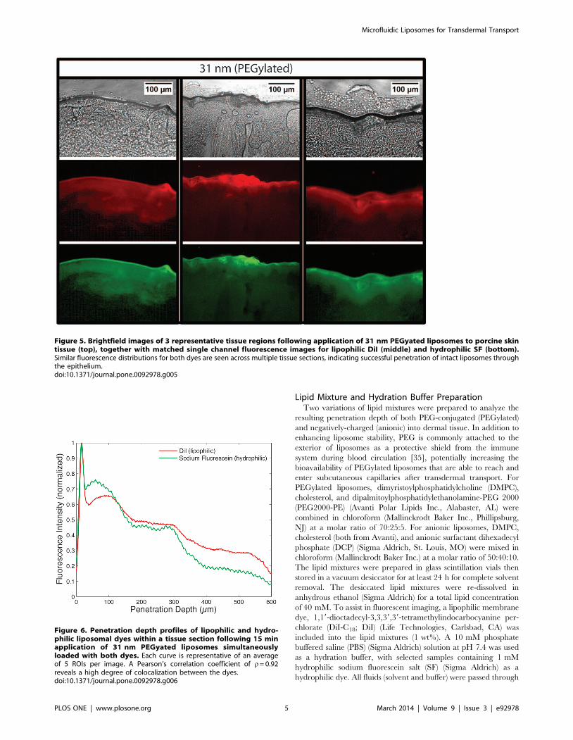
presence of intact vesicles. For the case of 31 nm liposomes, two
color imaging of the exposed tissue sections reveals strong
agreement between the distributions of hydrophilic (green) and
lipophilic (red) signals through the SC and into the epidermis for
all samples, as revealed through both the images (Fig. 5) and the
dye penetration depth profiles taken through the depth of the
tissue (Fig. 6). Using Pearson’s correlation coefficient (r) as a
measure of the degree of linear dependence between the spatial
distributions of each dye, an average value of r= 0.92 was
determined for the 31 nm PEGylated liposomes, indicating a high
degree of correlation between the dye locations. While not
conclusive, this evidence strongly suggests that the small liposomes
Figure 2. Brightfield/fluorescence image overlays (top) and single-channel fluorescence images (bottom) for microtomed tissuesections following 15 min application of PEGylated or anionic liposome samples of varying diameters containing DiI lipophilic dye.Significant dye penetration past the SC is observed with the smallest liposomes (31 nm diameter PEGylated and 41 nm anionic liposomes), while dyefrom the larger vesicles does not appear to cross the SC, indicating size-based passive transport independent of surface charge.doi:10.1371/journal.pone.0092978.g002
Microfluidic Liposomes for Transdermal Transport
PLOS ONE | www.plosone.org 3 March 2014 | Volume 9 | Issue 3 | e92978

successfully penetrate through the SC intact with minimal leakage
of their cargo. Experiments performed using larger liposomes
resulted in measured values of r= 0.81 and r= 0.75 for 308 nm
and 105 nm liposomes, respectively. This relatively poor correla-
tion, together with the overall lack of significant dye penetration
(Fig. 3), indicates that some degree of vesicle degradation and free
dye diffusion occurs for these larger liposomes.
In conclusion, we have leveraged a microfluidic liposome
synthesis technique to evaluate size-dependent transdermal delivery
of liposomes through ex vivo porcine tissues. Compared to larger
vesicles, where dye penetration across the SC is presumed to occur
primarily through a combination of vesicle rupture and transport
along follicular pathways, the smaller 31 nm diameter and 41 nm
diameter liposomes traverse and transport their intra-liposomal
contents across the full surface of the SC and into deep dermal
tissues, with penetration depths of at least several hundred
micrometers observed with a short 15 min incubation. Multicolor
fluorescence imaging of hydrophilic and hydrophobic dyes
incorporated into the liposomes during synthesis further reveals that
the smallest 31 nm liposomes are able to traverse dermal layers
intact, with implications for clinical applications requiring co-
delivery of therapeutic reagents with dissimilar chemical properties,
nanoparticle-mediated drug release, or transport of intact nano-
carriers to the bloodstream for systemic delivery. The results
presented here also represent the first demonstration of passive
transdermal diffusion of nanoscale, microfluidic-generated lipo-
somes, opening the door to the use of these nanoparticles for
effective delivery of lipophilic, hydrophilic, and amphipathic
compounds to underlying dermal layers. The transport of nano-
particles through the SC is a matter of much debate, and the
findings of the present study will require additional validation using
complementary methods to confirm the size-dependent behavior
described here and assess the fate of both liposomes and cargo.
Materials and Methods
Certain commercial equipment, instruments, or materials are
identified in this paper to foster understanding. Such identification
does not imply recommendation or endorsement by the National
Institute of Standards and Technology, nor does it imply that the
materials or equipment identified are necessarily the best available
for the purpose.
Ethics StatementThis research involved Yorkshire piglets sacrificed as part of a
separate parallel study approved by the Institutional Animal Care
and Use Committee (IACUC) at the Children’s National Medical
Center. All procedures were performed in accordance with the
National Institutes of Health Guide for the Care and Use of
Animals in Research.
Figure 3. DiI fluorescence intensity plot profiles for (a)PEGylated liposomes and (b) anionic liposomes as a functionof porcine skin tissue penetration depth. Measurements wereperformed 15 minute following liposome application. Each curve isrepresentative of an average of 5 ROIs per image.doi:10.1371/journal.pone.0092978.g003
Figure 4. Percentage of total DiI fluorescence signal seenbelow the SC for the different sizes of PEGylated and anionicliposomes. Each plot reflects the average profile extracted from 5 ROIsper tissue section, with error bars reflecting standard deviation. SCthickness, estimated from averaged manual measurements usingbrightfield images of each tissue, ranged from 15 mm to 40 mm, ingeneral agreement with previously reported values for porcine skin [32].The small 31 nm PEGylated liposomes pass the SC in large numbers(91%), which is up to 590% greater than the larger 105 nm to 308 nmdiameter liposomes. The small 41 nm anionic liposomes also reveal 65%of their total DiI signal under the SC, which is 200% greater thanobserved with 256 nm diameter liposomes of the same composition.doi:10.1371/journal.pone.0092978.g004
Microfluidic Liposomes for Transdermal Transport
PLOS ONE | www.plosone.org 4 March 2014 | Volume 9 | Issue 3 | e92978
Lipid Mixture and Hydration Buffer PreparationTwo variations of lipid mixtures were prepared to analyze the
resulting penetration depth of both PEG-conjugated (PEGylated)
and negatively-charged (anionic) into dermal tissue. In addition to
enhancing liposome stability, PEG is commonly attached to the
exterior of liposomes as a protective shield from the immune
system during blood circulation [35], potentially increasing the
bioavailability of PEGylated liposomes that are able to reach and
enter subcutaneous capillaries after transdermal transport. For
PEGylated liposomes, dimyristoylphosphatidylcholine (DMPC),
cholesterol, and dipalmitoylphosphatidylethanolamine-PEG 2000
(PEG2000-PE) (Avanti Polar Lipids Inc., Alabaster, AL) were
combined in chloroform (Mallinckrodt Baker Inc., Phillipsburg,
NJ) at a molar ratio of 70:25:5. For anionic liposomes, DMPC,
cholesterol (both from Avanti), and anionic surfactant dihexadecyl
phosphate (DCP) (Sigma Aldrich, St. Louis, MO) were mixed in
chloroform (Mallinckrodt Baker Inc.) at a molar ratio of 50:40:10.
The lipid mixtures were prepared in glass scintillation vials then
stored in a vacuum desiccator for at least 24 h for complete solvent
removal. The desiccated lipid mixtures were re-dissolved in
anhydrous ethanol (Sigma Aldrich) for a total lipid concentration
of 40 mM. To assist in fluorescent imaging, a lipophilic membrane
dye, 1,19-dioctadecyl-3,3,39,39-tetramethylindocarbocyanine per-
chlorate (DiI-C18; DiI) (Life Technologies, Carlsbad, CA) was
included into the lipid mixtures (1 wt%). A 10 mM phosphate
buffered saline (PBS) (Sigma Aldrich) solution at pH 7.4 was used
as a hydration buffer, with selected samples containing 1 mM
hydrophilic sodium fluorescein salt (SF) (Sigma Aldrich) as a
hydrophilic dye. All fluids (solvent and buffer) were passed through
Figure 5. Brightfield images of 3 representative tissue regions following application of 31 nm PEGyated liposomes to porcine skintissue (top), together with matched single channel fluorescence images for lipophilic DiI (middle) and hydrophilic SF (bottom).Similar fluorescence distributions for both dyes are seen across multiple tissue sections, indicating successful penetration of intact liposomes throughthe epithelium.doi:10.1371/journal.pone.0092978.g005
Figure 6. Penetration depth profiles of lipophilic and hydro-philic liposomal dyes within a tissue section following 15 minapplication of 31 nm PEGyated liposomes simultaneouslyloaded with both dyes. Each curve is representative of an averageof 5 ROIs per image. A Pearson’s correlation coefficient of r= 0.92reveals a high degree of colocalization between the dyes.doi:10.1371/journal.pone.0092978.g006
Microfluidic Liposomes for Transdermal Transport
PLOS ONE | www.plosone.org 5 March 2014 | Volume 9 | Issue 3 | e92978

0.22 mm filters (Millipore Corp., New Bedford, MA) before being
introduced to the microfluidic device.
Liposome synthesis and characterizationPEGylated and anionic liposomes were prepared using methods
described previously [17–20]. Briefly, a flow-focusing microchan-
nel network for liposome synthesis was fabricated following our
previous work [36]. All microchannels in the final device were
nominally 50 mm wide and 300 mm tall. The prepared lipid-
ethanol solution was injected into the microfluidic device between
two sheath flows of the aqueous buffer. The flow rate ratio (FRR),
defined as the ratio of the volumetric flow rate of the aqueous
buffer to the volumetric flow rate of the ethanol, was varied
between 5–50 to produce liposomes with modal diameters ranging
from 31 nm to 308 nm. Total average linear flow velocity for all
FRRs was kept constant (0.125 m/s) for a total volumetric flow
rate of 112 mL/min. To enable the formation of smaller vesicles,
the temperature of the microfluidic device was controlled by
contacting the glass slide of the device with a hot plate at 50uCthroughout the entire synthesis process [37]. The resulting
liposome populations were characterized for size via dynamic
light scattering (Nano ZSP, Malvern Instruments Ltd., UK). Size
distribution plots were generated by fitting spline curves to the
binned distribution data imported from the dynamic light
scattering instrument.
The microfluidic-generated liposomes contained lipophilic DiI
in their bilayers and hydrophilic SF in their cores to enable
fluorescence imaging of tissue penetration depth. To remove any
remaining dye not incorporated into the liposomes during the
synthesis process, all liposome samples were purified via size
exclusion chromatography on Sephadex G-25 PD-10 columns
(GE Healthcare, Piscataway, NJ) equilibrated with PBS immedi-
ately before application to the tissue. Gel filtration using the PD-10
columns provides efficient buffer exchange for removal of ethanol
used in the liposome formation process, thereby preventing
variations in ethanol concentration (2–16%) used for different
liposome populations from affecting skin permeation experiments.
Final lipid concentrations following gel filtration ranged from
0.56 mM to 4.76 mM, depending on the FRR used for liposome
synthesis.
Tissue exposure and cryosectioningPorcine ear tissue from Yorkshire piglets (4 weeks, 5 kg) was
selected due to its morphological and functional resemblance to
human skin. Porcine ear skin in vitro has shown remarkably similar
biophysical properties to human skin in vivo, particularly in terms
of the diffusivity and permeability coefficient of water across the
SC [38]. Studies have also indicated that porcine skin is extremely
similar both structurally and chemically to its human counterpart,
exhibits chemical properties which are rather consistent across
different samples and stable over time at room temperature,
therefore porcine skin is a valuable tool for investigating diffusion
dynamics of materials with human skin [39]. One ear from each
animal was removed following general anesthesia. Liposome
solutions were immediately applied in 50 mL aliquots for each
size in different locations on the outside of the ear, resulting in spot
areas ranging from 0.25–0.5 cm2, and incubated for 15 min at
room temperature. This exposure method was chosen over the use
of a perfusion cell since the focus of this study is on short-term SC
transport rather than long-term behavior of the nanoparticles
within the dermis. For the characterization of size-dependent
transport, all liposome solutions covering the full range of size
distributions were deposited on ears from a single animal to
minimize the influence of tissue morphology variations between
animals. Different animals were used for each set of experiments
characterizing PEGylated liposome transport, anionic liposome
transport, and co-distribution of lipophilic and hydrophilic dyes.
Following incubation, the ear tissue was placed in a plastic petri
dish and frozen. The frozen tissue was bulk sectioned, embedded
using Tissue-Tek Cryo-OCT compound (Fisher Scientific, Pitts-
burgh, PA), and frozen at 280uC. The frozen tissue blocks were
then sectioned into smaller slices, nominally 30 mm thick and
revealing dermal tissues at least 300 mm from the surface, using a
HM550 cryostat microtome (Richard Allan Scientific, Kalamazoo,
MI) and placed onto gelatin-treated glass slides for imaging.
Sections were procured from the tissue directly beneath each of
the applied liposome volumes, with the plane of each section
aligned through the center of its corresponding droplet. Sectioning
was performed with the blade oriented perpendicular to the skin
surface and the blade path in the direction of the SC to prevent
artifacts that could result from mechanical displacement of
liposomes, dye, or tissue normal to the SC layer.
Fluorescence microscopy and image processingThe 30 mm thick tissue sections were imaged using a TE-2000 S
inverted epifluorescence microscope (Nikon, Melville, NY). Bright-
field images and fluorescence images at 528 nm–553 nm (green
filter; DiI) and 465 nm–495 nm (blue filter; SF) excitation
wavelengths were acquired and overlaid to confirm and evaluate
the extent of liposome penetration into the dermal tissue and to
assess colocalization of the lipophilic and hydrophilic dyes.
ImageJ software (National Institutes of Health, Bethesda, MD)
was used to analyze the images. Fluorescence intensity profiles
were extracted using 10 mm wide regions of interest (ROIs), with
data from multiple ROIs combined to generate quantitative
profiles of liposome penetration depth. The intensity data was
averaged across 5 ROIs per sample, then normalized to peak
intensity and aligned to reveal the average fluorescence signal seen
within each tissue sample below the SC. Dye colocalization was
analyzed using the JACoP plugin with ImageJ [40]. In all
experiments, image analysis was performed independently for
each dye.
Acknowledgments
The authors thank Dr. Nobuyuki Ishibashi of Children’s National Medical
Center for supplying piglet ear tissue, support of the Maryland
NanoCenter FabLab, Dr. Peter Swaan at the University of Maryland
School of Pharmacy for Malvern Zetasizer access, and Dr. Adam Hsieh of
the University of Maryland College Park for cryostat microtome access.
Author Contributions
Conceived and designed the experiments: RH EK MJ WV ZQ JF DD.
Performed the experiments: RH EK. Analyzed the data: RH EK DD.
Contributed reagents/materials/analysis tools: MJ WV ZQ JF. Wrote the
paper: RH EK DD. Reviewed manuscript: RH EK MJ WN ZQ JF DD.
References
1. Prausnitz MR, Langer R (2008) Transdermal drug delivery. Nat Biotechnol 26:
1261–1268. Available: http://www.pubmedcentral.nih.gov/articlerender.
fcgi?artid = 2700785&tool = pmcentrez&rendertype = abstract. Accessed 27
February 2013.
2. Schreier H, Bouwstra J (1994) Liposomes and niosomes as topical drug carriers:
dermal and transdermal drug delivery. J Control Release 30: 1–15. Available:
http://linkinghub.elsevier.com/retrieve/pii/0168365994900396. Accessed 4
July 2013.
Microfluidic Liposomes for Transdermal Transport
PLOS ONE | www.plosone.org 6 March 2014 | Volume 9 | Issue 3 | e92978

3. Cevc G (2004) Lipid vesicles and other colloids as drug carriers on the skin. Adv
Drug Deliv Rev 56: 675–711. Available: http://www.ncbi.nlm.nih.gov/pubmed/15019752. Accessed 6 March 2013.
4. Karande P, Jain A, Ergun K, Kispersky V, Mitragotri S (2005) Design principles
of chemical penetration enhancers for transdermal drug delivery. PNAS 102:4688–4693.
5. Bouwstra JA, Honeywell-Nguyen PL, Gooris GS, Ponec M (2003) Structure ofthe skin barrier and its modulation by vesicular formulations. Prog Lipid Res 42:
1–36.
6. Ryman-Rasmussen JP, Riviere JE, Monteiro-Riviere NA (2006) Penetration ofintact skin by quantum dots with diverse physicochemical properties. Toxicol Sci
91: 159–165.7. Chu M, Wu Q, Wang J, Hou S, Miao Y, et al. (2007) In vitro and in vivo
transdermal delivery capacity of quantum dots through mouse skin. Nanotech-nology 18: 455103.
8. Schleich N, Preat V (2012) Nanostructured Biomaterials for Overcoming
Biological Barriers. Nanostructured Biomaterials for Overcoming BiologicalBarriers. doi:10.1039/9781849735292-00316.
9. Torchilin VP (2005) Recent advances with liposomes as pharmaceutical carriers.Nat Rev Drug Discov 4: 145–160. doi:10.1038/nrd1632.
10. Du Plessis J, Ramachandran C, Weiner N, Muller DG (1994) The influence of
particle size of liposomes on the deposition of drug into skin. Int J Pharm 103:277–282. doi:10.1016/0378-5173(94)90178-3.
11. Prow TW, Grice JE, Lin LL, Faye R, Butler M, et al. (2011) Nanoparticles andmicroparticles for skin drug delivery. Adv Drug Deliv Rev 63: 470–491.
Available: http://www.ncbi.nlm.nih.gov/pubmed/21315122. Accessed 23 Jan-uary 2014.
12. Sudhakar CK, Upadhyay N, Jain S, Charyulu RN (2012) Ethosomes as Non-
Invasive Loom for Transdermal Drug Delivery Systems. In: Sebastian M, NinanN, Haghi AK, editors. Nanomedicine and Drug Delivery.Apple Academic Press.
pp. 1–15.13. Sentjurc M, Vrhovnik K, Kristl J (1999) Liposomes as a topical delivery system:
the role of size on transport studied by the EPR imaging method. J Control
Release 59: 87–97.14. Cevc G, Gebauer D, Stieber J, Schatzlein A, Blume G (1998) Ultraflexible
vesicles, Transfersomes, have an extremely low pore penetration resistance andtransport therapeutic amounts of insulin across the intact mammalian skin.
Biochim Biophys Acta 1368: 201–215. Available: http://www.ncbi.nlm.nih.gov/pubmed/9459598. Accessed 4 July 2013.
15. Touitou E, Dayan N, Bergelson L, Godin B, Eliaz M (2000) Ethosomes - novel
vesicular carriers for enhanced delivery: characterization and skin penetrationproperties. J Control Release 65: 403–418. Available: http://www.ncbi.nlm.nih.
gov/pubmed/10699298. Accessed 28 August 2013.16. Brewer J, Bloksgaard M, Kubiak J, Sørensen JA, Bagatolli L a (2013) Spatially
resolved two-color diffusion measurements in human skin applied to transdermal
liposome penetration. J Invest Dermatol 133: 1260–1268. Available: http://www.ncbi.nlm.nih.gov/pubmed/23223136. Accessed 25 April 2013.
17. Jahn A, Vreeland WNNWN, Gaitan M, Locascio LEELE (2004) Controlledvesicle self-assembly in microfluidic channels with hydrodynamic focusing. J Am
Chem Soc 126: 2674–2675. Available: http://pubs.acs.org.proxy-um.researchport.umd.edu/doi/pdf/10.1021/ja0318030.
18. Jahn A, Vreeland WN, DeVoe DL, Locascio LE, Gaitan M (2007) Microfluidic
Directed Formation of Liposomes of Controlled Size. Langmuir 23: 6289–6293.Available: http://www.ncbi.nlm.nih.gov/pubmed/17451256. Accessed 3 Feb-
ruary 2014.19. Jahn A, Stavis SM, Hong JS, Vreeland WN, DeVoe DL, et al. (2010)
Microfluidic Mixing and the Formation of Nanoscale Lipid Vesicles. ACS Nano
4: 2077–2087. doi:10.1021/nn901676x.20. Hood RR, Shao C, Omiatek DM, Vreeland WN, DeVoe DL (2013)
Microfluidic Synthesis of PEG- and Folate-Conjugated Liposomes for One-Step Formation of Targeted Stealth Nanocarriers. Pharm Res 30: 1597–1607.
Available: http://www.ncbi.nlm.nih.gov/pubmed/23386106. Accessed 7
March 2013.21. Batzri S, Korn ED (1973) Single bilayer liposomes prepared without sonication.
Biochim Biophys Acta 298: 1015–1019. Available: http://www.ncbi.nlm.nih.gov/pubmed/4738145. Accessed 4 September 2013.
22. Maulucci G, De Spirito M, Arcovito G, Boffi F, Castellano AC, et al. (2005)Particle size distribution in DMPC vesicles solutions undergoing different
sonication times. Biophys J 88: 3545–3550. Available: http://www.
pubmedcentral.nih.gov/articlerender.fcgi?artid = 1305501&tool = pmcentrez&rendertype = abstract. Accessed 4 September 2013.
23. Vogt A, Combadiere B, Hadam S, Stieler KM, Lademann J, et al. (2006) 40 nm,
but not 750 or 1,500 nm, nanoparticles enter epidermal CD1a+ cells aftertranscutaneous application on human skin. J Invest Dermatol 126: 1316–1322.
Available: http://www.ncbi.nlm.nih.gov/pubmed/16614727. Accessed 6March 2013.
24. Kuchler S, Radowski MR, Blaschke T, Dathe M, Plendl J, et al. (2009)
Nanoparticles for skin penetration enhancement—a comparison of a dendriticcore-multishell-nanotransporter and solid lipid nanoparticles. Eur J Pharm
Biopharm Off J Arbeitsgemeinschaft fur Pharm Verfahrenstechnik eV 71: 243–250.
25. Verma DD, Verma S, Blume G, Fahr A (2003) Particle size of liposomesinfluences dermal delivery of substances into skin. Int J Pharm 258: 141–151.
Available: http://linkinghub.elsevier.com/retrieve/pii/S0378517303001832.
Accessed 7 March 2013.26. Barenholzt Y, Amselem S, Lichtenberg D (1979) A new method for preparation
of phospholipid vesicles (liposomes) - French press. FEBS Lett 99: 210–214.27. Hamilton RL, Goerke J, Guo LS, Williams MC, Havel RJ (1980) Unilamellar
liposomes made with the French pressure cell: a simple preparative and
semiquantitative technique. J Lipid Res 21: 981–992.28. Elsayed MM a, Abdallah OY, Naggar VF, Khalafallah NM (2007) Lipid vesicles
for skin delivery of drugs: reviewing three decades of research. Int J Pharm 332:1–16. Available: http://www.ncbi.nlm.nih.gov/pubmed/17222523. Accessed 6
August 2013.29. Gao S, Lan X, Liu Y, Shen Z, Lu J, et al. (2004) Characteristics of blood
fluorescence spectra using low-level, 457.9-nm excitation from Ar+ laser.
Chinese Opt Lett 2: 160–161.30. Rolland A, Wagner N, Chatelus A, Shroot B, Schaefer H (1993) Site-specific
drug delivery to pilosebaceous structures using polymeric microspheres. PharmRes 10: 1738–1744. Available: http://www.ncbi.nlm.nih.gov/pubmed/
8302759. Accessed 1 August 2013.
31. Mordon S, Sumian C, Devoisselle JM (2003) Site-specific methylene bluedelivery to pilosebaceous structures using highly porous nylon microspheres: an
experimental evaluation. Lasers Surg Med 33: 119–125. Available: http://www.ncbi.nlm.nih.gov/pubmed/12913884. Accessed 1 August 2013.
32. Jacobi U, Kaiser M, Toll R, Mangelsdorf S, Audring H, et al. (2007) Porcine earskin: an in vitro model for human skin. Skin Res Technol 13: 19–24. Available:
http://www.ncbi.nlm.nih.gov/pubmed/17250528. Accessed 7 March 2013.
33. Akomeah FK, Martin GP, Brown MB (2007) Variability in human skinpermeability in vitro: comparing penetrants with different physicochemical
properties. J Pharm Sci 96: 824–834.34. Mitragotri S (2003) Modeling skin permeability to hydrophilic and hydrophobic
solutes based on four permeation pathways. J Control Release 86: 69–92.
Available: http://www.ncbi.nlm.nih.gov/pubmed/12490374.35. Immordino ML, Dosio F, Cattel L (2006) Stealth liposomes: review of the basic
science, rationale, and clinical applications, existing and potential. Int JNanomedicine 1: 297–315. Available: http://www.pubmedcentral.nih.gov/
articlerender.fcgi?artid = 2426795&tool = pmcentrez&rendertype = abstract. Ac-cessed 3 July 2013.
36. Hood RR, Andar AU, Vreeland WN, DeVoe DL (2013) Microfluidic synthesis
of PEGylated and folate receptor-targeted liposomes. Pharm Res in press.doi:10.1007/s11095-013-0998-3.
37. Zook JM, Vreeland WN (2010) Effects of temperature, acyl chain length, andflow-rate ratio on liposome formation and size in a microfluidic hydrodynamic
focusing device. Soft Matter 6: 1352. doi:10.1039/b923299k.
38. Sekkat N, Kalia YN, Guy RH (2002) Biophysical study of porcine ear skin invitro and its comparison to human skin in vivo. J Pharm Sci 91: 2376–2381.
Available: http://www.ncbi.nlm.nih.gov/pubmed/12379922. Accessed 30 Jan-uary 2014.
39. Kong R, Bhargava R (2011) Characterization of porcine skin as a model for
human skin studies using infrared spectroscopic imaging. Analyst 136: 2359–2366. Available: http://www.ncbi.nlm.nih.gov/pubmed/21509377. Accessed 7
March 2013.40. Bolte S, Cordelieres FP (2006) A guided tour into subcellular colocalization
analysis in light microscopy. J Microsc 224: 213–232. doi:10.1111/j.1365-2818.2006.01706.x.
Microfluidic Liposomes for Transdermal Transport
PLOS ONE | www.plosone.org 7 March 2014 | Volume 9 | Issue 3 | e92978