microglia function in central nervous system development and...
TRANSCRIPT

Microglia Function in Central Nervous SystemDevelopment and Plasticity
Dorothy P. Schafer and Beth Stevens
Department of Neurology, F.M. Kirby Neurobiology Center, Boston Children’s Hospital, Harvard MedicalSchool, Boston, Massachusetts 02115
Correspondence: [email protected]
The nervous system comprises a remarkably diverse and complex network of different celltypes, which must communicate with one another with speed, reliability, and precision.Thus, the developmental patterning and maintenance of these cell populations and theirconnections with one another pose a rather formidable task. Emerging data implicate mi-croglia, the resident myeloid-derived cells of the central nervous system (CNS), in the spa-tial patterning and synaptic wiring throughout the healthy, developing, and adult CNS.Importantly, new tools to specifically manipulate microglia function have revealed thatthese cellular functions translate, on a systems level, to effects on overall behavior. In thisreview, we give a historical perspective of work to identify microglia function in the healthyCNS and highlight exciting new work in the field that has identified roles for these cells inCNS development, maintenance, and plasticity.
Microglia are one of the most enigmatic andunderstudied populations in the brain.
Until recently, most of what was known abouttheir function has been associated with theirrapid and robust responses to disease and injury(Ransohoff and Perry 2009; Graeber 2010; Ran-sohoff and Cardona 2010). The idea that microg-lia could be performing normal, homeostaticfunctions is a relatively new concept, galvanizedby pioneering in vivo imaging studies, whichrevealed that the processes of “resting” microgliaare highly motile in the intact, healthy adultbrain (Davalos et al. 2005; Nimmerjahn et al.2005). Remarkably, it is estimated that these mi-croglial processes survey the entire brain paren-chyma within a matter of hours, raising many
questions about the significance of this immunesurveillance system.
Since these initial findings, there has been asurge in the field to examine functional rolesof microglia in the healthy central nervous sys-tem (CNS), with a primary focus on postnataldevelopment. This focus was, to a large extent,incited by a landmark fate-mapping study in themouse showing that microglia develop fromprimitive myeloid progenitors in the embryonicyolk sac and begin to colonize the brain duringearly embryonic development (approximatelyembryonic day 9.5 [�E9.5] in the mouse) (Gin-houx et al. 2010). Given this early colonization,microglia are poised to play important roles inshaping the developing CNS and contributing
Editors: Ben A. Barres, Marc R. Freeman, and Beth Stevens
Additional Perspectives on Glia available at www.cshperspectives.org
Copyright # 2015 Cold Spring Harbor Laboratory Press; all rights reserved; doi: 10.1101/cshperspect.a020545
Cite this article as Cold Spring Harb Perspect Biol 2015;7:a020545
1
on May 29, 2018 - Published by Cold Spring Harbor Laboratory Press http://cshperspectives.cshlp.org/Downloaded from

to overall nervous system function. Indeed, re-cent work has shown that microglia in the de-veloping CNS can physically interact with neu-ronal soma and synapses in response to changesin neural activity, and data implicate microgliain many functions required to build and wirethe developing CNS ranging from neurogenesisto synaptic pruning (Tremblay 2011; Tremblayet al. 2011; Kettenmann et al. 2013; Schafer et al.2013; Wake et al. 2013; Salter and Beggs 2014).Furthermore, emerging work in the juvenileand adult reveal that these interactions and func-tions observed in the postnatal brain occur morebroadly to affect plasticity over the life span ofthe animal, ultimately affecting behavior.
In this chapter, we review the latest findingsin the field on microglia function in CNS devel-opment and plasticity. Our goal is to give a com-prehensive and critical perspective of this rela-tively new area of research and highlight newquestions. Furthermore, we discuss novel strat-egies to manipulate microglia function that willcontribute to our understanding of these cells inthe healthy nervous system and, ultimately, helpto identify mechanisms of disease.
SPATIAL PATTERNING IN THE DEVELOPINGAND MATURE CNS
In the context of CNS disease and injury, mi-croglia are appreciated as “professional” phago-cytes, known for their rapid and efficient abilityto clear dead or dying cells and cellular debris(Wyss-Coray and Mucke 2002; Napoli and Neu-mann 2009; Ransohoff and Perry 2009; Ranso-hoff and Cardona 2010; Kettenmann et al. 2011;Sierra et al. 2013). Furthermore, microglia pro-duce many factors that are known to activelytrigger neuronal apoptosis, including tumor ne-crosis factora (TNF-a), reactive oxygen species,glutamate, etc. (Bessis et al. 2007). Thus, microg-lia not only play a role in dead cell clearance,but they may also play a more active role ininitiating cell death or driving the cell death pro-gram in cells that are already rendered vulnera-ble/damaged. Consistent with this idea, recentwork has shown in a range of disease states (gli-oma, brain ischemia, Alzheimer’s disease, etc.)that microglia have the capacity to phagocytose
live cells, including viable but stressed neurons,via a number of different microglial phagocyticsignaling pathways (e.g., TREM2, MERTK, andMFG-E8) (Fig. 1C, top) (Brown and Neher2014). Concomitant to this cytotoxic potential,microglia produce trophic, mitogenic, and anti-inflammatory factors (Bessis et al. 2007; Pollard2009). As a result, microglia are poised to playmany diverse roles in patterning and maintain-ing the intricate and diverse cell populations thatcomprise the CNS. The following section re-views studies demonstrating a role for microgliain regulating CNS cell numbers during develop-ment and in adulthood.
Regulation of Cell Death
Essential to the development and spatial pat-terning of all organ systems is programmed celldeath (PCD), a process by which many cellsundergo apoptosis to achieve the cellular ar-chitecture characteristic of the mature system(Vaux and Korsmeyer 1999). When this processis absent or dysfunctional, a broad spectrum ofdevelopmental abnormalities and diseases canoccur (Meier et al. 2000). Given its diverse andcomplex cell populations, the nervous system isparticularly reliant on PCD and, thus, a modelsystem for studying the process (Oppenheim1991; Yeo and Gautier 2004; Rogulja-Ortmannet al. 2007). Indeed, during vertebrate develop-ment, �50% of neurons born must undergoPCD and the corpses must be cleared, a massiveand necessary task to maintain CNS homeosta-sis. In addition, the importance of this process isfurther emphasized given that it is highly con-served across vertebrates and invertebrate spe-cies (Sulston and Horvitz 1977; Abrams et al.1993; Bangs and White 2000).
The best-characterized role for microglia inPCD is their large capacity to phagocytose deadcell corpses during development (Fig. 1, top).One of the first descriptions of microglia carry-ing out this phenomenon was in the postnatalrat cerebral cortex, where, during the first weekof rat postnatal development, a large amount ofneuronal cell death occurs, particularly in thesubplate and layers II/III (Ferrer et al. 1990).During this time, globular, vacuolated phago-
D.P. Schafer and B. Stevens
2 Cite this article as Cold Spring Harb Perspect Biol 2015;7:a020545
on May 29, 2018 - Published by Cold Spring Harbor Laboratory Press http://cshperspectives.cshlp.org/Downloaded from

cytes, which are now assumed to be residentmicroglial cells, were found in increased num-bers and had engulfed dead cells throughout thecortical and subplate regions. Since these orig-inal observations, microglia have been shown tophagocytose dead cells throughout the healthy,developing CNS (Bessis et al. 2007; Sierra et al.2013). However, the mechanisms underlyingthese phagocytic events have remained unclear.Recent work in other model organisms mayoffer insight. For example, in Drosophila, glialcells, albeit not microglia, engulf dead cellcorpses and axons through a Draper/dCED-6-dependent signaling pathway, a pathway previ-ously found in Caenorhabditis elegans (Zhouet al. 2001). In addition, microglia engulf apo-ptotic neurons in developing zebrafish via an-
other pathway, the v0-ATPase a1 subunit (Lo-gan and Freeman 2007; Peri and Nusslein-Volhard 2008; Kurant 2011; Schafer and Stevens2013). It remains to be determined whether thesame or similar mechanisms are involved inPCD in the mammalian CNS. Furthermore, al-though studies show that microglia play an im-portant role in the removal of large amountsof cellular debris produced by PCD, in itself adaunting task, it was unclear whether microgliawere playing a role in the initiation of apoptoticevents. Recent work suggests a more active roleby which microglia initiate or, perhaps, propa-gate the cell death program before phagocytosis(Fig. 1A,C, top).
The concept of phagocytes initiating celldeath before phagocytosis was first conceived
Microglia
NPC or neuron
Apoptotic cell
IGF-1R-expressingNPC
Matureneuron
b
aIGF-1R
IGF-1R
Prosurvival
c
b
a
A
B
Prodeath
NGF, O2–, TNF
?
IGF-1
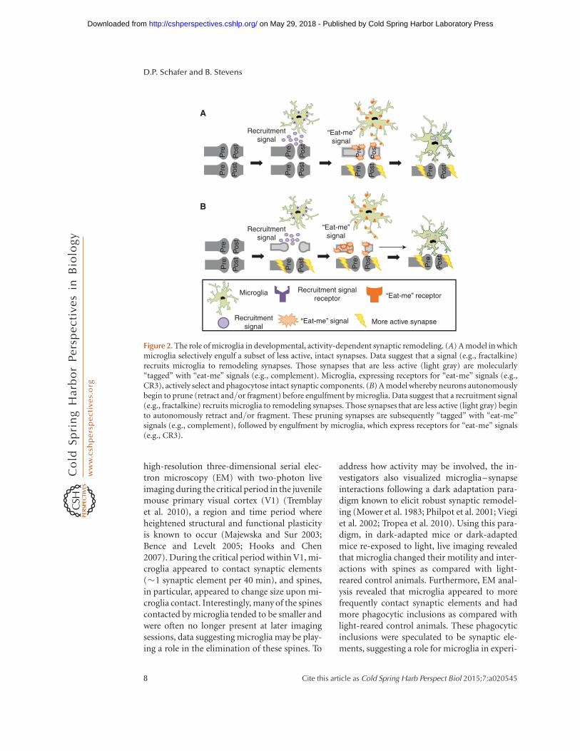
Figure 1. Mechanisms involved in microglia-mediated spatial patterning of neurons. (A) The role of microglia inprogrammed cell death. (a) Microglia can initiate the cell death program through a variety of soluble (e.g., nervegrowth factor [NGF], O2
–, and tumor necrosis factor [TNF]) and membrane-bound (e.g., CD11b and DAP12)factors followed by phagocytosis. (b) Alternatively, apoptosis of neurons or neural precursor cells (NPCs) isinduced by unknown mechanisms followed by microglia-mediated phagocytosis of debris. (c) “Phagoptosis”has been described in injury/disease paradigms known to induce inflammation (Brown and Neher 2014) inwhich microglia actively engulf live cells. Evidence suggests that something similar may be happening in thehealthy developing and adult central nervous system (CNS). (B) Microglia can also promote survival of NPCs inthe developing CNS. One mechanism identified was insulin-like growth factor (IGF)-1, a soluble factor madeand released by microglia, which is believed to bind IGF-1R receptors expressed by a subset of NPCs. It isspeculated that those NPCs expressing the receptor survive (a), whereas those that do not express the receptorundergo apoptosis (b).
Microglia in Development and Plasticity
Cite this article as Cold Spring Harb Perspect Biol 2015;7:a020545 3
on May 29, 2018 - Published by Cold Spring Harbor Laboratory Press http://cshperspectives.cshlp.org/Downloaded from

in the late 1800s by Elie Metchnikoff, who firstdescribed phagocytosis, and early work showedthat peripheral tissue macrophages could per-form such a function in developmental tissueremodeling (Aliprantis et al. 1996). For exam-ple, a study in the developing rodent retinashowed that, following genetic depletion of eyeand peritoneal macrophage subtypes, there wasa persistence of ocular tissues that are normallydevelopmentally transient, the hyaloid vascula-ture and pupillary membrane (Lang and Bishop1993). Since this time, microglia, the residentCNS myeloid-derived cells, have been shown toperform similar roles. The first evidence was inthe chick retina, where optic cups grown in theabsence of microglia resulted in a nearly three-fold reduction in retinal cell death as comparedwith optic cups grown in the presence of microg-lia (Frade and Barde 1998). Furthermore, whenpurified microglia were added back to the opticcup cultures, retinal cell death was increased.The molecular mechanism underlying this ef-fect was identified as microglial-derived nervegrowth factor (NGF). NGF expression was large-ly restricted to microglia during this develop-mental window, and retinal PCD was blockedwith NGF antibodies. It was proposed that mi-croglial-derived NGF initiates cell death when itbinds the neurotrophin receptor p75 (p75NTR),which is expressed by nearly all cells in the chickretina. Using a similar in vitro approach, microg-lia were also shown to initiate the PCD of Pur-kinje neurons in the developing mouse cerebel-lum and motoneurons in the developing ratspinal cord (Marin-Teva et al. 2004; Sedel etal. 2004). Using cerebellar slices, ablation of mi-croglia using clorodronate-filled liposomes re-sulted in a reduction in Purkinje neuron PCD(Marin-Teva et al. 2004). This cytotoxic effectwas mediated by microglial respiratory bursts,which produce superoxide ions (O2
2). However,the investigators also observed in vivo that.60% of Purkinje neurons that were contactedby microglial processes, but not yet phagocy-tosed, were already expressing molecules associ-ated with damage/PCD (i.e., activated caspase-3). These data suggest a role for microglia indriving the cell death program in neurons thatare already rendered vulnerable, a concept that
has been suggested in other model organisms(C. elegans and Drosophila) where dying, butnot yet dead, cells are engulfed by neighboringcells, including glia (Logan and Freeman 2007;Kinet and Shaham 2014). Similarly, using cul-tured spinal cord explants from developing ratembryos, microglial ablation, or blocking ofmicroglial-derived TNF-a signaling resulted ina decrease in motoneuron PCD (Sedel et al.2004). However, microglial TNF-a expressionwithin the spinal cord was transient and down-regulated before the cell death, data suggestingthat microglial-derived TNF-a may not direct-ly induce cell death but rather render moto-neurons more competent to die via other,yet-to-be-identified, neurotrophic factor–me-diated mechanisms.
Although intriguing and consistent withmacrophage studies, these initial studies sug-gesting that a more active role for microglia inPCD were all in vitro or ex vivo, which, in them-selves, induce injury during preparation. Tobetter address this question under more phys-iological conditions, more recent studies havetaken advantage of strategies to geneticallyand/or pharmacologically manipulate microg-lia in vivo. One study assessed PCD in the neo-natal hippocampus, where microgliawere foundto associate with neurons undergoing apoptosis(Wakselman et al. 2008). To determine an un-derlying mechanism, the investigators assessedmice deficient in CD11b (CD11b2/2), a surfacereceptor integrin, and mice with loss of functionin DAP12 (DAP12KI), a transmembrane sig-nal transduction adaptor molecule. These twomolecules are expressed by peripheral immunecells, including macrophages, where they carryout effector functions such as cell adhesion andphagocytosis (Mocsai et al. 2006). Important-ly, in the context of normal development, thesereceptors are almost exclusivelyexpressed by mi-croglia in the CNS. In CD11b2/2 and DAP12KI
mice, there was a significant, although not com-plete, reduction in neuronal apoptosis. In addi-tion, the investigators provided evidence thatCD11b and DAP12 regulate the production ofO2
2 by microglia, data consistent with earlierin vitro work in the cerebellum (Marin-Tevaet al. 2004). However, future work is necessary
D.P. Schafer and B. Stevens
4 Cite this article as Cold Spring Harb Perspect Biol 2015;7:a020545
on May 29, 2018 - Published by Cold Spring Harbor Laboratory Press http://cshperspectives.cshlp.org/Downloaded from
to show whether microglia initiate neuronalPCD in other CNS regions in vivo and whetherCD11b, DAP12, and/or O2
2 are involved. Fur-thermore, neuronal PCD, albeit reduced, wasstill observed in the hippocampus in DAP12KI
mice (Wakselman et al. 2008), and neuronsin the cerebellum appeared to already expressmarkers of cell death in the cerebellum beforeengulfment (Marin-Teva et al. 2004). Thus, al-though microglia may play a role in the initia-tion of cell death in a subset of neurons, theymay also be involved in facilitating the progres-sion of cell death in another subset that arealready rendered vulnerable by other mecha-nisms. The latter is particularly intriguing giventhat, in an otherwise healthy milieu, the precisetiming of PCD is critical. Microglia may offera mechanism by which this cell death timingis regulated. In addition, these data also suggestthat other molecular and/or cellular mecha-nisms (e.g., astrocyte-mediated phagocytosis)must also be involved, which are critical areasof investigation going forward.
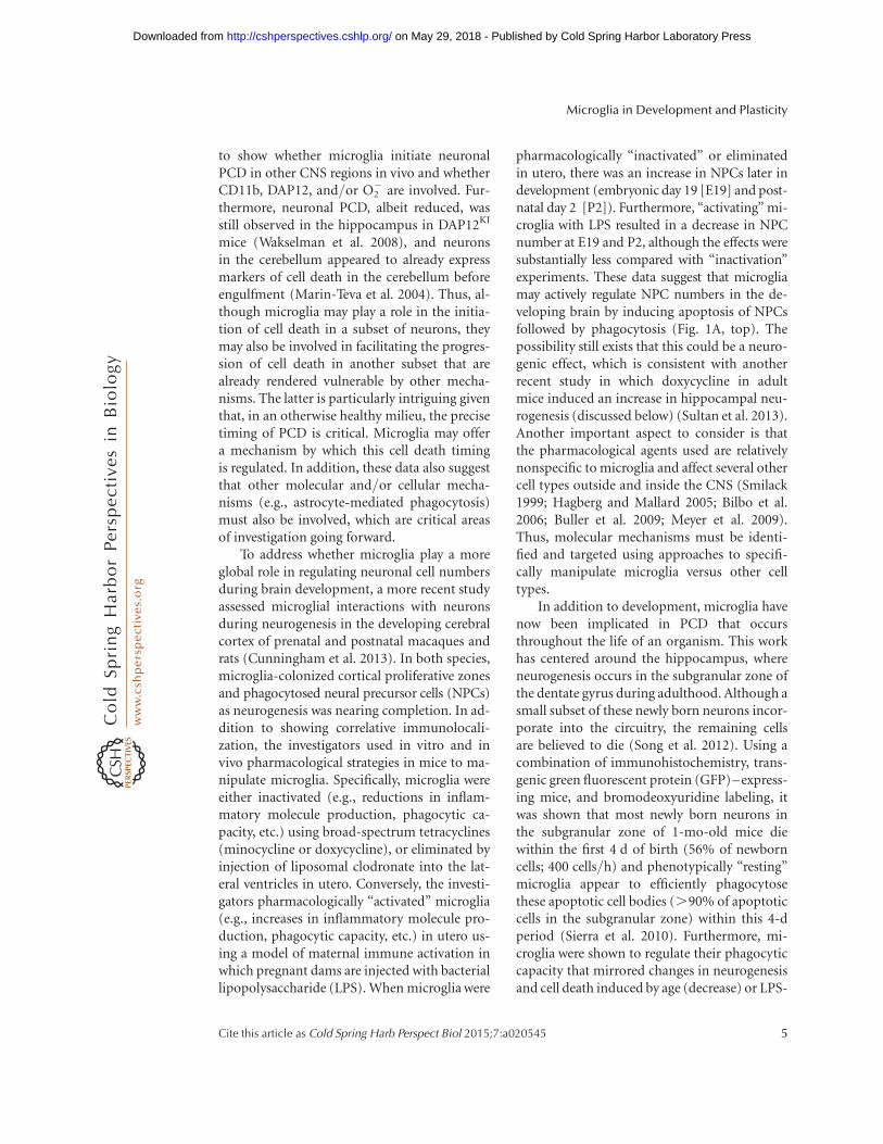
To address whether microglia play a moreglobal role in regulating neuronal cell numbersduring brain development, a more recent studyassessed microglial interactions with neuronsduring neurogenesis in the developing cerebralcortex of prenatal and postnatal macaques andrats (Cunningham et al. 2013). In both species,microglia-colonized cortical proliferative zonesand phagocytosed neural precursor cells (NPCs)as neurogenesis was nearing completion. In ad-dition to showing correlative immunolocali-zation, the investigators used in vitro and invivo pharmacological strategies in mice to ma-nipulate microglia. Specifically, microglia wereeither inactivated (e.g., reductions in inflam-matory molecule production, phagocytic ca-pacity, etc.) using broad-spectrum tetracyclines(minocycline or doxycycline), or eliminated byinjection of liposomal clodronate into the lat-eral ventricles in utero. Conversely, the investi-gators pharmacologically “activated” microglia(e.g., increases in inflammatory molecule pro-duction, phagocytic capacity, etc.) in utero us-ing a model of maternal immune activation inwhich pregnant dams are injected with bacteriallipopolysaccharide (LPS). When microglia were
pharmacologically “inactivated” or eliminatedin utero, there was an increase in NPCs later indevelopment (embryonic day 19 [E19] and post-natal day 2 [P2]). Furthermore, “activating” mi-croglia with LPS resulted in a decrease in NPCnumber at E19 and P2, although the effects weresubstantially less compared with “inactivation”experiments. These data suggest that microgliamay actively regulate NPC numbers in the de-veloping brain by inducing apoptosis of NPCsfollowed by phagocytosis (Fig. 1A, top). Thepossibility still exists that this could be a neuro-genic effect, which is consistent with anotherrecent study in which doxycycline in adultmice induced an increase in hippocampal neu-rogenesis (discussed below) (Sultan et al. 2013).Another important aspect to consider is thatthe pharmacological agents used are relativelynonspecific to microglia and affect several othercell types outside and inside the CNS (Smilack1999; Hagberg and Mallard 2005; Bilbo et al.2006; Buller et al. 2009; Meyer et al. 2009).Thus, molecular mechanisms must be identi-fied and targeted using approaches to specifi-cally manipulate microglia versus other celltypes.
In addition to development, microglia havenow been implicated in PCD that occursthroughout the life of an organism. This workhas centered around the hippocampus, whereneurogenesis occurs in the subgranular zone ofthe dentate gyrus during adulthood. Although asmall subset of these newly born neurons incor-porate into the circuitry, the remaining cellsare believed to die (Song et al. 2012). Using acombination of immunohistochemistry, trans-genic green fluorescent protein (GFP)–express-ing mice, and bromodeoxyuridine labeling, itwas shown that most newly born neurons inthe subgranular zone of 1-mo-old mice diewithin the first 4 d of birth (56% of newborncells; 400 cells/h) and phenotypically “resting”microglia appear to efficiently phagocytosethese apoptotic cell bodies (.90% of apoptoticcells in the subgranular zone) within this 4-dperiod (Sierra et al. 2010). Furthermore, mi-croglia were shown to regulate their phagocyticcapacity that mirrored changes in neurogenesisand cell death induced by age (decrease) or LPS-
Microglia in Development and Plasticity
Cite this article as Cold Spring Harb Perspect Biol 2015;7:a020545 5
on May 29, 2018 - Published by Cold Spring Harbor Laboratory Press http://cshperspectives.cshlp.org/Downloaded from

mediated inflammation (increase). Althoughthis study demonstrated a role for microglia inthe elimination of apoptotic NPCs, anotherstudy has shown that NPCs, themselves, canphagocytose dying cells during adult hippo-campal neurogenesis (Lu et al. 2011). Thus, itappears that microglia and NPCs may be work-ing together to eliminate apoptotic NPCs. Inaddition, unlike the developing CNS, it remainsunclear whether microglia and/or neighboringNPCs are involved in inducing apoptosis inthese newly born neurons in the adult hippo-campus. Evidence that apoptotic neurons existin the absence of microglial or NPC contact sug-gest that this may be a neuron-autonomousevent followed by phagocytosis (Fig. 1B, top).However, there is some evidence in the adultmouse dentate gyrus that microglia appear toinfiltrate small cytoplasmic openings of granulecells before cell death and that these microglialprocesses closely appose the nuclei of granulecells, data suggesting a more active role in initi-ating or contributing to progression of the deathprogram (Ribak et al. 2009).
Regulation of Cell Survival, Proliferation,and Differentiation
Microglia have also been implicated in spatialpatterning of the developing and adult CNS bypromoting cell survival, proliferation, and/ordifferentiation (Fig. 1, bottom). Some of the firstlines of evidence for this were in vitro studies inwhich microglia-conditioned media was addedto neuronal cultures, resulting in enhanced pro-genitor cell proliferation and enhanced neuronsurvival and/or maturation (Nagata et al. 1993;Chamak et al. 1994; Morgan et al. 2004). How-ever, other recent work has shown that culturingNPCs isolated from cortices that lack microglia(PU.12/2) had no effect on neuron survival orneurogenesis but rather resulted in decreases inneural progenitor cell proliferation and astro-genesis, effects that were rescued by addingwild-type (WT) microglia back to the cultures(Antony et al. 2011). Conversely, another studycultured NPCs from adult hippocampus andobserved a reduction in progenitor cell prolifer-ation in the presence of microglia (Gebara et al.
2013). In the same study, the investigators usedmodels of enhanced (exercise) or impaired (ag-ing) hippocampal neurogenesis to show an in-verse correlation between the number of mi-croglia and the number of progenitor cells infixed tissue. There are several explanations forthese disparate results. The most plausible expla-nations are differences in culture conditions,ages of mice used for the study, and/or region-specific effects. Furthermore, although it is clearfrom all of these studies that microglia have thecapacity to regulate progenitor cell numbers, invivo strategies and careful developmental anal-yses are necessary.
A more recent study has addressed whethermicroglia can promote neuron cell survival invivo in the developing mouse cortex (Fig. 1,bottom) (Ueno et al. 2013). The investigatorsshowed that, between P3 and P5, “inactivating”microglia with minocycline or ablating microg-lia by injecting diphtheria toxin into mice ex-pressing the diphtheria toxin receptor under thecontrol of the CD11b promoter resulted in anincrease in the number of apoptotic neuronsspecifically in layer Vof the cerebral cortex. Fur-thermore, microglia still appeared to engulfapoptotic debris after minocycline treatment,suggesting that microglial phagocytic capacityis not inhibited. In contrast, the data suggestthat microglia provide trophic support for neu-rons in the early postnatal CNS. Consistent withthis idea, the investigators show that mice defi-cient in the fractalkine receptor (CX3CR1),which is expressed almost exclusively by micro-glia in the context of the postnatal CNS, have asimilar reduction in layer V neurons. Further-more, CX3CR1 knockout mice had a reductionin free insulin-like growth factor 1 (IGF-1) andan increase in IGF-1-binding proteins, data sug-gesting that IGF-1 may be a factor downstreamfrom CX3CR1 that promotes neuron survival.When in vitro and in vivo approaches were usedto block IGF signaling (pharmacology andsmall interfering RNA), there was a significantincrease in cortical neuron cell death. However,none of the strategies used by these investigatorswere completely specific to microglia, as othercell types, particularly outside the CNS, expressCX3CR1 and IGF-1. Future work using Cre-lox
D.P. Schafer and B. Stevens
6 Cite this article as Cold Spring Harb Perspect Biol 2015;7:a020545
on May 29, 2018 - Published by Cold Spring Harbor Laboratory Press http://cshperspectives.cshlp.org/Downloaded from

technology to gain better cell specificity is nec-essary.
Taken together, the data demonstrate a rolefor microglia in spatial patterning of nervoussystem tissue by clearing apoptotic cells (neu-rons and neural progenitors). In addition, ex-periments also suggest a role for microglia ineither initiating the cell death program or pro-gressing cell death in a neuron previously ren-dered vulnerable. At the same time, there is alsonew evidence that microglia can also promoteneuron and NPC survival and/or proliferation.Given that these seemingly disparate functions(pro-cell death and pro-survival/proliferation)occur during very similar time frames, it re-mains unclear how microglia carry out thesefunctions simultaneously and raises the in-triguing possibility that microglia are a hetero-geneous cell population with several differentsubtypes of cells performing vastly differentfunctions. In addition, although work using ab-lation techniques to get rid of microglia duringspecific developmental time points has shownsome modest effects on cell numbers, ablationof microglia in the adult brain has shown nosignificant effect (Parkhurst et al. 2013; Elmoreet al. 2014). Several different explanations couldaccount for this, such as the importance ofcontext/timing; the developing CNS may be par-ticularly sensitive to these manipulations. In ad-dition, there may be overlapping functions withastrocytes, which can also phagocytose. In fact, ithas been shown that these cells can perform asimilar role as microglia in the context of syn-aptic pruning (Chung et al. 2013). Future workusing more specific in vivo strategies to manip-ulate microglia is necessary. Pharmacologicalstrategies (LPS, minocycline, and doxycycline)are nonspecific, with effects on many cell types.Furthermore, although data are promising inmice deficient in CD11b, DAP12, CX3CR1,and IGF-1 signaling, other cell types also expressthese molecules and are, thus, affected by genetictargeting. Recently, Cx3cr1creER mice, in whichcreER is expressed under the control of the frac-talkine receptor (CX3CR1), are an exciting newtool that will enable the analysis of microglia-specific effects (Goldmann et al. 2013; Parkhurstet al. 2013; Yona et al. 2013).
SYNAPTIC WIRING IN THE CNS
Similar to the excess of neurons born early indevelopment, an excess of synaptic contacts areformed initially, many of which are later prunedaway by phagocytic microglia during synapseelimination, a process critical for precise synap-tic connectivity (Fig. 2) (Purves and Lichtman1980; Lichtman and Colman 2000; Kano andHashimoto 2009). In addition, new researchsuggests that microglia can also influence brainwiring by regulating the number, maturation,and plasticity of synapses in development andthroughout the life span. The following sectionreviews these findings.
Activity-Dependent Interactionswith Synapses
One of the first clues that suggested a role formicroglia in synaptic pruning was the observa-tion that process-bearing, phagocytic microgliawere enriched in brain regions undergoing ac-tive synaptic remodeling, including cerebellum,hippocampus, and the visual system (Perry et al.1985; Milligan et al. 1991; Dalmau et al. 1998;Maslinska et al. 1998; Wierzba-Bobrowicz et al.1998; Schafer et al. 2012; Schwarz et al. 2012).However, it remained unclear if these cells wereperforming functions at developing synapses.Indeed, recent in vivo live imaging studies inthe adult and juvenile mouse cortex show thatmicroglia processes dynamically interact withdeveloping synapses and can detect local changesin neuronal activity (Tremblay et al. 2010; Wakeet al. 2013). For example, in layers II/III ofthe adult somatosensory and visual cortex, mi-croglial processes briefly contacted synaptic el-ements at a rate of �1 synapse/h (Wake et al.2013). Interestingly, this study also showed thatmicroglia–synapse contact was reduced whenneuronal activity was blocked by tetrodotoxin(TTX), enucleation of an eye, or lowering thetemperature. However, it remains to be deter-mined whether these activity-dependent inter-actions influenced synaptic remodeling.
To examine activity-dependent microglia–synapse interactions under more physiologicalconditions, Tremblay and colleagues combined
Microglia in Development and Plasticity
Cite this article as Cold Spring Harb Perspect Biol 2015;7:a020545 7
on May 29, 2018 - Published by Cold Spring Harbor Laboratory Press http://cshperspectives.cshlp.org/Downloaded from
high-resolution three-dimensional serial elec-tron microscopy (EM) with two-photon liveimaging during the critical period in the juvenilemouse primary visual cortex (V1) (Tremblayet al. 2010), a region and time period whereheightened structural and functional plasticityis known to occur (Majewska and Sur 2003;Bence and Levelt 2005; Hooks and Chen2007). During the critical period within V1, mi-croglia appeared to contact synaptic elements(�1 synaptic element per 40 min), and spines,in particular, appeared to change size upon mi-croglia contact. Interestingly, many of the spinescontacted by microglia tended to be smaller andwere often no longer present at later imagingsessions, data suggesting microglia may be play-ing a role in the elimination of these spines. To
address how activity may be involved, the in-vestigators also visualized microglia–synapseinteractions following a dark adaptation para-digm known to elicit robust synaptic remodel-ing (Mower et al. 1983; Philpot et al. 2001; Viegiet al. 2002; Tropea et al. 2010). Using this para-digm, in dark-adapted mice or dark-adaptedmice re-exposed to light, live imaging revealedthat microglia changed their motility and inter-actions with spines as compared with light-reared control animals. Furthermore, EM anal-ysis revealed that microglia appeared to morefrequently contact synaptic elements and hadmore phagocytic inclusions as compared withlight-reared control animals. These phagocyticinclusions were speculated to be synaptic ele-ments, suggesting a role for microglia in experi-
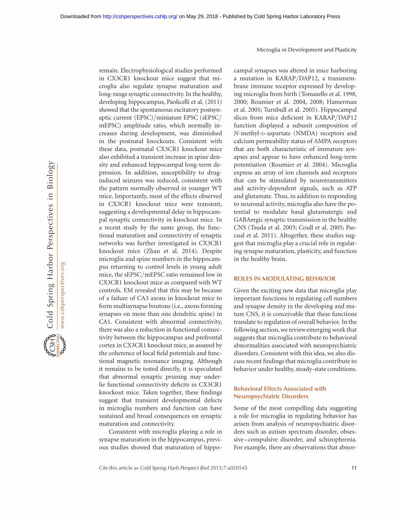
“Eat-me”signal
“Eat-me”signal
“Eat-me” signal
“Eat-me” receptor
Recruitmentsignal
A
B
Recruitmentsignal
Recruitmentsignal
Recruitment signalreceptor
More active synapse
MicrogliaP
re
Pre
Pre
Pre
Pre
Pre
Pre Pre Pre
Pre
Pre
Pre
Pos
t
Pos
t
Pos
t
Pos
t
Pos
t
Pos
tP
ost
Pos
t
Pos
t
Pos
t
Pos
t
eee
Figure 2. The role of microglia in developmental, activity-dependent synaptic remodeling. (A) A model in whichmicroglia selectively engulf a subset of less active, intact synapses. Data suggest that a signal (e.g., fractalkine)recruits microglia to remodeling synapses. Those synapses that are less active (light gray) are molecularly“tagged” with “eat-me” signals (e.g., complement). Microglia, expressing receptors for “eat-me” signals (e.g.,CR3), actively select and phagocytose intact synaptic components. (B) A model whereby neurons autonomouslybegin to prune (retract and/or fragment) before engulfment by microglia. Data suggest that a recruitment signal(e.g., fractalkine) recruits microglia to remodeling synapses. Those synapses that are less active (light gray) beginto autonomously retract and/or fragment. These pruning synapses are subsequently “tagged” with “eat-me”signals (e.g., complement), followed by engulfment by microglia, which express receptors for “eat-me” signals(e.g., CR3).
D.P. Schafer and B. Stevens
8 Cite this article as Cold Spring Harb Perspect Biol 2015;7:a020545
on May 29, 2018 - Published by Cold Spring Harbor Laboratory Press http://cshperspectives.cshlp.org/Downloaded from

ence- dependent synaptic remodeling in the ma-ture brain. It remains to be determined if thesehighly dynamic and activity-dependent interac-tions have functional significance during brainwiring.
To test the hypothesis that microglia phago-cytose excess or exuberant synaptic connectionsduring postnatal development, Schafer et al.took advantage of the mouse retinogeniculatesystem, a visual pathway and classic model sys-tem in which to study activity-dependent syn-apse elimination (Schafer et al. 2012). In thissystem postsynaptic thalamic relay neurons with-in the dorsal lateral geniculate nucleus (dLGN)are initially innervated by multiple weak reti-nal ganglion cell inputs originating from the ret-ina. A subset of these presynaptic inputs are latereliminated, while the remaining inputs aremaintained and strengthened (Chen and Re-gehr 2000; Hooks and Chen 2006; Huberman2007; Huberman et al. 2008). Using this system,the investigators observed microglial processesclosely apposed to presynaptic inputs under-going remodeling within the dLGN, data con-sistent with previous live imaging and EM data(Tremblay et al. 2010). Subsequently, a novelin vivo assay was developed in mice to deter-mine if microglia were engulfing inputs (Schaferet al. 2012, 2014). In this engulfment assay,presynaptic inputs within the dLGN are fluores-cently labeled by intraocular injection of anter-ograde dyes that are highly stable and resistantto lysosomal degradation (i.e., cholera toxin b
subunit conjugated to Alexa dyes). Further-more, this assay allows one to distinguish inputsfrom the left versus right eye by injecting dyetracers with different excitation and emissionspectra (e.g., cholera toxin b subunit conjugat-ed to Alexa 594 and Alexa 647, respectively).During the height of presynaptic input remod-eling (P5 in the mouse), microglia containedpresynaptic inputs within their processes andwithin lysosomal compartments. Furthermore,EM analysis revealed engulfed retinal ganglioncell axonal terminals throughout the dLGN.
Do microglia engulf specific synapses or dothey act as the cleanup crew that remove synap-tic debris (Fig. 2)? Synaptic pruning is believedto result from competition between neighbor-
ing axons for postsynaptic territory based ondifferences in patterns or levels of activity, suchthat weaker or less active synapses lose territo-ry while stronger or more active synaptic in-puts are elaborated and strengthened (Katz andShatz 1996; Sanes and Lichtman 1999; Hua andSmith 2004). Recent data in the retinogeniculatesystem support the hypothesis that microgliapreferentially engulf less active synapses duringactivity-dependent synaptic competition. Forexample, manipulation of retinal activity withTTX to block activity or forskolin, a cAMP an-alog that increases retinal activity, revealed thatmicroglia preferentially engulf presynaptic in-puts in the dLGN from the less active eye (Scha-fer et al. 2012). Together these data reveal thatmicroglia, indeed, engulf synapses during de-velopmental synaptic pruning in an activity-dependent manner. These data now raise ques-tions about the underlying mechanisms. Howdo microglia know which synapses to target?How do certain synapses become destined forelimination over others? Are synapses, indeed,intact at the time of engulfment (Fig. 2)?
Functional Consequences of Microglia–Synapse Interactions: Synaptic Pruning
One molecular pathway that has been proposedto mediate microglia–synapse interactions andsynaptic pruning is the classical complementcascade (Stevens et al. 2007; Schafer and Stevens2010; Alexander et al. 2012). Classical comple-ment cascade proteins C1q and C3 are localizedto synaptic compartments and mediate synapticpruning in the developing retinogeniculate sys-tem (Stevens et al. 2007). In the innate immunesystem, C1q and/or C3 bind cellular material,inducing its removal by several mechanisms,including phagocytic pathways (Lambris andTsokos 1986; Gasque 2004; van Lookeren Cam-pagne et al. 2007) Together, these findings raisedthe hypothesis that complement C1q and C3target synapses for elimination by microglia,which, in the context of the healthy postnatalbrain, are the only cells to express the high-af-finity receptor for C3, complement receptor 3(CR3; Cd11b) (Schafer et al. 2012). Consistentwith this hypothesis, C3 protein is highly ex-
Microglia in Development and Plasticity
Cite this article as Cold Spring Harb Perspect Biol 2015;7:a020545 9
on May 29, 2018 - Published by Cold Spring Harbor Laboratory Press http://cshperspectives.cshlp.org/Downloaded from

pressed and localized to synapses in the postna-tal dLGN, and CR3 is expressed by microglia atparticularly high levels during the peak of mi-croglia-mediated synaptic engulfment (P5). C3and CR3 knockout mice have reduced engulf-ment of presynaptic retinal inputs and a sus-tained deficit in eye-specific segregation thatphenocopied C1q and C3 knockout mice (Scha-feret al. 2012), implicating phagocytic microgliaas a key downstream cellular mediator of com-plement-dependent synapse elimination. Im-portantly, genetic disruption of CR3/C3 sig-naling resulted in sustained deficits in synapsenumber and synaptic connectivity. These find-ings show that microglia–synapse interactions,indeed, have functional consequences on synap-tic pruning that are sustained. Furthermore, thedata provide some insight into how synapsesmight be molecularly targeted for eliminationby microglia (Fig. 2). However, it is not yetknown if complement proteins are localized tospecific (i.e., less active/weaker) synapses andwhether changes in neuronal activity impactthis process. Finally, the identities of specific re-ceptors for complement at the synapse remainelusive. A recent in vitro study suggests that ter-minal sugar residues in the extracellular matrixof glycoproteins surrounding neurons mediateC1q binding to neurites (Linnartz et al. 2012). Infuture work, it will be interesting to test whethersialic acid residues in axons play a similar role invivo and whether the complement cascade me-diates synapse elimination in the developinghippocampus and other brain regions.
In addition to complement, recent data havealso implicated microglia in synaptic remodel-ing in the hippocampus via a CX3CR1-depen-dent mechanism (Tremblay et al. 2010; Paolicelliet al. 2011). Paolicelli and colleagues showed thatmice deficient in CX3CR1 (CX3CR1 knockouts)exhibited a transient increase in spine densityin postnatal weeks 2 and 3 in the hippocam-pus, and, similar to the case in the developingvisual system, high-resolution imaging and EMrevealed a concomitant increase in postsynap-tic density protein 95 (PSD95)-positive immu-noreactivity within microglia (Paolicelli et al.2011). Interestingly, there were significantlyfewer microglia detected in the postnatal hip-
pocampus of CX3CR1 knockout mice, and theseabnormalities in microglia number and synapsedensity returned to normal levels by adulthood.These data suggest that fractalkine signaling reg-ulates microglia number and/or recruitment tosynaptic sites in the early postnatal brain (Fig. 2).Another study showed different defects in syn-aptic function in the mature hippocampus ofCX3CR1 knockout mice, raising the possibilitythat fractalkine signaling may play different rolesdepending on the context (Rogers et al. 2011).In this case, long-term potentiation inductionwas reduced in organotypic hippocampal slicesprepared from adult CX3CR1 knockout miceas compared with WT littermates (Rogers et al.2011). Importantly, these deficits were coinci-dent with impairments in learning and memoryas assessed by Morris water maze and contextualand cued fear conditioning.
It is not yet known whether microglia engulfsynapses as part of an activity-dependent devel-opmental pruning process in the hippocampus,as was demonstrated in the visual system. Fur-thermore, it remains unclear whether fractalkineand complement systems are working coopera-tively to contribute to synaptic pruning. Thereis also an open question as to whether theseare, indeed, microglia-specific effects, as manyimmune cells, albeit localized to the peripheryand not the healthy CNS, express these receptors.Along these same lines, it has also recently beenshown that, similar to microglia, astrocytes inthe retinogeniculate system can also contributeto synaptic pruning by phagocytosing develop-ing synapses in an activity-dependent manner(Chung et al. 2013). These data suggest that thesetwo phagocytic cell populations could be work-ing cooperatively to achieve proper brain wiring.Experiments designed to addressthese questionswill be important areas of future investigation(Ransohoff and Stevens 2011).
Functional Consequences of Microglia–Synapse Interactions: Modulation ofSynapse Maturation and Function
Although synapse formation and pruning arekey features of circuit development, so too arethe function and maturation of the synapses that
D.P. Schafer and B. Stevens
10 Cite this article as Cold Spring Harb Perspect Biol 2015;7:a020545
on May 29, 2018 - Published by Cold Spring Harbor Laboratory Press http://cshperspectives.cshlp.org/Downloaded from
remain. Electrophysiological studies performedin CX3CR1 knockout mice suggest that mi-croglia also regulate synapse maturation andlong-range synaptic connectivity. In the healthy,developing hippocampus, Paolicelli et al. (2011)showed that the spontaneous excitatory postsyn-aptic current (EPSC)/miniature EPSC (sEPSC/mEPSC) amplitude ratio, which normally in-creases during development, was diminishedin the postnatal knockouts. Consistent withthese data, postnatal CX3CR1 knockout micealso exhibited a transient increase in spine den-sity and enhanced hippocampal long-term de-pression. In addition, susceptibility to drug-induced seizures was reduced, consistent withthe pattern normally observed in younger WTmice. Importantly, most of the effects observedin CX3CR1 knockout mice were transient,suggesting a developmental delay in hippocam-pal synaptic connectivity in knockout mice. Ina recent study by the same group, the func-tional maturation and connectivity of synapticnetworks was further investigated in CX3CR1knockout mice (Zhan et al. 2014). Despitemicroglia and spine numbers in the hippocam-pus returning to control levels in young adultmice, the sEPSC/mEPSC ratio remained low inCX3CR1 knockout mice as compared with WTcontrols. EM revealed that this may be becauseof a failure of CA3 axons in knockout mice toform multisynapse boutons (i.e., axons formingsynapses on more than one dendritic spine) inCA1. Consistent with abnormal connectivity,there was also a reduction in functional connec-tivity between the hippocampus and prefrontalcortex in CX3CR1 knockout mice, as assayed bythe coherence of local field potentials and func-tional magnetic resonance imaging. Althoughit remains to be tested directly, it is speculatedthat abnormal synaptic pruning may under-lie functional connectivity deficits in CX3CR1knockout mice. Taken together, these findingssuggest that transient developmental defectsin microglia numbers and function can havesustained and broad consequences on synapticmaturation and connectivity.
Consistent with microglia playing a role insynapse maturation in the hippocampus, previ-ous studies showed that maturation of hippo-
campal synapses was altered in mice harboringa mutation in KARAP/DAP12, a transmem-brane immune receptor expressed by develop-ing microglia from birth (Tomasello et al. 1998,2000; Roumier et al. 2004, 2008; Hamermanet al. 2005; Turnbull et al. 2005). Hippocampalslices from mice deficient in KARAP/DAP12function displayed a subunit composition ofN-methyl-D-aspartate (NMDA) receptors andcalcium permeability status of AMPA receptorsthat are both characteristic of immature syn-apses and appear to have enhanced long-termpotentiation (Roumier et al. 2004). Microgliaexpress an array of ion channels and receptorsthat can be stimulated by neurotransmittersand activity-dependent signals, such as ATPand glutamate. Thus, in addition to respondingto neuronal activity, microglia also have the po-tential to modulate basal glutamatergic andGABAergic synaptic transmission in the healthyCNS (Tsuda et al. 2003; Coull et al. 2005; Pas-cual et al. 2011). Altogether, these studies sug-gest that microglia play a crucial role in regulat-ing synapse maturation, plasticity, and functionin the healthy brain.
ROLES IN MODULATING BEHAVIOR
Given the exciting new data that microglia playimportant functions in regulating cell numbersand synapse density in the developing and ma-ture CNS, it is conceivable that these functionstranslate to regulation of overall behavior. In thefollowing section, we review emerging work thatsuggests that microglia contribute to behavioralabnormalities associated with neuropsychiatricdisorders. Consistent with this idea, we also dis-cuss recent findings that microglia contribute tobehavior under healthy, steady-state conditions.
Behavioral Effects Associated withNeuropsychiatric Disorders
Some of the most compelling data suggestinga role for microglia in regulating behavior hasarisen from analysis of neuropsychiatric disor-ders such as autism spectrum disorder, obses-sive–compulsive disorder, and schizophrenia.For example, there are observations that abnor-
Microglia in Development and Plasticity
Cite this article as Cold Spring Harb Perspect Biol 2015;7:a020545 11
on May 29, 2018 - Published by Cold Spring Harbor Laboratory Press http://cshperspectives.cshlp.org/Downloaded from

mal microglial cell activation exists along withdeficits in synaptic connectivity in patients withthese disorders (Arnold 1999; Belmonte et al.2004; Vargas et al. 2005; Woo and Crowell 2005;Monji et al. 2009; Morgan et al. 2010; Melomand Littleton 2011; Waites and Garner 2011).At the same time, there are now several animalmodels of these neuropsychiatric disorders inwhich there appear to be roles for microglia inonset and/or progression of behavioral pheno-types (Bilbo and Schwarz 2012; Derecki et al.2012a; Blank and Prinz 2013). For example,in mouse models of autism spectrum disorder(Mecp2 null) and obsessive-compulsive disor-der (Hoxb null), transplantation of WT bonemarrow into irradiated null hosts results in at-tenuation of behavioral phenotypes (Chen et al.2010; Derecki et al. 2012b). In the case of theMecp2 null, this attenuation was concomitantwith the colonization of host CNS by bonemarrow–derived myeloid cells with a microgliaphenotype (Derecki et al. 2012b). Another ex-ample of microglial involvement in behavioralabnormalities associated with neuropsychiatricdisorders is rodent models of maternal immuneactivation. In maternal immune activation,mice are subjected to early-life infection inutero and offspring develop several behavioralphenotypes associated with schizophrenia (e.g.,deficits in prepulse inhibition and acoustic star-tle response and decreases in exploratory be-havior and social interactions) and/or memoryand learning loss, particularly if combined witha later-life stress or immune challenge (i.e.,peripheral LPS administration) (Bilbo andSchwarz 2009; Meyer et al. 2009; Patterson2009; Harvey and Boksa 2012). These behavior-al phenotypes are accompanied by changes inmicroglia including activation state and num-bers in a brain region–specific manner (Bilboet al. 2005, 2007; Williamson et al. 2011; Gio-vanoli et al. 2013). These data provide evidencethat microglia may be “primed” prenatally dur-ing early-life infection and that a subsequentchallenge can significantly enhance their effectson later-life behavior. This has been furthersubstantiated by showing that microglial-derivedinterleukin-1b is a primary mechanism by whichdeficits in hippocampal-dependent learning
and memory are induced following in utero im-mune challenge followed by later-life stress(Williamson et al. 2011). Furthermore, a similarcytokine-mediated “priming” mechanism mayalso contribute to age-dependent memory lossassociated with aging and neurodegenerativedisease (Barrientos et al. 2006; Perry et al. 2007).
Behavioral Effects in the Healthy CNS
In addition to neuropsychiatric disorders, thereis also new evidence that microglia can af-fect baseline behavior in the absence of disease.For example, increased numbers of activatedmicroglia were observed in the preoptic nucleusof healthy male rats as compared to females(Lenz et al. 2013). When minocycline was ad-ministered to males, there was a decrease in ac-tivated microglia in males, which more closelyresembled female microglia, and a coincidentdisplay of feminine behaviors. Likewise, if fe-males were administered estradiol, which in-creased prostaglandin E2 and microglial activa-tion, masculine behaviors were observed. Thiswork suggests that microglia could be playingkey roles in regulating sexual behavior. Thisconcept is further substantiated by sexual di-morphisms that exist in microglial coloniza-tion and total numbers throughout the CNS(Schwarz and Bilbo 2012; Schwarz et al. 2012).More direct, genetic evidence that microgliacould be playing roles in baseline behavior in-clude work in CX3CR1-deficient mice, whichhave deficits in long-term potentiation accom-panied by significant deficits in contextual fearlearning and performance in the Morris watermaze (Rogers et al. 2011). However, anotherstudy found the opposite result, with very littleeffect on behavioral performance (Maggi et al.2009). This may be attributed to differences inparadigms used for behavioral measurements inthe two studies.
A recent study provided more direct evi-dence for a role of microglia in regulating be-havior using a strategy to manipulate gene ex-pression specifically in microglia (Cx3cr1creER)(Parkhurst et al. 2013). This system takes advan-tage of the relatively low turnover rate of microg-lia versus other peripheral CX3CR1þ immune
D.P. Schafer and B. Stevens
12 Cite this article as Cold Spring Harb Perspect Biol 2015;7:a020545
on May 29, 2018 - Published by Cold Spring Harbor Laboratory Press http://cshperspectives.cshlp.org/Downloaded from

cells, which turn over regularly (within a week)during hematopoiesis. After tamoxifen treat-ment, peripheral cells that underwent recom-bination turned over within 1 wk, resulting inmicroglia-specific, Cre-mediated expression ofgenes (Goldmann et al. 2013; Parkhurst et al.2013; Yona et al. 2013). Using these mice, mi-croglia were depleted using a diphtheria toxinstrategy, or brain-derived neurotrophic factor(BDNF) was specifically knocked out in mi-croglia. In either case, the result was a significantdeficit in performance on a rotarod as well asin a fear-conditioning paradigm. Furthermore,these effects were accompanied by a decrease indendritic spine formation, specifically in themotor cortex. Although it is not yet clear howmanipulation of BDNF in microglia impactsmotor learning or synaptic function, this wasthe first evidence that manipulating a gene spe-cifically in microglia had effects on synapticstructure and, ultimately, on behavior. In con-trast, when another recent study pharmaco-logically depleted microglia from adult mice,no significant behavioral deficits were observed(Elmore et al. 2014). The discrepancies in thesedata may be explained in several ways, includingthat a critical period (before adulthood) mayexist in which the rodent brain is particularlysensitive to microglial ablation. In addition, thebehavioral measures used for the latter studywere rather limited, and more extensive analysesmay be necessary to observe differences follow-ing pharmacological ablation in the adult.
Taken together, the data demonstrate a crit-ical function for microglia in regulating behav-ior, particularly if these cells are manipulatedduring development (postnatal through juve-nile stages). In addition, recent publicationshave advanced the field significantly by pro-viding genetic mouse models (Parkhurst et al.2013; Yona et al. 2013). The use of these micewill be critical going forward to specifically ma-nipulate microglia and understand their func-tion in the healthy and diseased CNS.
CONCLUDING REMARKS
The idea that microglia, compared with otherglial cell types, perform important functions in
the healthy nervous system is a relatively newconcept. This line of thinking was propagatedby experiments that used live imaging to deter-mine that microglial processes were highly mo-tile, often contacting synapses, in the healthy,intact brain. Other work described microgliain close apposition to and/or phagocytosingdead/dying neurons, neural progenitor cells,and synapses. Consistent with these cellular in-teractions having a more functional role, phar-macological and more specific genetic strategiesto target microglial phagocytic function result insignificant deficits in developmental neuronalPCD and synaptic pruning. On a behaviorallevel, abnormal microglial activation has beenobserved in humans and mouse models of neu-ropsychiatric disorders. Furthermore, when WT,microglia-like cells are introduced into mousemodels of these disorders, some behavioral def-icits are attenuated. Despite these intriguingfindings, it remains unclear how microglia con-tribute to behavioral phenotypes associated withthese disorders. One idea is that there are abnor-malities in microglia–synapse interactions dur-ing development. However, this remains to betested. Furthermore, microglia-specific molec-ular mechanisms are largely unknown. This waslargely because of the inability to manipulatemicroglia in a specific way without at least af-fecting peripheral immune cells of the same lin-eage and expressing a similar repertoire of genes(i.e., peripheral macrophages). New advances inCre-lox technology have resulted in Cx3cr1creER
mice, which will allow for specific genetic ma-nipulation in microglia after 1 wk of tamoxifentreatment. In addition, recent work identifyinggenes to distinguish microglia from their pe-ripheral cell counterparts offers exciting new ad-vances for manipulating microglia in more spe-cific ways (Chiu et al. 2013; Butovsky et al. 2014;Zhang et al. 2014). We are now entering a veryexciting time. The identification of molecularmechanisms underlying interactions betweenmicroglia and other CNS cell types and trans-lation of these mechanisms to functional con-sequences will offer enormous advancements.These advancements will range from under-standing of basic principles of nervous systemdevelopment and overall function to transla-
Microglia in Development and Plasticity
Cite this article as Cold Spring Harb Perspect Biol 2015;7:a020545 13
on May 29, 2018 - Published by Cold Spring Harbor Laboratory Press http://cshperspectives.cshlp.org/Downloaded from

tional neuroscience in which these basic mech-anisms can be applied to neurological disease.
ACKNOWLEDGMENTS
All work performed by authors is supportedby grants from the Smith Family Foundation(B.S.), the Dana Foundation (B.S.), the JohnMerck Scholars Program (B.S.), the NationalInstitute of Neurological Disorders and Stroke(RO1-NS-07100801, B.S.; F32-NS-066698,D.P.S.), the Nancy Lurie Marks Foundation(D.P.S.), and the National Institute of MentalHealth (K99-MH-102351, D.P.S.).
REFERENCES
Abrams JM, White K, Fessler LI, Steller H. 1993. Pro-grammed cell death during Drosophila embryogenesis.Development 117: 29–43.
Alexander A, Barres B, Stevens B. 2012. The complementsystem: An unexpected role in synaptic pruning duringdevelopment and disease. Annu Rev Neurosci 35: 369–389.
Aliprantis AO, Diez-Roux G, Mulder LC, Zychlinsky A, LangRA. 1996. Do macrophages kill through apoptosis?Immunol Today 17: 573–576.
Antony JM, Paquin A, Nutt SL, Kaplan DR, Miller FD.2011. Endogenous microglia regulate development ofembryonic cortical precursor cells. J Neurosci Res 89:286–298.
Arnold SE. 1999. Neurodevelopmental abnormalities inschizophrenia: Insights from neuropathology. Dev Psy-chopathol 11: 439–456.
Bangs P, White K. 2000. Regulation and execution of apo-ptosis during Drosophila development. Dev Dyn 218:68–79.
Barrientos RM, Higgins EA, Biedenkapp JC, Sprunger DB,Wright-Hardesty KJ, Watkins LR, Rudy JW, Maier SF.2006. Peripheral infection and aging interact to impairhippocampal memory consolidation. Neurobiol Aging27: 723–732.
Belmonte MK, Allen G, Beckel-Mitchener A, Boulanger LM,Carper RA, Webb SJ. 2004. Autism and abnormal devel-opment of brain connectivity. J Neurosci 24: 9228–9231.
Bence M, Levelt CN. 2005. Structural plasticity in the devel-oping visual system. 147: 125–139.
Bessis A, Bechade C, Bernard D, Roumier A. 2007. Microg-lial control of neuronal death and synaptic properties.Glia 55: 233–238.
Bilbo SD, Schwarz JM. 2009. Early-life programming oflater-life brain and behavior: A critical role for the im-mune system. Front Behav Neurosci 3: 14.
Bilbo SD, Schwarz JM. 2012. The immune system and de-velopmental programming of brain and behavior. FrontNeuroendocrinol 33: 267–286.
Bilbo SD, Levkoff LH, Mahoney JH, Watkins LR, Rudy JW,Maier SF. 2005. Neonatal infection induces memory im-pairments following an immune challenge in adulthood.Behav Neurosci 119: 293–301.
Bilbo SD, Rudy JW, Watkins LR, Maier SF. 2006. A behav-ioural characterization of neonatal infection-facilitatedmemory impairment in adult rats. Behav Brain Res 169:39–47.
Bilbo SD, Newsum NJ, Sprunger DB, Watkins LR, Rudy JW,Maier SF. 2007. Differential effects of neonatal handlingon early life infection-induced alterations in cognition inadulthood. Brain Behav Immun 21: 332–342.
Blank T, Prinz M. 2013. Microglia as modulators of cogni-tion and neuropsychiatric disorders. Glia 61: 62–70.
Brown GC, Neher JJ. 2014. Microglial phagocytosis of liveneurons. Nat Rev Neurosci 15: 209–216.
Buller KM, Carty ML, Reinebrant HE, Wixey JA. 2009. Min-ocycline: A neuroprotective agent for hypoxic-ischemicbrain injury in the neonate? J Neurosci Res 87: 599–608.
Butovsky O, Jedrychowski MP, Moore CS, Cialic R, LanserAJ, Gabriely G, Koeglsperger T, Dake B, Wu PM, DoykanCE, et al. 2014. Identification of a unique TGF-b-depen-dent molecular and functional signature in microglia.Nat Neurosci 17: 131–143.
Chamak B, Morandi V, Mallat M. 1994. Brain macrophagesstimulate neurite growth and regeneration by secretingthrombospondin. J Neurosci Res 38: 221–233.
Chen C, Regehr WG. 2000. Developmental remodeling ofthe retinogeniculate synapse. Neuron 28: 955–966.
Chen SK, Tvrdik P, Peden E, Cho S, Wu S, Spangrude G,Capecchi MR. 2010. Hematopoietic origin of patholog-ical grooming in Hoxb8 mutant mice. Cell 141: 775–785.
Chiu IM, Morimoto ET, Goodarzi H, Liao JT, O’Keeffe S,Phatnani HP, Muratet M, Carroll MC, Levy S, Tavazoie S,et al. 2013. A neurodegeneration-specific gene-expressionsignature of acutely isolated microglia from an amyotro-phic lateral sclerosis mouse model. Cell Rep 4: 385–401.
Chung WS, Clarke LE, Wang GX, Stafford BK, Sher A, Chak-raborty C, Joung J, Foo LC, Thompson A, Chen C, et al.2013. Astrocytes mediate synapse elimination throughMEGF10 and MERTK pathways. Nature 504: 394–400.
Coull JA, Beggs S, Boudreau D, Boivin D, Tsuda M, Inoue K,Gravel C, Salter MW, De Koninck Y. 2005. BDNF frommicroglia causes the shift in neuronal anion gradientunderlying neuropathic pain. Nature 438: 1017–1021.
Cunningham CL, Martinez-Cerdeno V, Noctor SC. 2013.Microglia regulate the number of neural precursor cellsin the developing cerebral cortex. J Neurosci 33: 4216–4233.
Dalmau I, Finsen B, Zimmer J, Gonzalez B, Castellano B.1998. Development of microglia in the postnatal rat hip-pocampus. Hippocampus 8: 458–474.
Davalos D, Grutzendler J, Yang G, Kim JV, Zuo Y, Jung S,Littman DR, Dustin ML, Gan WB. 2005. ATP mediatesrapid microglial response to local brain injury in vivo.Nat Neurosci 8: 752–758.
Derecki NC, Cronk JC, Kipnis J. 2012a. The role of microgliain brain maintenance: Implications for Rett syndrome.Trend Immunol 34: 144–150.
D.P. Schafer and B. Stevens
14 Cite this article as Cold Spring Harb Perspect Biol 2015;7:a020545
on May 29, 2018 - Published by Cold Spring Harbor Laboratory Press http://cshperspectives.cshlp.org/Downloaded from

Derecki NC, Cronk JC, Lu Z, Xu E, Abbott SB, Guyenet PG,Kipnis J. 2012b. Wild-type microglia arrest pathology in amouse model of Rett syndrome. Nature 484: 105–109.
Elmore MR, Najafi AR, Koike MA, Dagher NN, Spangen-berg EE, Rice RA, Kitazawa M, Matusow B, Nguyen H,West BL, et al. 2014. Colony-stimulating factor 1 receptorsignaling is necessary for microglia viability, unmaskinga microglia progenitor cell in the adult brain. Neuron82: 380–397.
Ferrer I, Bernet E, Soriano E, del Rio T, Fonseca M. 1990.Naturally occurring cell death in the cerebral cortex of therat and removal of dead cells by transitory phagocytes.Neuroscience 39: 451–458.
Frade JM, Barde YA. 1998. Microglia-derived nerve growthfactor causes cell death in the developing retina. Neuron20: 35–41.
Gasque P. 2004. Complement: A unique innate immunesensor for danger signals. Mol Immunol 41: 1089–1098.
Gebara E, Sultan S, Kocher-Braissant J, Toni N. 2013. Adulthippocampal neurogenesis inversely correlates with mi-croglia in conditions of voluntary running and aging.Front Neurosci 7: 145.
Ginhoux F, Greter M, Leboeuf M, Nandi S, See P, Gokhan S,Mehler MF, Conway SJ, Ng LG, Stanley ER. 2010. Fatemapping analysis reveals that adult microglia derive fromprimitive macrophages. Science 330: 841.
Giovanoli S, Engler H, Engler A, Richetto J, Voget M, WilliR, Winter C, Riva MA, Mortensen PB, Schedlowski M, etal. 2013. Stress in puberty unmasks latent neuropatho-logical consequences of prenatal immune activation inmice. Science 339: 1095–1099.
Goldmann T, Wieghofer P, Muller PF, Wolf Y, Varol D, YonaS, Brendecke SM, Kierdorf K, Staszewski O, Datta M, etal. 2013. A new type of microglia gene targeting showsTAK1 to be pivotal in CNS autoimmune inflammation.Nat Neurosci 16: 1618–1626.
Graeber MB. 2010. Changing face of microglia. Science 330:783–788.
Hagberg H, Mallard C. 2005. Effect of inflammation oncentral nervous system development and vulnerability.Curr Opin Neurol 18: 117–123.
Hamerman JA, Tchao NK, Lowell CA, Lanier LL. 2005.Enhanced Toll-like receptor responses in the absence ofsignaling adaptor DAP12. Nat Immunol 6: 579–586.
Harvey L, Boksa P. 2012. Prenatal and postnatal animal mod-els of immune activation: Relevance to a range of neuro-developmental disorders. Dev Neurobiol 72: 1335–1348.
Hooks BM, Chen C. 2006. Distinct roles for spontaneousand visual activity in remodeling of the retinogeniculatesynapse. Neuron 52: 281–291.
Hooks BM, Chen C. 2007. Critical periods in the visualsystem: Changing views for a model of experience-depen-dent plasticity. Neuron 56: 312–326.
Hua JY, Smith SJ. 2004. Neural activity and the dynamicsof central nervous system development. Nat Neurosci 7:327–332.
Huberman AD. 2007. Mechanisms of eye-specific visual cir-cuit development. Curr Opin Neurobiol 17: 73–80.
Huberman AD, Feller MB, Chapman B. 2008. Mechanismsunderlying development of visual maps and receptivefields. Annu Rev Neurosci 31: 479–509.
Kano M, Hashimoto K. 2009. Synapse elimination in thecentral nervous system. Curr Opin Neurobiol 19: 154–161.
Katz LC, Shatz CJ. 1996. Synaptic activity and the construc-tion of cortical circuits. Science 274: 1133–1138.
Kettenmann H, Hanisch U, Noda M, Verkhratsky A. 2011.Physiology of microglia. Physiol Rev 91: 461–553.
Kettenmann H, Kirchhoff F, Verkhratsky A. 2013. Microglia:New roles for the synaptic stripper. Neuron 77: 10–18.
Kinet MJ, Shaham S. 2014. Noncanonical cell death in thenematode Caenorhabditis elegans. Methods Enzymol545: 157–180.
Kurant E. 2011. Keeping the CNS clear: Glial phagocyticfunctions in Drosophila. Glia 59: 1304–1311.
Lambris J, Tsokos G. 1986. The biology and pathophysiol-ogy of complement receptors. Anticancer Res 6: 515–523.
Lang RA, Bishop JM. 1993. Macrophages are required forcell death and tissue remodeling in the developing mouseeye. Cell 74: 453–462.
Lenz KM, Nugent BM, Haliyur R, McCarthy MM. 2013.Microglia are essential to masculinization of brain andbehavior. J Neurosci 33: 2761–2772.
Lichtman J, Colman H. 2000. Synapse elimination and in-delible memory. Neuron 25: 269.
Linnartz B, Kopatz J, Tenner AJ, Neumann H. 2012. Sialicacid on the neuronal glycocalyx prevents complement C1binding and complement receptor-3-mediated removalby microglia. J Neurosci 32: 946–952.
Logan MA, Freeman MR. 2007. The scoop on the fly brain:Glial engulfment functions in Drosophila. Neuron GliaBiol 3: 63–74.
Lu Z, Elliott MR, Chen Y, Walsh JT, Klibanov AL, Ravichan-dran KS, Kipnis J. 2011. Phagocytic activity of neuronalprogenitors regulates adult neurogenesis. Nat Cell Biol13: 1076–1083.
Maggi L, Trettel F, Scianni M, Bertollini C, Eusebi F,Fredholm BB, Limatola C. 2009. LTP impairment byfractalkine/CX3CL1 in mouse hippocampus is mediatedthrough the activity of adenosine receptor type 3 (A3R).J Neuroimmunol 215: 36–42.
Majewska A, Sur M. 2003. Motility of dendritic spines invisual cortex in vivo: Changes during the critical periodand effects of visual deprivation. Proc Natl Acad Sci 100:16024–16029.
Marin-Teva JL, Dusart I, Colin C, Gervais A, van Rooijen N,Mallat M. 2004. Microglia promote the death of devel-oping Purkinje cells. Neuron 41: 535–547.
Maslinska D, Laure-Kamionowska M, Kaliszek A. 1998.Morphological forms and localization of microglial cellsin the developing human cerebellum. Folia Neuropathol36: 145–151.
Meier P, Finch A, Evan G. 2000. Apoptosis in development.Nature 407: 796–801.
Melom JE, Littleton JT. 2011. Synapse development inhealth and disease. Curr Opin Genet Dev 21: 256–261.
Meyer U, Feldon J, Fatemi SH. 2009. In vivo rodent modelsfor the experimental investigation of prenatal immuneactivation effects in neurodevelopmental brain disorders.Neurosci Biobehav Rev 33: 1061–1079.
Milligan CE, Cunningham TJ, Levitt P. 1991. Differentialimmunochemical markers reveal the normal distribution
Microglia in Development and Plasticity
Cite this article as Cold Spring Harb Perspect Biol 2015;7:a020545 15
on May 29, 2018 - Published by Cold Spring Harbor Laboratory Press http://cshperspectives.cshlp.org/Downloaded from

of brain macrophages and microglia in the developing ratbrain. J Comp Neurol 314: 125–135.
Mocsai A, Abram CL, Jakus Z, Hu Y, Lanier LL, Lowell CA.2006. Integrin signaling in neutrophils and macrophagesuses adaptors containing immunoreceptor tyrosine-based activation motifs. Nat Immunol 7: 1326–1333.
Monji A, Kato T, Kanba S. 2009. Cytokines and schizophre-nia: Microglia hypothesis of schizophrenia. PsychiatryClin Neurosci 63: 257–265.
Morgan SC, Taylor DL, Pocock JM. 2004. Microglia releaseactivators of neuronal proliferation mediated by activa-tion of mitogen-activated protein kinase, phosphatidyli-nositol-3-kinase/Akt and delta–Notch signalling cas-cades. J Neurochem 90: 89–101.
Morgan JT, Chana G, Pardo CA, Achim C, Semendeferi K,Buckwalter J, Courchesne E, Everall IP. 2010. Microglialactivation and increased microglial density observed inthe dorsolateral prefrontal cortex in autism. Biol Psychi-atry 68: 368–376.
Mower G, Christen W, Caplan C. 1983. Very brief visualexperience eliminates plasticity in the cat visual cortex.Science 221: 178–180.
Nagata K, Nakajima K, Takemoto N, Saito H, Kohsaka S.1993. Microglia-derived plasminogen enhances neuriteoutgrowth from explant cultures of rat brain. Int J DevNeurosci 11: 227–237.
Napoli I, Neumann H. 2009. Microglial clearance functionin health and disease. Neuroscience 158: 1030–1038.
Nimmerjahn A, Kirchhoff F, Helmchen F. 2005. Resting mi-croglial cells are highly dynamic surveillants of brain pa-renchyma in vivo. Science 308: 1314–1318.
Oppenheim RW. 1991. Cell death during development ofthe nervous system. Annu Rev Neurosci 14: 453–501.
Paolicelli RC, Bolasco G, Pagani F, Maggi L, Scianni M, Pan-zanelli P, Giustetto M, Ferreira TA, Guiducci E, Dumas L,et al. 2011. Synaptic pruning by microglia is necessary fornormal brain development. Science 333: 1456–1458.
Parkhurst CN, Yang G, Ninan I, Savas JN, Yates JR III, La-faille JJ, Hempstead BL, Littman DR, Gan WB. 2013.Microglia promote learning-dependent synapse forma-tion through brain-derived neurotrophic factor. Cell155: 1596–1609.
Pascual O, Ben Achour S, Rostaing P, Triller A, Bessis A.2011. Microglia activation triggers astrocyte-mediatedmodulation of excitatory neurotransmission. Proc NatlAcad Sci 109: E197–E205.
Patterson PH. 2009. Immune involvement in schizophreniaand autism: Etiology, pathology and animal models. Be-hav Brain Res 204: 313–321.
Peri F, Nusslein-Volhard C. 2008. Live imaging of neuronaldegradation by microglia reveals a role for v0-ATPase a1in phagosomal fusion in vivo. Cell 133: 916–927.
Perry VH, Hume DA, Gordon S. 1985. Immunohisto-chemical localization of macrophages and microglia inthe adult and developing mouse brain. Neuroscience 15:313–326.
Perry VH, Cunningham C, Holmes C. 2007. Systemic infec-tions and inflammation affect chronic neurodegenera-tion. Nat Rev Immunol 7: 161–167.
Philpot B, Sekhar A, Shouval H, Bear M. 2001. Visual expe-rience and deprivation bidirectionally modify the com-
position and function of NMDA receptors in visual cor-tex. Neuron 29: 157–169.
Pollard JW. 2009. Trophic macrophages in development anddisease. Nat Rev Immunol 9: 259–270.
Purves D, Lichtman JW. 1980. Elimination of synapses in thedeveloping nervous system. Science 210: 153–157.
Ransohoff RM, Cardona AE. 2010. The myeloid cells of thecentral nervous system parenchyma. Nature 468: 253–262.
Ransohoff RM, Perry VH. 2009. Microglial physiology:Unique stimuli, specialized responses. Annu Rev Immu-nol 27: 119–145.
Ransohoff RM, Stevens B. 2011. Neuroscience. How manycell types does it take to wire a brain? Science 333: 1391–1392.
Ribak CE, Shapiro LA, Perez ZD, Spigelman I. 2009. Microg-lia-associated granule cell death in the normal adultdentate gyrus. Brain Struct Funct 214: 25–35.
Rogers JT, Morganti JM, Bachstetter AD, Hudson CE, PetersMM, Grimmig BA, Weeber EJ, Bickford PC, Gemma C.2011. CX3CR1 deficiency leads to impairment of hippo-campal cognitive function and synaptic plasticity. JNeurosci 31: 16241–16250.
Rogulja-Ortmann A, Luer K, Seibert J, Rickert C, TechnauGM. 2007. Programmed cell death in the embryonic cen-tral nervous system of Drosophila melanogaster. Develop-ment 134: 105–116.
Roumier A, Bechade C, Poncer JC, Smalla KH, TomaselloE, Vivier E, Gundelfinger ED, Triller A, Bessis A. 2004.Impaired synaptic function in the microglial KARAP/DAP12-deficient mouse. J Neurosci 24: 11421–11428.
Roumier A, Pascual O, Bechade C, Wakselman S, Poncer JC,Real E, Triller A, Bessis A. 2008. Prenatal activation ofmicroglia induces delayed impairment of glutamatergicsynaptic function. PloS ONE 3: e2595.
Salter MW, Beggs S. 2014. Sublime microglia: Expandingroles for the guardians of the CNS. Cell 158: 15–24.
Sanes JR, Lichtman JW. 1999. Development of the vertebrateneuromuscular junction. Annu Rev Neurosci 22: 389–442.
Schafer DP, Stevens B. 2010. Synapse elimination duringdevelopment and disease: Immune molecules take centrestage. Biochem Soc Trans 38: 476–481.
Schafer DP, Stevens B. 2013. Phagocytic glial cells: Sculptingsynaptic circuits in the developing nervous system. CurrOpin Neurobiol 23: 1034–1040.
Schafer DP, Lehrman EK, Kautzman AG, Koyama R, Mar-dinly AR, Yamasaki R, Ransohoff RM, Greenberg ME,Barres BA, Stevens B. 2012. Microglia sculpt postnatalneural circuits in an activity and complement-dependentmanner. Neuron 74: 691–705.
Schafer DP, Lehrman EK, Stevens B. 2013. The “quad-par-tite” synapse: Microglia-synapse interactions in the de-veloping and mature CNS. Glia 61: 24–36.
Schafer DP, Lehrman EK, Heller CT, Stevens B. 2014. Anengulfment assay: A protocol to assess interactions be-tween CNS phagocytes and neurons. J Vis Exp doi:10.3791/51482.
Schwarz JM, Bilbo SD. 2012. Sex, glia, and development:Interactions in health and disease. Horm Behav 62:243–253.
D.P. Schafer and B. Stevens
16 Cite this article as Cold Spring Harb Perspect Biol 2015;7:a020545
on May 29, 2018 - Published by Cold Spring Harbor Laboratory Press http://cshperspectives.cshlp.org/Downloaded from

Schwarz JM, Sholar PW, Bilbo SD. 2012. Sex differences inmicroglial colonization of the developing rat brain. JNeurochem 120: 948–963.
Sedel F, Bechade C, Vyas S, Triller A. 2004. Macrophage-derived tumor necrosis factor a, an early developmentalsignal for motoneuron death. J Neurosci 24: 2236–2246.
Sierra A, Encinas JM, Deudero JJ, Chancey JH, EnikolopovG, Overstreet-Wadiche LS, Tsirka SE, Maletic-Savatic M.2010. Microglia shape adult hippocampal neurogenesisthrough apoptosis-coupled phagocytosis. Cell Stem Cell7: 483–495.
Sierra A, Abiega O, Shahraz A, Neumann H. 2013. Janus-faced microglia: Beneficial and detrimental consequencesof microglial phagocytosis. Front Cell Neurosci 7: 6.
Smilack JD. 1999. The tetracyclines. Mayo Clin Proc 74:727–729.
Song J, Christian KM, Ming GL, Song H. 2012. Modifica-tion of hippocampal circuitry by adult neurogenesis. DevNeurobiol 72: 1032–1043.
Stevens B, Allen NJ, Vazquez LE, Howell GR, Christopher-son KS, Nouri N, Micheva KD, Mehalow AK, HubermanAD, Stafford B, et al. 2007. The classical complementcascade mediates CNS synapse elimination. Cell 131:1164–1178.
Sulston JE, Horvitz HR. 1977. Post-embryonic cell lineagesof the nematode, Caenorhabditis elegans. Dev Biol 56:110–156.
Sultan S, Gebara E, Toni N. 2013. Doxycycline increasesneurogenesis and reduces microglia in the adult hippo-campus. Front Neurosci 7: 131.
Tomasello E, Olcese L, Vely F, Geourgeon C, Blery M, Moq-rich A, Gautheret D, Djabali M, Matteii M, Vivier E. 1998.Gene structure, expression pattern, and biological activ-ity of mouse killer cell activating receptor-associated pro-tein (KARAP)/DAP-12. J Biol Chem 273: 34115–34119.
Tomasello E, Desmoulins P, Chemin K, Guia S, Cremer H,Ortaldo J, Love P, Kaiserlian D, Vivier E. 2000. Combinednatural killer cell and dendritic cell functional deficiencyin KARAP/DAP12 loss-of-function mutant mice. Im-munity 13: 355–364.
Tremblay ME. 2011. The role of microglia at synapses in thehealthy CNS: Novel insights from recent imaging studies.Neuron Glia Biol 7: 67–76.
Tremblay ME, Lowery RL, Majewska AK. 2010. Microglialinteractions with synapses are modulated by visual expe-rience. PLoS Biol 8: e1000527.
Tremblay ME, Stevens B, Sierra A, Wake H, Bessis A, Nim-merjahn A. 2011. The role of microglia in the healthybrain. J Neurosci 31: 16064–16069.
Tropea D, Majewska AK, Garcia R, Sur M. 2010. Structuraldynamics of synapses in vivo correlate with functionalchanges during experience-dependent plasticity in visualcortex. J Neurosci 30: 11086–11095.
Tsuda M, Shigemoto-Mogami Y, Koizumi S, Mizokoshi A,Kohsaka S, Salter M, Inoue K. 2003. P2X4 receptors in-duced in spinal microglia gate tactile allodynia after nerveinjury. Nature 424: 778–783.
Turnbull IR, McDunn JE, Takai T, Townsend RR, Cobb JP,Colonna M. 2005. DAP12 (KARAP) amplifies inflamma-tion and increases mortality from endotoxemia and sep-tic peritonitis. J Exp Med 202: 363–369.
Ueno M, Fujita Y, Tanaka T, Nakamura Y, Kikuta J, Ishii M,Yamashita T. 2013. Layer V cortical neurons require mi-croglial support for survival during postnatal develop-ment. Nat Neurosci 16: 543–551.
van Lookeren Campagne M, Wiesmann C, Brown EJ. 2007.Macrophage complement receptors and pathogen clear-ance. Cell Microbiol 9: 2095–2102.
Vargas DL, Nascimbene C, Krishnan C, Zimmerman AW,Pardo CA. 2005. Neuroglial activation and neuroinflam-mation in the brain of patients with autism. Ann Neurol57: 67–81.
Vaux DL, Korsmeyer SJ. 1999. Cell death in development.Cell 96: 245–254.
Viegi A, Cotrufo T, Berardi N, Mascia L, Maffei L. 2002.Effects of dark rearing on phosphorylation of neurotro-phin Trk receptors. Eur J Neurosci 16: 1925–1930.
Waites CL, Garner CC. 2011. Presynaptic function in healthand disease. Trends Neurosci 34: 326–337.
Wake H, Moorhouse AJ, Miyamoto A, Nabekura J. 2013.Microglia: Actively surveying and shaping neuronal cir-cuit structure and function. Trends Neurosci 36: 209–217.
Wakselman S, Bechade C, Roumier A, Bernard D, Triller A,Bessis A. 2008. Developmental neuronal death in hip-pocampus requires the microglial CD11b integrin andDAP12 immunoreceptor. J Neurosci 28: 8138–8143.
Wierzba-Bobrowicz T, Kosno-Kruszewska E, Gwiazda E,Lechowicz W. 1998. The comparison of microglia matu-ration in different structures of the human nervous sys-tem. Folia Neuropathol 36: 152–160.
Williamson LL, Sholar PW, Mistry RS, Smith SH, Bilbo SD.2011. Microglia and memory: Modulation by early-lifeinfection. J Neurosci 31: 15511–15521.
Woo TU, Crowell AL. 2005. Targeting synapses and myelinin the prevention of schizophrenia. Schizophr Res 73:193–207.
Wyss-Coray T, Mucke L. 2002. Inflammation in neurode-generative disease—A double-edged sword. Neuron 35:419–432.
Yeo W, Gautier J. 2004. Early neural cell death: Dying tobecome neurons. Dev Biol 274: 233–244.
Yona S, Kim KW, Wolf Y, Mildner A, Varol D, Breker M,Strauss-Ayali D, Viukov S, Guilliams M, Misharin A, etal. 2013. Fate mapping reveals origins and dynamics ofmonocytes and tissue macrophages under homeostasis.Immunity 38: 79–91.
Zhan Y, Paolicelli RC, Sforazzini F, Weinhard L, Bolasco G,Pagani F, Vyssotski AL, Bifone A, Gozzi A, Ragozzino D,et al. 2014. Deficient neuron-microglia signaling resultsin impaired functional brain connectivity and social be-havior. Nat Neurosci 17: 400–406.
Zhang Y, Chen K, Sloan SA, Bennett ML, Scholze AR,O’Keeffe S, Phatnani HP, Guarnieri P, Caneda C, Ruder-isch N, et al. 2014. An RNA-sequencing transcriptomeand splicing database of glia, neurons, and vascular cellsof the cerebral cortex. J Neurosci 34: 11929–11947.
Zhou Z, Hartwieg E, Horvitz HR. 2001. CED-1 is a trans-membrane receptor that mediates cell corpse engulfmentin C. elegans. Cell 104: 43–56.
Microglia in Development and Plasticity
Cite this article as Cold Spring Harb Perspect Biol 2015;7:a020545 17
on May 29, 2018 - Published by Cold Spring Harbor Laboratory Press http://cshperspectives.cshlp.org/Downloaded from

17, 20152015; doi: 10.1101/cshperspect.a020545 originally published online JulyCold Spring Harb Perspect Biol
Dorothy P. Schafer and Beth Stevens PlasticityMicroglia Function in Central Nervous System Development and
Subject Collection Glia
MaintenanceThe Nodes of Ranvier: Molecular Assembly and
Matthew N. Rasband and Elior Peles
Oligodendrocyte Development and PlasticityDwight E. Bergles and William D. Richardson
Microglia in Health and DiseaseRichard M. Ransohoff and Joseph El Khoury Support
Oligodendrocytes: Myelination and Axonal
Mikael Simons and Klaus-Armin NaveThe Astrocyte: Powerhouse and Recycling Center
Bruno Weber and L. Felipe Barros Central Nervous System GliaDrosophila
Marc R. Freeman
Development and PlasticityMicroglia Function in Central Nervous System
Dorothy P. Schafer and Beth StevensSynapse: Adaptable, Multitasking Glial CellsPerisynaptic Schwann Cells at the Neuromuscular
Chien-Ping Ko and Richard Robitaille
the Central Nervous SystemOligodendrocyte Development and Myelination in Transcriptional and Epigenetic Regulation of
Ben Emery and Q. Richard Lu
and EliminationAstrocytes Control Synapse Formation, Function,
Won-Suk Chung, Nicola J. Allen and Cagla Eroglu
ControversiesOrigin of Microglia: Current Concepts and Past
Florent Ginhoux and Marco Prinz
Schwann Cell MyelinationJames L. Salzer
Remyelination−−Glia Disease and RepairRobin J.M. Franklin and Steven A. Goldman Repair
Schwann Cells: Development and Role in Nerve
LloydKristján R. Jessen, Rhona Mirsky and Alison C.
Astrocytes in Neurodegenerative DiseaseHemali Phatnani and Tom Maniatis
Perineurial GliaSarah Kucenas
http://cshperspectives.cshlp.org/cgi/collection/ For additional articles in this collection, see
Copyright © 2015 Cold Spring Harbor Laboratory Press; all rights reserved
on May 29, 2018 - Published by Cold Spring Harbor Laboratory Press http://cshperspectives.cshlp.org/Downloaded from