microrna biogenesis and hedgehog-patched signaling
TRANSCRIPT
HIGHLIGHTED ARTICLE| INVESTIGATION
MicroRNA Biogenesis and Hedgehog-PatchedSignaling Cooperate to Regulate an Important
Developmental Transition in GranuleCell Development
Lena Constantin,*,1 Myrna Constantin,† and Brandon J. Wainwright**Institute for Molecular Bioscience and †Queensland Alliance for Agriculture and Food Innovation, University of Queensland,
St. Lucia, Queensland 4072, Australia
ORCID IDs: 0000-0003-1492-3845 (L.C.); 0000-0002-3287-4891 (M.C.)
ABSTRACT The Dicer1, Dcr-1 homolog (Drosophila) gene encodes a type III ribonuclease required for the canonical maturation and functioningof microRNAs (miRNAs). Subsets of miRNAs are known to regulate normal cerebellar granule cell development, in addition to the growth andprogression of medulloblastoma, a neoplasm that often originates from granule cell precursors. Multiple independent studies have alsodemonstrated that deregulation of Sonic Hedgehog (Shh)-Patched (Ptch) signaling, through miRNAs, is causative of granule cell pathologies.In the present study, we investigated the genetic interplay between miRNA biogenesis and Shh-Ptch signaling in granule cells of the cerebellumby way of the Cre/lox recombination system in genetically engineered models of Mus musculus (mouse). We demonstrate that, although themiRNA biogenesis and Shh-Ptch-signaling pathways, respectively, regulate the opposing growth processes of cerebellar hypoplasia and hyper-plasia leading to medulloblastoma, their concurrent deregulation was nonadditive and did not bring the growth phenotypes toward an expectedequilibrium. Instead, mice developed either hypoplasia or medulloblastoma, but of a greater severity. Furthermore, some genotypes were bistable,whereby subsets of mice developed hypoplasia or medulloblastoma. This implies that miRNAs and Shh-Ptch signaling regulate an importantdevelopmental transition in granule cells of the cerebellum. We also conclusively show that the Dicer1 gene encodes a haploinsufficient tumorsuppressor gene for Ptch1-induced medulloblastoma, with the monoallielic loss of Dicer1more severe than biallelic loss. These findings exemplifyhow genetic interplay between pathways may produce nonadditive effects with a substantial and unpredictable impact on biology. Furthermore,these findings suggest that the functional dosage of Dicer1 may nonadditively influence a wide range of Shh-Ptch-dependent pathologies.
KEYWORDS Dicer1 protein; mouse; genes; tumor suppressor; Hedgehog proteins; heterozygote; medulloblastoma; nervous system malformations
MicroRNAs (miRNAs) are a class of �22-nucleotidenoncoding RNA molecules that function as sequence-
specific guides. They direct an Argonaute (Ago) protein-containing RNA-induced silencing complex (RISC) to targetmessenger RNAs (mRNAs) (Hutvagner and Zamore 2002;Mourelatos et al. 2002) by way of Watson–Crick pairing ofthe miRNA seed sequence to complementary elements usu-
ally found within the 39 untranslated region of target mRNAs(Bartel 2009). This results in translational repression and/ormRNA destabilization and decay (see Iwakawa and Tomari2015 for a recent review). MicroRNAs are first transcribed aslong primary transcripts (pri-miRNAs) with a characteristichairpin-like secondary structure (Mourelatos et al. 2002). Inthe canonical bioprocessing pathway, this hairpin-like struc-ture is recognized by the microprocessor–enzyme complex,through Ribonuclease 3 (protein Drosha) and Microproces-sor complex subunit DGCR8 (Lee et al. 2003; Denli et al.2004; Han et al. 2004), which cleaves the pri-miRNA intoan �70-nucleotide precursor miRNA (pre-miRNA). Thepre-miRNA is further processed, once exported to the cyto-plasm (Bohnsack et al. 2004; Lund et al. 2004), by the RNaseIII enzyme Dicer, into a mature �22-nucleotide RNA duplex
Copyright © 2016 by the Genetics Society of Americadoi: 10.1534/genetics.115.184176Manuscript received October 27, 2015; accepted for publication January 10, 2016;published Early Online January 13, 2016.Supporting information is available online at www.genetics.org/lookup/suppl/doi:10.1534/genetics.115.184176/-/DC1.1Corresponding author: Otto Hirschfeld Bldg. 81, Room 619, Chancellors Place,University of Queensland, St. Lucia, Queensland 4072, Australia.E-mail: [email protected]
Genetics, Vol. 202, 1105–1118 March 2016 1105
with 2-nucleotide overhangs at both 39 hydroxyl termini(Blaszczyk et al. 2001; Hutvagner et al. 2001). The endori-bonuclease Dicer, in addition to cleaving pre-miRNAs, partic-ipates in the loading of miRNAs into RISC (Chendrimadaet al. 2005). Both the endoribonuclease Dicer and proteinDrosha are thought to cleave double-stranded RNAs by form-ing an intramolecular pseudodimer between two tandemRNase III domains (a/b) (Han et al. 2004; Zhang et al.2004). In mice, a lysine residue (K1790) within the ribonucle-ase (RNase) IIIb domain of endoribonuclease Dicer appearscritical for double-stranded RNA cleavage (Du et al. 2008).
MicroRNAs, and by extension endoribonuclease Dicerfunction, are implicated in virtually every biological processin a wide range of organisms, and changes in their expressionare associated with a plethora of human pathologies. In thecerebellum, miRNA dysfunction, via ablation of endoribonu-clease Dicer function, has been linked to movement disorders(Lee et al. 2008; Constantin and Wainwright 2015), neuro-degeneration (Schaefer et al. 2007), and the malignant neo-plasm, medulloblastoma (Zindy et al. 2015). MicroRNAs playa prominent role in medulloblastoma, given that distinctmiRNA expression signatures are able to classify the molec-ular (Cho et al. 2011) and histopathological (Ferretti et al.2009) subtypes of human medulloblastoma, and dozens ofindependent studies have identified the functional networksby which specific miRNAs contribute to the growth and pro-gression of this disease. For example, the miR-17�92 clustercausally contributes to medulloblastoma and granule cellprecursor proliferation through Sonic Hedgehog (Shh)-Patched (Ptch) signaling (Northcott et al. 2009; Uziel et al.2009). Similarly, miR-106b has been shown to promote theproliferation, migration, and invasion potential of medullo-blastoma (Li et al. 2015), presumably via the principal medi-ator of the transcriptional response of the Shh-Ptch-signalingpathway, GLI-Kruppel family member GLI2 (Gli2) (Constantinand Wainwright 2015).
The cerebellum is located anteriorly on the brainstem,close to the midbrain–hindbrain boundary. It is essential forthe fine motor control of movement and posture and, on thebasis of human lesion and functional neuroimaging studies,appears to contribute to a broad range of high-order non-motor functions (see Buckner 2013 for review). A principalcell type of the cerebellum, and the most numerous in thecentral nervous system, is the granule cell. Granule cells areof critical importance to cerebellar function, as exemplifiedby the severity of motor coordination, language, and cogni-tive deficits observed in patients with congenital granule celldegeneration (Pascual-Castroviejo et al. 1994). Precursors ofgranule cells arise from the rhombic lip, a thin strip of neuro-epithelium that borders the non-neuronal roof plate of thefourth ventricle (Alder et al. 1996). During embryogenesis,granule cell precursors leave the rhombic lip and migrateanteriorly over the outer surface of the cerebellum to forma second germinal structure, termed the “external germinallayer” (see Hatten and Roussel 2011 for review). After birth,granule cell precursors undergo rapid proliferation in the
external germinal layer. Proliferation is maintained until peri-natal life by way of multiple symmetric mitoses in single-fated germinal cells (Espinosa and Luo 2008). Once granulecells become postmitotic, they extend parallel fiber axonsthat synapse to the dendrites of GABAergic inhibitory Pur-kinje neurons, while the soma of the granule cells migrateinteriorly to their final destination in the internal granularlayer (Komuro and Rakic 1998). Multiple mitogenic path-ways are known to drive the rapid expansion of granule cellprecursors in the external germinal layer, the most potent ofwhich is Shh (Dahmane and Ruiz i Altaba 1999; Lewis et al.2004). In the central nervous system, the binding of Shh tothe transmembrane protein Ptch relieves the inhibitory ac-tion of Ptch on the transmembrane protein Smoothened(Johnson et al. 2000; Briscoe et al. 2001). Active-stateSmoothened is then able to translocate to the primary cilium(Corbit et al. 2005), where it promotes the formation of theactive Gli family of transcription factors, which in turn in-duces the expression of target genes such as Gli1, Ptch1,and Nmyc (Humke et al. 2010; Tukachinsky et al. 2010).Therefore, the removal of Ptch from the plasma membrane,or the binding of Ptch to Shh, activates Shh-Ptch signaling.
Although some miRNAs are known to interact withHedgehog-Ptch signaling, the magnitude and directionalityof this interaction is not well understood, even within thecerebellum. In the present study, we investigated the genedose-dependent interaction between miRNA biogenesis andShh-Ptch signaling in cerebellar granule cell precursors. Theinactivation of Dicer1 in granule cell precursors is hypoplastic(Constantin and Wainwright 2015; Zindy et al. 2015). Alter-natively, constitutive activation of Shh-Ptch signaling in gran-ule cell precursors is well known to produce hyperplasia thatresembles human medulloblastoma (Goodrich et al. 1997;Pietsch et al. 1997; Raffel et al. 1997). In fact, themost widelyused animal model for medulloblastoma is the targeted mu-tation of the Ptch1 gene, either heterozygous null (Goodrichet al. 1997; Hahn et al. 1998; Zibat et al. 2009) or conditionalknockouts associated with the granule neuron lineage (Elliset al. 2003; Yang et al. 2008). Given that the two biologicalprocesses of hypoplasia and hyperplasia are on opposinggrowth spectrums, we hypothesized that the co-deregulationof miRNA biogenesis and Shh-Ptch-signaling pathwayswould attenuate overall growth and lead to reduced severityof murinemedulloblastoma. To inactivatemiRNA biogenesis,we used the conditional Dicer1 allele (Dicer1flox) with theknock-in mouse model, Atonal homolog 1-Cre recombinase(Atoh1-Cre, also known as Math1-Cre) to confer specificityto the granule cell lineage. Similarly, we constitutively acti-vated Shh-Ptch signaling through the conditional Ptch1 allele(Ptch1flox), together with Atoh1-Cre. Indeed, we establishedthat a strong genetic interaction exists between miRNA bio-genesis and Shh-Ptch signaling; however, this interaction didnot bring the growth phenotypes toward equilibrium. In-stead, combined deregulation exasperated hypoplasia in asubset of animals and exasperated medulloblastoma in theremaining subset. These findings are of interest because they
1106 L. Constantin, M. Constantin, and B. J. Wainwright
suggest that the miRNA biogenesis and Shh-Ptch-signalingpathways positively cooperate with one another to combina-torially regulate an important developmental transition ingranule cell development.
Materials and Methods
Mice
Dicer1flox, Ptch1flox, and Gt(ROSA)26Sor;lacZ mice havepreviously been described (Soriano 1999; Ellis et al. 2003;Harfe et al. 2005). Atoh1-Cre mice (line 8) were generatedin the Rowitch laboratory at the Institute for RegenerationMedicine (San Francisco) and are modified from Atoh1-Cremice previously described (Schuller et al. 2007).
Atoh1-Cre and Gt(ROSA)26Sor;lacZ mice were interbredto test the recombination efficiency and expression pattern ofthe Atoh1-Cre transgene. Dicer1flox/wt;Ptch1flox/flox andAtoh1-Cre;Dicer1flox/wt;Ptch1flox/wt mice were interbredto generate the experimental cohort with littermate controls.Experimental cohorts were monitored daily for signs of dis-ease. Adult and neonatal mice with a severe disease score inany one of the following phenotypes—head swelling, respi-ratory changes, weight loss, unresponsiveness, poor groom-ing, loss of balance and/or correct posture, or abnormalmovements—were euthanatized by decapitation, and imme-diately tissues of interest were harvested. All procedures in-volving animals were approved by the University ofQueensland Animal Ethics Committee and were performedin accordance with the Australian Code of Practice for theCare and Use of Animals for Scientific Purposes.
Tissue processing
Mouse brains were dissected in ice-cold phosphate-bufferedsaline using a stereomicroscope. One-half of the brain, in themidsagittal plane, was immersed in 4% paraformaldehyde for12–16 hr at 4� and then processed and embedded in paraffin.From the remaining half of the brain, a 2- to 3-mm2biopsy of thesuperficial-medial tumor mass or cerebellum was removed forprotein harvest; the remaining tumor mass or cerebellum wasstored in RNAlater reagent (Qiagen) for downstream DNA andRNA analyses. For b-galactosidase staining, embryonic tissuesderived from Atoh1-Cre;Gt(ROSA)26Sor;lacZ mice were gentlyfixed in 2% formaldehyde and 0.2%glutaraldehyde for 2.5 hr at4� with agitation. Following fixation, tissues were washed inphosphate-buffered saline, incubated with agitation in 30% su-crose in phosphate-buffered saline overnight at 4–10�, and thenembedded in Tissue-Tek O.C.T. compound (Sakura).
To extract genomic DNA, a 2- to 3-mm2 biopsy was re-moved from RNAlater-preserved tumors or cerebellums. Bi-opsies were homogenized with a sterile razor, digested with500 mg/ml of proteinase K in 60 mM Tris, 100 mM ethyl-enediaminetetraacetic acid, and 0.5% sodium dodecyl sulfateat 55� for 4 hr and then were purified using a standard phe-nol/chloroform method. The miRNeasy mini kit (Qiagen)was used to extract total RNA from a 2- to 3-mm2 biopsy ofRNAlater-preserved tissue.
Polymerase chain reaction analyses
All polymerase chain reaction (PCR) products were amplifiedusing standard touchdown cycling conditions with a finalannealing temperature of 55� and Taq DNA polymerase (LifeTechnologies). Cre-mediated recombination of genomicDicer1flox was measured using the following primers: for-ward (59-CCT GAC AGT GAC GGT CCA AAG-39) and reverse(59-CCT GAG TAA GGC AAG TCA TTC-39). The wild-typeallele produced an �1200-nt PCR product, the undisruptedloxP allele produced an �1400-nt PCR product, and therecombined gene produced an �350-nt PCR product. LoxPrecombination of genomic Ptch1 was measured using thefollowing primers: forward (59-TGG TTG TGG GTC TCCTCA TAT T-39) and reverse (59-TGA CTG CAA ACT TTCCCATCT-39). The undisrupted gene was too large to amplify(and was therefore undetectable), while the recombinedgene produced a 499-nt PCR product.
LoxP recombination ofDicer1 complementary DNA (cDNA)was measured using the following primers: forward (59-AGACGCAGAGAAAACCCTCA-39) and reverse (59-TCTCCGCTGGGC TAA ACT T-39). The undisrupted gene produced a 656-ntPCR product while the recombined gene produced a 387-ntPCR product. Alternatively, loxP recombination of Ptch1 cDNAwas measured using the following primers: forward (59-TGGTTG TGG GTC TCC TCATAT T-39) and reverse (59-CAC CGTAAA GGA GGC TTA CCT A-39). The undisrupted gene pro-duced a 463-nt product while the recombined gene produceda 273-nt PCR product.
Complementary DNA was synthesized from 10 ng/ml oftotal RNA primed with Oligo(dT) (Life Technologies) by wayof the SuperScript III system (Life Technologies) followingthe manufacturer’s instructions. MicroRNA cDNA was syn-thesized using a TaqMan miRNA reverse transcription kit(Applied Biosystems). Each RNA sample (5 ng/ml) wasreverse-transcribed in quadruplicate with specific miRNAstem-loop primers (Applied Biosystems), including the con-trol gene RNA, U6 small nuclear 6, pseudogene (Rnu6-2).Quantitative reverse transcriptase PCR was performed usingthe TaqMan universal PCR master mix with no AmpEraseUNG (Applied Biosystems), in technical triplicate, followingthemanufacturer’s instructions. An ABI Prism 7000 SequenceDetection system with SDS version 1.2.3 software was usedto detect and analyze relative expression levels. Analysis wasperformed using the relative threshold cycle (CT) method.
Histology and imaging
Whole-mount dorsal views of the mouse cerebellum werecaptured during dissection using the Olympus SZX12 stereo-microscope.Gross histological analyseswere performedusingstandard (Luna et al. 1968) hematoxylin and eosin Y (H&E)staining for 3.5 min and 45 sec, respectively, on 7-mm-thickmidsagittal sections of paraffin-preserved mouse brains.Paired box protein Pax-6 (Pax6) and neuronal nuclei (NeuN)co-immunolabeling was performed sequentially. Specifically,7-mm-thick midsagittal sections of paraffin-preserved mouse
Pathway Interactions in the Cerebellum 1107
brains were boiled for 3 min in citric acid-based AntigenUnmasking Solution (Vector Laboratories). Sections wereblocked in 10% animal serum, 2% bovine serum albumin,and 0.025% Triton X-100 in phosphate-buffered saline for1 hr and then incubated overnight at 4–10� with the firstprimary antibody, Pax6 (1:200, Covance), and then with sec-ondary antibody conjugated to Alexa Fluro 555 (1:250, Invi-trogen) for 1 hr at room temperature. Sections were againblocked in 10% animal serum, 2% bovine serum albumin,and 0.025% Triton X-100 in phosphate-buffered saline for1 hr at room temperature and incubated for 2 hr at roomtemperature with the second primary antibody NeuN(1:200, Merck/Millipore) and then with secondary antibodyconjugated to Alexa 488 (1:250, Invitrogen) for 1 hr at roomtemperature. Sections were counterstained with 0.1 mg/mlof 49,6-diamidino-2-phenylindole, mounted in FluorescentMounting (Dako) medium, and visualized using the LSM510 Meta UV (Zeiss) confocal microscope.
For b-galactosidase (lacZ) staining, 7-mm-thick midsagit-tal cryopreserved sections were incubated in 0.2% gluteral-dehyde, 50 mM ethylene glycol tetraacetic acid, and 100 mMmagnesium chloride in phosphate-buffered saline for 10 minat 4� and were then washed in lacZ wash (2 mMmagnesiumchloride, 0.1% sodiumdeoxycholate, 0.02%NP40 in phosphate-buffered saline). Subsequently, sections were incubated atroom temperature with 0.5 mg/ml X-gal, 5 mM potassiumferracynaide, and 5 mM potassium ferricynaide in lacZ washuntil color development. Sections were counterstained withNuclear Fast Red (Sigma-Aldrich) for 4 min.
Western blot
Directly after dissection, tissue biopsies were homogenizedwith a 27G 1/2-inch needle in 20 mM Tris–hydrochloride(pH 7.5) with protease inhibitors. Protein concentrationswere estimated using the BCA (Pierce) protein assay kit. Fivemicrograms of denatured protein were separated on a 4.5%resolving gel by sodium dodecyl sulfate polyacrylamide gelelectrophoresis. Proteins were then transferred to a 0.45-mmImmobilon-P (Merck Millipore) polyvinylidene fluoridemembrane by electroblotting at 30 mA for 16 hr at 4�. Mem-branes were blocked in 5% skim milk in TBS-Tween for 1 hr,incubated with the primary antibodies Dicer 349 (1:5000;kind gift fromWitold Filipowicz) or glyceraldehyde-3-phosphatedehydrogenase (GAPDH) (1:7000, Trevigen), and then in-cubated with IgG and HRP-conjugated antibodies (1:5000,Sigma) for 2 hr at room temperature. Proteins were visual-ized with ECL Plus (GE Healthcare) Western blotting detec-tion reagents and imaged with the GS-800 CalibratedDensitometer (Bio-Rad). Theoretical molecular weights werecalculated using the ExPASy server and known amino acidsequence.
In situ hybridization
Fourteen-micrometer-thick RNase-free sagittal sections of thecentral nervous system, medial to the vermis, were labeledusing 500 ng of the Atoh1 riboprobe as previously described
(Adolphe et al. 2004). The Atoh1 riboprobe was generatedusing the primers SP6 forward (59-GCG ATT TAG GTG ACACTA TAG GAC CAC CAT CAC CTT CGC-39) and T7 reverse(59-GCG TGT ATT CTG GGT CCG CCC TAT AGT GAG TCGTAT TAA CAT CG-39) to create a 877-nt product.
DICER1 expression vector
Inverse PCR was performed on pBluescript II SK(+)-DICER1(Zhang et al. 2002) with KOD hot start DNA polymerase(Merck) and primers oriented in the reverse direction of,yet flanking, exons 24 and 25 of human DICER1 to amplifythe entire construct but without exons 24 and 25 (Dex24-25).Primers used were exon 26 forward [59(P)-AAA AGT TTTCTG CAA ATG TACC-39] and exon 23 reverse (59-CAG TGATAG TAT TGT AGT GG-39). Modifications to the manufac-turer’s guidelines were the addition of 1.5 mM of magnesiumsulfate, an annealing temperature of 52�, and an extension of5 m and 30 sec over 20 cycles. DICER1Dex24-25 vs. DICER1wild-type products were screened by XbaI and NotI restric-tion enzyme digest to identify 3229- vs. 3661-nt DNA prod-ucts, respectively, and then sequencing of the describedfragment was performed. To reduce the number of potentialmutations outside of the region sequenced, a confirmed XbaI-and NotI-digested DICER1Dex24-25 fragment was subclonedinto the original pBluescript II SK(+)-DICER1 construct. Tomeasure transfection efficiencywith an enhancedGFP (eGFP),wild-type andmutantDICER1were excised from pBluescript IISK(+) using the restriction enzyme KpnI and NotI, bluntedusing T4 DNA polymerase (New England Biolabs), and thenligated into an EcoRV restriction site of the pCIG2-IRES-eGFPvector (Grainger et al. 2010).
Tissue culture
Control and Dicer1 null sarcoma (Ravi et al. 2012) and mes-enchymal stem [American Type Culture Collection (ATCC)CRL-3221] cells were kindly donated by the Phillip Sharplaboratory (Massachusetts Institute of Technology). Thesecells were maintained in culture as previously described (Raviet al. 2012; Gurtan et al. 2013). COS-7 cells (ATCC CRL-1651)were maintained as described by the ATCC. For transfectionexperiments, cell lines were seeded in a 24-well plate andtransfected at �70% confluence with Lipofectamine LTXand Plus reagents (Life Technologies) and 500 ng of pCIG2-IRES-eGFP, wild-type pCIG2-DICER1-IRES-eGFP, or mutantpCIG2-DICER1Dex24-25-IRES-eGFP. Total RNAwas harvestedfrom COS-7 and sarcoma cells 24 hr after transfection and36 hr following transfection in mesenchymal stem cells.
Statistics
Tumor-free survival of Atoh1-Cre; Ptch1flox/flox; Dicer1flox/wt and Atoh1-Cre; Ptch1flox/flox; Dicer1flox/flox, when com-pared to Atoh1-Cre; Ptch1flox/flox mice, was statistically sig-nificant according to both the log-rank (Mantel–Cox) andGehan–Breslow–Wilcoxon tests. Deaths not resulting frommedulloblastoma were censored in Kaplan–Meier survivalestimates (see Supporting Information, Table S1).
1108 L. Constantin, M. Constantin, and B. J. Wainwright
Results
Dicer1 function was efficiently disrupted in Atoh1-Cre;Ptch1flox/flox mice
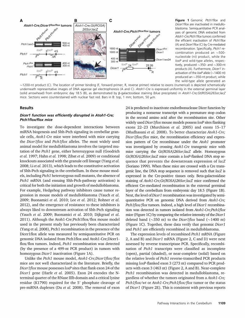
To investigate the dose-dependent interactions betweenmiRNA biogenesis and Shh-Ptch signaling in cerebellar gran-ule cells, Atoh1-Cre mice were interbred with mice carryingthe Dicer1flox and Ptch1flox alleles. The most widely usedanimal model for medulloblastoma involves the targeted mu-tation of the Ptch1 gene, either heterozygous null (Goodrichet al. 1997; Hahn et al. 1998; Zibat et al. 2009) or conditionalknockouts associatedwith the granule cell lineage (Yang et al.2008; Li et al. 2013),which leads to the constitutive activationof Shh-Ptch signaling in the cerebellum. In these mouse mod-els, including Ptch1 heterozygous null mutants, the absence ofPtch1 mRNA (and ensuing Shh-Ptch-pathway activation) iscritical for both the initiation and growth ofmedulloblastoma.For example, Hedgehog pathway inhibitors cause tumor re-gression in mouse models of medulloblastoma (Yauch et al.2009; Buonamici et al. 2010; Lee et al. 2012; Rohner et al.2012), and the emergence of resistance to these inhibitors isalways liked to downstream activation of Shh-Ptch signaling(Yauch et al. 2009; Buonamici et al. 2010; Dijkgraaf et al.2011). Although the Atoh1-Cre;Ptch1flox/flox mouse modelused in the present study has previously been characterized(Yang et al. 2008), Ptch1 recombination in the presence of theDicer1flox allele was measured by semiquantitative PCR ongenomic DNA isolated from Ptch1flox and Atoh1-Cre;Dicer1-flox/flox tumors. Indeed, Ptch1 recombination was detected(by the presence of a 499-nt PCR product) in tumors withhomozygous Dicer1 inactivation (Figure 1A).
Unlike the Ptch1 mouse model, Atoh1-Cre;Dicer1flox/floxmice are not well characterized in the literature. Briefly, theDicer1floxmouse possesses loxP sites that flank exon 24 of theDicer1 gene (Harfe et al. 2005). Exon 24 encodes the N-terminal quarter of the RNase IIIb domain and a critical lysineresidue (K1790) required for the 59 phosphate cleavage ofpre-miRNA duplexes (Du et al. 2008). The removal of exon
24 is predicted to inactivate endoribonuclease Dicer function byproducing a nonsense transcript with a premature stop codonin the second amino acid after the recombination site. Otherwidely usedDicer1floxmousemodels possess loxP sites flankingexons 22–23 (Murchison et al. 2005) and exons 15–17(Mudhasani et al. 2008). To better characterize Atoh1-Cre;Dicer1flox/flox mice, the recombination efficiency and expres-sion pattern of Cre recombinase under the Atoh1 promoterwas investigated by crossing Atoh1-Cre transgenic mice withmice carrying the Gt(ROSA)26Sor;lacZ allele. HomozygousGt(ROSA)26Sor;lacZ mice contain a loxP-flanked DNA stop se-quence that prevents the downstream expression of lacZ(Soriano 1999). When these mice are crossed with a Cre trans-genic line, the DNA stop sequence is removed such that lacZ isexpressed in the Cre-positive tissues only. Beta-galactosidasestaining of Atoh1-Cre;Gt(ROSA)26Sor;lacZ mice confirmed theefficient Cre-mediated recombination in the external germinallayer of the cerebellum from embryonic day 18.5 (Figure 1B).Next, the level ofDicer1 recombinationwas investigatedby semi-quantitative PCR on genomic DNA derived from Atoh1-Cre;Ptch1flox/flox tumors. Indeed, a high level of Dicer1 recombina-tion was detected in tumors isolated from Atoh1-Cre;Ptch1floxmice (Figure1C)by comparing the relative intensity of theDicer1deleted band (�350 nt) to the Dicer1flox band (�1400 nt)(Figure 1C). Together, these data verify that genomic Dicer1and Ptch1 are efficiently recombined in medulloblastoma.
The expression levels of recombined Ptch1 mRNA (Figure2, A and B) and Dicer1 mRNA (Figure 2, C and D) were nextassessed by reverse transcriptase PCR. Specifically, recombi-nation of Ptch1 transcripts were classified as incomplete(open), partial (shaded), or near-complete (solid) based onthe relative levels of Ptch1 reverse-transcribed PCR productsmissing loxP-flanked exon 3 (273 nt) compared to PCR prod-ucts with exon 3 (463 nt) (Figure 2, A and B). Near-completePtch1 recombination was detected in medulloblastoma, re-gardless of whether the tumors originated from a Atoh1-Cre;Ptch1flox/wt or Atoh1-Cre;Ptch1flox/flox tumor or the statusof Dicer1 (Figure 2E). This is consistent with previous reports
Figure 1 Genomic Ptch1flox andDicer1flox are inactivated in medullo-blastoma. Semiquantitative PCR anal-yses of genomic DNA extracted fromAtoh1-Cre;Ptch1flox tumors confirmedthe efficient inactivation of Ptch1flox(A) and Dicer1flox (C) by Cre-mediatedrecombination. Specifically, Ptch1 re-combination produced an �500-nucleotide (nt) product, while theloxP and wild-type alleles, respec-tively, produced �350- and �300-ntproducts (A). Furthermore, Dicer1 in-activation of the loxP allele (�1400 nt)produced an �350-nt product, whilethe wild-type allele generated an
�1200-nt product (C). The location of primer binding (F, forward primer; R, reverse primer) relative to exons (numerical) is depicted schematicallyunderneath representative images of DNA agarose gel electrophoresis (A and C). Atoh1-Cre is expressed uniformly in the external germinal layer(solid arrowhead) from embryonic day 18.5 (B), as demonstrated by b-galactosidase staining (blue precipitate) in Atoh1-Cre;Gt(ROSA)26Sor;lacZmice. Sections were counterstained with nuclear fast red. Bars in B: top, 1 mm; bottom, 50 mm.
Pathway Interactions in the Cerebellum 1109
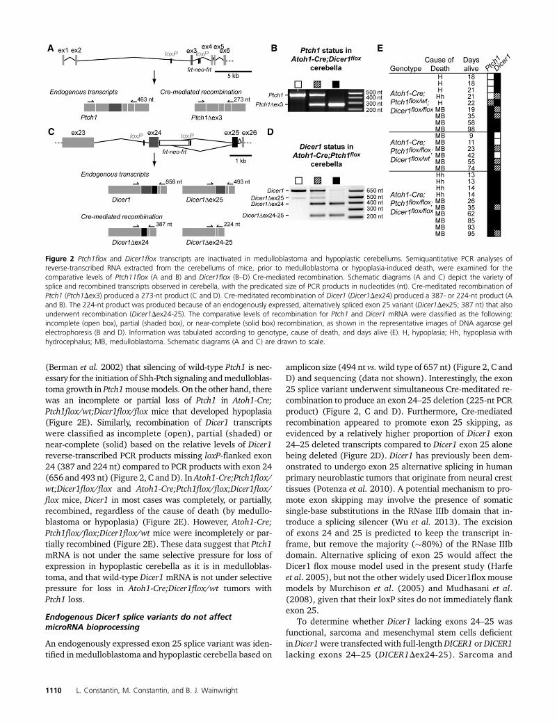
(Berman et al. 2002) that silencing of wild-type Ptch1 is nec-essary for the initiation of Shh-Ptch signaling andmedulloblas-toma growth in Ptch1mousemodels. On the other hand, therewas an incomplete or partial loss of Ptch1 in Atoh1-Cre;Ptch1flox/wt;Dicer1flox/flox mice that developed hypoplasia(Figure 2E). Similarly, recombination of Dicer1 transcriptswere classified as incomplete (open), partial (shaded) ornear-complete (solid) based on the relative levels of Dicer1reverse-transcribed PCR products missing loxP-flanked exon24 (387 and 224 nt) compared to PCR products with exon 24(656 and 493 nt) (Figure 2, C andD). In Atoh1-Cre;Ptch1flox/wt;Dicer1flox/flox and Atoh1-Cre;Ptch1flox/flox;Dicer1flox/flox mice, Dicer1 in most cases was completely, or partially,recombined, regardless of the cause of death (by medullo-blastoma or hypoplasia) (Figure 2E). However, Atoh1-Cre;Ptch1flox/flox;Dicer1flox/wt mice were incompletely or par-tially recombined (Figure 2E). These data suggest that Ptch1mRNA is not under the same selective pressure for loss ofexpression in hypoplastic cerebella as it is in medulloblas-toma, and that wild-type Dicer1mRNA is not under selectivepressure for loss in Atoh1-Cre;Dicer1flox/wt tumors withPtch1 loss.
Endogenous Dicer1 splice variants do not affectmicroRNA bioprocessing
An endogenously expressed exon 25 splice variant was iden-tified in medulloblastoma and hypoplastic cerebella based on
amplicon size (494 nt vs.wild type of 657 nt) (Figure 2, C andD) and sequencing (data not shown). Interestingly, the exon25 splice variant underwent simultaneous Cre-meditated re-combination to produce an exon 24–25 deletion (225-nt PCRproduct) (Figure 2, C and D). Furthermore, Cre-mediatedrecombination appeared to promote exon 25 skipping, asevidenced by a relatively higher proportion of Dicer1 exon24–25 deleted transcripts compared to Dicer1 exon 25 alonebeing deleted (Figure 2D). Dicer1 has previously been dem-onstrated to undergo exon 25 alternative splicing in humanprimary neuroblastic tumors that originate from neural cresttissues (Potenza et al. 2010). A potential mechanism to pro-mote exon skipping may involve the presence of somaticsingle-base substitutions in the RNase IIIb domain that in-troduce a splicing silencer (Wu et al. 2013). The excisionof exons 24 and 25 is predicted to keep the transcript in-frame, but remove the majority (�80%) of the RNase IIIbdomain. Alternative splicing of exon 25 would affect theDicer1 flox mouse model used in the present study (Harfeet al. 2005), but not the other widely used Dicer1flox mousemodels by Murchison et al. (2005) and Mudhasani et al.(2008), given that their loxP sites do not immediately flankexon 25.
To determine whether Dicer1 lacking exons 24–25 wasfunctional, sarcoma and mesenchymal stem cells deficientin Dicer1were transfected with full-length DICER1 or DICER1lacking exons 24–25 (DICER1Dex24-25). Sarcoma and
Figure 2 Ptch1flox and Dicer1flox transcripts are inactivated in medulloblastoma and hypoplastic cerebellums. Semiquantitative PCR analyses ofreverse-transcribed RNA extracted from the cerebellums of mice, prior to medulloblastoma or hypoplasia-induced death, were examined for thecomparative levels of Ptch11flox (A and B) and Dicer1flox (B–D) Cre-mediated recombination. Schematic diagrams (A and C) depict the variety ofsplice and recombined transcripts observed in cerebella, with the predicated size of PCR products in nucleotides (nt). Cre-meditated recombination ofPtch1 (Ptch1Dex3) produced a 273-nt product (C and D). Cre-meditated recombination of Dicer1 (Dicer1Dex24) produced a 387- or 224-nt product (Aand B). The 224-nt product was produced because of an endogenously expressed, alternatively spliced exon 25 variant (Dicer1Dex25; 387 nt) that alsounderwent recombination (Dicer1Dex24-25). The comparative levels of recombination for Ptch1 and Dicer1 mRNA were classified as the following:incomplete (open box), partial (shaded box), or near-complete (solid box) recombination, as shown in the representative images of DNA agarose gelelectrophoresis (B and D). Information was tabulated according to genotype, cause of death, and days alive (E). H, hypoplasia; Hh, hypoplasia withhydrocephalus; MB, medulloblastoma. Schematic diagrams (A and C) are drawn to scale.
1110 L. Constantin, M. Constantin, and B. J. Wainwright
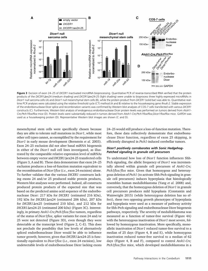
mesenchymal stem cells were specifically chosen becausethey are able to tolerate null mutations in Dicer1, while mostother cell types cannot, as exemplified by the requirement forDicer1 in early mouse development (Bernstein et al. 2003).Exon 24–25 exclusion did not alter basal miRNA biogenesisin either of the Dicer1 null cell lines investigated, as illus-trated by the comparable relative expression level of miRNAsbetween empty vector andDICER1Dex24-25 transfected cells(Figure 3, A and B). These data demonstrate that exon 24–25exclusion produces a loss-of-function mutation equivalent tothe recombination ofDicer1flox (i.e., exon 24 excision) alone.To further validate that the various DICER1 constructs lack-ing exons 24 and/or 25 produced stable protein products,Western blot analyses were performed. Indeed, all constructsproduced protein products of the expected size that wasbased on the predicted amino acid sequence of the endoribo-nuclease Dicer: 217 kDa for DICER1 (estimated 216 kDa),192 kDa for DICER1Dex24 (estimated 206 kDa), 207 kDafor DICER1Dex25 (estimated 210 kDa), and 212 kDa forDICER1Dex24-25 (estimated 200 kDa) (Figure 3C). Interest-ingly, in primary Atoh1-Cre;Ptch1flox/flox tumors, regardlessof the status of Dicer1flox, splice variants for exon 24 and/or25 were not detected (Figure 3D), even though they weredetectable at the transcript level (Figure 2, C–E). This doesnot preclude the possibility that low levels of alternativelyspliced endoribonuclease Dicer would be able to influencetumor growth; however, given that DICER1Dex24-25 is func-tionally equivalent to Dicer1flox (i.e., exon 24 excision), lowundetectable levels of endoribonuclease Dicer lacking exons
24–25would still produce a loss-of-functionmutation. There-fore, these data collectively demonstrate that endoribonu-clease Dicer function, regardless of exon 25 skipping, isefficiently disrupted in Ptch1-induced cerebellar tumors.
Dicer1 positively corroborates with Sonic Hedgehog-Patched signaling in granule cell precursors
To understand how loss of Dicer1 function influences Shh-Ptch signaling, the allelic frequency of Dicer1 was incremen-tally altered within granule cell precursors of Atoh1-Cre;Ptch1flox/flox mice. Given that homozygous and heterozy-gous deletion of Ptch1 (to activate Shh-Ptch signaling in gran-ule cell precursors) induces hyperplasia that histologicallyresembles human medulloblastoma (Yang et al. 2008) and,conversely, that the homozygous deletion ofDicer1 in granulecell precursors produces mild hypoplasia (Constantin andWainwright 2015) (while heterozygous deletion has no ef-fect), these two opposing growth phenotypes of hyperplasiaand hypoplasia were used as a measure of pathway activityfor Shh-Ptch signaling and endoribonuclease Dicer biogenesispathways, respectively. The severity of medulloblastoma wasmeasured as a function of tumor-free survival (Figure 4A)with the heterozygous inactivation of Dicer1most severe, fol-lowed by homozygous inactivation. More specifically, mono-allelic inactivation of Dicer1 reduced tumor-free survival to amedian of 25 days (Figure 4, B and E), while homozygousinactivation reduced tumor-free survival to a median of 62days (Figure 4, B and F), compared to control Atoh1-Cre;Ptch1flox/flox mice, which developed medulloblastoma in a
Figure 3 Excision of exon 24–25 of DICER1 inactivated microRNA bioprocessing. Quantitative PCR of reverse-transcribed RNA verified that the proteinproducts of the DICER1Dex24 (medium shading) and DICER1Dex24-25 (light shading) were unable to bioprocess three highly expressed microRNAs inDicer1 null sarcoma cells (A) and Dicer1 null mesenchymal stem cells (B), while the protein product from DICER1 (solid bar) was able to. Quantitative real-time PCR analyses were calculated using the relative threshold cycle (CT) method (A and B) relative to the housekeeping gene Rnu6-2. Stable expressionof the endoribonuclease Dicer splice and recombination variants was confirmed by Western blot analyses of COS-7 cells transfected with various DICER1constructs (C). Furthermore, Western blot analysis of endogenous endoribonuclease Dicer protein levels was performed on tumors derived from Atoh1-Cre;Ptch1flox/flox mice (D). Protein levels were substantially reduced in tumors derived from Atoh1-Cre;Ptch1flox/flox;Dicer1flox/flox mice. GAPDH wasused as a housekeeping protein (D). Representative Western blot images are shown (C and D).
Pathway Interactions in the Cerebellum 1111
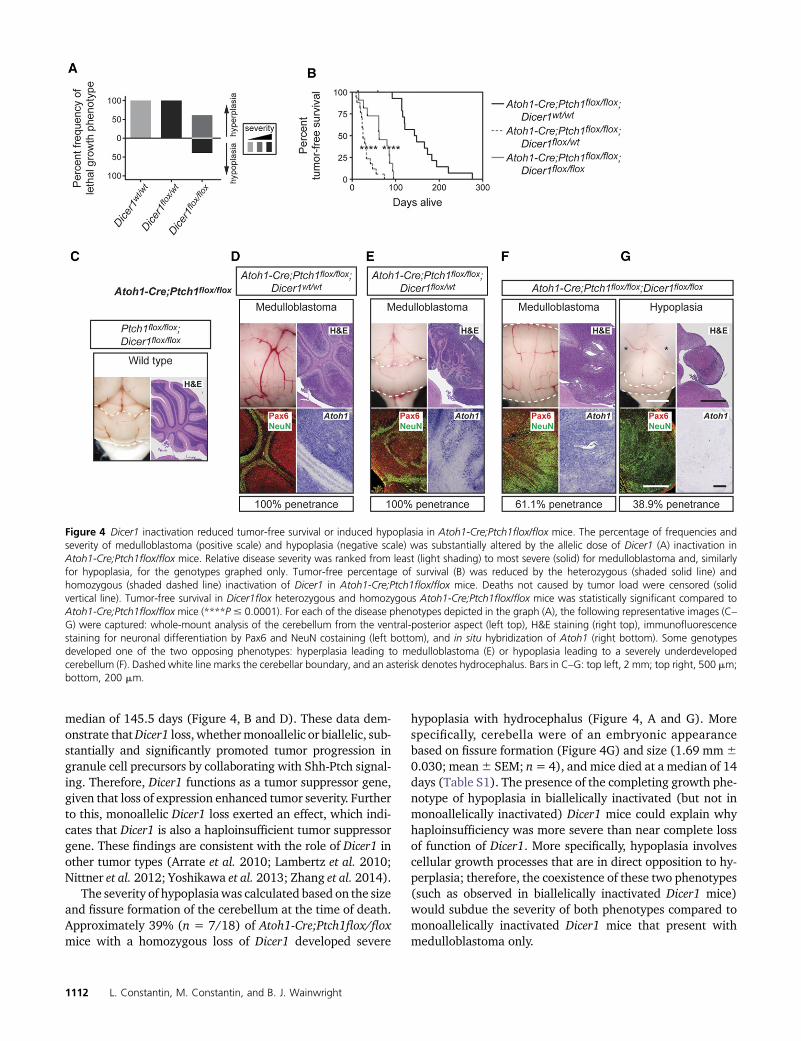
median of 145.5 days (Figure 4, B and D). These data dem-onstrate thatDicer1 loss, whethermonoallelic or biallelic, sub-stantially and significantly promoted tumor progression ingranule cell precursors by collaborating with Shh-Ptch signal-ing. Therefore, Dicer1 functions as a tumor suppressor gene,given that loss of expression enhanced tumor severity. Furtherto this, monoallelic Dicer1 loss exerted an effect, which indi-cates that Dicer1 is also a haploinsufficient tumor suppressorgene. These findings are consistent with the role of Dicer1 inother tumor types (Arrate et al. 2010; Lambertz et al. 2010;Nittner et al. 2012; Yoshikawa et al. 2013; Zhang et al. 2014).
The severity of hypoplasiawas calculated based on the sizeand fissure formation of the cerebellum at the time of death.Approximately 39% (n = 7/18) of Atoh1-Cre;Ptch1flox/floxmice with a homozygous loss of Dicer1 developed severe
hypoplasia with hydrocephalus (Figure 4, A and G). Morespecifically, cerebella were of an embryonic appearancebased on fissure formation (Figure 4G) and size (1.69 mm60.030; mean6 SEM; n= 4), and mice died at a median of 14days (Table S1). The presence of the completing growth phe-notype of hypoplasia in biallelically inactivated (but not inmonoallelically inactivated) Dicer1 mice could explain whyhaploinsufficiency was more severe than near complete lossof function of Dicer1. More specifically, hypoplasia involvescellular growth processes that are in direct opposition to hy-perplasia; therefore, the coexistence of these two phenotypes(such as observed in biallelically inactivated Dicer1 mice)would subdue the severity of both phenotypes compared tomonoallelically inactivated Dicer1 mice that present withmedulloblastoma only.
Figure 4 Dicer1 inactivation reduced tumor-free survival or induced hypoplasia in Atoh1-Cre;Ptch1flox/flox mice. The percentage of frequencies andseverity of medulloblastoma (positive scale) and hypoplasia (negative scale) was substantially altered by the allelic dose of Dicer1 (A) inactivation inAtoh1-Cre;Ptch1flox/flox mice. Relative disease severity was ranked from least (light shading) to most severe (solid) for medulloblastoma and, similarlyfor hypoplasia, for the genotypes graphed only. Tumor-free percentage of survival (B) was reduced by the heterozygous (shaded solid line) andhomozygous (shaded dashed line) inactivation of Dicer1 in Atoh1-Cre;Ptch1flox/flox mice. Deaths not caused by tumor load were censored (solidvertical line). Tumor-free survival in Dicer1flox heterozygous and homozygous Atoh1-Cre;Ptch1flox/flox mice was statistically significant compared toAtoh1-Cre;Ptch1flox/flox mice (****P # 0.0001). For each of the disease phenotypes depicted in the graph (A), the following representative images (C–G) were captured: whole-mount analysis of the cerebellum from the ventral-posterior aspect (left top), H&E staining (right top), immunofluorescencestaining for neuronal differentiation by Pax6 and NeuN costaining (left bottom), and in situ hybridization of Atoh1 (right bottom). Some genotypesdeveloped one of the two opposing phenotypes: hyperplasia leading to medulloblastoma (E) or hypoplasia leading to a severely underdevelopedcerebellum (F). Dashed white line marks the cerebellar boundary, and an asterisk denotes hydrocephalus. Bars in C–G: top left, 2 mm; top right, 500 mm;bottom, 200 mm.
1112 L. Constantin, M. Constantin, and B. J. Wainwright
In addition to gross histological analyses of these pheno-types through H&E staining, cerebella were costained withantibodies against the granule cell lineage marker Pax6 andthe neuronal differentiation marker NeuN. By identifyingPax6-immunopositive and NeuN-immunonegative cells vs.Pax6-immunopositive and NeuN-immunopositive cells, thepopulations of undifferentiated granule cell precursors vs.differentiated granule cells could, respectively, be determined.Pax6 and NeuN colocalization has previously been used toexamine changes in the population of proliferating and dif-ferentiating granule cell precursors (Flora et al. 2009; Con-stantin andWainwright 2015). As expected, therewas a largenumber of Pax6-immunopositive and NeuN-immunonegativecells in all tumors (regardless of the allelic frequency ofDicer1), which demarcate precursors of the granule lineage(Figure 4, D–F). On the other hand, the hypoplasticphenotype was composed of Pax6-immunopositive andNeuN-immunopositive cells, which indicates the presenceof differentiated cells in the majority of the hypoplastic struc-ture with a thin layer of precursor cells superficially (Figure4G). This suggests that the hypoplasia is a developmentaldefect rather than a small tumormass. High Atoh1 expressionis consistently and exclusively associated with the SHH sub-type of medulloblastoma (Swartling et al. 2012). All tumors,regardless of the allelic frequency of Dicer1, were positive forAtoh1 (Figure 4, D–F), which indicates that these tumorslikely belong to the Shh subtype.
Hedgehog-Patched signaling collaborates withmicroRNA biogenesis in granule cell precursors
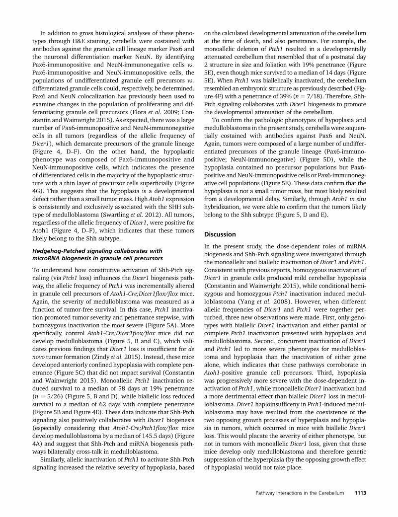
To understand how constitutive activation of Shh-Ptch sig-naling (via Ptch1 loss) influences the Dicer1 biogenesis path-way, the allelic frequency of Ptch1 was incrementally alteredin granule cell precursors of Atoh1-Cre;Dicer1flox/flox mice.Again, the severity of medulloblastoma was measured as afunction of tumor-free survival. In this case, Ptch1 inactiva-tion promoted tumor severity and penetrance stepwise, withhomozygous inactivation the most severe (Figure 5A). Morespecifically, control Atoh1-Cre;Dicer1flox/flox mice did notdevelop medulloblastoma (Figure 5, B and C), which vali-dates previous findings that Dicer1 loss is insufficient for denovo tumor formation (Zindy et al. 2015). Instead, thesemicedeveloped anteriorly confined hypoplasia with complete pen-etrance (Figure 5C) that did not impact survival (Constantinand Wainwright 2015). Monoallelic Ptch1 inactivation re-duced survival to a median of 58 days at 19% penetrance(n = 5/26) (Figure 5, B and D), while biallelic loss reducedsurvival to a median of 62 days with complete penetrance(Figure 5B and Figure 4E). These data indicate that Shh-Ptchsignaling also positively collaborates with Dicer1 biogenesis(especially considering that Atoh1-Cre;Ptch1flox/flox micedevelopmedulloblastoma by amedian of 145.5 days) (Figure4A) and suggest that Shh-Ptch and miRNA biogenesis path-ways bilaterally cross-talk in medulloblastoma.
Similarly, allelic inactivation of Ptch1 to activate Shh-Ptchsignaling increased the relative severity of hypoplasia, based
on the calculated developmental attenuation of the cerebellumat the time of death, and also penetrance. For example, themonoallelic deletion of Ptch1 resulted in a developmentallyattenuated cerebellum that resembled that of a postnatal day2 structure in size and foliation with 19% penetrance (Figure5E), even though mice survived to a median of 14 days (Figure5E). When Ptch1 was biallelically inactivated, the cerebellumresembled an embryonic structure as previously described (Fig-ure 4F) with a penetrance of 39% (n= 7/18). Therefore, Shh-Ptch signaling collaborates with Dicer1 biogenesis to promotethe developmental attenuation of the cerebellum.
To confirm the pathologic phenotypes of hypoplasia andmedulloblastoma in the present study, cerebellawere sequen-tially costained with antibodies against Pax6 and NeuN.Again, tumors were composed of a large number of undiffer-entiated precursors of the granule lineage (Pax6-immuno-positive; NeuN-immunonegative) (Figure 5D), while thehypoplasia contained no precursor populations but Pax6-positive and NeuN-immunopositive cells or Pax6-immunoneg-ative cell populations (Figure 5E). These data confirm that thehypoplasia is not a small tumor mass, but most likely resultedfrom a developmental delay. Similarly, through Atoh1 in situhybridization, we were able to confirm that the tumors likelybelong to the Shh subtype (Figure 5, D and E).
Discussion
In the present study, the dose-dependent roles of miRNAbiogenesis and Shh-Ptch signaling were investigated throughthe monoallelic and biallelic inactivation of Dicer1 and Ptch1.Consistent with previous reports, homozygous inactivation ofDicer1 in granule cells produced mild cerebellar hypoplasia(Constantin and Wainwright 2015), while conditional hemi-zygous and homozygous Ptch1 inactivation induced medul-loblastoma (Yang et al. 2008). However, when differentallelic frequencies of Dicer1 and Ptch1 were together per-turbed, three new observations were made. First, only geno-types with biallelic Dicer1 inactivation and either partial orcomplete Ptch1 inactivation presented with hypoplasia andmedulloblastoma. Second, concurrent inactivation of Dicer1and Ptch1 led to more severe phenotypes for medulloblas-toma and hypoplasia than the inactivation of either genealone, which indicates that these pathways corroborate inAtoh1-positive granule cell precursors. Third, hypoplasiawas progressively more severe with the dose-dependent in-activation of Ptch1, while monoallelic Dicer1 inactivation hada more detrimental effect than bialleic Dicer1 loss in medul-loblastoma. Dicer1 haploinsufficeny in Ptch1-induced medul-loblastoma may have resulted from the coexistence of thetwo opposing growth processes of hyperplasia and hypopla-sia in tumors, which occurred in mice with biallelic Dicer1loss. This would placate the severity of either phenotype, butnot in tumors with monoallelic Dicer1 loss, given that thesemice develop only medulloblastoma and therefore geneticsuppression of the hyperplasia (by the opposing growth effectof hypoplasia) would not take place.
Pathway Interactions in the Cerebellum 1113
MicroRNA biogenesis and Hedgehog-Patched-signalingpathways genetically interact to accentuate eithermedulloblastoma or hypoplasia
Several miRNA subsets are known to regulate components ofShh-Ptch signaling in the cerebellum. For example,miR-106bpositively regulatesGli2 transcription to promote granule cellexpansion (Constantin and Wainwright 2015). The miR-183�96�182 cluster synergizes with exogenous Shh ligandto promote the proliferation of Ptch1-induced medulloblas-toma (Zhang et al. 2013); similarly, themiR-17�92 cluster isnecessary for Ptch1-induced medulloblastoma (Zindy et al.
2014) through N-myc (Northcott et al. 2009). Conversely,miR-125b,miR-326, andmiR-324-5p antagonize the pathwayactivators Smoothened and/or Gli1 to inhibit granule cellprecursor growth (Ferretti et al. 2008). Therefore, it followsthat the deletion of miR-106b, miR-183�96�182, and miR-17�92 likely induced hypoplasia in Atoh1-Cre;Ptch1flox/flox;Dicer1flox/flox mice, while the loss of miR-125b, miR-326,and miR-324-5p promoted medulloblastoma. Given thatDicer1 regulates the bioprocessing of hundreds of miRNAs,and in turn each miRNA may regulate hundreds of targets,Dicer1 ablation would also affect many other pathways in
Figure 5 Ptch1 inactivation reduced tumor-free survival or promoted hypoplasia in Atoh1-Cre;Dicer1flox/flox mice. The percentage of frequencies andseverity of medulloblastoma (positive scale) and hypoplasia (negative scale) were substantially altered by the allelic dose of Ptch1 (A) inactivation inAtoh1-Cre;Dicer1flox/flox mice. Relative disease severity was ranked from least (light shading) to most severe (solid) for medulloblastoma and, similarlyfor hypoplasia, for the genotypes graphed only. Tumor-free percentage of survival (B) was reduced by the heterozygous (shaded line) and homozygous(shaded dashed line) inactivation of Ptch1 in Atoh1-Cre;Dicer1flox/flox mice. Deaths not caused by tumor load were censored (solid line). Tumor-freesurvival in Ptch1 heterozygous and homozygous Atoh1-Cre;Dicer1flox/flox mice was statistically significant compared to Atoh1-Cre;Dicer1flox/flox mice(*P = 0.0251; ****P# 0.0001). For each of the disease phenotypes depicted in the graph (A), the following representative images (C–E) were captured:whole-mount analysis of the cerebellum from the ventral-posterior aspect (left top), H&E staining (right top), immunofluorescence staining for neuronaldifferentiation by Pax6 and NeuN costaining (left bottom), and in situ hybridization of Atoh1 (right bottom). Some genotypes developed one of the twoopposing phenotypes: hyperplasia leading to medulloblastoma (D) or hypoplasia leading to a severely underdeveloped cerebellum (E). Dashed white linemarks the cerebellar boundary. Bars in C–E: top left, 2 mm; top right, 500 mm; bottom, 200 mm.
1114 L. Constantin, M. Constantin, and B. J. Wainwright
addition to Shh-Ptch signaling. Also, Dicer1 ablation may in-fluence Shh-Ptch signaling noncanonically, for example, byway of transforming growth factor b (Dennler et al. 2009).Although this body of work did not specify how or what thenature of the interactions between the endoribonucleaseDicer and Shh-Ptch signaling were, it did characterize thebroader effects of this genetic interaction. Through doing soit established that Dicer bioprocessing and Shh-Ptch signal-ing, together, have a substantial and unexpected biologicalimpact on the developing cerebellum (as measured by tumorlatency and growth attenuation) and that this interaction is aprincipal driving event during cerebellar development.
Bistability of phenotypes suggests that the associatedpathways regulate an importantdevelopmental transition
A bistable phenotype was induced in Atoh1-Cre;Ptch1flox/flox;Dicer1flox/floxmice, which strongly suggests that miRNAbiogenesis and Hedgehog-Patched signaling together regu-late an important developmental transition in granule cellprecursors. Based on the developmental stage of cerebellarattenuation in Atoh1-Cre;Ptch1flox/flox;Dicer1flox/flox mice,such an event likely occurred in late embryonic development.Further support of themolecular switch phenomenon includesprevious findings that miRNAs regulate transcription factorsassociated with Hedgehog-Patched signaling. More specifi-cally, interactions between miRNAs and transcription factorspermit feedback and feed-forward loops to be established,which are required in genetic switches (Tsang et al. 2007).A similar phenomenon may occur in granule cell precursors,whereby miRNA-mediate control of Shh-Ptch transcriptionfactors, such as Gli1/2 and N-myc, allows feedback andfeed-forward loops to be produced that in turn mediate hypo-plasia over medulloblastoma, or vice versa. The next stepwould be to identify the specific miRNAs and intracellularcomponents involved in regulating such a developmental tran-sition. This would not only cultivate a more thorough under-standing of early granule cell precursor development by, forexample, shedding light on relatively unstudied molecularprocesses involved in establishing the developmental com-partment-like units of the cerebellum (Hutvagner and Zamore2002), but may also yield novel insights into the molecularbasis of Shh-induced medulloblastoma.
Dicer1 functions as a haploinsufficient tumor suppressorgene in Ptch1-induced medulloblastoma
DICER1 has previously been reported to function as ahaploinsufficient tumor suppressor gene in human tumors(Karube et al. 2005; Chiosea et al. 2007; Merritt et al. 2008;Grelier et al. 2009; Faggad et al. 2010; Lin et al. 2010;Khoshnaw et al. 2012; Jafarnejad et al. 2013) and geneticallyengineeredmousemodels (Kumar et al. 2009; Lambertz et al.2010), with only a handful of exceptions (Arrate et al. 2010;Sabbaghian et al. 2012; Zhang et al. 2013). The precise roleof DICER1 in medulloblastoma is less clear. For example,subsets ofmiRNAs have been shown as either growth-inhibitory
(Ferretti et al. 2008; Li et al. 2009; Venkataraman et al. 2013;Jin et al. 2014; Hemmesi et al. 2015) or oncogenic (Grunderet al. 2011; Weeraratne et al. 2012; Li et al. 2015). In partic-ular, the miR-17�92 cluster has been shown as both neces-sary and sufficient to initiate medulloblastoma (Zindy et al.2014), which suggests an oncogenic role for Dicer1 on thebasis of miR-17�92 alone. On the other hand, the global re-duction of miRNAs in medulloblastoma (Ferretti et al. 2008),together with the identification of a germline DICER1 trun-cation mutation in a medulloblastoma cohort (Slade et al.2011), suggests that DICER1 is tumor-suppressive andhaploinsufficient.
Recently, Dicer1 loss was shown to accelerate tumorgrowth in a genetically engineered mouse model of Ptch1-induced medulloblastoma (Zindy et al. 2015); however,biallelic loss was never investigated because of the wide-spread apoptosis and ensuing perinatal lethality induced byDicer1 ablation in the Murchison et al. (2005) mouse model.In the present study, we conclusively demonstrate that boththe heterozygous and homozygous loss of Dicer1 dramaticallydecreases the latency of tumor-free survival in a geneticallyengineered mouse model of Ptch1-induced medulloblas-toma—different from that used by Zindy et al. (2015)—andthat heterozygous Dicer1 inactivation is most severe. The datapresented here are consistent with the role of DICER1 in themajority of human tumors. Interestingly, to date, only a singlegermline mutation (of 86 cases) has been reported for DICER1inmedulloblastoma and infratentorial primitive neuroectoder-mal tumors (Slade et al. 2011),which suggests that the geneticinteraction between miRNAs and Shh-Ptch signaling is per-haps more modest in humans or that mutations may affectthis pathway downstream/post-transcriptionally to DICER1(mRNA). Further to this, our data imply a mechanism as towhy monoallelic Dicer1 inactivation is more severe than bial-lelic inactivation. That is, given that hypoplasia is on the op-posite growth spectrum to medulloblastoma, and that onlyhomozygous Dicer1 inactivation induces hypoplasia, the bial-lelic loss of Dicer1 function would genetically suppress theseverity of hyperplasia, leading to medulloblastoma. Thiswould result in the less severe tumor phenotype in mice withbiallelic Dicer1 loss, as was observed. This may also explainhaploinsufficiency in other developmental pathologies.
Tumor suppressor genes can be designated as gatekeepergenes, for those involved in the control of cell proliferation orcell death, or as caretaker genes, for those involved in thecontrol of DNA integrity, as reviewed by Kinzler and Vogelstein(1997). The inactivation of a caretaker gene often does notinitiate tumor formation, but fosters transformation by ren-dering the cell genetically unstable. The endoribonucleaseDicer shares similarities with the majority of caretaker genesin that it cannot initiate tumors de novo, as illustrated here bythe absence of medulloblastoma in Atoh1-Cre;Dicer1flox/floxmice. Furthermore, the endoribonuclease Dicer has recentlybeen shown to maintain genomic stability in Schizosacchar-omyces pombe by promoting transcription termination at sitesassociated with replication stress and DNA damage (Castel
Pathway Interactions in the Cerebellum 1115
et al. 2014). This is likely to occur in higher eukaryotes.Therefore, we extrapolate that the tumor suppressor functionof Dicer1 would be complex and would not involve the clas-sical gatekeeper approach of directly regulating tumors bytargeting effects that inhibit growth or promote cell death.
We propose that more studies that characterize the geneticinterplay between principal pathways, such as here, should beconducted, given that pathway interactions may produce non-additivephenotypes that substantially differ fromderegulationof a pathway in isolation. Here, we successfully demonstratedthat the two principal pathways ofmiRNA biogenesis and Shh-Ptch signaling combinatorially andnonadditively influence thelatency and severity of cerebellar hypoplasia and medullo-blastoma. Novel insights were reached on the basis of theseobservations, such as a potential mode of action for Dicer1haploinsufficiency in Ptch1-induced medulloblastoma, andindications thatmiRNAs and Shh-Ptch signaling together reg-ulate an early developmental transition in granule cells.
Acknowledgments
We thank Witold Filipowicz for the Dicer 349 antibody andfor his intellectual input. Confocal imaging was performedin the Australian Cancer Research Foundation’s ImagingFacility at the Institute for Molecular Bioscience. This workwas supported by the National Health and Medical ResearchCouncil of Australia.
Literature Cited
Adolphe, C., M. Narang, T. Ellis, C. Wicking, P. Kaur et al., 2004 Anin vivo comparative study of sonic, desert and Indian hedgehogreveals that hedgehog pathway activity regulates epidermal stemcell homeostasis. Development 131: 5009–5019.
Alder, J., N. K. Cho, and M. E. Hatten, 1996 Embryonic precursorcells from the rhombic lip are specified to a cerebellar granuleneuron identity. Neuron 17: 389–399.
Arrate, M. P., T. Vincent, J. Odvody, R. Kar, S. N. Jones et al.,2010 MicroRNA biogenesis is required for Myc-induced B-celllymphoma development and survival. Cancer Res. 70: 6083–6092.
Bartel, D. P., 2009 MicroRNAs: target recognition and regulatoryfunctions. Cell 136: 215–233.
Berman, D. M., S. S. Karhadkar, A. R. Hallahan, J. I. Pritchard, C. G.Eberhart et al., 2002 Medulloblastoma growth inhibition byhedgehog pathway blockade. Science 297: 1559–1561.
Bernstein, E., S. Y. Kim, M. A. Carmell, E. P. Murchison, H. Alcornet al., 2003 Dicer is essential for mouse development. Nat.Genet. 35: 215–217.
Blaszczyk, J., J. E. Tropea, M. Bubunenko, K. M. Routzahn, D. S.Waugh et al., 2001 Crystallographic and modeling studies ofRNase III suggest a mechanism for double-stranded RNA cleav-age. Structure 9: 1225–1236.
Bohnsack, M. T., K. Czaplinski, and D. Gorlich, 2004 Exportin 5 isa RanGTP-dependent dsRNA-binding protein that mediates nu-clear export of pre-miRNAs. RNA 10: 185–191.
Briscoe, J., Y. Chen, T. M. Jessell, and G. Struhl, 2001 A hedgehog-insensitive form of patched provides evidence for direct long-range morphogen activity of sonic hedgehog in the neural tube.Mol. Cell 7: 1279–1291.
Buckner, R. L., 2013 The cerebellum and cognitive function: 25years of insight from anatomy and neuroimaging. Neuron 80:807–815.
Buonamici, S., J. Williams, M. Morrissey, A. Wang, R. Guo et al.,2010 Interfering with resistance to smoothened antagonists byinhibition of the PI3K pathway in medulloblastoma. Sci. Transl.Med. 2: 51ra70.
Castel, S. E., J. Ren, S. Bhattacharjee, A. Y. Chang, M. Sanchez et al.,2014 Dicer promotes transcription termination at sites of rep-lication stress to maintain genome stability. Cell 159: 572–583.
Chendrimada, T. P., R. I. Gregory, E. Kumaraswamy, J. Norman, N.Cooch et al., 2005 TRBP recruits the Dicer complex to Ago2 formicroRNA processing and gene silencing. Nature 436: 740–744.
Chiosea, S., E. Jelezcova, U. Chandran, J. Luo, G. Mantha et al.,2007 Overexpression of Dicer in precursor lesions of lung ad-enocarcinoma. Cancer Res. 67: 2345–2350.
Cho, Y. J., A. Tsherniak, P. Tamayo, S. Santagata, A. Ligon et al.,2011 Integrative genomic analysis of medulloblastoma iden-tifies a molecular subgroup that drives poor clinical outcome.J. Clin. Oncol. 29: 1424–1430.
Constantin, L., and B. J. Wainwright, 2015 MicroRNAs promotegranule cell expansion in the cerebellum through Gli2. Cerebel-lum 14: 688–698.
Corbit, K. C., P. Aanstad, V. Singla, A. R. Norman, D. Y. Stainieret al., 2005 Vertebrate Smoothened functions at the primarycilium. Nature 437: 1018–1021.
Dahmane, N., and A. Ruiz i Altaba, 1999 Sonic hedgehog regu-lates the growth and patterning of the cerebellum. Development126: 3089–3100.
Denli, A. M., B. B. Tops, R. H. Plasterk, R. F. Ketting, and G. J.Hannon, 2004 Processing of primary microRNAs by the Micro-processor complex. Nature 432: 231–235.
Dennler, S., J. Andre, F. Verrecchia, and A. Mauviel, 2009 Cloningof the human GLI2 promoter: transcriptional activation by trans-forming growth factor-beta via SMAD3/beta-catenin coopera-tion. J. Biol. Chem. 284: 31523–31531.
Dijkgraaf, G. J., B. Alicke, L. Weinmann, T. Januario, K. West et al.,2011 Small molecule inhibition of GDC-0449 refractorysmoothened mutants and downstream mechanisms of drug re-sistance. Cancer Res. 71: 435–444.
Du, Z., J. K. Lee, R. Tjhen, R. M. Stroud, and T. L. James,2008 Structural and biochemical insights into the dicingmechanism of mouse Dicer: a conserved lysine is critical fordsRNA cleavage. Proc. Natl. Acad. Sci. USA 105: 2391–2396.
Ellis, T., I. Smyth, E. Riley, S. Graham, K. Elliot et al.,2003 Patched 1 conditional null allele in mice. Genesis 36:158–161.
Espinosa, J. S., and L. Luo, 2008 Timing neurogenesis and differ-entiation: insights from quantitative clonal analyses of cerebel-lar granule cells. J. Neurosci. 28: 2301–2312.
Faggad, A., J. Budczies, O. Tchernitsa, S. Darb-Esfahani, J. Sehouliet al., 2010 Prognostic significance of Dicer expression in ovar-ian cancer-link to global microRNA changes and oestrogen re-ceptor expression. J. Pathol. 220: 382–391.
Ferretti, E., E. De Smaele, E. Miele, P. Laneve, A. Po et al.,2008 Concerted microRNA control of Hedgehog signalling incerebellar neuronal progenitor and tumour cells. EMBO J. 27:2616–2627.
Ferretti, E., E. De Smaele, A. Po, L. Di Marcotullio, E. Tosi et al.,2009 MicroRNA profiling in human medulloblastoma. Int. J.Cancer 124: 568–577.
Flora, A., T. J. Klisch, G. Schuster, and H. Y. Zoghbi,2009 Deletion of Atoh1 disrupts Sonic Hedgehog signalingin the developing cerebellum and prevents medulloblastoma.Science 326: 1424–1427.
Genovesi, L. A., C. G. Ng, M. J. Davis, M. Remke, M. D. Taylor et al.,2013 Sleeping Beauty mutagenesis in a mouse medulloblastoma
1116 L. Constantin, M. Constantin, and B. J. Wainwright
model defines networks that discriminate between human molec-ular subgroups. Proc. Natl. Acad. Sci. USA 110: E4325–E4334.
Goodrich, L. V., L. Milenkovic, K. M. Higgins, and M. P. Scott,1997 Altered neural cell fates and medulloblastoma in mousepatched mutants. Science 277: 1109–1113.
Grainger, L., L. Cicchini, M. Rak, A. Petrucelli, K. D. Fitzgerald et al.,2010 Stress-inducible alternative translation initiation of hu-man cytomegalovirus latency protein pUL138. J. Virol. 84:9472–9486.
Grelier, G., N. Voirin, A. S. Ay, D. G. Cox, S. Chabaud et al.,2009 Prognostic value of Dicer expression in human breastcancers and association with the mesenchymal phenotype. Br.J. Cancer 101: 673–683.
Grunder, E., R. D’Ambrosio, G. Fiaschetti, L. Abela, A. Arcaro et al.,2011 MicroRNA-21 suppression impedes medulloblastomacell migration. Eur. J. Cancer 47: 2479–2490.
Gurtan, A. M., A. Ravi, P. B. Rahl, A. D. Bosson, C. K. JnBaptisteet al., 2013 Let-7 represses Nr6a1 and a mid-gestation devel-opmental program in adult fibroblasts. Genes Dev. 27: 941–954.
Hahn, H., L. Wojnowski, A. M. Zimmer, J. Hall, G. Miller et al.,1998 Rhabdomyosarcomas and radiation hypersensitivity ina mouse model of Gorlin syndrome. Nat. Med. 4: 619–622.
Han, J., Y. Lee, K. H. Yeom, Y. K. Kim, H. Jin et al., 2004 TheDrosha-DGCR8 complex in primary microRNA processing.Genes Dev. 18: 3016–3027.
Harfe, B. D., M. T. McManus, J. H. Mansfield, E. Hornstein, andC. J. Tabin, 2005 The RNaseIII enzyme Dicer is required formorphogenesis but not patterning of the vertebrate limb. Proc.Natl. Acad. Sci. USA 102: 10898–10903.
Hatten, M. E., and M. F. Roussel, 2011 Development and cancerof the cerebellum. Trends Neurosci. 34: 134–142.
Hemmesi, K., M. L. Squadrito, P. Mestdagh, V. Conti, M. Cominelliet al., 2015 miR-135a inhibits cancer stem cell-driven medul-loblastoma development by directly repressing Arhgef6 expres-sion. Stem Cells 33: 1377–1389.
Humke, E. W., K. V. Dorn, L. Milenkovic, M. P. Scott, and R. Rohatgi,2010 The output of Hedgehog signaling is controlled by thedynamic association between Suppressor of Fused and the Gliproteins. Genes Dev. 27: 670–682.
Hutvagner, G., and P. D. Zamore, 2002 A microRNA in a multiple-turnover RNAi enzyme complex. Science 297: 2056–2060.
Hutvagner, G., J. McLachlan, A. E. Pasquinelli, E. Balint, T. Tuschlet al., 2001 A cellular function for the RNA-interference en-zyme Dicer in the maturation of the let-7 small temporal RNA.Science 293: 834–838.
Iwakawa, H. O., and Y. Tomari, 2015 The functions of micro-RNAs: mRNA decay and translational repression. Trends CellBiol. 25: 651–665.
Jafarnejad, S. M., G. S. Ardekani, M. Ghaffari, M. Martinka, andG. Li, 2013 Sox4-mediated Dicer expression is critical forsuppression of melanoma cell invasion. Oncogene 32: 2131–2139.
Jin, Y., A. Xiong, Z. Zhang, S. Li, H. Huang et al., 2014 MicroRNA-31 suppresses medulloblastoma cell growth by inhibiting DNAreplication through minichromosome maintenance 2. Oncotar-get 5: 4821–4833.
Johnson, R. L., L. Milenkovic, and M. P. Scott, 2000 In vivo func-tions of the patched protein: requirement of the C terminus fortarget gene inactivation but not Hedgehog sequestration. Mol.Cell 6: 467–478.
Karube, Y., H. Tanaka, H. Osada, S. Tomida, Y. Tatematsu et al.,2005 Reduced expression of Dicer associated with poor prog-nosis in lung cancer patients. Cancer Sci. 96: 111–115.
Khoshnaw, S. M., E. A. Rakha, T. M. Abdel-Fatah, C. C. Nolan, Z.Hodi et al., 2012 Loss of Dicer expression is associated withbreast cancer progression and recurrence. Breast Cancer Res.Treat. 135: 403–413.
Kinzler, K. W., and B. Vogelstein, 1997 Cancer-susceptibilitygenes. Gatekeepers and caretakers. Nature 386(761): 763.
Komuro, H., and P. Rakic, 1998 Distinct modes of neuronal mi-gration in different domains of developing cerebellar cortex. J.Neurosci. 18: 1478–1490.
Kumar, M. S., R. E. Pester, C. Y. Chen, K. Lane, C. Chin et al.,2009 Dicer1 functions as a haploinsufficient tumor suppressor.Genes Dev. 23: 2700–2704.
Lambertz, I., D. Nittner, P. Mestdagh, G. Denecker, J. Vandesompeleet al., 2010 Monoallelic but not biallelic loss of Dicer1 pro-motes tumorigenesis in vivo. Cell Death Differ. 17: 633–641.
Lee, M. J., B. A. Hatton, E. H. Villavicencio, P. C. Khanna, S. D.Friedman et al., 2012 Hedgehog pathway inhibitor saridegib(IPI-926) increases lifespan in a mouse medulloblastoma model.Proc. Natl. Acad. Sci. USA 109: 7859–7864.
Lee, Y., C. Ahn, J. Han, H. Choi, J. Kim et al., 2003 The nuclearRNase III Drosha initiates microRNA processing. Nature 425:415–419.
Lee, Y., R. C. Samaco, J. R. Gatchel, C. Thaller, H. T. Orr et al.,2008 miR-19, miR-101 and miR-130 co-regulate ATXN1 levelsto potentially modulate SCA1 pathogenesis. Nat. Neurosci. 11:1137–1139.
Lewis, P. M., A. Gritli-Linde, R. Smeyne, A. Kottmann, and A. P.McMahon, 2004 Sonic hedgehog signaling is required for ex-pansion of granule neuron precursors and patterning of themouse cerebellum. Dev. Biol. 270: 393–410.
Li, K. K., J. C. Pang, A. K. Ching, C. K. Wong, X. Kong et al.,2009 miR-124 is frequently down-regulated in medulloblas-toma and is a negative regulator of SLC16A1. Hum. Pathol.40: 1234–1243.
Li, K. K., T. Xia, F. M. Ma, R. Zhang, Y. Mao et al., 2015 miR-106bis overexpressed in medulloblastomas and interacts directlywith PTEN. Neuropathol. Appl. Neurobiol. 41: 145–164.
Li, P., F. Du, L. W. Yuelling, T. Lin, R. E. Muradimova et al.,2013 A population of Nestin-expressing progenitors in the cer-ebellum exhibits increased tumorigenicity. Nat. Neurosci. 16:1737–1744.
Lin, R. J., Y. C. Lin, J. Chen, H. H. Kuo, Y. Y. Chen et al.,2010 MicroRNA signature and expression of Dicer and Droshacan predict prognosis and delineate risk groups in neuroblas-toma. Cancer Res. 70: 7841–7850.
Luna, L. G. (Editor), 1968 Manual of Histologic Staining Methodsof the Armed Forces Institute of Pathology. Blakiston Division,McGraw-Hill, New York.
Lund, E., S. Guttinger, A. Calado, J. E. Dahlberg, and U. Kutay,2004 Nuclear export of microRNA precursors. Science 303:95–98.
Merritt, W. M., Y. G. Lin, L. Y. Han, A. A. Kamat, W. A. Spannuthet al., 2008 Dicer, Drosha, and outcomes in patients with ovar-ian cancer. N. Engl. J. Med. 359: 2641–2650.
Mourelatos, Z., J. Dostie, S. Paushkin, A. Sharma, B. Charrouxet al., 2002 miRNPs: a novel class of ribonucleoproteins con-taining numerous microRNAs. Genes Dev. 16: 720–728.
Mudhasani, R., Z. Zhu, G. Hutvagner, C. M. Eischen, S. Lyle et al.,2008 Loss of miRNA biogenesis induces p19Arf-p53 signalingand senescence in primary cells. J. Cell Biol. 181: 1055–1063.
Murchison, E. P., J. F. Partridge, O. H. Tam, S. Cheloufi, and G. J.Hannon, 2005 Characterization of Dicer-deficient murine em-bryonic stem cells. Proc. Natl. Acad. Sci. USA 102: 12135–12140.
Nittner, D., I. Lambertz, F. Clermont, P. Mestdagh, C. Kohler et al.,2012 Synthetic lethality between Rb, p53 and Dicer or miR-17–92 in retinal progenitors suppresses retinoblastoma forma-tion. Nat. Cell Biol. 14: 958–965.
Northcott, P. A., L. A. Fernandez, J. P. Hagan, D. W. Ellison, W.Grajkowska et al., 2009 The miR-17/92 polycistron is up-regulated in sonic hedgehog-driven medulloblastomas and
Pathway Interactions in the Cerebellum 1117
induced by N-myc in sonic hedgehog-treated cerebellar neu-ral precursors. Cancer Res. 69: 3249–3255.
Pascual-Castroviejo, I., M. Gutierrez, C. Morales, I. Gonzalez-Mediero, A. Martinez-Bermejo et al., 1994 Primary degenera-tion of the granular layer of the cerebellum. A study of 14 patientsand review of the literature. Neuropediatrics 25: 183–190.
Pietsch, T., A. Waha, A. Koch, J. Kraus, S. Albrecht et al.,1997 Medulloblastomas of the desmoplastic variant carry mu-tations of the human homologue of Drosophila patched. CancerRes. 57: 2085–2088.
Potenza, N., U. Papa, P. Scaruffi, N. Mosca, G. P. Tonini et al.,2010 A novel splice variant of the human dicer gene is ex-pressed in neuroblastoma cells. FEBS Lett. 584: 3452–3457.
Raffel, C., R. B. Jenkins, L. Frederick, D. Hebrink, B. Alderete et al.,1997 Sporadic medulloblastomas contain PTCH mutations.Cancer Res. 57: 842–845.
Ravi, A., A. M. Gurtan, M. S. Kumar, A. Bhutkar, C. Chin et al.,2012 Proliferation and tumorigenesis of a murine sarcoma cellline in the absence of DICER1. Cancer Cell 21: 848–855.
Rohner, A., M. E. Spilker, J. L. Lam, B. Pascual, D. Bartkowski et al.,2012 Effective targeting of Hedgehog signaling in a medullo-blastoma model with PF-5274857, a potent and selectiveSmoothened antagonist that penetrates the blood-brain barrier.Mol. Cancer Ther. 11: 57–65.
Sabbaghian, N., N. Hamel, A. Srivastava, S. Albrecht, J. R. Priestet al., 2012 Germline DICER1 mutation and associated loss ofheterozygosity in a pineoblastoma. J. Med. Genet. 49: 417–419.
Schaefer, A., D. O’Carroll, C. L. Tan, D. Hillman, M. Sugimori et al.,2007 Cerebellar neurodegeneration in the absence of micro-RNAs. J. Exp. Med. 204: 1553–1558.
Schuller, U., Q. Zhao, S. A. Godinho, V. M. Heine, R. H. Medemaet al., 2007 Forkhead transcription factor FoxM1 regulates mi-totic entry and prevents spindle defects in cerebellar granuleneuron precursors. Mol. Cell. Biol. 27: 8259–8270.
Slade, I., C. Bacchelli, H. Davies, A. Murray, F. Abbaszadeh et al.,2011 DICER1 syndrome: clarifying the diagnosis, clinical fea-tures and management implications of a pleiotropic tumour pre-disposition syndrome. J. Med. Genet. 48: 273–278.
Soriano, P., 1999 Generalized lacZ expression with the ROSA26Cre reporter strain. Nat. Genet. 21: 70–71.
Swartling, F. J., V. Savov, A. I. Persson, J. Chen, C. S. Hackett et al.,2012 Distinct neural stem cell populations give rise to dispa-rate brain tumors in response to N-MYC. Cancer Cell 21: 601–613.
Tsang, J., J. Zhu, and A. van Oudenaarden, 2007 MicroRNA-mediated feedback and feedforward loops are recurrent net-work motifs in mammals. Mol. Cell 26: 753–767.
Tukachinsky, H., L. V. Lopez, and A. Salic, 2010 A mechanism forvertebrate Hedgehog signaling: recruitment to cilia and dissoci-ation of SuFu-Gli protein complexes. J. Cell Biol. 191: 415–428.
Uziel, T., F. V. Karginov, S. Xie, J. S. Parker, Y. D. Wang et al.,2009 The miR-17�92 cluster collaborates with the SonicHedgehog pathway in medulloblastoma. Proc. Natl. Acad. Sci.USA 106: 2812–2817.
Venkataraman, S., D. K. Birks, I. Balakrishnan, I. Alimova, P. S.Harris et al., 2013 MicroRNA 218 acts as a tumor suppressorby targeting multiple cancer phenotype-associated genes in me-dulloblastoma. J. Biol. Chem. 288: 1918–1928.
Weeraratne, S. D., V. Amani, N. Teider, J. Pierre-Francois, D. Winteret al., 2012 Pleiotropic effects of miR-183�96�182 convergeto regulate cell survival, proliferation and migration in medul-loblastoma. Acta Neuropathol. 123: 539–552.
Wu, M. K., N. Sabbaghian, B. Xu, S. Addidou-Kalucki, C. Bernardet al., 2013 Biallelic DICER1 mutations occur in Wilms tu-mours. J. Pathol. 230: 154–164.
Yang, Z. J., T. Ellis, S. L. Markant, T. A. Read, J. D. Kessler et al.,2008 Medulloblastoma can be initiated by deletion of Patchedin lineage-restricted progenitors or stem cells. Cancer Cell 14:135–145.
Yauch, R. L., G. J. Dijkgraaf, B. Alicke, T. Januario, C. P. Ahn et al.,2009 Smoothened mutation confers resistance to a Hedgehogpathway inhibitor in medulloblastoma. Science 326: 572–574.
Yoshikawa, T., M. Otsuka, T. Kishikawa, A. Takata, M. Ohno et al.,2013 Unique haploinsufficient role of the microRNA-processingmolecule Dicer1 in a murine colitis-associated tumorigenesismodel. PLoS One 8: e71969.
Zhang, B., H. Chen, L. Zhang, O. Dakhova, Y. Zhang et al., 2014 Adosage-dependent pleiotropic role of Dicer in prostate cancergrowth and metastasis. Oncogene 33: 3099–3108.
Zhang, H., F. A. Kolb, V. Brondani, E. Billy, and W. Filipowicz,2002 Human Dicer preferentially cleaves dsRNAs at their ter-mini without a requirement for ATP. EMBO J. 21: 5875–5885.
Zhang, H., F. A. Kolb, L. Jaskiewicz, E. Westhof, and W. Filipowicz,2004 Single processing center models for human Dicer andbacterial RNase III. Cell 118: 57–68.
Zhang, Z., S. Li, and S. Y. Cheng, 2013 The miR-183�96�182cluster promotes tumorigenesis in a mouse model of medullo-blastoma. J. Biomed. Res. 27: 486–494.
Zibat, A., A. Uhmann, F. Nitzki, M. Wijgerde, A. Frommhold et al.,2009 Time-point and dosage of gene inactivation determinethe tumor spectrum in conditional Ptch knockouts. Carcinogen-esis 30: 918–926.
Zindy, F., D. Kawauchi, Y. Lee, O. Ayrault, L. Ben Merzoug et al.,2014 Role of the miR-17�92 cluster family in cerebellar andmedulloblastoma development. Biol. Open 3: 597–605.
Zindy, F., Y. Lee, D. Kawauchi, O. Ayrault, L. B. Merzoug et al.,2015 Dicer is required for normal cerebellar development andto restrain medulloblastoma formation. PLoS One 10: e0129642.
Communicating editor: S. K. Sharan
1118 L. Constantin, M. Constantin, and B. J. Wainwright
GENETICSSupporting Information
www.genetics.org/lookup/suppl/doi:10.1534/genetics.115.184176/-/DC1
MicroRNA Biogenesis and Hedgehog-PatchedSignaling Cooperate to Regulate an Important
Developmental Transition in GranuleCell Development
Lena Constantin, Myrna Constantin, and Brandon J. Wainwright
Copyright © 2016 by the Genetics Society of AmericaDOI: 10.1534/genetics.115.184176

Table S1. Cause of death and days alive in Atoh1-Cre;Dicer1flox;Ptch1flox mice. e, euthanized at end of trial; h, hydrocephalus; H, hypoplasia; Hh, hypoplasia accompanied with hydrocephalus; MB, hyperplasia that manifested as medulloblastoma; O, other/unrelated. Available for download as a .docx file at http://www.genetics.org/lookup/suppl/doi:10.1534/genetics.115.184176/-/DC1/TableS1.docx