microscopic studies of cavity formation by soft rot fungi
TRANSCRIPT

STUDIA FORESTALIA SUECICA
Microscopic studies of cavity formation by soft rot fungi Allescheria terrestris Apinis, Margarinomyces luteo-viridis v. Beyma and Phialophora richardsiae (NannE) C onant
Mikroskopiska studier over kavitetsbildningen hos mogel- rotesvamparna Allescheria terrestris Apinis, Margarinomyces luteo-viridis v. Beyma och Phia lophora richardsiae (Nannf.) Conant
HANS L U N D S T R ~ M Department of Forest Products
SKOGSH~GSKOLAN ROYAL COLLEGE OF FORESTRY
STOCKHOLM

Abstract
T h e ability o f three soft rot fungi to form cavities in birch sapwood (Betula sp.), was investigated, viz. Allescheria terrestris Apinis (strain Apinis and strain H63-I), Margarinon7yce.s luteo-viridis v. Beyrna (strain Begma and strain M74-IV) as ivell as Phialophora richardsiae (Nannf.) Conant (strain RR40-V). T h e primary aim of the studies was to elucidate how rapidly the fungal hyphae form cavities after infection of the wood. T h e role o f nitrogen in cavity formation was studied by comparing the time taken for cavity formation in untreated wood and in wood impregnated with various nitrogen compounds. Phialophora richardsiae reacted nzost strongly to the addition o f nitrogen coinpounds to the wood, forming cavities after three days, as compared with six duys in untreated wood. Allescheria terrestris arzd Margarinoinycer luteo-viridis fornzed cavities two duys after infection o f the wood, regardless o/ whether nitrogen loas employed or not. However, in ~zitroge~z-in7pregnated wood, cavity formation began at a more uniform rate and to a greater extent than in untreated wood. T h e "T-shaped branching" form of attaclc frequently occzlrred, in various forms, in that the bore-holes were not always synnzetrically related to the long edge of the cavity. T h e ability o f Allescheria terresrris and illargarinonzyces luteo-viridis to form cavities in 25 different plants with lignified cells was studied. Cavity fornza- tion dijfered n.idely between softwood where there was little and hardwood where cavities were very nunzero~~s , although with some sinall variations.
Ms. received 9 February 1972
Ailmanna Forlaget
ISBN 9 1-38-00282-5
Berling5ka Boktryckeriet. kund 1972
Contents
. . . . . . . . . . P Introduction 5 5 Cavity formation in lignified stems . . 11
2 Materials and methods . . . . . . 6 6 Discussion . . . . . . . . . . . 13
3 Cavity formation in birch wood . . . 7 Acknowledgements . . . . . . . . . 15 . . . . . . . . . . . . . . 3.1 The preliminary stage of cavities 7 References 16
. . . 3.2 Observation of the first cavity 7 3.3 The occurrence of cavity chains . . . 9 $ammanfattming . . . . . . . . . . 17
4 The effect of temperature on cavity formation . . . . . . . . . . . 10

1 Introduction
The ability of fungi hyphae to develop in the cell walls of wood and so destroy them, thus forming cavities, was described as early as the 1850s by Schacht (1850, 1863). Bailey and Vestal (1937) demonstrated that the hyphae develop in the secondary walls of the wood cell and that wood from both gymnosperms and angiosperms is attacked. Savory (1954), who gave this type of rot its English name "soft rot", isolated a large number of cavity-forming fungi in wood which had been attacked by soft rot. He found that the fungi isolated belonged to the Ascomycetes and Fungi Imperfecti. Later studies have shown that two Basidio- mycetes display a rotting effect typical of soft rot, namely Porin nigrescens (Duncan, 1960) and Stereuin purpureum (Nilsson, personal communication).
In addition to cavity formation, erosion of the cell wall from the lumen can also occur in soft rot. However, this type of attack has not been studied in detail in the present paper.
The penetration of a hypha into a wood cell and its method of forming a cavity by means of a T-shaped attack has been de- scribed by Corbett and Levy (1963). The
variations in the form of cavities in dif- ferent wood cells and types of wood were studied by Courtois (1963). Very little study has been devoted to the time taken for cavity formation after a hypha has come into contact with the wood. Greaves and Levy (1965) state that after a week Chaetoiniurn globosunz formed cavities in Fagus sylvntica, after two weeks in Betuln sp. and after four weeks in Pinus sylvestris.
The time required for cavity formation by some soft rot fungi in wood cells has been especially studied in this investiga- tion. The soft rot fungi employed are a thermophilic Ascomycete: Allescheria ter- restris Apinis (strain Apinis and strain H63-I), two Fungi Imperfecti: Mnrgarino- myces luteo-viridis v. Beyma (strain Beyma and strain M74-IV), as well as Phialophorn richnrdsiae (Nannf.) Conant (strain BB40- V). Strain H63-1 was isolated from aspen chips in 1965 and strain M74-1V and strain BB40-V from birch chips in 1964. The collection was made by T. Nilsson. Strain Apinis has been supplied by A.E. Apinis and strain Beyma has been obtained from C B S in Baarn.

2 Materials and methods
The test fungi were cultivated in sloping agar tubes. The following temperatures and cultivation times before rotting were em- ployed for the various fungi: Allescheria terrestris strain H63-1 and strain Apinis at 45" C for five days, Margarinomyces luteo- viridis strain M74-IV and strain Beyma at 30" C for approximately 20 days and Phialophorn richardsiae strain BB40-V at 25" C for approximately 20 days. The same temperatures were employed both for the cultivation of the mycelium and for the tests and were determined on the basis of the optimal temperatures for extension growth.
Blocks ( 2 ~ 0.5 x 0.5 cm) of Betula ver- rucosa Ehrh. or Betula pubescens Ehrh. were placed on the developed mycelium. The fresh wood samples were taken at a height of 0-2 m from the stems of trees with a maximum diameter of 25 cm. The stored air-dry wood samples were taken at an unknown stem height from trees with an unknown diameter. These samples were
then either sterilised in an autoclave or not sterilised at all. When impregnating the wood with nitrogen, the following com- pounds were employed: ammonium chlo- ride, ammonium nitrate, ammonium tar- trate, asparagine and casein hydrolysate. The nitrogen impregnation was carried out in a desiccator under air suction for 50 minutes and vacuum for 50 minutes. The nitrogen compounds were mainly in a 0.6 per cent aqueous solution. The blocks were sliced with a razor blade for microscopic study. The cut was stained with a safranin solution (safranin dissolved in 70 per cent alcohol). A light microscope was primarily employed for the microscopic studies, but a Cambridge Stereoscan Electron Micro- scope was also used to a lesser extent. A method of studying cavity formation by the mycelium in the wood in vitro was also developed and utilised (Lundstrom, 1970). "Kjeldahl's macro-Se method" was employ- ed for analyses of the total nitrogen con- tent of the wood.

3 Cavity formation in birch wood
The studies of cavity formation primarily concern Alleschevia terrestris. Margarino- myces luteo-viridis and Phialophora ri- chardsiae have been utilised for some com- parative tests. This was done in order to determine the range of the variation in time required for cavity formation by soft rot fungi with different temperature re- quirements for optimal extension growth. No large and clearly demonstrated differ- ences in the time required for cavity forma- tion were discovered among the types and strains of fungi studied here, with the exception of Phialophora richardsiae (see Table 1).
In some of the tests, cavities tended to develop more rapidly in sapwood than in the inner parts of the stem (including the pith). This may possibly depend on varia- tions in the amounts of nitrogen. Merrill and Cowling (1966) investigated the radial distribution of nitrogen in a number of wood species and found wide variations. The highest amounts of nitrogen were found in the outer sapwood and the inner parts of the stem (pith region) and sig- nificantly smaller amounts in the wood between these two regions. They also found that early wood has a higher nitrogen con- tent than late wood.
Two birches (Betula verrucosa) from which a major portion of the test blocks were obtained, were analysed for their nitrogen content (see Table 2). The nitrogen content proved to be lower in the sapwood than in the inner parts of the stem where cavity formation was somewhat poorer. The tendency towards rather slow cavity formation in the inner parts of the stem, observed in some cases, was probably caused by factors other than the nitrogen, and these are evidently of importance for cavity formation.
After impregnation of the sapwood with nitrogen, thus increasing the nitrogen con- tent considerably (see Table 2), cavity formation began more rapidly and to a greater extent than in untreated wood (see Table 1). Only sapwood was employed in the studies of cavity formation referred to here. The first cavities in the blocks were formed on the sides which were in direct contact with the mycelium.
Since approximately 60-70 per cent of birch wood consists of fibres (Thunell and Perem, 1952), it was considered the simplest way to follow cavity formation in them. The studies have therefore almost exclusively come to concern cavity formation in fibres.
3.1 The preliminary stage of cavities
The initial stage of cavity formation in the wood cells caused by fungal hyphae can- not always be easily distinguished in the light microscope. A very thin, oblong formation (see Figure 1) often appears and may be designated as a pre-cavity stage. Such preliminary stages can be en- countered in wood fibres 24 hours after the mycelium has come into contact with the wood.
3.2 Observation of the first cavity
Two days (50 hours) after the mycelium came into contact with the wood, cavities were encountered in both the untreated and the nitrogen-impregnated wood (Figure 2). Greaves and Levy (1965) observed cavities in birch wood (Betula sp.) after a two-week attack by Chaetorniunt globosum. The shape of the cavities varies but the short ends are usually more or less pointed at an angle of approximately 45". The term "cavity" was not employed in this study
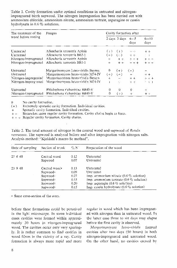
Table 1. Cavity formation under optimal conditions in untreated and nitrogen- impregnated birch sapwood. The nitrogen impregnation has been carried out with ammonium chloride, ammonium nitrate, ammonium tartrate, asparagine or casein hydrolysate in 0.6 70 solutions.
The treatment of the Fungus wood before rotting
Cavity formation after
2 days 3 days 4-5 6 1 0 days days
Untreated Allescheria terrestris Apinis Untreated Allescheria terrestris H63-1 Nitrogen-impregnated Allescheria terrestris Apinis Nitrogen-impregnated Allescheria terrestris H63-1
Untreated Margarinomyces luteo-viridis Beyma Untreated Margarinomyces luteo-viridis M74-IV Nitrogen-impregnated Margarinomyces luteo-~iridis Beyma Nitrogen-impregnated Margarinomyces luteo-viridis M74-IV
Untreated Phialophora richardsiae BB40-V Nitrogen-impregnated Phialophora richardsiae BB40-V
0 No cavity formation. (+) Extremely sporadic cavity formation. Individual cavities. + Sporadic cavity formation. Individual cavities. + + Somewhat more regular cavity formation. Cavity chains begin to form. + s + Regular cavity formation. Cavity chains.
Table 2. The total amount of nitrogen in the central wood and sapwood of Betuln vewucosn. The sapwood is analysed before and after impregnation with nitrogen salts. Analysis method: "Kjeldahl's macro-Se method".
Date of sampling Section of trunk 70 N Preparation of the wood
27 6 68 Central wood 0.12 Untreated Sapwood 0.07 Untreated
23 9 68 Central woo& 0.13 Untreated Sapwooda 0.09 Untreated Sapwooda 0.27 Imp. ammonium nitrate (0.6 % solution) Sapwooda 0.15 Imp. ammonium tartrate (0.6 70 solution) Sapwooda 0.20 Imp. asparagin (0.6 9'0 solution) Sapwooda 0.15 Imp. casein hydrolysate (0.6 9'0 solution)
a Same cross-section of the stem.
before these formations could be perceived in the light microscope. I n some individual cases cavities were formed within approxi- mately 30 hours in nitrogen-impregnated wood. The cavities occur only very sparing- ly. I t is rather common to find cavities in wood fibres in the vicinity of a ray. Cavity formation is always more rapid and more
regular in wood which has been impregnat- ed with nitrogen than in untreated wood. In the latter case three to six days may elapse before the first cavity is observed.
Margarinomyces luteo-viridis formed cavities after two days (50 hours) in both nitrogen-impregnated and untreated wood. On the other hand, no cavities caused by

Phialophora richardsiae were encountered until after three days in nitrogen-impreg- nated wood and after six days in untreated wood. The rapid cavity formation described above took place under optimal tempera- tures for extension growth.
Enlargement of the cavity takes place after its formation. This has been deter- mined by an in vitro study of the cavity formation of Margarinomyces luteo-viridis (Lundstrom, 1970). It is uncertain how long the process continues. However, en- largement appears to continue after new cavities have been formed at the ends of the initial cavity. When an initial cavity is formed, it is normal that the fungal hyphae grow into secondary wall of the wood fibre at an angle of 90" (Figure 3). It was im- possible to study the penetration of the outermost hypha tip into the initial cavity on the cut. Having penetrated the second- ary wall, the hypha branches upwards and downwards and the cavity is enlarged rather similarly in both directions. This T- shaped penetration of the hypha into the wood cell was first described by Corbett and Levy (1963) in respect of an attack by Chaetonziunz globosum in Pinus sylvestris. The T-shaped penetration has been studied by Findlay (1970) with the electron micro- scope.
The position of the bore-hole in relation to the cavity is, however, not always near the centre and at right-angles to the long side of the cavity (see Figures 4, 9 and 11). Sometimes several bore-holes may be found in conjunction with one cavity (Figures 5, 6). In such cases it often appears as if cavity chains were not formed. Instead, the hypha grows further in the cell wall and into an adjacent wood cell after having formed only a single cavity. In some cases, it was observed that the bore-hole con- tinued directly to the opposite long side of the cavity (Figure 7). The majority of the initial cavities occur without bore-holes. It is probable that they exist, but that their position in relation to the cavity is such that they cannot be perceived in the light microscope. The mycelium may possibly have grown inward from the lumen so that
the bore-hole is very short, in contrast to the cases where the mycelium grows through the entire cell wall.
In addition to the cavities in wood fibres, cavities have also been observed in vessels after 100 hours (Figure 8). However, such cavities seldom occur. Levy and Stevens (1966) have made the same observation, i.e. that soft rot fungi attack fibres rather than vessels in hardwoods.
3.3 The occurrence o f cavity chains
The cavity chains develop in two different ways: 1) a new cavity is formed at both ends of the initial cavity (see Figure 9), 2) new cavities are formed at one end of the initial cavity (see Figure 10). Cavity chains with three cavities were formed in nitrogen-impregnated wood (see Figure 11) 70 hours after the mycelium had come into contact with the wood and cavity chains with four cavities had been formed after 100 hours (see Figure 12). Cavity chains with three cavities were encountered in untreated wood after 100 hours of attack (see Figure 9). As in the case of the initial cavity, cavity formation here took place at a slower rate if the wood had not been supplied with nitrogen.
After 100 hours it began to be difficult to follow cavity formation in nitrogen- impregnated wood. New formation of initial cavities took place continuously and short and long cavity chains began to coalesce.
The formation of cavities in the cell wall sometimes took place very close to the lumen, so that the cavity shape could be observed directly from the lumen (Figure 13). It is probable that the thin wall be- tween the cavity and the lumen was de- stroyed during the growth of the cavity, or that cavities were also formed in the wall of the lumen. Since the photograph was taken during studies with a Cambridge Stereoscan Electron Microscope, a thin wall may possibly have been destroyed during the preparation of the specimen or by the electron beam. Levy (1971) has pointed out this possibility.

4 The effect of temperature on cavity formation
Earlier observations have indicated that Allesclzeria terrestris does not form cavities in birch sapwood at temperatures between 35" C and 55" C. At these temperatures it has been reported that the fungus only erodes the cell walls (Bergman and Nilsson, 1967, 1968).
In the present investigation Alleschevia tervestris formed cavities in birch sapwood between 25" C and S O 0 C-temperatures which closely approach the minimum and maximum temperatures, respectively, for
the growth of the fungus. When Allescherin terrestris attacked birch wood at 50" C, the mycelium, in addition to forming cavities, also greatly eroded the cell wall.
Mnrgnrinomyces luteo-viridis formed cavities at temperatures between 5" C and 35" C and Phinlophova riclzardsiae between 5" C and 30" C-temperatures which ap- proach the minimum and maximum tem- peratures for the growth limits of these fungi.

Figure 1 Pre-stage of a cavity formed by Allescheria terrestris in birch sapwood. Im- pregnated with a 0.6 per cent solution of ammonium tartrate. Approx. 875 >< .
Figure 2 Cavity formed by Allescheria ter- r e s t r i .~ in birch sapwood after 50 hours of attack. Impregnated with a 0.6 per cent solution of asparagine. Appros. 8 7 5 x .

Figure 3 T-formed penetration (Corbett's Type I attack) by Al1escher.i~ terrestris in a birch wood fibre. Approx. 875X.
Figure 4 Cavity formed by Allescheria ter- restris in sallow wood (Salix caprea) where the bore-hole adjoins one end of the cavity. Approx. 875 x .
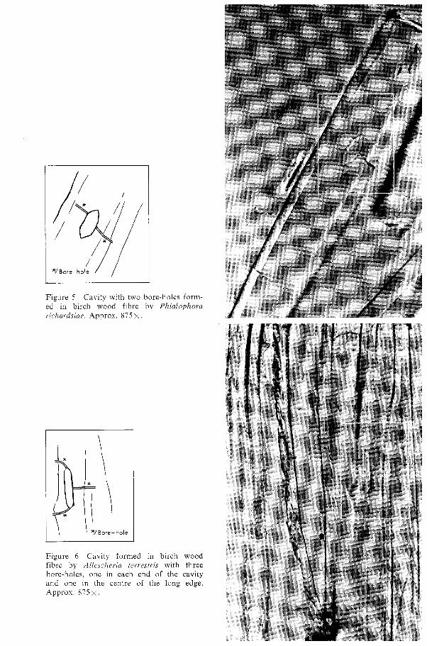
Figure 5 Cavity with two bore-holes ed in birch wood fibre by Plzialc richardsiae. Approx. 875 X.
form- ~ p h o r a
Figure 6 Cavity formed in birch fibre by Al le sc l~er in rerresrris with bore-holes. one in each end of the and one in the centre of the long Appros. 875 x .
wood three
cavity edge.

Figure 7 Cavity formed by Allescheria ter- restris with bore-holes opposite each other on the long edges of the cavity. Approx. 875x.
Figure 8 A cavity formed in a birch wood vessel after 100 hours of attack by Alle- .rchc.ria terrestris. Approx. 875 X .

5' Bore -hole
Figure 9 Growth of a cavity chain by means of new formation of cavities on both short ends of the initial cavity. Birch sap- wood (not treated with nitrogen) attacked 100 hours by Allescheria terrestris. NOTE: The bore-hole is not at right-angles to the cavity. Approx. 875x
Figure 10 Growth of cavity chains by means of cavities formed at one end of the initial cavity. Allescheria t e r r e ~ f r i ~ has been allowed to attack birch sapwood treated with a 0.6 per cent solution of asparagine for 100 hours. Approx. 8 7 5 ~ .

*J Bore- hole
Figure 11 A. Cavity chain with three cavi- ties formed by Allescheria terrestris after 70 hours' attack of birch sapwood impreg- nated with a 0.6 per cent solution of am- monium tartrate. B. Cavity with bore-hole at one of the cavity's short ends. Approx. 8 7 5 x .
Figure 12 Cavity chain with four cavities formed by Allescheria terrestris after 100 hours' attack on birch sapwood impregnated with a 0.6 per cent solution of ammonium tartrate. Approx. 875 X .

Figure 13 Cavity formed by Allescheria terrestris in the wall of the lumen. Approx. 10 600 x . (Cambridge Stereoscan Electron Microscope.)
Figure 14 Cavities formed by Margarirzo- myces lutco-viridis in Juniperis cornmunis. Approx. 875 >< .

Figure 15 Cavity formed by Mul-garino- rnyces luteo-viridis in Ledurn palustre. Approx. 8 7 5 ~ .

5 Cavity formation in lignified stems
A large number of plants with lignified stems were infected with Allescheria ter- restlfs and Margarinoinyces luteo-viridis (see Table 3) . A total of approximately 300 samples was studied. The summary in Table 3 shows that the ability to form cavities in the various woods differs be- tween the two fungi tested. This is especial- ly true of softwood, where the capacity of
Allescheria terrestris to form cavities is poorer than that of Mnrgarinornyces luteo- viridis.
A number of investigations, such as those by Savory (1954), Greaves and Levy (1965) and Findlay (1970), have shown that cavity formation in softwood is slower than that in hardwood and may be completely absent. Variations in the intensity of cavity forma-
Table 3. Cavity formation in plants with lignified cells, exposed to attack by Allescheria terrestris at 45" C and Mnrgarinornyces luteo-viridis at 30" C for approx. 1 month.
Species Allescheria Margarinomyces terrestris luteo-viridis
Acer platanoides L. Andromeda polifolia L. Alnus glutinosa (L.) Gaertn. Betula nana L. Betula tortuosa Ledeb. Corylus avellana L. Fagus silwtica L. Fraxinus excelsior L. Hedera sp. Ledum palustre L. Parthenocissus tricuspidata (Siebold & Zucc.) Planchon. Populus tremula L. Prunus spinosa L. Quercus sp. Salix caprea L. Salix fragilis L. Salix pentandra L. Sambucus racemosa L. Sorbus aucuparia L. Tilia sp. Ulmus sp. Vaccinium myrtillus L. Juniperus communis L. Picea abies (L.) H. Karst. Pinus silvestris L.
+ sparse Cavities not found with complete certainty. + + occasional Only upon impregnation with 0.6 70 NH4N03. + + + plentiful After 2 months of rotting. 0 none occurring - not studied

tion in hardwood are also discussed. The fact that softwood is more resistant to the attack of soft rot fungi than hardwood is assumed to be due to the higher lignin content, to a different chemical composi- tion of the lignin, etc.
The present study, however, showed that even if there were great differences in the intensity of cavity formation, the general appearance of the cavities formed in the various plant species did not differ from those formed in birch wood.
The orientation of the cavities and cavity chains varied widely among the various types of plant but also within the same species of plant. On the other hand, in Vaccinium myrtill~is, Juniperus conzrnuizis, Andromeda polifolin, Parthenocissus tri- cuspidatn and Ledum pallistre the cavities
encountered were always orientated at an angle of approximately 30" to 60" in the wood cell (Figures 14 and 15). The cavities were usually orientated in the same manner in Betula nana. Orientation of the cavities and cavity chains is probably highly de- pendent on the direction of the micro- fibrils in the cell wall (Courtois 1963, Levy 1965). It should therefore be possible to determine approximately the direction of the fibrils in the cell walls of the attacked wood fibres, on the basis of the orientation of the cavities. Thus, the microfibrils in the four above-mentioned plants should pri- marily be found within the approximate angle of 30" to 60" in the section of the secondary cell wall where the cavities were formed.

6 Discussion
If rot conditions are very favourable, with a high moisture content in the wood and an optimal growth temperature, the soft rot fungi employed in this study form cavities in birch wood fibres very rapidly after the mycelium has grown into the wood. Alle- scheria terrestris, with which the study primarily deals, forms individual cavities two days after the wood comes into con- tact with the mycelium. After an additional day, some individual cavity chains are formed. A week after the penetration of the mycelium into the wood, cavities and cavity chains appear in great numbers in the wood which was first attacked. Marga- rirzomyces luteo-viuidis forms cavities just as rapidly as Allescheria terrestris, while Phialophorn richardsiae is a soft rot fungus which forms cavities at a somewhat slower rate. The holes in the form of cavities and bore-holes, which the mycelium creates in the walls of the wood cells, reduce the strength of the wood even at an early stage of attack. This occurs long before a weight loss can be registered in the wood (Liese and Pechmann 1959, Liese and Ammer 1964, Henningsson 1967). This is also true of several of the soft rot fungi studied.
Two types of attack, 1 (cavity formation) and 2 (erosion) were described by Corbett (1965). Of these, type 1 occurred most frequently in the wood fibres during this study. Type 1, with the typical "T-shaped branching" appearance, often occurs in somewhat different forms in that the bore- hole is not always symmetrically situated on the long edge of the cavity. The hypha which forms the lateral cavity sometimes grows farther along the opposite long side of the cavity after, or in connection with, cavity formation.
The fungal hypha's actual penetration of the cell wall, especially by means of
pits, and whether this is passive or active, has been studied and discussed by Corbett (1965), Levy (1965), Greaves and Levy (1965), Levy and Stevens (1966), Findlay (1970) and others. The problem of study- ing the hypha itself before and during penetration of the cell wall and the forma- tion of an initial cavity with the aid of a light microscope, is technically rather dif- ficult. However, it is apparent that cel- lulolytic activity exists in the hypha when the initial cavity is enlarged.
The role of the plasmodesmata during a hypha attack (type 1) on a wood cell has been discussed by Levy (1965) and Levi (1966) and others. Since at that time no known studies of the plasmodesmata in- dicated that they passed through the fully formed walls of the wood cells, the sig- nificance of the plasmodesmata for the hypha's penetration of the cell wall was uncertain. Corbett (1965) considered that the hypha only attacks enzymatically and is not dependent on plasmodesmata or any other "physical discontinuity".
According to recent literature plasmo- desmata form during the embryonic devel- opment of the cells and constitute the connections between the cells. During the growth of the cells, the plasmodesmata col- lect within certain limited areas and form the pits of the cells, Cronshaw (1964), Clowes and Juniper (1968).
The question is whether all plasmodes- mata in the cell are utilised for pit forma- tion or if some remain and are incorporated into the cell wall. Robards's (1968) electron- microscopic photographs of the plasmo- desmata in the ray cells of Salix fragilis show that the plasmodesmata are present in very large numbers, which is a prerequisite for the existance of surplus plasmodesmata after pit formation. Burgess (1971) has

established that the plasmodesmata are reduced during the growth of the cells, which should indicate that they are in- corporated into the cell walls.
The existance of plasmodesmata which constitute a break in the heavily lignified sections of the cell walls, is an important condition which is of advantage to the hyphae when attacking the cell walls.
The following very hypothetical explana- tion can be given for the right-angled bore- holes made in most cases by the mycelium in the cell wall. In cases where diagonal penetration occurs, this may also be the result of plasmodesmata since there are anastomic forms (Robards, 1971). As the wood cell ages, new layers are laid down on the cell wall, and the plasmodesmata which are not incorporated into the pits become covered. Within these plasmodes- mata the endoplasmic reticulum passes through the plasmodesmata (Robards, 1971, and others) and possibly at their openings, substances may be stored which may con- ceivably be diffusible and attractive to the mycelium. At moisture contents in excess of the fibre saturation point, the diffusible elements expand and penetrate the rein- forced cell walls, entirely or in part. The mycelium is stimulated and grows up to the plasmodesmata and through them, penetrating the middle lamella and thus forming the central right-angled bore-hole. Once in the new wood cell, the mycelium can either branch out or grow further towards the lumen. Branching out may be presumed to occur when the mycelium must again find its way along the new cell wall.
Type 2, erosion of the cell wall from the lumen, was not observed during the earliest attack of the mycelium on the cell wall, where only cavities were noted. On the other hand, erosion of the cell walls appears after a longer period of attack. In certain cases it is difficult to determine whether pure erosion takes place. A larger number of hyphae can sometimes develop in the S,
layer and destroy the cell wall towards the lumen, so that an erosion-like pattern is formed in the walls of the wood cell.
The rate at which the mycelium forms cavities in the test blocks has not yet been attained in the thin wood shavings which were used for studies of cavity formation in vitro. This may result from several causes. The intense heat radiation during microscopy and photographing may disturb the growth of the mycelium by drying out the wood. The mycelium itself may be damaged by the light. Since there are fewer intact wood cell rows in a shaving some 25 ,u thick, there may have been too few wood cells. The hyphae simply grow on the outside of the wood.
Findlay (1970) has also described a tech- nique for studying cavity formation in vitro. The procedure is simple in itself. Thin wood shavings are placed on a sheet of glass. Another sheet of glass is placed over the wood shaving. The wood shaving is moistened by means of a wick from a water container. The basic idea is the same as Lundstrom's (1970), i.e. to study cavity formation by means of thin wood shavings. Unfortunately, Findlay's cultivation tech- nique has not proved very successful. The reason for this may again be that the myce- lium is damaged by the light.
Existing studies show that soft rot fungi possess great cavity-forming capacity in lignified cells of various plants. Schacht noted cavities in wood cells of plants as early as 1863. Cavity formation in wood cells from a large number of trees has been reported by Duncan (19601, Courtois (19631, Levy (1969), Findlay (1970) and others. All of the cases referred to here concern dead wood cells. Cavity formation in wood cells from living plants is almost unknown. Oliver (1959) has, however, re- ported formations resembling cavities in living Guarea sp. and Lovon klaineniza Pierre.

Acknowledgements
My sincere gratitude is due to Professor Thomas Nilsson, and Professor Aino Kaarik Per Nylinder, head of the Department of of the Royal College of Forestry. Forest Products at the Royal College of I also wish to express my thanks to Mrs. Forestry, Stockholm, Sweden for his con- Gunnel Lundstrom, Miss Christina Ohlsson tinued interest in my work. and Mrs. Inga Bergholm for valuable aid.
For stimulating discussions and valuable For the careful work of the translation into advice in the work, the author wishes to English I wish to thank Anne Bergstrom. thank Professor Bjorn Henningsson, Dr.

References
Bailey, I. W., Vestal, M. R. 1937: The sig- nificance of certain wood-destroying fungi in the study of the enzymatic hydrolysis of cellulose. J. Am. Arb. 18. 196-205.
Bergman, o., Nilsson, T. 1967: On outside storage of aspen chips at Htirnefors' sul- phite mill. R. Coll. For., Dept. For. Prod., Res. Notes R 55, Stockholm.
- 1968: On outside storage of birch chips at Morrum's sulphate mill. R. Coll. For., Dept. For. Prod., Res. Notes R 60, Stock- holm.
Burgess, J. 1971 : Observations on structure and differentiation in Plasmodesmata. Protoplasma. 73. 83-95.
Clowes, F. A. L., Juniper, B. E. 1968: Plant cells. Botanical monographs. Vol. 8. Black- well Scientific Publications. Oxford and Edinburgh.
Corbett, N . H. 1965 : Micro-morphological studies on the degradation of lignified cell walls by Ascomycetes and Fungi Imperfecti. J. Inst. Wood Sci. 14. 18-29.
Corbett, N. H., Levy, J. F. 1963: Penetration of tracheid walls of Pinz~s sylvestris L. (Scots pine) by Chaetorniu~n globosunz Kunz. Nature. 198. 1322-1323.
Courtois, H. 1963 : Mikromorphologische Be- fallsymptome beim Holzabbau durch Moderfaulepilze. Holzforschung u. Holz- verwertung. 15. 5. 88-101.
Cronshaw, J. 1965 : Cytoplasmic fine structure and cell wall development in differentiat- ing xylem elements. Cellular ultrastructure of woody plants. 99-124. New York.
Duncan, C. G. 1960: Wood-attacking capacities and physiology of soft-rot fungi. U.S. Dept. Agric. For. Prod. Lab. Rep. 2173.
Findlay, G. W. D. 1970: Microscopic studies on soft rot in wood. Ph. D. Thesis. Univer- sity of London.
Greaves, H., Levy, J. F. 1965: Comparative degradation of the sapwood of Scots pine, beech and birch by Lenzites trabea, Poly- sfictus versicolor, Chaetomiurn globosum and Bacillus polyrnysa. J. Inst. Wood Sci. 3. 15. 55-63.
Hemningsson, B. 1967 : Changes in impact bending strength, weight and alkali solu- bility following fungal attack on birch wood. Stud. For. Suec. 41.
Levi, M. P. 1966: Decay patterns produced by Chaetonziu~n globosunz in beechwood fibers.
A chemical and microscopic study. Material u. Organismen, Beih. 1. "Holz und Orga- nismen." 119-126.
Levy, J. F. 1965: The soft rot fungi: Their mode of action and significance in the degradation of wood. Adv. in Bot. Res. 2. 323-357.
- 1969: The spectrum of interaction between fungi and wood. B.W.P.A. Annual Conven- tion 1969. 71-78 (Paper 4).
- 1971 : Further basic studies on the inter- action of fungi, wood preservatives and wood. B.W.P.A. Annual Convention 1971. 63-75 (Paper 4).
Levy, J. F., Stevens, M. G. 1966: The initia- tion of attack by soft rot fungi in wood. J. Inst. Wood Sci. 3. 16. 49-55.
Liese, W., Ammer, U. 1964: Uber den Einfluss von Moderfaulepilzen auf die Schlagbie- gefestigkeit von Buchenholz. Holz als Roh- u. Werkstoff. 22. 455-459.
Liese, W., von Pechmann, M. 1959: Unter- suchungen iiber den Einf l~~ss von Moder- faulepilzen auf die Holzfestigkeit. Forst- wiss. Cbl. 78. 271-279.
Lundstriim, H. 1970: Cavity formation in soft rot-A method for microscopic direct study. Holzforschung. 24.4. 132-1 33.
Merrill, W., Cowling, E. B. 1966: Role of nitrogen in wood deterioration: Amounts and distribution of nitrogen in tree stems. Can. J. Bot. 44. 1555-1580.
Oliver, A. C. 1959: Soft rot. A summary of existing knowledge. The Timber Develop- ment Assoc. (London) Inform. Bull BiIBi2.
Robards, A. W. 1968 : A new interpretation of plasmodesmatal ultrastructure. Planta. 82. 200-210.
- 1971 : The ultrastructure of plasmodesmata. Protoplasma. 72. 315-323.
Savory, 9. G. 1954: Breakdown of timber by Ascomycetes and Fungi Imperfecti. Ann. appl. Biol. 41.2. 336-347.
Schacht, H. 1850: Uber eigentiimliche. bisher noch nicht beobachtete Erscheinungen in den Verdickungsschichten gewisse Holzzel- len. Botan. Zeitung. 8. 697-702, 713-719.
- 1863: Uber die Veranderungen durch Pilze in abgestorbenen Pflanzenzellen. Jahrb. f. Wiss. Botan. 3. 442-483.
Thunell, B., Berem, E. 1952: Svenskt tra. C. A. Stromberg AB, Stockholm.

Sammanfattning
Mogelrotesvamparna angriper vedceller p i tvh olika satt, dels genom att erodera cell- vaggen direkt inifrin cellhiligheten varvid cellvaggen fortunnas, dels genom att vaxa fram i cellvaggens S2-skikt och dar bilda hiligheter s.k. kaviteter. Variationer av dessa angreppsbilder forekommer hos olika mogelrotesvampar. De hiligheter i form av kaviteter och borrhil, som mycelet skapar i vedcellernas vaggar redan i tidigt skede vid rotangrepp, sanker vedens hillfasthet avse- vart och detta lingt innan storre viktsfor- luster av veden registrerats.
I denna studie har tidsforloppet speciellt studerats ifrln det att en mogelrotesvamp infekterat bjorkved till dess att mycelet bildat de forsta kaviteterna. Studierna har utforts p i laboratoriet under olika optimala rotbetingelser for testsvamparna.
De studerade mogelrotesvamparna ar en termofil Ascomycet Allescheria terrestris Apinis stam Apinis och stam H63-1, tv i Fungi Imperfecti Margarinonzyces luteo- viridis v. Beyma stam Beyma och stam M74-IV samt Phialophora richardsine (Nannf.) Conant stam BB40-V. Studierna har framst gallt Alleschevia terrestris varvid de tv i ovriga mogelrotesvamparna har an- vants i jamforande studiesyfte.
Svamparna framodlades p i snedagarror i den optimala langdtillvaxttemperaturen for varje svamp. Klotsar (2 x 0,5 x 0,5 cm) f r in splintveden av Betula verrucosa Ehrh. eller Bet~da pubescens Ehrh. lades darefter ut ph det utvuxna mycelet. Vid mikroskopstudier- na har klotsarna snittats med rakblad och fargats in med safranin lost i 70 9'0 alkohol. Ett Cambridge Stereoscan Electron Micro- scope har ocksh anvants i mindre omfatt- ning.
Den sparsamma tillghngen p i kvave i veden har visat sig vara en begransande faktor for rotsvamparnas formiga att an-
gripa veden. Darfor har kvavets roll vid kavitetsbildningen undersokts. Darvid har kavitetsbildningen tidsmassigt jamforts i obehandlad resp. kvaveimpregnerad (ammo- niumklorid, ammoniumnitrat, ammonium- tartrat, asparagin och kaseinhydrolysat) ved.
Phialophorn richardsiae piverkades mest om kvave tillsattes i veden, d i svampen bildade kaviteter efter 3 dygn mot 6 dygn i obehandlad ved. Allescheria terrestris och Margavinomyces luteo-viridis bildade kavi- teter 2 dygn efter det att de infekterat veden oberoende om kvave tillsattes eller ej (figur 2). I kvaveimpregnerad ved kom dock kavitetsbildningen i glng jamnare och i storre omfattning an i obehandlad ved (tabell 1).
En vanlig angreppstyp vid mogelrote- svampars angrepp p i cellvaggen, ar att hyfen vaxer in i en mer eller mindre 90" vinkel in i cellvaggen och darefter forgrenar sig i fiberns bagge langdriktningar. Darmed bildas ett "T-format angrepp" i vedcellens sekundba cellvagg (figur 3). Hyfen och det efterlamnade borrhilet kommer darvid att ansluta mitt p i initialkaviteten. Denna an- greppsbild ar mycket vanlig och har be- skrivits tidigare. I denna undersokning har det emellertid visat sig att borrhiilet kan ansluta var som helst utefter kavitetens sida (figurerna 4, 5, 9, 11).
En metod att folja kavitetsbildningen in vitro har utarbetats och publicerats separat (Lundstrom, 1970). Den gl r ut p i att lhta mycelet angripa tunna traspin utan att stora kavitetsbildningen. Traspinet sitter monterat over ett vertikalt fastsatt glasror, som ar fast i botten p l en glaspetriskil. Svampen ympas p i maltagar som har in- gjutits i petriskilen. Glasroret hiller undan maltagarn s i att traspinet kan belysas underifrln. Odlingen sker helt sterilt och fukten i traspinet (som ar en absolut nod-
vandighet for att mycelet skall angripa l~lteo-viridis kavitetsbildande formlga har veden) uppsugs f r ln agarn. Vissa problem studerats i 25 olika vaxter med forvedade kvarstlr dock att losa i samband med dessa celler (figurerna 14, 15). Kavitetsbildningen studier, d l mycelet stors av vissa yttre fak- visade stora skillnader mellan barrved, dar torer slsom ljus, varme m.m. den var sparsam, och lovved, dar den oftast
Allescherin terrestris och Mm.garinomyces skedde rikligt (tabell 3).
Electronic version 0 Studia Forestalia Suecica 2002 Edited by J.G.K.Flower-Ellis