microscopie électronique en transmission i. instrument histoire ruska & knoll (1931) prix nobel...
TRANSCRIPT
Microscopie électronique en transmissiontransmissionI. Instrument
Nadi BraidyProfesseur adjointGénie chimique et Génie biotechnologiqueUniversité de Sherbrooke
8 mars 2011

Plan
� I. Instrument
� II. Imagerie
� III. Microscopie analytique
2
� III. Microscopie analytique

Plan- I. Instrument
� A. Introduction
� B. Instrumentation
� C. Composantes
3
� C. Composantes
� D. Préparation d’échantillons
� E. Porte-objets

A.1 Résolution spatiale vs chimique
4www.npl.co.uk/nanoscience/surface-nanoanalysis/research/

A.2 Résolution vs longueur d’onde1 m
100 mm
10 mm
1 mm
100 µm
10 µm
Radio
Micro-onde
Infra-rouge
Critère de Rayleigh:longueur d’onde
NA
λδ
61.0=
5
Images:w3.lmp.ualberta.ca/resources/pathoimages
http://pro.corbis.com/imagesw3.healthinitiative.org/
w3.scq.ubc.ca/
10 µm
1 µm
100 nm
10 nm
1 nm
1 Å
visible
Ultra-violet
Rayons X
Rayons gamma
difficile à
converger

A.3 Dualité onde-particule
1 m
100 mm
10 mm
1 mm
100 µm
Radio
Micro-onde
Infra-rouge
onde particule
masse = 10-30 kg
charge = -1.6 x 10⁻¹⁹ C0
2
m
eUv
vm
h
=
=λ
6
10 µm
1 µm
100 nm
10 nm
1 nm
1 Å
visible
Ultra-violet
Rayons X
Rayons gamma
e- e-
e- � 0.02 – 1 Å
0m
v =
U
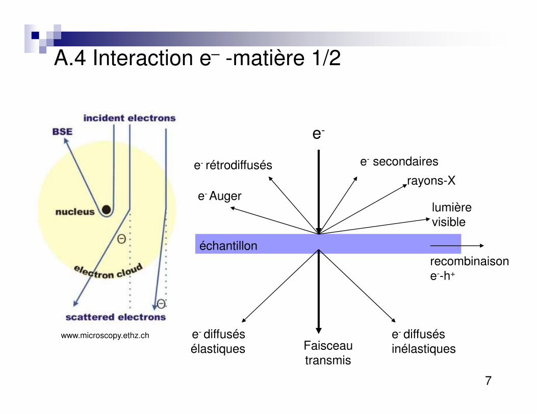
A.4 Interaction e– -matière 1/2
e-
e- secondaires
rayons-X
lumière
e- rétrodiffusés
e- Auger
7
lumière visible
Faisceau transmis
e- diffusés élastiques
e- diffusés inélastiques
échantillonrecombinaison e--h+
www.microscopy.ethz.ch

A.5 SEM vs TEM
Au massif Au 10 nm
8
Simulé avec Casino : http://www.gel.usherbrooke.ca/casino/What.html
Différence entre MEB et MET: épaisseur de l’échantillon

A.6 Histoire
Ruska & Knoll (1931) Prix Nobel 1986
1897 Découverte de l’électron par J.J. Thompson1924 P. De Broglie: dualité onde-particule1927 Hans Busch: focalisation des électrons à l’aide d’un champ magnétique inhomogène.1931 Ernst Ruska et Max Knoll construisent le premier MET
1938 Premier microscope électronique à balayage en transmission (M. von Ardenne)1939 Siemens lance le premier microscope commercial (Ruska, von Borries)~1940 Bases de l’optique des électrons et des lentilles magnétiques (W. Glaser, O. Scherzer) 1943 Spectroscopie des électrons en perte d’énergie
9Microscope moderne (zmb.uzh.ch)
1943 Spectroscopie des électrons en perte d’énergie (EELS, J. Hillier)1951 Spectroscopie des rayons X (R. Castaing)1956 Première image de haute résolution (J. Menter)1957 Méthode de simulation multi-couche (J. Cowley, A. Moodie)1964 Premier SEM commercial (Cambridge Instruments)~1970 Microscope HRTEM ayant une résolution de 4 Å1986 E. Ruska gagne le prix Nobel (partagé avec G. Binning and H. Rohrer, inventeurs du microscope à effet tunnel)~2003 MET avec correcteur d’aberration sphérique résolution sub-Ångstrøm
traduction libre, www.microscopy.ethz.ch

B.1 Aperçu de l’instrument: 1-5 M$canon e-
échantillon
1 m
3 mm
10
écran
www.iopb.res.in
www.gatan.com
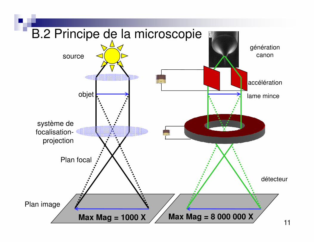
B.2 Principe de la microscopie
source
objet
système de
générationcanon
accélération
lame mince
11
système de focalisation-
projection
Plan image
Plan focal
détecteur
Max Mag = 1000 X Max Mag = 8 000 000 X

B.2 Principe de la microscopie
12: www.lab.anhb.uwa.edu.au

B.3 Sources d’électrons
� Thermoionique�W ou LaB6
�e- arraché à la surface = φ fonction de travail�Courant de chauffage pour vibration
13
� Émission de champs�φ obtenue en appliquant champs électrique� lorsque barrière suffisemment fine � tunnel�Requiert vide + poussé

W
B.4 Types de filamentsLaB6 FEG
14
Williams &Carter. Transmission Electron Microscopy. Plenum Press, NY 1996. p.73-76

B.5 Lentille électromagnétique
� Trajectoire en spirale� Converge vers l’axe optique
� Lentille àélectrons
e- e-
15
électrons
� équivalentoptique

B.5 Lentille électromagnétique
� Point focal contrôlé par courant
� Focalisation ou condensation
16
� Aberrations�sphérique�chromatique�astigmastime
� Refroidi à l’eauwww.matter.org.uk/tem

B.6 Diaphrames
� Sélectionner e-
�ayant un certain angle�région particulière de l’image
� Condenseur
17
� Condenseur� limite divergence�contrôle intensité sur échantillon
Williams &Carter. Transmission Electron Microscopy. Plenum Press, NY 1996.

C. Composantes principales
Condenseur
Illumination
18
Objectif
Projecteur
www.mah.se

C.1 Système condenseur
� Composante�2 lentilles�1diaphragme
� Contrôle de façon ~
19
� Contrôle de façon ~ indépendante� Intensité�Angle de convergence�Taille du faisceau
www.matter.org.uk/tem

C.2 Système objectif
� Composantes�2 lentilles�1 diaphragme
� Plan focal arrière
20
�où convergent faisceaux parallèles
� Plan image�Première image
formée
www.microscopy.ethz.ch

D. Préparation d’échantillons
� Film mince�d’autant plus vrai que kV bas�à tout prix, < 100 nm� idéal: 10-20 nm�autour de 50 nm... dépend de la densité
21
�éviter endommagement lors amincissement
� Polissage mécanique� Électro-polissage� Amincissement ionique� Ultramicrotomie
http://temsamprep.in2p3.fr/techniques.php?lang=fr

D.1 Polissage électrochimique
� Idéal pour métaux
� Produit chimique corrosifs
� Attaque préférentielle zone pré-amincie
Goodhew & Humphreys (1988) Electron microscopy and analysis.Taylor & Francis, London.
22
pré-amincie
� Chaque métal à sa propre recette
� Idéal pour alliages de Al, Mg, Fe
www.mpie.de

D.2 Ultramicrotomie
� Idéal pour échantillon biologique
� et métaux ductile (introduit déformation !)
� Enrober dans la résine
Goodhew & Humphreys (1988) Electron microscopy and analysis.Taylor & Francis, London.
23
� Couteau au diamant� Art... temsamprep.in2p3.fr

D.3 Amincissement ionique
� Bombardement Ar+
Goo
dhew
& H
umph
reys
(198
8) E
lect
ron
mic
rosc
opy
and
anal
ysis
.Tay
lor
&
Fra
ncis
, Lon
don.
24www.wintech-nano.com/services_fa
� Faisceau d’ions focalisé (FIB)�comp.
microélectronique�analyse de
rupture

E.1 Porte-objet
� Goniomètre (pourquoi rotation ?)�±10 U-HRTEM...espace lentilles�± obj. ±30 conventionel�± 70 tomographie �Double-tilt
25
�Double-tilt
http://www.gatan.com/products/specimen_holders/

E.2 Porte-objets in-situ� Contrôle de température
� Refroidissant� N: 77 K� He: 4 K
� Platine chauffante� 1000°C, 1 min
� Transfert sous vide
26
� Transfert sous vide� Cellules environmentales
� Essai de traction intégré� Premières dislocations� Extrait de H. Nakashima, U. Kyushu
� Mo et W ont les même propriétés de durcissement structural sur la ferrite
http://www.msm.cam.ac.uk/phasetrans/2002/dislocations.movies.html

27

R. Feynman (1959) “There is plenty of room at the bottom”
� The electron microscope is not quite good enough, with the greatest care and effort, it can only resolve about 10 angstroms. I would like to try and impress upon you while I am talking about all of these things on a small scale, the importance of improving the electron microscope by a hundred times. It is not impossible; it is not against the laws of diffraction of the electron. The wave length of the electron in such a microscope is only 1/20 of an angstrom. So it should be possible to see the individual atoms. What good would it be to see individual atoms distinctly?
28
...
� And I know that there are theorems which prove that it is impossible, with axially symmetrical stationary field lenses, to produce an f-value any bigger than so and so; and therefore the resolving power at the present time is at its theoretical maximum. But in every theorem there are assumptions. Why must the field be symmetrical? I put this out as a challenge: Is there no way to make the electron microscope more powerful?