microsporidia and cryptosporidium in horses and … kvetoˇ nov ᡠc, lihua xiaod ... province and...
TRANSCRIPT

MAs
ADAa
b
c
Cd
e
f
g
a
ARRA
KHDCEEM
R
0
Veterinary Parasitology 208 (2015) 135–142
Contents lists available at ScienceDirect
Veterinary Parasitology
jo u r nal homep age: www.elsev ier .com/ locate /vetpar
icrosporidia and Cryptosporidium in horses and donkeys inlgeria: Detection of a novel Cryptosporidium hominisubtype family (Ik) in a horse
bd Elkarim Laatamnaa,g, Pavla Wagnerováb,c, Bohumil Sakc,ana Kvetonovác, Lihua Xiaod, Michael Roste, John McEvoyf,hmed Rachid Saadig, Meriem Aissig, Martin Kvácb,c,∗
Faculty of Natural Sciences and Life, University of Djelfa, Moudjbara, BP 3117, Djelfa, AlgeriaFaculty of Agriculture, University of South Bohemia in Ceské Budejovice, Studentská 13, 370 05 Ceské Budejovice, Czech RepublicBiology Centre of the Academy of Sciences of the Czech Republic, Institute of Parasitology, Branisovská 31, 370 05 Ceské Budejovice,zech RepublicCenters for Disease Control and Prevention, Atlanta, GA, USAFaculty of Economics, University of South Bohemia in Ceské Budejovice, Studentská 13, 370 05 Ceské Budejovice, Czech RepublicDepartment of Veterinary and Microbiological Sciences, North Dakota State University, Fargo, ND, USAHigher National School of Veterinary, BP 161 Hacène Badi, EL Harrach, Algiers, Algeria
r t i c l e i n f o
rticle history:eceived 24 June 2014eceived in revised form 6 January 2015ccepted 8 January 2015
eywords:orsesonkeysryptosporidium spp.ncephalitozoon spp.nterocytozoon bieneusiolecular prevalence
a b s t r a c t
A total of 219 and 124 individual fecal samples of horses and donkeys, respectively, werescreened for the presence of Cryptosporidium spp., Encephalitozoon spp., and Enterocytozoonbieneusi DNA by genus-specific nested PCR. Isolates were genotyped by sequence analysisof SSU rRNA, GP60, TRAP-C1, COWP, and HSP70 loci in Cryptosporidium, and the ITS regionin microsporidia. Cryptosporidium spp. was detected on 3/18 horse farms and 1/15 farmswhere donkeys were kept. Overall, five (2.3%) horse and two (1.6%) donkey specimens werePCR positive for Cryptosporidium. Genotyping at SSU and GP60 loci revealed that threeisolates from horses and donkeys were C. parvum subtype family IIaA16G1R1, one isolatefrom a horse was, C. muris RN66, and one isolate from a donkey was C. muris TS03. Anisolate from a horse shared 99.4% and 99.3% similarity with Cryptosporidium hominis and C.cuniculus, respectively, at the SSU locus. This isolate shared 100% identity with C. hominisat the TRAP-C1, COWP, and HSP70 loci, and it was from the novel gp60 subtype familyIkA15G1.
Microsporidia were found on 6/18 horse and 2/15 donkey farms. E. bieneusi was identifiedin 6.8% (15/219) and 1.6% (2/124), and Encephalitozoon cuniculi was identified in 1.8% (4/219)and 1.6% (2/124), of horses and donkeys, respectively. Three genotypes of E. cuniculi (I, IIand III) were detected in horses, and E. cuniculi genotype II was detected in donkeys. Four
genotypes of E. bieneusi (horse1, horse 2, CZ3, D) were described in horses. An additional fivehorses and two donkeys were positive for E. bieneusi, but the isolated were not genotyped.Neither Cryptosporidium nor microsporidia prevalence were affected by sex, age, type ofbreeding, or whether the∗ Corresponding author at: Biology Centre of the Academy of Sciences of theepublic. Tel.: +420 387775419; fax: +420 385310388.
E-mail address: [email protected] (M. Kvác).
http://dx.doi.org/10.1016/j.vetpar.2015.01.007304-4017/© 2015 Elsevier B.V. All rights reserved.
host was a horse or a donkey.
© 2015 Elsevier B.V. All rights reserved.Czech Republic, v.v.i., Branisovská 31, 370 05 Ceské Budejovice, Czech
ry Para
136 A.E. Laatamna et al. / Veterina1. Introduction
Both horses and donkeys are used worldwide for work,food, and social activities. Both wild and domestic equinesare exposed to a complex mixture of multicellular andunicellular parasites. Although most of these parasites areequine-specific, horses and donkeys can be infected byhost-nonspecific and zoonotic parasites including Cryp-tosporidium and several microsporidia (Grinberg et al.,2003; Kouam et al., 2010; Santín et al., 2010).
Approximately 1200 species of microsporidia areknown, and they infect all major animal groups. Mostof these ubiquitous obligate intracellular parasites infectinvertebrates and fish, but 14 species in eight genera infectmammals (Didier et al., 1995; Didier and Weiss, 2006).Although equine microsporidiosis is known to cause abor-tion (Patterson-Kane et al., 2003; Szeredi et al., 2007; vanRensburg et al., 1991), microsporidia in horses remains onthe periphery of scientific interest. Three recent reportshave described the prevalence of Enterocytozoon bieneusiand Encephalitozoon cuniculi and the course of infection ofE. cuniculi in horses from Colombia and the Czech Republic(Santín et al., 2010; Wagnerová et al., 2012, 2013).
Protozoans of the genus Cryptosporidium are parasitesinhabiting the digestive tract and/or respiratory systemsof birds, fish, reptiles, and mammals, including equines(Mtambo et al., 1997; Ryan et al., 2003). Twenty five speciesand more than 40 genotypes of Cryptosporidium have beendescribed to date (Ryan and Xiao, 2014), and only threeof these – C. parvum, C. erinacei (previously known asCryptosporidium hedgehog genotype) and Cryptosporidiumhorse genotype – are known to infect horses (Chalmerset al., 2005; Laatamna et al., 2013; Ryan et al., 2003). How-ever, only eight of the 30 studies of horse cryptosporidiosisreported the species/genotype present (Burton et al., 2010;Grinberg et al., 2008, 2003, 2009; Chalmers et al., 2005;Imhasly et al., 2009; Ryan et al., 2003; Veronesi et al., 2010),so the actual number of Cryptosporidium taxa infectinghorses may be greater.
A number of Cryptosporidium spp. (C. parvum, C. erina-cei and Cryptosporidium horse genotype) and microsporidia(Encephalitozoon intestinalis, E. cuniculi genotypes I andII, and E. bieneusi – genotypes D, EbpA, G and WL15)detected in horses and donkeys to date have been shownto cause human infection (Didier et al., 2000; Mathiset al., 2005; Nichols et al., 2014). Therefore, there is aneed to better understand the zoonotic potential of horsemicrosporidiosis and cryptosporidiosis. This study exa-mines the prevalence of Cryptosporidium and microsporidiain wild and domestic horses and donkeys from Algeria.
2. Materials and methods
2.1. Origin of samples and animals
The research was performed from November 2011to May 2013 on 4 horse farms in an urban area and
20 small private farms of horses and donkeys in ruralareas located in 4 provinces in Algeria. The farms wereselected without previous knowledge of parasitologicalstatus, with the exception of one farm (no. 1), wheresitology 208 (2015) 135–142
four cases of Cryptosporidium erinacei infection (previouslyknown as Cryptosporidium hedgehog genotype) were pre-viously reported (Laatamna et al., 2013). Farm 1, situatedin province of Tiaret in west Algeria, is a national centerof horse breeding that keeps up to 280 horses and servesas a supplier of horses to other farms located in the differ-ent provinces of Algeria. The management system at Farm1 depended on the season: breeding stallions, mares, andfoals spent most of the time on the pasture, except dur-ing winter, when they were housed in stables. Equestriancenters 2, 3 and 4, focus exclusively on horse breeding,are located in the capital Algiers, and breed approximately50, 70, and 90 horses, respectively. Horses are used forhorseback riding, horseracing, and show jumping and aremaintained in individual boxes. Twenty farms in rural areasincluded 12 private farms in Bourdj Bou Arréridj (B.B.A)province and eight in Setif province. These rural areas areregions of high plateaus that are almost exclusively agricul-tural, and horses live in close association with cattle, otherruminants, and domestic birds. Donkeys live unrestrictedin these areas and are used for transport and work. Bothhorses and donkeys are on pasture during the entire year,except during winter, when they are primarily housed instables.
2.2. Sample collection
Fecal samples from horses and donkeys were collecteddirectly from the rectum or from the ground immediatelyafter defecation. Specifically, all horse fecal samples fromfarms 1–3 (n = 94), 40 out of 55 horse samples from farm4, and three donkey samples from farm 15 were collecteddirectly from rectum (Table 1). Each sample was individu-ally placed into a sterile plastic container without fixatives,and transported in an isotherm box to the laboratory. Norepeated analyses of the same animals were included in thesurvey to prevent cumulative prevalence.
2.3. DNA isolation and molecular analyses
Total DNA was extracted from 200 mg of feces bybead disruption for 60 s at 5.5 m/s using 0.5 mm glassbeads in a FastPrep®24 Instrument (MP Biomedicals, CA,USA) followed by isolation/purification using a commer-cially available kit in accordance with the manufacturer’sinstructions (QIAamp® DNA Stool Mini Kit, Qiagen, Hilden,Germany). Purified DNA was stored at −20 ◦C prior tobeing used for PCR. Due to the predicted low numberof oocysts (<100) in examined samples, a nested PCRwas required (Smith, 2008). The nested PCR approachwas used to amplify a region of the small subunit ofrRNA gene (SSU; ∼830 bp; Jiang et al., 2005; Xiao et al.,1999) and 60 kDa glycoprotein (GP60; ∼830 bp; Alveset al., 2003) in all samples. In addition, nested PCRamplifying a region of Cryptosporidium Oocyst Wall Pro-tein (COWP; ∼550 bp; Pedraza-Diaz et al., 2001; Spanoet al., 1997), Thrombospondin-Related Adhesive Protein
of Cryptosporidium-1 (TRAP-C1; ∼780 bp; Spano et al.,1998) and Heat Shock Protein (HSP70; ∼1950 bp; Sulaimanet al., 2000) were used in case of detection of a novelisolate of Cryptosporidium. Negative and positive controls
A.E. Laatamna et al. / Veterinary Parasitology 208 (2015) 135–142 137
Table 1A survey of Cryptosporidium spp. and microsporidia in fecal samples of horses and donkeys.
Province No. offarm
Animal No. ofexaminedsamples
Cryptosporidium Microsporidia
No. of positivesamples
Species(strain/family)
No. of positivesamples
E. bieneusi E. cuniculi
Tiaret 1 Horse 62 3 1× C. parvum(IIaA16G1R1)1× C. hominis(IkA15G1)1× C. muris(RN66)
9 6× horse11× horse21*
1× E.cuniculi I
Algiers2 Horse 15 – – – – –3 Horse 17 – – 1 1* –4 Horse 55 – – 2 1* 1× E.
cuniculi I
BourdjBouArréridj
5 Horse 2 – – 1 – 1× E.cuniculi II
6 Horse 6 – 1 1* –7 Horse 5 – – – – –8 Donkey 3 – – – – –9 Donkey 3 – – – – –
10 Donkey 5 – – –
11Horse 8 – – 3 1* 1× CZ3 1× E.
cuniculi IIIDonkey 29 – – 1 – 1 × E.
cuniculi II
12Horse 6 – – 1 1 × D –Donkey 3 – – – – –
13Horse 7 1 1× C. parvum
(IIaA16G1R1)– – –
Donkey 28 2 1× C. muris(TS03) 1× C.parvum(IIaA16G1R1)
1 1* –
14Horse 3 – – – – –Donkey 3 – – – – –
15Horse 2 – – – – –Donkey 4 – – – – –
16Horse 2 1 1× C. parvum
(IIaA16G1R1)– – –
Donkey 13 – – 1 1* –
Setif
17 Horse 5 – – – – –18 Horse 7 – – – – –19 Donkey 3 – – – – –20 Donkey 23 – – 1 – 1× E.
cuniculi II21 Donkey 1 – – – – –
22Horse 11 – – 1 1× CZ3 –Donkey 1 – – – – –
23Horse 3 – – – – –Donkey 1 – – – –
24Horse 3 – – – – –Donkey 4 – – – – –
TotalHorse 219 5 19 15 4Donkey 124 2 4 2 2All 343 7 23 17 6
(at(da
* Unsuccessful re-sequencing at ITS locus.
DNA of C. tyzzeri family IXa) were included in each PCRmplification. To identify microsporidia, a nested PCR pro-
ocol was used to amplify the internal transcribed spacerITS) region of the rRNA gene of E. bieneusi, as previouslyescribed Buckholt et al. (2002). Two primer sets, INT580Fnd INT580R (Didier et al., 1995) for primary and MSP3and MSP4A (Katzwinkel-Wladarsch et al., 1996) for sec-ondary PCR reaction, were used to amplify the ITS region
of Encephalitozoon spp. Positive control DNA isolated fromE. intestinalis and E. bieneusi genotype D spores and nega-tive controls were included in each run. Both primary andsecondary PCR reactions were carried out in a volume of
ry Para
138 A.E. Laatamna et al. / Veterina20 �l; the primary reaction contained 2 �l of genomic DNA(or water as a negative control) and the secondary reac-tion contained 2 �l of the primary reaction as template.PCR amplicons were electrophoresed in 1% agarose gels andvisualized with 0.2 mg/ml ethidium bromide under ultra-violet light. Purified secondary products were sequencedin both directions with an ABI 3130 genetic analyser(Applied Biosystems, Foster City) using the secondary PCRprimers and the BigDye1TerminatorV3.1cycle sequencingkit (Applied Biosystems, Foster City) in 10 �l reactions.DNA extraction and the amplification and sequencing ofeach locus were repeated twice for each positive sam-ple.
2.4. Phylogenetic analyses
The nucleotide sequences of each gene obtained inthis study were manually edited using the program Chro-masPro 1.7.5 (Technelysium, Pty, Ltd.), and aligned withpreviously published sequences using the MAFFT ver-sion 7 online server using the Q-INS-i algorithm forSSU sequences, L-INS-i algorithm for GP60 sequences,and the FFT-NS-1 algorithm for COWP, TRAP-C1 andHSP70 sequences (http://mafft.cbrc.jp/alignment/server/)(Katoh and Standley, 2013). Alignment adjustments weremade manually to remove artificial gaps using BioEdit7.0.5.3. Phylogenetic relationships of aligned sequenceswas inferred using Neighbor-Joining (NJ) methods (Saitouand Nei, 1987) based on the Kimura 2-parameter (K2P)distances model (Kimura, 1980) with pairwise dele-tions. Phylogenetic analyses were performed using MEGA5(Tamura et al., 2011). The reliability of branches in treeswas assessed using the bootstrap analysis with 1000 pseu-doreplicates. Bootstrap values above 50% were reported.Phylograms were constructed using MEGA5 and visuallyedited for style using CorelDrawX5 (without changingthe tree topography). Sequences generated in this studywere deposited in GenBank under the accession numbersKJ941130–KJ941151.
2.5. Statistical analyses
A two-sample z-test for proportions (independentgroups) was used to assess relationships between par-asite detection (Cryptosporidium species/genotype ormicrosporidia) and animal attributes (age, sex, and species)or management practices. Odds ratios were used todetermine the significance of potential risk factor vari-ables. The predictor variables were gender, age, housing,and host species. These were considered separately, notsimultaneously. All computations were performed with R2.15.1.
3. Results
A total of 343 individual fecal samples from 219 horses
and 124 donkeys on 18 and 15 farms, respectively, wereexamined using molecular tools for the presence of Cryp-tosporidium and microsporidia (Encephalitozoon spp. andE. bieneusi) specific DNA (Table 1). No samples collectedsitology 208 (2015) 135–142
from the ground were positive for Cryptosporidium ormicrosporidia.
3.1. Cryptosporidium in horses and donkeys
Overall, Cryptosporidium was detected on 3 of 18 exam-ined horse farms and on one farm in a rural area wheredonkeys were kept. The prevalence of Cryptosporidium DNAin fecal samples on all farms and the distribution by age cat-egory and housing system is described in Table 2. Targetingthe SSU gene, Cryptosporidium was detected in five horses(2.3%) and two donkeys (1.6%). Partial SSU sequencesshared 100% identity with sequences of the followingspecies in GenBank: 4× C. parvum (GenBank accessionnumber: AF093493), 1× C. muris TS03 (EU245043) and1× C. muris RN66 (EU245045). Additionally, novel SSUsequence from a horse (isolate 12328) shared 99.4% and99.3% similarity, respectively, with Cryptosporidium homi-nis and C. cuniculus (Table 1, Fig. 1A). Sequences of COWP,TRAP-C1, and HSP70 genes from isolate 12328 shared 100%identity with C. hominis (Fig. 1B–E). All C. parvum gp60sequences were subtype A16G1R1 in the family IIa. TheC. hominis gp60 subtype, A15G1, belonged to a new sub-type family, Ik, which clustered with the C. hominis monkeygenotype family Ii and C. cuniculus family Vb (Fig. 1D). Allgenotypes are based on sequences from three independentDNA extractions from positive samples. Cryptosporidiumprevalence did not vary significantly with age, sex, or farm-ing practices of equines (all resulted in non-significant oddsratios; P > 0.05). There were no differences in Cryptosporid-ium detection between horses and donkeys in rural areas.No Cryptosporidium positive horses or donkeys showedsigns of diarrhea at the time of sampling.
3.2. Entrocytozoon bieneusi and Encephalitozooncuniculi in horses and donkeys
Nineteen (8.7%) of 219 horses were positive formicrosporidia. Of these, 15 (6.8%) were positive for E.bieneusi and four (1.8%) were positive for Encephalito-zoon spp. Two (1.6%) out of 124 donkeys were positiveeither for E. bieneusi or Encephalitoozon spp. Sequence anal-ysis of the ITS region of Encephalitozoon spp. revealedthe presence of three genotypes of E. cuniculi, I (n = 2), II(n = 3), and III (n = 1) (Table 1; GenBank accession num-bers: KJ941140–KJ941142). All three E. cuniculi genotypeswere found in horses but only genotype II was found indonkeys (Table 1). Genotypes of E. cuniculi are based on ITSsequences from three independent DNA extractions frompositive samples.
Ten genotypes were identified from 17 E. bieneusi posi-tive samples: eight in horses and two in donkeys (Table 1).Genotypes are based on sequences from three independentDNA extractions from positive samples. Sequencing wasnot reproducible in the remaining seven E. bieneusi posi-tive samples, so isolates from these samples are identifiedonly as E. bieneusi.
Horses were positive for E. bieneusi genotype horse 1,horse 2, D and CZ3, previously found also in horses. Inaddition, five horses and two donkeys were positive for E.bieneusi (Table 1). The prevalence of microsporidial DNA in

A.E. Laatamna et al. / Veterinary Parasitology 208 (2015) 135–142 139
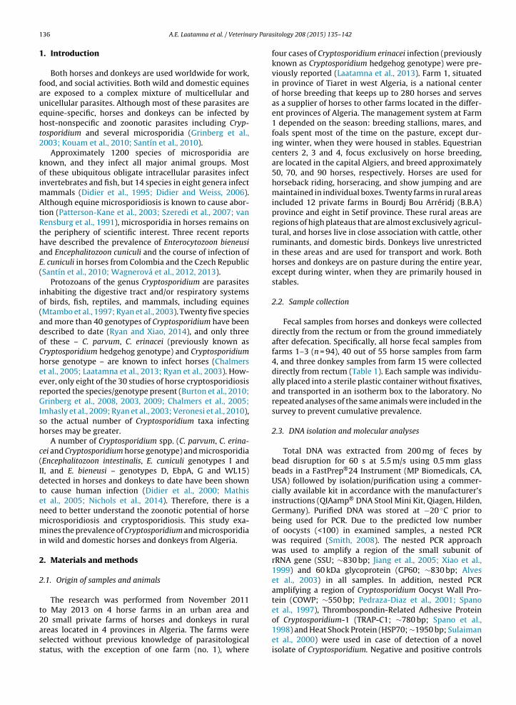
Fig. 1. Neighbor-joining trees depicting phylogenetic relationships among Cryptosporidium spp. and genotypes, including those detected in present study(highlighted), inferred from a partial fragment of (A) small subunit of ribosomal gene (743 base positions in the final dataset), (B) COWP (384 base positionsin the final dataset), (C) TRAP-C1 (523 base positions in the final dataset), (D) GP60 (859 base positions in the final dataset) and (E) HSP70 (277 basepositions in the final dataset) genes. The Kimura 2-parameter (K2P) model was used in all trees. The percentage of replicate trees in which the associatedtaxa clustered together in the bootstrap test (1000 replicates). Numbers at the nodes represent bootstrap values for the nodes gaining more than 50%support. Scale bar included in each tree.

140 A.E. Laatamna et al. / Veterinary Parasitology 208 (2015) 135–142
Table 2Distribution of Cryptosporidium spp. and microsporidia in horses and donkeys by housing management, age and sex.
Animal Variable Cryptosporidium Microsporidia
n No. of positive animals (%) No. of positive animals (%)
Horses
GenderMale 98 3 (3.7) 8 (8.2)Female 121 2 (1.7) 11 (9.1)
Age>3 yr 147 2 (1.4) 10 (6.8)6 mth–3 yr 26 1(3.8) 2 (7.7)<6 mth 46 2 (4.3) 7 (15.2)
HousingCommercial 149 3 (2.0) 12 (8.1)Rural areas 70 2 (2.9) 7 (10.0)
Donkeys
GenderMale 11 0 (0.0) 0 (0.0)Female 113 2 (1.8) 4 (3.5)
Age
>3 yr 1146 mth–3 yr 2
<6 mth 8
fecal samples on all farms and the distribution by age cate-gory and housing system is described in Table 2. Although E.bieneusi was 2.5-times more likely to occur in horses thandonkeys, the difference was not statistically different. Inaddition, the prevalence of microsporidial genera in horsesand donkeys showed no sex, age, or management depend-ent association (all resulted in non-significant odds ratios;P > 0.05).
4. Discussion
The prevalence of Cryptosporidium and microsporidial inhorses and donkeys in Algeria was low in this study. Lowoccurrence of microsporida is in agreement with previousstudies performed in Colombia and Czech Republic (Santínet al., 2010; Wagnerová et al., 2012). The low prevalenceof Cryptosporidium is in agreement with most previousstudies (e.g. Cole et al., 1998; De Souza et al., 2009; Epeet al., 2004; Laatamna et al., 2013; Majewska et al., 2004,1999; Sturdee et al., 2003). In contrast, some studies havereported prevalence rates between 10–31% (Caffara et al.,2013; Grinberg et al., 2009; Netherwood et al., 1996; Xiaoand Herd, 1994). However, relative to cattle and pigs, whereprevalence often exceeds 50–60%, and cumulative preva-lence approaches 100% (e.g. Sak et al., 2008; Santín et al.,2008), horses are rarely infected with Cryptosporidium andmicrosporidia. While most previous studies have shown ahigher prevalence of Cryptosporidium, primarily C. parvum,in foals worldwide (see Burton et al., 2010), any variabilityin presence of Cryptosporidium spp. between age categorieswas not observed in this study. Similarly, this study foundthat Cryptosporidium prevalence did not differ betweenmales and females. Santín et al. (2010) found a signifi-cantly higher prevalence of E. bieneusi in horses youngerthan 1 year of age; however, similar to Wagnerová et al.(2012), any dependence of microsporidial presence on age
of equines was not detected in the present study. In addi-tion, microsporidia prevalence did not differ between thesexes in the present study, which is consistent with previ-ous findings in horses (Santín et al., 2010).2 (1.8) 4 (3.5)0 (0.0) 0 (0.0)0 0
Cryptosporidium parvum is the only Cryptosporidiumspp. reported to cause diarrheal disease in horses:similar to cattle, disease is associated with younger animals(Caffara et al., 2013; Grinberg et al., 2009; Perrucci et al.,2011). None of the species identified in this study, includingC. parvum, was associated with diarrheal disease in horsesor donkeys. The association between microsporidia infec-tion and diarrhea in horses remains unclear. No signs ofdiarrhea were reported in microsporidia positive horsesin the present and previous studies (Santín et al., 2010;Wagnerová et al., 2012). Microsporidia, primarily E. cuni-culi, has been reported to cause reproductive problemsin horses, including placentitis, abortion, and stillbirth(Patterson-Kane et al., 2003; van Rensburg et al., 1991).
This is the first report of C. muris DNA detected in horsefeces. Although this species has been found in wide spec-trum of vertebrate hosts including mammals, birds, andreptiles (Kvác et al., 2014), it is considered to be relativelyspecific for some rodents and camels and its presence inhosts such as pigs, snakes, and raptors is due to mechan-ical transport (Kvác et al., 2012). By targeting DNA in thepresent study, it was not possible to differentiate activeinfections from passive carriage. Further research will berequired to determine the extent to which horses can beactively infected with C. muris.
Cryptosporidium parvum belonging to gp60 family IIa,subtypes A15G2R1 and A18G3R1, have been reported pre-viously in horses (Díaz et al., 2012; Grinberg et al., 2008).Cryptosporidium parvum isolates from horses and donkeysin the present study were identified as IIaA16G1R1 sub-type at gp60 locus. Although this subtype is common indomestic ruminants and humans worldwide (Herges et al.,2012; Robertson et al., 2014), this is the first report on itsoccurrence in horses and donkeys.
Considering the high specificity of C. hominis forhumans, detecting this species in horses (isolate 12328) in
the present study was unexpected. Based on SSU data, it ispossible that isolate 12328, which shares 99.4% similaritywith C. hominis and 99.3% similarity with C. cuniculus,represents a distinct species/genotype. There is precedent
ry Para
frssiCisosHAfCrtaCm1
oooigbtnm2WbColkb(
fttg1Wt
shWfwfigzinfw
A.E. Laatamna et al. / Veterina
or closely related Cryptosporidium SSU genotypes beingecognized as separate species: C. andersoni and C. murishare 99.1% similarity, and C. bovis and C. xiaoi share 99.5%imilarity at the SSU locus. However, the findings thatsolate 12328 did not differ from C. hominis at the HSP70,OWP, and TRAP-C1 loci supports the conclusion that
t is C. hominis. Other Cryptosporidium spp. varies intra-pecifically at the SSU locus, probably due to the presencef divergent gene paralogs; for example, C. ubiquitum SSUequences share 96.3–99.8% similarity, although COWP,SP70, and actin sequences of this species are identical.t the gp60 locus, isolate 12328 formed a novel subtype
amily, Ik, which clustered with C. cuniculus family Vb and. hominis monkey genotype family Ii. Isolate 12328 couldepresent a recently horse-adapted lineage of C. hominishat has not yet diverged sufficiently to be recognized as
separate species. It is worth noting that rabbit-adapted. cuniculus and human-adapted C. hominis are onlyarginally more divergent at the HSP70 locus, and share
00% similarity at the COWP locus.A phylogenetic analysis of all ITS sequences, performed
n a multiple alignment that included representativesf E. bieneusi genotypes accessible in GenBank, revealedccurrences of E. bieneusi genotypes previously reportedn horses (D, CZ3, Horse 1 and 2). With the exceptionenotype horse 2, all genotypes detected in this studyelonged to group 1, which comprises numerous geno-ypes from diverse hosts worldwide, including human andon-human primates, and domestic, captive, and wild ani-als (Thellier and Breton, 2008). Genotypes horse 1 and
have been detected only in horses (Santín et al., 2006;agnerová et al., 2012). In contrast, genotype CZ3 also has
een described from mice and humans in Germany andzech Republic, respectively, and genotype D, which is onef the most widespread genotypes, has been reported in ateast 15 hosts, including human, gorilla, cynomolgus mon-ey, cattle, alpaca, domestic pig, wild boar, cat, fox, dog,eaver, muskrat, raccoon, falcon, and pigeons worldwideSak et al., 2011; Santín and Fayer, 2011).
Genotype horse 1 (detected in 6 horses) occurred morerequently than genotype horse 2 (detected in 1 horse onhe same farm) in the present study. This is consistent withhe findings of a study of South American horses, whereenotypes horse 1 and horse 2 were found in 62% and9% of horses, respectively (Santín et al., 2010). In contrast,agnerová et al. (2012) detected genotype horse 2 at twice
he frequency of genotype horse 1 in the Czech Republic.Analysis of the ITS region of Encephalitozoon spp.
howed the presence of all three E. cuniculi genotypes inorses; whereas only genotype II was found in donkeys.hile E. cuniculi genotype I was found on suburban farms
ocused exclusively on horse breeding, genotypes II and IIIere detected in rural areas. Wagnerová et al. (2012) also
ound differences in the distribution of E. cuniculi genotypesn horses: genotype I predominated in stabled horses andenotype II predominated in horses on pasture. Encephali-ozoon cuniculi genotypes infect a wide range of mammals,
ncluding rodents, rabbits, carnivores, swine, human andonhuman primates, and birds (Mathis et al., 2005); there-ore, differences in genotype distribution may be associatedith different sources of infection.
sitology 208 (2015) 135–142 141
5. Conclusion
The finding of C. hominis and C. muris in horses increasesthe number of Cryptosporidium spp. reported in horsesfrom three to five. Although the relationship betweenCryptosporidium and microsporidia of horses and diseaseremains unclear, this study has shown that horses anddonkeys have the potential to transmit human-pathogenicparasites. However, given the low prevalence of all parasitictaxa, this risk appears to be low.
Acknowledgements
The authors would like to thank Hadouch Zohra(responsible veterinarian in the farm of Tiaret), SamariHousam (private veterinarian in the province of BBA),Badeh Nadir (private veterinarian in the rural area OuledBraheme) for their participation on material sampling. Thisstudy was funded by the Grant of the Czech Science Foun-dation (15-01090S) and project of the Grant Agency ofUniversity of South Bohemia (011/2013/Z). The opinionsexpressed by authors contributing to this journal do notnecessarily reflect the opinions of the Centers for DiseaseControl and Prevention or the institutions with which theauthors are affiliated.
References
Alves, M., Xiao, L.H., Sulaiman, I., Lal, A.A., Matos, O., Antunes, F., 2003.Subgenotype analysis of Cryptosporidium isolates from humans, cattle,and zoo ruminants in Portugal. J. Clin. Microbiol. 41, 2744–2747.
Buckholt, M.A., Lee, J.H., Tzipori, S., 2002. Prevalence of Enterocytozoonbieneusi in swine: an 18-month survey at a slaughterhouse in Mas-sachusetts. Appl. Environ. Microbiol. 68, 2595–2599.
Burton, A.J., Nydam, D.V., Dearen, T.K., Mitchell, K., Bowman, D.D., Xiao,L., 2010. The prevalence of Cryptosporidium, and identification of theCryptosporidium horse genotype in foals in New York State. Vet. Para-sitol. 174, 139–144.
Caffara, M., Piva, S., Pallaver, F., Iacono, E., Galuppi, R., 2013. Molecularcharacterization of Cryptosporidium spp. from foals in Italy. Vet. J. 198,531–533.
Chalmers, R.M., Thomas, A.L., Butler, B.A., Morel, M.C., 2005. Identificationof Cryptosporidium parvum genotype 2 in domestic horses. Vet. Rec.156, 49–50.
Cole, D.J., Cohen, N.D., Snowden, K., Smith, R., 1998. Prevalence of and riskfactors for fecal shedding of Cryptosporidium parvum oocysts in horses.J. Am. Vet. Med. Assoc. 213, 1296–1302.
De Souza, P.N., Bomfim, T.C., Huber, F., Abboud, L.C., Gomes, R.S., 2009. Nat-ural infection by Cryptosporidium sp., Giardia sp. and Eimeria leuckartiin three groups of equines with different handlings in Rio de Janeiro.Braz. Vet. Parasitol. 160, 327–333.
Díaz, P., Castagnetti, C., Marchesi, B., Soilán, M., López, C.M., Díez-Banos, P.,Morrondo, P., Poglayen, G., 2012. Investigation of the zoonotic poten-tial of Cryptosporidium in a diarrhoeic foal. In: Mappe ParassitologicheXXVII Congresso Nazionale Società Italiana di Parassitologia (Alghero),p. 251.
Didier, E.S., Didier, P.J., Snowden, K.F., Shadduck, J.A., 2000. Microsporid-iosis in mammals. Microbes Infect./Inst. Pasteur 2, 709–720.
Didier, E.S., Vossbrinck, C.R., Baker, M.D., Rogers, L.B., Bertucci, D.C.,Shadduck, J.A., 1995. Identification and characterization of threeEncephalitozoon cuniculi strains. Parasitology 111 (Pt 4), 411–421.
Didier, E.S., Weiss, L.M., 2006. Microsporidiosis: current status. Curr. Opin.Infect. Dis. 19, 485–492.
Epe, C., Coati, N., Schnieder, T., 2004. Results of parasitological exami-nations of faecal samples from horses, ruminants, pigs, dogs, cats,hedgehogs and rabbits between 1998 and 2002. Dtsch. Tierarztl.
Wochenschr. 111, 243–247.Grinberg, A., Learmonth, J., Kwan, E., Pomroy, W., Lopez Villalobos, N.,Gibson, I., Widmer, G., 2008. Genetic diversity and zoonotic potentialof Cryptosporidium parvum causing foal diarrhea. J. Clin. Microbiol. 46,2396–2398.

ry Para
142 A.E. Laatamna et al. / VeterinaGrinberg, A., Oliver, L., Learmonth, J.J., Leyland, M., Roe, W., Pomroy, W.E.,2003. Identification of Cryptosporidium parvum ‘cattle’ genotype froma severe outbreak of neonatal foal diarrhoea. Vet. Rec. 153, 628–631.
Grinberg, A., Pomroy, W.E., Carslake, H.B., Shi, Y., Gibson, I.R., Drayton, B.M.,2009. A study of neonatal cryptosporidiosis of foals in New Zealand.N. Z. Vet. J. 57, 284–289.
Herges, G.R., Widmer, G., Clark, M.E., Khan, E., Giddings, C.W., Brewer,M., McEvoy, J.M., 2012. Evidence that Cryptosporidium parvum popu-lations are panmictic and unstructured in the Upper Midwest of theUnited States. Appl. Environ. Microbiol. 78, 8096–8101.
Imhasly, A., Frey, C.F., Mathis, A., Straub, R., Gerber, V., 2009. Cryptosporid-iose (C. parvum) in a foal with diarrhea. Schweiz. Arch. Tierheilkd. 151,21–26.
Jiang, J., Alderisio, K.A., Xiao, L., 2005. Distribution of Cryptosporidiumgenotypes in storm event water samples from three watersheds inNew York. Appl. Environ. Microbiol. 71, 4446–4454.
Katoh, K., Standley, D.M., 2013. MAFFT multiple sequence alignment soft-ware version 7: improvements in performance and usability. Mol. Biol.Evol. 30, 772–780.
Katzwinkel-Wladarsch, S., Lieb, M., Helse, W., Loscher, T., Rinder, H.,1996. Direct amplification and species determination of microsporid-ian DNA from stool specimens. Trop. Med. Int. Health 1, 373–378.
Kimura, M., 1980. A simple method for estimating evolutionary ratesof base substitutions through comparative studies of nucleotidesequences. J. Mol. Evol. 16, 111–120.
Kouam, M.K., Diakou, A., Kanzoura, V., Papadopoulos, E., Gajadhar, A.A.,Theodoropoulos, G., 2010. A seroepidemiological study of expo-sure to Toxoplasma, Leishmania, Echinococcus and Trichinella inequids in Greece and analysis of risk factors. Vet. Parasitol. 170,170–175.
Kvác, M., Kestránová, M., Kvetonová, D., Kotková, M., Ortega, Y., McEvoy,J., Sak, B., 2012. Cryptosporidium tyzzeri and Cryptosporidium murisoriginated from wild West-European house mice (Mus musculusdomesticus) and East-European house mice (Mus musculus musculus)are non-infectious for pigs. Exp. Parasitol. 131, 107–110.
Kvác, M., McEvoy, J., Stenger, B., Clark, M., 2014. Cryptosporidiosis in othervertebrates. In: Cacciò, S.M., Widmer, G. (Eds.), Cryptosporidium: Par-asite and Disease. Springer, pp. 237–326.
Laatamna, A.E., Wagnerová, P., Sak, B., Kvetonová, D., Aissi, M., Rost,M., Kvác, M., 2013. Equine cryptosporidial infection associated withCryptosporidium hedgehog genotype in Algeria. Vet. Parasitol. 197,350–353.
Majewska, A.C., Solarczyk, P., Tamang, L., Graczyk, T.K., 2004. Equine Cryp-tosporidium parvum infections in western Poland. Parasitol. Res. 93,274–278.
Majewska, A.C., Werner, A., Sulima, P., Luty, T., 1999. Survey on equinecryptosporidiosis in Poland and the possibility of zoonotic transmis-sion. Ann. Agric. Environ. Med. 6, 161–165.
Mathis, A., Weber, R., Deplazes, P., 2005. Zoonotic potential of themicrosporidia. Clin. Microbiol. Rev. 18, 423–445.
Mtambo, M.M., Sebatwale, J.B., Kambarage, D.M., Muhairwa, A.P., Maeda,G.E., Kusiluka, L.J., Kazwala, R.R., 1997. Prevalence of Cryptosporidiumspp. oocysts in cattle and wildlife in Morogoro region, Tanzania. Prev.Vet. Med. 31, 185–190.
Netherwood, T., Wood, J.L.N., Townsend, H.G.G., Mumford, J.A., Chanter,N., 1996. Foal diarrhoea between 1991 and 1994 in the United King-dom associated with Clostridium perfringens, rotavirus, Strongyloideswesteri and Cryptosporidium spp. Epidemiol. Infect. 117, 375–383.
Nichols, G.L., Chalmers, R.M., Hadfield, S.J., 2014. Molecular epidemiol-ogy of human cryptosporidiosis. In: Cacciò, S.M., Widmer, G. (Eds.),Cryptosporidium: Parasite and Disease. Springer, pp. 237–326.
Patterson-Kane, J.C., Caplazi, P., Rurangirwa, F., Tramontin, R.R., Wolfs-dorf, K., 2003. Encephalitozoon cuniculi placentitis and abortion in aquarterhorse mare. J. Vet. Diagn. Invest. 15, 57–59.
Pedraza-Diaz, S., Amar, C., Nichols, G.L., McLauchlin, J., 2001. Nested poly-merase chain reaction for amplification of the Cryptosporidium oocystwall protein gene. Emerg. Infect. Dis. 7, 49–56.
Perrucci, S., Buggiani, C., Sgorbini, M., Cerchiai, I., Otranto, D., Traversa, D.,
2011. Cryptosporidium parvum infection in a mare and her foal withfoal heat diarrhoea. Vet. Parasitol. 182, 333–336.Robertson, L.J., Björkman, C., Axén, C., Fayer, R., 2014. Cryptosporidiosis infarmed animals. In: Cacciò, S.M., Widmer, G. (Eds.), Cryptosporidium:Parasite and Disease. Springer, pp. 149–236.
sitology 208 (2015) 135–142
Ryan, U., Xiao, L., 2014. Taxonomy and molecular taxonomy. In: Cac-ciò, S.M., Widmer, G. (Eds.), Cryptosporidium: Parasite and Disease.Springer, pp. 3–42.
Ryan, U., Xiao, L., Read, C., Zhou, L., Lal, A.A., Pavlásek, I., 2003. Identificationof novel Cryptosporidium genotypes from the Czech Republic. Appl.Environ. Microbiol. 69, 4302–4307.
Saitou, N., Nei, M., 1987. The neighbor-joining method: a new method forreconstructing phylogenetic trees. Mol. Biol. Evol. 4, 406–425.
Sak, B., Brady, D., Pelikánová, M., Kvetonová, D., Rost, M., Kostka, M.,Tolarová, V., Huzová, Z., Kvác, M., 2011. Unapparent microsporidialinfection among immunocompetent humans in the Czech Republic. J.Clin. Microbiol. 49, 1064–1070.
Sak, B., Kvác, M., Hanzlíková, D., Cama, V., 2008. First report of Entero-cytozoon bieneusi infection on a pig farm in the Czech Republic. Vet.Parasitol. 153, 220–224.
Santín, M., Fayer, R., 2011. Microsporidiosis: Enterocytozoon bieneusi indomesticated and wild animals. Res. Vet. Sci. 90, 363–371.
Santín, M., Trout, J.M., Fayer, R., 2008. A longitudinal study of cryptospo-ridiosis in dairy cattle from birth to 2 years of age. Vet. Parasitol. 155,15–23.
Santín, M., Trout, J.M., Vecino, J.A., Dubey, J.P., Fayer, R., 2006.Cryptosporidium Giardia and Enterocytozoon bieneusi in cats fromBogota (Colombia) and genotyping of isolates. Vet. Parasitol. 141,334–339.
Santín, M., Vecino, J.A., Fayer, R., 2010. A zoonotic genotype of Enterocyto-zoon bieneusi in horses. J. Parasitol. 96, 157–161.
Smith, H., 2008. Diagnostics. In: Fayer, R., Xiao, L. (Eds.), Cryptosporidiumand Cryptosporidiosis. CRC Press, Boca Raton, FL, pp. 173–208.
Spano, F., Putignani, L., McLauchlin, J., Casemore, D.P., Crisanti, A., 1997.PCR-RFLP analysis of the Cryptosporidium oocyst wall protein (COWP)gene discriminates between C. wrairi and C. parvum, and between C.parvum isolates of human and animal origin. FEMS Microbiol. Lett.150, 209–217.
Spano, F., Putignani, L., Naitza, S., Puri, C., Wright, S., Crisanti, A., 1998.Molecular cloning and expression analysis of a Cryptosporidiumparvum gene encoding a new member of the thrombospondin family.Mol. Biochem. Parasitol. 92, 147–162.
Sturdee, A.P., Bodley-Tickell, A.T., Archer, A., Chalmers, R.M., 2003. Long-term study of Cryptosporidium prevalence on a lowland farm in theUnited Kingdom. Vet. Parasitol. 116, 97–113.
Sulaiman, I.M., Morgan, U.M., Thompson, R.C., Lal, A.A., Xiao, L., 2000.Phylogenetic relationships of Cryptosporidium parasites based on the70-kilodalton heat shock protein (HSP70) gene. Appl. Environ. Micro-biol. 66, 2385–2391.
Szeredi, L., Pospischil, A., Dencso, L., Mathis, A., Dobos-Kovacs, M., 2007. Acase of equine abortion caused by Encephalitozoon sp. Acta Vet. Hung.55, 525–532.
Tamura, K., Peterson, D., Peterson, N., Stecher, G., Nei, M., Kumar, S., 2011.MEGA5: molecular evolutionary genetics analysis using maximumlikelihood, evolutionary distance, and maximum parsimony methods.Mol. Biol. Evol. 28, 2731–2739.
Thellier, M., Breton, J., 2008. Enterocytozoon bieneusi in human and ani-mals, focus on laboratory identification and molecular epidemiology.Parasite 15, 349–358.
van Rensburg, I.B., Volkmann, D.H., Soley, J.T., Stewart, C.G., 1991.Encephalitozoon infection in a still-born foal. J. S. Afr. Vet. Assoc. 62,130–132.
Veronesi, F., Passamonti, F., Caccio, S., Diaferia, M., Piergili Fioretti, D.,2010. Epidemiological survey on equine cryptosporidium and giardiainfections in Italy and molecular characterization of isolates. ZoonosesPublic Health 57, 510–517.
Wagnerová, P., Sak, B., Kvetonová, D., Bunatová, Z., Civisová, H., Marsálek,M., Kvác, M., 2012. Enterocytozoon bieneusi and Encephalitozoon cuni-culi in horses kept under different management systems in the CzechRepublic. Vet. Parasitol. 190, 573–577.
Wagnerová, P., Sak, B., Kvetonová, D., Marsálek, M., Langrová, I., Kvác, M.,2013. Humoral immune response and spreading of Encephalitozooncuniculi infection in experimentally infected ponies. Vet. Parasitol.197, 1–6.
Xiao, L., Herd, R.P., 1994. Epidemiology of equine Cryptosporidium and
Giardia infections. Equine Vet. J. 26, 14–17.Xiao, L., Morgan, U.M., Limor, J., Escalante, A., Arrowood, M., Shulaw, W.,Thompson, R.C., Fayer, R., Lal, A.A., 1999. Genetic diversity withinCryptosporidium parvum and related Cryptosporidium species. Appl.Environ. Microbiol. 65, 3386–3391.