microwave assisted synthesis of ag-zno particles and … analysis of agzno ... composition was...
TRANSCRIPT

Microwave Assisted Synthesis of Ag-ZnO Particles and Their
Antibacterial Properties
BAZANT PAVEL1, 2
, KURITKA IVO1, 2
, MACHOVSKY MICHAL1, 2
, SEDLACEK TOMAS1, 2
PASTOREK MIROSLAV1, 2
1Centre of Polymer Systems, University Institute
Tomas Bata University in Zlín
Nad Ovcirnou 3685, 760 01 Zlín,
CZECH REPUBLIC 2 Polymer Centre, Faculty of Technology
Tomas Bata University in Zlín
Nam. T. G. Masaryka 275, 762 72 Zlín
CZECH REPUBLIC
[email protected] http://cps.utb.cz
Abstract: Hybrid Ag-ZnO micro-structured nanoparticles were prepared by two different microwave techniques
from silver nitrate and zinc nitrate within 15 minutes. Crystalline structures of obtained Ag-ZnO powders were
characterized by X-ray diffractometer. Scanning electron microscopy was used for characterization of structure,
morphology, particle size Ag-ZnO filler. Elemental analysis of Ag-ZnO filler was made by Energy dispersive
X-ray analysis. Growth mechanism of particles was elucidated. The antibacterial activity was evaluated by
inhibition zone test; one of the materials performed well against Staphylococcus aureus, and Candida albicans.
Key-Words: microwave synthesis, hybrid filler, Ag-ZnO, antibacterial activity
1 Introduction Hybrid materials, especially metal-modified oxide
semiconductors, are of great interest amongst the
researchers and bring interesting possibilities of
property design, thus, they open new application
fields. Materials that combine silver and zinc oxide
(ZnO) have attracted attention because they were
successfully used in chemical and biological
sensors, electronics and photoelectronics devices
and have considerable bio-activity as well. [1-3].
Nowadays, various techniques are studied for
preparation of novel Ag-ZnO hybrid systems and
their antibacterial activity is tested [4-6]. The strong
antibacterial effects of both metallic Ag and Ag+
ions have been known for a long time [7, 8]. Zinc
oxide is another inorganic agent which, in form of
nanoparticles, exhibit strong antibacterial activity on
a broad spectrum of bacteria although its effect on
micro-organisms is not fully understood yet [9].
Preparation of such hybrid systems can be
significantly enhanced by the introduction of
microwave (MW) into synthesis process.
Microwave irradiation has attracted wide interest as
heating mechanisms in materials synthesis due to its
many advantages, including a very short reaction
time, and the ability to produce small inorganic
particles with narrow particle size distribution and
high purity [3, 10, 11]. In this paper, two different
facile microwave preparation methods of Ag-ZnO
nanostructured microparticles are presented using
hexamethylenetetramine (HMT) as precipitation and
reducing agent, with further aim to obtain new
antibacterial additives for prospective applications
in polymer medical devices.
2 Experimental 2.1 Material Materials silver nitrate AgNO3 (purum, ≥99.5%)
and zinc nitrate hexahydrate Zn(NO3)2·6H2O
(purum, >99%) were purchased from Penta (Prague,
Czech Republic). Hexamethylenetetramine (HMT)
C6H12N4 (purum, >99%, Fluka) was purchased from
Sigma-Aldrich (Prague, Czech Republic) and used
as precipitation and reduction agent and growth
modifier. Demineralised water was used in these
experiments.
2.2 Synthesis of Ag-ZnO For preparation of powders two different microwave
apparatures were used.
First material (sample 1) was prepared in the
microwave open vessel system MWG1K-10 (Radan,
Czech Republic, 800W, 2.45 GHz) based on
Mathematical Methods and Techniques in Engineering and Environmental Science
ISBN: 978-1-61804-046-6 341

modification of domestic oven by drilling a hole on
the ceiling for external cooler and equipped with
external MW power source that enables operate the
instrument in continuous mode. All the chemical
reagents were dissolved in demineralised water. The
solution of 0.015 mol Zn(NO3)2·6H2O was mixed
together with solution of 0.01 mol AgNO3. The total
amount of used water was 100 mL. The obtained
solution was placed into the microwave oven cavity
and heated for 2 minutes then a solution of 0.015
mol C6H12N4 in 50 mL water was added quickly
through the dropping system and microwave heating
continued for another 15 minutes. Obtained
suspension was cooled freely to room temperature.
For preparation of the second material (sample 2)
a commercial pressurized MW system Mars5 (CEM
Corporation) was used. Identical solutions were
prepared in smaller amounts, as they needed to be
mixed prior synthesis and filled in a Teflon reaction
vessel (XP-1500 Plus), sealed and heated in the
microwave oven. The total amount of used water
was 60 mL. The reaction solution in the vessel was
irradiated by MW at moderate power of 400 W to
ensure temperature regulation. The solution was
heated to 90°C for 15 minutes. When the reaction
was completed, the vessel was left to cool naturally
and then unsealed. A suspension was obtained.
Both suspensions were filtered, washed with
demineralised water and finally collected powders
were dried in the air at 37°C for over night.
2.3 Characterization The crystalline phase structure of obtained powders
was characterized by X-ray diffractometer
PANalytical X´Pert PRO (PANalytical, The
Netherlands) using Cu Kα1 radiation (λ = 0.1542
nm) operating at 40 kV and 30 mA with detector
PIXcel. Both materials were measured in
transmission mode with fixed setting and screen of
range angle 25-85° (2θ) and step 0.0263°. The phase
composition was evaluated by the software
PANalytical X'Pert High Score using normalized
RIR method. The RIR is the ratio between the
integrated intensities of the peak of interest and that
of a known standard [11].
The morphology of the products was investigated by
scanning electron microscope Vega II LMU
(Tescan, Czech Republic) with beam acceleration
voltage set at 10 kV, after coating with
gold/palladium by a high-resolution SEM sputter
coater SC 7640 (Quorum Technologies Ltd, UK).
SEM equipment includes Energy dispersive X-ray
analyser (Oxford INCA) used for elemental
analysis.
2.4 Testing antibacterial activity Antibacterial testing was performed against
Staphylococcus aureus CCM 4516, Escherichia coli
CCM 4517 and Candida albicans CCM 8215.
Amount of 0.1 g of obtained Ag-ZnO powder was
dispersed in 1 mL demineralised water and
homogenised in ultrasonic bath for 15 minutes.
Then 10 µL of suspension was applied on filter
paper with diameter 8 mm. Prepared specimens
were dried at the room temperature. Specimens
(diameter 8 mm) were put into nutrient agar which
was inoculated with bacteria (approximately 108
colony forming unit (CFU) per millilitre). The
bacteria used in this study were S. aureus (CCM
4517), E. coli and C. albicans (CCM 8215). After
24 h incubation at 37°C, the diameters of the
inhibition zones, which appeared on the surface,
were measured in five directions. Average values
were used for calculation of the inhibition zone area,
which was the measure of the antibacterial activity
of the studied samples. Each test was conducted
with six specimens. Blank samples without
suspension were prepared and tested in the same
way.
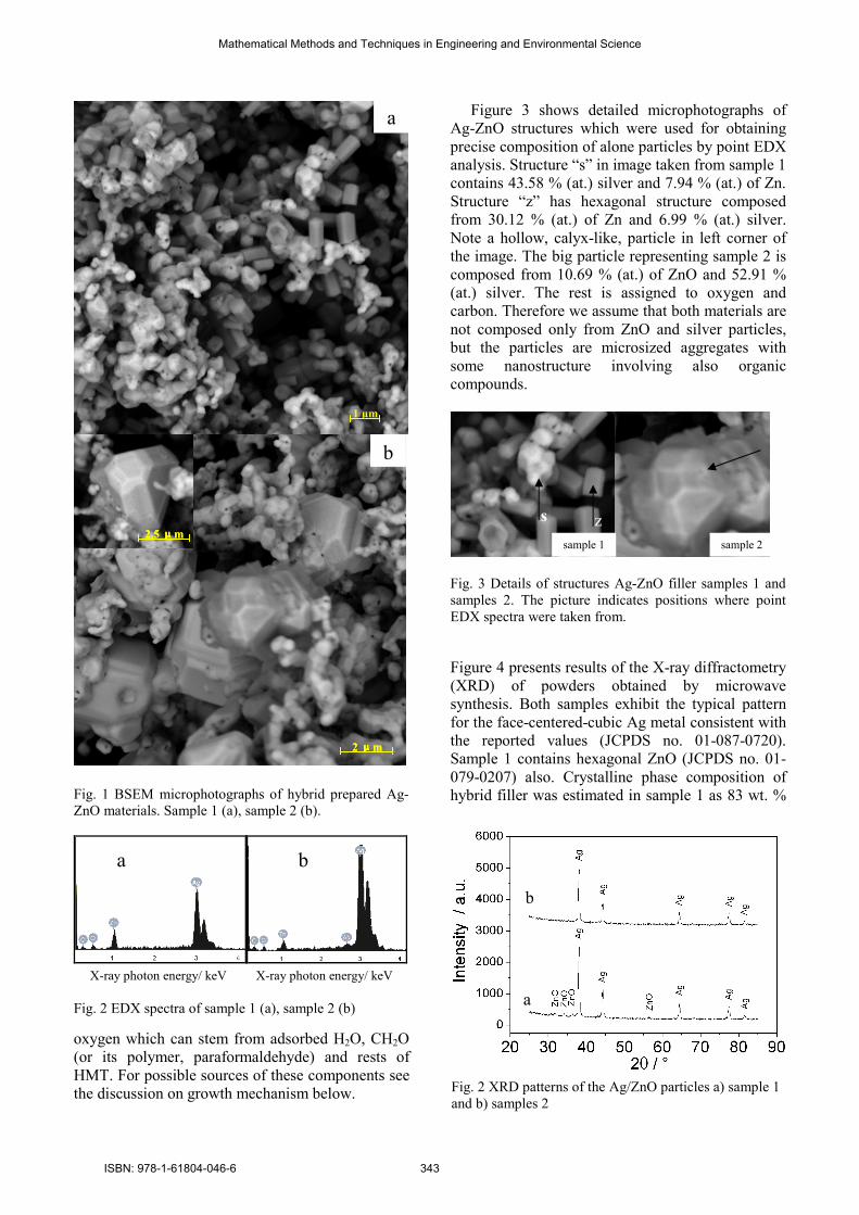
3 Results and discussion 3.1 Morphology and structure of prepared
Ag-ZnO particles SEM microphotographs are shown in Figure 1a and
1b. The images were taken by BSE detector that
allows distinguish composition of particles by
material (greyscale) contrast showing heavier
elements brighter. For sample 1 (a) it is apparent,
that the zinc oxide particles are hexagonal
microrods structures with the size up to 2 µm and
silver structures are aggregates of spherical particles
with the diameter up to 200 nm. In sample 2 (b) Ag-
ZnO particles of polyhedral shape with size up to 3
µm together with similar particle aggregates as in
sample 1. In left top corner of Figure 1b can be seen
a detail of one bigger particle revealing its
hexagonal symmetry. This kind of pyramid-like
particles can be considered as the final product of
the reaction, as similar growth was presented Huang
A.S. et al. (2010) who demonstrated grow of ZnO
hexagonal pyramids from hexagonal disks by
solvothermal reaction during 10 days. [16].
Figure 2 gives typical EDX spectra recorded for
the obtained powders. The EDX spectrum indicates
that samples 1 and 2 are composed of Ag, Zn, C and
O. Sample 1 contains 9.87 % (at.) of Zn and 32.36
% (at.) silver. Sample 2 contains 3.75 % (at.) of Zn
and 39.63 % (at.) of silver. Both materials contain
approximately 18% (at.) carbon and 40 % (at.)
Mathematical Methods and Techniques in Engineering and Environmental Science
ISBN: 978-1-61804-046-6 342
oxygen which can stem from adsorbed H2O, CH2O
(or its polymer, paraformaldehyde) and rests of
HMT. For possible sources of these components see
the discussion on growth mechanism below.
Figure 3 shows detailed microphotographs of
Ag-ZnO structures which were used for obtaining
precise composition of alone particles by point EDX
analysis. Structure “s” in image taken from sample 1
contains 43.58 % (at.) silver and 7.94 % (at.) of Zn.
Structure “z” has hexagonal structure composed
from 30.12 % (at.) of Zn and 6.99 % (at.) silver.
Note a hollow, calyx-like, particle in left corner of
the image. The big particle representing sample 2 is
composed from 10.69 % (at.) of ZnO and 52.91 %
(at.) silver. The rest is assigned to oxygen and
carbon. Therefore we assume that both materials are
not composed only from ZnO and silver particles,
but the particles are microsized aggregates with
some nanostructure involving also organic
compounds.
Fig. 3 Details of structures Ag-ZnO filler samples 1 and
samples 2. The picture indicates positions where point
EDX spectra were taken from.
Figure 4 presents results of the X-ray diffractometry
(XRD) of powders obtained by microwave
synthesis. Both samples exhibit the typical pattern
for the face-centered-cubic Ag metal consistent with
the reported values (JCPDS no. 01-087-0720).
Sample 1 contains hexagonal ZnO (JCPDS no. 01-
079-0207) also. Crystalline phase composition of
hybrid filler was estimated in sample 1 as 83 wt. %
2μμμμm
2.5 μμμμm
b
2μμμμm
2.5 μμμμm
2μμμμm2μμμμm
2.5 μμμμm2.5 μμμμm
b
Fig. 1 BSEM microphotographs of hybrid prepared Ag-
ZnO materials. Sample 1 (a), sample 2 (b).
X-ray photon energy/ keV X-ray photon energy/ keV
Fig. 2 EDX spectra of sample 1 (a), sample 2 (b)
Fig. 2 XRD patterns of the Ag/ZnO particles a) sample 1
and b) samples 2
a b
a
sample 2
s z
sample 1
b
a
1 µm
Mathematical Methods and Techniques in Engineering and Environmental Science
ISBN: 978-1-61804-046-6 343

of Ag and 17 wt. % of ZnO. Sample 2 was
characterized as 100 wt. % crystalline Ag. Overall
intensity of both observed spectra was relatively low
because X-ray diffraction patterns were measured in
transmitting mode; therefore diffraction lines
belonging to minor ZnO phase in sample 2 could be
hidden in background. However, these results seem
to be somewhat contradictory with respect to EDX
observation. It must be noted, that XRD allows to
analyse crystalline phase only, so it is reasonable to
expect, that at least certain portion of prepared
materials is amorphous, or the disorder introduced
by mutual doping of Ag and ZnO destroyed
periodicity of materials structure.
3.2 Mechanism of microwave assisted
synthesis Ag-ZnO powders were prepared by hydrothermal
microwave synthesis by two different microwave
devices, which involved the precipitation of ZnO
and the formation of Ag nanoparticles using HMT
as precipitation and reducing agent. During short
synthesis time were formed structures of Ag
containing ZnO with containing Ag nanoparticles.
Growth and morphology of ZnO and Ag particles
are dependent of many factors. Ashfold, M.N.R. et
al. 2007 describe the role of pH, zinc ion and HMT
concentration of the solution [13]. For controlled
growths of ZnO structure Baruah, S. and Dutta, J.,
2008 explain another parameters e.g. type of used
chemical (Zinc acetate, zinc nitrate and other),
reduction, precipitation agents or precursor and
surfactant (HMT, glucose, NH3, PEG, PVA, CTAB)
experimental parameters (temperature, time) and
describe basic role HMT [14]. This can be
summarized in the following equations:
(CH2)6N4 + 6H2O ↔ 6HCHO + 4NH3 (1)
NH3 +H2O ↔ NH4+ + OH
− (2)
2OH− + Zn
2+→ ZnO(s) + H2O (3)
Ye, X. Y. et al. 2008 refer that controlled growth
of ZnO structure can be influenced the presence Ag+
[4]. Zhang, Y.Y. and Mu, J. 2007 observed that the
morphology of ZnO varied from pillar-like to rod-
like with the increased concentration of Ag+. The
reactions involved in the formation of metallic silver
in basic conditions are believed to be as follows:
Ag+ + 2NH3 → [Ag(NH3)2]
+ (4)
HCHO + 2[Ag(NH3)2]+ + H2O →
HCOO- + 2Ag(s) + 3NH4
+ + NH3 (5)
The source of formaldehyde (reducing agent) is
decomposition of HMT at elevated temperature in
water solution as described in equation (1). The
addition of the silver nitrate changes the equilibrium
between zinc ions and ammonia due to the
coordination of silver ions by ammonia and
subsequently influences the morphology of ZnO and
efficiency of its growth. Increase the concentration
of HMT results in increase of the concentration of
ammonia in the reaction system, which will prevent
the influence of the silver ion on the equilibrium
between zinc ion and ammonia. [15]
The main difference between both synthesis
methods was the pressure in the reaction system. As
the sample 2 was prepared in sealed Teflon vessel it
is assumed that all chemical equilibria reactions
involving change in molarity were shifted to that
side where lower sum of stoichiometric indexes is.
Reactions (1) and (5) are shifted to the left side, the
reactions (3) and (4) shifts to the right side, reaction
(2) has the same number of moles on both sides.
Normally, elevated pressure would strongly enhance
reaction with solid phase formation, but it must not
be at the expenses of increase of other by-products
as in (5).
Observed composition of prepared materials, i.e.
higher content of Ag in sample 2 than in sample 1,
testifies for larger influence of silver nitrate addition
and competition for ammonia due to silver ions
complexation than the pressure effect on the
reaction system has, namely on HMT
decomposition (1) and ZnO formation (3), which
would prefer the growth of ZnO.
3.3 Antibacterial activity of Ag-ZnO filler Antibacterial activity of Ag-ZnO filler is shown in
Table 1. S. aureus represents gram positive bacteria
and seems to be sensitive towards sample 1,
whereas E. coli is a gram negative bacteria and did
not show inhibition zone. These two groups differ in
the structure of their cell walls since the cell walls of
gram positive bacteria contain more peptidoglycan
layers than gram negative bacteria. Gram positive
bacteria are more sensitive against ZnO than gram
negative. [17, 18] Sample 1 performed well also in
case of C. albicans. However, no effect of silver
present in sample 2 was observed on all tested
organisms. It can be expected that there is not
enough of free Ag+ ions available for diffusion
through agar plate to form the “halo” zone effect. To
complete the assessment of antibacterial properties
another more predicative test is needed which would
take into account contact inhibition effect of silver
surface on bacteria.
Mathematical Methods and Techniques in Engineering and Environmental Science
ISBN: 978-1-61804-046-6 344
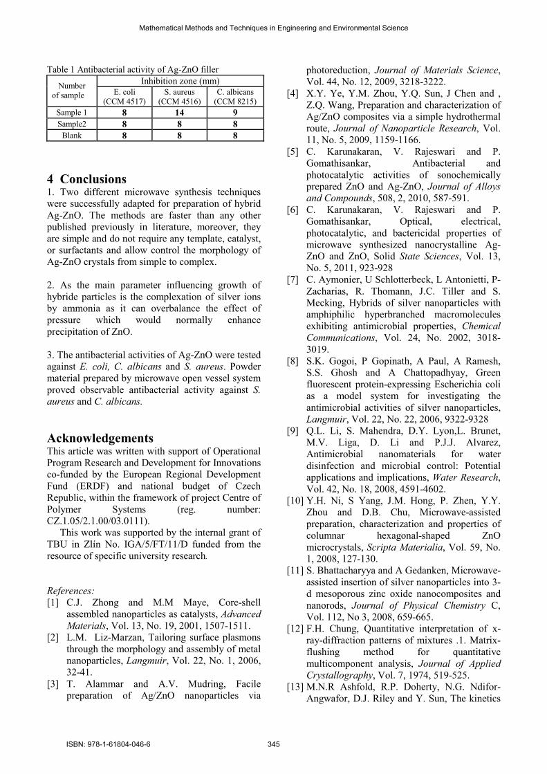
Table 1 Antibacterial activity of Ag-ZnO filler
Inhibition zone (mm) Number
of sample E. coli
(CCM 4517)
S. aureus
(CCM 4516)
C. albicans
(CCM 8215)
Sample 1 8 14 9
Sample2 8 8 8
Blank 8 8 8
4 Conclusions 1. Two different microwave synthesis techniques
were successfully adapted for preparation of hybrid
Ag-ZnO. The methods are faster than any other
published previously in literature, moreover, they
are simple and do not require any template, catalyst,
or surfactants and allow control the morphology of
Ag-ZnO crystals from simple to complex.
2. As the main parameter influencing growth of
hybride particles is the complexation of silver ions
by ammonia as it can overbalance the effect of
pressure which would normally enhance
precipitation of ZnO.
3. The antibacterial activities of Ag-ZnO were tested
against E. coli, C. albicans and S. aureus. Powder
material prepared by microwave open vessel system
proved observable antibacterial activity against S.
aureus and C. albicans.
Acknowledgements This article was written with support of Operational
Program Research and Development for Innovations
co-funded by the European Regional Development
Fund (ERDF) and national budget of Czech
Republic, within the framework of project Centre of
Polymer Systems (reg. number:
CZ.1.05/2.1.00/03.0111).
This work was supported by the internal grant of
TBU in Zlín No. IGA/5/FT/11/D funded from the
resource of specific university research.
References:
[1] C.J. Zhong and M.M Maye, Core-shell
assembled nanoparticles as catalysts, Advanced
Materials, Vol. 13, No. 19, 2001, 1507-1511.
[2] L.M. Liz-Marzan, Tailoring surface plasmons
through the morphology and assembly of metal
nanoparticles, Langmuir, Vol. 22, No. 1, 2006,
32-41.
[3] T. Alammar and A.V. Mudring, Facile
preparation of Ag/ZnO nanoparticles via
photoreduction, Journal of Materials Science,
Vol. 44, No. 12, 2009, 3218-3222.
[4] X.Y. Ye, Y.M. Zhou, Y.Q. Sun, J Chen and ,
Z.Q. Wang, Preparation and characterization of
Ag/ZnO composites via a simple hydrothermal
route, Journal of Nanoparticle Research, Vol.
11, No. 5, 2009, 1159-1166.
[5] C. Karunakaran, V. Rajeswari and P.
Gomathisankar, Antibacterial and
photocatalytic activities of sonochemically
prepared ZnO and Ag-ZnO, Journal of Alloys
and Compounds, 508, 2, 2010, 587-591.
[6] C. Karunakaran, V. Rajeswari and P.
Gomathisankar, Optical, electrical,
photocatalytic, and bactericidal properties of
microwave synthesized nanocrystalline Ag-
ZnO and ZnO, Solid State Sciences, Vol. 13,
No. 5, 2011, 923-928
[7] C. Aymonier, U Schlotterbeck, L Antonietti, P-
Zacharias, R. Thomann, J.C. Tiller and S.
Mecking, Hybrids of silver nanoparticles with
amphiphilic hyperbranched macromolecules
exhibiting antimicrobial properties, Chemical
Communications, Vol. 24, No. 2002, 3018-
3019.
[8] S.K. Gogoi, P Gopinath, A Paul, A Ramesh,
S.S. Ghosh and A Chattopadhyay, Green
fluorescent protein-expressing Escherichia coli
as a model system for investigating the
antimicrobial activities of silver nanoparticles,
Langmuir, Vol. 22, No. 22, 2006, 9322-9328
[9] Q.L. Li, S. Mahendra, D.Y. Lyon,L. Brunet,
M.V. Liga, D. Li and P.J.J. Alvarez,
Antimicrobial nanomaterials for water
disinfection and microbial control: Potential
applications and implications, Water Research,
Vol. 42, No. 18, 2008, 4591-4602.
[10] Y.H. Ni, S Yang, J.M. Hong, P. Zhen, Y.Y.
Zhou and D.B. Chu, Microwave-assisted
preparation, characterization and properties of
columnar hexagonal-shaped ZnO
microcrystals, Scripta Materialia, Vol. 59, No.
1, 2008, 127-130.
[11] S. Bhattacharyya and A Gedanken, Microwave-
assisted insertion of silver nanoparticles into 3-
d mesoporous zinc oxide nanocomposites and
nanorods, Journal of Physical Chemistry C,
Vol. 112, No 3, 2008, 659-665.
[12] F.H. Chung, Quantitative interpretation of x-
ray-diffraction patterns of mixtures .1. Matrix-
flushing method for quantitative
multicomponent analysis, Journal of Applied
Crystallography, Vol. 7, 1974, 519-525.
[13] M.N.R Ashfold, R.P. Doherty, N.G. Ndifor-
Angwafor, D.J. Riley and Y. Sun, The kinetics
Mathematical Methods and Techniques in Engineering and Environmental Science
ISBN: 978-1-61804-046-6 345

of the hydrothermal growth of ZnO
nanostructures, Thin Solid Films, Vol. 515, No.
24, 2007, 8679-8683.
[14] S. Baruah and J Dutta, Hydrothermal growth of
ZnO nanostructures, Science and Technology of
Advanced Materials, 2009, Vol. 10, No. 1.
[15] Y.Y. Zhang and J. Mu, One-pot synthesis,
photoluminescence, and photocatalysis of
Ag/ZnO composites. Journal of Colloid and
Interface Science, 309, 2, 2007, 478-484.
[16] A.S. Huang and J. Caro, Controlled growth of
zinc oxide crystals with tunable shape, Journal
of Crystal Growth, Vol. 312, No. 7, 2010, 947-
952.
[17] T. Galya, V. Sedlarik, I Kuritka, J. Sedlarikova
and P. Saha, Characterization of antibacterial
polymeric films based on poly(vinyl alcohol)
and zinc nitrate for biomedical applications,
International Journal of Polymer Analysis and
Characterization, Vol. 13, No. 4, 2008, 241-
253.
[18] V. Sedlarik, T. Galya, J Sedlarikova, P.
Valasek and P. Saha, The effect of preparation
temperature on the mechanical and antibacterial
properties of poly(vinyl alcohol)/silver nitrate
films Polymer Degradation and Stability, , Vol.
95, No. 3, 2010, 399-404
Mathematical Methods and Techniques in Engineering and Environmental Science
ISBN: 978-1-61804-046-6 346