minimal sequences necessary for imprinted expression...
TRANSCRIPT

MINIMAL SEQUENCES NECESSARY FOR IMPRINTED EXPRESSION OF THE
PRADER-WILLI LOCUS
By
CHRISTOPHER FUTTNER
A DISSERTATION PRESENTED TO THE GRADUATE SCHOOL OF THE UNIVERSITY OF FLORIDA IN PARTIAL FULFILLMENT
OF THE REQUIREMENTS FOR THE DEGREE OF DOCTOR OF PHILOSOPHY
UNIVERSITY OF FLORIDA
2007

Copyright 2007
by
Christopher R. Futtner

iii
ACKNOWLEDGMENTS
I would like to thank both past and present members of the lab, including Stormy
Chamberlain, Edwin Peery, Jessica Walrath, Mike Elmore, Amanda DuBose, Emily
Smith, Lori Kellam, and my favorite Scotswoman, Karen Johnstone.
I would also like to thank my mentor Jim Resnick, not only for his scientific
expertise, but also for his excellent advise on why old school is better than new school,
how to have a happy marriage, and why the Yankees are the best team in baseball.
I would like to thank my parents for their love and encouragement. Without them I
would be biologically impossible.
I am also extremely thankful to Cami Brannan. The experiments contained within
this work would not have been possible without her ideas and vision.
Lastly, I would like to thank my lovely wife, Danielle Maatouk, whose scientific
coattails I will be riding to greatness.

iv
TABLE OF CONTENTS page
ACKNOWLEDGMENTS ................................................................................................. iii
LIST OF FIGURES ........................................................................................................... vi
ABSTRACT...................................................................................................................... vii
CHAPTER
1 INTRODUCTION ........................................................................................................1
Genomic Imprinting......................................................................................................1 Prader-Willi and Angelman Syndromes.......................................................................4 Purpose of Imprinting ...................................................................................................6 The PWS/AS Locus......................................................................................................8
2 MATERIALS AND METHODS ...............................................................................17
Lambda Red Recombineering ....................................................................................17 Transformation of Recombineering Strains ........................................................17 Transformation of Targeting Vector and Induction of Recombination...............19
BAC preparation for injection ....................................................................................20 Generation of Transgenic Animals by Pronuclear Injection ......................................23 Genotyping .................................................................................................................23
Identification of Transgenic Founders.................................................................23 Identification of Castaneous C7 Homozygotes ...................................................23
Southern Blot ..............................................................................................................24 RNA Isolation.............................................................................................................25 RT-PCR ......................................................................................................................25 Preparation of High Molecular Weight Genomic DNA for Bisulfite Conversion .....26 Bisulphite Sequencing of Genomic DNA...................................................................27
Bisulphite Conversion .........................................................................................27 Bisulphite Polymerase Chain Reaction ...............................................................27 Purification of PCR Products ..............................................................................28 Cloning and Sequencing of PCR Products ..........................................................28
Sequencing..................................................................................................................29
3 IMPRINTED TRANSGENE......................................................................................31
v
Introduction.................................................................................................................31 Results.........................................................................................................................34
BAC Modification ...............................................................................................34 Production of Transgenic Mice ...........................................................................37 Analysis of 425Δ5−7 Transgenic Lines ..............................................................38
Discussion...................................................................................................................41
4 REFINEMENT OF THE PWS-IC..............................................................................50
Introduction.................................................................................................................50 Results.........................................................................................................................53
BAC Modification ...............................................................................................53 Production of Transgenic Mice ...........................................................................54 Analysis of 425Δ30kb Transgenic Lines.............................................................55
Discussion...................................................................................................................56
5 DEFININING THE LOCATION OF THE AS-IC.....................................................62
Introduction.................................................................................................................62 Results.........................................................................................................................67
BAC modification ...............................................................................................67 Production of Transgenic Mice ...........................................................................69 Analysis of 425ΔU1-U3 and 425ΔU2/U3 Transgenic Lines ..............................69
Discussion...................................................................................................................70
6 CONCLUSIIONS AND FUTURE DIRECTIONS....................................................79
LIST OF REFERENCES...................................................................................................82
BIOGRAPHICAL SKETCH .............................................................................................89
vi
LIST OF FIGURES
Figure page 1-1 The life cycle of an imprint. .....................................................................................12
1-2 Mapping the Imprinting Center (IC). .......................................................................13
1-3 Molecular classes of PWS and AS. ..........................................................................14
1-4 Gene organization of the PWS/AS locus.. ...............................................................15
1-5 Paternal only model of imprinting regulation.. ........................................................16
3-1 Schematic and expression of BAC transgenics........................................................44
3-2 425Δ5−7 targeting. ...................................................................................................45
3-3 rt-PCR analysis of 425Δ5−7mouse lines..................................................................46
3-4 Cast c7 breeding scheme.. ........................................................................................47
3-5 Bisulfite sequence analysis of maternally transmitted transgene.............................48
3-6 Bisulfite sequence analysis of paternally transmitted transgene ..............................49
4-1 Schematic diagram of three nested deletions aimed at minimizing the PWS-IC.....59
4-2 rt-PCR analysis of 425Δ30kb mouse lines. ..............................................................60
4-3 Bisulfite sequence analysis of maternally transmitted 425Δ30kb transgene. ..........61
5-1 Schematic diagram of human upstream exons .........................................................74
5-2 Schematic diagram of upstream exons in the mouse. ..............................................75
5-3 Compensation model of AS-IC activity. ..................................................................76
5-4 Schematic diagram of upstream exon deletions. ......................................................77
5-5 RT-PCR analysis of 425ΔU1-U3 mouse lines. ........................................................78
vii
Abstract of Dissertation Presented to the Graduate School of the University of Florida in Partial Fulfillment of the Requirements for the Degree of Doctor of Philosophy
MINIMAL SEQUENCES NECESSARY FOR IMPRINTED EXPRESSION OF THE PRADER-WILLI LOCUS
By
CHRISTOPHER FUTTNER
May 2007
Chair: James Resnick Major Department: Medical Sciences⎯Genetics
Prader-Willi (PWS) ands Angelman (AS) syndromes are neurodevelopmenal
disorders arising from the improper expression of oppositely imprinted genes located on
human chromosome 15 q11-q13. Imprint regulation of this region is under the control of
a bi-partite imprinting center consisting of an Angelman Imprinting Center (AS-IC)
located approximately 35kb upstream of the paternally expressed Snrpn exon 1 and a
Prader-Willi Imprinting Center (PWS-IC) located 5’ to and including Snrpn exon 1. The
PWS-IC has been shown to be a positive element promoting expression of a set of genes
on the paternal allele, while the AS-IC provides suppression of the PWS-IC on the
maternal allele thereby suppressing expression of the same set of genes and allowing
expression of the maternal program. Both are required for proper establishment and/or
maintenance of the imprint in the germline. In the mouse, both gene order and the
imprinted expression pattern have been conserved, with the syntenic region being located
viii
on murine chromosome 7C. While the location of the PWS-IC has also been conserved,
the position of the murine AS-IC remains unknown.
We have taken a transgenic approach to locating the AS-IC and further dissecting
out components of the PWS-IC. Using a recombineering method that utilizes the lambda
phage Red genes, we have created a series of deletions within a Snrpn containing
bacterial artificial chromosome (BAC) that we have shown recapitulates the imprinted
expression of the endogenous locus. First we have created a deletion of 30 kb just 5’ to
Snrpn exon 1 and show that imprinted expression is retained. This shows that the
components required for establishment and/ or maintenance of the imprint on Snrpn are
located within the retained sequence. Second, we have created a series of deletions that
remove several alternative upstream exons of Snrpn in various combinations and show
that they contain the AS-IC element and each can act as an independent AS-IC when the
other are deleted. These findings are significant because they further restrict the minimal
necessary imprinting center functional unit and may suggest a possible mechanism of
action.
1
CHAPTER 1 INTRODUCTION
Genomic Imprinting
A consequence of sexual reproduction in mammals is the inheritance of both a
maternal and paternal set of genes (alleles), which are virtually identical in sequence and
for the most part expressed at similar levels. However, a certain subset of genes is
expressed from only one allele with the other being silenced. This phenomenon has been
termed genomic imprinting. Imprinting is defined as an epigenetic phenomenon whereby
otherwise identical parental alleles are differentially expressed in a parent of origin
specific manner. This difference in functionality is brought about by heritable marks
placed upon the DNA without changes to gene sequence.
The first evidence that the paternal and maternal genetic contributions were not
functionally equal came from the labs of Solter (McGrath and Solter, 1984) and Surani
(Barton et al., 1984). Via pronuclear transplantation experiments, both labs independently
came to the conclusion that embryos derived from two paternal pronuclei (biparental
androgenones) or two maternal pronucleii (biparental gynogenones) were incapable of
completing normal embryogenesis. Zygotes derived from two maternal genomes resulted
in some embryonic growth but lacked the necessary extraembryonic tissues necessary for
implantation. This is similar to the formation of ovarian teratomas in humans, tumors
derived from the spontaneous parthenogenesis of female germ cells. Like the
gynogenetic zygote, ovarian teratomas develop the various embryonic tissues but lack
extraembryonic ones (Linder et al., 1975; Ohama et al., 1985). Biparental andregenones

2
on the other hand develop substantial extraembryonic tissues but lack a significant
embryo. The human equivalent to this is a hydatidiform mole, a conceptus with a
completely paternal genome. Complete hydatidiform moles are characterized by
hypertrophy of the trophoblast and a complete lack of embryonic tissues (Jacobs et al.,
1980).
Further evidence for the inequality of the maternal and paternal genetic
contribution came from the labs of Cattanach (Cattanach, 1986; Cattanach and Kirk,
1985) and Searle (Searle and Beechey, 1978). By crossing mice heterozygous for certain
Robertsonian translocations, they were able to create nondisjunction events that resulted
in maternal or paternal duplications in specific regions of individual chromosomes with
the corresponding loss of the other parents’ alleles. Many duplications proved to have no
effect as is the case with chromosomes 1, 4, 5, 9, 13, 14, and 15. However, duplications
on several other chromosomes resulted in embryonic lethality or other developmental
abnormalities. This showed that the genomic parental nonequivalancy was a regional
phenomenon rather than a global one and suggested a role for imprinted genes.
Central to the topic of genomic imprinting is the idea of an “epigenetic mark.”
This mark contains information about the expression status of a particular allele.
Importantly however it does not involve a change in sequence between the maternal and
paternal allele. While the effects of imprinting can be seen in gene expression, the actual
imprint or mark that designates a gene to be imprinted is still unknown. What is known
however is that the imprint has three phases in its life cycle:
1. establishment - The mark must be placed within cells of the developing germline in order to identify the allele as expressed or silenced.
2. maintenance – The mark must be stable through mitosis so that it is propagated throughout all the somatic cells of the growing embryo.
3
3. erasure – The mark must be initially erased as cells of the germline are first developing so that a new mark may be established according to the sex of the growing embryo (Figure 1-1).
Current evidence suggests that chemical modifications such as DNA methylation and
histone modifications are responsible for distinguishing maternal and paternal alleles.
DNA methylation possesses these features making it an attractive candidate for the
imprint mark (Reik and Dean, 2001). First, methylation at imprinted loci is typically
restricted to the allele of only one parent. These methylation marks are typically
established during gametogenesis and survive the genome wide wave of demethylation
that occurs during the first cleavage stages of embryogenesis. This differential
methylation is maintained during the global de-novo methylation that occurs post
implantation. Second, DNA replication results in unmethylated CpG dinucleotides on the
daughter strand. These hemimethylated sites quickly become fully methylated by specific
DNA methyltransferases (DNMTs) thereby retaining the mark during DNA synthesis and
restricting it to previously methylated sites. Third, methylation of imprinted loci is
erased during gametogenesis and subsequently re-established during maturation of the
oocyte and spermatocyte, resetting the methylation mark to that of the sex of the
developing embryo. Evidence supporting methylation as the mark comes in the form of
Dnmt1 (Howell et al., 2001) and Dnmt3L (Hata et al., 2002) knockouts that result in
biallelic expression of previously imprinted genes .
It has been suggested that histone modifications may also be involved in
maintenance and establishment of differential expression of imprinted genes. Differential
methylation patterns of histones associated with the PWS-IC and SNRPN have been
reported (Xin et al., 2001) and histone methyltransferases (Xin et al., 2003) have been
shown to be required for maintenance of DNA methylation of the PWS-IC CpG island in

4
cell culture. This type of mark could be both erasable and heritable fulfilling the
requirements of an imprinting mark.
Prader-Willi and Angelman Syndromes
Prader-Willi (PWS) and Angelman (AS) syndromes are both neuro-developmental
disorders linked to the same region of chromosome 15. Both occur at a frequency of
approximately 1 in 15,000 live births. Initially, typical PWS patients exhibit neonatal
hypotonia, poor suckle, failure to thrive, hypogonadism, and cryptorchidism. After the
first few years of life the hypotonia and failure to thrive give way to obesity and
hyperphagia (a strong desire to eat). PWS is also characterized by moderate mental
retardation and obsessive-compulsive disorder (Holm et al., 1993) (Cassidy and
Ledbetter, 1989). Obesity, complicated by a decreased caloric requirement and
hyperphagia, is a major cause of morbidity among PWS patients. AS on the other hand is
characterized by severe mental retardation, an ataxic gait, inappropriate laughter, happy
affect, and almost absent speech (Clayton-Smith and Pembrey, 1992).
While the two disorders are clinically distinct, both can be linked to disruptions in
gene expression from chromosome 15q11-q13 (Knoll et al., 1989b). Most often PWS
and AS can be attributed to large deletions of 3−4 mb encompassing this locus (category
I). This region contains the paternally expressed genes MKRN3, MAGEL2, NDN,
SNURF/SNRPN, several sno RNAs, and the UBE3A antisense transcript. It also contains
the maternally expressed genes UBE3A and ATP10C. Prader-Willi Syndrome is due to a
paternal deletion of this region (Butler and Palmer, 1983) (Ledbetter et al., 1981) while
Angelman Syndrome is due to a maternal deletion (Magenis et al., 1987) (Knoll et al.,
1989a). These de-novo deletions are believed to be facilitated by homologous

5
recombination between large genomic duplications of the gene HERC2 that flank 15q11-
q13 (Amos-Landgraf et al., 1999).
A second category of mutation that creates a functional loss is uniparental disomy
(UPD) of chromosome 15 (category II). Paternal duplication results in AS (Malcolm et
al., 1991) (Knoll et al., 1991) while maternal duplication results in PWS (Nicholls et al.,
1989). UPD is thought to occur due to a non-disjunction event followed by reduction
within the zygote to remain viable. Since both chromosome fifteens are inherited from
one parent they both act in that manner and imprinted expression is lost from the other
parents’ allele.
A third class of mutations are only found in AS. These patients inherit an intact
chromosome 15 from each parent but have mutations within the UBE3A gene (Kishino et
al., 1997) (Sutcliffe et al., 1997) or lesions of unknown origin (category IV and V). Thus,
functional loss of UBE3A appears to be sufficient to cause the main clinical features of
AS. Currently no PWS cases have been described which can be attributed to defects in a
single gene. Rather it is believed that Prader-Willi is a contiguous gene syndrome in
which multiple genes within 15q11-q13 must be lost to cause PWS.
A fourth less frequent but important class of PWS and AS are imprinting center
mutations and microdeletions (category III) (Horsthemke et al., 1997). The imprinting
center (IC) is a cis acting regulatory region that controls the parental specific expression
of genes in the locus. In this category both a maternal and paternal chromosome 15 are
inherited, however loss of a functional IC results in both behaving maternally in the case
of PWS or paternally in the case of AS. Mapping of microdeletions in PWS and AS
patients has led to the identification of two shortest regions of overlap (SRO) indicating

6
the boundaries of the PWS-IC and AS-IC (Figure 1-2). The first, a 4.3kb region that is
located just 5’ to and includes the first exon of SNRPN, is associated with Prader-Willi
syndrome (PWS-IC) (Buiting et al., 1995) (Saitoh et al., 1996). The second, an 880bp
region located approximately 35 kb upstream of SNRPN, is associated with Angleman
syndrome (AS-IC) (Buiting et al., 1995) (Buiting et al., 1999). Evidence suggests that
the PWS-IC is a positive element that drives expression of the paternal genes. Physical
or functional loss of this element results in loss of the expression from the paternal allele.
The AS-IC, on the other hand, seems to be a negative element silencing the activity of the
PWS-IC on the maternal chromosome. Loss of this element results in loss of expression
from the maternal allele (Figure 1-3).
The severity of phenotype within AS is dependent upon the molecular defect
inherited (Lossie et al., 2001) (Butler et al., 2004). Category I deletions produce the most
severe phenotypes, showing high incidence of early onset seizures, microcephaly, and
hypopigmentation while patients with UPD or IC mutations are much less likely to
present with these symptoms. Patients with class IV and V mutations fall somewhere in
between. The severity of category I is thought to be due to the physical loss of non-
imprinted genes that fall between the breakpoints of the deletion but are outside of the
imprinting locus.
Function of Imprinting
It is generally understood that in mammals the diploid state has the benefit of
conferring protection against deleterious recessive mutations. What then could be the
purpose of genomic imprinting, which in effect results in a haploid state at certain loci?
Numerous hypothesis have been put forth to try to explain the possible benefits of
imprinting. Four are discussed below.
7
The rheostat model was first suggested by the lab of Arthur Beaudet. It suggests
that silencing a single allele in an individual shelters it away from the effects of natural
selection (Beaudet and Jiang, 2002). This temporary and reversible state of haploidy
allows mutations that may be beneficial to the population to quickly accumulate while
those that are deleterious are hidden. Lethal mutations that are silenced may eventually
mutate again and become advantageous. Beaudet suggests that this model allows for
relatively rapid adaptations to environmental changes, however this is difficult to prove.
A second hypothesis, termed the ovarian time bomb hypothesis, suggests that
imprinting has evolved as a means of protection for the mother against ovarian
trophoblastic disease (Varmuza and Mann, 1994). Those genes that are silenced within
the maternal genome are suggested to be important in development of the trophectoderm.
Without expression of these genes ovarian teratomas that develop parthenogenetically
within an ovary lose their invasiveness and remain relatively benign. While this
hypothesis holds true for some “male” genes it does not explain the presence of
imprinting in all cases and does not explain the presence of silenced paternal alleles.
A third hypothesis is that imprinting of vital genes ensures sexual reproduction and
therefore genetic diversity through hybrid vigor (Driscoll, 1994). By requiring a genetic
contribution from two parents, this ensures protection from the risk of homozygosity for
deleterious recessive mutations. If parthenogenesis were possible in humans, the risk of
acquiring these recessive alleles would increase, decreasing the fitness of the species.
The hypothesis that has gained the most traction within the imprinting community
was put forth by David Haig in 1991. His Kinship theory suggests a genomic “tug-of-
war” between maternal and paternal interests (Moore and Haig, 1991). Here paternally
8
expressed genes favor embryonic and neonatal growth as well as other traits that
maximize survivability at the expense of the mother and future offspring. In order to
protect herself and future offspring, the maternal genome silences those genes within the
growing fetus that compromise the mothers reproductive fitness and the survival of future
pregnancies and expresses those that are growth inhibiting.
Of the suggested models, the kinship theory is supported best by observable
evidence. First, the only animals in which imprinting has been observed are placental
mammals. This is an important argument to the kinship theory in that female eutherians
are more resource indebted to their young than egg laying species. The second line of
evidence comes from analysis of those genes that are imprinted. Indeed many of them
are involved in fetal and prenatal growth and behavior.
None of the current explanations for imprinting can perfectly account for its
existence. Many imprinted genes may in fact be “hitch-hikers,” genes that just happened
to fall within an imprinted loci’s range of influence. Also not all genes may be imprinted
for the same reasons. It seems likely that a host of factors probably played a role in the
evolution of imprinting.
The PWS/AS Locus
The PWS/AS locus is highly conserved between man and mouse, both in gene
order and imprinted expression making the mouse an excellent system in which to study
the imprinting mechanism (Leff et al., 1992) (Lee et al., 2000) (MacDonald and Wevrick,
1997) (de los Santos et al., 2000) (Jong et al., 1999a) (Chamberlain and Brannan, 2001).
The locus consists of two clusters of genes on chromosome 15q11-q13, an upstream
cluster and a downstream cluster in relation to the imprinting center (IC). The syntenic
region in the mouse is located on chromosome 7C (Figure 1-4).
9
In humans and mice, the upstream cluster contains three paternally expressed
intronless genes, MAGEL2/Magel2 (Boccaccio et al., 1999), and NDN/Ndn (MacDonald
and Wevrick, 1997), both Mage family genes, and MKRN3/Mkrn3 (Jong et al., 1999a;
Jong et al., 1999b), a putative zinc finger protein. Both NDN/Ndn and MAGEL2/Magel2
contain differentially methylated CpG islands within their 5’ promoter regions as is
frequently seen in imprinted genes. Both are hypomethylated on the paternal allele and
hypermethylated on the maternal allele. MKRN3/Mkrn3 also contains a 5’ CpG island,
however it has yet to be shown to be differentially methylated. It is also associated with
an anti-sense transcript of unknown function. The mouse contains a fourth paternally
expressed intronless gene, Frat3, which is differentially methylated as well. Frat3 seems
to have been acquired by the locus due to a relatively recent L1 mediated
retrotransposition event (Chai et al., 2001). Upon insertion into the locus, Frat3 appears
to have become an innocent bystander having adopted the imprinted expression pattern of
its neighboring genes.
The downstream cluster consists of a single 460kb long transcript from which
multiple paternally expressed gene products are spliced (Le Meur et al., 2005). Most
proximal to the promoter is the bicistronic gene SNRPN/Snrpn that encodes both SmN, a
component of the spliceosome (Shemer et al., 1997), and SNURF/Snurf, SNRPN
upstream reading frame, a protein of unknown function (Gray et al., 1999). Additionally,
this transcript encodes for several different snoRNAs; HBII-436/MBII-436, HBII-
13/MBII-13, HBII-437, HBII-438A and B, and tandem repeat arrays of HBII-52/MBII-52,
and HBII-85/MBII-85 (Runte et al., 2001). These snoRNAs are processed from introns
contained in the long transcript. Most distally encoded is anti-sense UBE3A which is

10
believed to be important for silencing the paternal copy of UBE3A (Runte et al., 2001).
Two maternally expressed genes are also contained within the downstream cluster,
UBE3A/Ube3a, and ATP10C/Atp10c. Ube3A is implicated as being the Angelman
syndrome gene as deletion of UBE3A is sufficient to cause the disorder.
Important to the region is the Imprinting Center (IC). The purpose of the imprinting
center is to regulate mono-allelic expression within the locus. ICs are often found in
imprinted loci however their mechanism of action is not always the same. As discussed
earlier, the IC is bipartite in nature consisting of a PWS-IC and an AS-IC. The PWS-IC
is located just 5’ to and includes exon 1 of SNRPN/Snrpn. Paternal deletion of this
region in mice leads to the loss of expression of the paternal set of genes and concurrent
biallelic expression of UBE3A/Ube3A (Yang et al., 1998) (Chamberlain and Brannan,
2001). The AS-IC is approximately 35kb upstream of the PWS-IC in humans.
Microdeletions lead to biallelic expression of the paternal genes and loss of expression of
UBE3A/Ube3a. The AS-IC has yet to be located in the mouse and is one focus of this
work. Our lab has suggested a model (Brannan and Bartolomei, 1999) whereby the
PWS-IC is a positive force, driving expression of the upstream and downstream paternal
genes on the paternal allele by some unknown mechanism. The AS-IC on the other hand
is a negative force, silencing the PWS-IC on the maternal allele and therefore silencing
expression of the paternally expressed genes there (Figure 1-5). Several observations lead
to this model. First, paternal inheritance of an AS-IC deletion is benign suggesting its
function is only on the maternal allele. Likewise maternal inheritance of a PWS-IC
deletion is also benign suggesting that it functions only on the paternal allele. A third
observation is that PWS-IC deletions that include the AS-IC cause PWS whether

11
maternally or paternally inherited. This shows that the function of the AS-IC is
secondary to that of the PWS-IC. Indeed, if the function of the AS-IC is only to silence
the PWS-IC, loss of both would have no effect on the maternal allele since there is no
PWS-IC to drive expression of the set of paternal genes on both the maternal and paternal
alleles.
12
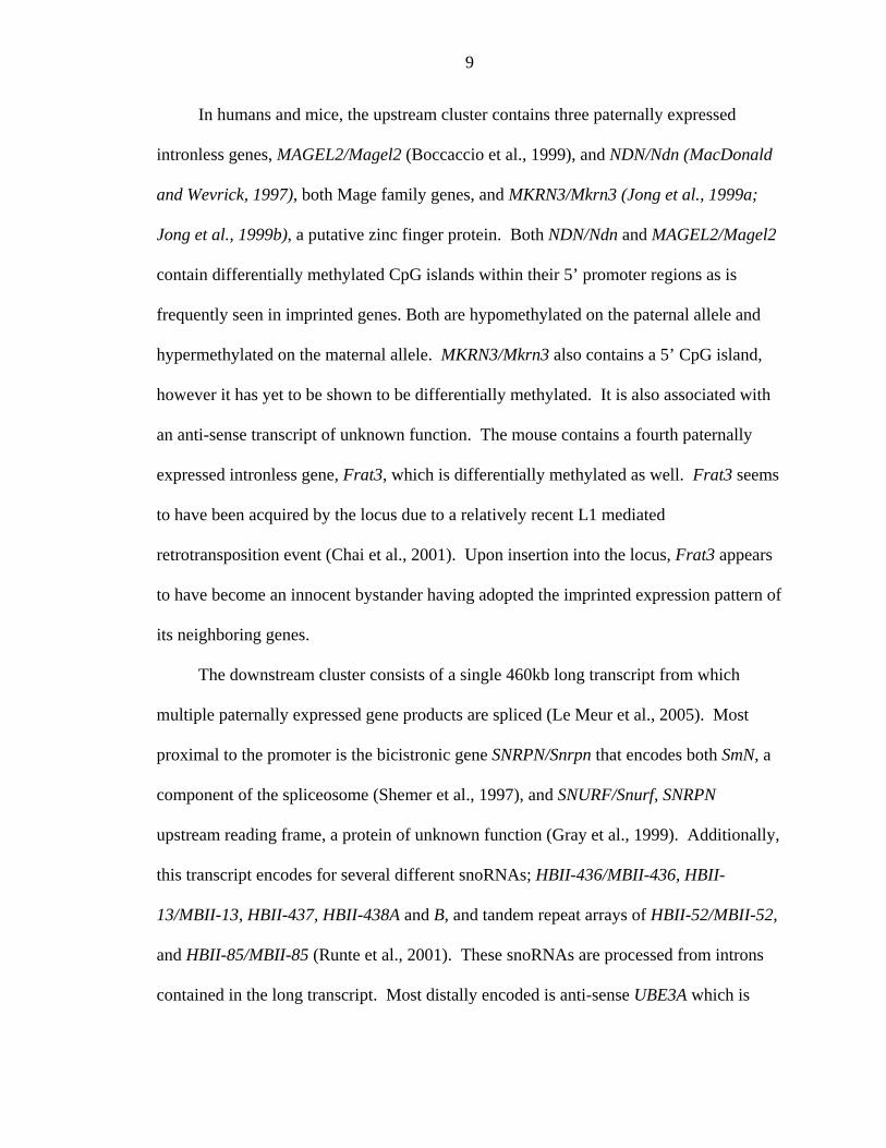
Figure 1-1 The life cycle of an imprint. There are three phases in the life of an imprint. First the imprint must be established in the germline. This imprinting mark can be distinguished between the maternal and paternal alleles. Second the mark must be maintained within the somatic tissues throughout the life of the animal. Third the imprint must be erased during gametogenesis of a developing embryo so that it can be reset according to the sex of the embryo.

13
Figure 1-2. Mapping the Imprinting Center (IC). Mapping in humans using Shortest Region of Overlap (SRO) shows that the IC is bi-partite in structure. The PWS-IC is located within 4.3 kb interval that includes exon 1 of SNRPN. The AS-IC is located 35kb upstream of SNRPN exon 1 within a .88 kb interval
14
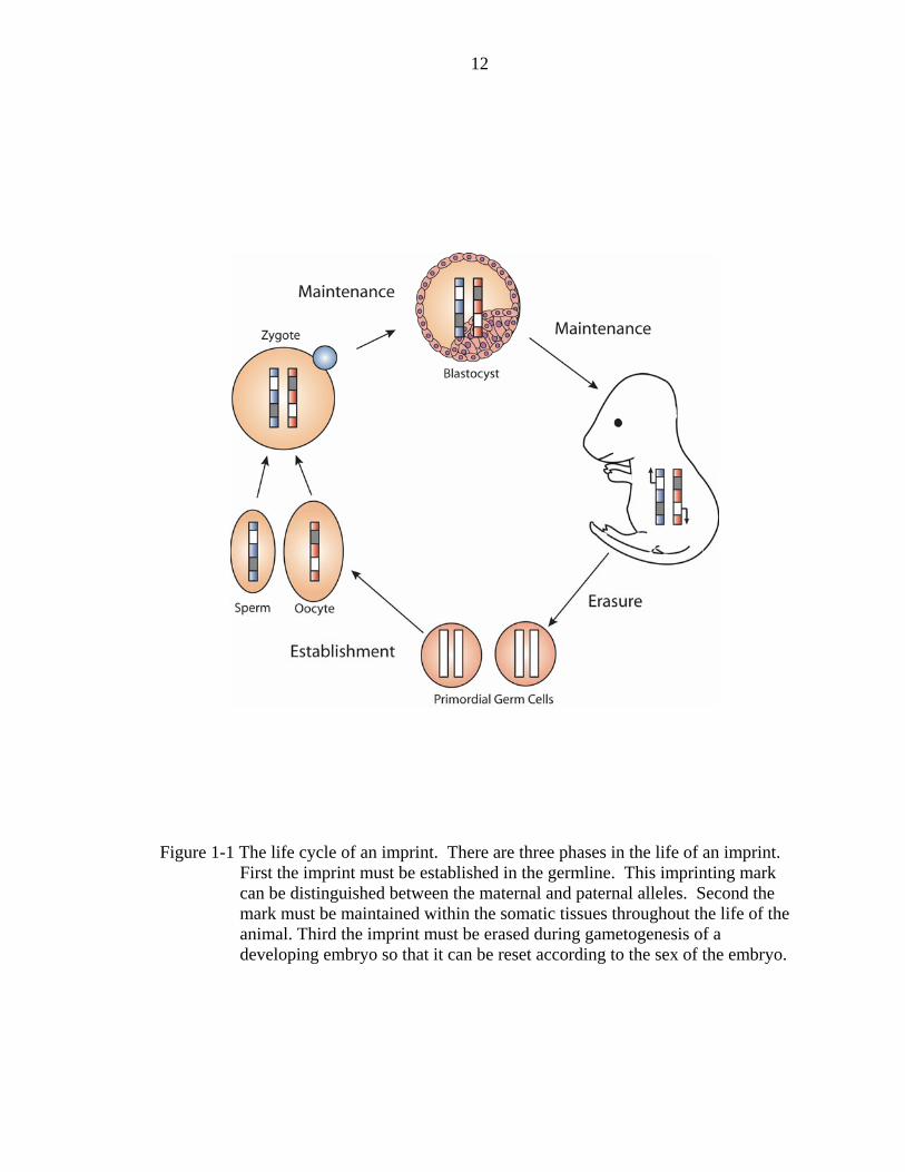
Figure 1-3. Molecular classes of PWS and AS. There are four different molecular classes that can result in PWS and AS. First, large deletions of 3−4 Mb result in loss of the all genes within the locus. This deletion occurs on the paternal allele in the case of PWS and the maternal allele in the case of AS. Second, uniparental disomy results in two alleles that act in the same manner. Maternal uniparental disomy results in PWS and Paternal uniparental disomy results in AS. A third class, single gene mutation, only occurs in the case of AS. This occurs when the gene UBE3A is mutated or deleted. The fact that this class does not exist in the case of PWS suggests that it is a contiguous gene disorder, requiring the loss of multiple genes to occur. A fourth class, IC microdeletions, occurs when the imprinting control elements are missing or mutated.

15
Figure 1-4. Gene organization of the PWS/AS locus. The PWS/AS locus, located on human chromosome 15q11-q13, consists of several genes upstream of the IC and a single long transcript originating from the SNRPN gene downstream of the IC. The syntenic region in the mouse exists on mouse chromosome 7C and is conserved both in gene order and imprinted expression pattern.

16
Figure 1-5. Paternal only model of imprinting regulation. The paternal only model of regulation suggests that the PWS-IC is active only on the paternal allele driving expression of the paternal genes. The AS-IC on the other hand is active only on the maternal allele and acts to silence the PWS-IC on the maternal there.

17
CHAPTER 2 MATERIALS AND METHODS
Lambda Red Recombineering
Transformation of Recombineering Strains
In order to create targeted deletions within the BAC of interest, it must first be
transformed into the appropriate recombineering E-coli strain. Three strains are available.
DY380 is a modified DH10B strain containing the defective lambda prophage required
for recombineering. EL350 is a modified DH10B strain containing the defective lambda
prophage and an arabinose inducible cre gene used for cre/lox recombination. EL250 is a
modified DH10B strain containing the defective lambda prophage and an arabinose
inducible flpe gene used for flpe/frt recombination.
Fresh BAC is prepared by alkaline lysate method. Briefly, single colonies of
DH10B containing the BAC of interest are picked and grown overnight in 3 mL Luria
Broth (LB) with 12.5 ug/mL chloramphenicol at 32oC, 175 rpm. The next day cultures
are pelleted in a microcentrifuge at 7000 rpm, resuspended by vortexing in 100 uL
solution I (50 mM dextrose, 10mM EDTA, 25mM Tris pH 8.0) and allowed to sit at
room temperature for 3−5 minutes. Next, 200 uL of solution II (.2M NaOH, 1% SDS) is
added, the samples are mixed by inversion and then placed on ice. After 5 minutes 150
uL of solution III (29.4g KOAc, 11.5 ml. conc. HOAc, H20 to 100ml.) is added, the
samples are shaken and then placed back on ice. 400 uL of prepared phenol chloroform is
added to the tubes and the samples are shaken to mix and then centrifuged at 13000 rpm
for 5 minutes. After centrifugation the aqueous layer is transferred to a fresh tube and 1
18
mL of 100% EtOH is added. The samples are then shaken and centrifuged again at 13000
rpm. The supernatant is poured off and 1ml of 70% EtOH is added. The samples are
shaken and centrifuged again at 13000 rpm. The resulting DNA pellet is resuspended in
50ul of sterile H2O.
In order to transform the BAC into the DY380 E.coli, a single colony of the
appropriate recombineering strain is grown up overnight in 3 mL LB at 32oC, 175 rpm.
The next day 700ul of culture is diluted in 70 mL of LB with 12.5ug/mL chloramphenicol
and incubated 3−5 hours at 32oC, 175 rpm, until the culture density reaches an OD600 of
.5−.6. The culture is next transferred in 10 mL aliquots to 15 mL falcon tubes and then
pelleted by cold (~5oC) centrifugation at 5000 rpm. Supernatant is poured off and the
pellet is resuspended in 10 mL ice cold 10% glycerol. The tubes are spun again at 5000
rpm, 5oC, for 5 minutes and the supernatant poured off. The pellet is resuspended this
time in 1 mL 10%glycerol and transferred to a 1.5 mL eppendorf tube. The tubes are spun
at 13000 rpm in a tabletop microcentrifuge for 30 seconds and the supernatant poured off.
The pellets are again resuspended in 1 mL 10% glycerol and spun again at 13000 rpm.
This time all of the supernatant is carefully drawn off with a pipette and the pellet is
resuspended in 80ul of 10% glycerol. The tubes containing the now electrocompetent
cells are stored on ice until use. Fresh BAC DNA is mixed with the electrocompetent
cells to give a final volume of 50ul. Usually DNA volumes of 2, 5, and 10 uL mixed with
48, 45, and 40 uL competent cells will give enough of a range to produce good results.
The DNA, cell mixture is transferred to a pre-cooled .1 cm Bio-Rad electroporation
cuvette; catalog number 165−2089. Electroporation is performed using a Bio-Rad Gene
Pulser set at 1.8kV, 25uF, with the pulse controller set at 200 ohms. The electroporated

19
cells are then immediately diluted with 1 mL of LB and allowed to incubate at 32oC, 175
rpm, for 1 hour. After incubation, the cells are pelleted by centrifugation at 7000 rpm and
the supernatant drawn off with a pipette, leaving approximately 100ul total volume in the
tube. The remaining volume is plated to LB agar plates containing 12.5 ug/mL
chloramphenicol and incubated overnight at 32oC. Resulting colonies are screened by
digesting with numerous different restriction enzymes and comparing them to the original
BAC.
Transformation of Targeting Vector and Induction of Recombination
Recombination within the BAC is achieved by electroporating a targeting construct
into the BAC containing recombineering strain. The targeting construct should contain a
selectable marker such as neomycin resistance and should be linearized by restriction
digest or amplified by polymerase chain reaction (pcr) prior to transformation. The night
before transformation a single colony of the BAC containing strain is grown up overnight
in 3 mL LB at 32oC, 175 rpm. The next day 700ul of culture is diluted in 70 mL of LB
with 12.5ug/mL chloramphenicol and incubated 3−5 hours at 32oC, 175 rpm, until the
culture density reaches an OD600 of .5−.6. At this point 10 mL of the culture should be
drawn off and reserved on ice to serve as an uninduced control. The remaining 60 mL is
induced to express the lambda RED genes by shaking in a 42oC water bath for 15
minutes. After induction the flask is transferred to an ice slurry bath and swirled for 10
minutes to turn off the defective lambda phage. The induced and uninduced control
cultures are then made to be electrocompetent in the same way in which the
recombineering strains were made to accept the BAC, by a series of cold 10% glycerol
washes. Targeting vector DNA is mixed with the electrocompetent cells to give a final
20
volume of 50ul. Usually DNA volumes of 2, 5, and 10 uL mixed with 48, 45, and 40 uL
competent cells will give enough of a range to produce good results. The DNA, cell
mixture is transferred to a pre-cooled .1 cm Bio-Rad electroporation cuvette; catalog
number 165−2089. Electroporation is performed using a Bio-Rad Gene Pulser set at
1.8kV, 25uF, with the pulse controller set at 200 ohms. The electroporated cells are then
immediately diluted with 1 mL of LB and allowed to incubate at 32oC, 175 rpm, for 1
hour. After incubation, the cells are pelleted by centrifugation at 7000 rpm and the
supernatant drawn off with a pipette, leaving approximately 100ul total volume in the
tube. The remaining volume is plated to LB agar plates containing the appropriate
antibiotic and incubated overnight at 32oC. The resulting colonies are screened by
Southern blot.
BAC preparation for injection
In order to inject BAC’s as transgenes, the DNA must be prepared in a way that
removes all traces of impurities and leaves the BAC as intact supercoils if possible. The
DNA may be linearized by restriction digest, but this requires an additional purification
step. BAC preparation by cesium prep and linearization are described below.
Two nights before the DNA prep, a single bacterial colony containing the
appropriate BAC is picked and cultured overnight in 3 mL LB with 12.5 ug/mL
chloramphenicol at 32oC, 175 rpm. The next day, 1 mL of this starter culture is
inoculated into 3 1000ml flasks containing 500 mL LB with 12.5 ug/mL
chloramphenicol. These cultures are incubated overnight at 32oC, 175 rpm. The next day,
the cultures are poured into 500ml bottles and spun in a centrifuge at 5000 rpm. The
resulting pellets are resuspended in 25 mL solution I each (50 mM dextrose, 10 mM

21
EDTA, 25mM Tris pH 8.0) and allowed to sit at room temperature for 3−5 minutes. 50
mL solution II (.2M NaOH, 1% SDS) is added to each bottle and then inverted to mix.
After 4 minutes 37.5 mL of solution III is added (29.4 g KOAc, 11.5ml glacial acetic
acid, H2O to 100 ml), the bottles are mixed by inversion and placed on ice for 10 minutes.
In order to remove as much cellular debris as possible the mixture is poured into clean
bottles through a layer of gauze and then spun in a centrifuge at 5000 rpm for 5 minutes.
The now cleared solution is additionally filtered through medium porosity filter paper
(Fisher catalog number 09−802−1a). Each bottle now should contain approximately 112
mL of BAC DNA solution. In order to precipitate the DNA the solution is transferred to 3
250 mL bottles which are then filled to the neck with 100% EtOH. The EtOH/DNA
mixture is spun in a centrifuge at 10,000 rpm for 20 minutes. The supernatant is poured
off, the bottles filled with 70% EtOH and then spun again at 10,000 rpm for 10 minutes.
The ethanol is poured off and any residual ethanol is removed from the bottles using a
pipette. The pellets are then air dried for approximately 20 minutes and resuspended in a
total of 8 mL TE with RNase when combined.
To perform a gradient the density of the solution must be brought up to 1.55 g/mL
by adding the proper amount of cesium. 1.05 grams of cesium are added per each mL of
solution to achieve this density. Transfer the solution to a 15 mL conical tube and to this
tube add 8.4 g of cesium. Mix the solution by inversion until all the cesium dissolves.
The density should be checked by weighing 1 mL of solution on an analytical balance.
The weight should equal between 1.55−1.57 g. The cesium/DNA mixture is then divided
between 2 ultracentrifuge tubes. One hundred uL ethidium bromide is added and the
volume brought up to the top of the tube using pre-made 1.55 g/mL TE-RNase solution

22
stored in the freezer, making sure to keep the 2 tubes at an equal weight. Seal the tubes
spin them overnight at 55,000 rpm using an NVT-90 fixed angle rotor in an
ultracentrifuge. The next day, remove the tubes from the rotor and extract the DNA,
which should be present as a visible red band within the tube, using an 18 gauge needle
and syringe. Combine the DNA from the 2 tubes into one and bring up the volume using
the pre-made 1.55g/mL TE-RNase solution. Seal the tube and spin again for 4 hours at
78,000 rpm using the same rotor. Again, extract the DNA with an 18 gauge needle and
syringe. It should be present now as a thicker red band, and transfer to a 15 mL conical
tube. There should be a total volume of 1−1.5 ml.
Salt water saturated butanol is used to extract the ethidium from the DNA solution.
To make the butanol solution, in a 250 mL bottle add NaCl to 50 mL of sterile water until
the solution becomes completely saturated and the salt no longer dissolves. One hundred
and fifty mL butanol is added to the bottle and mixed by shaking. The butanol is allowed
to completely separated from the aqueous phase before using the solution. Two mL
butanol is added to the BAC DNA solution and mixed by inverting 10 times. Allow the
aqueous layer to resolve and then remove the upper butanol layer. Continue to repeat the
addition and removal of butanol until the lower DNA solution layer is completely clear
when looked at in front of a white background.
The ethidium free BAC DNA solution is next transferred into a 4 inch long piece of
Spectra/Por dialysis tubing (MWCO 12,000−14,000 daltons, cat.# 132676) for dialysis
against the DNA injection buffer. The tightly sealed dialysis bag is allowed to float in a
bucket of 4 liters of injection buffer (10mM tris pH7.5, .1mM EDTA, 100mM NaCl)
overnight at a temperature of 4oC with a slowly spinning stirbar. The next day the DNA

23
solution is removed from the dialysis tubing and the concentration is measured on a
spectrophotometer.
Generation of Transgenic Animals by Pronuclear Injection
Injections were expertly done by the Chris Futtner Injection Core (CFIC) at The
University of Florida. Briefly DNA was microinjected into the pronucleus of fertilized
mouse zygotes derived from superovulated, 5 week old FVB female mice at a
concentration of 2.5−3 ng/ul. Injected zygotes were allowed to mature overnight to the
two cell stage at which point they were transferred into the infundibula of pseudo
pregnant (B6D2)F1 female mice.
Genotyping
Identification of Transgenic Founders
PCR genotyping was used to identify founder animals carrying the various
transgene constructs. At 2 weeks of age pups born to (B6D2)F1 foster moms were ear
punched and tail clipped for screening. Genomic DNA was prepared from each tail piece
by digestion with 10ug/mL protienase K in tail lysis buffer (100mM Tris pH 8.5, 5mM
EDTA, .2% SDS, 200mM NaCl). PCR was performed to screen for the T7 BAC arm and
adjoining mouse sequence using the following primers: T7F 5’-GTA ATA CGA CTC
ACT ATA GGC-3’ and 425T7R1 5’- CTC CAA TCA TGT TCA ACT GTC-3’.
Founders were further screened by Southern blot, probing for Snrpn.
Identification of Castaneous C7 Homozygotes
PCR genotyping followed by restriction endonuclease digestion was used to
identify transgenic animals that were also homozygous for Castaneous C7 at the PWS
locus. This was done by screening for a polymorphic AvaII site between Mus musculus
castaneous and Mus musculus domesticus that results from a cytosine nucleotide rather

24
than a thymine nucleotide at position 117 of the Ndn gene. AvaII cuts the domesticus but
not the castaneous allele due to the presence of the cytosine. PCR primers flanking the
polymorphic site with the following sequence were used: NdnpolyF 5’-ACA AAG TAA
GGA CCT GAG CGA CC-3’ and NdnpolyR 5’-CAA CAT CTT CTA TCC GTT CTT
CG-3’. The amplified PCR product was gel purified on a 2% agarose gel, extracted from
the gel using the Promega Wizard Prep kit, and digested with AvaII. The digested
products were then electrophoresed on a 4.8% agarose gel (2:1 low-melt agarose:
agarose).
Southern Blot
Southern blotting was performed as described (Sambrook et al., 1989). Agarose
gels were placed on a ultra violet (UV) light box to nick DNA. After 5 minutes, gels
were soaked in alkali solution (1.5 M NaCl and 0.5 N NaOH) for 45 minutes followed by
neutralizing solution (1.5 M NaCl and 1 M Tris pH 7.4) for 1.5 hours (Sambrook et al.,
1989). Gels were then blotted using 10X SSC overnight allowing for transfer of the
DNA from the gel to the nylon membrane. The following day, the membrane was rinsed
in 2X SSC, baked at 80oC for several hours and hybridized with 20 mL of Church and
Gilbert hybridization buffer (Church and Gilbert, 1985) (2.5% BSA, 1 mM EDTA pH
8.0, 0.25 M sodium phosphate buffer pH 7.2, and 7% SDS) at 65oC for 2 hours. The
prehybridization buffer was then poured off and another 5 mL of buffer, containing the
denatured probe, was added to the hybridization tube and incubated at 65oC overnight.
The membrane is washed three times for 15 minutes each with 2X SSC and 0.1% SDS,
then wrapped in saran wrap and exposed to film.
25
RNA Isolation
RT-PCR was performed to detect the expression of the transgenic Snrpn minigene.
Total RNA was isolated from complete brains obtained from neonatal mice. RNA was
extracted using RNA-zol (Tel-Test, Inc). Briefly, tissue samples were homogenized with
4 ml. of RNA-zol using a Polytron homogenizer. To this homogenate .4ml of chloroform
is added, the samples are shaken vigorously for 15 seconds and then put on ice for 5
minutes. After 5 minutes the tubes are then centrifuged for 15 minutes at 12,000g after
which the homogenate forms two distinct layers: a lower blue phenol chloroform phase
and an upper colorless aqueous phase containing the RNA. The aqueous phase is added
to a new tube and to it an equal volume of isopropanol is added and the samples vortexed
then place on ice for 15 minutes. After 15 minutes the samples are centrifuged for 15
minutes at 12,000g. The resulting white RNA pellet is then washed with 75%ethanol and
centrifuged again at 12,000 g for 10 minutes. After removal of the ethanol the pellet is
allowed to air dry and then resuspended in .2 mL of DEPC H2O.
RT-PCR
RNA isolated from neonatal mouse brains was analyzed for transgene expression
by RT-PCR. Five ug RNA is mixed with 1ul of 500 ug/mL random primers and dH2O to
reach a final volume of 25.8ul. This mixture is then heated to 68oC for 3 minutes and
then placed on ice. To each tube is added 3.2ul dNTP mixture, 8ul of reverse
transcriptase buffer, 2ul 100mM DTT, 1ul RNase inhibitor and 1ul SuperscriptII and then
incubated at 37oC for 60 minutes. Two uL of this reaction is then used to seed a PCR
reaction using the following primers: N2.1F 5’-CCC CGA GTA TTA AGG ATC TTG-3’
and N6.2R 5’-GCA ACA GTG CCT CTT CCC TG-3’. PCR amplification condition
26
were 95oC for 5 min., followed by 30 cycles of 95oC for 30 sec., 57oC for 30 sec., 72oC
for 1 min. The cycle was followed by an extension step of 72oC for 10 min.
Preparation of High Molecular Weight Genomic DNA for Bisulfite Conversion
High molecular weight brain DNA was isolated from neonatal mouse brain to be
used for bisulfite PCR. Two and one half mL of homogenizing solution (1X SSC, 1%
SDS, .25mg/mL Pronase E) was added to a dounce homogenizer along with a single
frozen neonatal mouse brain. Tissue was homogenized by gently moving the pestle up
and down within the dounce. The homogenate was collected in a 15 mL polyproplene
tube and the homogenizer washed with an additional 2.5 mL of solution that was then
combined with the homogenate. The homogenate was vortexed and then incubated for 1
hour in a 37oC water bath. After 1 hour, 5 mL of phenol chloroform was added and the
mixture was vortexed and then spun in a centrifuge at 2500 rpm for 5 min to extract the
DNA. The top aqueous layer was collected to a new 15 mL tube and the extraction was
repeated twice more as above, first with phenol chloroform and then with chloroform
alone. After the aqueous layer was removed from the final chloroform extraction, 0.5 mL
RNase A was added and then incubated for 30 min. at 37oC. One half mL of pronase E
was then added and the mixture was incubated an addition 30 min at 37oC. The DNA
was extracted again as above, first with phenol chloroform and then with chloroform
only. After extraction the DNA was precipitated by the addition of 12 mL 95% ethanol
followed by vortexing. The now visible DNA strands were spooled onto a glass rod and
placed into a 1.5 mL tube to dry. Once the DNA appeared to be mostly dry, 200 mL of
water was added to resuspend the DNA which was then stored at –20oC.

27
Bisulphite Sequencing of Genomic DNA
Bisulphite Conversion
Bisulphite conversion was carried out as described (Clark et al., 1994). DNA was
denatured with 4.2 µl of 3 M NaOH (final concentration is 0.3 M in 42 µl) and incubated
for 37oC for 30 minutes. Freshly prepared 2M sodium metabisulphite and 10 mM
hydroquinone (final concentrations 1.55 M and 0.5 mM respectively) was added to the
denatured DNA to a final volume of 240 µl. The bisulphite reaction was inefficient on
double stranded DNA. Samples were then incubated in the dark for 16−20 hours. A
water control was also prepared and treated with the bisulphite stock for later use as a
PCR control.
Following overnight incubation, free bisulphite ion was removed by passing the
sample through a desalting column (Promega Wizard DNA Clean-up System) and eluted
in 45 µl of water. Freshly prepared 3 M NaOH was added and the samples were
incubated at 37oC for 15 minutes. The DNA was then precipitated overnight at -80oC by
adding 25 µl 7.5M Ammonium Acetate and 200 µl 100% Ethanol. The following day
DNA was pelleted by centrifugation at 13,000 rpm for 30 minutes at 4oC followed by a
70% Ethanol wash. The final pellet was dried by vacuum centrifugation and resuspended
in 100 µl of water.
Bisulphite Polymerase Chain Reaction
Bisulphite primers were designed against the bisulphite converted DNA. In order
to avoid any amplification of unconverted DNA, primers were designed in regions, which
contained numerous converted cytosines (now thymines), predominantly at the 3' end.
Primers were designed in regions in which no CpG dinucleotides were present, however
if a primer did include one or two CpGs a degenerate nucleotide (cytosine or thymine)

28
was included at those positions. Care was also taken to include as many guanine residues
as possible to increase the Tm of the primer. Converted sequences have very few
cytosines causing melting temperatures to be relatively low. Primer lengths were
occasionally increased to account for this. Primers were synthesized by Integrated DNA
Technologies (IDT) and purified using standard desalting methods. Primer sequences are
listed in Table A-1.
PCR was performed on bisulphite treated brain DNA preparations with HotStar
Taq (Qiagen) using the following PCR conditions (final): 1X PCR buffer (with 15 mM
MgCl2), 200 µM each dNTP, 1 µM each primer, and 1.5 U HotStar Taq DNA
polymerase. Bisulphite converted brain DNA preparations were not quantitated for DNA
concentration and 2 µl was used per 25 µl reaction. PCR reactions were performed using
the following cycling conditions: An initial denaturation at 95oC for 15 minutes was
required for activation of HotStar Taq polymerase. A denaturation at 95oC for 45
seconds was followed by a 53oC annealing for 30 seconds followed by a 72oC elongation
for 1.5 minutes and all three steps repeated 35 times followed by a final 72oC elongation
for 10 minutes.
Purification of PCR Products
PCR products were purified using the Quiagen Quiaquik PCR cleanup kit as per kit
directions. Once the PCR product was purified and resuspended in 45 ul, 10 uL was taken
and run out on a 2% agarose gel to check for quality before cloning.
Cloning and Sequencing of PCR Products
PCR products were cloned using TA cloning with pGEM-T Easy Vector Systems
(Promega). Ligations were performed as suggested by the kit protocol. Ligations were
transformed into chemically competent, subcloning efficiency XL-1 Blue (Stratagene)
29
E.coli cells. Transformations were plated onto Luria-Bertani (LB) plates (1% tryptone,
0.5% yeast extract, 1% sodium chloride and 1.5% agar) containing 50 µg/mL ampicillin
and supplemented with 100 µl of 100 mM IPTG and 40 µl of 40 mg/mL X-Gal per plate
(Gold Biotechnology, Inc) for blue-white colony selection (Sambrook et al., 1989).
Plates were incubated overnight at 37oC and the following morning white colonies were
picked into 3 mL of culture media (1% tryptone, 0.5% yeast extract, and 0.5% sodium
chloride). Liquid cultures were grown overnight at 37oC and plasmid DNA extracted by
the alkaline lysis method (Sambrook et al., 1989). Plasmid sequencing was carried out
using SP6 primer for using the standard plasmid sequencing methods (described below).
Sequences were analyzed using Sequencher 4.2 (Gene Codes Corporation).
Sequencing
DNA sequencing was carried out using ABI Prism BigDye terminator
(PerkinElmer) which uses the AmpliTaq DNA polymerase. Once sequence reactions
were completed the samples were sent to the Center for Mammalian Genetics DNA
Sequence Core (Florida) where the samples are run on the ABI Prism 377XL Automated
DNA Sequencer (PerkinElmer). Sequence files which were received from the
sequencing core were then analyzed using Sequencher 4.2 (Gene Codes Corporation).
Sequencing samples were prepared as suggested by the manufacturer
(PerkinElmer): 2 µl BigDye Terminator, 2 µl 5X sequencing buffer, 1 µl 3.2 pmole/µl
primer, 2 µl water and 3 µl plasmid DNA (approximately 3 µg). Sequencing PCR was
carried out by 24 cycles of s of 96oC for 30 seconds, 50oC for 15 seconds and 60oC for 4
minutes. Reactions were purified using Performa DTR Gel Filtration Columns (Edge
Biosytems) and dried by vacuum centrifugation. Alternatively, sequencing reactions
were purified by Ethanol precipitation where 2 volumes of 100% ethanol and 1/2

30
volumes of 7.5 mM ammonium acetate were added to the sequencing reactions. Samples
were washed with 70% ethanol and dried by vacuum centrifugation.

31
CHAPTER 3 IMPRINTED TRANSGENE
Introduction
Imprinted loci are often organized as clusters of genes controlled by cis-acting
regulatory elements called imprinting centers (IC) (Brannan and Bartolomei, 1999).
These IC’s can often be found large distances away from the genes they control making it
difficult to localize and understand their mechanisms of action. Transgenes are excellent
tools for the study of imprinted loci in that they serve to isolate genes and their regulatory
elements and relocate them to ectopic locations in the genome allowing for the study of
those sequences without the influence of other adjacent elements. BAC transgenes have
an additional advantage over conventional plasmid based transgenes. Smaller plasmid
transgenes are often influenced by the chromatin into which they randomly insert. These
insertional effects often result in inconsistencies in expression between individual
transgenic mouse lines. The inherent greater size of BAC transgenes make them much
less susceptible to position effects most likely due to the fact that they contain essential
regulatory elements necessary for maintaining an open chromatin structure. For the study
of imprinted loci, this greater size also increases the chance of inclusion of IC’s and other
necessary sequences required for proper expression patterns.
BAC transgenes have been successfully used in the past for the study of imprinting.
In one example Yevtodiyenko showed that a 178 kb BAC transgene expressed the Gene-
trap locus 2 (Gtl2) gene only upon maternal inheritance within the mouse embryo and
placenta (Schmidt et al., 2000; Yevtodiyenko et al., 2004). This is similar to the
32
endogenous locus on distal mouse chromosome 12 and human 14q32 suggesting that the
178 kb interval contains all the elements necessary to imprint Gtl2. They were also able
to show that levels of expression were similar to endogenous levels only in the brain but
not other regions, localizing the enhancer elements necessary for brain expression but not
other tissues to within the BAC. Lastly they showed that a second gene present within
the BAC, Delta-like1 (Dlk1), was not expressed in an imprinted fashion, as is the
endogenous locus, indicating that additional elements are required for imprinting Dlk1.
The Surani lab reports using a 120 kb BAC transgene to localize imprinting elements
required for paternal expression of the Peg3 gene which encodes a C2H2 type zinc-finger
protein involved in maternal behavior (Li et al., 2000; Szeto et al., 2004). In this study
they showed that imprinting is linked to differential methylation of a region upstream of
the gene. A second maternally expressed gene Zim1, located within the same cluster and
present on the BAC was not imprinted correctly indicating that other cis acting elements
are required for imprint regulation of the locus. The Surani lab also used BAC transgenes
to locate dispersed cis-regulatory elements necessary for paternal expression of
Nueronatin (Nnat) (John et al., 2001). By modifying the BAC they showed that these
elements were located within an 80kb interval mostly upstream to Nnat.
As other labs have shown, BAC transgenes can be a powerful tool for localization
of IC elements. In the case of the PWS locus, current understanding of IC function in the
mouse is limited to a 35 kb region in which the PWS-IC is located. The AS-IC in the
mouse has yet to be identified. Our lab has previously produced several transgenic lines
aimed at further elucidating the imprinting machinery of the PWS locus (Figure 1). The
first of these transgenic lines was developed from mouse and human phage (P1) clones

33
carrying 85 and 76 kb of genomic sequence respectively (Blaydes et al., 1999). The
mouse transgene contained 33 kb of 5’ and 30 kb of 3’ flanking DNA while the human
clone carried 45kb of 5’ and 7 kb of 3’ flanking sequence. Five lines were produced from
the human P1 clone, four of which were single copy and the fifth present as five copies.
This clone was chosen due to the fact that it contained the complete interval in which
both the PWS-IC and the AS-IC were located. All lines were expressed biallelically
suggesting that the human PWS-IC, or at least the Snrpn promoter, is functional in the
mouse, while the AS-IC is not. Two lines were derived from the mouse phage clone, a
line containing a single copy of the transgene and a line containing two copies. The
single copy line was biallelically expressed while the two-copy line was correctly
imprinted as it was expressed only upon paternal inheritance. The most likely
explanation for this two-copy effect is that the mouse clone was large enough to contain
the entire PWS-IC since it was capable of driving expression of Snrpn. However the AS-
IC probably lies outside of this interval since the single copy line was unable to silence
Snrpn expression upon maternal inheritance. The presence of a second copy somehow
overcomes this deficiency, either by acting as a buffer to the insertion site effects of
integration or it adds back some needed sequences lost to the single copy line. This is
similar to the effect seen in BAC transgenic studies of the maternally expressed H19 gene
where a 14 kb transgene requires multiple copies to imprint, however a 130kb BAC
transgene imprints in single copy (Bartolomei et al., 1993; Pfeifer et al., 1996).
More recently the lab has created multiple transgenic lines using BAC transgenes
which are much greater in size than the P1 clones. The Roswell Park RPCI-23 murine
BAC library was screened for clones containing Snrpn. Three were identified that carried
34
sufficient sequence upstream of Snrpn; 215A9 which extends 120 kb 5’ and 20 kb 3’,
380J10 which extends 8 kb 5’ and 140 kb 3’, and 425D18 which extends approximately
100 kb 5’ and 65kb 3’. These three BACS were injected as transgenes and were then
tested for imprinted expression patterns (Figure3-1). The 380J10 transgene was
expressed upon both maternal and paternal inheritance suggesting that the complete
PWS-IC was present but the AS-IC was lacking. The 425 and 215 transgenes on the
other hand showed correct imprinted expression. They were paternally expressed and
maternally silenced. Interestingly the 215B line suffered from a truncation that leaves
only approximately 40 kb of sequence 5’ to Snrpn. This establishes the minimal genomic
sequence necessary to establish imprinted expression in the locus as approximately 40 kb
upstream and 20 kb downstream of Snrpn.
This project attempts to further delineate the sequences necessary to confer an
imprinted expression pattern within the PWS locus. To do this a modified BAC
transgene was developed in order to simplify expression analysis. Later chapters will
discuss additional modifications that were used to minimize the PWS-IC interval and
locate the AS-IC.
Results
BAC Modification
Previously, analysis of transgene expression required a complex breeding scheme
in order to distinguish the transgenic allele from the endogenous alleles. Transgenic
founders were mated to mice carrying a targeted deletion of exons 5−7 of Snrpn (ΔSmN)
that results in a larger fusion transcript that include exons 1−4 and the neomycin
resistance gene (Yang et al., 1998). In this way the transgenic allele could be
35
distinguished as a smaller transcript than the endogenous one. In order to simplify
expression analysis, we decided to mark the transgene in a way that it could be
distinguished immediately. This was done using a technique called lambda red mediated
homologous recombination developed by Daiguan Yu and Don Court at the National
Cancer Institute (Lee et al., 2001). This method takes advantage of a defective lambda
prophage from which the genes allowing entry into a lytic life cycle have been removed.
What is left are only the minimal elements required to express the lambda Red genes exo,
beta and gam. Exo and bet are required for recombining a double stranded (ds) linear
DNA targeting construct into BAC DNA via homologous recombination. Exo encodes a
5’-3’ exonuclease that acts upon the 5’ ends of linear dsDNA to create 3’ single stranded
(ss) ends. Bet encodes a pairing protein that binds the 3’ssDNA ends and promotes
annealing to the complementary strand on the BAC. The third gene, gam, inhibits E. coli
RecBCD exonuclease activity that destabilizes linear dsDNA. The defective prophage is
integrated into the E. Coli DH10B (DY380) host bacterial chromosome and is under the
control of the strong PL promoter. The promoter in turn is under the control of the
temperature sensitive lambda cI857 repressor. Transiently shifting the culture
temperature from 32˚C to 42˚C results in expression of these three genes and subsequent
homologous recombination between the linear targeting construct and BAC DNA.
The 425D18 BAC was chosen for modification since a good amount of both 5’ and
3’ sequence was present. Since the previous SmN deletion had shown no effects on
expression it was decided to create the same deletion on the BAC (Figure 3-2). Exons
5−7 of Snrpn were replaced with a floxed neomycin resistance cassette using a targeting
construct (170 floxed neo) containing a 2 kb 5’ arm of homology and a 6.2 kb 3’ arm of

36
homology. The targeting construct was linearized with Not I and then electroporated into
induced DY380 recombination competent cells. Integration of the targeting construct
was selected for by plating the transformed cells onto LB agar plates supplemented with
neomycin and incubation overnight at 32oC. The next day 12 neomycin resistant colonies
were picked and grown overnight in 3 mL of LB broth supplemented with neomycin at
32oC. After sufficient growth the cultures were subjected to alkaline lysis to extract the
BAC DNA. BAC DNA was then cut with SacI restriction endonuclease and Southern
blotted. The blot was probed with a 2.2kb EcoRI/EcoRV genomic DNA fragment
obtained from a restriction digest of lab stock genomic DNA Snrpn clones. Owing to the
extreme efficiency of this recombineering method, all of the clones picked were positive
for the exon 5−7 deletion (Figure 3-2). Following successful targeting of Snrpn, the
floxed neomycin cassette was to be removed by expression of Cre recombinase. To do
this, the targeted 425 BAC (425Δ5−7neo) was transferred to an E. Coli host strain
carrying the Cre recombinase gene under the control of an arabinose inducible promoter
(EL350). However, after Cre had been induced, a severe undesired truncation event
occurred, making the BAC unusable (data not shown). Investigation into the composition
of the BAC vector (pBACe3.6) into which the genomic DNA is cloned revealed a
previously unknown third lox-p site. Induction of Cre resulted in recombination between
one or both of the neomycin cassette flanking lox-ps with the third lox-p resulting in
massive rearrangements. To deal with this, a second targeting construct, designed to
replace the third lox-p site with a blasticydin resistance cassette, was built.
Recombination with this construct was induced as before and efficient removal of lox-p
number three was achieved (data not shown). The doubly modified 425Δ5−7 BAC was

37
again subjected to Cre mediated recombination. Successful deletion of the floxed
neomycin cassette was determined by restriction digest with EcoRI and SacI followed by
Southern blot. The Southern blot was then probed with the same 2.2kb EcoRI/EcoRV
genomic DNA fragment as before (Figure).
Production of Transgenic Mice
Transgenic mice were produced to test of the suitability of the 425Δ5−7 BAC as a
model system for the study of imprinting in the PWS locus. If the modified BAC was to
be a reliable system, the truncated Snrpn transcript it contains must be expressed only
upon paternal inheritance. When inherited from the mother it should be silenced. BAC
DNA was prepared for injection by large-scale alkaline lysis followed by cesium chloride
banding. Both nicked and supercoiled DNA were collected from the cesium prep and
dialyzed against 4 liters of injection buffer (1.0 mM Tris pH7.5, .1 mM EDTA, 100 mM
NaCl) for approximately 16 hours. After dialysis, the BAC DNA was diluted to 2.5 ng/ul
and injected into the male pronucleus of fertilized oocytes derived from superovulated
FVB/N female mice. The injected oocytes were then implanted into pseudopregnant
B6D2 F1 female mice. The resulting pups were allowed to mature to 3 weeks of age at
which time they were ear punched for identification and tail clipped to obtain DNA for
genotyping. Genomic tail DNA was then digested with AvaII and Southern blotted. The
blot was then probed with a 1.5 kb genomic DNA HindII restriction digest fragment to
both screen for the presence of the transgene and estimate copy number. Ten founder
animals were obtained. Line A and D appeared to be single copy when compared to the
endogenous band. Lines B, E, F, G1, H, I, and J appeared to be present in multiple
copies. Line J was discarded, as it appeared to contain an immediately observable
38
rearrangement. Lines A, B, D, E, H, and I were retained and mated as they seemed to
contain the lowest numbers of copies. Lines D and E failed to mate so they were
discarded as well. The remaining four lines were analyzed for expression and
methylation status.
Analysis of 425Δ5−7 Transgenic Lines
Since the modified BAC transgene could be easily distinguished from the
endogenous locus, expression was easily testable after a single generation. Founder
animals were initially mated to wild type FVB/N males and females to establish the lines
and test for animals that may be chimeric. Chimerism results from late integration of the
transgene into the recipient zygotes genome after the first cleavage has occurred. If the
cell that receives the late integration does not contribute to the founder animal’s germ
line, the transgene will not be transmitted to future generations and that line is worthless.
All four lines transmitted the transgene (data not shown). Transgenic males and females
from each line were next set up in matings with FVB/N males and females to test for
expression of the transgene upon both maternal and paternal inheritance. Brains were
collected from the resulting F2 day old neonatal pups. RNA was extracted from the
brains using RNAzol. The RNA was used as a template to produce cDNA that was then
used to program pcr reactions that spanned exons 4−8 of Snrpn. Since the modification
to the 425Δ5−7 BAC removes exons 5−7 the resulting transgenic transcript appears as a
band of approximately 350 base pairs (bp). This includes 242 bp of exonic sequence plus
the additional lox-p left over from targeting. The endogenous band spans 513 bp. In all
four lines transgenic Snrpn was expressed upon paternal inheritance and silenced upon
maternal inheritance (Figure 3-3). This is in accord with expression of the endogenous

39
alleles in which only paternal Snrpn is expressed. Interestingly, a faint signal was
amplified from the transgene upon maternal inheritance. This will be discussed later on
in this chapter.
As a second readout of the imprinted status of the 425Δ5−7 transgene, the
methylation status of Snrpn was analyzed using sodium bisulfite genomic sequencing,
hereafter referred to as the bisulfite sequencing. This method uses sodium bisulfite to
convert cytosines to uracils within genomic DNA. Methylated cytosines are protected
from this conversion and remain as cytosines. Converted DNA is PCR amplified across
the region of interest and the resulting products cloned and sequenced. When the
sequence is read, methylated cytosines remain as cytosines, however unprotected
cytosines appear as thymines. In this way the methylation status across an entire CpG
island can be determined.
Bisulfite sequencing was used to examine a CpG island present within the promoter
region of Snrpn. This region has been consistently shown to be hypomethylated on the
paternal allele and hypermethylated on the maternal allele (Brannan and Bartolomei,
1999; Hershko et al., 2001). In order to distinguish between the transgenic and
endogenous alleles, the 425Δ5−7 transgene was crossed onto a B6.Cast.c7 (Cast c7)
background in order to take advantage of a single base pair polymorphism within Snrpn
that can be differentiated during sequence analysis (Figure3-4). The Cast.c7 strain is a
congenic line that contains a region of Mus musculus castaneous chromosome 7 on a Mus
musculus domesticus C57BL/6J background. 425Δ5−7 transgenic males from the A and
H lines were mated to Cast c7 females and their offspring screened for transmission of
the transgene. Male and female N1, transgene positive animals were then subjected to a

40
second round of matings to the Cast c7 line. Brains from P1 neonatal offspring were
collected from these matings and tail tips from each were then screened for both the
transgene and Cast c7 homozygosity. Cast c7 homozygosity was determined by PCR
amplification of the Necdin (Ndn) gene using the following primers: NdnpolyF 5’-ACA
AAG TAA GGA CCT GAG CGA CC-3’ A and NdnpolyR 5’-CAA CAT CTT CTA
TCC GTT CTT CG-3’. The product was subsequently digested with the AvaII restriction
endonuclease. Ndn contains a polymorphic cytosine at residue 117 of the domesticus
allele that results in recognition and subsequent cutting by AvaII. The castaneous allele
has a thymine residue at position 117 and does not cut. Cast c7 homozygosity is
therefore recognizable by a single castaneous band when electrophoresed on a 4.8%
agarose gel. Four animals from the H line were identified that contained the 425Δ5−7
transgene and were homozygous for the Cast c7 allele. Brain DNA from two of these
animals, one with a paternally transmitted transgene and the other with a maternally
transmitted transgene, was subjected to bisulfite conversion. Primers flanking the Cast c7
polymorphism were used to PCR amplify a 364 bp amplicon spanning the Snrpn
promoter CpG island. Primer sequences for this region were as follows: W18 5’-GTA
GTA GGA ATG TTT AAG TAT TTT TTT TGG-3’ and W19 5’-CCA ATT CTC AAA
AAT AAA AAT ATC TAA ATT-3’. The products of this reaction were cloned and
sequenced producing data for 15 clones representing a maternally transmitted transgene
(Figure 3-5) and 11 clones representing a paternally transmitted transgene (Figure3-6).
Transgenic domesticus alleles were identified by a “G” at position 69 versus a “T”
present in the endogenous castaneous alleles. In both cases 14 endogenous allele clones
were sequenced as well. In summary, 11 of fifteen maternal allele clones showed greater
41
than 93% methylation while three were completely unmethylated. In contrast nine of
eleven paternal allele clones were less than 7% methylated while two clones were 35%
and 93% methylated. The endogenous clones contained a mixture of both methylated
and unmethylated clones as both maternal and paternal alleles were represented.
Discussion
As will be discussed in later chapters, much work has gone into trying to
understand the mechanism by which the PWS/AS locus is imprinted. Much of the data
that has come out of this work is confusing and contradictory. As a means to simplify the
system, we have created a BAC transgenic mouse model that contains 90 kb of upstream
sequence and 65 kb of downstream sequence in relation to the Snrpn gene. In this way
we can now study imprinting of Snrpn and its long transcript in isolation from the
upstream gene cluster which, while it probably shares some regulatory elements, most
likely has a different imprint mechanism. This BAC transgene model can now be
modified by recombineering to determine where specific IC elements may lay.
These initial proof of principal experiments show that this model does indeed
behave as the endogenous locus. Expression analysis shows that the 425Δ5−7 transgene
is expressed when paternally inherited and silenced when maternally inherited. This data
shows that the interval contained within the BAC is enough to confer correct imprinted
expression upon Snrpn, and it can be therefore be inferred that all the cis-regulatory
sequences, the PWS-IC and AS-IC, are present within. Additionally, bisulfite sequence
analysis of the CpG island within the Snrpn promoter region shows a hypermethylated
maternal allele and a hypomethylated paternal allele. This is also in agreement with what
is currently known about the locus.

42
Interestingly, analysis of expression from the transgene reveals a very low level of
expression when maternally inherited. Bisulfite analysis backs this up as 3 of the 15
clones sequenced were completely unmethylated and one can presume, expressed. This
at first was concerning since it suggested that the BAC did not contain enough sequence
to silence the maternally inherited transgene at all times. However, previous data from
our lab suggests that this may be the case for the endogenous locus as well. Analysis of
endogenous expression of Snrpn on a PWS-IC deletion background shows leaky
expression from the maternal allele that can be detected when the PWS-IC deletion is
inherited from the father (Chamberlain et al., 2004). It is important to note that this
deletion also removed the Snrpn promoter, therefore expression could only be coming
from the maternal allele. The bisulfite data is also important in this observation. The
majority of maternally inherited clones were completely methylated and four others were
at least 85%. The three unmethylated clones were unlikely to be due to experimental
artifact, the result of incomplete conversion, since all other cytosines within the sequence
were converted to thymines. This suggests that low level leaky expression may be
coming from cells that do not maintain the original imprint. Whether this is of biological
significance or simply the result of sloppy maintenance within isolated cells is unknown.
It is also possible that an as yet to be discovered small population of cells within the brain
may express Snrpn biallelically. However that is beyond the scope of this work.
In conclusion, we have created a BAC transgenic model that recapitulates the
endogenous imprinted expression pattern of Snrpn. This BAC contains all the regulatory
elements required to establish and maintain the imprint within its interval. The next two

43
chapters will describe modifications to the BAC that were used to further refine the
location of the PWS-IC and locate the AS-IC.

44
Figure 3-1. Schematic and expression of BAC transgenics. (A) Schematic representation of various BAC transgenic lines and their location in relationship to Snrpn.. (B) Northern blot analysis of BAC transgene expression. The 425A and 215B BAC transgenic lines express Snrpn when inherited from the father (C) but not when inherited from the mother. The 380 BAC transgene is expressed when maternally inherited and therefore not imprinted. These lines are all derived from unmodified BACs.

45
B.
Figure 3-2. 425Δ5−7 targeting. (A) The relationship of the 425Δ5−7 targeting construct to Snrpn. A 2.2kb probe recognizes the 3’end of a SacI restriction fragment. The unmodified BAC appears as a 16.8kb band. Replacement of exons with a neomycin resistance cassette results in an additional SacI site and a 15.2kb band. Cre-lox removal of the neo cassette results in a 10.5kb band. (B) Southern blot shows efficient targeting of 12 clones with the Δ5−7 construct.
A.

46
Figure 3-3. Rt-PCR analysis of 425Δ5−7mouse lines. rt-PCR analysis of four transgenic mouse lines derived with the 425Δ5−7 BAC reveals proper imprinted expression. The Snrpn minigene contained on the transgene is expressed when paternally inherited and silenced when maternally inherited and is seen as a smaller band when compared to the endogenous band found in all lanes..
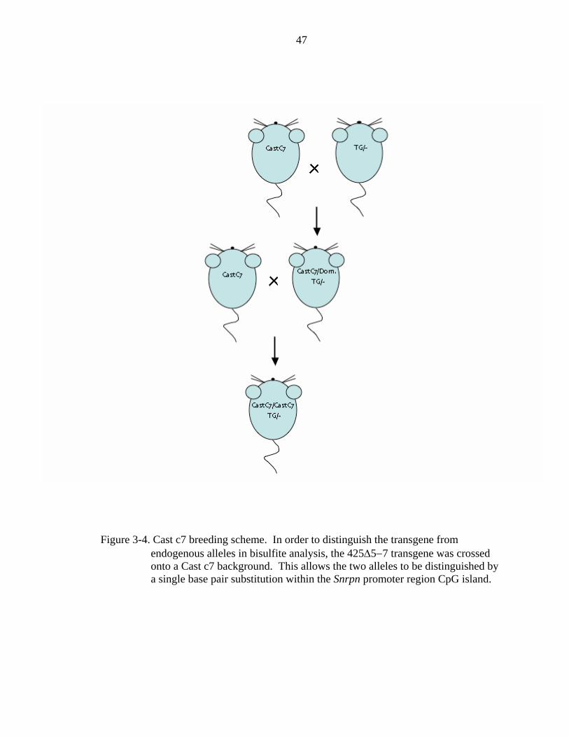
47
Figure 3-4. Cast c7 breeding scheme. In order to distinguish the transgene from endogenous alleles in bisulfite analysis, the 425Δ5−7 transgene was crossed onto a Cast c7 background. This allows the two alleles to be distinguished by a single base pair substitution within the Snrpn promoter region CpG island.

48
Figure 3-5. Bisulfite sequence analysis of maternally transmitted transgene. Sequence analysis of genomic DNA converted by sodium bisulfite reveals that the majority of transgenic alleles are methylated at the Snrpn promoter CpG island when inherited from the mother. A minority of alleles escape methylation, perhaps accounting for the leaky expression seen from both the transgene and endogenous alleles.

49
Figure 3-6. Bisulfite sequence analysis of paternally transmitted transgene. Sequence analysis of genomic DNA converted by sodium bisulfite reveals that the majority of transgenic alleles are unmethylated at the Snrpn promoter CpG island when inherited from the father.

50
CHAPTER 4 REFINEMENT OF THE PWS-IC
Introduction
Considerable effort has been made to understand the cis elements that make up the
PWS-IC. Much of this work is in the context of targeted deletions of the region
surrounding exon 1 of Snrpn. Initially the location of the PWS-IC was mapped to a
region in humans that includes both the promoter and first exon of SNRPN. This was
accomplished by fine mapping of paternal microdeletions that resulted in PWS. The
smallest region of overlap (SRO) between these deletions defined the location of the
PWS-IC within humans (Saitoh et al., 1996). Deletion of this region is associated with
absent expression of all the paternally expressed genes within the locus.
In 1998, our lab published a murine PWS-IC deletion model. This model contained
a 35kb deletion of the syntenic region within the mouse on chromosome 7 centered
around exon 1 of Snrpn (Yang et al., 1998) encompassing 16 kb of sequence 5’ and 19 kb
3’ to exon 1. Mice which inherit this deletion from the father lack expression of the
paternal genes Mkrn3, Ndn, and Ipw, as well as the Snrpn long transcript, and exhibit
several phenotypes common to PWS infants, including poor suckle, failure to thrive, and
lethality after approximately seven days. In contrast, mice inheriting a smaller intergenic
Snrpn deletion that removed the 3’ part of exon 5, exon 6 and the 5’ part of exon 7 had no
obvious phenotypic abnormalities when inherited from either the father or the mother.
These data suggested that both the location and function of the PWS-IC were conserved

51
between mouse and man and that the mouse could serve as a model with which to study
the mechanism of imprinting within the PWS locus.
With the goal of further refining the location of the PWS-IC the Beaudet lab
created two smaller deletions also centered on the first exon of Snrpn (Bressler et al.,
2001). The first, encompassing 0.9 kb, removed both the promoter and a portion of the
Snrpn differentially methylated region 1 (DMR1). These mice were phenotypically
normal upon either maternal or paternal inheritance. Analysis of DNA methylation at
both Snrpn and Ndn revealed no disturbance of the normal pattern as measured by
methylation sensitive restriction digests. This suggests that the sequence elements
necessary to confer imprinted expression upon the locus do not reside within this interval.
The second deletion removed a larger 4.8 kb segment also centered on exon 1. These
mice demonstrated a partial or mosaic phenotype with substantial postnatal lethality
(~40−50%) and growth deficiency when the deletion was inherited from the father. In
contrast, maternal inheritance resulted in no phenotype as would be expected in a PWS
IC deletion. Expression analysis was limited to the upstream genes as the entire Snrpn
promoter was removed in this deletion. Ndn and Mkrn3 showed reduced levels of
expression and increased DNA methylation when the deletion was paternally inherited.
Although the partial imprinting defect makes these results difficult to interpret, one can
make the assumption that some elements of the PWS-IC do lie within the deleted
interval.
The Yang lab recently documented several transcription binding sites within the
Snrpn promoter region (Rodriguez-Jato et al., 2005). Using in-vivo footprinting and
chromatin immunoprecipitation (ChIP) analysis they were able to show that several sites
52
within this region are occupied. Three footprints were shown to be active on the paternal
allele. Footprint P2 appears to be a potential E2F binding site. This family of
transcription factors participates in transcriptional activation and suppression. Footprint
P5 was shown to associate with NRF-1. This site is conserved between mouse and
human and also overlaps with a previously reported seven base pair element (SBE), a cis
acting element shown to be required for promoter activity (Hershko et al., 2001).
Footprint P6/M6 was shown to be active on both the paternal and maternal alleles,
although the patterns were distinctly different. P6/M6 correspond to overlapping NRF-1
and CTCF binding sites however only the NRF-1 site on the paternal allele was
confirmed to be occupied by ChIP. Three other footprints were analyzed within the
promoter but do not appear to be occupied or correspond to known transcription binding
sites. The Yang lab also showed that elements with in the first intron of Snrpn were
preferentially bound only on the paternal allele. These three binding sites for the
transcription factors NRF-1, YY1, and SP1 are phylogenetically conserved between
mouse and man. NRF-1 and YY-1 have been shown to interact with chromatin
modification enzymes suggestive of their possible role in maintaining or establishing the
imprinted expression pattern in the locus. This agrees with the observation that this
conserved intronic region has been shown to be associated with an open, active chromatin
state. How these various factors may work to coordinate an imprinted expression pattern
is unknown and additional work is needed to understand their roles.
With the location of the PWS-IC established in the mouse, understanding the
epigenetic changes that occur within the IC could provide insight into the mechanism of
imprinting at the PWS/AS locus. As discussed earlier, differential methylation at CpG

53
islands is typically found within imprinted loci. The PWS/AS locus is no exception.
Two differentially methylated regions (DMR’s) exist within the PWS-IC, one within the
promoter of Snrpn (DMR1) and one within the first intron (DMR2) (Shemer et al., 1997).
These DMR’s are hypomethylated on the paternal allele and hypermethylated on the
maternal allele. Methylation of CpG’s within the PWS-IC of mice occurs during
oogenesis, however in humans it occurs after fertilization suggesting that this is not the
primary imprint(El-Maarri et al., 2001). It has also been shown that parent specific
differential methylation of histone H3 exists in the same region (Xin et al., 2001). H3
Lys9 is methylated on the maternal copy of the PWS-IC, while H3 Lys4 is methylated on
the paternal copy. H3 Lys-9 methylation is correlated with transcriptional silencing
while H3 Lys4 methylation is correlated with transcriptional activity suggesting a
possible epigenetic switch. Furthermore CpG methylation has been tied to the G9a H3
Lys9/Lys27 methyltransferase (Xin et al., 2003). It’s been hypothesized that H3 Lys9
methylation could be the primary imprint and may trigger CpG methylation of the PWS-
IC through G9a leading to maternal silencing of the paternal genes on the maternal allele
after fertilization.
This chapter discusses modifications to the 425Δ5−7 BAC designed to further
elucidate the PWS-IC.
Results
BAC Modification
As a means to further define the minimal cis elements necessary to establish and
maintain imprinted expression in the locus I have created a series of 3 nested deletions
within the imprinted 425Δ5−7 BAC (Figure 4-1). The 5’ anchor point of all three

54
deletions was positioned at approximately 30kb upstream of Snrpn exon 1. This point
was selected because targeted deletions made within the lab that extend an additional 12
kb further upstream of this point had proven to have no effect on imprinting (Peery et al.,
manuscript in preparation). The largest deletion, 425Δ30kb, extends to 133 bp upstream
of exon 1, removing all but the smallest minimal promoter (SMP) of Snrpn (Hershko et
al., 2001). This point was selected as to allow for transcription of the Snrpn minigene
reporter. The smallest deletion, 425Δ17kb, extends to 11.5kb upstream of exon 1 and is
approximately 17kb in length. This point was selected based on reports from the Beaudet
lab of a 100kb targeted deletion anchored at the 3’ end at the same position (Wu et al.,
2006). As will be discussed in the next chapter, this deletion had no effect on imprinting.
The third deletion, 425Δ23kb, extends to approximately 6kb upstream of exon 1 and is
23kb in length. This was selected as a midway point between the largest and smallest
deletions. The 425Δ5−7 BAC was targeted to produce these modifications using the
Lambda recombineering system previously discussed.
Production of Transgenic Mice
The 425Δ30kb BAC was chosen as the first construct to inject as it was supposed
that this deletion would have the greatest effect. Fresh 425Δ30kb BAC DNA was
prepared as previously described and was injected into the pronuclei of fertilized FVB
oocytes at a concentration of 2.5 ng/ul. Injected oocytes were transplanted at the two cell
stage into the oviducts of pseudopregnant B6D2 F1 mice. The resulting pups were
allowed to mature to 3 weeks of age at which time they were ear punched for
identification and tail clipped to obtain DNA for genotyping. Genomic tail DNA was
used as a template for PCR amplification of the transgene to screen for founders. The
55
primers T7F 5’- GTA ATA CGA CTC ACT ATA GGG C – 3’ and T7R1 5’ – CTC CAA
TCA TGT TCA ACT GTC C – 3’ amplify a region of the BAC adjacent to the vector
arms and are transgene specific. Five founders were obtained; lines C, D, E, F, and G.
Of these only lines C, D, and E transmitted to their offspring. Lines F and G were
discarded.
Analysis of 425Δ30kb Transgenic Lines
Founder animals were mated to FVB/N males or females to establish breeding
colonies and test for chimerism. Lines C, D, and E transmitted through the germ line and
the resulting F1 transgenic pups were allowed to mature to six weeks of age. At six
weeks, F1 transgenic males and females from each line were set up in matings with
FVB/N males and females to test for expression of the transgene upon both maternal and
paternal inheritance. Brains were collected from the resulting F2 day old neonatal pups.
RNA was extracted from the brains using RNAzol. The RNA was used as a template to
produce cDNA that was then used to program PCR reactions that spanned exons 4−8 of
Snrpn. Since the modification to the 425Δ5−7 BAC removes exons 5−7 the resulting
transgenic transcript appears as a band of approximately 350 base pairs (bp). This
includes 242 bp of exonic sequence plus the additional lox-p left over from targeting.
The endogenous band spans 513 bp. Unexpectedly, the 425Δ30kb transgene was
silenced in lines D, and E when maternally inherited. Line D never transmitted the
transgene paternally however line E expressed the Snrpn reporter gene upon paternal
transmission. Line C expressed the transgene biallelically, suggesting that a linearization
of the transgene had occurred at a point incompatible with imprinted expression.

56
Bisulfite sequencing was performed on the C and E lines to test for methylation at
the Snrpn DMR1 as was done with the 425Δ5−7 lines. However, the 30kb deletion made
it possible to amplify DMR1 without first backcrossing the transgene onto the Cast.c7
line of mice. The resulting bisulfite amplicon retains 10 of the original 15 CG
dinucleotides present within the CpG island. Twenty five micrograms of DNA of
genomic brain DNA from the C and E lines was bisulfite converted and then used to
program PCR reactions with the following primer set; 425.06 bisulf F2 5’- GTT TTA
GGA GTT RGA GGT TTT TT – 3’ and 425.06 bisulf R2 5’ – AAC CCA AAT CTA
AAA TAT TTT AAT C – 3’. The products from these reactions were cloned and
sequenced. Data from 11 clones representing a paternally transmitted transgene and 9
clones representing a maternally transmitted transgene was obtained from the E line. In
summary, all paternally transmitted E alleles were completely unmethylated. In contrast,
five of nine maternal E alleles were completely methylated. Two of nine alleles were
methylated at nine sites. One of nine alleles was methylated at only site, and one allele
was completely unmethylated.
Discussion
The lack of data from multiple lines makes drawing strong conclusions difficult for
these experiments. However, it seems highly unlikely that imprinted expression of the
425Δ30kb transgene from line E could be due to an insertional effect. Therefore some
inferences can be made. First, line E expressed the Snrpn reporter in a correctly
imprinted fashion and line D was silenced when maternally inherited. This suggests that
all the PWS-IC cis-regulatory elements necessary for imprinting Snrpn are within the
region between intron 1 and 133bp upstream of exon 1. Since imprinting was preserved

57
in this, the longest deletion of the three construct built, the other two BACs were not
injected. The footprint and ChIP data reported by Rodriguez-Jato et-al demonstrate that
there are conserved elements within this region that are actively occupied, some in a
parent of origin specific manner (Rodriguez-Jato et al., 2005). These include NRF-1,
YY1, and SP1 sites located within intron 1 and an NRF-1 site just upstream of exon one
that had previously been show to be necessary for promoter activity (Hershko et al.,
2001). It can also be presumed that the AS-IC is still intact within this construct since the
transgene is silenced upon maternal transmission. A likely location would be upstream of
the deletion since such a small interval of sequence remains downstream.
A second observation is that the mechanism governing the imprinted expression of
Snrpn likely differs from that governing the upstream cluster of genes. As just
mentioned, Bressler et-al showed the region encompassing the first 600 bp upstream of
Snrpn exon 1 was dispensable for imprinting as judged by expression and methylation of
Ndn. Only when an additional 4kb of sequence upstream was deleted did they see an
imprinting defect. Although expression of the upstream genes is not measurable in this
transgenic system, it’s fairly obvious that this more proximal region is important for
imprinting Snrpn since only 133bp of upstream sequence confer imprinted expression on
the E line. It seems possible that maternal silencing of the Snrpn long transcript may
simply be due to methylation of the promoter, thereby blocking access to trans-acting
factors required for transcription. Imprinting of the upstream genes however, would
require a longer range mechanism, perhaps similar to the active chromatin hub seen in the
beta-globin locus (Patrinos et al., 2004; Zhou et al., 2006). Looping of the upstream
genes to the PWS-IC has already been demonstrated in human cell culture (Arnold

58
Heggestad personal communication). The cis-regulatory elements that would control this
type of mechanism have yet to be identified.
A third point that can be made comes from the observation that the 425Δ30kb C
line expresses transgenic Snrpn in a biallelic fashion. Since the AS-IC is the element that
silences the maternal allele, mapping the point at which the BAC linearized before
integration may provide clues to the location of this element.
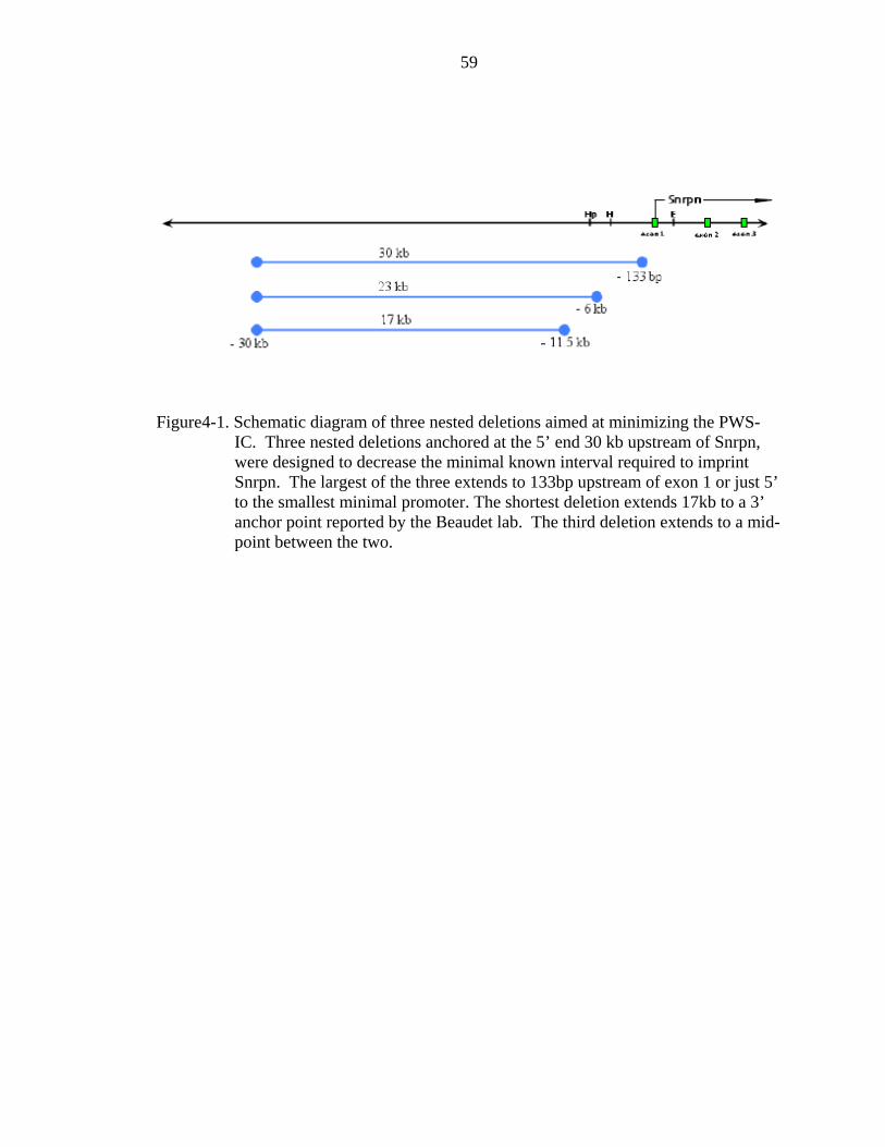
59
Figure4-1. Schematic diagram of three nested deletions aimed at minimizing the PWS-IC. Three nested deletions anchored at the 5’ end 30 kb upstream of Snrpn, were designed to decrease the minimal known interval required to imprint Snrpn. The largest of the three extends to 133bp upstream of exon 1 or just 5’ to the smallest minimal promoter. The shortest deletion extends 17kb to a 3’ anchor point reported by the Beaudet lab. The third deletion extends to a mid-point between the two.

60
Figure 4-2. rt-PCR analysis of 425Δ30kb mouse lines. rt-PCR analysis of two transgenic mouse lines derived with the 425Δ30kb BAC reveals proper imprinted expression of the E line and biallelic expression of the C line. Proper expression of the E line suggests all cis-regulatory elements required for imprinted expression of Snrpn are contained within its interval. Biallelic expression of line C may be due to linearization of the BAC that is incompatible with imprinted expression.
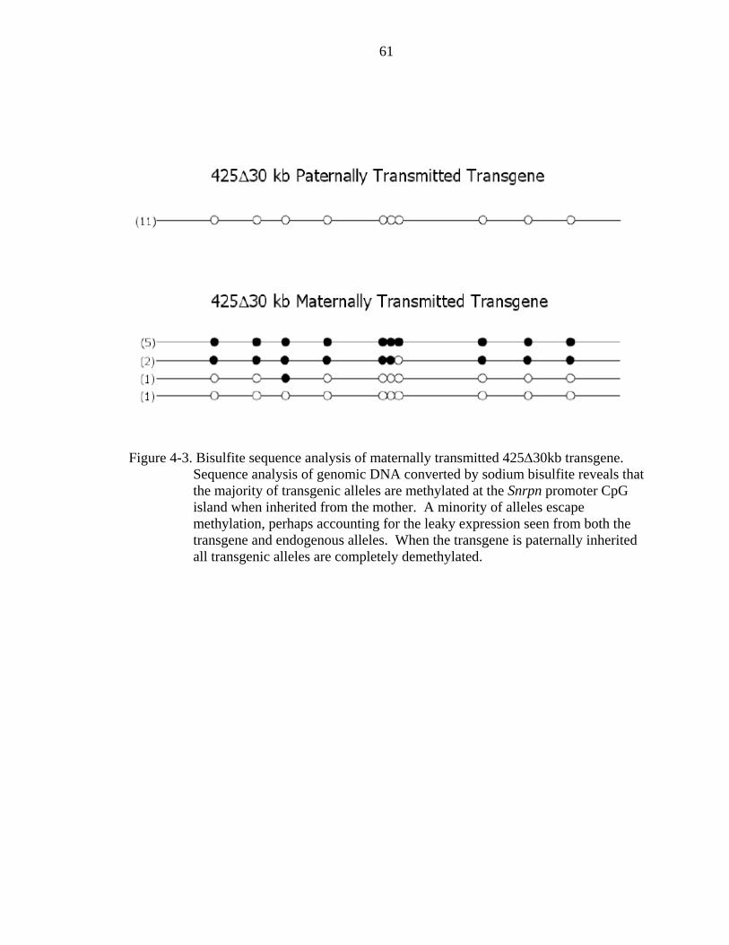
61
Figure 4-3. Bisulfite sequence analysis of maternally transmitted 425Δ30kb transgene. Sequence analysis of genomic DNA converted by sodium bisulfite reveals that the majority of transgenic alleles are methylated at the Snrpn promoter CpG island when inherited from the mother. A minority of alleles escape methylation, perhaps accounting for the leaky expression seen from both the transgene and endogenous alleles. When the transgene is paternally inherited all transgenic alleles are completely demethylated.
62
CHAPTER 5 DEFINING THE LOCATION OF THE MURINE AS-IC
Introduction
Imprinting in the PWS/AS locus is dependent upon coordinate control by a
bipartite imprinting center, consisting of a PWS-IC that drives expression from the
paternal allele and an AS-IC that is responsible for silencing the PWS-IC on the maternal
allele. While the position of the AS-IC has been localized to a region approximately
35kb upstream of Snrpn exon 1 in humans, its location in the mouse is still unknown,
hampering efforts to understand its mechanism. Targeted deletions in the mouse, based
on a similar distance from Snrpn in the human, failed to disturb the differential
methylation and imprinted expression patterns of Snrpn and Ndn suggesting that the
location is not conserved. This chapter will focus on efforts to define the murine AS-IC
For years it has been postulated that silencing of the maternal allele may involve
expression of a series of alternative SNRPN transcripts that arise from a set of
infrequently used upstream exons. The first evidence for this came from analysis of
Angelman Syndrome patients inheriting microdeletions in a region upstream of exon 1.
A consequence of these deletions is a block in the paternal to maternal imprint switch
during gametogenesis resulting in a paternal epigenotype on the maternal chromosome
and biallelic expression of the paternal set of genes. Several upstream alternative SNRPN
exons were then isolated from this region (Dittrich et al., 1996; Farber et al., 1999) which
extends >700kb upstream of exon 1 (Figure 5-1). These upstream exons share a degree
of sequence similarity to each other and in turn are also similar to exon one, implying that
63
they arose from duplications of exon one. Alternative transcripts initiate from the
paternal allele from two of these exons, u1A and u1B and give rise to various splice
variants. All splice variants skip exon one and splice to exon two. Maternal specific
methylation is also present in the regions upstream of these exons. What is most
interesting about these upstream exons is that exon u5 is deleted in all AS patients with
an IC deletion. Exon u5 is contained within multiple alternative transcripts so the
argument could be made that these transcripts could be somehow involved in the paternal
to maternal imprint switch. An alternative explanation is that the location of exon u5 is
merely a coincidence and the actual AS-IC is a cis regulatory element located in close
proximity. An argument in favor of this hypothesis is that no alternative transcripts to
date have been identified which initiate at exon u5. If transcription of u5 were necessary
for AS-IC function, patients with deletions encompassing exons u1A or u1B should be
present since these are the exons at which those transcripts identified to date initiate. It is
just as likely however that as yet unidentified transcripts that do initiate at u5 are present
within developing gametes, tissue which has not yet been tested.
Studying these issues in humans is complicated by the fact that the tissues in which
the imprints are being established, developing gametes, are difficult if not impossible to
acquire. As discussed earlier, gene content and the imprinted expression pattern within
the PWS/AS locus is conserved in the mouse making it an attractive model system.
However the location of the murine AS-IC is still not known. Given that the AS-IC is
located at approximately 35kb upstream of SNRPN in humans our lab targeted deletions
at the corresponding location in the mouse. Nested deletions of 7 and 12kb had no effect
on methylation or expression of the paternally expressed genes suggesting that the
64
location of the AS-IC relative to Snrpn is not conserved in the mouse (E. Peery personal
communication). Closer examination of this region reveals that the mouse also contains
several exons upstream to Snrpn that extend approximately 500kb upstream. Rt-pcr
analysis reveals that these upstream exons are also involved in alternative transcripts
similar to humans (Bressler et al., 2001; Landers et al., 2004). Nine U exons have been
identified to date. They participate in multiple splice variants that fall into two
categories, those that join to Snrpn exon 2 and those that exclude Snrpn and splice further
downstream to the non-coding Ipw exons. As is the case in humans, it appears that the
upstream exons are the result of multiple duplication events as there is a high degree of
homology to the region surrounding exon 1. Sequence is highly conserved among the
murine exons, more so than in humans, approaching 90%, however there is very little
conservation between human and mouse. The function that these alternative exons play
in imprinting is yet to be determined. In fact conflicting results from two labs either
supports or excludes alternative transcripts from playing a role. Extensive work by
LeMeur et-al characterized the temporal and spatial regulation of these transcripts (Le
Meur et al., 2005). Transcripts containing various U exons were found to be expressed
within embryos 10.5 days post-coitum (dpc) and later exclusively within the nervous
system. This group however failed to detect expression of the U exons within testis and
ovary samples suggesting that alternative transcripts are not produced within developing
gametes. This would preclude a role for alternative transcripts in establishing imprints
within the PWS/AS locus during a time when these marks are placed. Mapendano et-al
however did detect expression of alternative transcripts arising from exon U2 and
splicing to Snrpn exon 2 within the ovary by rt-pcr (Mapendano et al., 2006). In-situ
65
hybridization, utilizing exon U2 as a probe, detected expression within both oocytes and
their supporting granulosa cells within Graafian follicles. Stronger signals were detected
within oocytes and granulosa cells of secondary and developing follicles. While this
contradicts the observations by Le Meur et-al, it supports the idea that alternative
transcripts may be important to imprint establishment.
In a recently published report, Wu et al targeted deletions to the murine region
upstream of Snrpn (Wu et al., 2006). Two lines of mice were generated mutating the
region 5’ of the Snrpn promoter. The first was an anchor mutation that introduced a
targeting vector 13kb upstream of Snrpn exon 1. The insertion was comprised of the 3’
portion of an Hprt selectable cassette, a loxP site, a puromycin selectable marker, and a
6kb duplication of genomic sequence. This mutation is referred to as AS-ICan. The
second mutation was an 80kb deletion of genomic sequence extending upstream of the 3’
anchor created by AS-ICan. Paternal inheritance of either mutation resulted in a normal
pattern of methylation at the Snrpn and Ndn CpG islands. Maternal inheritance of the
AS-ICan mutation resulted in an imprint defect with both maternal and paternal alleles
being unmethylated at Snrpn. This was correlated with inappropriate maternal expression
of Snrpn. Ndn was also affected with a partial or mosaic pattern of methylation on the
maternal allele. All subsequent breeding of AS-ICan mutant mice resulted in an AS like
imprinting defect, with inappropriate hypomethylation of the maternal allele. Maternal
inheritance of the 80kb deletion resulted in an imprinting defect with incomplete
penetrance. Three groups of mice with different methylation patterns were observed.
The first (3 of 8 heterozygous mice) exhibited a normal pattern of methylation as
compared to wild type litter-mates. A second group (2 of 8) showed partial methylation

66
of the maternal allele. The third group of three was completely unmethylated. In trying
to understand the disruption of imprinting in both mutant lines, it is important to note that
the puromycin cassette is present and transcribes in the opposite orientation to Snrpn in
the AS-ICan mutant. In the 80kb deletion, the puromycin cassette is deleted by cre-lox,
but the Hprt promoter is present and expresses in the same direction as Snrpn. In the
context of the upstream exons, the anchor insertion alters the distance between the
putative AS-IC and the PWS-IC and perhaps the distance between the two regulatory
elements is important to coordinating imprinted expression. A second explanation is that
transcription from the puromycin cassette interferes with alternate transcripts from the
upstream exons. This second explanation may also be valid in the case of the 80kb
deletion. Transcription towards Snrpn from the Hprt promoter may interfere with the
normal transcription signal being sent from upstream.
More evidence regarding the location of the AS-IC comes from our lab in the form
of imprinted transgenes (Figure 5-2). Multiple transgenic lines have been created and
characterized in our lab extending variable distances upstream of Snrpn. As mentioned
earlier, a phage clone carrying 33kb of murine sequence 5’ to Snrpn did not imprint
correctly as a single copy transgene suggesting that the AS-IC is not within that interval.
The 425Δ5−7 BAC transgene however does imprint correctly suggesting that the AS-IC
is somewhere within the 100kb interval upstream of Snrpn. The 215B BAC transgenic
line also is imprinted correctly. What is interesting about this line is that it has suffered a
truncation at approximately 40kb 5’ to Snrpn. The location of mouse upstream exon 1 is
at 43kb 5’ to Snrpn. Whether the 215B line does contain U1 is unknown and is currently
being investigated.
67
These observations have led to the following hypothesis (Figure 5-3). First, the
upstream exons or an element nearby act as the AS-IC and establishes the primary
imprint upon the PWS-IC. Second, the high degree of homology between the murine
upstream exons allows for compensation to occur when one is deleted. This may explain
the variable penetrance seen in the 80kb deletion. Exons further upstream may not be as
efficient in a role as the AS-IC as the closer U1. Lastly, only deletion of all U exons will
result in a complete AS like phenotype. This would prove to be difficult in a targeted
deletion strategy as it may require individually targeting each of nine identical sequences
over a distance greater than 500kb. The BAC transgenic model provides an excellent
opportunity to ask these question as only three upstream exons are present within the
425Δ5−7 BAC interval.
In order to investigate the role that murine upstream Snrpn exons play in imprinted
expression of the locus several modifications to the 425Δ5−7 BAC were made. This
chapter characterizes those modifications in an attempt to understand AS-IC function.
Results
BAC modification
The 425Δ5−7 BAC transgene contains 3 upstream exons, U1, U2, and U3 at 43kb,
75kb, and 96 kb upstream of Snrpn exon 1 respectively. In order to test their role in AS-
IC function, a 2499 bp region of homology in which each U exon is imbedded, was
targeted for deletion in a way as to leave the surrounding DNA intact. This 2.5kb region
of homology likely represents the region of Snrpn exon 1 duplicated to form the U exons
and may contain cis regulatory element required for silencing the PWS-IC. Targeting
vectors to be used to delete each upstream exon by BAC recombineering were designed
68
as follows. Primers were designed to flank each region of homology with approximately
1kb of additional sequence in either direction. The primer sets used are as follows; U1 –
U1delF1 5’- GAC TTC AGT ACA TGT TCC AAC –3’ and U1delR2 5’- TAG ATA
GTC CAG TAC CAG GAC T –3’, U2 – U2delF 5’- CCA GAA GAT GTT CCA ACT
GG –3’ and U2delR 5’- CCA CCT AAC CAA TGG ACA GA 3’, U3 – U3delF1 5’-
CCA GAT AGT GTA CCT CAT GTC –3’ and U3delR1 5’- GAG GCT TCC ACA TAA
GGT CTT –3’. The products amplified with these primers were cloned into the pGEM T
easy cloning vector from Promega. In order to delete the region containing each U exon,
restriction endonuclease cleavage sites were identified and used to remove the portion of
sequence containing the regions of homology leaving 1kb of genomic sequence in either
direction intact. The U1 T-easy vector was cleaved with AflII and PacI to release a
2753bp fragment. The U2 T-easy vector was cleaved with BamHI to release a 2515bp
fragment. The U3 T-easy vector was cleaved with BssHII and Csp45I to release a
2608bp fragment. Each linearized vector was blunted with Klenow and a loxP flanked
neomycin resistance cassette was ligated in. In order to avoid cross-reaction between the
various targeted areas when multiple modifications were made to a single BAC, mutant
loxP sites were used to flank the neo cassette in the U2 and U3 targeting vectors. FRT
sites were used to flank the neo cassette in the U1 targeting vector. Lambda red
recombineering was used to make targeted deletions of the three U exons in the 425Δ5−7
BAC using these vectors. Transgene 425ΔU1-U3 has all three U exons deleted (Figure
5-4A). If our compensation hypothesis is correct, all three exons must be deleted to see a
complete imprinting defect. Transgene 425ΔU2/U3 has only U2 and U3 deleted (Figure
5-4B). This is important to show that exon U1, or the region immediately adjacent to it,

69
serves as the AS-IC. Two more constructs, 425ΔU1/U3 and 425ΔU1/U2 leave exon U2
or U3 intact in order to demonstrate compensation.
Production of Transgenic Mice
The 425ΔU1-U3 and 425ΔU2/U3 BAC’s were chosen as the first constructs to
inject. Fresh BAC DNA was prepared as previously described and was injected into the
pronuclei of fertilized FVB oocytes at a concentration of 2.5 ng/ul. Injected oocytes were
transplanted at the two cell stage into the oviducts of pseudopregnant B6D2 F1 mice. The
resulting pups were allowed to mature to 3 weeks of age at which time they were ear
punched for identification and tail clipped to obtain DNA for genotyping. Genomic tail
DNA was used as a template for pcr amplification of the transgene to screen for founders.
The primers T7F 5’- GTA ATA CGA CTC ACT ATA GGG C – 3’ and T7R1 5’ – CTC
CAA TCA TGT TCA ACT GTC C – 3’ amplify a region of the BAC adjacent to the
vector arms and are transgene specific. Four founders were obtained for the 425ΔU1-U3
line and two founders were obtained for the 425ΔU2/U3 line. All were found to transmit
the transgene and were consequently mated to Cast.c7 males and females to establish
colonies.
Analysis of 425ΔU1-U3 and 425ΔU2/U3 Transgenic Lines
Founder animals were mated to Castc7 males or females to establish breeding
colonies. Lines A, B, C, and D transmitted through the germ line and the resulting N1
transgenic pups were allowed to mature to six weeks of age. At six weeks, N1 transgenic
males and females from each line were set up in matings with FVB/N males and females
to test for expression of the transgene upon both maternal and paternal inheritance.
Brains were collected from the resulting N2 day old neonatal pups. RNA was extracted
70
from the brains using RNAzol. The RNA was used as a template to produce cDNA that
was then used to program pcr reactions that spanned exons 4−8 of Snrpn. Since the
modification to the 425Δ5−7 BAC removes exons 5−7 the resulting transgenic transcript
appears as a band of approximately 350 base pairs (bp). This includes 242 bp of exonic
sequence plus the additional lox-p left over from targeting. The endogenous band spans
513 bp. Line A did not express the transgene upon paternal or maternal inheritance,
possibly due to insertional effects or breakage of the transgene at a point incompatible to
expression prior to insertion. Line D on the other hand expressed the transgene from both
the paternal and maternal alleles, suggesting that the AS-IC had been deleted by
removing the upstream exons. Lines B and C have only been tested for maternal
expression at this point. Line B expressed the maternally inherited transgene while line C
did not. Conclusions on these two lines cannot be made until expression data from the
paternally inherited alleles can be analyzed
Discussion
Due to the overlap of human Snrpn exon u5 with the AS-SRO, it has been
hypothesized that the upstream exons have some role in AS-IC function. However,
testing this hypothesis in the mouse has proven to be difficult, possibly due to the high
degree of homology among the nine murine upstream exons. Using our BAC transgenic
model provides a simpler model since only three U exons are contained within its
interval.
Since this data set is not yet complete, no firm conclusions can be made, however
biallelic expression from the 425ΔU1-U3 D line and maternal expression from the B line
suggest that the U exons do play a role in silencing the maternal allele. Expression from
71
the B and C paternally inherited alleles remains to be analyzed and this will prove to be
important in determining the imprinting status of this construct. Also it will be important
to determine whether or not the BAC remained intact during integration. If the transgene
is indeed intact throughout the region 5’ of Snrpn, this raises the possibility that the U
exons also serve some sort of insulating purpose. All four 425Δ5-7 transgenic lines
proved to be capable of silencing maternally inherited Snrpn suggesting that the BAC
contains sufficient sequence to overcome the chromatin into which it inserts. The
425ΔU1-U3 lines do not seem to have this capability as the A line is completely silenced
upon both paternal and maternal inheritance. Southern blots spanning the region 5’ to
Snrpn are currently underway to determine the whether the transgenes have remained
intact.
The 425ΔU2-U3 lines still remain to be analyzed. Determining their imprinting
status will provide conclusive evidence about whether the AS-IC resides within the
upstream exons. Cloning of additional BAC constructs 425ΔU1/U3 and 425ΔU1-U2 has
been completed however they have not yet been injected as transgenes. Data from these
lines should provide evidence about whether U2 and U3 can compensate for AS-IC
function when U1 is deleted.
The data presented in this chapter provides preliminary evidence that the AS-IC
resides within the upstream exons. However, this data does not suggest a mechanism by
which the AS-IC imprints the maternal allele. Two possibilities immediately come to
mind. First and probably the most obvious is that transcription factor binding sites lie
within the region that includes U1. At this point in time however, this would be difficult

72
to test. Additional work would need to be done to further limit the interval in which the
AS-IC is contained in order for an efficient search for active binding sites could occur.
A second intriguing hypothesis is that active transcription from the upstream
promoter is what triggers heterochromatin formation and subsequent silencing of the
maternal allele. There is precedence for this type of mechanism in multiple systems.
First, Mayer et al have shown that transcripts from intergenic spacer sequences between
rRNA genes are important for establishing and maintaining a specific heterochromatin
conformation at the promoter of a subset of rDNA arrays (Mayer et al., 2006). These
150-300 nucleotide transcripts interact with TIP5, the large subunit of the chromatin
remodeling complex NoRC and guide the complex to its target via sequence
complementarity to the rDNA promoter. A second example comes from the analysis of a
patient suffering from α-thalesemia, an inherited form of anemia. This patient was found
to have a deletion that resulted in the juxtaposition a structurally normal α-globin gene
(HBA2) to LUC7L (Tufarelli et al., 2003). Expression of LUC7L from the opposite
strand from HBA2 resulted in an anti-sense transcript that was capable of mediating
methylation and silencing of HBA2 during development. Through the use of transgenic
mice they were also able to show that methylation and modification of chromatin occurs
in cis. A third mechanism by which transcription could result in gene silencing is siRNA
mediated DNA methylation. Until recently this mechanism had only been known to be
active in plants. Kawasaki et al were able to show that mammalian cells are also capable
of siRNA mediated silencing within a cell culture model (Kawasaki and Taira, 2004).
Vector based siRNA’s targeting CpG islands within the E-cadherin and erbB2 genes were
73
shown to be capable of inducing DNA methylation and histone H3 lysine 9 methylation
resulting in repression of transcription.
The work contained in this chapter is ongoing and the data presented preliminary.
Expression from the paternal allele of the 425ΔU1-U3 lines B and C transgenes and
analysis of the 425ΔU2-U3 transgene remains to be done. Also, determination of the
breakpoints for these transgenes will be necessary before any firm conclusions can be
made.

74
Figure 5-1. Schematic diagram of human upstream exons. The PWS-IC region has undergone duplication events resulting in the establishment of several upstream exons. Low levels of paternal transcripts arise from these exons as shown by the arrow heads. Lollipops represent the differential methylation status at specific methylation sensitive restriction sites. Filled circles represent methylated CpG’s while empty circles represent unmethylated CpG’s. (adapted from Farber et al.)
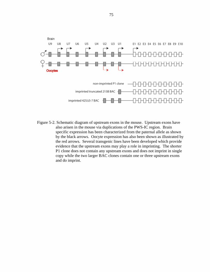
75
Figure 5-2. Schematic diagram of upstream exons in the mouse. Upstream exons have also arisen in the mouse via duplications of the PWS-IC region. Brain specific expression has been characterized from the paternal allele as shown by the black arrows. Oocyte expression has also been shown as illustrated by the red arrows. Several transgenic lines have been developed which provide evidence that the upstream exons may play a role in imprinting. The shorter P1 clone does not contain any upstream exons and does not imprint in single copy while the two larger BAC clones contain one or three upstream exons and do imprint.

76
Figure 5-3. Compensation model of AS-IC activity. Alternative exons further upstream can compensate for loss of those closer to Snrpn. Only by deleting all upstream exons will there be an imprinting defect.

77
Figure 5-4. Schematic diagram of upstream exon deletions. In order to elicit an AS-IC imprinting defect it may be necessary to delete all upstream exons to avoid compensation by those that remain.
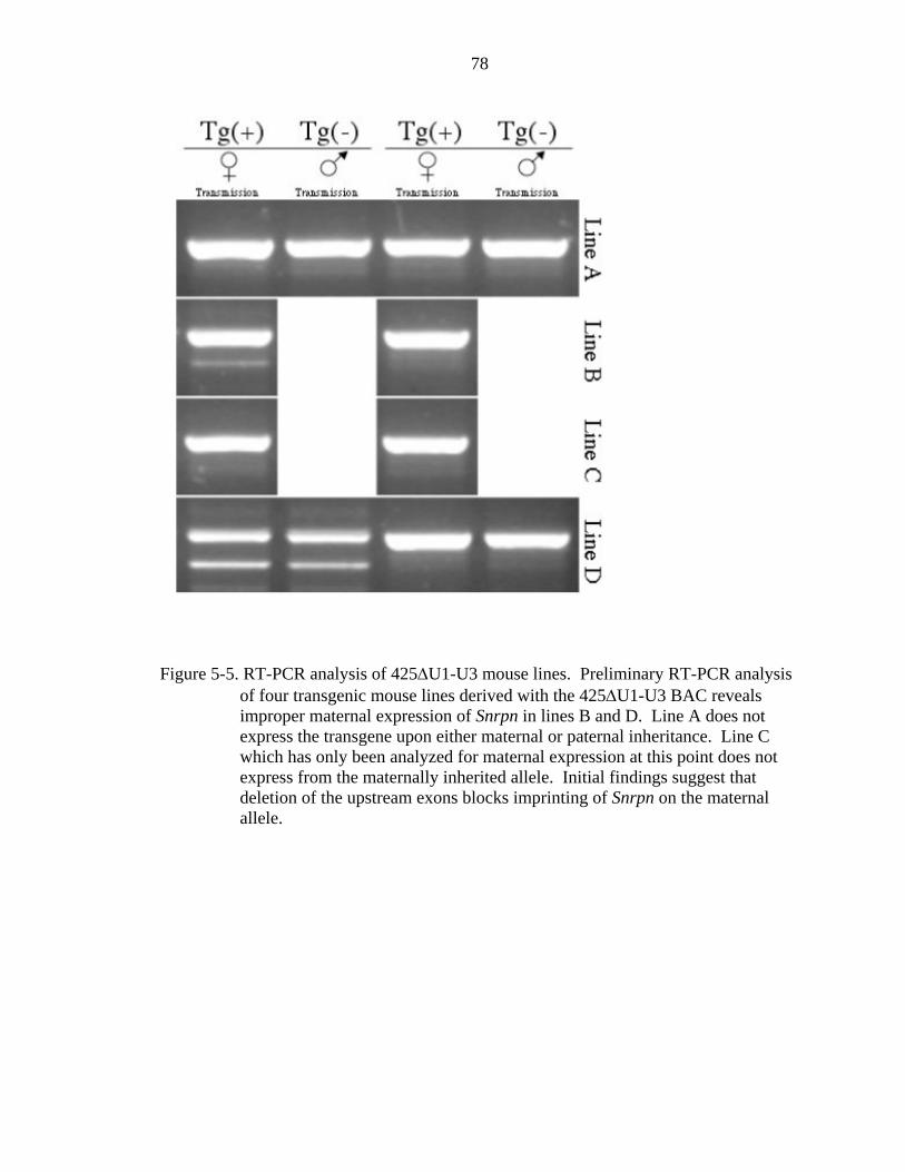
78
Figure 5-5. RT-PCR analysis of 425ΔU1-U3 mouse lines. Preliminary RT-PCR analysis of four transgenic mouse lines derived with the 425ΔU1-U3 BAC reveals improper maternal expression of Snrpn in lines B and D. Line A does not express the transgene upon either maternal or paternal inheritance. Line C which has only been analyzed for maternal expression at this point does not express from the maternally inherited allele. Initial findings suggest that deletion of the upstream exons blocks imprinting of Snrpn on the maternal allele.

79
CHAPTER 6 CONCLUSIONS AND FUTURE DIRECTIONS
The mechanism by which imprinted expression occurs within the Prader-Willi /
Angelman locus is currently unknown. Several animal models have been put forth in
order to further our understanding of this phenomenon, however the regulatory elements
that coordinate parental specific expression of this set of genes remains a mystery. This
work describes a new transgenic model, which will hopefully further our understanding
of the locus.
BAC transgenes have historically been shown to be good models of the genomic
loci, which they represent. Our BAC transgenic model faithfully recapitulates the
imprinted expression of the endogenous Snrpn therefore it must contain all the cis
regulatory elements required for establishment and maintenance of the imprint.
The 425Δ30kb modified BAC shortens the interval known to contain the PWS-IC
considerably and leaves us with a manageable amount of sequence within which to look
for the cis elements of the PWS-IC. Data from Rodriguez-Jato et al has already shown
that conserved transcription binding sites within this interval are occupied in a parent of
origin specific manner and may play a role. Further modifications to the 425Δ5−7 BAC
whereby these individual sites are mutated will help to identify those that are important.
The inability to identify the AS-IC in the mouse has hampered efforts to understand
how it silences the PWS-IC. Observations in humans have suggested that alternative
transcripts arising from exons upstream of Snrpn exon 1 may play a role, however
definitive evidence supporting this hypothesis has yet to be shown. The BAC transgenic

80
model makes it possible to study these upstream exons in isolation from the rest of the
locus. Preliminary evidence presented here suggests that exon U1 or the sequence in
within the duplicated region may serve as the AS-IC. The mechanism by which this
works is still not understood. It is possible that elements within this region attract trans-
acting factors to the locus. These factors could then create a silenced chromatin
conformation which spreads outward to silence the PWS-IC and thereby the paternally
expressed genes on the maternal allele. A second compelling hypothesis is that
transcription arising from the upstream exons regulates the imprint establishment at the
PWS-IC. Transcription of non-coding RNA has been shown to regulate epigenetic states
using a variety of mechanisms in both plant and mammalian cells. Determination of
whether the AS-IC functions via attraction of trans-acting factors or through a
transcription based mechanism is well within the capabilities of this model system.
Experiments are currently under way to insert an oocyte specific promoter in front of
Snrpn exon 1 within a non-imprinted BAC. It will be interesting to see if transcription
through the PWS-IC within the context of a growing oocyte will confer imprinted status
to this transgene. Other obvious directions are to take a “promoter bashing” approach,
deleting larger and larger portions of exon U1 to reveal the relevant sequences.
The work contained within this document will certainly add to the understanding of
imprinting within the PWS/AS, however understanding how the locus is controlled has
further implications. Mouse models for PWS and AS will be important in developing
treatments for these disorders. Understanding the elements that make up the PWS-IC and
AS-IC will be important in developing for developing these models. .Many imprinted
loci are contained within the genome, but only in a small portion is the mechanism of

81
parental gene regulation understood. Furthering the understanding of this locus may
provide clues to how imprinting works at other regions. Also as the possibility of
therapeutic cloning becomes a reality, understanding gene regulation will become
essential. Disrupted imprinting has been suggested to play a role in the low survival rate
of animals cloned by somatic cell nuclear transfer. It is therefore likely that the genetic
reprogramming necessary to clone therapeutic tissues may also prove to be highly error
prone. Understanding how imprinting is controlled will be important to develop these
therapeutic technologies. There have also been reports of disrupted imprinting in
children conceived by intracytoplasmic sperm injection (ICSI). As assisted reproduction
techniques become more commonplace, understanding how imprints are acquired and
maintained will be important in reducing the risks associated with these procedures.
82
LIST OF REFERENCES
Amos-Landgraf, J. M., Ji, Y., Gottlieb, W., Depinet, T., Wandstrat, A. E., Cassidy, S. B., Driscoll, D. J., Rogan, P. K., Schwartz, S. and Nicholls, R. D. (1999). Chromosome breakage in the Prader-Willi and Angelman syndromes involves recombination between large, transcribed repeats at proximal and distal breakpoints. Am J Hum Genet 65, 370-86. Bartolomei, M. S., Webber, A. L., Brunkow, M. E. and Tilghman, S. M. (1993). Epigenetic mechanisms underlying the imprinting of the mouse H19 gene. Genes Dev 7, 1663-73. Barton, S. C., Surani, M. A. and Norris, M. L. (1984). Role of paternal and maternal genomes in mouse development. Nature 311, 374-6. Beaudet, A. L. and Jiang, Y. H. (2002). A rheostat model for a rapid and reversible form of imprinting-dependent evolution. Am J Hum Genet 70, 1389-97. Blaydes, S. M., Elmore, M., Yang, T. and Brannan, C. I. (1999). Analysis of murine Snrpn and human SNRPN gene imprinting in transgenic mice. Mamm Genome 10, 549-55. Boccaccio, I., Glatt-Deeley, H., Watrin, F., Roeckel, N., Lalande, M. and Muscatelli, F. (1999). The human MAGEL2 gene and its mouse homologue are paternally expressed and mapped to the Prader-Willi region. Hum Mol Genet 8, 2497-505. Brannan, C. I. and Bartolomei, M. S. (1999). Mechanisms of genomic imprinting. Curr Opin Genet Dev 9, 164-70. Bressler, J., Tsai, T. F., Wu, M. Y., Tsai, S. F., Ramirez, M. A., Armstrong, D. and Beaudet, A. L. (2001). The SNRPN promoter is not required for genomic imprinting of the Prader-Willi/Angelman domain in mice. Nat Genet 28, 232-40. Buiting, K., Lich, C., Cottrell, S., Barnicoat, A. and Horsthemke, B. (1999). A 5-kb imprinting center deletion in a family with Angelman syndrome reduces the shortest region of deletion overlap to 880 bp. Hum Genet 105, 665-6. Buiting, K., Saitoh, S., Gross, S., Dittrich, B., Schwartz, S., Nicholls, R. D. and Horsthemke, B. (1995). Inherited microdeletions in the Angelman and Prader-Willi syndromes define an imprinting centre on human chromosome 15. Nat Genet 9, 395-400.

83
Butler, M. G., Bittel, D. C., Kibiryeva, N., Talebizadeh, Z. and Thompson, T. (2004). Behavioral differences among subjects with Prader-Willi syndrome and type I or type II deletion and maternal disomy. Pediatrics 113, 565-73. Butler, M. G. and Palmer, C. G. (1983). Parental origin of chromosome 15 deletion in Prader-Willi syndrome. Lancet 1, 1285-6. Cassidy, S. B. and Ledbetter, D. H. (1989). Prader-Willi syndrome. Neurol Clin 7, 37-54. Cattanach, B. M. (1986). Parental origin effects in mice. J Embryol Exp Morphol 97 Suppl, 137-50. Cattanach, B. M. and Kirk, M. (1985). Differential activity of maternally and paternally derived chromosome regions in mice. Nature 315, 496-8. Chai, J. H., Locke, D. P., Ohta, T., Greally, J. M. and Nicholls, R. D. (2001). Retrotransposed genes such as Frat3 in the mouse Chromosome 7C Prader-Willi syndrome region acquire the imprinted status of their insertion site. Mamm Genome 12, 813-21. Chamberlain, S. J. and Brannan, C. I. (2001). The Prader-Willi syndrome imprinting center activates the paternally expressed murine Ube3a antisense transcript but represses paternal Ube3a. Genomics 73, 316-22. Chamberlain, S. J., Johnstone, K. A., DuBose, A. J., Simon, T. A., Bartolomei, M. S., Resnick, J. L. and Brannan, C. I. (2004). Evidence for genetic modifiers of postnatal lethality in PWS-IC deletion mice. Hum Mol Genet 13, 2971-7. Church, G. M. and Gilbert, W. (1985). The genomic sequencing technigque. In Prog. Clin. Biol. Res., vol. 177 (ed., pp. 17-21. Clark, S. J., Harrison, J., Paul, C. L. and Frommer, M. (1994). High sensitivity mapping of methylated cytosines. In Nucleic Acids Res, vol. 22 (ed., pp. 2990-7. Clayton-Smith, J. and Pembrey, M. E. (1992). Angelman syndrome. J Med Genet 29, 412-5. de los Santos, T., Schweizer, J., Rees, C. A. and Francke, U. (2000). Small evolutionarily conserved RNA, resembling C/D box small nucleolar RNA, is transcribed from PWCR1, a novel imprinted gene in the Prader-Willi deletion region, which Is highly expressed in brain. Am J Hum Genet 67, 1067-82.

84
Dittrich, B., Buiting, K., Korn, B., Rickard, S., Buxton, J., Saitoh, S., Nicholls, R. D., Poustka, A., Winterpacht, A., Zabel, B. et al. (1996). Imprint switching on human chromosome 15 may involve alternative transcripts of the SNRPN gene. Nat Genet 14, 163-70. Driscoll, D. J. (1994). Genomic imprinting in humans. Mol Genet Med 4, 37-77. El-Maarri, O., Buiting, K., Peery, E. G., Kroisel, P. M., Balaban, B., Wagner, K., Urman, B., Heyd, J., Lich, C., Brannan, C. I. et al. (2001). Maternal methylation imprints on human chromosome 15 are established during or after fertilization. Nat Genet 27, 341-4. Farber, C., Dittrich, B., Buiting, K. and Horsthemke, B. (1999). The chromosome 15 imprinting centre (IC) region has undergone multiple duplication events and contains an upstream exon of SNRPN that is deleted in all Angelman syndrome patients with an IC microdeletion. Hum Mol Genet 8, 337-43. Gray, T. A., Saitoh, S. and Nicholls, R. D. (1999). An imprinted, mammalian bicistronic transcript encodes two independent proteins. Proc Natl Acad Sci U S A 96, 5616-21. Hata, K., Okano, M., Lei, H. and Li, E. (2002). Dnmt3L cooperates with the Dnmt3 family of de novo DNA methyltransferases to establish maternal imprints in mice. Development 129, 1983-93. Hershko, A. Y., Finberg, Y., Kantor, B., Shemer, R. and Razin, A. (2001). The mouse Snrpn minimal promoter and its human orthologue: activity and imprinting. Genes Cells 6, 967-75. Holm, V. A., Cassidy, S. B., Butler, M. G., Hanchett, J. M., Greenswag, L. R., Whitman, B. Y. and Greenberg, F. (1993). Prader-Willi syndrome: consensus diagnostic criteria. Pediatrics 91, 398-402. Horsthemke, B., Dittrich, B. and Buiting, K. (1997). Imprinting mutations on human chromosome 15. Hum Mutat 10, 329-37. Howell, C. Y., Bestor, T. H., Ding, F., Latham, K. E., Mertineit, C., Trasler, J. M. and Chaillet, J. R. (2001). Genomic imprinting disrupted by a maternal effect mutation in the Dnmt1 gene. Cell 104, 829-38. Jacobs, P. A., Wilson, C. M., Sprenkle, J. A., Rosenshein, N. B. and Migeon, B. R. (1980). Mechanism of origin of complete hydatidiform moles. Nature 286, 714-6.

85
John, R. M., Aparicio, S. A., Ainscough, J. F., Arney, K. L., Khosla, S., Hawker, K., Hilton, K. J., Barton, S. C. and Surani, M. A. (2001). Imprinted expression of neuronatin from modified BAC transgenes reveals regulation by distinct and distant enhancers. Dev Biol 236, 387-99. Jong, M. T., Carey, A. H., Caldwell, K. A., Lau, M. H., Handel, M. A., Driscoll, D. J., Stewart, C. L., Rinchik, E. M. and Nicholls, R. D. (1999a). Imprinting of a RING zinc-finger encoding gene in the mouse chromosome region homologous to the Prader-Willi syndrome genetic region. Hum Mol Genet 8, 795-803. Jong, M. T., Gray, T. A., Ji, Y., Glenn, C. C., Saitoh, S., Driscoll, D. J. and Nicholls, R. D. (1999b). A novel imprinted gene, encoding a RING zinc-finger protein, and overlapping antisense transcript in the Prader-Willi syndrome critical region. Hum Mol Genet 8, 783-93. Kawasaki, H. and Taira, K. (2004). Induction of DNA methylation and gene silencing by short interfering RNAs in human cells. Nature 431, 211-7. Kishino, T., Lalande, M. and Wagstaff, J. (1997). UBE3A/E6-AP mutations cause Angelman syndrome. Nat Genet 15, 70-3. Knoll, J. H., Glatt, K. A., Nicholls, R. D., Malcolm, S. and Lalande, M. (1991). Chromosome 15 uniparental disomy is not frequent in Angelman syndrome. Am J Hum Genet 48, 16-21. Knoll, J. H., Nicholls, R. D. and Lalande, M. (1989a). On the parental origin of the deletion in Angelman syndrome. Hum Genet 83, 205-7. Knoll, J. H., Nicholls, R. D., Magenis, R. E., Graham, J. M., Jr., Lalande, M. and Latt, S. A. (1989b). Angelman and Prader-Willi syndromes share a common chromosome 15 deletion but differ in parental origin of the deletion. Am J Med Genet 32, 285-90. Landers, M., Bancescu, D. L., Le Meur, E., Rougeulle, C., Glatt-Deeley, H., Brannan, C., Muscatelli, F. and Lalande, M. (2004). Regulation of the large (approximately 1000 kb) imprinted murine Ube3a antisense transcript by alternative exons upstream of Snurf/Snrpn. Nucleic Acids Res 32, 3480-92. Le Meur, E., Watrin, F., Landers, M., Sturny, R., Lalande, M. and Muscatelli, F. (2005). Dynamic developmental regulation of the large non-coding RNA associated with the mouse 7C imprinted chromosomal region. Dev Biol 286, 587-600. Ledbetter, D. H., Riccardi, V. M., Airhart, S. D., Strobel, R. J., Keenan, B. S. and Crawford, J. D. (1981). Deletions of chromosome 15 as a cause of the Prader-Willi syndrome. N Engl J Med 304, 325-9.

86
Lee, E. C., Yu, D., Martinez de Velasco, J., Tessarollo, L., Swing, D. A., Court, D. L., Jenkins, N. A. and Copeland, N. G. (2001). A highly efficient Escherichia coli-based chromosome engineering system adapted for recombinogenic targeting and subcloning of BAC DNA. Genomics 73, 56-65. Lee, S., Kozlov, S., Hernandez, L., Chamberlain, S. J., Brannan, C. I., Stewart, C. L. and Wevrick, R. (2000). Expression and imprinting of MAGEL2 suggest a role in Prader-willi syndrome and the homologous murine imprinting phenotype. Hum Mol Genet 9, 1813-9. Leff, S. E., Brannan, C. I., Reed, M. L., Ozcelik, T., Francke, U., Copeland, N. G. and Jenkins, N. A. (1992). Maternal imprinting of the mouse Snrpn gene and conserved linkage homology with the human Prader-Willi syndrome region. Nat Genet 2, 259-64. Li, L. L., Szeto, I. Y., Cattanach, B. M., Ishino, F. and Surani, M. A. (2000). Organization and parent-of-origin-specific methylation of imprinted Peg3 gene on mouse proximal chromosome 7. Genomics 63, 333-40. Linder, D., McCaw, B. K. and Hecht, F. (1975). Parthenogenic origin of benign ovarian teratomas. N Engl J Med 292, 63-6. Lossie, A. C., Whitney, M. M., Amidon, D., Dong, H. J., Chen, P., Theriaque, D., Hutson, A., Nicholls, R. D., Zori, R. T., Williams, C. A. et al. (2001). Distinct phenotypes distinguish the molecular classes of Angelman syndrome. J Med Genet 38, 834-45. MacDonald, H. R. and Wevrick, R. (1997). The necdin gene is deleted in Prader-Willi syndrome and is imprinted in human and mouse. Hum Mol Genet 6, 1873-8. Magenis, R. E., Brown, M. G., Lacy, D. A., Budden, S. and LaFranchi, S. (1987). Is Angelman syndrome an alternate result of del(15)(q11q13)? Am J Med Genet 28, 829-38. Malcolm, S., Clayton-Smith, J., Nichols, M., Robb, S., Webb, T., Armour, J. A., Jeffreys, A. J. and Pembrey, M. E. (1991). Uniparental paternal disomy in Angelman's syndrome. Lancet 337, 694-7. Mapendano, C. K., Kishino, T., Miyazaki, K., Kondo, S., Yoshiura, K., Hishikawa, Y., Koji, T., Niikawa, N. and Ohta, T. (2006). Expression of the Snurf-Snrpn IC transcript in the oocyte and its putative role in the imprinting establishment of the mouse 7C imprinting domain. J Hum Genet 51, 236-43. Mayer, C., Schmitz, K. M., Li, J., Grummt, I. and Santoro, R. (2006). Intergenic transcripts regulate the epigenetic state of rRNA genes. Mol Cell 22, 351-61.
87
McGrath, J. and Solter, D. (1984). Completion of mouse embryogenesis requires both the maternal and paternal genomes. Cell 37, 179-83. Moore, T. and Haig, D. (1991). Genomic imprinting in mammalian development: a parental tug-of-war. Trends Genet 7, 45-9. Nicholls, R. D., Knoll, J. H., Butler, M. G., Karam, S. and Lalande, M. (1989). Genetic imprinting suggested by maternal heterodisomy in nondeletion Prader-Willi syndrome. Nature 342, 281-5. Ohama, K., Nomura, K., Okamoto, E., Fukuda, Y., Ihara, T. and Fujiwara, A. (1985). Origin of immature teratoma of the ovary. Am J Obstet Gynecol 152, 896-900. Patrinos, G. P., de Krom, M., de Boer, E., Langeveld, A., Imam, A. M., Strouboulis, J., de Laat, W. and Grosveld, F. G. (2004). Multiple interactions between regulatory regions are required to stabilize an active chromatin hub. Genes Dev 18, 1495-509. Pfeifer, K., Leighton, P. A. and Tilghman, S. M. (1996). The structural H19 gene is required for transgene imprinting. Proc Natl Acad Sci U S A 93, 13876-83. Reik, W. and Dean, W. (2001). DNA methylation and mammalian epigenetics. Electrophoresis 22, 2838-43. Rodriguez-Jato, S., Nicholls, R. D., Driscoll, D. J. and Yang, T. P. (2005). Characterization of cis- and trans-acting elements in the imprinted human SNURF-SNRPN locus. Nucleic Acids Res 33, 4740-53. Runte, M., Huttenhofer, A., Gross, S., Kiefmann, M., Horsthemke, B. and Buiting, K. (2001). The IC-SNURF-SNRPN transcript serves as a host for multiple small nucleolar RNA species and as an antisense RNA for UBE3A. Hum Mol Genet 10, 2687-700. Saitoh, S., Buiting, K., Rogan, P. K., Buxton, J. L., Driscoll, D. J., Arnemann, J., Konig, R., Malcolm, S., Horsthemke, B. and Nicholls, R. D. (1996). Minimal definition of the imprinting center and fixation of chromosome 15q11-q13 epigenotype by imprinting mutations. Proc Natl Acad Sci U S A 93, 7811-5. Sambrook, J., Fritsch, E. F. and Maniatis, T. (1989). Molecular Cloning, (ed.: Cold Spring Harbor Laboratory Press. Schmidt, J. V., Matteson, P. G., Jones, B. K., Guan, X. J. and Tilghman, S. M. (2000). The Dlk1 and Gtl2 genes are linked and reciprocally imprinted. Genes Dev 14, 1997-2002.

88
Searle, A. G. and Beechey, C. V. (1978). Complementation studies with mouse translocations. Cytogenet Cell Genet 20, 282-303. Shemer, R., Birger, Y., Riggs, A. D. and Razin, A. (1997). Structure of the imprinted mouse Snrpn gene and establishment of its parental-specific methylation pattern. Proc Natl Acad Sci U S A 94, 10267-72. Sutcliffe, J. S., Jiang, Y. H., Galijaard, R. J., Matsuura, T., Fang, P., Kubota, T., Christian, S. L., Bressler, J., Cattanach, B., Ledbetter, D. H. et al. (1997). The E6-Ap ubiquitin-protein ligase (UBE3A) gene is localized within a narrowed Angelman syndrome critical region. Genome Res 7, 368-77. Szeto, I. Y., Barton, S. C., Keverne, E. B. and Surani, A. M. (2004). Analysis of imprinted murine Peg3 locus in transgenic mice. Mamm Genome 15, 284-95. Tufarelli, C., Stanley, J. A., Garrick, D., Sharpe, J. A., Ayyub, H., Wood, W. G. and Higgs, D. R. (2003). Transcription of antisense RNA leading to gene silencing and methylation as a novel cause of human genetic disease. Nat Genet 34, 157-65. Varmuza, S. and Mann, M. (1994). Genomic imprinting--defusing the ovarian time bomb. Trends Genet 10, 118-23. Wu, M. Y., Chen, K. S., Bressler, J., Hou, A., Tsai, T. F. and Beaudet, A. L. (2006). Mouse imprinting defect mutations that model Angelman syndrome. Genesis 44, 12-22. Xin, Z., Allis, C. D. and Wagstaff, J. (2001). Parent-specific complementary patterns of histone H3 lysine 9 and H3 lysine 4 methylation at the Prader-Willi syndrome imprinting center. Am J Hum Genet 69, 1389-94. Xin, Z., Tachibana, M., Guggiari, M., Heard, E., Shinkai, Y. and Wagstaff, J. (2003). Role of histone methyltransferase G9a in CpG methylation of the Prader-Willi syndrome imprinting center. J Biol Chem 278, 14996-5000. Yang, T., Adamson, T. E., Resnick, J. L., Leff, S., Wevrick, R., Francke, U., Jenkins, N. A., Copeland, N. G. and Brannan, C. I. (1998). A mouse model for Prader-Willi syndrome imprinting-centre mutations. Nat Genet 19, 25-31. Yevtodiyenko, A., Steshina, E. Y., Farner, S. C., Levorse, J. M. and Schmidt, J. V. (2004). A 178-kb BAC transgene imprints the mouse Gtl2 gene and localizes tissue-specific regulatory elements. Genomics 84, 277-87. Zhou, G. L., Xin, L., Song, W., Di, L. J., Liu, G., Wu, X. S., Liu, D. P. and Liang, C. C. (2006). Active chromatin hub of the mouse alpha-globin locus forms in a transcription factory of clustered housekeeping genes. Mol Cell Biol 26, 5096-105.

89
BIOGRAPHICAL SKETCH
Chris Futtner was born in Hartford Connecticut in 1971. He graduated with honors
from Eastbury Elementary School at the age of ten; it was then that he became certain
that he would become a famous baseball player. Unfortunately, baseball didn’t work out
for him, so he moved to Ft. Collins, CO, were he received a BS in microbiology at
Colorado State University in the year 1998. Settling down and earning large sums of
money did not appeal to Chris so he decided to return to school and pursue a PhD at the
University of Florida which he received in May of 2007. With his degree, Chris hopes
one day to become a contestant on the Wheel of Fortune, and then parlay his winnings to
become a cattle baron.