mining the genomes of actinomycetes: identification of ... · technique of staining bacteria to...
TRANSCRIPT

Mining the genomes of actinomycetes:
Identification of metabolic pathways
and regulatory networks
Der Naturwissenschaftlichen Fakultät
der Friedrich-Alexander-Universität Erlangen-Nürnberg
zur
Erlangung des Doktorgrades Dr. rer. nat.
vorgelegt von
Johannes Amon
aus Bamberg

Vergleichende Genomanalyse von
Actinomyceten: Identifikation von Stoffwechselwegen
und regulatorischen Netzwerken
Der Naturwissenschaftlichen Fakultät
der Friedrich-Alexander-Universität Erlangen-Nürnberg
zur
Erlangung des Doktorgrades Dr. rer. nat.
vorgelegt von
Johannes Amon
aus Bamberg

Als Dissertation genehmigt von der Naturwissen-
schaftlichen Fakultät der Friedrich-Alexander-Universität
Erlangen-Nürnberg
Tag der mündlichen Prüfung: 1. April 2010
Vorsitzender der
Promotionskommission: Prof. Dr. Eberhard Bänsch
Erstberichterstatter: Prof. Dr. Andreas Burkovski
Zweitberichterstatter: Priv.-Doz. Dr. Knut Jahreis

Meinen Eltern
I may not have gone where I intended to go,
but I think I have ended up where I needed to be.
Douglas Adams, british humorist & science fiction novelist (1952 - 2001)

Danksagungen
Diese Arbeit wurde von Januar 2007 bis März 2010 in der Arbeitsgruppe Burkovski am
Department für Mikrobiologie, Institut für Biologie der Friedrich-Alexander-Universität
Erlangen-Nürnberg angefertigt.
Mein besonderer Dank gilt den Herren Professores Dr. Andreas Burkovski und Dr. Fritz Titgemeyer mit Familien für die Überlassung der Arbeit, die gemeinsame Teilnahme an Kongressen und Fortbildungen und die guten Gespräche. Weiterhin möchte ich mich beim Departmentsleiter Prof. Dr. Wolfgang Hillen für die freundliche Aufnahme an seinem Lehrstuhl und die Mai Thais im Zen bedanken.
Herrn PD Dr. Knut Jahreis danke ich für die freundliche Übernahme des Zweitgutachtens.
Ein ganz herzlicher Dank geht an das Prüfungsamt Biologie, namentlich Roswitha Hahn und Petra Schmitt für die langjährige Begleitung und Unterstützung in allen Studien- und Lebenslagen, und natürlich auch für den weihnachtlichen Glühwein.
Herrn Dr. Rösch mit Familie, Frau Wehr, Manuela Hanuschik, Anja Zabel und Markus Müller mit Familie möchte ich dafür danken, daß sie mir und allen Mitarbeitern am Lehrstuhl nicht nur den akademischen Alltag leichter gemacht haben.
Für ein wunderbares Arbeitsklima am Lehrstuhl möchte ich mich ganz herzlich bei allen derzeitigen Mitgliedern der „MiBi“ bedanken, im besonderen und in willkürlicher Reihenfolge: Kristin Hasselt, Nadine Rehm, Christoph Stöckle, Cornelius „Wendelin“ Wimmer, Dr. Gerald Seidel, Dr. Sanya Ün, Andreas Schmidt, Nadja Jeßberger, Lisa Ott, Eva Hiery, Andrea Wünsche, Katrin Pfeffer, Nadine Steinert und all die anderen.
Ein herzlicher Gruß geht an meine lieben ehemaligen, jetzt in alle Welt verstreuten Mitstreiter: Skype ist eine wunderbare Erfindung! Die Doktores Dagmar Goeke, Sabine Fischer, Maximilian Schlicht, Manfred Beleut, Eva Hänßler & Britta Walter (viva colonia!), Didi Hillmann, Dave Mitzner, Johannes Bail, Yinhua Lu, Marius Majewski, Markus Steber, Michael Vockenhuber, Julia Weigand, Mareen Sprehe etc. pp.
Was hätte ich ohne euch gemacht: Dr. Janko Daam mit Ehefrau Tanja und mein Patenkind Kira.
Ganz besonderer Dank geht an meine neuen Kollegen von Symanto für ihre Geduld während der Endphase meiner Promo: Khaleeq, Alex und insbesondere Michael & Susi Klieber mit Töchterchen Johanna.
Ein herzlicher Gruß gilt Dr. Peter Richter und Martin Schuster von meinem alten Lehrstuhl und natürlich der gesamten Belegschaft der Bio-Werkstatt für ein paar wirklich unvergeßliche Weihnachtsfeiern, Katzenstreu inklusive.
Schlußendlich gilt mein größter Dank meinem Vater Dr. Hans Amon, der meinen langen Weg zum Titel all die Jahre geduldig ertragen und mich bis zum Äußersten unterstützt hat: I love you, Dad!

Table of contents
1 Introduction .................................................................................................. 8
1.1 Gram-positive bacteria: the order Actinomycetales .................................................... 8
1.2 Actinomycetes: The era of genomics ........................................................................ 14
1.3 The basics of nitrogen metabolism and regulation .................................................... 17
1.4 The basics of carbohydrate uptake and regulation .................................................... 20
1.5 Proteolysis: An alternative route for carbohydrate and nitrogen sources .....................
and its role in pathogenicity ....................................................................................... 22
1.6 Aim ............................................................................................................................ 25
2 Results & Discussion .................................................................................. 26
2.1 Regulation of nitrogen metabolism in Mycobacterium smegmatis ........................... 26
2.1.1 Nitrogen-dependent expression of ammonium transport and ...............................
assimilation proteins depends on the OmpR-type regulator GlnR ..................... 26
2.1.2 The role and function of AmtR in M. smegmatis and S. avermitilis .................. 27
2.1.3 The search for the GlnR interaction partner ....................................................... 29
2.2 Comparative genomic analysis of nitrogen metabolism and .........................................
control in mycobacteria ............................................................................................ 31
2.3 The glucose permease and glucose kinase of M. smegmatis ..................................... 33
2.4 Carbohydrate transport systems of mycobacteria ...................................................... 37
2.5 Carbohydrate transport systems of Bifidobacterium longum .................................... 39
2.6 Comparative genomic analysis of the proteolytic potential in corynebacteria .......... 41
3 Summary / Zusammenfassung .................................................................. 43
3.1 Summary .................................................................................................................... 43

3.2 Zusammenfassung ..................................................................................................... 44
4 References ................................................................................................... 45
5 Own publications ........................................................................................ 56
6 Appendix ..................................................................................................... 57
6.1 Full species names ..................................................................................................... 57
6.2 Publications ............................................................................................................... 59

Introduction
1 Introduction
1.1 Gram-positive bacteria: the order Actinomycetales
In 1884, the Danish scientist Hans-Christian Gram (1853 – 1938) published a
technique of staining bacteria to distinguish two species with similar clinical symptoms,
namely Streptococcus pneumoniae and Klebsiella pneumoniae. This staining method, which
is until today one of the most basic and widely used laboratory techniques for the
characterization of a bacterial organism, is based on the chemical and physical properties of
the cell wall and allows to differentiate bacterial species into two large groups, namely the
Gram-negative and the Gram-positive bacteria. While the technique of Gram-staining for
exact identification purposes in modern environmental or molecular microbiology is now
mostly superseded by advanced techniques such as genetic sequencing, Gram-stains remain a
reliable and fast detection method in medical laboratories when a bacterial infection is
suspected and the patient’s treatment (e.g. the appropriate antibiotics choice) has to be
determined.
Gram‐positivecell wall
Gram‐negativecell wall
PeptidoglycanPlasma membrane
Periplasmic space
Outer membrane
Cytoplasm
Figure 1: Gram-positive and -negative cell wall structure. Note that Gram-positive organisms feature a much thicker peptidoglycan layer containing lipoteichoic acid (LTA), while Gram-negative bacteria have an outer membrane containing lipopolysaccharide (LPS).
Apart from the shortcomings of this technique such as some organisms being Gram-
variable or not susceptible to staining at all, the differentiation of bacteria into Gram-negative
and Gram-positive organisms proves itself valid even today in the era of genomics where
high-throughput sequencing allows the rapid creation and detailed phylogenetic analysis of
8

Introduction
whole genome sequences. This is mostly owed to the fact that Gram-staining exploits one of
the most fundamental differences between bacterial organisms, namely the occurrence of a
thick peptidoglycan cell wall for Gram-positive organisms in comparison to the thin cell wall
and outer membrane of Gram-negative bacteria (Fig. 1). Gram-positive bacteria lack the outer
membrane and associated lipopolysaccharide (LPS) that is present in Gram-negative
organisms. In Gram-positive bacteria, the peptidoglycan layer is thicker and contains teichoic
acids. An overview of bacterial phylogeny is given in Fig. 2.
Figure 2: Overview of bacterial phylogeny. Note that within all given phyla, only the Actinobacteria and Firmicutes are Gram-positive. Source: NCBI Bacterial Genomes Tree BLAST group, National Institutes Of Health, Bethesda, MD, USA.
Focussing on the group of Gram-positive organisms, another subdivision was created
which is based on the genomic content of the nucleotides guanine and cytosine; this resulted
in two major groups, namely the phyla of low G+C Firmicutes (e.g. bacilli, clostridia, cocci)
and high G+C Actinobacteria (e.g. streptomycetes, corynebacteria, mycobacteria,
bifidobacteria; for review on phylogenetics, see Ventura et al., 2007b). Despite sharing the
thick peptidoglycan cell wall, the two phyla show only distant relationship on a phylogenetic
level, which is also reflected on 16S rRNA analyses (Fig. 3).
9

Introduction
Mycobacterium tuberculosis H37rv
Mycobacterium smegmatis MC2155
Corynebacterium glutamicum ATCC13032
Streptomyces coelicolor A3
Escherichia coli K12
Salmonella enterica ssp. enterica
Agrobacterium tumefaciens
Anabaena variabilis
Clostridium butyricum
Clostridium beijerinckii
Clostridium perfringens
Clostridium botulinum
Clostridium tetani
Bacillus subtilis 168
Staphylococcus aureus RF122
Listeria monocytogenes
Enterococcus faecalis v583
Streptococcus pneumoniae R4
Lactococcus lactis ssp. lactis
Lactobacillus acidophilus
Lactobacillus casei ATCC 3340.1
Actinomycetes
Proteobacteria
Cyanobacteria
Clostridiales
Bacillales
Lactobacillales
Firmicutes
Figure 3: Phylogenetic tree of selected bacteria based on 1,500 nucleotides of 16S rRNA. Scale bar, substitutions of 5 nucleotides. Note that the Proteobacteria and Cyanobacteria are Gram-negative, while the Actinobacteria and Firmicutes are Gram-positive organisms. Phyla and orders are indicated.
Within the phylum Actinobacteria, the streptomycetes take a special role in many
respects. The most distinguishable feature is their capability of morphological differentiation:
most of them grow vegetatively in a mycelium-like filamentous fashion and are able to form
aerial branches which in turn produce spores as a result of nutrient depletion (Fig. 4; Chater,
1993). Sporulation of aerial hyphae is connected to the other distinct feature of
streptomycetes, the activation of secondary metabolism and production of antibiotics
(Hopwood, 1988). This capability has turned streptomycetes into the primary antibiotics-
producing organisms for the pharmaceutical industry (Berdy, 2005).
10

Introduction
Figure 4: Scanning electron micrograph of the aerial hyphae and spores of Streptomyces coelicolor. Source: Dr. Paul Hoskisson, University of Strathclyde, Glasgow, UK.
Other human related, industrial relevant Actinobacteria belong to the deep-branching
family of Bifidobacteriaceae. Isolated from ecological niches such as the intestine, the oral
cavity and food, they show a far simpler morphology than the streptomycetes (Ventura et al.,
2007a). Of special interest are the gastrointestinal tract (GIT) inhabitants due to their probiotic
properties. GIT species such as Bifidobacterium breve or Bifidobacterium longum (biotype
longum and biotype infantis) are able to uptake and ferment a large variety of di-, tri- and
oligosaccharides, especially those that are not digested by their hosts (Ventura et al., 2007a).
While mammalians commonly consume plant-based foods which have a high rate of complex
polysaccharides that contain sugars such as glucose, fructose, or arabinose, mammalian
genomes apparently lack coding sequences for enzymes that are capable of degrading most of
these glycans. GIT bifidobacteria on the other side are equipped with a multitude of glycosyl
hydrolases that are predicted to be involved in the degradation of these higher order poly- and
oligosaccharides (Schell et al., 2002).
Inside the Actinobacteria, the genera Corynebacterium, Mycobacterium and Nocardia
form a monophyletic taxon, the so-called CMN group (Embley & Stackebrandt, 1994). These
bacteria are classified as Gram-positive bacteria, but they have features of both Gram-positive
and Gram-negative organisms. Members of the CMN group share an unusual cell envelope
11

Introduction
composition, characterized by the presence of a wax-like cell envelope containing mycolic
acids (Fig. 6). Thus, the production of mycolic acids seems to be a genuine trait unique to this
phylogenetic group (Embley & Stackebrandt, 1994).
Figure 5: Scanning electron micrograph of cells of Corynebacterium glutamicum immobilized on carrier material. Source: Forschungszentrum Jülich.
The genus Corynebacterium comprises human relevant species such as
Corynebacterium jeikeium, a lipid-requiring commensal organism of the human skin flora that
was also associated with nosocomial infections (Coyle & Lipsky, 1990), and one of the most
notorious human pathogens, Corynebacterium diphtheriae, the causative agent of diphtheria
(Hadfield et al., 2000). The disease is caused by exotoxin-producing C. diphtheriae strains
and characterized by the formation of a pharyngal pseudomembrane. The diphtheria toxin,
which is encoded by corynephages, belongs to the family of AB toxins that inhibit protein
synthesis and finally kill susceptible host cells (Holmes, 2000). Far more beneficial members
of this genus are Corynebacterium glutamicum, where genetically modified high-performance
strains are widely used in the industrial production of amino acids such as L-glutamic acid, L-
lysine, L-serine and L-threonine (Leuchtenberger et al., 2005), and the close relative
Corynebacterium efficiens, strains of which are able to grow at higher temperatures than C.
glutamicum; this makes C. efficiens an interesting target for biotechnological use in
fermenters and as a supplemental amino acid-producing organism for C. glutamicum as these
strains might reduce the need for cooling during the process of fermentation (Fudou et al.,
2002).
12

Introduction
Figure 6: Schematic diagram of the mycobacterial cell wall. 1: outer lipids, 2: mycolic acids, 3: polysaccharides (arabinogalactan), 4: peptidoglycan, 5: plasma membrane, 6: lipoarabinomannan (LAM), 7: phosphatidylinositol mannoside, 8: cell wall skeleton. Figure used under license of the GNU Free Documentation License FDL 1.2.
Moving along in the CMN group, the genus Mycobacterium is divided between slow-
growing and fast-growing organisms. The slow-growing members are mostly the pathogenic
mycobacteria including the causative agents of tuberculosis and leprosy, which are both
intracellular parasites. Not much is known about the exact virulence and pathogenicity of
Mycobacterium leprae, also due to the fact that the only known animal model susceptible to
leprosy is the nine-banded armadillo (Job, 2003). This not only complicates research, but also
emphasizes the importance of one or more yet unknown genetic host factors that allow
susceptibility to the leprosy bacillus (Ranque et al., 2008). Also, being an obligate
intracellular parasite, the genome of M. leprae shows the most serious case of gene loss and
decay, a process termed “reductive evolution” (Gómez-Valero et al., 2007). A more complete
picture of pathogenicity and virulence factors is available for Mycobacterium tuberculosis,
which is replicating within modified phagosomes of macrophages. In this location, the
bacterial cells prevent formation of phagolysosomes and inhibit the macrophage responses to
infection such as apoptosis and secretion of inflammatory cytokines (Flannagan et al., 2009).
These activities depend on cell wall constituents, such as plasma membrane
13

Introduction
lipoarabinomannans, which consist of phosphatidylinositol mannosides covalently linked to
arabinogalactans that extend out from the cell surface (Figure 6). As cells of M. tuberculosis
only divide every 15-20 hours, which seems to be a distinct feature also of other pathogenic
Mycobacterium species such as Mycobacterium bovis (causing the cattle tuberculosis) or
Mycobacterium avium ssp. paratuberculosis (causing Johne’s disease in ruminants), the fast-
growing non-pathogenic soil bacterium Mycobacterium smegmatis is often used in research as
a model organism to study common features of mycobacterial biology such as metabolic and
regulatory pathways or the genus-typical cell wall composition.
1.2 Actinomycetes: The era of genomics
Figure 7: Circular representation of the C. glutamicum ATCC 13032 chromosome. The concentric circles denote (from outward to inward): coding sequences (CDS) transcribed clockwise and counter-clockwise, relative G/C content, and GC skew. For details, see Kalinowski et al., 2003.
Many of the recent advances in biology would not have been possible without the availability
of genome sequencing. Especially in the field of microbiology, the number of available
whole-genome sequences of various species grew nearly exponentially over the last few
years. At the time of writing (Feb. 2010), the servers at the National Center for Biotechnology
Information (NCBI) contain the completed and annotated genomes of 1021 microbial
14

Introduction
organisms (ftp://ftp.ncbi.nih.gov/genomes/Bacteria/), with 2260 prokaryotic genome
sequencing projects available as draft assembly or in progress
(http://www.ncbi.nlm.nih.gov/genomes/lproks.cgi). This increase is mostly owed to the
ground-breaking pyrosequencing method (Ronaghi et al., 1998), which is commercially
available in the large-scale DNA sequencing systems by 454 Life Sciences. The lengths of
individual reads of DNA sequence are significantly shorter than with the classic chain
termination method (Sanger sequencing; Sanger & Coulson, 1975), but this limitation is
mostly redeemed by the high-throughput and the consequent manifold coverage that is
available by this technique. These array-based pyrosequencing platforms are able to generate
400 million nucleotide data in a 10 hour run, thus best suited for genome sequencing and
metagenomics applications, so that the processing of the raw sequence data is now the more
time consuming process.
Prokaryotic DNA is especially well suited for sequencing since it has some advantages
compared to eukaryotic DNA. The bacterial chromosome is usually organized circular (Fig.
7), so replication is possible without the need for telomeres; furthermore, the majority of
bacteria possess only one chromosome, not counting movable DNA elements such as
plasmids, and as such is of much smaller size than eukaryotic chromosomes. Prokaryotic
organisms divide by a simple process called binary fission, where, after DNA replication and
chromosome segregation, the cell simply divides in the middle, resulting in two identical
daughter cells. The multiplication is usually asexually, so there is no need for a distinction
between mitosis or meiosis, which in turn results in no cell phases or cell differentiation as in
eukaryotes. Thus the prokaryotic chromosome is not compacted by histones into various
states of aggregation but for the most part simply packed by supercoiling, also if recent
advances reported some unanticipated structural and functional complexity (Thanbichler et
al., 2005).
Dependent on the computed coverage of the whole bacterial genome, the number of all
obtained contigs (gapless long sequencing reads) and the number of non-overlapping contigs,
the post-processing in form of additional Sanger sequencing to manually fill the gaps between
two adjacent contigs and the bioinformatics assembly and annotation pipeline can very well
pose the more time-consuming process. This process is often sped up by using so-called
scaffold genomes: these are high-quality genomes of closely related organisms that were done
using traditional sequencing methods and that have been carefully manually annotated.
Examples of these scaffold genomes are the whole genome sequences of Escherichia coli K12
15

Introduction
(Blattner et al., 1997) for proteobacterial genome projects; Bacillus subtilis 168 (Kunst et al.,
1997) for Gram-positive low GC genome projects; M. tuberculosis H37rv (Cole et al., 1998)
for mycobacterial genomes; and C. glutamicum ATCC 13032 (Fig. 7; Ikeda et al., 2003;
Kalinowski et al., 2003) for corynebacterial genome projects. After the fully automated step
of determining open reading frames (coding sequences, CDS) and other features like
ribosomal RNA sequences for phylogeny analyses inside the whole genome sequence (Fig.
7), homology analyses with the protein products of the predicted reading frames have to be
done to determine putative homologs from other closely related organisms. This process,
which is also mostly automated, involves BLAST searches with databases for already
described and highly similar protein products (Altschul & Lipman, 1990) as well as domain
prediction analyses involving Hidden Markov Models (HMMs) with domain databases such
as the PFAM (Finn et al., 2010).
Figure 8: Sample screenshot of the M. smegmatis MC2155 genome, viewed with Artemis Version 11 (Carver
et al., 2008). Automatically annotated CDS shown in the upper part, raw sequence with corresponding reading frames in the middle, gene names with location information and predicted products in the lower part.
While the automated annotation might give a rough overview of the coding potential of an
organism’s genome sequence, manual inspection and maintenance has to be done afterwards
16

Introduction
to find errors and mismatches in the CDS and their predicted products. These errors comprise,
among others, wrong start and stop codons, false homology hits and incorrect gene names.
Furthermore, advanced coding features in the intergenic spaces such as terminator structures,
sRNAs or regulator binding sites are still not being annotated automatically, which requires
manual screening of the sequences of interest with specialized bioinformatics tools and
servers.
Summarizing, whole genome sequences of microorganisms represent an invaluable
treasure of information to the microbiologist interested in the coding capability of a bacterium
of interest, whether he is interested in pathogenicity and virulence features or the genetic
potential for uptake and metabolism of various substrates, among many other possibilities.
1.3 The basics of nitrogen metabolism and regulation
For long time, nitrogen control in Gram-negative enterobacteria was the paradigm of
nitrogen metabolism and regulation in bacteria. The pathways used for example by E. coli to
assimilate ammonium depend on the concentration of this nitrogen source in the medium and
are under strict control (for review, see Merrick & Edwards, 1995; Leigh & Dodsworth,
2007). During growth in ammonium-rich medium, this nitrogen source is primarily
assimilated by glutamate dehydrogenase (GDH) while at ammonium concentrations below 1
mM, affinity of GDH to ammonium is too low and in this situation, the glutamine
synthetase/glutamate synthase (GS/GOGAT) system takes over. The flux through GS is
subject to regulation on the level of enzyme activity and expression of the GS-encoding glnA
gene. In enteric bacteria, the activity of GS is regulated by adenylylation/deadenylylation,
depending on the nitrogen availability. This modification is catalyzed by the bifunctional
enzyme adenylyltransferase (ATase/GlnE), the activity of which is controlled by the signal
transduction protein PII (GlnK/GlnB/NrgB). PII occupies a pivotal position in nitrogen
regulatory networks as a sensory protein and signal transducer (for recent reviews, see
Forchhammer 2007; 2008). The nitrogen status of the cell, as sensed by uridylyltransferase
(GlnD), is signalled to PII by adjusting the degree of uridylylation of the latter. Native PII
indicates a nitrogen-rich status, whereas PII-UMP flags a nitrogen-poor status of the cell.
From the PII protein, the signal is transferred to ATase, which regulates GS activity, and to
the NtrC protein, which is part of a two-component system involved in the transcriptional
regulation of genes under nitrogen control (Weiss et al., 2002). Two PII-type proteins are
17

Introduction
present in E. coli, the glnB (Bueno et al., 1985; van Heeswijk et al., 2009) and glnK gene
product (van Heeswijk et al., 1995; 1996). PII and GlnK form heterotrimers (Forchhammer et
al., 1999) that are proposed to be important for fine tuning the nitrogen regulatory cascade
(van Heeswijk et al., 2000). The complete E. coli signal transduction cascade was investigated
in a reconstituted system (Jiang et al., 1998a; 1998b; 1998c). These experiments verified that
the E. coli glnD product, the uridylyltransferase, senses glutamine as a nitrogen signal.
Glutamine inhibits uridylylation of PII by affecting the rate of UMP transfer and, in the
presence of Mg2+, stimulates deuridylylation of PII-UMP. Interaction of PII with NtrB (see
below) is inhibited by uridylylation and, in addition, by binding of oxoglutarate to PII. Low
levels of oxoglutarate stimulate the interaction of PII and NtrB, leading to an inhibition of
kinase activity and an activation of phosphatase activity of NtrB.
PNtrC GlnE
AMP
GSNtrB
AmtB
GlnD
UMPGlnK
amtB
glnAglnG glnL
glnQglnPglnH
= 10 genes
nac
UMPGlnB
glnK
Nac
Figure 9: Transcriptional regulation of nitrogen metabolism in E. coli. Arrows indicate the relative gene length and organization on the chromosome with the respective gene name. Dotted black arrows illustrate the regulatory interactions of the respective transcription factor (orange) including its function in activating (plus) the target gene(s). Grey lines with circles indicate post-translational interactions. Blue, glutamine synthetase and GS ATase; yellow, transport systems and PII proteins; grey, further genes invoilved in nitrogen metabolism. glnHPQ: components of the E.coli glutamine permease. NtrC-dependent transcriptional activation is σ54-dependent; NtrBC equivalent to NRII-NRI. For accessibility reasons, protein modifications such as de-/uridylylation, de-/adenylylation or de-/phosphorylation are not fully shown. Note that the genes glnA-glnL-glnG actually form an operon. For details about gene names and products, see text.
18

Introduction
The expression of glnA and several other genes involved in nitrogen metabolism in
enterobacteria is controlled by the Ntr nitrogen regulation system (Fig. 9). This two-
component signal transduction system activates transcription of σ54-dependent promoters in
response to nitrogen limitation. Phosphorylation of the response regulator NtrC by the NtrB
kinase is controlled by the PII protein depending on the cellular glutamine concentration (via
uridylylation/deuridylylation of PII by UTase) and depending on the cellular oxoglutarate
concentration (Jiang & Ninfa, 2009a; 2009b; 2009c). High oxoglutarate concentrations
prevent binding of PII to NtrB leading to a decrease of NtrB phosphatase and a stimulation of
NtrB kinase activity. As a consequence, NtrC is phosphorylated and Ntr-regulated genes are
expressed.
Subsequent work and comparative genomics analyses with a growing number of
whole genome sequences showed a rather uniform regulation of nitrogen metabolism in the
Gram-negative proteobacteria similar to the system described in detail above for E. coli
(Leigh & Dodsworth, 2007). Outside this group, the situation is more complex and diverse. In
the last decade, nitrogen control in several Gram-negative and especially Gram-positives was
analyzed and it soon became clear that for virtually every phylum of the bacteria, different
strategies of transcriptional regulation evolved, while the assimilatory enzymes (e.g.
glutamine synthetase and glutamate synthase) as well as signal transduction proteins
(adenylyltransferase, uridylyltransferase, PII) are widely conserved. For example, the Gram-
negative cyanobacteria evolved the Crp-Fnr family regulator NtcA (Herrero et al., 2001;
Muro-Pastor et al., 2005) for the transcriptional control of ammonium assimilation, whereas
the post-translational signal transduction via the PII system - while sporting some unique
features - is also for cyanobacteria highly conserved (Forchhammer, 2004; Osanai & Tanaka,
2007).
For the Actinobacteria, the regulation of nitrogen metabolism was already studied in
detail for Streptomyces coelicolor (Reuther & Wohlleben, 2007; Tiffert et al., 2008) and the
genus Corynebacterium (Burkovski, 2007; Walter et al., 2007; Hänßler & Burkovski, 2008),
while not much was known about the regulatory networks of nitrogen metabolism in the
mycobacteria, e.g. M. smegmatis and its pathogenic relatives M. tuberculosis and M. leprae.
19

Introduction
1.4 The basics of carbohydrate uptake and regulation
Initial work about sugar uptake and regulation focussed on the glucose-specific
phosphoenolpyruvate-dependent phosphotransferase system (PTS IIGlc) in E. coli (Erni &
Zanolari, 1986; for review, see Jahreis et al., 2008). Since then, four more major uptake
systems for glucose were discovered in E. coli (Fig. 10) including the mannose - PTS IIMan
(Gutknecht et al., 1999), the N-Acetylglucosamine - PTS IINag (Postma et al., 1993), the
galactose permease of the major facilitator superfamily (MFS) GalP (Weickert & Adhya,
1993; El Qaidi et al., 2009), and the methyl-galactoside permease MglBAC (Death & Ferenci,
1994; Ferenci, 1996). The PTS is the major uptake system for carbohydrates in enteric
bacteria and consists of conserved components: The cytoplasmic proteins Enzyme I (EI,
encoded by ptsI) and the phosphocarrier protein HPr (encoded by ptsH) are at the beginning
of the PTS-specific phosphorylation chain. After autophosphorylation of EI with the
phosphate group of phosphoenolpyruvate (PEP), the phosphate group gets transferred from
HPr to the hydrophilic membrane-associated proteins Enzymes IIA and IIB. The specific
carbohydrate is then, by simultaneous phosphorylation from IIB, transported into the cell via
the transmembrane permease components IIC and (optionally) IID. These proteins are present
in the cell either as a single multidomain proteins or as several individual polypeptides,
depending on the specific PTS and organism (Figure 10; Jahreis et al., 2008). For example,
while the E. coli Enzyme IIAGlc is encoded by the crr gene and the Enzyme IICBGlc is the
product of the ptsG gene, the B. longum ptsG gene encodes the complete Enzyme IIABCGlc.
From the three E. coli PTS systems that are suited for the uptake of glucose, the PTS
IIGlc and the PTS IIMan transport glucose very efficiently (Km of up to 20µM) while the
wildtype PTS IINag exhibits only a marginal glucose transport capacity that can be
dramatically enhanced by a single mutation (Jahreis et al., 2008). While EI and HPr (ptsI and
ptsH, respectively) are common to bacterial phosphotransferase systems, the EII components
downstream of HPr are variable in constitution, occurrence and specificity to various
substrates.
The glucose-specific PTS of enteric bacteria also links transport to regulation of other
relevant proteins: i) At low glucose concentrations, phosphorylated EIIA accumulates and
activates membrane-bound adenylate cyclase; the resulting increase in intracellular cAMP
levels activates the catabolite activator protein CAP, which in turn causes the transcription of
cAMP-CAP dependent genes and operons, a process termed “carbon-catabolite repression”
(Park et al., 2006; Bettenbrock et al., 2007). ii) At high glucose concentrations,
20

Introduction
unphosphorylated EIIA accumulates, which acts as an allosteric inhibitor to proteins involved
in the uptake of other carbohydrates such as the lactose permease LacY or the glycerol kinase
GlpK, among others. By this, unphosphorylated EIIA prevents the uptake of metabolites
which act as inducers for other carbohydrate uptake systems, a process termed “inducer
exclusion” (Brückner & Titgemeyer, 2002). For further details about PTS-dependent
regulation, see Jahreis et al., 2008. Besides the mentioned PTS systems, glucose can also be
internalized by the galactose permease GalP, a proton symporter of the major facilitator
superfamily (MFS), and the methyl-galactoside permease MglBAC, belonging to the ATP-
binding cassette (ABC) family of transporters (Ferenci et al., 1996). As glucose is not
phosphorylated during transport in contrast to PTS-mediated glucose uptake, this is done by
the glucose kinase GlkA, an ATP-consuming process that is energetically unfavourable in
comparison to the glucose uptake via the PTS (Fig. 10).
PTSMFS
ABCEscherichia coli
glucose-6-P
f ructose-6-P
fructose-1,6-bis-P
PEP
HPr-P
EI-P
ATP
Streptomyces coelicolor Bifidobacterium longumMycobacterium smegmatis
Bifidobacterium longumCorynebacterium glutamicumMycobacterium smegmatis
glucose
Escherichia coli
Figure 10: Major glucose uptake systems of Escherichia coli K12 and selected Actinobacteria. Note that S. coelicolor possesses no glucose-specific PTS IIGlc but components of a fructose-PTS IIFru (among others, not shown). The E. coli mannose-PTS IIMan and N-Acetylglucosamine-PTS IINag are also able to transport glucose. Other ABC (ATP-binding cassette) sugar permeases and transport systems are left out for accessibility. For details, see text and Parche et al., 2007; Titgemeyer et al., 2007; Jahreis et al., 2008. The transporter families are coloured as follows: MFS (major facilitator superfamily), light blue; PTS (phosphotransferase system), yellow; ABC transporter, red. Adapted from Jahreis et al., 2008.
21

Introduction
For Actinobacteria, research was focused on the GlcP glucose permease of
streptomycetes (van Wezel et al., 2005) and the PTS system of corynebacteria (Parche et al.,
2001a; 2001b; Moon et al., 2007), while not much was known about sugar uptake systems of
mycobacteria, including the pathogenic M. tuberculosis and its soil-living relative M.
smegmatis, or the industrial relevant B. longum.
1.5 Proteolysis: An alternative route for carbohydrate and nitrogen
sources and its role in pathogenicity
Proteolytic enzymes catalyze the cleavage of peptide bonds and represent
approximately two percent of the total number of proteins in all types of organisms (Rao et
al., 1998). This means that even in bacteria with strong reduction of genome size, such as in
Mycoplasma species, several proteases can be found. The multiplicity of these enzymes in
cells is caused by the fact that proteases differ significantly in respect to their physiological
function. For example, proteases can catalyze the total degradation of their substrate.
Resulting amino acids or small peptides can subsequently be used as building blocks for new
proteins or as carbon and energy source. In this case, typically a set of rather unspecific endo-
and exoproteases work together to ensure a fast and complete hydrolysis of their targets.
Proteins used as growth substrates cannot be transported into a bacterial cell. Therefore,
proteases are often transported across the cytoplasmic membrane and either bound to the
surface of the cell or released into the surrounding medium. The resulting hydrolysis products
are subsequently transported into the cell by specific amino acid or peptide uptake systems.
Other proteases have a housekeeping function and degrade misfolded proteins or proteins
damaged by heat, radiation or other detrimental factors (Wickner et al., 1999; Hengge and
Bukau, 2003). Accessory subunits help to identify target proteins and to prevent hydrolysis of
functional proteins in this case. Highly specific proteases are also involved in the maturation
of preproteins (van Roosmalen et al., 2004) and in the regulatory proteolysis of signal
transduction proteins or transcriptional regulators (Gottesman, 1999). Furthermore, in
pathogens, proteases are involved in virulence and can damage host cells and tissues (Travis
et al., 1995; Armstrong, 2006; Ribeiro-Guimaraes et al., 2007). Due to the multiple functions
of proteases, all bacteria have a broad spectrum of these enzymes (for a topical list of
peptidases in various species see MEROPS database (Rawlings et al., 2006)). Interestingly,
the proteolytic capacity differs significantly between different bacteria.
22

Introduction
0
50
100
150
200
250
149
224
199 199
56
110121
70 63 64 58
Figure 11: The proteolytic potential of selected Gram-positive bacteria. Shown are the total numbers of predicted putative peptidases according to the MEROPS database (Rawlings et al., 2006). Adapted from Amon et al., 2007.
When major groups of Gram-positive bacteria are compared, namely bacilli, streptomycetes,
mycobacteria, and corynebacteria (Fig. 11), it becomes obvious that the spore-forming
Bacillus species are proteolytically the most active group, especially when the number of
proteases is correlated with genome size. Forty eight protease-encoding genes were annotated
per Mbp of Bacillus thuringiensis chromosomal DNA and 36 genes per Mbp in the genome of
B. subtilis. The high number of proteins with putative proteolytic functions in B. thuringiensis
might be the result of its pathogenic lifestyle. This bacterium kills insect larvae by a toxin,
which destroys the gut epithelium of the larvae, and uses the decaying bodies as nutrient
source (Crickmore, 2005; Bravo et al., 2007). Due to their high proteolysis activity and ability
to secrete proteolytic enzymes into the surrounding medium, species like B. subtilis, Bacillus
licheniformis or Bacillus halodurans are important industrial producers of alkaliphilic
proteases as additives for laundry detergents, amongst various other secreted enzymes
(Schallmey et al., 2004; Westers et al., 2004). Despite their large genome sizes of about 9
Mbps, streptomycetes possess only a moderate number of proteases. In the S. coelicolor
genome approximately 23 protease-encoding genes were found per Mbp chromosomal DNA,
in case of Streptomyces avermitilis 22 genes. Taking the complex life cycle of streptomycetes
and their various metabolic capabilities into account this is surprising. Polypeptides seem to
be a less preferred growth substrate of these saprophytic soil bacteria, as streptomycetes can
use various other growth substrates including the abundant polysaccharides chitin, xylan and
23

Introduction
cellulose (Schrempf, 2001; Bertram et al., 2004). Mycobacteria are also only moderately
proteolytically active, especially M. smegmatis, which features a comparatively small amount
of proteins with peptidase function in respect to its genome size of about 7 Mbp. In this
organism, only approximately 19 protease-encoding genes were annotated per Mbp of
chromosomal DNA. An even more reduced number of proteases was found in M. leprae with
only 17 proteases encoded per Mbp. The reason might be the lifestyle of this obligate
intracellular parasite resulting in severe gene decay termed reductive evolution (Eiglmeier et
al., 2001). Nevertheless, some of the genes coding for proteins with putative proteolytic
activity contribute to the process of human infection and leprosy skin lesions (Ribeiro-
Guimaraes et al., 2007). This is also the case for the major human pathogen of the same
genus, M. tuberculosis, where proteases are studied as important pharmaceutical research
targets in concerning infection and virulence (Ribeiro-Guimaraes & Pessolani, 2007). M.
tuberculosis has the highest number of genes encoding proteases in this genus and
approximately 30 of these genes are encoded per Mbp of chromosomal DNA, indicating a
more important role of this enzyme class for survival of this bacterium.
For the genus Corynebacterium, it was already shown that that at least three different
proteases influence the degradation of the PII-protein GlnK, namely FtsH, the ClpCP and the
ClpXP protease complex in C. glutamicum (Strösser et al., 2004); furthermore, the
transcriptional regulator ClgR was reported to control the transcription of ClpCP and other
genes involved in proteolysis (Engels et al., 2005). Nevertheless, a detailed comparative
genomic analysis of the corynebacterial proteolytic potential with the reported proteome maps
of the species C. jeikeium (Hansmeier et al., 2007), C. efficiens (Hansmeier et al., 2006a) and
its putative role in virulence and pathogenicity of C. diphtheriae (Hansmeier et al., 2006b)
was missing.
24

Introduction
1.6 Aim
The aim of this work was to uncover new metabolic pathways and regulatory networks of
Gram-positive low G+C species belonging to the phylum Actinobacteria. This approach was
based on in silico analyses of whole-genome sequences of various industrial relevant and
pathogenic model species such as Corynebacterium glutamicum, Bifidobacterium longum,
Streptomyces avermitilis, Mycobacterium smegmatis, Mycobacterium tuberculosis and
Mycobacterium leprae.
With the help of available genome sequences and homology searches with already described
systems of close relatives, the up to now mostly unknown repertoire of nitrogen metabolism-
related genes in the genus Mycobacterium was to be discovered and putative pathways and
regulatory networks to be constructed and compared. Focussing on the non-pathogenic M.
smegmatis, the putative regulators of nitrogen metabolism GlnR and AmtR were to be
characterized by a combined in silico, in vitro and in vivo approach. By this, direct target
genes for both regulators were to be verified to construct a regulon for both transcription
factor. Furthermore, the interaction partner of GlnR was to be identified in order to gain a
more detailed view on nitrogen sensing in Actinobacteria and the role of AmtR in nitrogen-
dependent regulation outside of the genus Corynebacterium was to be investigated for M.
smegmatis and S. avermitilis.
In respect to carbon metabolism, the up to now mostly unknown repertoire of carbohydrate
uptake systems in the genus Mycobacterium was to be discovered in order to provide
predictions about the comparative growth potential of M. smegmatis and M. tuberculosis on
various carbon sources. Furthermore, the putative glucose permease GlcP and the glucose
kinase GlkA of M. smegmatis were to be studied in greater detail based on comprehensive
sequence and homology analyses. In a similar approach, the genome of the industrially
relevant Bifidobacterium longum was to be examined. By this approach, a detailed analysis of
carbohydrate uptake systems was to result in an overview about the growth potential of this
organism on various carbohydrates.
In a comparative genomics in silico project, the proteolytic potential of various
Corynebacterium species based on all available genome sequences was to be unraveled in
order to gain a more complete view on the housekeeping protease systems as well as on
proteases involved in pathogenicity and virulence of C. diphtheriae.
25
Results & Discussion
2 Results & Discussion
2.1 Regulation of nitrogen metabolism in Mycobacterium smegmatis
2.1.1 Nitrogen-dependent expression of ammonium transport and assimilation proteins
depends on the OmpR-type regulator GlnR
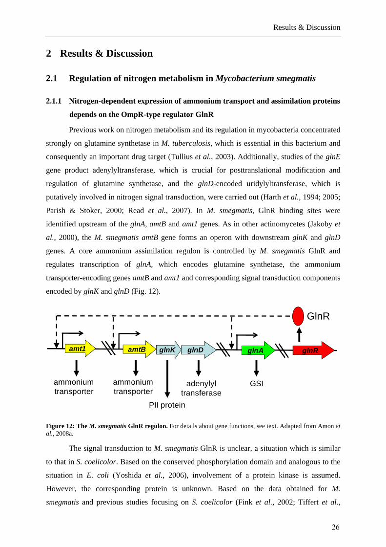
Previous work on nitrogen metabolism and its regulation in mycobacteria concentrated
strongly on glutamine synthetase in M. tuberculosis, which is essential in this bacterium and
consequently an important drug target (Tullius et al., 2003). Additionally, studies of the glnE
gene product adenylyltransferase, which is crucial for posttranslational modification and
regulation of glutamine synthetase, and the glnD-encoded uridylyltransferase, which is
putatively involved in nitrogen signal transduction, were carried out (Harth et al., 1994; 2005;
Parish & Stoker, 2000; Read et al., 2007). In M. smegmatis, GlnR binding sites were
identified upstream of the glnA, amtB and amt1 genes. As in other actinomycetes (Jakoby et
al., 2000), the M. smegmatis amtB gene forms an operon with downstream glnK and glnD
genes. A core ammonium assimilation regulon is controlled by M. smegmatis GlnR and
regulates transcription of glnA, which encodes glutamine synthetase, the ammonium
transporter-encoding genes amtB and amt1 and corresponding signal transduction components
encoded by glnK and glnD (Fig. 12).
glnRglnDglnKamtB glnAamt1
GlnR
ammoniumtransporter
PII protein
adenylyl transferase
GSIammoniumtransporter
Figure 12: The M. smegmatis GlnR regulon. For details about gene functions, see text. Adapted from Amon et al., 2008a.
The signal transduction to M. smegmatis GlnR is unclear, a situation which is similar
to that in S. coelicolor. Based on the conserved phosphorylation domain and analogous to the
situation in E. coli (Yoshida et al., 2006), involvement of a protein kinase is assumed.
However, the corresponding protein is unknown. Based on the data obtained for M.
smegmatis and previous studies focusing on S. coelicolor (Fink et al., 2002; Tiffert et al.,
26

Results & Discussion
2008; Wray & Fisher, 1993; Wray et al., 1991) and Amycolatopsis mediterranei (Yu et al.,
2006; 2007), GlnR seems to be a major regulator of ammonium assimilation in actinomycetes
(see also Fig. 16). This conclusion is supported by the results of electrophoretic mobility shift
assays, which showed that S. coelicolor GlnR is able to bind to the glnA promoter regions of
B. longum, Frankia sp. strain EAN1, M. tuberculosis, Nocardia farcinica, Nocardioides sp.
strain JS614, Propionibacterium acnes and Rhodococcus sp. strain RHA1 in vitro (Tiffert et
al., 2008).
In contrast to the situation in most other actinomycetes, GlnR does not play a role in
nitrogen control in the genus Corynebacterium. In this genus, no GlnR homologs were
observed; instead, AmtR is the central nitrogen control protein (Walter et al., 2007).
Compared to the recently described extended S. coelicolor GlnR regulon (Tiffert et al., 2008),
M. smegmatis GlnR has a reduced number of target genes. For the NADP-dependent
glutamate dehydrogenase gene gdhA (msmeg_5442), the urease operon (msmeg_2627 to
msmeg_3622), and the nitrite reductase genes (msmeg_0427 and msmeg_0428), genes that are
under the control of GlnR in S. coelicolor, no GlnR cis elements were found in this study.
2.1.2 The role and function of AmtR in M. smegmatis and S. avermitilis
C. glutamicum 1 --------MAGA-VGRPRRSAPRRAGKNPREEILDASAELFTRQGFATTSTHQIADAVGIRQASLYYHFPSKTEIFLTLLKSTVEPSTVLC. efficiens 1 --------MAGA-VGRPRRSAPRRAGKNPREEILDASAELFTRQGFATTSTHQIADAVGIRQASLYYHFPSKTEIFLTLLKSTVEPSMVLC. diphtheriae 1 --------MAGA-VGRPRKNSPRRRGSTAREEILDASAELFTTQGFATTSTHQIADAVGIRQASLYYHFPSKTEIFLTLLQSTVAPSTALS. avermitilis 1 ------MGTSGRRVGRPRAAQRPDSGLSPRDELLTAAAELFTTRGYAATTTRAVAERAGMRQASMYHYVSGKEELLAALLESTVTPSLALM. smegmatis 1 -----MTTTSGR--GRPRLEQPRRPGQTAREEILDAAAELFTTHGYGSTSTRRIADEVGVRQASLYHHFATKDDILDALLAGTVDEPLELN. farcinica 1 MRQNGEVTSLGP--GRPRLAPRRRQGRTPRAEILDAAAELFTTQGYASTSTRAVADAVGIRQASLYHHFAAKDDILEALLAETVSGPLALA. aurescens 1 ------MTTAGP--GRPRKQQAVRPGATARDEILDAAAELFTGQGFANTSTRAIADAVGIRQSSLYHHFSTKDEILGELLGGTVSTSLDFRhodococcus sp. 1 -------MTTGP--GRPRLTSQRRPGQTTPEEILDAAGELFTTKGFAATSTRQIAEMVGIRQASLYHHFPNKEEILAALLEETVSPALAA
C. glutamicum 82 AEDLS---TLDAGPEMRLWAIVASEVRLLLSTKWNVGRLYQLPIVGSEEFAEYHSQREALTNVFRDLATEIVG---------DDPRAELPC. efficiens 82 AGDLA---NLEASPELRLWALVAAEVRLLLSTKWNVGRLYQLPIVASEEFEEYHTQRATLTDTFRSLATEIVGE--------DDPRAELPC. diphtheriae 82 AEAFA---DNEAPAALRLWALTATECRLLLSTRWNVGRLYQLPVAASAEFASYQTQRDQLRQTFKNIASEILNP--------DDPRTDLPS. avermitilis 85 ARHLLA--EDAAPAESRLWELCRTDVELLCGGPHNLGGLYLLPEVHTERFAGFHAVRAELKDTYRQLLAATAVGGALAKS-ELDLRTDLVM. smegmatis 84 AHGLL---GESGPAAPRLHALVIYDASQLCAGRWNLGALYLLPELRTDRFAPFRRRRAELRSAYRSLAAAVIAECGGP----PE-ADDLPN. farcinica 89 AERLR---AEPVAPAVRLYALARFDVRQLCSARWNLGALYLLPELRSARFAAFRRQRDDLRGHYERFAAEVLAAARAAH---AEGAELLPA. aurescens 83 ARAIRQHSADAVSAAARLHAVVLFDGSQLCNSRWNLGVLYHLPEARAEIFQPFMAARKELRTIYSELGRELARVSDAD-----QGLGDTARhodococcus sp. 82 AGRLA---DAAAPATVRLHALATYDVTQLTATKWNLGALYLLPELLTDRFEPFRAQRTLLRRHYRQLADQALTEITDDSGDTSPALTDLP
C. glutamicum 160 FHITMSVIEMRRNDGKIPSPLSADSLPETAIMLADASLAVLGAPLPADRVEKTLELIKQADAK------------C. efficiens 161 FHITMSAIEMRRNDGKVPSPLSEDSLPDTAVMLADAALAVLGADLPGDRVERTLELLRQADAK------------C. diphtheriae 161 FHIALSVIEMRSNDGVVPEPLRDDELPVLAIMLADAALAVVGAELPDDRVEWTLNLIRTLND-------------S. avermitilis 172 FGLIEGVILVHRSDP--ERPVSAF-----AEATADAALRIVGV--------------------------------M. smegmatis 166 FRLVESVINSRSDD----AVVPPE----QPWVIGEGALRVLGFDGDFAELAAATASRLGVRPPGRAAR-------N. farcinica 173 FRMVESVINMRSDE----GTAPDY----AERLIPEAILHLLGHHDALGAVRAAADDLLD-RLDG-----------A. aurescens 168 FRLVESLINLRAD-----GLISTD----SASTTADTVMILAGLKRELPAVRTASRDLIS-RFGDVPERVSSMKSARhodococcus sp. 169 FRIVESAIATRADIE--RGLLPGPENEGAAGLLADACLRALGWTKPMDEIRAKSRALLAETAGGTPLRL------
TetR-N
Figure 13: Sequence alignment of selected AmtR-homologs. Amino acid residues identical in all sequences are shaded in black, other conserved amino acids in gray. Black bar marks the conserved N-terminal TetR DNA binding domain. Adapted from Muhl et al., 2009. For full species names see appendix.
27

Results & Discussion
Preliminary data indicate that there may be nitrogen-dependent regulation of the nitrite
reductase and urease, and AmtR might be an interesting candidate for a second nitrogen
regulator, considering the presence of a gene encoding an AmtR homolog in the genome of
M. smegmatis and the fact that for C. glutamicum the urease-encoding genes - among many
others - are under the control of the AmtR repressor (Beckers et al., 2005). Together with N.
farcinica, Rhodococcus sp. strain RHA1 and Arthrobacter aurescens, M. smegmatis seems to
be one of the few actinomycetes besides the members of the genus Corynebacterium to have
an AmtR homolog (Figure 13).
Co-occurrence searches done with the STRING server at the EMBL, Heidelberg,
suggested a putative role of AmtR in repression of an operon that might be involved in amino
acid uptake and assimilation, which is conserved in the genomes of M. smegmatis and close
relatives. This operon comprises, among two hypopthetical reading frames of unknown
function, genes encoding a predicted amino acid permease and enzymes putatively involved
in amino acid degradation (Table 1).
Table 1: Distribution of AmtR and putative target genes in M. smegmatis and close relatives. Hypothetical proteins msmeg_2185 & msmeg_2186 not shown. For C. efficiens, AmtR binding motifs in the upstream regions are predicted. For full species names, see appendix.
aminoacidpermease
ureahydrolase amidase familyprotein
AmtR
M. smegmatis msmeg_2184 msmeg_2187 msmeg_2189 msmeg_4300
C. efficiens* ce0711 ce0713 ce0710 ce0939
S. avermitilis sav6709 sav6698 sav6697 sav6701
Rhodococcus sp. RHA1_ro06919 RHA1_ro06922 RHA1_ro02136 RHA1_ro06918
N. farcinica nfa22220 nfa22190 nfa22180 nfa22230
A. aurescens AAur_0190 AAur_0187 AAur_0186 AAur0192
C. glutamicum n/a n/a n/a cg0986
C. diphtheriae n/a n/a n/a DIP0846
Early experimental results not only confirm the bioninformatics predictions but also
point to the hypothesis that regulation of this operon might in fact be a result of dual
regulation, namely by AmtR and the main nitrogen regulator GlnR (Fig. 14).
28

Results & Discussion
msmeg_2184
GlnRGlnR AmtR
Figure 14: Model of dual transcriptional regulation by GlnR and AmtR. Note that AmtR as a member of the TetR family is a transcriptional repressor that is supposed to bind to DNA as a dimer while GlnR, an activator, binds to DNA in a mode termed “galloping model” (Yoshida et al., 2006). Binding sites might actually overlap.
AmtR and GlnR double mutants support this hypothesis for both M. smegmatis and S.
avermitilis (Dr. Yinhua Lu, personal communication). Ongoing transcriptome studies and
footprinting of the exact DNA binding sites for AmtR and GlnR will result in more evidence
and an expanded model of the dual regulation of nitrogen uptake and metabolism in M.
smegmatis and S. avermitilis.
2.1.3 The search for the GlnR interaction partner
Suggesting OmpR as a model system for GlnR, it is to be expected that GlnR as a
typical response regulator of a bacterial two-component system is being phosphorylated.
Indeed, in an alignment with several homologous GlnR proteins, various conserved aspartate
residues have been identified that putatively get phosphorylated by a yet unknown histidine
kinase (Figure 15).
MSMEG_5784 ----M LLLLTV PHP- SVLPSLSLLAHTVRTAPT VSSLL TGS-------A VAIV ART LAAARGL RLLGTTG---TSVPVVAVIN GGLVAVNH WGL ILL 95MAP_0649 ----M LLLLTP LHP- PVLPSLSLLAHTVRTAPP PSSLL AGT-------A AVIV ART LSSARGL RLLSTAG---RSVPVLAVVS GGLVAVNS WGL ILL 95Mb0841 ---ML LLLLTS LYP- PVLPALSLLPHTVRTAPA ASSLL AGN-------A AVLV ARN LSSGRGL RLLSSTG---RSIPVLAVVS GGLVAVSA WGL ILL 96Rv0818 ---ML LLLLTS LYP- PVLPALSLLPHTVRTAPA ASSLL AGN-------A AVLV ARN LSSGRGL RLLSSTG---RSIPVLAVVS GGLVAVSA WGL ILL 96SCO4159 ---MSSLLLLTNALQPST VLPALGLLLHNVRVAPA GPALV TPG-------A VILV GRR LPQIRSL QLLRSTG---LS PLVLIVT GGLAAVTA WGI VLL 97SAV4042 ---MSSLLLLTNALQPST VLPALGLLLHNVRVAPA GPALV TPG-------A VILI GRR LPQVRSL QLLRSTG---PG PLILVVT GGLAAVTA WGI VLL 97nfa5980 ----M LLLLTS PNP- SVLPSLALLAHTVRPAPT VASLL AGT-------A VALV ART LAAARGL RLLGSTG---SSVPVVAVLT GGLVAVNA WGL ILL 95RHA1_ro04860 MGG V LLLLTS PNP- AVLPALALLAHSVRPAPT VSSLL ASS-------A IALV ART LAAARGL RLLGSTG---SAVPVVAVLT GGLVAVNS WGL ILL 99FRAAL0936 --------MLTNAPGPSA SFPSLGLLTHTVRVAPL ASALL APP-------V VIVV GRR LVIARGL RLLRTTG---LTTPLLALVT GGLAAVSA WGV VVL 92Francci3_0443 ---MSVVLLLTNAPGPSA SFPSLGLLTHTVRVAPL ASALL APP-------V VIVV GRR LVIARGL RLLRTTG---LTTPLLALVT GGLAAVSA WGV VVL 97Tfu_2737 ---MSHLLLLTNSN PS QILPALGLLLHSVRVAPA ASALLTPGP NAAGSPV AILV ARTNLVAARNL RLLNTTG---L PLIAVLT GGAAALTP WQVT FLL 104BL0392 ---MT LTLMTFAS P-ATVLPSLALLSHRVRVLPM AASLVKMP N-------TILFL AR LATAKTL RLIHASG---LSTPIVLVLT GGFTVVNSQWGIA VVV 96AAur_2986 ---MSHILLLTNSPGSSV ILPAL LLNHRVHILPA PTALL T P-------T IVFL ARK LVGARSLTQLLKATG---LSAPLMLILT GGMAAVSSAWAV IVL 97Lxx18370 ---MAQLLILTSAPG--A VLPSLALLSHRTRQIPA PASLVNAPN------- LIFV ARR LASAKSL KILTTT ---ISVPLLLVLT GGLTAVSA WRAS VVL 95ANA_1784 ----MRIIMFTR S A-T VLPATAFL SPV LPPMPSSYA V S-------A VVMI ARG LTRARAL QLFTGPM --- PPIILIV GGAAAVQI WGAA FVM 95AAN77733 --MSL LLVLTA A A-TAVLPAL LLPHTVRVRAP VTALL AGH-------R VILL ARS LASAKSL RLLKGTG AATPIIAVVG GGLVAVSA WRT ILL 100Clustal Consensus ::* :*: :* .. . .: .:*.* :* : * ::: . *:: :: * ** ..: * :.::
D D E E E D D D E E DEE E D E E D D D E D DEE E D E E D D D E D DEE E D E E D D D E D DE
E E D D D D E D DDE E D D D D E D DD
E D E E E D D D E D DDE D E E E D D D E D DD
E E D D D E E D DDE E D D D E E D DD
D D E D D D D E E DD D D E D DD E D
D E E E D D D D E DDE E D D D D E D D
E D D D E E D D D D D EE D DD E D D E D D D D EDE E E DD
CCCCC CC CC
C CCCC CC
C CC C C
C
Figure 15: Alignment of N-termini of selected GlnR homologs. Given are the exact annotation numbers according to the full genome sequences of the respective organisms. Red, conserved aspartate residues.
Up until now, no specific phosphorylation site could be determined, as an exchange to
alanine of each of the three C-terminal aspartate residues results in a complete loss of function
of the regulator, while phosphorylation assays in fact point to a phosphorylation of the protein
(N. Jessberger, personal communication). In parallel, a genomic screening was undertaken
based on co-occurrence searches and following prerequisites: a) The putative histidine kinase
is found in every actinobacterial genome that also features a GlnR homolog; b) the kinase
29

Results & Discussion
must not be found in corynebacterial genomes as AmtR is the regulator of nitrogen
metabolism in this genus; and c) it is supposed to be an orphan kinase to the orphan regulator
GlnR, i.e. the kinase may not occur as a typical two-component system pair with its cognate
response regulator genomically. The screening resulted in a comprehensive analysis of
histidine kinases encoded in the genomes of M. smegmatis and other Actinobacteria, which is
presented in table 2.
Table 2: Analysis of the histidine kinases of M. smegmatis. Given are the predicted open reading frames with their corresponding annotation number for the respective genome and their identity in percentage according to BLASTP results. On the right, the putative role based on literature research and homology searches is given. n/a, not available.
M. smegmatis other mycobacteria
N. farcinica streptomycetes corynebacteria putative role
MSMEG_5158 n/a NFA17980 (69%)
SCO1217 (49%)
SAV7118 (50%)
n/a LytS (regulation of cell autolysis)
MSMEG_2793 yes (all) NFA6630 (46%)
n/a n/a PrrB
(mult. copies) MSMEG_5663
MSMEG_0246
MSMEG_5304 n/a NFA46340 (54%)
SCO5435 (48%)
SAV2816 (47%)
CG0089 (33%) CitA regulating citrate/malate metabolism ?
MSMEG_3239
(no orphan)
MAP_3274 (65%)
NFA54920 (48%)
SCO2121 (38%)
SAV6081 (39%)
n/a MSMEG_3240 LuxR-type regulator, conserved across species…
MSMEG_5870 yes (all except ML)
NFA5450 (54%)
SAV4197 (46%) SCO4021 (49%)
CG2887 (44%) [JK0342 (48%), DIP1935 (45%), CE2493 (41%)]
PhoR ribonucleotide biosynthesis (mult. copies)
MSMEG_4989
MSMEG_0106
(no orphan)
yes (all except ML)
n/a SCO1369 (33%) CG2201 28% ?
MSMEG_6864 n/a NFA12320 (32%)
SCO7562 (35%) CG3388 25% ?
MSMEG_2248 n/a n/a n/a n/a ?
MSMEG_4307 yes (all) NFA16340 (64%)
n/a CG2457 (49%) DIP1680 (53%), JK0667 (51%), CE2135 (50%)
?
MSMEG_4211 n/a n/a SCO1137 (43%) n/a similar MSMEG_530
30

Results & Discussion
no orphan, cit? 4
MSMEG_1918 yes (all) NFA45810 (64%)
SCO5239 (46%) SAV3017 (46%)
n/a ? (VERY good candidate)
MSMEG_2915
(no orphan!)
yes (all except ML)
n/a … … similar to MSMEG_4989 /5870…
MSMEG_0980
(no orphan)
n/a n/a … … similar to MSMEG_2248
MSMEG_3448
(no orphan)
MSMEG_2804
(no orphan)
MSMEG_5241 yes (all except ML)
NFA28940 (63% BLASTN)
SCO0203 (61%)
SAV4257 (59%)
n/a “GAF family protein”; orphan, also very good candidate
msmeg_0854 n/a nfa7820 sco6163/6424/5784 n/a
msmeg_1493 (no orphan)
n/a n/a n/a n/a
msmeg_4968 (no orphan)
n/a n/a sco4597/4598 n/a
msmeg_0936
no orphan!
rv0490 (all) nfa51870 sco4229/sav3973 cg0483/ce0424 “SenX3”
From all identified histidine kinases, two candidates were selected for experimental
validation, namely Msmeg_1918 and Msmeg_5241. Experimental work is ongoing, but
deletion mutants of both kinases showed no specific influence on nitrogen-dependent
regulation of GlnR target genes (personal communication, N. Jessberger).
2.2 Comparative genomic analysis of nitrogen metabolism and control in
mycobacteria
M. smegmatis is equipped with a variety of genes enabling the uptake and assimilation of
nitrogen sources. Compared to the fast-growing M. smegmatis, all slow-growing pathogenic
members of the genus exhibit a reduced number of genes encoding proteins for nitrogen
31

Results & Discussion
uptake and assimilation. This is also due to the fact that M. smegmatis seems to have acquired
an astonishingly wide range of nitrogen-related genes and gene regions via horizontal gene
transfer from a variety of other bacteria, such as Agrobacterium, Burkholderia, and
Pseudomonas species. This includes among others a second urease operon, additional
ammonium transporters, and a broad variety of glutamine synthetases of various classes and
origins. According to the genomic data, M. smegmatis is capable of the active uptake and
assimilation of a comparatively wide range of substrates for the extraction of ammonium and
further assimilation into central metabolites such as glutamate and glutamine (Amon et al.,
2009), which is in good concordance to the situation found for the uptake and assimilation of
carbohydrates in M. smegmatis (Titgemeyer et al., 2007) and thus exhibits a similar repertoire
of nitrogen-related genes to that of C. glutamicum (Burkovski, 2007; Hänssler & Burkovski,
2008).
AmtR(Cg0986)
GlnR(Sco4159)
S. coelicolor
Bifidobacterium sp.
T. fusca
P. acnes
A. naeslundii
L. xyli
C. glutamicum
C. efficiens
C. diphtheriae
M. smegmatis
M. tuberculosisM. bovisM. avium
N. farcinica
Rhodococcus sp.
Arthrobacter sp.
Bold: experimentally verified
A. mediterranei
S. avermitilis
C. matruchotii
C. aurimucosum
C. lipophiloflavum
C. striatum
C. pseudogenitalium
C. accolens
S. griseus
T. curvata
G. obscurus
S. roseum C. acidiphila
A. cellulolyticus
K. flavida
K. radiotolerans
Frankia sp.
Janibacter sp.
C. michiganensis
M. abscessus
T. paurometabola
G. bronchialis
S. arenicolaR. salmoninarum
N. multipartita
G. vaginalis
K. rhizophila
……
Figure 16: Distribution of putative nitrogen-dependent transcription regulators GlnR and AmtR in selected Actinobacteria, based on current genomics data. Bold species names indicate experimental evidence for nitrogen control by the corresponding protein. In M. smegmatis only the function of GlnR is characterized, while AmtR function remains unclear. The references for experimentally verified systems are as follows: Amon et al., 2008a; Fink et al., 2002; Jakoby et al., 2000; Nolden et al., 2002; Yu et al., 2006. Adapted from Amon et al., 2009. For a full list of species names, see appendix.
Another interesting fact is the co-occurrence of homologs of both regulators of the
nitrogen metabolism in actinomycetes in the genome of M. smegmatis, namely AmtR and
GlnR (Amon et al., 2009). While the transcriptional repressor AmtR is the global regulator of
32

Results & Discussion
nitrogen metabolism in corynebacteria (Walter et al., 2007), this function was only recently
shown for the M. smegmatis GlnR and its respective target genes amt1, amtB, and glnA
(Amon et al., 2008a); as only homologs of GlnR are found in the genomes of other
mycobacteria (Fig. 16), the role of AmtR for M. smegmatis remains to be further explored.
All mycobacteria investigated exhibit the subunits for a respiratory nitrate reductase
(which are nonfunctional pseudogenes in M. leprae), while especially the tuberculoid
members possess multiple homologs of the E. coli nitrite/nitrate antiporters, NarK and NarU
(Amon et al., 2009). For M. tuberculosis it has already been shown that nitrate respiration
plays an important role during hypoxia (Sohaskey, 2008), but the additional occurrence of a
nitrite reductase, besides its role in detoxification by reduction of nitrite, points to a possible
involvement of the enzyme in the complete reduction of nitrate to ammonium and following
assimilation, which has been demonstrated for M. smegmatis (Khan et al., 2008).
M. leprae unsurprisingly reveals the strongest reduction of nitrogen metabolism-related genes
as a process of gene decay termed ‘reductive evolution’ (Gómez-Valero et al., 2007),
resulting in a minimal set of genes required for a functional nitrogen metabolism. This set
comprises the genes of the GS/GOGAT pathway (gltBD, glnA) as well as the GS ATase
(glnE) (Amon et al., 2009). While no nitrogen-specific transport systems besides one
nitrite/nitrate antiporter (NarK1) have been found, the genome of M. leprae features putative
amino acid and oligopeptide transporters and permeases; these substrates could very well
represent the main nitrogen sources, taking into account the lifestyle of M. leprae as an
obligate intracellular parasite (Sassetti et al., 2003; Vissa & Brennan, 2001).
2.3 The glucose permease and glucose kinase of M. smegmatis
The protein database of the M. smegmatis genome was scanned with several known
glucose permease protein sequences using BLASTP as implemented at The Institute for
Genomic Research (TIGR) (Parche et al., 2006; van Wezel et al., 2005). Only one gene
(msmeg4187) was found that encodes a predicted glucose permease of the major facilitator
super family (MFS) (Pao et al., 1998). The deduced protein shares 53% amino acid identity to
the well-characterized glucose permease GlcP of Streptomyces coelicolor (Titgemeyer et al.,
2007; van Wezel et al., 2005). Analysis of the gene locus of msmeg4187 (hereafter named
glcP) indicated that the gene is transcribed as a monocistronic mRNA (Pimentel-Schmitt et
al., 2009). glcP is flanked downstream by a putative dicistronic hisEG operon of genes
33

Results & Discussion
involved in histidine biosynthesis, and upstream by an unknown open reading frame. While
the hisEG operon is conserved in mycobacteria, it appears that the slow-growing
mycobacteria, e.g. M. tuberculosis, M. bovis, and M. avium, do not have an obvious
homologue of glcP. The multiple alignment includes four homologous glucose permeases
which have been experimentally described (Parche et al., 2006; van Wezel et al., 2005;
Weisser et al., 1995; Zhang et al., 1989). The range of protein identity is from 34 to 53%,
showing 67 conserved residues in these sequences. Hydrophobicity profiles showed that all
five sequences have 12 predicted transmembrane helices that are at equivalent positions
(Pimentel-Schmitt et al., 2009). To establish the phylogenetic relationships of GlcP, 19
protein sequences of the MFS were selected which have been characterized by experimental
investigation including those from diverse mycobacteria (Fig. 17).
Figure 17: Phylogenetic tree of the glcP family. An unrooted phylogenetic tree was computed with the CLUSTALW software making use of the implemented neighbor joining method with the function for evolutionary distance correction. Evolutionary distances are proportional to the branch length. 19 protein sequences were selected as indicated in the figure. The references for experimentally verified transporters are as follows: Chaillou et al., 1998; Krispin et al., 1998; Martin et al., 1994; Parche et al., 2006; van Wezel et al., 2005; Weisser et al., 1995; Zhang et al., 1989. Adapted from Pimentel-Schmitt et al., 2009. For full species names see appendix.
GlcP clusters with the S. coelicolor homolog and the one from the cyanobacterium
Synechocystis (van Wezel et al., 2005; Zhang et al., 1989). Interestingly, the xylose permease
XylE of E. coli is more closely related to GlcP than expected, while the bifidobacterial GlcP
is more distant but closely associated with another xylose permease, XylT of Lactobacillus
34

Results & Discussion
brevis (Chaillou et al., 1998). This suggests that glucose and xylose transporter genes may
have evolved following two different branches from the common origins. The glucose-
specific transport protein GlcP from M. smegmatis encoded by open reading frame
msmeg4187 is the first reported mycobacterial sugar permease. Interestingly, no GlcP
homolog is present in slow-growing mycobacteria, notably M. tuberculosis and M. leprae
(Titgemeyer et al., 2007). In these bacteria, glucose may enter either by simple diffusion,
which could be efficient enough to meet the nutritional demands of these extremely slow-
growing bacteria (generation time of weeks), or through another, yet unknown permease
(Titgemeyer et al., 2007).
[-4] [4] [433] [69]
fabD2ghyA1356MSMEG1354 1355glkA rplJ rplL
1357 1358
MT0676 0677 0679ghyA glkAfabD2
0680 0681rplJ rplL
[-4] [-1] [330] [36]
Rv0648 06500649glkAfabD2ghyA
[-75] [-1] [330] [36]
0651 0652rplJ rplL
Mb0667 0668 0669ghyA fabD2 glkA
[-4] [-1] [330] [36]
0670 0671rplJ rplL
MAP4121 4122 4123ghyA fabD2 glkA
[-4] [4] [9] [10] [25]
rplLurf4125rplJ
41264124
Figure 18: Genetic organization of the glkA gene in M. smegmatis. The highly conserved depicted glkA region (black) includes the genes ghyA (encoding a glycosyl hydrolase), fabD2 (malonyl-CoA transacylase), and the genes for the ribosomal proteins L10 and L7/L12 (gray). Note that M. avium paratuberculosis additionally carries a gene encoding a protein of unknown function (urf, MAP4124). The genomes of M. smegmatis mc2 155 (MSMEG), M. tuberculosis H37Rv (Rv), M. tuberculosis CDC1551 (MT), M. bovis subsp. bovis AF2122/97 (Mb), and M. avium paratuberculosis (MAP) were sequenced and annotated by TIGR. The arrows indicate length and transcriptional orientation of annotated genes and open reading frames. Numbers in brackets show the lengths of intergenic regions in bp. Adapted from Pimentel-Schmitt et al., 2007.
Furthermore, genomes of M. smegmatis and M. tuberculosis H37RV were screened for
the presence of putative glucose kinase genes. A protein BLASTP search with the Glk protein
sequence of S. coelicolor revealed the presence of seven homologues in the genome of M.
smegmatis (Pimentel-Schmitt et al., 2007). These sequences are members of the ROK family,
which comprises bacterial transcription factors and sugar kinases (Titgemeyer et al., 1994a;
1994b). Three of the seven genes are sugar kinases. Of these, the gene product MSMEG1356
exhibited with 37% identical and 58% similar amino acid residues the closest homologue.
Analyzing the genome of M. tuberculosis, three ROK family members have been found, of
35

Results & Discussion
which one gene, Rv0650, was close to glk of S. coelicolor and MSMEG1356. Hence,
MSMEG1356 (hereafter termed glkA) was chosen as the prime candidate for a possible
glucose kinase (Pimentel-Schmitt et al., 2007). A comparison of the glkA region with other
mycobacterial genomes revealed a very similar genetic context (Fig. 18), in which glkA is the
third gene of a putative tricistronic operon.
The first one is a possible glycosyl hydrolase, suggesting that the enzyme together
with GlkA participates in the same pathway, which could be the metabolism of glucose-
containing saccharides. The second gene appears to encode a putative malonyl-CoA
transacylase found as a unique conserved mycobacterial gene (Huang et al., 2006). Two genes
that encode the ribosomal proteins L10 and L7/L12 are located immediately downstream of
glkA. Such a conserved gene order is a good hint for equivalent functions in closely related
species. A multiple alignment of homologous glucose kinases from diverse bacteria reveals
that GlkA shows 71% protein identity to the one from the M. tuberculosis H37Rv reference
strain and 28–52% to the others that were chosen from related actinomycetes and from
Bacillus subtilis (Pimentel-Schmitt et al., 2007). An unrooted tree shows that GlkA is most
closely related to the mycobacterial homologues, then to SCO1077 (GlkII) of S. coelicolor
and then equally distant to other glucose kinases (Pimentel-Schmitt et al., 2007).
The occurrence of a glucose kinase gene in all so far sequenced mycobacterial
genomes indicates an important function. Multiple alignment analysis revealed 39 amino acid
residues, which are conserved in all six selected species. Among these residues are several
that have been identified to participate in ATP, glucose, and zinc binding in distantly related
species (Hansen et al., 2002; Lunin et al., 2004; Mesak et al., 2004; Schiefner et al., 2005;
Titgemeyer et al., 1994a). Future research should be focused on the question: which protein or
metabolic pathways in M. smegmatis are affected by glkA? In particular, it will be important
to examine whether glkA plays a direct metabolic role in glucose utilization and/or carbon
regulation. So far, GlkA may not be required to maintain a functional virulence cycle. This at
least can be inferred from a global microarray-based analysis of M. tuberculosis (Sassetti et
al., 2003; Sassetti & Rubin, 2003). Assuming that specific inhibitors of protein kinases have
been successfully developed for therapeutic usage against a variety of diseases (Shawver et
al., 2002), future investigation for drug development could be directed to the selection of
GlkA of mycobacterium, as a means of interfering with growth and possible regulatory
processes. M. smegmatis and also the slowgrowing mycobacterial species possess a glucose
kinase that converts the incoming glucose to glucose-6-phosphate (Pimentel-Schmitt et al.,
36

Results & Discussion
2007; Titgemeyer et al., 2007). Apart from its catalytic function, this enzyme confers global
carbon regulation in closely related streptomycetes (Angell et al., 1994). It has been shown in
S. coelicolor that glucose kinase binds to glucose permease GlcP (van Wezel et al., 2007).
This protein-protein interaction is thus thought to influence the regulatory function of glucose
kinase. For this reason, it would be worthwhile to study, whether GlcP and GlkA of M.
smegmatis also interact with each other and whether this has similar consequences regarding
nutritional control. This in turn would help to understand how nutrition and virulence may be
linked in mycobacteria.
2.4 Carbohydrate transport systems of mycobacteria
It has been widely documented that M. smegmatis can grow on many carbon sources
such as polyols, pentoses and hexoses (Edson, 1951; Franke & Schillinger, 1944; Izumori et
al., 1978). Multiple inner membrane transport systems for all three of these classes of
carbohydrates by bioinformatic analysis have been identified (Titgemeyer et al., 2007). This
provides the molecular basis for the adaptability of M. smegmatis to different environments in
the soil and water. Often, the integrated bioinformatic approach enabled us to propose a
specific substrate for particular uptake proteins (Fig. 19). Since the specificity of transport
proteins can be altered by the modification of a few residues, the suggested substrates rather
represent a hypothesis for experiments such as transport measurements with gene deletion
mutants or analysis of the induction of gene expression. Analysis of the induction of both the
transport activity and transcription of genes confirmed the substrate predictions for a fructose-
and glucose-specific PTS as well as for the predicted glycerol operon. Since glucose uptake
was constitutive, at least one more system for glucose transport must occur in M. smegmatis.
This was predicted to be GlcP. Indeed, cloning and heterologous expression of glcP in E. coli
revealed that it is a glucose-specific permease (Pimentel-Schmidt et al., 2009).
The discovery of homologs of all components of a PTS in M. smegmatis contradicts a
previous report that did not find biochemical evidence for the existence of a PTS (Romano et
al., 1970) and many repeating statements (Connell & Nikaido, 1994; Content et al., 2005;
Ratledge & Stanford, 1982). Components of the PTS play a key role in the global control of
sugar metabolism to achieve the hierarchical utilization of carbon sources in bacteria
(Brückner & Titgemeyer, 2002), where two different mechanisms have evolved. In E. coli and
other closely related gram-negative bacteria, the enzyme IIAGlc is dephosphorylated mainly
37

Results & Discussion
under repressing conditions and mediates inducer exclusion. Under nonrepressing conditions,
phosphorylated IIAGlc stimulates cyclic AMP (cAMP) synthesis and thereby triggers the
activation of catabolite- repressed genes by a global regulator, the cAMP-dependent catabolite
activator protein (see 1.4).
In low G+C Gram-positive bacteria such as Bacillus subtilis, HPr is a central switch of carbon
catabolite repression. Under repressing conditions, HPr is phosphorylated mainly at serine 46
by a unique HPr kinase/phosphatase mediating inducer exclusion and carbon catabolite
repression/activation (Brückner & Titgemeyer, 2002; Monedero et al., 2007; Reizer et al.,
1998).
HPr
EI
sugar-P
fructose-6-P
fructose-1,6BP
PEP
ATP
Figure 19: Predicted sugar transport systems of M. smegmatis. Shown are the permeases of the ABC (orange), PTS (yellow), MIP (violet), MFS (green) and SSS (blue) families. The derived putative substrates are inferred from in silico analyses in combination with experimental data. Adapted from Titgemeyer et al., 2007.
Under non-repressing conditions, HPr is phosphorylated at histidine 15 and activates
PTS-dependent sugar transport, glycerol kinase, and substrate-specific regulators. The
apparent absence of a protein in M. smegmatis similar to the HPr kinase/phosphatase argues
against the mechanism found in low-GC Gram-positive bacteria. On the other hand, M.
smegmatis apparently does not produce proteins with significant sequence similarities to the
cAMP receptor protein (CRP) (catabolite activator protein) of E. coli, which is crucial for
38

Results & Discussion
carbon catabolite repression in Gram-negative bacteria. By contrast, the coordination of the
few operons involved in the uptake and degradation of carbohydrates by M. tuberculosis
(Titgemeyer et al., 2007) may not require a global control mechanism, as suggested by the
lack of PTS homologs. Alternatively, a completely different mechanism for the global control
of carbon metabolism may have evolved in M. tuberculosis to adapt to its specific
environment inside the phagosome of human macrophages. Indeed, M. tuberculosis has eight
orthologs of CRP-like transcriptional regulators (McCue et al., 2000), one of which, Rv3676,
was experimentally described (Bai et al., 2005). Furthermore, the large number and the
different subcellular localization of the 15 putative nucleotide cyclases in M. tuberculosis
imply that this organism may have the ability to sense and respond to many intracellular and
extracellular signals through a second messenger system based on cyclic nucleotide
monophosphates (McCue et al., 2000). This is in strong contrast to E. coli and other Gram-
negative bacteria, which have only one CRP and one adenylate cyclase. CRP homologs have
been identified in streptomycetes (Derouaux et al., 2004), where the regulator plays a role in
germination, and in corynebacteria, where CRPs have been associated with global carbon
regulation (Moon et al., 2007). Although the potential mechanisms of global control of carbon
metabolism in both M. smegmatis and M. tuberculosis are not evident from the bioinformatic
analysis of their genomes, these findings provide hypotheses for further experiments.
2.5 Carbohydrate transport systems of Bifidobacterium longum
In silico analysis was done by conducting BLASTP analyses at the BLAST server of
the TIGR institute following previously described strategies (Bertram et al., 2004). The
detected gene products from B. longum NCC2705 and those of the adjacent genes were then
searched with BLASTP at various genome servers, including the one of the Transporter
Classification Database (TCDB; www.tcdb.org), in order to identify all carbohydrate
transporter genes (Saier et al., 2006). The search led to the identification of 19 B. longum loci
predicted to encode carbohydrate transporters (Parche et al., 2007; Fig. 20). They belong to
the ATP binding cassette family (ABC), the phosphotransferase system (PTS), the major
facilitator superfamily (MFS), the glycoside-pentoside-hexuronide cation symporter family
(GPH), and the major intrinsic protein family (MIP). The majority was formed by 13 ABC
permeases (ATP-binding cassette), which include porters for di-, tri- and higher order
oligosaccharides. Three permeases of the MFS, which include proton symporters and
facilitators, are predicted to transport glucose, lactose, and sucrose. Also found was one
39

Results & Discussion
complete glucose-specific PTS. It comprises the general phosphotransferases enzyme I (EI)
and HPr, which operate in global carbon regulation in many bacteria (Brückner &
Titgemeyer, 2002), and a single predicted glucose- specific enzyme II permease. Finally, a
putative glycerol permease of the MIP and one permease of the GPH family completed the list
of detected NCC2705 sugar transport systems (Parche et al., 2007). To gain more hints on
possible substrates, the genomic neighborhood was inspected for associated metabolic genes,
which are usually colocalized in an operon structure, and for possible regulators, which often
occur adjacent to the target operon.
maltose
carbohydrate-P
fructose-6-P
fructose-1,6-bis-P
PEP
pyruvate lactic acid
HPr-P
EI-P
MIP
PTS
MFS
glucose
H+
H+
sucrose
unknown
GPH
glucose
0264
GlkA
0144014301
41
ATP
0034
ABC
carbohydrate
MsiK0673
lactose, FOS
FOS
raffinose
unknown
disaccharides
ribose
multiple sugar, fructose, mannose
maltoseraffinose
glucose
1692 glucose
lactose
H+
glycerol
mannose-containingoligosaccharides
lactose, FOS
FOS
unknown
xylosides, xylose
Figure 20: Predicted sugar transport systems of B. longum NCC2705. Shown are the permeases of the ABC (red), PTS (yellow), MFS (green), MIP (violet) and GPH family (blue). FOS, fructooligosaccharide. The derived putative substrates are inferred from in silico analyses in combination with experimental data. Experimentally verified systems are denoted by an asterisk. Adapted from Parche et al., 2007.
The combined analysis of the carbohydrate transporter systems of B. longum
NCC2705 revealed 19 transport systems and provided a number of interesting conclusions:
(1) B. longum can transport a variety of disaccharides and oligosaccharides like oligofructose,
which are described as growth-promoting prebiotics. Compared to the initial publication of
40

Results & Discussion
the genome sequence (Schell et al., 2002), in which 7 oligosaccharide systems were
presented, our more extensive analysis revealed that up to 15 such systems may exist in
NCC2705. (2) More than half of the transport systems are ATP-dependent ABC-type
permeases, a feature that is also found in other actinomycetes such as S. coelicolor (Bertram
et al., 2004) and Mycobacterium smegmatis (Titgemeyer et al., 2007). (3) PTSs are present in
bifidobacteria and occur either as glucose- or fructose-specific PTS. It remains to be
established whether these PTS components serve additional regulatory function besides sugar
transport as they do in other bacteria (Brückner & Titgemeyer, 2002; Rigali et al., 2006).
2.6 Comparative genomic analysis of the proteolytic potential in
corynebacteria
2647 26202645 2644
tig clpP1 clpP2 clpX
[188] [30]
0543 05460544 0545
tig clpP1 clpP2 clpX
[207] [3] [141]
1793 17901792 1791tig clpP1 clpP2 urf clpX
[125] [17] [89] [146]1789
2313 22912312 2311tig clpP1 clpP2 clpX
[188] [34]
ce
jk
dip
cg
100 aa
Figure 21: As an example of a conserved proteolytic system, the genetic organization of clp gene clusters in corynebacteria is shown. Arrows indicate the lengths and transcriptional orientations of annotated genes and predicted ORFs. Genes are shown by their annotation number, with the prefix of the respective organism. The gene names are assigned according to the annotations given by TIGR (http://www.tigr.org). Numbers in square brackets refer to the intergenic distance between two genes. Gene designations are as follows: urf, unknown reading frame; tig, trigger factor; clpP1, clpP2, proteolytic subunits; clpX, ATPase subunit; cg, C. glutamicum; ce, C. efficiens; jk, C. jeikeium; dip, C. diphtheriae. Adapted from Amon et al., 2008b.
Based on published genome sequences of the Corynebacterium species C. diphtheriae
(Cerdeno-Tarraga et al., 2003), Corynebacterium efficiens (Fudou et al., 2002), C.
glutamicum (Ikeda & Nakagawa, 2003; Kalinowski et al., 2003) and C. jeikeium (Tauch et
al., 2005), a comparative genomics analysis of genes encoding proteins with putative
proteolytic functions was performed. A complete overview of protease genes found in each of
the corynebacterial genomes is given in Amon et al., 2008b. A total number of 53, 51, 56, and
42 protease-encoding genes was predicted in the genomes of C. glutamicum, C. diphtheriae,
41

Results & Discussion
C. efficiens, and C. jeikeium. Correlated to the genome size, C. efficiens has the lowest density
of protease encoding genes on the chromosome (less than 20 per Mbp), followed by C.
jeikeium (approximately 23 genes per Mbp), C. glutamicum (about 29 genes per Mbp) and C.
diphtheriae (more than 30 genes per Mbp). A core subset of 39 homologous proteases is
common to all corynebacterial species. These findings are in good accordance with the
numbers recently published for the closely related mycobacterial genus (Ribeiro-Guimaraes et
al., 2007), comprising a similar set of highly conserved homologous proteases.
Compared to other bacteria, corynebacteria seem to have only moderate proteolytic activity.
Nevertheless, proteases obviously play a crucial role in many cellular processes. First
investigations of C. glutamicum Clp protease complexes (Fig. 21) and FtsH hint to interesting
regulatory functions of these AAA+ enzymes in this species. However, knowledge on this
class of proteases is far from completeness and for example even the degradation signals are
still unknown. The same is true for the function of putative degradation tags or amino acid
residues influencing protein stability (Mogk et al., 2007). As discussed above, already the
increased density of protease-encoding genes in the C. diphtheriae genome compared to other
corynebacterial genomes and their partial localization on genomic islands suggest a role of
these enzymes in pathogenicity. In fact, C. diphtheriae sortases were already shown to be
crucial for pili assembly and adhesion to hosts cells, an effect also observed for two other
secreted C. diphtheriae proteases. However, also in this case, only limited information is
available. Future approaches might shed more light on the diverse function of known and
uncharacterized proteases in different corynebacteria and hopefully elucidate the up to now
completely unknown target recognition mechanisms of these enzymes.
42

Summary / Zusammenfassung
3 Summary / Zusammenfassung
3.1 Summary
Based on available genome sequences and homology searches with already described systems
of close relatives, the up to now mostly unknown repertoire of nitrogen metabolism-related
genes in the genus Mycobacterium was discovered and putative pathways and regulatory
networks have been constructed and compared. Focussing on the non-pathogenic
Mycobacterium smegmatis, the regulator of nitrogen metabolism GlnR was characterized by a
combined in silico, in vitro and in vivo approach. By this, direct target genes for the regulator
were verified and a basic regulon of nitrogen metabolism was constructed. Furthermore,
putative interaction partners of GlnR were identified, while the experimental validation of
these candidates is ongoing. The role of AmtR in nitrogen-dependent regulation has been
investigated for M. smegmatis and Streptomyces avermitilis and putative target genes have
been found. The experimental validation is ongoing.
In respect to carbohydrate metabolism, the repertoire of carbohydrate uptake systems in the
genus Mycobacterium was studied and predictions about the comparative growth potential of
M. smegmatis and Mycobacterium tuberculosis on various carbon sources were provided.
Furthermore, the glucose permease GlcP and the glucose kinase GlkA of M. smegmatis have
been studied in greater detail based on comprehensive sequence and homology analyses. In a
similar approach, the genome of the industrial relevant probiotic Bifidobacterium longum has
been examined for carbohydrate uptake systems, which resulted in a detailed analysis of
carbohydrate uptake systems and an overview about the growth potential of this organism on
various sugars.
Also, the proteolytic potential of various Corynebacterium species based on all available
genome sequences was compared and resulted in a more complete view on the housekeeping
protease systems as well as on proteases putatively involved in pathogenicity and virulence of
Corynebacterium diphtheriae.
43

Summary / Zusammenfassung
3.2 Zusammenfassung
Basierend auf Genomanalysen und vergleichenden Homologieanalysen konnte das bislang
weitgehend unbekannte Repertoire an Genen analysiert werden, das im Genus Mycobacterium
an der Aufnahme und Assimilation von Stickstoff beteiligt ist. Putative Stoffwechselwege und
übergeordnete regulatorische Netzwerke wurden konstruiert und einer vergleichenden
Analyse unterzogen. Für den nicht-pathogenen Mycobacterium smegmatis konnte der
Stickstoff-regulator GlnR durch einen kombinierten in silico, in vitro und in vivo Ansatz
charakterisiert werden. Hierbei konnten direkte Zielgene verifiziert und ein Stickstoff-
abhängiges Regulon konstruiert werden. Weiterhin wurden putative Interaktionspartner von
GlnR identifiziert, für die die experimentelle Verifikation zur Zeit in Arbeit ist. Die Rolle von
AmtR in der Stickstoff-abhängigen Regulation wurde für M. smegmatis und Streptomyces
avermitilis untersucht und putative Zielgene konnten gefunden werden. Die experimentelle
Validierung ist zur Zeit in Arbeit.
In Bezug auf den Kohlenhydrat Metabolismus wurde das Repertoire an Kohlenhydrat-
Aufnahmesystemen im Genus Mycobacterium untersucht und vergleichende Voraussagen
über das Wachstumspotential von M. smegmatis und Mycobacterium tuberculosis auf
verschiedenen Kohlenstoffquellen getroffen. Weiterhin wurden die Glucosepermease GlcP
und die Glucosekinase GlkA von M. smegmatis mittels vergleichender Sequenzanalyse und
Homologiestudien detailliert studiert. Das Genom des industriell relevanten Bifidobacterium
longum wurde auf Kohlenhydrat-Aufnahmesysteme untersucht. Ein kombinierter Ansatz
resultierte in einer detaillierten Analyse der Kohlenhydrat Aufnahmesysteme und einer
Übersicht über das Wachstumspotential dieses Organismus auf verschiedenen
Kohlenstoffquellen.
Schlußendlich wurde das proteolytische Potential verschiedener Corynebakterien mithilfe
verfügbarer Genomsequenzen verglichen und eine detaillierte Analyse sowohl der
housekeeping Proteasesysteme als auch der Proteasen, die möglicherweise involviert sind in
Pathogenität und Virulenz von Corynebacterium diphtheriae, erstellt.
44

References
4 References
Altschul, S.F. & Lipman, D.J. (1990) Protein database searches for multiple alignments. Proc Natl Acad Sci USA 87: 5509-5513. Amon, J., Bräu, T., Grimrath, A., Hänßler, E., Hasselt, K., Höller, M., Jeßberger, N., Ott, L., Szököl, J., Titgemeyer, F. & Burkovski, A. (2008a) Nitrogen control in Mycobacterium smegmatis: nitrogen-dependent expression of ammonium transport and assimilation proteins depends on the OmpR-type regulator GlnR. J Bacteriol 190: 7108-7116. Amon, J., Lüdke, A., Titgemeyer, F. & Burkovski, A. (2008b) General and regulatory proteolysis in corynebacteria, In: Corynebacteria: genomics and molecular biology, A. Burkovski (ed.) (Caister Academic Press, Norfolk, UK) pp. 183-201. Amon, J., Titgemeyer, F. & Burkovski, A. (2009) A genomic view on nitrogen metabolism and nitrogen control in mycobacteria. J Mol Microbiol Biotechnol 17: 20-29. Angell, S., Lewis, C.G., Buttner, M.J. & Bibb, M.J. (1994) Glucose repression in Streptomyces coelicolor A3(2): a likely regulatory role for glucose kinase. Mol Gen Genet 244: 135-143. Armstrong, P. (2006) Proteases and protease inhibitors: a balance of activities in host-pathogen interaction. Immunobiology 211: 263-281. Bai, G., McCue, L.A. & McDonough, K.A. (2005) Characterization of Mycobacterium tuberculosis Rv3676 (CRPMt), a cyclic AMP receptor protein-like DNA binding protein. J Bacteriol 187: 7795-7804. Beckers, G., Strösser, J., Hildebrandt, U., Kalinowski, J., Farwick, M., Krämer, R. & Burkovski, A. (2005) Regulation of AmtR-controlled gene expression in Corynebacterium glutamicum: mechanism and characterization of the AmtR regulon. Mol Microbiol 58: 580-595. Bérdy, J. (2005) Bioactive microbial metabolites. J Antibiot 58: 1-26. Bertram, R., Schlicht, M., Mahr, K., Nothaft, H., Saier, M.H. & Titgemeyer, F. (2004). In silico and transcriptional analysis of carbohydrate uptake systems of Streptomyces coelicolor A3(2). J Bacteriol 186: 1362-1373. Bettenbrock, K., Sauter, T., Jahreis, K., Kremling, A., Lengeler, J.W. & Gilles, E.D. (2007) Analysis of the correlation between growth rates, EIIACrr phosphorylation and intracellular cAMP levels in Escherichia coli K-12. J Bacteriol 189: 6891-6900. Blattner, F.R., Plunkett, G. 3rd, Bloch, C.A., Perna, N.T., Burland, V., Riley, M., Collado-Vides, J., Glasner, J.D., Rode, C.K., Mayhew, G.F., Gregor, J., Davis, N.W., Kirkpatrick, H.A., Goeden, M.A., Rose, D.J., Mau, B. & Shao, Y. (1997) The complete genome sequence of Escherichia coli K-12. Science 277: 1453-1462.
45

References
Bravo, A., Gill, S.S. & Soberon, M. (2007). Mode of action of Bacillus thuringiensis Cry and Cyt toxins and their potential for insect control. Toxicon 49: 423-435. Brückner, R. & Titgemeyer, F. (2002) Carbon catabolite repression in bacteria: choice of the carbon source and autoregulatory limitation of sugar utilization. FEMS Microbiol Lett 209: 141-148. Bueno, R., Pahel, G. & Magasanik, B. (1985) Role of glnB and glnD gene products in regulation of the glnALG operon of Escherichia coli. J Bacteriol 164: 816-822. Burkovski, A. (2007) Nitrogen control in Corynebacterium glutamicum: proteins, mechanisms, signals. J Microbiol Biotechnol 17: 187-194. Cerdeno-Tarraga, A.M., Efstratiou, A., Dover, L.G., Holden, M.T.G., Pallen, M., Bentley, S.D., Besra, G.S., Churcher, C., James, K.D., De Zoysa, A., Chillingworth, T., Cronin, A., Dowd, L., Feltwell, T., Hamlin, N., Holroyd, S., Jagels, K., Moule, S., Quail, M.A., Rabbinowitch, E., Rutherford, K.M., Thomson, N.R., Unwin, L., Whitehead, S., Barrell, B.G. & Parkhill, J. (2003) The complete genome sequence and analysis of Corynebacterium diphtheriae NCTC13129. Nucleic Acids Res 31: 6516-6523. Chaillou, S., Bor, Y.C., Batt, C.A., Postma, P.W. & Pouwels, P.H. (1998) Molecular cloning and functional expression in Lactobacillus plantarum 80 of xylT , encoding the D-xylose-H+ symporter of Lactobacillus brevis. Appl Environ Microbiol 64: 4720-4728. Chater, K.F. (1993) Genetics of differentiation in Streptomyces. Annu Rev Microbiol 47: 685-713. Cole, S.T., Brosch, R., Parkhill, J., Garnier, T., Churcher, C., Harris, D., Gordon, S.V., Eiglmeier, K., Gas, S., Barry, C.E. 3rd, Tekaia, F., Badcock, K., Basham, D., Brown, D., Chillingworth, T., Connor, R., Davies, R., Devlin, K., Feltwell, T., Gentles, S., Hamlin, N., Holroyd, S., Hornsby, T., Jagels, K., Krogh, A., McLean, J., Moule, S., Murphy, L., Oliver, K., Osborne, J., Quail, M.A., Rajandream, M.A., Rogers, J., Rutter, S., Seeger, K., Skelton, J., Squares, R., Squares, S., Sulston, J.E., Taylor, K., Whitehead, S. & Barrell, B.G. (1998) Deciphering the biology of Mycobacterium tuberculosis from the complete genome sequence. Nature 393: 537-544. Connell, N.D., & Nikaido, H. (1994) Membrane permeability and transport in Mycobacterium tuberculosis. In: Tuberculosis: pathogenesis, protection, and control. B.R. Bloom (ed.) (ASM Press, Washington, DC), p. 333-349. Content, J., Braibant, M., Connell, N. & Ainsa, J. A. (2005) Transport processes. In: Tuberculosis and the tubercle bacillus. S.T. Cole, K.D. Eisenach, D.N. McMurray & W.R. Jacobs (eds.) (ASM Press, Washington, DC), p. 379-404. Coyle, M.B. & Lipsky, B.A. (1990) Coryneform bacteria in infectious diseases: clinical and laboratory aspects. Clin Microbiol Rev 3: 227-246. Crickmore, N. (2005) Using worms to better understand how Bacillus thuringiensis kills insects. Trends Microbiol 13: 347-350.
46

References
Death, A. & Ferenci, T. (1994) Between feast and famine: endogenous inducer synthesis in the adaptation of Escherichia coli to growth with limiting carbohydrates. J Bacteriol 176: 5101-5107. Derouaux, A., Halici, S., Nothaft, H., Neutelings, T., Moutzourelis, G., Dusart, J., Titgemeyer, F. & Rigali, S. (2004) Deletion of a cyclic AMP receptor protein homologue diminishes germination and affects morphological development of Streptomyces coelicolor. J Bacteriol 186: 1893-1897. Edson, N. L. (1951) The intermediary metabolism of the mycobacteria. Bacteriol Rev 15: 147-182. Eiglmeier, K., Parkhill, J., Honore, N., Garnier, T., Tekaia, F., Telenti, A., Klatser, P., James, K.D., Thomson, N.R., Wheeler, P.R., Churcher, C., Harris, D., Mungall, K., Barrell, B.G. & Cole, S.T. (2001). The decaying genome of Mycobacterium leprae. Lepr Rev 72: 387-398. El Qaidi, S., Allemand, F., Oberto, J. & Plumbridge, J. (2009) Repression of galP, the galactose transporter in Escherichia coli, requires the specific regulator of N-acetylglucosamine metabolism. Mol Microbiol 71: 146-157. Embley, T.M. & Stackebrandt, E. (1994) The molecular phylogeny and systematics of the actinomycetes. Annu Rev Microbiol 48: 257-289. Engels, S., Ludwig, C., Schweitzer, J.-E., Mack, C., Bott, M. & Schaffer, S. (2005) The transcriptional regulator ClgR controls transcription of genes involved in proteolysis and DNA repair in Corynebacterium glutamicum. Mol Microbiol 57: 576-591. Erni, B. & Zanolari, B. (1986) Glucose-permease of the bacterial phosphotransferase system. Gene cloning, overproduction, and amino acid sequence of enzyme IIGlc. J Biol Chem 261: 16398-16403. Ferenci, T. (1996) Adaptation to life at micromolar nutrient levels: the regulation of Escherichia coli glucose transport by endoinduction and cAMP. FEMS Microbiol Rev 18: 301-317. Fink, D., Weisschuh, N., Reuther, J., Wohlleben, W. & Engels, A. (2002) Two transcriptional regulators GlnR and GlnRII are involved in regulation of nitrogen metabolism in Streptomyces coelicolor A3(2). Mol Microbiol 46: 331-347. Finn, R.D., Mistry, J., Tate, J., Coggill, P., Heger, A., Pollington, J.E., Gavin, O.L., Gunasekaran, P., Ceric, G., Forslund, K., Holm, L., Sonnhammer, E.L., Eddy, S.R. & Bateman, A. (2010) The Pfam protein families database. Nucleic Acids Res 38: D211-222. Flannagan, R.S., Cosío, G. & Grinstein, S. (2009) Antimicrobial mechanisms of phagocytes and bacterial evasion strategies. Nat Rev Microbiol 7: 355-366. Forchhammer, K., Hedler, A., Strobel, H. & Weiss, V. (1999) Heterotrimerization of PII-like signalling proteins: implications for PII-mediated signal transduction systems. Mol Microbiol 33: 338-349.
47

References
Forchhammer, K. (2004) Global carbon/nitrogen control by PII signal transduction in cyanobacteria: from signals to targets. FEMS Microbiol Rev 28: 319-333. Forchhammer, K. (2007) Glutamine signalling in bacteria. Front Biosci 12: 358-370. Forchhammer, K. (2008) P(II) signal transducers: novel functional and structural insights. Trends Microbiol 16: 65-72. Franke, W. & Schillinger, A. (1944) Zum Stoffwechsel der säurefesten Bakterien I. Orientierende aerobe Reihenversuche. Biochem Zeitung 319: 313-334. Fudou, R., Jojima, Y., Seto, A., Yamada, K., Kimura, E., Nakamatsu, T., Hiraishi, A. & Yamanaka, S. (2002) Corynebacterium efficiens sp. nov., a glutamic-acid-producing species from soil and vegetables. Int J Syst Evol Microbiol 52: 1127-1131. Gómez-Valero, L., Rocha, E.P., Latorre, A. & Silva, F.J. (2007) Reconstructing the ancestor of Mycobacterium leprae: the dynamics of gene loss and genome reduction. Genome Res 17: 1178-1185. Gottesman, S. (1999) Regulation by proteolysis: developmental switches. Curr Opin Microbiol 2: 142-147. Gutknecht, R., Flukiger, K., Lanz, R. & Erni, B. (1999) Mechanism of phosphoryl transfer in the dimeric IIABMan subunit of the Escherichia coli mannose transporter. J Biol Chem 274: 6091-6096. Hadfield, T.L., McEvoy, P., Polotsky, Y., Tzinserling, V.A. & Yakovlev, A.A. (2000) The pathology of diphtheria. J Infect Dis 181: S116-20. Hänßler, E. & Burkovski, A. (2008) Molecular mechanisms of nitrogen control in corynebacteria. In: Corynebacteria: genomics and molecular biology. A. Burkovski (ed) (Caister Academic Press, Norfolk, UK), p. 183-201. Hansmeier, N., Chao, T.C., Kalinowski, J., Pühler, A., & Tauch, A. (2006a) Mapping and comprehensive analysis of the extracellular and cell surface proteome of the human pathogen Corynebacterium diphtheriae. Proteomics 6: 2465-2476. Hansmeier, N., Chao, T.C., Pühler, A. & Tauch, A. (2006b) The cytosolic, cell surface and extracellular proteomes of the biotechnologically important soil bacterium Corynebacterium efficiens YS-314 in comparison to those of Corynebacterium glutamicum ATCC 13032. Proteomics 6: 233-250. Hansmeier, N., Chao, T.C., Daschkey, S., Müsken, M., Kalinowski, J., Pühler, A. & Tauch, A. (2007) A comprehensive proteome map of the lipid-requiring nosocomial pathogen Corynebacterium jeikeium K411. Proteomics 7: 1076-1096. Harth, G., Clemens, D.L. & Horwitz, A.A. (1994) Glutamine synthetase of Mycobacterium tuberculosis: extracellular release and characterization of its enzymatic activity. Proc Natl Acad Sci USA 91: 9342-9346.
48

References
Harth, G., Maslesa-Galic, S., Tullius, M.V. & Horwitz, A.A. (2005) All four Mycobacterium tuberculosis glnA genes encode glutamine synthetase activities but only GlnA1 is abundantly expressed and essential for bacterial homeostasis. Mol Microbiol 58: 1157-1172. Hengge, R. & Bukau, B. (2003) Proteolysis in prokaryotes: protein quality control and regulatory principles. Mol Microbiol 49: 1451-1462. Herrero, A., Muro-Pastor, A.M. & Flores, E. (2001) Nitrogen control in cyanobacteria. J Bacteriol 183: 411-425. Holmes, R.K. (2000) Biology and molecular epidemiology of diphtheria toxin and the tox gene. J Infect Dis 181: S156-167. Hopwood, D.A. (1987) The Leeuwenhoek lecture. Towards an understanding of gene switching in Streptomyces, the basis of sporulation and antibiotic production. Proc R Soc Lond B Biol Sci 235: 121-138. Huang, Y.S., Ge, J., Zhang, H.M., Lei, J.Q., Zhang, X.L. & Wang, H.H. (2006) Purification and characterization of the Mycobacterium tuberculosis FabD2, a novel malony-CoA:AcpM transacetylase of fatty acid synthase. Protein Expr Purif 45: 393-399. Ikeda, M. & Nakagawa, S. (2003) The Corynebacterium glutamicum genome: features and impacts on biotechnological processes. Appl Microbiol Biotechnol 62: 99-109. Izumori, K., Ueda, Y. & Yamanaka, K. (1978) Pentose metabolism in Mycobacterium smegmatis: comparison of L-arabinose isomerases induced by L-arabinose and D-galactose. J Bacteriol 133: 413-414. Jahreis, K., Pimentel-Schmitt, E.F., Brückner, R. & Titgemeyer, F. (2008) Ins and outs of glucose transport systems in eubacteria. FEMS Microbiol Rev 32: 891-907. Jakoby, M., Nolden, L., Meier-Wagner, J., Krämer, R. & Burkovski, A. (2000) AmtR, a global repressor in the nitrogen regulation system of Corynebacterium glutamicum. Mol Microbiol 37: 964-977. Jiang, P. & Ninfa, A.J. (2009a) Reconstitution of Escherichia coli glutamine synthetase adenylyltransferase from N-terminal and C-terminal fragments of the enzyme. Biochemistry 48: 415-423. Jiang, P. & Ninfa, A.J. (2009b) α-ketoglutarate controls the ability of the Escherichia coli PII signal transduction protein to regulate the activities of NRII (NtrB) but does not control the binding of PII to NRII. Biochemistry 48: 11514-11521. Jiang, P. & Ninfa, A.J. (2009c) Sensation and signaling of α-ketoglutarate and adenylylate energy charge by the Escherichia coli PII signal transduction protein require cooperation of the three ligand-binding sites within the PII trimer. Biochemistry 48: 11522-11531. Jiang, P., Peliska, J.A. & Ninfa, A.J. (1998a) Enzymological characterization of the signal-transducing uridylyltransferase/uridylyl-removing enzyme (EC 2.7.7.59) of Escherichia coli and its interaction with the PII protein. Biochemistry 37: 12782-12794.
49

References
Jiang, P., Peliska, J.A. & Ninfa, A.J. (1998b) Reconstitution of the signal-transduction bicyclic cascade responsible for the regulation of Ntr gene transcription in Escherichia coli. Biochemistry 37: 12795-12801. Jiang, P., Peliska, J.A. & Ninfa, A.J. (1998c) The regulation of Escherichia coli glutamine synthetase revisited: role of 2-ketoglutarate in the regulation of glutamine synthetase adenylylation state. Biochemistry 37: 12802-12810. Job, C.K. (2003) Nine-banded armadillo and leprosy research. Indian J Pathol Microbiol 46: 541-550. Kalinowski, J., Bathe, B., Bartels, D., Bischoff, N., Bott, M., Burkovski, A., Dusch, N., Eggeling, L., Eikmanns, B.J., Gaigalat, L., Goesmann, A., Hartmann, M., Huthmacher, K., Krämer, R., Linke, B., McHardy, A.C., Meyer, F., Möckel, B., Pfefferle, W., Pühler, A., Rey, D.A., Rückert, C., Rupp, O., Sahm, H., Wendisch, V.F., Wiegräbe, I. & Tauch, A. (2003) The complete Corynebacterium glutamicum ATCC 13032 genome sequence and its impact on the production of L-aspartate-derived amino acids and vitamins. J Biotechnol 104: 5-25. Khan, A., Akhtar, S., Ahmad, J. & Sarkar, D. (2008) Presence of a functional nitrate pathway in Mycobacterium smegmatis. Microb Pathog 44: 71-77. Krispin, O. & Allmansberger, R. (1998) The Bacillus subtilis AraE protein displays a broad substrate specificity for several different sugars. J Bacteriol 180: 3250-3252. Kunst, F., Ogasawara, N., Moszer, I., Albertini, A.M., Alloni, G., Azevedo, V., Bertero, M.G., Bessières, P., Bolotin, A., Borchert, S., Borriss, R., Boursier, L., Brans, A., Braun, M., Brignell, S.C., Bron, S., Brouillet, S., Bruschi, C.V., Caldwell, B., Capuano, V., Carter, N.M., Choi, S.K., Codani, J.J., Connerton, I.F., Danchin, A. et al. (1997) The complete genome sequence of the gram-positive bacterium Bacillus subtilis. Nature 390: 249-256. Leigh, J.A. & Dodsworth, J.A. (2007) Nitrogen regulation in bacteria and archaea. Annu Rev Microbiol 61: 349-377. Leuchtenberger, W., Huthmacher, K. & Drauz, K. (2005) Biotechnological production of amino acids and derivatives: current status and prospects. Appl Microbiol Biotechnol 69: 1-8. Martin, G.E., Seamon, K.B., Brown, F.M., Shanahan, M.F., Roberts, P.E. & Henderson, P.J. (1994) Forskolin specifically inhibits the bacterial galactose-H+ transport protein, GalP. J Biol Chem 269: 24870-24877. McCue, L.A., McDonough, K.A. & Lawrence, C.E. (2000) Functional classification of cNMP-binding proteins and nucleotide cyclases with implications for novel regulatory pathways in Mycobacterium tuberculosis. Genome Res 10: 204-219. Merrick, M.J. & Edwards, R.A. (1995) Nitrogen control in bacteria. Microbiol Rev 59: 604-622. Mogk, A., Schmidt, R. & Bukau, B. (2007) The N-end rule pathway for regulated proteolysis: prokaryotic and eukaryotic strategies. Trends Cell Biol 17: 165-172.
50

References
Monedero, V., Maze, A., Boel, G., Zuniga, M., Beaufils, S., Hartke, A. & Deutscher, J. (2007) The phosphotransferase system of Lactobacillus casei: regulation of carbon metabolism and connection to cold shock response. J Mol Microbiol Biotechnol 12: 20-32. Moon, M.W., Park, S.Y., Choi, S.K. & Lee, J.K. (2007) The phosphotransferase system of Corynebacterium glutamicum: features of sugar transport and carbon regulation. J Mol Microbiol Biotechnol 12: 43-50. Muhl, D., Jeßberger, N., Hasselt, K., Jardin, C., Sticht, H. & Burkovski, A. (2009) DNA binding by Corynebacterium glutamicum TetR-type transcription regulator AmtR. BMC Mol Biol 10: 73. Muro-Pastor, M.I., Reyes, J.C. & Florencio, F.J. (2005) Ammonium assimilation in cyanobacteria. Photosynth Res 83: 135-150. Nolden, L., Beckers, G. & Burkovski, A (2002) Nitrogen assimilation in Corynebacterium diphtheriae: pathways and regulatory cascades. FEMS Microbiol Lett 208: 287-293. Osanai, T. & Tanaka, K. (2007) Keeping in touch with PII: PII-interacting proteins in unicellular cyanobacteria. Plant Cell Physiol 48: 908-914. Pao, S.S., Paulsen, I.T. & Saier, M.H. jr. (1998) Major facilitator superfamily. Microbiol Mol Biol Rev 62: 1-34. Parche, S., Burkovski, A., Sprenger, G.A., Weil, B., Kramer, R. & Titgemeyer, F. (2001a) Corynebacterium glutamicum: a dissection of the PTS. J Mol Microbiol Biotechnol 3: 423-428. Parche, S., Thomae, A.W., Schlicht, M. & Titgemeyer, F. (2001b) Corynebacterium diphtheriae: a PTS view to the genome. J Mol Microbiol Biotechnol 3: 415-422. Parche, S., Beleut, M., Rezzonico, E., Jacobs, D., Arigoni, F., Titgemeyer, F. & Jankovic, I. (2006) Lactose-over-glucose preference in Bifidobacterium longum NCC2705: glcP, encoding a glucose transporter, is subject to lactose repression. J Bacteriol 188: 1260-1265. Parche, S., Amon, J., Jankovic, I., Rezzonico, E., Beleut, M., Barutçu, H., Schendel, I., Eddy, M.P., Burkovski, A., Arigoni, F. & Titgemeyer, F. (2007) Sugar transport systems of Bifidobacterium longum NCC2705. J Mol Microbiol Biotechnol 12: 9-19. Parish, T. & Stoker, N. G. (2000) glnE is an essential gene in Mycobacterium tuberculosis. J Bacteriol 182: 5715-5720. Park, Y.H., Lee, B.R., Seok, Y.J. & Peterkofsky, A. (2006) In vitro reconstitution of catabolite repression in Escherichia coli. J Biol Chem 281: 6448–6454. Pimentel-Schmitt, E.F., Jahreis, K., Eddy, M.P., Amon, J., Burkovski, A. & Titgemeyer, F. (2009) Identification of a glucose permease from Mycobacterium smegmatis mc2 155. J Mol Microbiol Biotechnol 16: 169-175.
51

References
Pimentel-Schmitt, E.F., Thomae, A.W., Amon, J., Klieber, M.A., Roth, H.M., Muller, Y.A., Jahreis, K., Burkovski, A. & Titgemeyer, F. (2007) A glucose kinase from Mycobacterium smegmatis. J Mol Microbiol Biotechnol 12: 75-81. Postma, P.W., Lengeler, J.W. & Jacobson, G.R. (1993) Phosphoenolpyruvate:carbohydrate phosphotransferase systems of bacteria. Microbiol Rev 57: 543–594. Ranque, B., Alter, A., Schurr, E., Abel, L. & Alcais, A.(2008) Leprosy: a paradigm for the study of human genetic susceptibility to infectious diseases. Med Sci 24: 491-497. Rao, M.B., Tanksale, A.M., Ghatge, M.S. & Deshpande, V.V. (1998) Molecular and biotechnological aspects of microbial proteases. Microbiol Mol Biol Rev 62: 597-635. Ratledge, C. & Stanford, J. (1982) Nutrition, growth, and metabolism. In: The biology of mycobacteria, C. Ratledge and J. Stanford (eds.) (Academic Press Inc. Ltd., London, United Kingdom), p.186-271. Rawlings, N.D., Morton, F.R. & Barrett, A.J. (2006) MEROPS: the peptidase database. Nucleic Acids Res 34: D270-272. Read, R., Pashley, C. A., Smith, D. & Parish, T. (2007) The role of GlnD in ammonium assimilation in Mycobacterium tuberculosis. Tuberculosis 87: 384-390. Reizer, J., Hoischen, C., Titgemeyer, F., Rivolta, C., Rabus, R., Stülke, J., Karamata, D., Saier, M.H. Jr., & Hillen, W. (1998) A novel protein kinase that controls carbon catabolite repression in bacteria. Mol Microbiol 27: 1157-1169. Reuther, J. & Wohlleben, W. (2007) Nitrogen metabolism in Streptomyces coelicolor: transcriptional and post-translational regulation. J Mol Microbiol Biotechnol 12: 139-146. Ribeiro-Guimaraes, M.L. & Pessolani, M.C. (2007) Comparative genomics of mycobacterial proteases. Microb Pathog 43: 5-6. Ribeiro-Guimaraes, M.L., Tempone, A.J., Amaral, J.J., Nery, J.A., Antunes, S.L. & Pessolani, M.C. (2007) Expression analysis of proteases of Mycobacterium leprae in human skin lesions. Microb Pathog 43: 249-254. Rigali, S., Nothaft, H., Noens, E.E., Schlicht, M., Colson, S., Müller, M., Joris, B., Körten, H.K., Hopwood, D.A., Titgemeyer, F. &van Wezel, G.P. (2006) The sugar phosphotransferase system of Streptomyces coelicolor is regulated by the GntR-family regulator DasR and links N-acetylglucosamine metabolism to the control of development. Mol Microbiol 61: 1237-1251. Romano, A.H., Eberhard, S.J., Dingle, S.L. & McDowell, T.D. (1970) Distribution of the phosphoenolpyruvate:glucose phosphotransferase system in bacteria. J Bacteriol 104: 808-813. Ronaghi, M., Uhlén, M. & Nyrén, P. (1998) A sequencing method based on real-time pyrophosphate. Science 281: 363, 365.
52

References
Saier, M.H. Jr., Tran, C.V. & Barabote, R.D. (2006) TCDB: the Transporter Classification Database for membrane transport protein analyses and information. Nucleic Acids Res 34: D181-186. Sanger, F. & Coulson, A.R. (1975) A rapid method for determining sequences in DNA by primed synthesis with DNA polymerase. J Mol Biol 94: 441-448. Sassetti, C.M., Boyd, D.H. & Rubin, E.J. (2003) Genes required for mycobacterial growth defined by high density mutagenesis. Mol Microbiol 48: 77-84. Sassetti, C.M. & Rubin, E.J. (2003) Genetic requirements for mycobacterial survival during infection. Proc Natl Acad Sci USA 100: 12989-12994. Schallmey, M., Singh, A. & Ward, O.P. (2004) Developments in the use of Bacillus species for industrial production. Can J Microbiol 50: 1-17. Schell, M.A., Karmirantzou, M., Snel, B., Vilanova, D., Berger, B., Pessi, G., Zwahlen, M.C., Desiere, F., Bork, P., Delley, M., Pridmore, R.D. & Arigoni, F. (2002) The genome sequence of Bifidobacterium longum reflects its adaptation to the human gastrointestinal tract. Proc Natl Acad Sci USA 99: 14422-14427. Schrempf, H. (2001) Recognition and degradation of chitin by streptomycetes. Antonie van Leuwenhook 79: 285-289. Shawver, L.K., Slamon, D. & Ullrich, A. (2002) Smart drugs: tyrosine kinase inhibitors in cancer therapy. Cancer Cell 1: 117-123. Sohaskey, C.D. (2008) Nitrate enhances the survival of Mycobacterium tuberculosis during inhibition of respiration. J Bacteriol 190: 2981-2986. Strösser, J., Lüdke, A., Schaffer, S., Krämer, R. & Burkovski, A. (2004) Regulation of GlnK activity: modification, membrane sequestration, and proteolysis as regulatory principles in the network of nitrogen control in Corynebacterium glutamicum. Mol Microbiol 54: 132-147. Tauch, A., Kaiser, O., Hain, T., Goesmann, A., Weisshaar, B., Albersmeier, A., Bekel, T., Bischoff, N., Brune, I., Chakraborty, T., Kalinowski, J., Meyer, F., Rupp, O., Schneiker, S., Viehoever, P. & Pühler, A. (2005) Complete genome sequence and analysis of the multiresistant nosocomial pathogen Corynebacterium jeikeium K411, a lipid-requiring bacterium of the human skin flora. J Bacteriol 187: 4671-4682. Thanbichler, M., Wang, S.C. & Shapiro, L. (2005) The bacterial nucleoid: a highly organized and dynamic structure. J Cell Biochem 96: 506-521. Tiffert, Y., Supra, P., Wurm, R., Wohlleben, W., Wagner, R. & Reuther, J. (2008) The Streptomyces coelicolor GlnR regulon: identification of new GlnR targets and evidence for a central role of GlnR in nitrogen metabolism in actinomycetes. Mol Microbiol 67: 861-880.
53

References
Titgemeyer, F., Reizer, J., Reizer, A. & Saier, M.H. Jr. (1994a) Evolutionary relationships between sugar kinases and transcriptional repressors in bacteria. Microbiology 140: 2349-2354. Titgemeyer, F., Walkenhorst, J., Cui, X., Reizer, J. & Saier, M.H. Jr. (1994b) Proteins of the phosphoenolpyruvate:sugar phosphotransferase system in Streptomyces : possible involvement in the regulation of antibiotic production. Res Microbiol 145: 89-92. Titgemeyer, F., Amon, J., Parche, S., Mahfoud, M., Bail, J., Schlicht, M., Rehm, N., Hillmann, D., Stephan, J., Walter, B., Burkovski, A. & Niederweis, M. (2007) A genomic view of sugar transport in Mycobacterium smegmatis and Mycobacterium tuberculosis. J Bacteriol 189: 5903-5915. Tullius, M V., Harth, G. & Horwitz, A.A. (2003) Glutamine synthetase GlnA1 is essential for growth of Mycobacterium tuberculosis in human THP-1 macrophages and guinea pigs. Infect Immun 71: 3927-3936. van Heeswijk, W.C., Stegeman, B., Hoving, S., Molenaar, D., Kahn, D. & Westerhoff, H.V. (1995) An additional PII in Escherichia coli: a new regulatory protein in the glutamine synthetase cascade. FEMS Microbiol Lett 132: 153-157. van Heeswijk, W.C., Hoving, S., Molenaar, D., Stegeman, B., Kahn, D. & Westerhoff, H.V. (1996) An alternative PII protein in the regulation of glutamine synthetase in Escherichia coli. Mol Microbiol 21: 133-146. van Heeswijk, W.C., Wen, D., Clancy, P., Jaggi, R., Ollis, D.L., Westerhoff, H.V. & Vasudevan, S.G. (2000) The Escherichia coli signal transducers PII (GlnB) and GlnK form heterotrimers in vivo: fine tuning the nitrogen signal cascade. Proc Natl Acad Sci USA 97: 3942-3947. van Heeswijk, W.C., Molenaar, D., Hoving, S. & Westerhoff, H.V. (2009) The pivotal regulator GlnB of Escherichia coli is engaged in subtle and context-dependent control. FEBS J 276: 3324-3340. van Roosmalen, M.L., Geukens, N., Jongbloed, J.D.H., Tjalsma, H., Dubois, J.-Y.F., Bron, S., van Dijl, J.M. & Anné, J. (2004) Type I signal peptidases of Gram-positive bacteria. Biochim Biophys Acta 1694: 279-297. van Wezel, G.P., Mahr, K., König, M., Traag, B.A., Pimentel-Schmitt, E.F., Willimek, A. & Titgemeyer, F. (2005) GlcP constitutes the major glucose uptake system of Streptomyces coelicolor A3(2). Mol Microbiol 55: 624-636. van Wezel, G.P., König, M., Mahr, K., Nothaft, H., Thomae, A.W., Bibb, M.J. & Titgemeyer, F. (2007) A new piece of an old jigsaw: glucose kinase is activated posttranslationally in a glucose transport-dependent manner in Streptomyces coelicolor A3 (2). J Mol Microbiol Biotechnol 12: 65-72. Ventura, M., Canchaya, C., Fitzgerald, G.F., Gupta, R.S. & van Sinderen, D. (2007a) Genomics as a means to understand bacterial phylogeny and ecological adaptation: the case of bifidobacteria. Antonie van Leeuwenhoek 91: 351-372.
54

References
Ventura, M., Canchaya, C., Tauch, A., Chandra, G., Fitzgerald, G.F., Chater, K.F. & van Sinderen, D. (2007b) Genomics of Actinobacteria: tracing the evolutionary history of an ancient phylum. Microbiol Mol Biol Rev 71: 495-548. Vissa, V.D. & Brennan, P.J. (2001) The genome of Mycobacterium leprae: a minimal mycobacterial genome set. Genome Biol 2: 1023.1-1023.8. Walter, B., Hänßler, E., Kalinowski, J. & Burkovski, A. (2007) Nitrogen metabolism and nitrogen control in corynebacteria: variations of a common theme. J Mol Microbiol Biotechnol 12: 131-138. Weickert, M.J. & Adhya, S. (1993) The galactose regulon of Escherichia coli. Mol Microbiol 10: 245-251. Weiss, V., Kramer, G., Dünnebier, T. & Flotho, A. (2002) Mechanism of regulation of the bifunctional histidine kinase NtrB in Escherichia coli. J Mol Microbiol Biotechnol 4: 229-233. Westers, L., Westers, H. & Quax, W.J. (2004) Bacillus subtilis as cell factory for pharmaceutical proteins: a biotechnological approach to optimize the host organism. Biochim Biophys Acta 1694: 299-310. Wickner, S., Maurizi, M.R. & Gottesman, S. (1999). Posttranslational quality control: folding, refolding, and degrading proteins. Science 286: 1888-1893. Wray, L.V. & Fisher, S.H. (1993) The Streptomyces coelicolor glnR gene encodes a protein similar to other bacterial response regulators. Gene 130: 145-150. Wray, L.V., Atkinson, M.R. & Fisher, S.H. (1991) Identification and cloning of the glnR locus, which is required for transcription of the glnA gene in Streptomyces coelicolor A3(2). J Bacteriol 173: 7351-7360. Yoshida, T., Qin, L., Egger, L.A. & Inouye, M. (2006) Transcription regulation of ompF and ompC by a single transcription factor, OmpR. J Biol Chem 281: 17114-17123. Yu, H., Peng, W., Liu, Y., Wu, T., Yao, Y., Cui, M., Jiang, W. & Zhao, G.-P. (2006) Identification and characterization of glnA promoter and its corresponding trans-regulating protein GlnR in the rifamycin SV producing actinomycete, Amycolatopsis mediterranei U32. Acta Biochim Biophys Sin 38: 831-843. Yu, H., Yao, Y., Liu, Y., Jiao, R., Jiang, W. & Zhao, G.-P. (2007) A complex role of Amycolatopsis mediterranei GlnR in nitrogen metabolism and related antibiotics production. Arch Microbiol 188: 89-96. Zhang, C.C., Durand, M.C., Jeanjean, R. & Joset, F. (1989) Molecular and genetical analysis of the fructose-glucose transport system in the cyanobacterium Synechocystis PCC6803. Mol Microbiol 3: 1221-1229.
55

Own publications
5 Own publications
Amon, J., Bräu, T., Grimrath, A., Hänßler, E., Hasselt, K., Höller, M., Jeßberger, N., Ott, L.,
Szököl, J., Titgemeyer, F. & Burkovski, A. (2008a) Nitrogen control in Mycobacterium
smegmatis: nitrogen-dependent expression of ammonium transport and assimilation proteins
depends on the OmpR-type regulator GlnR. J Bacteriol 190: 7108-7116.
Amon, J., Lüdke, A., Titgemeyer, F, & Burkovski, A. (2008b) General and regulatory
proteolysis in corynebacteria, In: Corynebacteria: genomics and molecular biology, A.
Burkovski (ed.) (Caister Academic Press, Norfolk, UK) pp. 183-201.
Amon, J., Titgemeyer, F. & Burkovski, A. (2009) A genomic view on nitrogen metabolism
and nitrogen control in mycobacteria. J Mol Microbiol Biotechnol 17: 20-29.
Parche, S., Amon, J., Jankovic, I., Rezzonico, E., Beleut, M., Barutçu, H., Schendel, I., Eddy,
M.P., Burkovski, A., Arigoni, F. & Titgemeyer, F. (2007) Sugar transport systems of
Bifidobacterium longum NCC2705. J Mol Microbiol Biotechnol 12: 9-19.
Pimentel-Schmitt, E.F., Jahreis, K., Eddy, M.P., Amon, J., Burkovski, A. & Titgemeyer, F.
(2009) Identification of a glucose permease from Mycobacterium smegmatis mc2 155. J Mol
Microbiol Biotechnol 16: 169-175.
Pimentel-Schmitt, E.F., Thomae, A.W., Amon, J., Klieber, M.A., Roth, H.M., Muller, Y.A.,
Jahreis, K., Burkovski, A. & Titgemeyer, F. (2007) A glucose kinase from Mycobacterium
smegmatis. J Mol Microbiol Biotechnol 12: 75-81.
Titgemeyer, F., Amon, J., Parche, S., Mahfoud, M., Bail, J., Schlicht, M., Rehm, N.,
Hillmann, D., Stephan, J., Walter, B., Burkovski, A. & Niederweis, M. (2007) A genomic
view of sugar transport in Mycobacterium smegmatis and Mycobacterium tuberculosis. J
Bacteriol 189: 5903-5915.
56

Appendix
6 Appendix
6.1 Full species names
Table 3: Full taxonomic names of species used throughout the manuscript.
Actinobacteria:
Acidothermus cellulolyticus
Actinomyces naeslundii
Amycolatopsis mediterranei
Arthrobacter aurescens
Bifidobacterium longum
Catenulispora acidiphila
Clavibacter michiganensis ssp. michiganensis
Clavibacter michiganensis ssp. sepedonicus
Corynebacterium accolens
Corynebacterium aurimucosum
Corynebacterium diphtheria
Corynebacterium efficiens
Corynebacterium glutamicum
Corynebacterium lipophiloflavum
Corynebacterium matruchotii
Corynebacterium pseudogenitalium
Corynebacterium striatum
Gardnerella vaginalis
Geodermatophilus obscurus
Gordonia bronchialis
Kineococcus radiotolerans
Kocuria rhizophila
Kribbella flavida
Lactobacillus brevis
Leifsonia xyli
Mycobacterium abscessus
57

Appendix
Mycobacterium avium paratuberculosis
Mycobacterium bovis
Mycobacterium smegmatis
Mycobacterium tuberculosis
Nakamurella multipartita
Nocardia farcinica
Propionibacterium acnes
Renibacterium salmoninarum
Rhodococcus erythropolis
Rhodococcus jostii
Rhodococcus opacus
Salinispora arenicola
Streptomyces avermitilis
Streptomyces coelicolor
Streptomyces griseus
Streptosporangium roseum
Thermobifida fusca
Thermonospora curvata
Tsukamurella paurometabola
Zymomonas mobilis
58

Appendix
6.2 Publications
59

Fax +41 61 306 12 34E-Mail [email protected]
Research Article
J Mol Microbiol Biotechnol DOI: 10.1159/000159195
A Genomic View on Nitrogen Metabolism and Nitrogen Control in Mycobacteria
Johannes Amon Fritz Titgemeyer Andreas Burkovski
Lehrstuhl für Mikrobiologie, Friedrich-Alexander-Universität Erlangen-Nürnberg, Erlangen , Deutschland
tory proteins were found, namely GlnR and AmtR, while in other mycobacteria, AmtR was absent and GlnR seems to be the nitrogen transcription regulator protein.
Copyright © 2008 S. Karger AG, Basel
Introduction
The genus Mycobacterium includes saprophytic soil bacteria like Mycobacterium smegmatis as well as human pathogens such as Mycobacterium leprae and, the most prominent member, Mycobacterium tuberculosis . The latter is responsible for an estimated eight million new infections and nearly two million deaths each year [Dye et al., 2005]. Due to the ability of M. tuberculosis to repli-cate within macrophages, these bacteria are able to escape the human immune system and can persist in the human lung for decades in a state of dormancy prior to reactiva-tion. Medical treatment of tuberculosis is further imped-ed by the appearance of strains, which are highly resistant against almost all standard antibiotics. For the design of new drugs, a more thorough understanding of the mo-lecular biology and physiology of mycobacteria becomes crucial. A milestone in this respect has been achieved by sequencing several mycobacterial genomes including M. tuberculosis , M. leprae , Mycobacterium avium and Mycobacterium bovis [Cole et al., 1998, 2001; Garnier et al., 2003; Li et al., 2005]. This is also the case for the fast-growing M. smegmatis (http://www.tigr.org/), which has
Key Words
Ammonium � Mycobacterium � Glutamine synthetase � Nitrogen control � Nitrogen regulation � GlnR
Abstract
Knowledge about nitrogen metabolism and control inthe genus Mycobacterium is sparse, especially compared to the state of knowledge in related actinomycetes like Strepto-myces coelicolor or the close relative Corynebacterium glu-tamicum . Therefore, we screened the published genome sequences of Mycobacterium smegmatis, Mycobacterium tu-berculosis, Mycobacterium bovis, Mycobacterium avium ssp. paratuberculosis and Mycobacterium leprae for genes encod-ing proteins for uptake of nitrogen sources, nitrogen assimi-lation and nitrogen control systems, resulting in a detailed comparative genomic analysis of nitrogen metabolism-re-lated genes for all completely sequenced members of the genus. Transporters for ammonium, nitrate, and urea could be identified, as well as enzymes crucial for assimilation of these nitrogen sources, i.e. glutamine synthetase, glutamate dehydrogenase, glutamate synthase, nitrate reductase, ni-trite reductase, and urease proteins. A reduction of genes encoding proteins for nitrogen transport and metabolism was observed for the pathogenic mycobacteria, especially for M. leprae . Signal transduction components identified for the different species include adenylyl- and uridylyltransfer-ase and a P II -type signal transduction protein. Exclusively for M. smegmatis , two homologs of putative nitrogen regula-
Published online: September 29, 2008
Andreas Burkovski Lehrstuhl für Mikrobiologie, Friedrich-Alexander-Universität Erlangen-Nürnberg Staudtstrasse 5, DE–91058 Erlangen (Germany) Tel. +49 9131 85 28086, Fax +49 9131 85 28082E-Mail [email protected]
© 2008 S. Karger AG, Basel1464–1801/08/0000–0000$24.50/0
Accessible online at:www.karger.com/mmb
60

Amon/Titgemeyer/Burkovski J Mol Microbiol Biotechnol2
been used as the basis of mycobacterial genetics [Pelicic et al., 1998] and as a non-pathogen substitute to study the metabolic and regulatory pathways of M. tuberculosis [Bange et al. , 1999; Titgemeyer et al., 2007].
We are especially interested in nitrogen metabolism and regulation in mycolic acids-containing actinomy-cetes. Since almost all of the macromolecules in a bacte-rial cell, e.g. proteins, nucleic acids and cell wall compo-nents, contain nitrogen, prokaryotes have developed elaborate mechanisms to provide an optimal nitrogen supply for cell maintenance and growth, and to overcome and survive situations of nitrogen starvation. In actino-mycetes, the uptake and assimilation of nitrogen sources has been studied intensively for the close relatives of my-cobacteria Corynebacterium glutamicum [for reviews, see Burkovski, 2003a, b, 2005, 2007; Hänßler and Burkovski, 2008] and Streptomyces coelicolor [Reuther and Wohlle-ben, 2007], while for the genus Mycobacterium , work con-centrated mainly on the glutamine synthetase GlnA1 and its adenylylase GlnE as potential drug targets [Carroll et al., 2008; Harth et al., 2005; Hotter et al., 2008; Nordqvist et al., 2008]. Using the information available for these well-studied model organisms, we compiled a list of ni-trogen metabolism-related genes based on in silico analy-ses of the published genome sequence of M. smegmatis mc 2 155 and their respective homologs in various patho-genic mycobacteria to provide comprehensive informa-tion for further experimental studies.
Results and Discussion
Genes Related to Ammonium Uptake and Assimilation Ammonium is the preferred nitrogen source of most
microorganisms and consequently ammonium uptake systems and assimilating enzymes are widely distributed among bacteria. A highly conserved apparent operon was found in all mycobacterial genomes comprising the genes amtB-glnK-glnD. The amtB gene encodes a putative am-monium uptake system, which has been functionally characterized in enterobacteria, Bacillus subtilis [Detsch and Stülke, 2003] and C. glutamicum [Jakoby et al., 1999; Meier-Wagner et al., 2001; Walter et al., 2008]. The two other genes are crucial for nitrogen-dependent signal transduction. The glnK gene is coding for a P II -type sig-nal transduction protein and glnD encodes a GlnK mod-ifying/demodifying enzyme, which works as a uridylyl-transferase in enterobacteria and acts as an adenylyl-transferase in S. coelicolor [Hesketh et al., 2002] and C.
glutamicum [Strösser et al., 2004]. In accordance with the massive gene decay observed in the leprosy bacillus, in M. leprae all genes of this operon are pseudogenes and carry mutations, which prevent synthesis of active proteins. For example, amtB exhibits a mutated start codon, thus pre-venting translation, and multiple frame shifts resulting in a nonfunctional reading frame.
In M. smegmatis , another two genes, designated amtA ( msmeg_4635 ) and amt1 ( msmeg_6259 ) were found, which encode additional putative ammonium transport-ers. While for amtA no apparent operon was observed, the latter one is organized in a cluster together with genes that apparently encode different functional domains of a class III glutamine synthetase (msmeg_6260) , a class II glutamine amidotransferase (msmeg_6261) , and domains of a glutamate synthase (msmeg_6262, msmeg_6263) . This conserved region seems to be of Pseudomonadales origin according to the BLAST results and is in the se-quenced actinomycetes only found in the genomes of M. smegmatis , Nocardia farcinica , and Rhodococcus sp.
Once ammonium has entered the cell via diffusion across the cytoplasmic membrane or by protein-depen-dent transport, this nitrogen source has to be assimilated. Most bacteria have two primary pathways to facilitate the incorporation of ammonium into the key nitrogen do-nors for biosynthetic reactions, L -glutamate and L -gluta-mine, namely the glutamate dehydrogenase (GDH) and the glutamine synthetase/glutamate synthase pathways (GS/GOGAT). Assimilation via glutamate dehydroge-nase is bioenergetically more favorable, as the GS/GO-GAT pathway utilizes an additional ATP per assimilated ammonium. Consequently, GDH is often preferentially used in ammonium-rich medium. However, when the cells face nitrogen limitation, assimilation via GDH is not sufficient due to the low affinity of the enzyme and the GS/GOGAT pathway is recruited for ammonium assimi-lation. Interestingly, only the genome of M. smegmatis features a gdhA gene coding for an assimilatory NADPH-dependent glutamate dehydrogenase. This gene is miss-ing in the other four mycobacterial species investigated here and consequently ammonium assimilation depends on the GS/GOGAT pathway in these bacteria.
The genes glnA1 and glnA2 are found in all mycobac-terial genomes in a conserved region together with glnE , encoding an adenylyltransferase that is essential in M. tuberculosis [Parish et al., 2000] and was shown to regu-late glutamine synthetase activity [Carroll et al., 2008]. Homologs of the GlnA3 and GlnA4 proteins were found in all mycobacterial genomes except in M. leprae , while for GlnA4 at least three homologous proteins were iden-
61

Nitrogen Metabolism in Mycobacteria J Mol Microbiol Biotechnol 3
tified in the genome of M. smegmatis via BLASTP(MSMEG_2595, MSMEG_3828, MSMEG_1116), of which MSMEG_2595 shares the highest identity to the S. coelicolor GlnA4 protein (62%, SCO1613 [Rexer et al., 2006]). Besides homologs of glnA1, glnA2, glnA3 and glnA4 , M. smegmatis possesses various additional genes that seem to encode homologs of glutamine synthetases (see below and table 1 ), and which are missing in other mycobacterial genomes. MSMEG_5374 shares with 51% the highest identity to glutamine synthetases-like pro-teins in the soil-dwelling � proteobacteria Rhodopseudo-monas palustris CGA009 (RPA0984) and Bradyrhizo-bium japonicum USDA 110 (BLL1069). Furthermore, msmeg_5374 features with 63% a GC-content significant-ly below the average 67% G+C content of the genome of M. smegmatis . For MSMEG_3827, only weak identities (about 30%) to various not further characterized gluta-mine synthetase-like proteins in proteobacteria were found. MSMEG_6260, which also shares 69% identity to a putative glutamine synthetase of N. farcinica (NFA21040), seems to be a type III glutamine synthetase with around 45% identity to GS III enzymes of various Synechococcus and Pseudomonas species, also indicated by the BLASTP results of the corresponding putative operon (msmeg_6260–6264) . Another putative type III GS might be en-coded by msmeg_6693 , exhibiting 35–45% identical ami-no acids to various rhizobial GS III enzymes and 33% identity to the glutamine synthetase III of Agrobacterium tumefaciens (Atu4230). While it was already shown for M. tuberculosis that only GlnA1 is abundantly expressed and essential for bacterial homeostasis [Harth et al., 2005], the physiological role and function of the various other glutamine synthetase-encoding genes, especially in the genome of M. smegmatis , remain to be verified.
In all mycobacterial genomes, a highly conserved ap-parent operon encoding the large and small subunit of the glutamate synthase (GOGAT; gltB and gltD ) was ob-served. In addition, the genome of M. smegmatis features several additional copies of gltB (msmeg_5594, msmeg_6263, msmeg6459) and gltD (msmeg_6262 , msmeg_6458) that are not found in other mycobacteria.
Genes Encoding Transporters of Alternative Nitrogen Sources and Assimilatory Enzymes Ammonium cannot only be provided directly, but also
generated from alternative nitrogen sources. A promi-nent example is urea, which is degraded to ammonium and carbon dioxide by ureases. For M. smegmatis , two putative urease-encoding operons were found, of which only one (msmeg_3623–3627) exhibits homology to the
ure gene clusters in M. tuberculosis and M. bovis based on gene identity and arrangement. The second one exhibits striking similarities to the urease subunits – encoding genes from � proteobacteria (e.g. Pseudomonas syringae, Helicobacter pylori, and various Burkholderia species) with DNA sequence identities between 60 and 70%, an identical operon arrangement and a ureAB fusion gene. No urease operon or urease-related genes were found in M. avium and M. leprae . Also, M. smegmatis is the only mycobacterial species to feature a distinct operon ( ms-meg_2978–2982 ) encoding the subunits of a putative urea ABC transporter, emphasizing – together with the pres-ence of the different urease-encoding operons – the im-portance of urea as a nitrogen source for this species.
Furthermore, we found genes encoding the necessary components for the complete reduction of nitrate to am-monium via nitrate reductase (NarGHJI, NarX) and ni-trite reductase (NirBD), including putative nitrite/nitrate transporters (homologs of Escherichia coli NarK and NarU), in the genomes of all screened mycobacteria ex-cept M. leprae , which seems to possess a nitrate/nitrite antiporter (ML0844) but is missing genes encoding the corresponding enzymes for reduction and assimilation.
Signal Transduction Among mycobacteria, proteins involved in signalling
and posttranslational modification are best character-ized in M. tuberculosis [Parish and Stoker, 2000]. Since glutamine synthetase in M. tuberculosis is essential for this bacterium and consequently an important drug tar-get [Harth et al., 1994, 2005; Nordqvist et al., 2008; Tul-lius et al., 2003], work concentrated first on the glnE gene product adenylyltransferase, which is involved in post-translational modification and regulation of this enzyme. It was shown that glnE is essential in this organism [Par-ish and Stoker, 2000], an observation that is in agreement with the crucial function of adenylyltransferase in regu-lation of GS activity [Carroll et al., 2008]; furthermore, the M. tuberculosis glnE promoter is upregulated in am-monia- or glutamine-containing media, at least in the heterologous host M. smegmatis [Pashley et al., 2006], while the transcriptional organization of the glnA1 - glnE - glnA2 gene cluster remains to be elucidated further [Hot-ter et al., 2008].
GlnD seems to have no crucial function in the regula-tion of ammonium assimilation in M. tuberculosis [Read et al., 2007]. This is in agreement with observations made in S. coelicolor and C. glutamicum [Hesketh et al., 2002; Strösser et al., 2004]. In contrast to the signal transfer via GlnD, GlnK and GlnE to GS as shown in E. coli , enzyme
62

Amon/Titgemeyer/Burkovski J Mol Microbiol Biotechnol4
Enzyme/function Genename
M. smegmatismc2 155
M. tuber-culosisH37rv
M. lepraeTN
M. aviumssp. para-tuberculosis
M. bovisssp. bovisAF2122/97
Reference/nearest homologs
Assimilatory nitrite reductase
nirB msmeg_0427 Rv0252 n/a MAP_3702 Mb0258 E. coli nitrite reductase operon[Wang and Gunsalus, 2000]nirD msmeg_0428 Rv0253 n/a MAP_3703 Mb0259
Hydrolysis of urea ureE msmeg_1091 n/a n/a n/a n/a Pseudomonas syringae urease operon(psyr_2200-2195)ureF msmeg_1092 n/a n/a n/a n/a
ureAB msmeg_1093 n/a n/a n/a n/aureC msmeg_1094 n/a n/a n/a n/aureG msmeg_1095 n/a n/a n/a n/aureD msmeg_1096 n/a n/a n/a n/a
Unknown glnA* msmeg_1116 n/a n/a n/a n/a [Harth et al., 2005]
Ammonium uptake amtB msmeg_2425 Rv2920c (ML1627) MAP_2988c Mb2944c amtB-glnK-glnD S. coelicolor[Fink et al., 2002]Signal transduction glnK msmeg_2426 Rv2919c (ML1626) MAP_2987c Mb2943c
Post-translational regulation of GlnK
glnD msmeg_2427 Rv2918c (ML1625) MAP_2986c Mb2942c
Unknown glnA4 msmeg_2595 Rv2860c n/a MAP_2931c Mb2885c [Harth et al., 2005]Urea uptake urtE msmeg_2978 n/a n/a n/a n/a C. glutamicum urease transport operon
[Beckers et al., 2004]urtD msmeg_2979 n/a n/a n/a n/aurtC msmeg_2980 n/a n/a n/a n/aurtB msmeg_2981 n/a n/a n/a n/aurtA msmeg_2982 n/a n/a n/a n/a
Ammonium assimilaton gltB msmeg_3225 Rv3859 ML0061 MAP_0172 Mb3888c C. glutamicum glutamate synthase gltD msmeg_3226 Rv3858 ML0062 MAP_0173 Mb3889c [Beckers et al., 2001; Schulz et al., 2001]
Unknown glnA3 msmeg_3561 Rv1878 n/a MAP_1599 Mb1910 [Harth et al., 2005]Hydrolysis of urea ureD n/a Rv1853 n/a n/a Mb1884 M. tuberculosis urease operon
[Clemens et al., 1995]ureG msmeg_3623 Rv1852 n/a n/a Mb1883ureF msmeg_3624 Rv1851 n/a n/a Mb1882ureC msmeg_3625 Rv1850 n/a n/a Mb1881ureB msmeg_3626 Rv1849 n/a n/a Mb1880ureA msmeg_3627 Rv1848 n/a n/a Mb1879
Unknown glnA* msmeg_3827 n/a n/a n/a n/a E. coli K12 putative glutamine synthetase (b1297)
Unknown glnA* msmeg_3828 n/a n/a n/a n/a [Harth et al., 2005]
Ammonium assimilation glnA1 msmeg_4290 Rv2220 ML0925 MAP_1962 Mb2244 [Harth et al., 2005]Post-translational regulation of GSI
glnE msmeg_4293 Rv2221c ML1630 MAP_1965c Mb2245c [Carroll et al., 2008]
Unknown glnA2 msmeg_4294 Rv2222c ML1631 MAP_1966c Mb2246c [Harth et al., 2005]Transcriptional regulation amtR msmeg_4300 n/a n/a n/a n/a amtR C. glutamicum [Jakoby et al., 2000]Ammonium uptake amtA msmeg_4635 n/a n/a n/a n/a Silicibacter pomeroyi (spo_1578)Assimilatory nitratereductase
narI msmeg_5137 Rv1164 (ML1499) MAP_2617c Mb1196 [Khan et al., 2008; Sohaskey, 2008; Sohaskey and Wayne, 2003]narJ msmeg_5138 Rv1163 (ML1500) MAP_2618c Mb1195
narH msmeg_5139 Rv1162 (ML1501) MAP_2619c Mb1194narG msmeg_5140 Rv1161 (ML1502) MAP_2620c Mb1193narX n/a Rv1736c n/a n/a Mb1765c
Nitrite/nitrate transporter narK msmeg_5141 n/a n/a n/a n/anarK1 n/a Rv2329c n/a MAP_2102c Mb2356cnarK2 n/a Rv1737c ML0844 n/a Mb1766cnarK3 msmeg_0433 Rv0261c n/a MAP_3707c Mb0267cnarU n/a Rv0267 n/a MAP_3712 Mb0273
Unknown glnA* msmeg_5374 n/a n/a n/a n/a Rhodopseudomonas palustris glnA (RPA0984)
Table 1. Nitrogen metabolism-related genes in mycobacteria: the given references were selected to provide information for represent-ative, well-studied homologs
63

Nitrogen Metabolism in Mycobacteria J Mol Microbiol Biotechnol 5
activity measurements in glnK deletion strains showed that the ATase GlnE works independently from GlnK in S. coelicolor [Hesketh et al., 2002] and C. glutamicum [Strösser et al., 2004].
Transcription Control In parallel to the bioinformatics analysis of genes en-
coding transporters and enzymes related to nitrogen metabolism, a search was carried out for mycobacterial homologs of known transcriptional regulators of nitro-gen control in actinomycetes, namely AmtR of C. glu-tamicum and GlnR of S. coelicolor ( table 1 ). Interestingly, both a homolog to the corynebacterial regulator AmtR (42% identity) and a protein with high identity to the regulator of streptomycetes, GlnR (60%) were found in M. smegmatis , while in all other examined mycobacte-rial species, only a homolog for GlnR was found ( fig. 1 ). A phylogenetic tree shows that GlnR homologs of patho-genic mycobacteria form a cluster, while M. smegmatis GlnR is more closely related to the corresponding N. far-cinica and Rhodococcus sp. RHA1 protein ( fig. 2 a). The M. smegmatis AmtR is more isolated, only AmtR homo-logs of corynebacteria are clustered and in summary AmtR proteins are less distributed in actinomycetes ( fig. 2 b).
When all mycobacterial genomes were screened for known cis -acting elements for AmtR and for GlnR, AmtR binding sites could not be identified in any of the myco-bacterial genomes. In contrast, using the GlnR motifs of S. coelicolor , Streptomyces avermitilis , and Streptomyces
Table 1 (continued)
Enzyme/function Genename
M. smegmatismc2 155
M. tuber-culosisH37rv
M. lepraeTN
M. aviumssp. para-tuberculosis
M. bovisssp. bovisAF2122/97
Reference/nearest homologs
Glutamate dehydrogenase gdh msmeg_5442 n/a n/a n/a n/a [Börmann et al., 1992]Ammonium fixation gltB msmeg_5594 n/a n/a n/a n/a C. glutamicum glutamate synthase
[Beckers et al., 2001; Schulz et al., 2001]Transcriptional regulation glnR msmeg_5784 Rv0818 (ML2194) MAP_0649 Mb0841 [Fink et al., 2002; Wray and Fisher, 1993;
Wray et al., 1991]Ammonium transporter amt1 msmeg_6259 n/a n/a n/a n/a Pseudomonas syringae (psyr_2277-2273)Similar to subunits ofglutamate synthetaseand glutamine synthase;for details, see text
glxA msmeg_6260 n/a n/a n/a n/aglxB msmeg_6261 n/a n/a n/a n/aglxC msmeg_6262 n/a n/a n/a n/aglxD msmeg_6263 n/a n/a n/a n/a
Ammonium fixation gltD msmeg_6458 n/a n/a n/a n/a C. glutamicum glutamate synthase[Beckers et al., 2001; Schulz et al., 2001]gltB msmeg_6459 n/a n/a n/a n/a
Unknown glnA* msmeg_6693 n/a n/a n/a n/a Agrobacterium tumefaciens glnA (Atu4230)
AmtR
GlnR
S. coelicolor
B. longum
T. fusca
P. acnes
A. naeslundii
L. xyli
C. glutamicum
C. efficiens
C. diphtheriae
M. smegmatis*
M. tuberculosis
M. bovis
M. avium
N. farcinicaRhodococcus sp.A. aurescens
A. mediterranei
S. avermitilisF. alni
Frankia sp.
Fig. 1. Distribution of putative nitrogen-dependent transcription regulators GlnR and AmtR in actinomycetes. Bold species names indicate experimental evidence for nitrogen control by the cor-responding protein. In M. smegmatis (indicated by an asterisk), only the function of GlnR is characterized [Amon et al., 2008], while AmtR function remains unclear [for references of experi-mentally verified systems see Fink et al., 2002; Jakoby et al., 2000; Nolden et al., 2002; Yu et al., 2006]. Full species names are as fol-lows: Actinomyces naeslundii , Amycolatopsis mediterranei , Ar-throbacter aurescens , Bifidobacterium longum , Corynebacterium diphtheriae , Corynebacterium efficiens , Corynebacterium glu-tamicum , Frankia alni , Leifsonia xyli , Mycobacterium avium , My-cobacterium bovis , Mycobacterium smegmatis , Mycobacterium tuberculosis , Nocardia farcinica , Propionibacterium acnes , Strep-tomyces avermitilis , Streptomyces coelicolor , and Thermobifida fusca.
64

Amon/Titgemeyer/Burkovski J Mol Microbiol Biotechnol6
scabies as query sequences, putative binding sites were detected in the available mycobacterial genomes, includ-ing three highly conserved cis elements in M. smegmatis ( fig. 3 ). These putative binding motifs were located up-stream of the glnA gene, coding for glutamine synthetase,
as well as upstream of amtB and amt1 , ammonium per-mease-encoding genes.
First experimental evidence supports the idea that GlnR is the crucial regulator of nitrogen control in my-cobacteria. As shown by in vitro experiments, purified S.
a
0.1
A. mediterranei(AAN77733.1)
M. smegmatis(MSMEG_5784)
N. farcinica(Nfa5980)
M. avium(MAP 0649)
M. bovis(Mb0841)
M. tuberculosis(Rv0818)
L. xyli(Lxx18370)
T. fusca(Tfu 2737)
Frankia sp. CCI3(Francci3_0443)
S. coelicolor(SCO4159)
S. avermitilis(SAV4042)
P. acnes(PPA0344)
A. naeslundii(ANA 1784)
B. longum(BL0392)
Rhodococcus sp. RHA1(RHA1_ro04860)
F. alni(FRAAL0936)
A. aurescens(Aaur_2986)
b
S. avermitilis(SAV6701)
C. diphtheriae(DIP0846)
C. glutamicum(cg0986) C. efficiens
(CE0939)
Rhodococcus sp.(RHA1_ro06918)
M. smegmatis(MSMEG_4300)
N. farcinica(nfa22230)
A. aurescens(AAur 0192)
1.0
Fig. 2. Phylogenetic trees of the ( a ) GlnR and ( b ) AmtR family of proteins. Unroot-ed phylogenetic trees were computed with the CLUSTALW software making use of the implemented neighbor joining meth-od with the function for evolutionary dis-tance correction. Evolutionary distances are proportional to the branch length. 17 protein sequences for GlnR and 8 for AmtR homologs were selected as indicated in the figure. For full species names, see figure legend 1.
65

Nitrogen Metabolism in Mycobacteria J Mol Microbiol Biotechnol 7
coelicolor GlnR is able to bind to the M. tuberculosis glnA promoter region in electrophoretic mobility shift assays [Tiffert et al., 2008]. Furthermore, deletion mutant anal-yses showed that transcription of M. smegmatis amt1 , amtB , glnK , glnD and glnA is controlled by GlnR and that the corresponding mutant strain is unable to respond to nitrogen limitation [Amon et al., 2008].
Comparison of the Nitrogen Metabolism-Related Gene Repertoire of Mycobacteria M. smegmatis is equipped with a variety of genes en-
abling the uptake and assimilation of nitrogen sources. Compared to the fast-growing M. smegmatis, all slow-growing pathogenic members of the genus exhibit a re-duced number of genes encoding proteins for nitrogen uptake and assimilation (see table 1 for details and fig. 4 for comparison of M. smegmatis and M. tuberculosis ). This is also due to the fact that M. smegmatis seems to have acquired an astonishingly wide range of nitrogen-related genes and gene regions via horizontal gene trans-fer from a variety of other bacteria, such as Agrobacteri-um , Burkholderia , and Pseudomonas species. This in-cludes among others a second urease operon, additional ammonium transporters, and a broad variety of gluta-mine synthetases of various classes and origins ( table 1 ). According to the genomic data, M. smegmatis is capable of the active uptake and assimilation of a comparatively wide range of substrates for the extraction of ammonium and further assimilation into central metabolites such as glutamate and glutamine, which is in good concordance to the situation found for the uptake and assimilation of
carbohydrates in M. smegmatis [Titgemeyer et al., 2007] and thus exhibits a similar repertoire of nitrogen-related genes to that of C. glutamicum [Burkovski, 2007; Hänssler and Burkovski, 2008]. Another interesting fact is the co-occurrence of homologs of both regulators of the nitro-gen metabolism in actinomycetes in the genome of M. smegmatis , namely AmtR and GlnR. While the tran-scriptional repressor AmtR is the global regulator of ni-trogen metabolism in corynebacteria [Walter et al., 2007], this function was only recently shown for the M. smeg-matis GlnR and its respective target genes amt1 , amtB , and glnA [Amon et al., 2008]; as only homologs of GlnR are found in the genomes of other mycobacteria ( fig. 1 ), the role of AmtR for M. smegmatis remains to be further explored.
All mycobacteria investigated exhibit the subunits for a respiratory nitrate reductase (which are nonfunctional pseudogenes in M. leprae ), while especially the tubercu-loid members possess multiple homologs of the E. coli nitrite/nitrate antiporters, NarK and NarU. For M. tuber-culosis it has already been shown that nitrate respiration plays an important role during hypoxia [Sohaskey, 2008], but the additional occurrence of a nitrite reductase, be-sides its role in detoxification by reduction of nitrite, points to a possible involvement of the enzyme in the complete reduction of nitrate to ammonium and follow-ing assimilation, which has been demonstrated for M. smegmatis [Khan et al., 2008].
M. leprae unsurprisingly reveals the strongest reduc-tion of nitrogen metabolism-related genes as a process of gene decay termed ‘ reductive evolution ’ [Gómez-Valero
msmeg-glnA CGTAACGTCGGCGCAACATCGGGTTGACGACTGCGCAACATCGTH37rv-glnA AGTAACGTCTGCGCAACACGGGGTTGACTGACGGGCAATATCGGbovis-glnA AGTAACGTCTGCGCAACACGGGGTTGACTGACGGGCAATATCGGavium-glnA CGTAACGTGCGCGCAACATCGGGTTGACTGACGGGCAACATCTGmsmeg-amtB GTTCACTTTCCGGAAACGCAACGGCAGCACCGGCGAAACGCCGGH37rv-amtB GTTAATCCTGATGAAACATGGCGGCACCATCGCCGCAACAACTAbovis-amtB GTTAATCCTGATGAAACATGGCGGCACCATCGCCGCAACAACTAavium-amtB GTTAATCCCGCCGAAACACAGCGGCACTATCGCCGAAACAACCAmsmeg-amt1 TTTAACCACGCTGCAACACTTGGCGACCATCTCCGTAACAGAAA
3´5´
Fig. 3. GlnR-binding motif in mycobacte-ria. Sequence logo [Crooks et al., 2004] of putative GlnR cis elements identified up-stream of the M. smegmatis (msmeg), M. tuberculosis (H37rv), M. bovis (bovis), and M. avium (avium) glnA , amtB , and amt1 genes ( amt1 is exclusively encoded in the M. smegmatis genome).
66

Amon/Titgemeyer/Burkovski J Mol Microbiol Biotechnol8
et al., 2007], resulting in a minimal set of genes required for a functional nitrogen metabolism. This set comprises the genes of the GS/GOGAT pathway (gltBD , glnA) as well as the GS ATase (glnE) . While we did not find any nitrogen-specific transport systems besides one nitrite/nitrate antiporter (NarK1), the genome of M. leprae fea-tures putative amino acid and oligopeptide transporters and permeases (data not shown); these substrates could very well represent the main nitrogen sources, taking into account the lifestyle of M. leprae as an obligate in-
tracellular parasite [Sassetti et al., 2003; Vissa and Bren-nan, 2001].
Concluding Remarks A growing number of genome sequences of different
actinomycetes and useful tools and databases for whole genome screening and comparison are already available for research. Due to different studies based on these da-tabases in combination with experimental approaches the transcriptional regulation of nitrogen control in acti-
a
Urea
NH3
GS*
NO3-/ NO2
-
GlnK GlnD GlnE
NH4+/ NH3
NO2-
?
NH3 + glutamate glutamine
GOGAT*
glutamateNH3 + oxoglutarate
GDH glutamate
H2N-CO-NH2
NH3 + oxoglutarate
??
NO3-/ NO2
-
b
Urea
NH3
GS*
NO3-/ NO2
-
GlnK GlnD GlnE
NH4+/ NH3
NO2-H2N-CO-NH2
?
NH3 + glutamate glutamine
GOGAT
glutamateNH3 + oxoglutarate?
??
NO3-/ NO2
-
?
Fig. 4. Comparison of proteins involved in nitrogen uptake, metabolism and control in ( a ) M. smegmatis and ( b ) M. tuberculo-sis . Given are the protein names of the re-spective transporters and enzymes as de-rived from the in silico analyses (for de-tails, see text). Question marks indicate putative protein-protein interactions and unknown signals. Multiple copies of en-zymes are indicated by asterisks.
67

Nitrogen Metabolism in Mycobacteria J Mol Microbiol Biotechnol 9
nomycetes is now well understood. However, there are still a number of open questions regarding the post-trans-lational interactions and modifications between the key players of nitrogen control ( fig. 4 ). For example, the sen-sory protein for the nitrogen status of the cell that puta-tively controls the GlnR regulator as well as the signal sensed by this protein is still unknown, and also the func-tion of the P II -type signal transduction proteins is un-clear. Furthermore, the existence of two different nitro-gen-dependent transcription regulators in the actinomy-cetes, namely GlnR and AmtR, is still a mystery. The here compiled overview of nitrogen-related genes might thus serve as a blueprint to direct future studies.
Experimental Procedures
In silico Analysis of Mycobacterial Genome Repertoire A bioinformatic screen of the available genome sequences of
M. smegmatis , M. tuberculosis , M. leprae , M. avium , and M. bovis was performed. For prediction of genes encoding proteins in-volved in uptake and assimilation of nitrogen sources as well as in nitrogen signal transduction and nitrogen control, genome data obtained from The Institute for Genomic Research (http://www.tigr.org) were screened. To find the most representative mycobacterial homologs of B. subtilis , E. coli , C. glutamicum and S. coelicolor proteins, we used single genome protein and nu-cleotide BLAST available at the following genome server sites: http://genolist.pasteur.fr, TigrBLAST (http://tigrblast.tigr.org/cmr-blast/), CoryneRegNet (http://www.cebitec.uni-bielefeld.de [Baumbach, 2007]) and ScoDB (http://streptomyces.org.uk). Pre-diction of the possible function was based on the following crite-
ria: (1) protein identity of the mycobacterial protein of more than 30% to the homologous protein of E. coli or B. subtilis , more than 50% to S. coelicolor or C. glutamicum, or more than 60% to other mycobacterial proteins, (2) when more than one gene of the cor-responding operon was conserved, and (3) most importantly, a solid biochemical analysis of the homologous protein was avail-able. Sequence alignments were conducted with CLUSTALW ap-plying pre-defined algorithms of The European Bioinformatics Institute (EBI) at The European Molecular Biology Laboratory (EMBL) (http://www.ebi.ac.uk/clustalw). Phylogenetic trees were calculated with the neighbour joining method as implemented in CLUSTALW and graphically visualized by importing phylip( * .ph) files into the TREEVIEW software (http://taxonomy.zool-ogy.gla.ac.uk/rod/treeview.hmtl).
Screening for Putative Transcriptional Regulator Binding Sites All mycobacterial genomes were screened for conserved cis
elements using the already published binding motifs of the cory-nebacterial AmtR [Beckers et al., 2005; Walter et al., 2007] and the Streptomyces GlnR [Reuther and Wohlleben, 2007] as query se-quences using the PreDetector program [Hiard et al., 2007]. The screening for putative cis elements was performed by first creating positional weight matrices with different lengths (in bp) for the known cis elements of AmtR and GlnR with the Weight Matrix Creation module of PreDetector, then using these weight matrices in the Regulon Prediction module with the genomes of interest. All genomes to be examined were obtained automatically from the NCBI GenBank and downloaded to hard disk for later use.
Acknowledgements
The authors were supported by the Deutsche Forschungsge-meinschaft (SFB473, C12 and D4).
References
Amon J, Bräu T, Grimrath A, Hänßler E, Hasselt K, Höller M, Jeßberger N, Ott L, Szököl J, Titgemeyer F, Burkovski A: Nitrogen control in Mycobacterium smegmatis : Nitrogen-de-pendent expression of ammonium transport and assimilation proteins depends on OmpR-type regulator GlnR. J Bacteriol 2008, Epub ahead of print.
Bange FC, Collins FM, Jacobs WR Jr: Survival of mice infected with Mycobacterium smegma-tis containing large DNA fragments from Mycobacterium tuberculosis . Tuber Lung Dis 1999; 79: 171–180.
Baumbach J: CoryneRegNet 4.0 – A reference database for corynebacterial gene regulatory networks. BMC Bioinformatics 2007; 8: 429.
Beckers G, Nolden L, Burkovski A: Glutamate synthase of Corynebacterium glutamicum is not essential for glutamate synthesis and is regulated by the nitrogen status. Microbiol-ogy 2001; 147: 2961–2970.
Beckers G, Bendt AK, Krämer R, Burkovski A: Molecular identification of the urea uptake system and transcriptional analysis of urea transporter- and urease-encoding genes in Corynebacterium glutamicum . J Bacteriol 2004; 186: 7645–7652.
Beckers G, Strösser J, Hildebrandt U, Kalinow-ski J, Farwick M, Krämer R, Burkovski A: Regulation of AmtR-controlled gene expres-sion in Corynebacterium glutamicum : mech-anism and characterization of the AmtR reg-ulon. Mol Microbiol 2005; 58: 580–595.
Börmann ER, Eikmanns BJ, Sahm H: Molecular analysis of the Corynebacterium glutamicum gdh gene encoding glutamate dehydroge-nase. Mol Microbiol 1992; 6: 317–326.
Burkovski A: I do it my way: Regulation of am-monium uptake and ammonium assimila-tion in Corynebacterium glutamicum . Arch Microbiol 2003a;179: 83–88.
Burkovski A: Ammonium assimilation and ni-trogen control in Corynebacterium glutami-cum and its relatives: an example for new regulatory mechanisms in actinomycetes. FEMS Microbiol Rev 2003b:27: 617–628.
Burkovski A: Nitrogen metabolism and its regu-lation; in Bott M, Eggeling L (eds): Hand-book of Corynebacterium glutamicum . Boca Raton, CRC Press LLC, 2005, pp 333–349.
Burkovski A: Nitrogen control in Corynebacte-rium glutamicum : proteins, mechanisms, signals. J Microbiol Biotechnol 2007; 17: 187–194.
Carroll P, Pashley CA, Parish T: Functional anal-ysis of GlnE, an essential adenylyl transfer-ase in Mycobacterium tuberculosis. J Bacte-riol 2008;DOI:10.1128/JB.00166-08.
Clemens DL, Lee BY, Horwitz MA: Purification, characterization, and genetic analysis of My-cobacterium tuberculosis urease, a potential-ly critical determinant of host-pathogen in-teraction. J Bacteriol 1995; 177: 5644–5652.
68

Amon/Titgemeyer/Burkovski J Mol Microbiol Biotechnol10
Cole ST, Brosch R, Parkhill J, Garnier T, Church-er C, Harris D, et al: Deciphering the biology of Mycobacterium tuberculosis from the complete genome sequence. Nature 1998; 393: 537–544.
Cole ST, Eiglmeier K, Parkhill J, James KD, Thomson NR, Wheeler PR, et al: Massive gene decay in the leprosy bacillus. Nature 2001; 409: 1007–1011.
Crooks GE, Hon G, Chandonia JM, Brenner SE: WebLogo: a sequence logo generator. Ge-nome Res 2004; 14: 1188–1190.
Detsch C, Stülke J: Ammonium utilization in Bacillus subtilis : transport and regulatory functions of NrgA and NrgB. Microbiology 2003; 149: 3289–3297.
Dye C, Watt CJ, Bleed DM, Hosseini SM, Ravi-glione MC: Evolution of tuberculosis control and prospects for reducing tuberculosis in-cidence, prevalence, and deaths globally. JAMA 2005; 293: 2767–2775.
Fink D, Weisschuh N, Reuther J, Wohlleben W, Engels A: Two transcriptional regulators GlnR and GlnRII are involved in regulation of nitrogen metabolism in Streptomyces coe-licolor A3(2). Mol Microbiol 2002; 46: 331–347.
Garnier T, Eiglmeier K, Camus JC, Medina N, Mansoor H, Pryor M, et al: The complete ge-nome sequence of Mycobacterium bovis . Proc Natl Acad Sci USA 2003; 100: 7877–7882.
Gómez-Valero L, Rocha EPC, Latorre A, Silva FJ: Reconstructing the ancestor of Mycobacte-rium leprae : The dynamics of gene loss and genome reduction. Genome Res 2007; 17: 1178–1185.
Hänßler E, Burkovski A: Molecular mechanisms of nitrogen control in corynebacteria; in Burkovski A (ed): Corynebacteria: Genom-ics and Molecular Biology. Wymondham, Caister Academic Press, 2008, pp 183–201.
Harth G, Clemens DL, Horwitz AA: Glutamine synthetase of Mycobacterium tuberculosis : extracellular release and characterization of its enzymatic activity. Proc Natl Acad Sci USA 1994; 91: 9342–9346.
Harth G, Maslesa-Galic S, Tullius MV, Horwitz AA: All four Mycobacterium tuberculosis glnA genes encode glutamine synthetase ac-tivities but only GlnA1 is abundantly ex-pressed and essential for bacterial homeosta-sis. Mol Microbiol 2005; 58: 1157–1172.
Hesketh A, Fink D, Gust B, Rexer H-U, Scheel B, Chater K, Wohlleben W, Engels A: The GlnD and GlnK homologues of Streptomyces coeli-color A3(2) are functionally dissimilar to their nitrogen regulatory system counter-parts from enteric bacteria. Mol Microbiol 2002; 46: 319–330.
Hiard S, Maree R, Colson S, Hoskisson PA, Titgemeyer F, van Wezel G, Joris B, Wehen-kel L, Rigali S: PREDetector: a new tool to identify regulatory elements in bacterial ge-nomes. Biochem Biophys Res Commun 2007; 357: 861–864.
Hotter GS, Mouat P, Collins DM: Independent transcription of glutamine synthetase ( glnA2) and glutamine synthetase adenylyl-transferase ( glnE) in Mycobacterium bovis and Mycobacterium tuberculosis . Tuberculo-sis DOI:10.1016/j.tube.2008.02.006.
Jakoby M, Krämer R, Burkovski A: Nitrogen reg-ulation in Corynebacterium glutamicum : Isolation of genes involved and biochemical characterization of the corresponding pro-teins. FEMS Microbiol Lett 1999; 173: 303–310.
Jakoby M, Nolden L, Meier-Wagner J, Krämer R, Burkovski A: AmtR, a global repressor in the nitrogen regulation system of Corynebacte-rium glutamicum . Mol Microbiol 2000; 37: 964–977.
Khan A, Akhtar S, Ahmad J, Sarkar D: Presence of a functional nitrate pathway in Mycobac-terium smegmatis . Microb Pathog 2008; 44: 71–77.
Li L, Bannantine JP, Zhang Q, Amonsin A, Max BJ, Alt D, Banerji N, Kanjilal S, Kapur V: The complete genome sequence of Mycobacteri-um avium subspecies paratuberculosis . Proc Natl Acad Sci USA 2005; 102: 12344–12349.
Meier-Wagner J, Nolden L, Jakoby M, Siewe R, Krämer R, Burkovski A: Multiplicity of am-monium uptake systems in Corynebacteri-um glutamicum : role of Amt and AmtB. Mi-crobiology 2001; 147: 135–143.
Nolden L, Beckers G, Burkovski A: Nitrogen as-similation in Corynebacterium diphtheriae : pathways and regulatory cascades. FEMS Microbiol Lett 2002; 208: 287–293.
Nordqvist A, Nilsson MT, Röttger S, Odell LR, Krajewski WW, Andersson CE, Larhed M, Mowbray SL, Karlén A: Evaluation of the amino acid binding site of Mycobacterium tu-berculosis glutamine synthetase for drug dis-covery. Bioorg Med Chem 2008; 6: 5501–5513.
Parish T, Stoker NG: glnE is an essential genein Mycobacterium tuberculosis . J Bacteriol 2000; 182: 5715–5720.
Pashley CA, Brown AC, Robertson D, Parish T: Identification of the Mycobacterium tuber-culosis glnE promoter and its response to ni-trogen availability. Microbiology 2006; 152: 2727–2734.
Pelicic V, Reyrat JM, Gicquel B: Genetic advanc-es for studying Mycobacterium tuberculosis pathogenicity. Mol Microbiol 1998; 28: 413–420.
Read R, Pashley CA, Smith D, Parish T: The role of GlnD in ammonium assimilation in My-cobacterium tuberculosis . Tuberculosis 2007; 87: 384–390.
Reuther J, Wohlleben W: Nitrogen metabolism in Streptomyces coelicolor : Transcriptional and post-translational regulation. J Mol Mi-crobiol Biotechnol 2007; 12: 139–146.
Rexer HU, Schäberle T, Wohlleben W, Engels A: Investigation of the functional properties and regulation of three glutamine synthe-tase-like genes in Streptomyces coelicolor A3(2). Arch Microbiol 2006; 186: 447–458.
Sassetti CM, Boyd DH, Rubin EJ: Genes required for mycobacterial growth defined by high density mutagenesis. Mol Microbiol 2003; 48: 77–84.
Schulz AA, Collett HJ, Reid SJ: Regulation of glu-tamine synthetase and glutamate synthase in Corynebacterium glutamicum ATCC 13032. FEMS Microbiol Lett 2001; 205: 361–367.
Sohaskey CD: Nitrate enhances the survival of Mycobacterium tuberculosis during inhibi-tion of respiration. J Bacteriol 2008; 190: 2981–2986.
Sohaskey CD, Wayne LG: Role of narK2X and narGHJI in hypoxic upregulation of nitrate reduction by Mycobacterium tuberculosis . J Bacteriol 2003; 185: 7247–7256.
Strösser J, Lüdke A, Schaffer S, Krämer R, Burkovski A: Regulation of GlnK activity: modification, membrane sequestration, and proteolysis as regulatory principles in the network of nitrogen control in Corynebacte-rium glutamicum. Mol Microbiol 2004; 54: 132–147.
Tiffert Y, Supra P, Wurm R, Wohlleben W, Wag-ner R, Reuther J: The Streptomyces coelicolor GlnR regulon: identification of new GlnR targets and evidence for a central role of GlnR in nitrogen metabolism in actinomy-cetes. Mol Microbiol 2008; 67: 436–446.
Titgemeyer F, Parche S, Mahfoud M, Amon J, Bail J, Schlicht M, Hillmann D, Rehm N, Stephan J, Walter B, Burkovski A, Nieder-weis M: Sugar transport in Mycobacterium smegmatis and Mycobacterium tuberculosis . J Bacteriol 2007; 189: 5903–5915.
Tullius MV, Harth G, Horwitz AA: Glutamine synthetase GlnA1 is essential for growth of Mycobacterium tuberculosis in human THP-1 macrophages and guinea pigs. Infect Im-mun 2003; 71: 3927–3936.
Vissa VD, Brennan PJ: The genome of Mycobac-terium leprae : a minimal mycobacterial ge-nome set. Genome Biol 2001; 2: 1023.1–1023.8.
Walter B, Hänßler E, Kalinowski J, Burkovski A: Nitrogen metabolism and nitrogen control in corynebacteria: variations of a common theme. J Mol Microbiol Biotechnol 2007; 12: 131–138.
Walter B, Küspert M, Ansorge A, Krämer R, Burkovski A: Dissection of ammonium up-take systems in Corynebacterium glutami-cum : mechanism and energetics of AmtA and AmtB. J Bacteriol 2008; 190: 2611–2614.
Wang H, Gunsalus RP: The nrfA and nirB nitrite reductase operons in Escherichia coli are ex-pressed differently in response to nitrate than to nitrite. J Bacteriol 2000; 182: 5813–5822.
Wray LV, Atkinson MR, Fisher SH: Identifica-tion and cloning of the glnR locus, which is required for transcription of the glnA gene in Streptomyces coelicolor A3(2). J Bacteriol 1991; 173: 7351–7360.
Wray LV, Fisher SH: The Streptomyces coelicolor glnR gene encodes a protein similar to other bacterial response regulators. Gene 1993; 130: 145–150.
Yu H, Peng W, Liu Y, Wu T, Yao Y, Cui M, Jiang W, Zhao G-P: Identification and character-ization of glnA promoter and its correspond-ing trans -regulating protein GlnR in the ri-famycin SV producing actinomycete, Amy-colatopsis mediterranei U32. Acta Biochim Biophys Sin 2006; 38: 831–843.
69

JOURNAL OF BACTERIOLOGY, Nov. 2008, p. 7108–7116 Vol. 190, No. 210021-9193/08/$08.00�0 doi:10.1128/JB.00855-08Copyright © 2008, American Society for Microbiology. All Rights Reserved.
Nitrogen Control in Mycobacterium smegmatis: Nitrogen-DependentExpression of Ammonium Transport and Assimilation Proteins
Depends on the OmpR-Type Regulator GlnR�†Johannes Amon, Tanja Brau, Aletta Grimrath, Eva Han�ler, Kristin Hasselt, Martina Holler,
Nadja Je�berger, Lisa Ott, Juraj Szokol, Fritz Titgemeyer, and Andreas Burkovski*Lehrstuhl fur Mikrobiologie, Friedrich-Alexander-Universitat Erlangen-Nurnberg, Erlangen, Germany
Received 23 June 2008/Accepted 28 July 2008
The effect of nitrogen regulation on the level of transcriptional control has been investigated in a variety ofbacteria, such as Bacillus subtilis, Corynebacterium glutamicum, Escherichia coli, and Streptomyces coelicolor;however, until now there have been no data for mycobacteria. In this study, we found that the OmpR-typeregulator protein GlnR controls nitrogen-dependent transcription regulation in Mycobacterium smegmatis.Based on RNA hybridization experiments with a wild-type strain and a corresponding mutant strain, real-timereverse transcription-PCR analyses, and DNA binding studies using cell extract and purified protein, the glnA(msmeg_4290) gene, which codes for glutamine synthetase, and the amtB (msmeg_2425) and amt1(msmeg_6259) genes, which encode ammonium permeases, are controlled by GlnR. Furthermore, sinceglnK (msmeg_2426), encoding a PII-type signal transduction protein, and glnD (msmeg_2427), coding for aputative uridylyltransferase, are in an operon together with amtB, these genes are part of the GlnR regulon aswell. The GlnR protein binds specifically to the corresponding promoter sequences and functions as anactivator of transcription when cells are subjected to nitrogen starvation.
Almost all of the macromolecules in a bacterial cell, includ-ing proteins, nucleic acids, and cell wall components, containnitrogen. Consequently, prokaryotes have developed elaboratemechanisms to provide an optimal nitrogen supply for metab-olism and to overcome and survive under nitrogen starvationconditions. We are especially interested in nitrogen metabolismand regulation in mycolic acid-containing actinomycetes. In theactinomycete group, the uptake and assimilation of nitrogensources have been studied most intensively for Corynebacteriumglutamicum (for reviews, see references 4, 5, 6, 9) and Streptomy-ces coelicolor (for a review, see reference 21).
In corynebacteria, like the amino acid-producing strains of C.glutamicum and Corynebacterium efficiens or the pathogenCorynebacterium diphtheriae, the expression of genes coding forproteins involved in uptake and assimilation of nitrogen sources isunder the control of a central regulatory protein, the TetR-typeregulator AmtR (14, 17, 28). In C. glutamicum AmtR blockstranscription of at least 33 genes (2), including genes which en-code ammonium transporters (amtA and amtB), ammonium as-similation enzymes (glnA and gltBD), creatinine (crnT and codA)and urea (urtABCDE and ureABCEFGD) transport and metab-olism, a number of biochemically uncharacterized enzymes andtransport systems, and signal transduction proteins (glnD andglnK).
For S. coelicolor, it has been shown that various genes en-coding nitrogen metabolism-related proteins are under the
control of a different regulatory protein, the OmpR-type reg-ulator GlnR (8, 26). GlnR acts as a transcriptional activator forat least 15 genes encoding proteins related to nitrogen uptake,metabolism, and regulation, as well as proteins with unknownfunctions. The GlnR-regulated genes include amtB encoding aputative ammonium uptake system, glnK and glnD coding forsignal transduction proteins, the glutamine synthetase-encod-ing glnA and glnII genes, ureA encoding urease subunit gamma,and nirB coding for a large subunit of a putative nitrite reduc-tase. Interestingly, a second GlnR regulator is encoded in theS. coelicolor genome; this regulator, GlnRII, also binds tothe upstream regions of glnA, glnII, amtB, glnK, and glnD (8).The regulatory function of this protein is still unknown.
Less functional information is available for mycobacteria (10).Previous work on nitrogen uptake and assimilation concentratedmainly on the major pathogenic member of the genus, Mycobac-terium tuberculosis. While extensive data are available for glu-tamine synthetases, especially the physiologically crucial GlnA1enzyme (12), the associated adenylyltransferase GlnE (7, 18), andthe uridylyl transferase GlnD (20), not much information is avail-able for the transcriptional regulation of nitrogen metabolism inmycobacteria (19). In Mycobacterium smegmatis, homologs of allpreviously characterized genes encoding glutamine synthetases inM. tuberculosis are present in the genome (GlnA1, MSMEG_4290; GlnA2, MSMEG_4294; GlnA3, MSMEG_3561; GlnA4,MSMEG_2595), as are open reading frames encoding additionalglutamine synthetase-like proteins with unknown physiologicalfunctions (e.g., MSMEG_1116, MSMEG_3827, MSMEG_5374,and MSMEG_6693).
In order to investigate the control of ammonium assimila-tion in M. smegmatis, we performed an in silico analysis usingthe previously described genome sequence of M. smegmatismc2155 encoding homologs of known nitrogen control proteins
* Corresponding author. Mailing address: Lehrstuhl fur Mikrobiolo-gie, Friedrich-Alexander-Universitat Erlangen-Nurnberg, Staudtstr. 5,91058 Erlangen, Germany. Phone: 49 9131 85 28086. Fax: 49 9131 8528082. E-mail: [email protected].
† Supplemental material for this article may be found at http://jb.asm.org/.
� Published ahead of print on 8 August 2008.
7108
by Wolfgang H
illen on October 19, 2008
jb.asm.org
Dow
nloaded from
70

and their corresponding cis-acting elements. The results of thisanalysis were verified experimentally.
MATERIALS AND METHODS
Bioinformatic analyses. To screen genes encoding well-investigated nitrogentranscriptional regulators (amtR of C. glutamicum, as well as glnR and glnRII ofS. coelicolor), genome data were obtained from The Institute for GenomicResearch (http://www.tigr.org). To find the most representative homologs, weused single-genome protein BLAST available at the following genome serversites for well-characterized bacterial species: TigrBLAST (http://tigrblast.tigr.org/cmr-blast/) for mycobacterial species, CoryneRegNet (http://www.cebitec.uni-bielefeld.de) (1) for corynebacteria, and the S. coelicolor database ScoDB (http://streptomyces.org.uk). Sequence alignment was performed with CLUSTALWby using predefined algorithms of The European Bioinformatics Institute at TheEuropean Molecular Biology Laboratory (www.ebi.ac.uk/clustalw).
In a parallel analysis, all mycobacterial genomes available at the NCBIGenBank were screened for conserved cis elements using the previously de-scribed binding motifs of corynebacterial AmtR (2, 28) and Streptomyces GlnR(21) as query sequences. The PreDetector program (13) was used to screen theavailable mycobacterial genomes after successful positive control analyses wereperformed with the AmtR and GlnR binding motifs in the genomes of C.glutamicum and S. coelicolor, respectively. Screening for putative cis elementswas performed by first creating positional weight matrices with different lengths(in bp) for the known cis elements of AmtR and GlnR with the Weight MatrixCreation module of PreDetector and then using these weight matrices in theRegulon Prediction module with the genomes of interest.
Strains and growth conditions. Bacteria were routinely grown at 37°C inbaffled flasks with agitation. Mycobacterial strains were grown in Middlebrook7H9 liquid medium (Difco Laboratories) (containing [per 900 ml] approximately0.5 g ammonium sulfate, 0.5 g L-glutamic acid, 0.1 g sodium citrate, 1.0 mgpyridoxine, 0.5 mg biotin, 2.5 g disodium phosphate, 1.0 g monopotassium phos-phate, 0.04 g ferric ammonium citrate, 0.05 g magnesium sulfate, 0.5 mg calciumchloride, 1.0 mg zinc sulfate, and 1.0 mg copper sulfate) supplemented with 0.2%glycerol and 0.05% Tween 80 or on Middlebrook 7H9 medium (Difco Labora-tories) containing 1.5% agar supplemented with 0.2% glycerol. When appropri-ate, antibiotics were added at the following concentrations: hygromycin, 200 �gml�1 for Escherichia coli and 50 �g ml�1 for M. smegmatis; kanamycin, 30 �gml�1 for E. coli and 10 �g ml�1 for M. smegmatis; and streptomycin, 400 �g ml�1
for M. smegmatis. In order to study the effects of nitrogen starvation, a fresh M.smegmatis culture was used to inoculate Middlebrook 7H9 medium for overnightgrowth. This culture, which had an optical density at 600 nm (OD600) of approx-imately 2 to 3 after overnight growth, was used to inoculate fresh Middlebrook7H9 medium to obtain an OD600 of approximately 0.2, and cells were grown for10 to 11 h until the exponential growth phase was reached (OD600, approximately0.6 to 0.8). To induce nitrogen starvation, cells were harvested by centrifugation,washed, and resuspended in prewarmed Middlebrook 7H9 medium without anitrogen source (Middlebrook 7H9 medium lacking ammonium sulfate andglutamic acid and containing iron citrate instead of iron ammonium citrate). Asa control, unstarved cells were harvested, washed, and transferred using Middle-brook 7H9 medium. Alternatively, nitrogen limitation was induced by addition ofmethionine sulfoximine (MSX) (final concentration, 200 �M) to Middlebrook7H9 medium. This substrate analog inhibits glutamine synthetase activity andconsequently blocks nitrogen metabolism (15, 16).
General molecular biology techniques. For plasmid isolation, transformation,and cloning, standard techniques were used (22). E. coli strain JM109 (31) wasused as the cloning host. Plasmids were subsequently transferred into competentM. smegmatis cells by electroporation. Chromosomal DNA was extracted from100-ml cultures grown to stationary phase as described previously (3). DNAsequence analyses were carried out using a BigDye Terminator V3.1 cycle se-quencing kit (Perkin Elmer).
Construction of a glnR deletion strain. In order to generate a glnR deletion,two DNA fragments flanking the glnR gene (1 kb up- and downstream of glnR)were PCR amplified using chromosomal DNA from M. smegmatis strain SMR5as the template. For subsequent cloning in the pML814 vector (ColE1 origin,FRT-hyg-FRT rpsL, Ampr Hygr; 6,220 bp; general deletion vector; kindly pro-vided by M. Niederweis), SwaI and PacI restriction sites were introduced into theprimer sequences used for amplification of the upstream sequence of glnR andSpeI and PmeI restriction sites were introduced into the primer sequences usedfor amplification of the downstream sequence of glnR (see Table S1 in thesupplemental material). The resulting plasmid, pML814�glnR, carried an FRT-hyg-FRT expression cassette (24) flanked by the sequences upstream and down-stream of glnR. M. smegmatis SMR5, a streptomycin-resistant derivative of M.
smegmatis mc2155, was transformed with plasmid pML814�glnR, and transfor-mants were selected on hygromycin-containing plates to obtain a single crossover(24). After verification of the single-crossover event using PCR, cells were se-lected on plates containing hygromycin and streptomycin. Clones on these platesshould have lost the vector and should have had the FRT-hyg-FRT cassetteintegrated into the chromosome. After verification of the double-crossover eventusing PCR, the FLP recombinase was used to specifically remove the hyg genefrom the chromosome, generating a marker-free deletion mutant and allowingthe hyg gene to be reused as resistance marker. Selection of clones was carriedout using plates containing hygromycin and streptomycin. Deletion of glnR in theresulting strain, MH1, was verified by PCR and Southern blotting (data notshown).
Construction of antisense probes. For generation of antisense probes, internalDNA fragments of the corresponding genes were amplified by PCR (the primersused for the different probes are shown in Table S1 in the supplemental mate-rial). The reverse primers encoded the promoter region for T7 polymerase,which allowed in vitro transcription of probes using T7 polymerase.
RNA preparation, hybridization analyses, and real-time reverse transcrip-tion-PCR (RT-PCR). M. smegmatis RNA was prepared from 6-ml culture sam-ples using a NucleoSpin RNA II kit (Macherey Nagel, Duren, Germany). Ifnecessary, a second DNase digestion was performed with Turbo DNase (Am-bion) to completely remove the chromosomal DNA. RNA samples were storedat �80°C.
Antisense probes that were between 0.2 and 0.5 kb long and were used foranalysis of gene transcription were generated by PCR and subsequent labelingwith a digoxigenin (DIG) RNA-labeling mixture (Roche, Mannheim, Germany)and T7 polymerase (NEB, Frankfurt, Germany). RNA (1 �g per time point) wasspotted onto nylon membranes using a Schleicher & Schuell (Dassel, Germany)Minifold I dot blotter. Hybridization of DIG-labeled RNA probes was detectedwith X-ray film (Amersham Hyperfilm MP; GE Healthcare) using alkaline phos-phatase-conjugated anti-DIG Fab fragments and chloro-5-substituted adaman-tyl-1,2-dioxetane phosphate (CSPD) as a light-emitting substrate as recom-mended by the supplier (Roche, Mannheim, Germany). All experiments werecarried out at least in duplicate with independent cultures (biological replicates).
For real-time RT-PCR, an MyiQ single-color real-time PCR detection system(Bio-Rad, Munich, Germany), a QuantiTect SYBR green RT-PCR kit (Qiagen,Hilden, Germany), primers at a concentration of 1 �M, and 100 ng of templateRNA were used. RT was carried out at 50°C for 30 min, and the reversetranscriptase was inactivated and the polymerase was activated by 15 min ofincubation at 95°C. The PCR was carried out by using 40 cycles of DNA dena-turation for 15 s at 94°C, primer annealing for 30 s at 60°C, and DNA polymer-ization for 15 s at 72°C. The PCR was followed by a melting curve program (55to 100°C with a heating rate of 1°C per 10 s) and then a cooling program (25°C).No-template controls were included with all reactions. Data were analyzed usingthe MyiQ single-color real-time PCR detection system software.
Construction of glnR-carrying plasmids. For complementation assays, theglnR gene was amplified by PCR using chromosomal DNA of M. smegmatis asthe template and oligonucleotides glnR-fw-PacI and glnR-rev-SwaI (see TableS1 in the supplemental material). The PCR products were ligated to plasmidpMN016 (psmyc-mspA, ColE1 origin, PAL5000 origin, Hygr; 6,164 bp), (25)using the SwaI and PacI sites, which was introduced by using the oligonucleotideprimers. The resulting plasmid, pMN016-glnR, was sequenced and used as acontrol.
For purification of GlnR for gel retardation assays, the glnR gene was ampli-fied by PCR (for the primers used, see Table S1 in the supplemental material)and ligated to plasmid pQE70 (Qiagen, Hilden, Germany) in order to add aC-terminal His6 tag to the expressed protein, resulting in plasmid pQE-glnR-His.For expression of His-tagged GlnR in M. smegmatis, the corresponding DNAfragment was excised from plasmid pQE-glnR-His by HindIII/SphI restrictionand ligated to dephosphorylated and HindIII/SphI-cut plasmid pMN016, result-ing in plasmid pMN016-glnR-His.
Purification of GlnR and gel retardation experiments. For gel shift assays,protein extracts were prepared from M. smegmatis wild-type strain SMR5 carry-ing plasmid pMN016-glnR-His. Cells were cultivated in Middlebrook 7H9 me-dium containing antibiotics as described above, harvested by centrifugation(4,000 � g, 15 min, 4°C), and suspended in 300 mM NaCl–50 mM NaH2PO4–20mM imidazole (pH [NaOH] 8.0) (2 ml g cells�1) containing lysozyme (2 mgml�1) and Complete protease inhibitor as recommended by the supplier (Roche,Mannheim, Germany). Cells were subsequently disrupted by ultrasonic treat-ment. Cell debris was removed by centrifugation (14,000 � g, 30 min, 4°C), andthe protein extract was loaded on a 5-ml Ni-nitrilotriacetic acid column in achromatography apparatus (Akta prime; GE Healthcare, Munich, Germany).Washing and subsequent elution with 300 mM NaCl–50 mM NaH2PO4–500 mM
VOL. 190, 2008 NITROGEN CONTROL IN M. SMEGMATIS 7109
by Wolfgang H
illen on October 19, 2008
jb.asm.org
Dow
nloaded from
71

imidazole (pH [NaOH] 8.0) were carried out as recommended by the supplier ofthe Ni-nitrilotriacetic acid matrix (GE Healthcare, Munich, Germany); the pu-rified protein was stored at �80°C.
Target DNA for the gel shift assays was synthesized by PCR (for the primersused, see Table S1 in the supplemental material) and was purified by agarose gelelectrophoresis. To label the DNA and to prepare the reaction mixture for thegel shift assay, a DIG gel shift kit (Roche, Mannheim) was used as recommendedby the supplier. Separation by gel electrophoresis was performed in native 6%polyacrylamide gels (Anamed Electrophorese GmbH, Darmstadt, Germany)using 0.5� Tris-borate-EDTA buffer as the running buffer. Subsequently, thelabeled DNA was transferred to a nylon membrane (Roche, Mannheim, Ger-many) by electroblotting as described in the protocol of the DIG gel shift kit(Roche, Mannheim, Germany). For detection of the labeled DNA, X-ray filmwas used.
DNA affinity purification of GlnR. For DNA affinity purification with magneticbeads, target DNAs were amplified by PCR using biotinylated oligonucleotides(see Table S1 in the supplemental material). For preparation of DNA-coatedmagnetic beads, M-280 Dynabeads coated with streptavidin (Dynal, Oslo, Nor-way) were washed and resuspended in 200 �l of 1� DNA binding buffer (5 mMTris-HCl [pH 7.5], 0.5 mM EDTA, 1 M NaCl), and biotinylated target DNA wasadded. After incubation for 30 min at room temperature to allow DNA bindingto the streptavidin-coated magnetic beads, the beads were washed three timeswith 500 �l of 1� DNA binding buffer and subsequently stored at 4°C inTris-EDTA buffer containing 0.02% NaN3. Before use, the DNA-coated mag-netic beads were washed three times in 1 ml ice-cold protein binding buffer(phosphate-buffered saline) (pH 7.4) (22). For preparation of protein, M. smeg-matis wild-type strain SMR5 was grown in 500 ml Middlebrook 7H9 mediumuntil the OD600 was approximately 2. Cells were harvested by centrifugation(4,000 � g, 15 min, 4°C) and resuspended in 4 ml ice-cold lysis buffer (50 mMTris-HCl, 70 mM KCl, 1 mM EDTA, 1 mM dithiothreitol, 10% glycerol, 400 �lRoche Complete protease inhibitor; pH 8.0). The cell suspension was transferredto tubes containing glass beads, and the cells were disrupted by vigorous shakingat 6.5 m s�1 for 1 min using a FastPrep FP120 instrument (Q-BIOgene, Heidel-berg, Germany). The cell debris and glass beads were removed by centrifugation(14,000 � g, 4 min, 4°C) before the membranes were separated by ultracentrif-ugation (267,000 � g, 30 min, 4°C). One-milliliter aliquots of the supernatantcontaining the cytoplasmic proteins were incubated with the DNA-coated beadsfor 45 min at 4°C with continuous agitation. The beads were separated from theprotein extract and washed twice with 500 �l ice-cold protein binding buffer. Thisprocedure was repeated three times. Finally, the proteins bound to the DNAwere eluted. To do this, the beads were resuspended in 20-�l portions of elutionbuffer (phosphate-buffered saline with the sodium chloride concentration in-creasing from 200 mM to 1,000 mM in 100 mM steps). The elution fractions wereanalyzed by sodium dodecyl sulfate-polyacrylamide gel electrophoresis (SDS-PAGE) (23), and proteins were identified by peptide mass fingerprinting.
Mass spectrometry. Matrix-assisted laser desorption ionization–time of flightmass spectrometry (MALDI-TOF MS) and peptide mass fingerprint analysiswere carried out by the bioanalytics service unit at the Center for MolecularMedicine of Cologne.
RESULTS
In silico analysis of transcription regulators and corre-sponding binding sites. A bioinformatic analysis was carriedout to identify M. smegmatis homologs of known global tran-scriptional regulators of nitrogen control in actinomycetes. In-terestingly, both a homolog of the C. glutamicum regulatorAmtR and a protein with a high level of identity to GlnR, theregulator of nitrogen metabolism in S. coelicolor, were found inM. smegmatis. M. smegmatis AmtR shows 42% identity to C.glutamicum AmtR, and GlnR exhibits 55% amino acid identityto S. coelicolor GlnR. However, when the previously describedgenome sequences of M. tuberculosis, Mycobacterium bovis, andMycobacterium avium were screened, only a homolog of glnRwas found. Obviously, GlnR proteins are highly conserved inactinobacteria (Fig. 1) (26), and based on their wide distribu-tion in different genera, including Actinomyces, Amycolatopsis,Arthrobacter, Bifidobacterium, Frankia, Leifsonia, Mycobacte-rium, Nocardia, Rhodococcus, Streptomyces, and Thermobifida,
they are most likely involved in nitrogen regulation in myco-bacteria. While no homologs of the S. coelicolor GlnR proteinhave been found in the available corynebacterial genomes, theorganization of the genome sequences of the genera men-tioned above is conserved and the genomes contain an appar-ently monocistronic operon with no open reading frames closeto the corresponding glnR genes.
Using a parallel approach, in which the genomes of M.smegmatis, M. tuberculosis, M. bovis, and M. avium werescreened for known cis-acting elements of AmtR (2, 28), cor-responding binding sites could not be identified. In contrast,using the GlnR motifs of S. coelicolor, Streptomyces avermitilis,and Streptomyces scabies (21) as query sequences, putativebinding sites were detected in the available mycobacterial ge-nomes, and these sites included three highly conserved ciselements in M. smegmatis (Fig. 2). These putative bindingmotifs were located upstream of the glnA gene (msmeg_4290),which codes for glutamine synthetase and is homologous to theM. tuberculosis glnA1 gene coding for the physiologically cru-cial GS enzyme, as well as upstream of amtB (msmeg_2425)and amt1 (msmeg_6259), which are ammonium permease-en-coding genes. No GlnR cis elements were detected upstreamof urease subunit-encoding genes (msmeg_1091 and msmeg_3627), glutamate synthase-encoding genes (msmeg_3225,msmeg_5594, and msmeg_6459), or other genes encodingputative glutamine synthetases (msmeg_1116, msmeg_2595,msmeg_3561, msmeg_3827, msmeg_4294, msmeg_5374, andmsmeg _6693).
The M. smegmatis amtB gene (msmeg_2425) is localized in agene cluster together with glnK (msmeg_2426), which encodesa PII-type signal transduction protein, and glnD (msmeg_2427), which encodes a putative uridylyltransferase. By usingRT-PCR, a common transcript of these three genes was de-tected (data not shown), adding glnK and glnD to the putativeGlnR regulon.
DNA affinity purification of GlnR. As described above, pu-tative GlnR binding motifs were detected upstream of the glnAgene, as well as upstream of amtB and amt1. The correspond-ing promoter regions, including the GlnR binding site, wereamplified by PCR and bound to magnetic beads. The beadswere incubated with M. smegmatis cytoplasmic proteins. Sub-sequently, bound proteins were eluted, separated by SDS-PAGE (Fig. 3), and identified by MALDI-TOF MS and pep-tide mass fingerprinting (Table 1). In addition to transcriptiontermination factor Rho, DNA topoisomerase I, and a proteinannotated as a hypothetical protein, GlnR was isolated usingthis approach. In fact, GlnR was purified with all promoterDNA sequences used, supporting the hypothesis that there istranscriptional control of these genes by this regulator. Al-though binding of GlnR to the promoter regions used seemedto be specific, as indicated by the high NaCl concentrationsnecessary for elution, in principle, binding of GlnR could alsobe explained by a general DNA binding property of the pro-tein. Therefore, genetic analyses were carried out to furthercharacterize the GlnR function.
GlnR-dependent transcriptional response to nitrogen star-vation. To validate the data obtained by in silico analyses andDNA affinity purification and to elucidate the putative role ofGlnR in nitrogen control in M. smegmatis, a glnR deletion wasintroduced into the M. smegmatis genome. During growth on
7110 AMON ET AL. J. BACTERIOL.
by Wolfgang H
illen on October 19, 2008
jb.asm.org
Dow
nloaded from
72

FIG. 1. Alignment of GlnR proteins from various actinomycetes. Abbreviations used for GlnR homologs with annotation numbers: MSMEG,Mycobacterium smegmatis; MAP, Mycobacterium avium subsp. paratuberculosis; Mb, Mycobacterium bovis; Rv, Mycobacterium tuberculosis H37rv;SCO, Streptomyces coelicolor; SAV, Streptomyces avermitilis; nfa, Nocardia farcinica; RHA1, Rhodococcus sp. strain RHA1; FRAAL, Frankia alniACN14a; Francci3, Frankia sp. strain Cci3; Tfu, Thermobifida fusca; BL, Bifidobacterium longum; AAur, Arthrobacter aurescens; Lxx, Leifsonia xylii;ANA, Actinomyces naeslundii; AAN77733, Amycolatopsis mediterranei. Identical amino acid residues are indicated by a black background, andsimilar amino acids are indicated by a gray background. M. smegmatis GlnR contains a putative phosphorylation pocket, located at amino acidresidues 43 to 53, and a C-terminal DNA binding domain (helix turn helix), located at amino acid residues 144 to 218 (indicated by a bar abovethe sequence). *, identical residues; :, conserved substitutions/residues; ., semiconserved substitutions.
VOL. 190, 2008 NITROGEN CONTROL IN M. SMEGMATIS 7111
by Wolfgang H
illen on October 19, 2008
jb.asm.org
Dow
nloaded from
73

Middlebrook 7H9 medium, no difference between the wild-type and mutant strains was observed. The doubling times werebetween 4.5 and 5 h (data not shown). The response to nitro-gen deprivation (i.e., the response after transfer of the bacteriato nitrogen-free Middlebrook 7H9 medium) was analyzed byperforming RNA hybridization experiments. The transcriptionprofiles of the M. smegmatis wild-type strain and �glnR strainMH1 were compared for the glnA, amtB, and amt1 genes. Asexpected based on the results of the bioinformatic analyses andDNA affinity purification experiments described above, thetranscript levels of these genes increased during nitrogen de-privation, while no increase in the level of transcription ofglnA, amtB, and amt1 was observed in glnR mutant strain MH1.This suggests that GlnR is in fact a nitrogen-dependent regu-lator in M. smegmatis and also indicates that GlnR works as anactivator of transcription (Fig. 4A). The putative expressionregulation of GlnR itself was tested by performing RNA hy-bridization using a glnR probe (Fig. 4B). Due to deletion ofglnR, strain MH1 showed no glnR transcription signal, whilethe wild-type samples exhibited constitutive expression, whichwas not significantly altered upon nitrogen deprivation. Thisresult indicates that glnR transcription is not subject to nitro-gen control.
The regulation of mRNA levels in response to nitrogenstarvation was also analyzed by performing real-time RT-PCRexperiments. For the wild type (Fig. 5), these experimentsshowed that, compared to the levels after growth in Middle-brook 7H9 medium, the levels of transcripts were significantlyincreased after the washing step in Middlebrook 7H9 mediumwithout a nitrogen source by factors of 50% � 1% for glnA,72% � 3% for amtB, and 56% � 3% for amt1. After 30 minthe transcript levels were increased by 64% � 4% for glnA,340% � 35% for amtB, and 537% � 95% for amt1. For alltime points, the upregulation factor was significantly higher forthe ammonium transporter-encoding genes amtB and amt1than for glnA. Again, regulation of glnR was not observed (datanot shown). For �glnR mutant MH1 no increases in mRNAlevels in response to nitrogen starvation were observed. In fact,when the steady-state level for the wild type (zero time) was
FIG. 2. GlnR binding motif in mycobacteria. Sequence logos of putative GlnR cis elements were identified upstream of the M. smegmatis(msmeg), M. tuberculosis (H37rv), M. bovis (bovis), and M. avium (avium) glnA, amtB, and amt1 genes (amt1 is present only in the M. smegmatisgenome). The standard code of the Weblogo server is shown in gray scale at the top.
FIG. 3. DNA affinity purification of GlnR. Magnetic beads coatedwith glnA, amtB, and amt1 promoter DNA were incubated with M.smegmatis cytoplasmic proteins. After washing steps, proteins bound tothe DNA fragments were eluted using buffers containing differentNaCl concentrations. Eluted proteins were separated by SDS-PAGEand subjected to tryptic in-gel digestion, MALDI-TOF MS, and pep-tide mass fingerprint analyses. The numbers indicate proteins identi-fied by this approach, as shown in Table 1.
7112 AMON ET AL. J. BACTERIOL.
by Wolfgang H
illen on October 19, 2008
jb.asm.org
Dow
nloaded from
74

defined as 100%, the levels of transcription in the mutant were19% � 2% for glnA, 12% � 1% for amtB, and 31% � 1% foramt1, which is consistent with an activator function of GlnR.Moreover, glutamine synthetase activity was reduced in themutant. While 0.230 � 0.016 U (mg protein)�1 was observed inthe wild type, only 0.148 � 0.009 U (mg protein)�1 was ob-served in strain MH1.
Induction of GlnR-dependent transcription response byMSX treatment. As an alternative approach for inducing ni-trogen starvation without changing the medium, the glutamateanalog MSX was tested. MSX is known to inactivate glutaminesynthetase and impair ammonium assimilation (15, 16). Thegrowth tests carried out showed that addition of MSX to a finalconcentration of 200 �M did not impair growth of the wildtype (doubling time, 4.5 to 5.0 h), while glnR mutant MH1stopped growing after approximately 4 h of MSX treatment. Atthe mRNA level, within 15 min after addition of MSX en-hanced transcription of glnA and amtB was observed in thewild type, while an increase in amt1 transcription was detectedafter 60 min (Fig. 6A), indicating that MSX in fact induces thecellular nitrogen starvation response at the level of transcrip-tion. As expected, this response at the level of transcription ofglnA, amtB, and amt1 was not observed in glnR mutant strainMH1. Expression regulation of GlnR itself was also tested by
TABLE 1. Proteins isolated by DNA affinity purificationa
Accession no. Protein No. Mol wt Sequencecoverage (%)
PromoterDNA
MSMEG_4954 Transcription termination factor Rho 01 71,762 60 glnAMSMEG_3081 Hypothetical protein 02 34,499 71 glnAMSMEG_5784 GlnR 03 27,933 39–60 glnAMSMEG_4954 Transcription termination factor Rho 04 71,762 60 amtBMSMEG_3081 Hypothetical protein 05 34,499 62–71 amtBMSMEG_5784 GlnR 06 27,933 47–54 amtBMSMEG_6157 DNA topoisomerase I 07 102,137 46 amt1MSMEG_4954 Transcription termination factor Rho 08 71,762 37 amt1MSMEG_5784 GlnR 09 27,933 26 amt1
aIdentification of proteins after DNA affinity purification was carried out by performing tryptic in-gel digestion, MALDI-TOF MS, and peptide mass fingerprintanalyses. The numbers are the same as the numbers in Fig. 3.
FIG. 4. Transcriptional responses of the M. smegmatis wild-typestrain and glnR deletion strain MH1 to nitrogen deprivation: hy-bridization of RNA isolated from M. smegmatis cells before andafter induction of nitrogen starvation by incubation in nitrogen-freemedium. (A) Transcription of the glutamine synthetase-encodingglnA gene, amtB and amt1 coding for ammonium uptake systems,and glnR in the wild type. The time points indicated (t0, t30, t90, andt150) are sampling times (in min) after resuspension of centrifugedand washed cells. One microgram of total RNA per spot was ap-plied. (B) Transcription of genes in M. smegmatis glnR deletionstrain MH1.
FIG. 5. Real-time RT-PCR of the M. smegmatis wild-type strain.Cells were grown until the exponential growth phase was reached(OD600, approximately 0.6 to 0.8). To induce nitrogen starvation, cellswere harvested, washed, and resuspended in prewarmed Middlebrook7H9 medium without a nitrogen source (�N); prewarmed standardMiddlebrook 7H9 medium was used as a control (�N). RNA wasisolated at the indicated time points, and transcription of amtB (openbars), amt1 (gray bars), and glnA (black bars) was monitored by real-time RT-PCR. For each gene the control value at zero time wasdefined as 1.
VOL. 190, 2008 NITROGEN CONTROL IN M. SMEGMATIS 7113
by Wolfgang H
illen on October 19, 2008
jb.asm.org
Dow
nloaded from
75

RNA hybridization using a glnR probe (Fig. 6B). As expected,wild-type samples exhibited constitutive expression of glnR,which was not significantly altered upon addition of MSX,confirming that glnR transcription is not subject to nitrogencontrol.
Complementation of strain MH1 by M. smegmatis glnR. Toexclude the possibility of a polar effect of the glnR deletion, acomplementation experiment was carried out. To do this,strain MH1 was transformed with plasmid pMN016 (control)and with the glnR-carrying plasmid pMN016-glnR (Fig. 7).Transcription of glnR was tested as a control (Fig. 7A). Whilethe control plasmid without the glnR gene had no influence ontranscription of glnA, amtB, and amt1, transcription of thesegenes in response to nitrogen starvation was increased as it wasin the wild type when glnR was supplied in trans by plasmidpMN016-glnR (Fig. 7B). Consequently, the lack of nitrogencontrol in strain MH1 was caused exclusively by the glnR de-letion and could be cured by complementation with a plasmid-encoded glnR gene.
Gel retardation assays. DNA affinity purification revealedthat GlnR is able to bind to the promoter regions of glnA,amtB, and amt1. As an independent approach to verify GlnRbinding, gel retardation experiments were carried out usingcorresponding promoter DNAs and cell extracts from the M.smegmatis wild-type strain and glnR deletion mutant MH1. Asexpected, a promoter DNA fragment of the amtB gene wasshifted by the wild-type cell extract, while identical amounts ofMH1 proteins had no influence on DNA mobility (data notshown). Nevertheless, these experiments with cell extracts didnot exclude the possibility of indirect effects of GlnR on DNAmotility. Therefore, GlnR was purified and used as an isolatedprotein in gel retardation experiments (Fig. 8). Again, the
promoter regions of amtB and glnA exhibited decreased mo-tility in the gel when GlnR protein was added (Fig. 8A and B),showing that this transcriptional regulator binds to the corre-sponding DNA. To exclude the possibility of nonspecific, gen-eral DNA binding activity of GlnR, the corresponding glnRpromoter DNA was tested. As shown by the RNA hybridiza-tion experiments described above (Fig. 4 and 6), glnR expres-sion was not autoregulated, and in fact, no binding of GlnRwas observed (Fig. 8C). Furthermore, promoter regions ofurease-encoding genes (ureE1 [msmeg_1091] and ureA2[msmeg_3627]) were tested, and no retardation was observedfor these DNA fragments (data not shown). Together, thesedata indicate that there was specific binding of GlnR to the
FIG. 6. Induction of the nitrogen starvation response by MSX:hybridization of RNA isolated from M. smegmatis cells before (t0) andafter addition of MSX. t15, 15 min after addition of MSX; t30, 30 minafter addition of MSX; t60, 60 min after addition of MSX. (A) Tran-scription of the glutamine synthetase-encoding glnA gene, as well asamtB and amt1 coding for ammonium uptake systems. (B) Transcrip-tion of M. smegmatis glnR.
FIG. 7. Complementation of M. smegmatis glnR deletion strainMH1: hybridization of RNA isolated from M. smegmatis cells carryingcontrol plasmid pMN016 or glnR delivery plasmid pMN016-glnR be-fore (t0) and after induction of nitrogen starvation by addition of MSX.t15, 15 min after addition of MSX; t30, 30 min after addition of MSX;t60, 60 min after addition of MSX. (A) Transcription control for M.smegmatis glnR. (B) Transcription of glnA, amtB, and amt1.
FIG. 8. Gel retardation assay. amtB (220 bp) (A), glnA (219 bp)(B), and glnR (249 bp) (C) apparent promoter fragments were incu-bated with different amounts of purified GlnR protein. Lanes 0, 1, 2, 3,4, and 5 contained 0, 1, 2, 3, 4, and 5 �l of a 0.3-�g �l�1 solution,respectively.
7114 AMON ET AL. J. BACTERIOL.
by Wolfgang H
illen on October 19, 2008
jb.asm.org
Dow
nloaded from
76

promoter regions of genes, showing that there was GlnR-de-pendent upregulation of transcription upon nitrogen depriva-tion.
DISCUSSION
Previous work on nitrogen metabolism and its regulation inmycobacteria concentrated strongly on glutamine synthetase inM. tuberculosis, which is essential in this bacterium and conse-quently an important drug target (11, 12, 27). Additionally,studies of the glnE gene product adenylyltransferase, which iscrucial for posttranslational modification and regulation of glu-tamine synthetase, and the glnD-encoded uridylyltransferase,which is putatively involved in nitrogen signal transduction,were carried out (11, 12, 18, 20). In this paper, we describe thefirst in vivo characterization of GlnR as a regulator of nitrogencontrol in mycobacteria. In M. smegmatis, GlnR binding siteswere identified upstream of the glnA, amtB, and amt1 genes.As in other actinomycetes (14), the M. smegmatis amtB geneforms an operon with downstream glnK and glnD genes. A coreammonium assimilation regulon seems to be controlled by M.smegmatis GlnR and to regulate transcription of glnA, whichencodes glutamine synthetase, the ammonium transporter-en-coding genes amtB and amt1, and corresponding signal trans-duction components encoded by glnK and glnD. The signaltransduction to M. smegmatis GlnR is unclear, a situationwhich is similar to that in S. coelicolor. Based on the conservedphosphorylation domain and analogous to the situation in E.coli (32), involvement of a protein kinase is assumed. However,the corresponding protein is unknown.
Based on the data obtained for M. smegmatis in this studyand the data obtained in previous studies focusing on S. coeli-color (8, 26, 29, 30) and Amycolatopsis mediterranei (33, 34),GlnR seems to be a major regulator of ammonium assimilationin actinomycetes. This conclusion is supported by the results ofelectrophoretic mobility shift assays, which showed that S.coelicolor GlnR is able to bind to the glnA promoter regions ofBifidobacterium longum, Frankia sp. strain EAN1, M. tubercu-losis, Nocardia farcinica, Nocardioides sp. strain JS614, Propi-onibacterium acnes, and Rhodococcus sp. strain RHA1 in vitro(26). In contrast to the situation in most other actinomycetes,GlnR does not play a role in nitrogen control in the genusCorynebacterium. In this genus, no GlnR homologs were ob-served; instead, AmtR is the central nitrogen control protein(28).
Compared to the recently described extended S. coelicolorGlnR regulon (26), M. smegmatis GlnR has a reduced numberof target genes. For the NADP-dependent glutamate dehydro-genase gene gdhA (msmeg_5442), the urease operon (msmeg_2627 to msmeg_3622), and the nitrite reductase genes(msmeg_0427 and msmeg_0428), genes that are under thecontrol of GlnR in S. coelicolor, no GlnR cis elements werefound in this study. In fact, preliminary data indicate that theremay be nitrogen-dependent regulation of the nitrite reductaseand urease (T. Brau, personal communication), and AmtRmight be an interesting candidate for a second nitrogen regu-lator, considering the presence of a gene encoding an AmtRhomolog in the genome of M. smegmatis and the fact that forC. glutamicum the urease-encoding genes are under the con-trol of the AmtR repressor (2). Together with N. farcinica,
Rhodococcus sp. strain RHA1, and Arthrobacter aurescens, M.smegmatis seems to be one of the few actinomycetes besidesthe members of the corynebacterium family to have an AmtRhomolog. Thus, future studies should focus on the role ofAmtR in M. smegmatis using comparisons of transcriptomedata for wild-type and deletion mutant strains.
ACKNOWLEDGMENTS
We thank D. Hillmann (Erlangen, Germany) for providing M. smeg-matis strains, M. Niederweis (Birmingham, AL) for providing plasmids,and A. Ludke (Erlangen, Germany) for help with protein analysis.
A.B., J.A., K.H., and F.T. were supported by the Deutsche For-schungsgemeinschaft (grant SFB 473), and J.S. received a fellowshipfrom the Deutscher Akademischer Austausch Dienst.
REFERENCES
1. Baumbach, J. 2007. CoryneRegNet 4.0—a reference database for coryne-bacterial gene regulatory networks. BMC Bioinformatics 8:429.
2. Beckers, G., J. Strosser, U. Hildebrandt, J. Kalinowski, M. Farwick, R.Kramer, and A. Burkovski. 2005. Regulation of AmtR-controlled gene ex-pression in Corynebacterium glutamicum: mechanism and characterization ofthe AmtR regulon. Mol. Microbiol. 58:580–595.
3. Belisle, J. T., and M. G. Sonnenberg. 1998. Isolation of genomic DNA frommycobacteria. Methods Mol. Biol. 101:31–44.
4. Burkovski, A. 2003. Ammonium assimilation and nitrogen control in Coryne-bacterium glutamicum and its relatives: an example for new regulatory mech-anisms in actinomycetes. FEMS Microbiol. Rev. 27:617–628.
5. Burkovski, A. 2005. Nitrogen metabolism and its regulation, p. 333–349. InM. Bott and L. Eggeling (ed.), Handbook of Corynebacterium glutamicum.CRC Press LLC, Boca Raton, FL.
6. Burkovski, A. 2007. Nitrogen control in Corynebacterium glutamicum: pro-teins, mechanisms, signals. J. Microbiol. Biotechnol. 17:187–194.
7. Carroll, P., C. A. Pashley, and T. Parish. 2008. Functional analysis of GlnE,an essential adenylyl transferase in Mycobacterium tuberculosis. J. Bacteriol.190:4894–4902.
8. Fink, D., N. Weisschuh, J. Reuther, W. Wohlleben, and A. Engels. 2002. Twotranscriptional regulators GlnR and GlnRII are involved in regulation ofnitrogen metabolism in Streptomyces coelicolor A3(2). Mol. Microbiol. 46:331–347.
9. Han�ler, E., and A. Burkovski. 2008. Molecular mechanisms of nitrogencontrol in corynebacteria, p. 183–201. In A. Burkovski (ed.), Corynebacteria:genomics and molecular biology. Caister Academic Press, Wymondham,United Kingdom.
10. Harper, C., D. Hayward, I. Wiid, and P. Van Helden. Regulation of nitrogenmetabolism in Mycobacterium tuberculosis: a comparison with mechanisms inCorynebacterium glutamicum and Streptomyces coelicolor. IUBMB Life, inpress.
11. Harth, G., D. L. Clemens, and A. A. Horwitz. 1994. Glutamine synthetase ofMycobacterium tuberculosis: extracellular realease and characterization of itsenzymatic activity. Proc. Natl. Acad. Sci. USA 91:9342–9346.
12. Harth, G., S. Maslesa-Galic, M. V. Tullius, and A. A. Horwitz. 2005. All fourMycobacterium tuberculosis glnA genes encode glutamine synthetase activi-ties but only GlnA1 is abundantly expressed and essential for bacterialhomeostasis. Mol. Microbiol. 58:1157–1172.
13. Hiard, S., R. Maree, S. Colson, P. A. Hoskisson, F. Titgemeyer, G. van Wezel,B. Joris, L. Wehenkel, and S. Rigali. 2007. PREDetector: a new tool toidentify regulatory elements in bacterial genomes. Biochem. Biophys. Res.Commun. 357:861–864.
14. Jakoby, M., L. Nolden, J. Meier-Wagner, R. Kramer, and A. Burkovski.2000. AmtR, a global repressor in the nitrogen regulation system of Coryne-bacterium glutamicum. Mol. Microbiol. 37:964–977.
15. Khan, A., S. Akhtar, J. Ahmad, and D. Sarkar. 2008. Presence of a functionalnitrate pathway in Mycobacterium smegmatis. Microb. Pathog. 44:71–77.
16. Manning, J. M., S. Moore, W. B. Rowe, and A. Meister. 1969. Identificationof L-methionine S-sulfoximine as the diastereoisomer of L-methionine SR-sulfoximine that inhibits glutamine synthetase. Biochemistry 8:2681–2686.
17. Nolden, L., G. Beckers, and A. Burkovski. 2002. Nitrogen assimilation inCorynebacterium diphtheriae: pathways and regulatory cascades. FEMS Mi-crobiol. Lett. 208:287–293.
18. Parish, T., and N. G. Stoker. 2000. glnE is an essential gene in Mycobacteriumtuberculosis. J. Bacteriol. 182:5715–5720.
19. Pashley, C. A., A. C. Brown, D. Robertson, and T. Parish. 2006. Identificationof the Mycobacterium tuberculosis glnE promoter and its response to nitrogenavailability. Microbiology 152:2727–2734.
20. Read, R., C. A. Pashley, D. Smith, and T. Parish. 2007. The role of GlnD inammonium assimilation in Mycobacterium tuberculosis. Tuberculosis 87:384–390.
VOL. 190, 2008 NITROGEN CONTROL IN M. SMEGMATIS 7115
by Wolfgang H
illen on October 19, 2008
jb.asm.org
Dow
nloaded from
77

21. Reuther, J., and W. Wohlleben. 2007. Nitrogen metabolism in Streptomycescoelicolor: transcriptional and post-translational regulation. J. Mol. Micro-biol. Biotechnol. 12:139–146.
22. Sambrook, J., E. F. Fritsch, and T. Maniatis. 1989. Molecular cloning: alaboratory manual, 2nd ed. Cold Spring Harbor Laboratory, Cold SpringHarbor, NY.
23. Schagger, H., and G. von Jagow. 1987. Tricine-sodium dodecyl sulfate-polyacrylamide gel electrophoresis for the separation of proteins in the rangefrom 1 to kDa. Anal. Biochem. 166:368–379.
24. Stephan, J., V. Stemmer, and M. Niederweis. 2004. Consecutive gene dele-tions in Mycobacterium smegmatis using the yeast FLP recombinase. Gene343:181–190.
25. Stephan, J., J. Bender, F. Wolschendorf, C. Hoffmann, E. Roth, C. Mailander,H. Engelhardt, and M. Niederweis. 2005. The growth rate of Mycobacteriumsmegmatis depends on sufficient porin-mediated influx of nutrients. Mol. Micro-biol. 58:714–730.
26. Tiffert, Y., P. Supra, R. Wurm, W. Wohlleben, R. Wagner, and J. Reuther.2008. The Streptomyces coelicolor GlnR regulon: identification of new GlnRtargets and evidence for a central role of GlnR in nitrogen metabolism inactinomycetes. Mol. Microbiol. 67:436–446.
27. Tullius, M. V., G. Harth, and A. A. Horwitz. 2003. Glutamine synthetaseGlnA1 is essential for growth of Mycobacterium tuberculosis in human THP-1macrophages and guinea pigs. Infect. Immun. 71:3927–3936.
28. Walter, B., E. Han�ler, J. Kalinowski, and A. Burkovski. 2007. Nitrogenmetabolism and nitrogen control in corynebacteria: variations of a commontheme. J. Mol. Microbiol. Biotechnol. 12:131–138.
29. Wray, L. V., and S. H. Fisher. 1993. The Streptomyces coelicolor glnR geneencodes a protein similar to other bacterial response regulators. Gene 130:145–150.
30. Wray, L. V., M. R. Atkinson, and S. H. Fisher. 1991. Identification andcloning of the glnR locus, which is required for transcription of the glnA genein Streptomyces coelicolor A3(2). J. Bacteriol. 173:7351–7360.
31. Yanisch-Perron, C., L. Vieira, and J. Messing. 1985. Improved M13 phagecloning vectors and host strains: nucleotide sequences of M13mp18 andpUC19 vectors. Gene 33:103–119.
32. Yoshida, T., L. Qin, L. A. Egger, and M. Inouye. 2006. Transcription regu-lation of ompF and ompC by a single transcription factor, OmpR. J. Biol.Chem. 281:17114–17123.
33. Yu, H., W. Peng, Y. Liu, T. Wu, Y. Yao, M. Cui, W. Jiang, and G.-P. Zhao.2006. Identification and characterization of glnA promoter and its corre-sponding trans-regulating protein GlnR in the rifamycin SV producing acti-nomycete, Amycolatopsis mediterranei U32. Acta Biochim. Biophys. Sin.38:831–843.
34. Yu, H., Y. Yao, Y. Liu, R. Jiao, W. Jiang, and G.-P. Zhao. 2007. A complexrole of Amycolatopsis mediterranei GlnR in nitrogen metabolism and relatedantibiotics production. Arch. Microbiol. 188:89–96.
7116 AMON ET AL. J. BACTERIOL.
by Wolfgang H
illen on October 19, 2008
jb.asm.org
Dow
nloaded from
78

JOURNAL OF BACTERIOLOGY, Aug. 2007, p. 5903–5915 Vol. 189, No. 160021-9193/07/$08.00�0 doi:10.1128/JB.00257-07Copyright © 2007, American Society for Microbiology. All Rights Reserved.
A Genomic View of Sugar Transport in Mycobacterium smegmatis andMycobacterium tuberculosis�
Fritz Titgemeyer,1* Johannes Amon,1 Stephan Parche,1 Maysa Mahfoud,1 Johannes Bail,1Maximilian Schlicht,1 Nadine Rehm,1 Dietmar Hillmann,1,2 Joachim Stephan,1
Britta Walter,1 Andreas Burkovski,1 and Michael Niederweis1,2*Lehrstuhl fur Mikrobiologie, Friedrich Alexander Universitat Erlangen-Nurnberg, Staudtstr. 5, D-91058 Erlangen, Germany,1 and
Department of Microbiology, University of Alabama at Birmingham, 613 Bevill Biomedical Research Building,845 19th Street South, Birmingham, Alabama 352942
Received 14 February 2007/Accepted 22 May 2007
We present a comprehensive analysis of carbohydrate uptake systems of the soil bacterium Mycobac-terium smegmatis and the human pathogen Mycobacterium tuberculosis. Our results show that M. smegmatishas 28 putative carbohydrate transporters. The majority of sugar transport systems (19/28) in M. smeg-matis belong to the ATP-binding cassette (ABC) transporter family. In contrast to previous reports, weidentified genes encoding all components of the phosphotransferase system (PTS), including permeasesfor fructose, glucose, and dihydroxyacetone, in M. smegmatis. It is anticipated that the PTS of M. smegmatisplays an important role in the global control of carbon metabolism similar to those of other bacteria. M.smegmatis further possesses one putative glycerol facilitator of the major intrinsic protein family, foursugar permeases of the major facilitator superfamily, one of which was assigned as a glucose transporter,and one galactose permease of the sodium solute superfamily. Our predictions were validated by geneexpression, growth, and sugar transport analyses. Strikingly, we detected only five sugar permeases in theslow-growing species M. tuberculosis, two of which occur in M. smegmatis. Genes for a PTS are missing inM. tuberculosis. Our analysis thus brings the diversity of carbohydrate uptake systems of fast- and aslow-growing mycobacteria to light, which reflects the lifestyles of M. smegmatis and M. tuberculosis in theirnatural habitats, the soil and the human body, respectively.
The growth and nutritional requirements of mycobacteriahave been intensely studied since the discovery of Mycobac-terium tuberculosis (32). This resulted in an overwhelmingbody of literature on the physiology of mycobacterial me-tabolism in the years before the dawn of molecular biology(20, 53, 54). Carbon metabolism of mycobacteria has at-tracted renewed interest since the discovery that M. tuber-culosis relies on the glyoxylate cycle for survival in mice (36,41). This observation indicates that M. tuberculosis uses lip-ids as the main carbon source during infection. On the otherside, genes that encode a putative disaccharide transporterwere essential for M. tuberculosis during the first week ofinfection, indicating that M. tuberculosis may switch its maincarbon source from carbohydrates to lipids with the onset ofthe adaptive immune response (61). However, the nutrientsand the corresponding uptake proteins are unknown for M.tuberculosis inside the human host. Surprisingly, this is alsotrue for M. tuberculosis growing in vitro and for Mycobacte-rium smegmatis, which is often used as a fast-growing, non-pathogenic model organism to learn more about basic my-
cobacterial physiology. There is no doubt that the uptakepathways have been adapted to the habitats of M. tubercu-losis and M. smegmatis, the human body and soil, respec-tively. Thus, much can be learned about the lifestyles of bothorganisms by a comparison of the complements of specificnutrient uptake proteins. Previously, 38 ATP-binding cas-sette (ABC) transport proteins have been identified in M.tuberculosis by bioinformatic analysis, 4 of which were as-signed a role in carbohydrate import (9).
The goal of this study was to compile a complete list ofpotential carbohydrate uptake systems of M. smegmatis and M.tuberculosis based on in silico analyses of their genomes. WhileM. tuberculosis has only 5 recognizable carbohydrate importsystems, M. smegmatis has 28 of such transporters at its dis-posal. In particular, we show that the genome of M. smegmatisencodes a phosphotransferase system (PTS) that plays a fun-damental role in the global control of sugar metabolism in bothgram-negative and gram-positive bacteria (12). Furthermore,we show by reverse transcription (RT)-PCR and uptake exper-iments that the PTS genes and other selected systems arefunctionally expressed in M. smegmatis.
MATERIALS AND METHODS
Chemicals and enzymes. Chemicals were purchased from Merck, Roth, orSigma at the highest purity available. Enzymes for DNA restriction and modifi-cation were obtained from New England Biolabs, MBI Fermentas, and Boehr-inger. Oligonucleotides were obtained from MWG-Biotech AG.
Bacterial strains and growth conditions. M. smegmatis mc2155 was grown inliquid cultures using Middlebrook 7H9 medium (Difco) supplemented with 0.2%glycerol and 0.05% Tween 80 or minimal Hartmans-de Bont (HB) medium (65)
* Corresponding author. Mailing address for Fritz Titgemeyer:Lehrstuhl fur Mikrobiologie, Friedrich Alexander Universitat Erlangen-Nurnberg, Staudtstr. 5, D-91058 Erlangen, Germany. Phone: 49 1774824821. Fax: 49 9131 8528082. E-mail: [email protected]. Mailing address for Michael Niederweis: Department ofMicrobiology, University of Alabama at Birmingham, 613 BevillBiomedical Research Building, 845 19th Street South, Birmingham,AL 35294. Phone: (205) 996 2711. Fax: (205) 934 9256. E-mail:[email protected].
� Published ahead of print on 8 June 2007.
5903
at UN
IVE
RS
ITA
ET
SB
IBLIO
ER
LAN
GE
N on M
arch 28, 2008 jb.asm
.orgD
ownloaded from
79
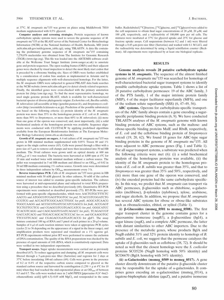
at 37°C. M. smegmatis mc2155 was grown on plates using Middlebrook 7H10medium supplemented with 0.5% glycerol.
Computer analyses and screening strategies. Protein sequences of knowncarbohydrate uptake systems were used to screen the genome sequence of M.smegmatis mc2155 at the BLAST server of the National Center for BiotechnologyInformation (NCBI) at the National Institutes of Health, Bethesda, MD (www.ncbi.nlm.nih.gov/sutils/genom_table.cgi), using TBLASTN. A data file contain-ing the preliminary genome sequence of M. smegmatis mc2155 containing7,278,076 nucleotides was obtained from The Institute for Genomic Research(TIGR) (www.tigr.org). This file was loaded into the ARTEMIS software avail-able at the Wellcome Trust Sanger Institute (www.sanger.ac.uk) to annotategene and protein sequences. The open reading frames (ORFs) and their adjacentgenes were checked by visual inspection to detect the most likely start codon thatis preceded by a ribosome binding site. Sizes of ORFs were further establishedby a consideration of codon bias analysis as implemented in Artemis and bymultiple sequence alignments with well-characterized homologs. For the latter,the M. smegmatis ORFs were subjected to general PBLAST data bank searchesat the NCBI website (www.ncbi.nlm.nih.gov) to detect closely related sequences.Finally, the identified genes were cross-checked with the primary annotationprotein list (http://cmr.tigr.org). To find the most representative homologs, weused single genome protein BLAST, which is available for well-characterizedbacterial species of diverse phylogenetic origins: Escherichia coli, Bacillus subtilis,M. tuberculosis (all accessible at http://genolist.pasteur.fr), and Streptomyces coeli-color (http://avermitilis.ls.kitasato-u.ac.jp). Prediction of the possible substrate(s)was based on the following criteria: (i) protein identity of the M. smegmatisprotein was more than 35% to the homologous protein of E. coli or B. subtilis,more than 50% to Streptomyces, or more than 60% to M. tuberculosis; (ii) morethan one gene of the operon was conserved; and, most importantly, (iii) a solidbiochemical analysis of the homologous protein was available. Sequence align-ments were conducted with CLUSTALW by applying predefined algorithmsavailable from the European Bioinformatics Institute at The European Molec-ular Biology Laboratory (www.ebi.ac.uk/clustalw).
Growth of M. smegmatis on sugars. A 4-ml culture of M. smegmatis mc2155 wasgrown in HB minimal medium supplemented with 0.05% Tween 80 and 1%sugars as the single carbon source (65). Cells were passed through a filter with apore size of 5 �m to remove cell clumps and were then inoculated into 50 ml HBmedium. The 50-ml cultures were grown until an optical density at 600 nm(OD600) of 1 was reached. The bacteria were harvested at 4,000 rpm at 4°C for10 min and washed twice with minimal medium without a carbon source. Thepellet was resuspended in 5 ml HB medium and diluted to an OD600 of 0.02 in50 ml HB medium containing 1% carbon source. Growth rates were determinedin three independent cultures by OD600 measurements every 3 h.
Reverse transcriptase PCR. Cells of M. smegmatis mc2155 were grown in HBminimal medium with 50 mM glycerol. In other cultures, 50 mM of the carbonsource of interest was added to examine gene-specific induction. The cultureswere harvested at mid-exponential phase and subjected to total RNA prepara-tion using a procedure that we described previously (68). Quantitative RT-PCRexperiments were conducted as described previously (73). RT-PCRs were per-formed with gene-specific oligonucleotides, which were AACTGTGCTTTCTCAACCG and ATGGCGTCGAGTTGGTGC for ptsI, TCACCGTCGGATCTGCCGTCG and ACCAGTTCGGCAACCTTGGC for ptsH, AGGCATCAACGTGGCCAAGG and ACCGCGTGATCGCATCGAGCG for fruK, ACCGAGTTCCTGTTCCTCG and CGAGCGTCGTGACCATCG for ptsI, GGGCATCCTCACGTCAGG and CAGCAGGTCGATCAGACC for ptsG, TCAGACCGTGACCATCACG and TGGACCAGCACTCCCAC for crr, and GCAAGGTGCTTCCGTTCAGC and CGAGACCGATGATCACCG for glpF1. The assaymixture contained 100 ng of RNA and 5 pmol of each primer in a 20-�l volume.Samples of 4 �l of each reaction mixture were taken at appropriate PCR cycles(cycles 21 to 36 depending on the appearance of a signal in the linear range), andamplification products were separated and visualized on a 1% agarose gel.RT-PCR experiments without prior RT were performed to ensure the exclusionof DNA contamination. The quality of the RNA preparations was checked by thepresence of equal amounts of 16S rRNA, which is constitutively expressed. Datawere verified in two independent experiments.
Transport assays. Sugar uptake measurements were carried out as previouslydescribed (67). To reduce aggregation and clumping, all M. smegmatis cells werefiltered through a 5-�m-pore-size filter (Sartorius) and regrown for 2 days at37°C before inoculating 100-ml cultures (69). Cells were grown in the presenceof 0.2 or 0.4% of the respective carbon source compared to glycerol as thestandard carbon source and harvested by centrifugation (1,250 � g at 4°C for 10min) when they had reached the mid-exponential phase at an OD600 of between0.5 and 0.7. The cells were washed once in 2 mM PIPES [piperazine-N,N�-bis(2-ethanesulfonic acid)] (pH 6.5)–0.05 mM MgCl2 and resuspended in the same
buffer. Radiolabeled [14C]fructose, [14C]glucose, and [14C]glycerol were added tothe cell suspension to obtain final sugar concentrations of 20 �M, 20 �M, and100 �M, respectively, and a radioactivity of 100,000 cpm per ml cells. Themixtures were incubated at 37°C for glycerol uptake and 25°C for glucose andfructose uptake assays. Cell suspensions (between 0.2 and 1 ml) were filteredthrough a 0.45-�m-pore-size filter (Sartorius) and washed with 0.1 M LiCl, andthe radioactivity was determined by using a liquid scintillation counter (Beck-man). All experiments were reproduced by at least one biological replicate.
RESULTS
Genome analysis reveals 28 putative carbohydrate uptakesystems in M. smegmatis. The sequence of the almost finishedgenome of M. smegmatis mc2155 was searched for homologs ofwell-characterized bacterial sugar transport systems to identifypossible carbohydrate uptake systems. Table 1 shows a list of28 putative carbohydrate permeases: 19 of the ABC family, 3of the PTS family, 1 of the major intrinsic protein family(MIP), 4 of the major facilitator superfamily (MFS), and oneof the sodium solute superfamily (SSS) (6, 47–49, 56).
ABC systems. Operons for carbohydrate-specific transport-ers of the ABC family always contain a gene encoding a sugar-specific periplasmic binding protein (6, 9). We have conductedTBLASTN analyses of the M. smegmatis genome with knownbinding proteins of ABC systems, such as the maltose- andribose-specific binding proteins MalE and RbsB, respectively,of E. coli and the cellobiose binding protein of Streptomycesreticuli (19, 20, 62). We found 18 genes for ABC-type sugarbinding proteins in the genome of M. smegmatis mc2155. Allwere adjacent to ABC permease genes (Fig. 1 and Table 1).For all sugar transport systems, a substrate was predicted whenthe following criteria were met (6): (i) a solid biochemicalanalysis of the homologous proteins was available, (ii) theidentity of the M. smegmatis protein to the homologous pro-teins of E. coli or B. subtilis was greater than 35%, and that toStreptomyces was greater than 35% and 50%, respectively, and(iii) more than one gene of the operon was conserved, and(iv) the encoded proteins had the same carbohydrate specific-ity. In this way, we predict that M. smegmatis can transport viaABC permeases, �-glucosides such as chitobiose, �-galacto-sides (melibiose), �-xylosides (xylobiose), xylose, arabinose,and sugar alcohols. In addition, we propose that M. smegmatishas several ABC systems for ribose or ribose-like substratessuch as ribonucleosides, ribitol, or xylitol (Table 1).
(i) �-Glucosides (msmeg_0501 to msmeg_0508). The firstsugar transport cluster in the genome contains genes for aglucosamine isomerase (nagB1), a �-glucosidase (bglA), asugar kinase (sugK), and a complete ABC permease, the latterwith distant similarities to other ABC importers. Due to thepresence of the metabolic genes, whose products BglA andNagB exhibit 31% and 32% protein identity to homologs of B.subtilis and E. coli, we suggest that the permease catalyzes theuptake of �-glucosides such as cellobiose (38, 72). It should benoted as well that the closest homologs were the S. coelicolorproteins SCO5236 (NagB homolog with 50% identity) andSCO6670 (BglA homolog with 54% identity).
(ii) �-Galactosides (msmeg_0509 to msmeg_0517). A genecluster directly downstream of the putative �-glucoside clustermay be responsible for the uptake of �-galactosides. It com-prises genes encoding an �-galactosidase (msmeg_0514), atagatose-bisphosphate aldolase (agaZ), and a putative isomerase
5904 TITGEMEYER ET AL. J. BACTERIOL.
at UN
IVE
RS
ITA
ET
SB
IBLIO
ER
LAN
GE
N on M
arch 28, 2008 jb.asm
.orgD
ownloaded from
80

(agaS) besides the genes for the ABC permease, agaEFG. Theoperon is likely controlled by AgaR, a regulator of the DeoRfamily (11, 51). Downstream of the aga region is the genemspB, which encodes a porin (69), which may indicate that this
porin is required for the entry of the substrate transported byAgaEFG. The proteins share, as many other proteins from M.smegmatis do, the highest identity to proteins from S. coelicolor(39 to 71%) (Table 1).
TABLE 1. Sugar transport systems of M. smegmatis
Family and predictedsubstrate(s) Gene designation(s) Locus tag(s)a Representative homolog(s)b
or descriptionReference(s) and/or
source(s)c
ABC�-Glucosides, chitobiose,
disaccharidesnagB1 bglA bglR sugK
bglEFGK0501–0508 nagB (SCO5236), bgl (SCO0670),
deoR, rbsK E. coli; abcEFG genesare distantly related to many ABCpermease genes; msiK (SCO4240)
6, S, C
�-Galactosides, melibiose agaRZSXPAEFGKmspB
0509–0517 SCO5848–SCO5851,SCO0538–SCO0541, msiK(SCO4240)
6, 11, S
Unknown abcEFGK 0553–0556 Distant similarity to many ABCpermease genes
S, C
Ribose, xylose rbsA1C1B1 gatY sugKrbsR1 pfkB
1372–1378 SCO6009–SCO6011, gatY(SCO5852), pfkB (SCO3197);distantly related to ribose andxylose ABC transporters
3, 6, S, C, T, B
Xylose xylF2G2H2 1704–1706 E. coli xylFGH 17, 70, CArabinose pho araR araGFKEBDA 1707–1715 E. coli ytf operon, B. subtilis araABD 60, B, CRibose, ribonucleosides gap pgk tpiA secG urf
rscA deoC rbsC2A2rbsR2 sugK sugDrbsB2 ppc pgl opcAzwf tal tkt
3084–3103 rbsH (SCO2747), rbsA (SCO2746),E. coli rbsB
3, 6, S, C
Unknown abcKGFE fabG sugK 3108–3113 Distant similarity to many ABCpermease genes
6, S, C
Unknown abcR sugKabcEFGK1K2 sugKrpiB
3264–3272 Distant similarity to many ABCpermease genes, SCO0580, rpiBE. coli
6, 66, S, C
Ribose rbsB3R3C3A3 3598–3602 SCO2747, B. subtilis ribose operon 75Sorbitol yphREKFB 3998–4002 yph operon E. coli 6, CRibose rbsA4C4B4R4 4170–4174 E. coli ribose operon rbsCBR 3, CUnknown uspGFE 4466–4468 M. tuberculosis usp operon 9, TUnknown abcRFGE 4655–4658 Distant similar to many ABC
permease genesS, C
Unknown sugKGFE 5058–5061 sug operon M. tuberculosis 9, T�-Xyloside bglG bxlRAEFG 5142–5147 S. coelicolorbxlEFG2 6Sugar alcohol smoKGFER 5571–5574 S. coelicolor smo operon 6Xylose xylG1F1E1A1R1 6018–6022 E. coli xyl operon 70Ribose rbsR5P5K5G5F5E5 6798–6805 SCO0723, SAV5702, SCO2747 6, S, C
PTSFructose ptsH fruAKR ptsI 0084–0088 S. coelicolor ptsH fruAKR ptsI 6, 45, SGlucose, trehalose
N-acetylglucosamineptsG crr nagB2A ptsR 2116–2120 S. coelicolor nagE2 crr nagAB 5, 44
Dihydroxyacetone ptsT dhaLKFR 2121–2125 S. coelicolor gyl operon, E. colidhaKLM
6, 25, S, C
MIPGlycerol glpK2RFK1D 6756–6760 S. coelicolor gyl operon 6, S
SSSGalactose galPRTK 3689–3692 S. coelicolor gal operon 6
MFSUnknown sugP1 2966 Distant similarity to sugar
permeases of the MFSS, C, B, T
Unknown sugP2 4078Glucose glcP 4182 S. coelicolor glcP 73Unknown sugP3 5559
a Only the numbers of the locus tags (msmeg_XXXX) of the M. smegmatis mc2155 genome are shown. Numbers are according to the revised annotation (www.tigr.org).b The column contains information on representatives homologs for which experimental information is available.c The following genome servers were used for BLASTP analysis: http://genolist.pasteur.fr/SubtiList (B) for B. subtilis, http://genolist.pasteur.fr/Colibri (C) for E. coli,
www.avermitilis.ls.kitasato-u.ac.jp/blast_local/index.html (S) for S. coelicolor, and http://genolist.pasteur.fr/Tuberculist (T) for M. tuberculosis.
VOL. 189, 2007 SUGAR TRANSPORT IN MYCOBACTERIA 5905
at UN
IVE
RS
ITA
ET
SB
IBLIO
ER
LAN
GE
N on M
arch 28, 2008 jb.asm
.orgD
ownloaded from
81

FIG. 1. Genetic organization of M. smegmatis carbohydrate transporters of the ABC family. The arrows indicate the lengths and transcriptionalorientations of annotated genes and predicted ORFs. Genes encoding transport systems are depicted in dark gray, carbohydrate metabolic genesare colored in light gray, and regulatory genes are highlighted in black, while other genes are white. Genes are shown by their number, with theprefix “msmeg_.” The gene names are assigned according to the annotations given by TIGR (http://www.tigr.org) and by us. Numbers in bracketsrefer to the intergenic distance between two genes. General gene designations are as follows: urf, unknown reading frame; sugD, sugardehydrogenase; sugK, sugar kinase; sugP, sugar permease; abcE, unspecified substrate binding protein of an ABC permease; abcF and abcG,unspecified membrane proteins of an ABC permease.
5906
at UN
IVE
RS
ITA
ET
SB
IBLIO
ER
LAN
GE
N on M
arch 28, 2008 jb.asm
.orgD
ownloaded from
82

(iii) Ribose and ribose-like carbohydrates (msmeg_1372 tomsmeg_1378, msmeg_3090 to msmeg_3095, msmeg_3598 tomsmeg_3602, msmeg_4170 to msmeg_4174, and msmeg_6798to msmeg_6804). Several of the analyzed ABC systems of M.smegmatis revealed similarities to the ribose ABC permeasesof E. coli and B. subtilis (3, 75). The best candidates fora ribose-specific ABC permease seem to be msmeg_3090,msmeg_3091, and msmeg_3095. The latter one is the substratebinding protein that shares the highest identity (30%) to theribose-specific periplasmic binding protein RbsB. Interestingly,this putative ribose permease is embedded within a cluster ofgenes involved in central carbon metabolism, such as glyco-lysis. The other four ABC systems with similarities to ribosepermeases may also transport ribose or a ribose derivative(Table 1).
(iv) Xylose and �-xylosides (msmeg_1704 to msmeg_1706,msmeg_5142 to msmeg_5147, and msmeg_6018 tomsmeg_6022). Xylose is usually taken up by an ABC permeaseand further metabolized by isomerization (xylose isomerase[XylA]) and phosphorylation (xylulokinase [XylB]) to enterthe pentose-phosphate shunt. The region msmeg_6018 tomsmeg_6022 was designated the xylGFEAR1 regulon since itencodes proteins sharing identities of 29% to 38% with thecorresponding proteins of E. coli (7, 20). The substrate bindingprotein XylF1 has the highest score, with 38% identity to the E.coli homolog (70). The regulon further contains the metabolicxylA1 gene. The missing XylB may be encoded bymsmeg_3257, having 50% and 32% identity to XylB of Coryne-bacterium glutamicum and E. coli, respectively (31, 33). A sec-ond potential operon for a xylose ABC transporter is encodedby msmeg_1704 to msmeg_1706 (xylFGH2). XylF2, XylG2, andXylH2 share 38% to 48% identity to the corresponding E. coliproteins.
A predicted �-xyloside permease is encoded by msmeg_5142to msmeg_5147. We have designated the genes bxlRAEFGaccording to their close relationship to the bxlEFG2 operon ofS. coelicolor, which encodes the transporter for xylobiose (6,27). The amino acid identities are in the range of 45% to 79%.
(v) Arabinose (msmeg_1708 to msmeg_1715). We detectedan araBDA operon that is most similar to the arabinose met-abolic genes of B. subtilis (40). AraB (L-ribulokinase), AraD(L-ribulose-5-phosphate-4-epimerase), and AraA (L-arabinoseisomerase) show 42 to 48% identity to the corresponding pro-teins of B. subtilis. The adjacent ABC permease (msmeg_1709to msmeg_1712) could be the uptake system for arabinose. Itsgene products, including the juxtaposed regulator, exhibit res-idues that are up to 43% identical to an unknown gene clusterof E. coli that is designated ytfRT yjfF. This suggests that the E.coli locus encodes a sugar-specific permease. Transport of ar-abinose has so far been described only for the E. coli protonsymporter AraE of the MFS (34, 47).
(vi) Sugar alcohols (msmeg_3998 to msmeg_4002 andmsmeg_5571 to msmeg_5575). Sugar alcohols like mannitol,glucitol (sorbitol), and xylitol are frequently consumed by bac-teria (52). We found two loci that could encode a sugar alco-hol-specific transport system. The operon comprisingmsmeg_3998 to msmeg_4002 is homologous to the yph operon ofE. coli. Although the permease proteins do not allow substrateprediction, they are associated with a sugar alcohol dehydro-genase (msmeg_4002), which is 44% identical to YphC and
more distantly related to GutB of B. subtilis (sorbitol dehydro-genase) and GatD (galactitol-1P dehydrogenase) of E. coli(43). The second region, msmeg_5571 to msmeg_5575, is ho-mologous to the smo operon of S. coelicolor, encoding a pos-sible permease for sugar alcohols. Here, the two substrate-specific binding SmoE proteins share 52% identity (6).
(vii) Less-well-defined ABC-type sugar transporters. Wefurther found six gene loci encoding ABC transport systems forcarbohydrates. The deduced substrate binding proteins werehomologous to known sugar substrate binding proteins in therange of about 30% protein identity. Due to this and due to theabsence of adjacent metabolic genes, prediction of substrateswas not possible. Among those are the only two permeases thatare common to M. smegmatis and M. tuberculosis, SugABC andUspABC (see below).
(viii) Lipid anchors of sugar binding proteins. In gram-negative bacteria, the substrate binding proteins of ABC trans-porters are soluble proteins that appear to move freely throughthe periplasmic space. By contrast, it has been shown for gram-positive bacteria that the substrate binding proteins are co-valently anchored to the outside of the cell membrane by fattyacids (63). Lipidation occurs via esterification of a conservedcysteine at the N terminus of the processed protein. Strikingly,all sugar binding proteins of M. smegmatis (Table 2) and M.tuberculosis (9) have a predicted lipoprotein signal peptide.
PTS permeases. (i) Glucose, trehalose, GlcNAc, and dihy-droxyacetone (msmeg_2116 to msmeg_2125). Two loci encod-ing components of a PTS were discovered in the genome of M.smegmatis. This is interesting, since the existence of PTS genesis in contrast to previous reports in which no evidence for PTSproteins in M. smegmatis was found in biochemical experi-ments (14, 15, 54, 59). Genes for a PTS of the glucose-sucrosesubfamily (Fig. 2) were detected in a cluster comprisingmsmeg_2116 to msmeg_2120 (52). The two divergently tran-scribed genes msmeg_2116 (ptsG) and msmeg_2117 (crr) en-code the IIBC and IIA permeases. The deduced protein se-quence of IIBC (496 amino acids [aa]) shares 49% identitywith the N-acetylglucosamine (GlcNAc)-specific IIBGlcNAc andIICGlcNAc of S. coelicolor A3(2) and 42 to 43% identity to PTSpermeases of E. coli and B. subtilis that transport glucose,GlcNAc, trehalose, or �-glucosides, respectively (52). The IIAprotein of M. smegmatis is 50% identical to the S. coelicolorhomolog IIACrr (30). Two metabolic genes for the metabolismof GlcNAc are situated downstream of the crr gene. Theyencode a putative glucosamine-6-phosphate deaminase,NagB2, and a GlcNAc deacetylase, NagA, that share 43% and33% identity to the characterized E. coli homologs and 59%and 47% to the ones from S. coelicolor, respectively (1). Thegene locus further contains a putative regulatory gene, ptsR,whose product shares low similarities to uncharacterized reg-ulators of S. coelicolor and M. tuberculosis. Thus, PtsR might bea possible regulator of the PTS genes.
Downstream of ptsR is a gene for a multiphosphoryl PTSphosphotransferase. The gene, which we have designated ptsT,encodes a protein of 808 aa that comprises all three generalphosphotransferases of a PTS: IIA (aa 1 to 150), HPr (aa 151to 251), and EI (aa 251 to 808). The protein resembles themultiphosphoryl phosphotransferase MTP from Rhodobactercapsulatus (76), which drives fructose uptake by phosphoryla-tion of the respective FruA PTS permease, in length and do-
VOL. 189, 2007 SUGAR TRANSPORT IN MYCOBACTERIA 5907
at UN
IVE
RS
ITA
ET
SB
IBLIO
ER
LAN
GE
N on M
arch 28, 2008 jb.asm
.orgD
ownloaded from
83

main structure. Hence, PtsT may be involved in the phosphor-ylation of the adjacent PTS permease PtsG. In addition, PtsTis a homolog of the multiphosphoryl phosphotransferaseDhaM of E. coli. DhaM undergoes phosphoenolpyruvate-de-pendent autophosphorylation and then phosphorylates ADPthrough its enzyme IIA domain. This ATP molecule is used bythe dihydroxyacetone kinase DhaKL for substrate phosphory-lation (21, 25). Strikingly, homologs of dhaKL are, as in E. coli,juxtaposed with ptsT (dhaM). An adjacent permease gene,which we have termed dhaF, does not exist in E. coli. Wepredict that PtsT serves as the shuttle for the transfer of phos-phate to DhaKL kinase, which in turn phosphorylates dihy-droxyacetone molecules that are imported through the MIPfamily facilitator DhaF.
(ii) Fructose (msmeg_0084 to msmeg_0088). The secondPTS locus comprises genes for a fructose-specific PTS com-posed of EI (ptsI), HPr (ptsH), and IIABCFru (fruA) (Fig. 2B).The locus further consists of a gene coding for a regulator ofthe DeoR family (51) and a fruK gene coding for a proteinsimilar to fructose-1-phosphate kinases (45). The gene orderfruR-fruK-fruA is the same as what we previously found in S.coelicolor, while the fru operons of other bacteria are under thecontrol of different regulators (52). The similarities of proteinsencoded by genes in these operons are highest between M.smegmatis and S. coelicolor, with identities of 51% for theDeoR-type regulator, 43% for FruK, and 51% for FruA(IIABCFru).
MIP permeases. Sugar permeases of the MIP family werescreened by BLASTP searches with the glycerol facilitatorprotein sequences of E. coli and S. coelicolor. We found aprotein, msmeg_6758 (GlpF), with identity scores of 42% and37% to the respective pendants from E. coli and S. coelicolor.msmeg_6758 is situated in an operon with genes for two glyc-erol kinases (glpA1 and glpA2) and a glycerol-3-phosphate de-hydrogenase (glpD) (Fig. 2). A putative regulatory gene (glpR)that is homologous to the glycerol operon regulator gene gylRof S. coelicolor is found upstream (26). Hence, the genes
msmeg_6756 to msmeg_6760 are the best candidates to encodeproteins for glycerol uptake and metabolism. As describedabove for PTS, we identified DhaF (msmeg_2124) as being asecond MIP family member that may serve as a facilitator fordihydroxyacetone in conjunction with PTS proteins.
MFS permeases. Well-characterized permeases of the MFSfamily are the xylose symporter XylE of E. coli and the glucose-specific symporter GlcP from S. coelicolor and the cyanobac-terium Synechocystis (34, 73, 77). We found four homologs(msmeg_2966, msmeg_4098, msmeg_4182, and msmeg_5559),of which msmeg_4182 exhibited 53% identity to glucose sym-porters (Fig. 2). msmeg_4182 is surrounded by genes that arenot related to sugar metabolism, and there is no putative reg-ulatory gene in the vicinity. Hence, glcP might encode a glucosepermease that is expressed constitutively in a monocistronicoperon. The other three candidates showed very distant simi-larities to sugar transporters, thus making a substrate predic-tion impossible.
Growth of M. smegmatis on carbohydrates as single carbonsources. According to our genome analysis, M. smegmatis pos-sesses uptake systems for a large number of sugars. This isconsistent with very early findings that M. smegmatis can growon many monosaccharides, organic acids, and sugar alcohols(20). In those studies, oxygen consumption of starved bacteriawith low endogenous respiration was measured after the addi-tion of single carbon sources to bacteria suspended either inphosphate buffer or in phosphate saline (57). However, inthose experiments, M. smegmatis may have lacked other ele-ments that may be required to consume the substrate of inter-est. Therefore, we conducted growth experiments in HB min-imal medium, which contains all essential nutrients and traceelements (65), and the carbohydrate of interest as the solecarbon source. M. smegmatis readily grew in HB minimal me-dium with glucose, glycerol, xylose, fructose, ribose, arabinose,or trehalose (Fig. 3 and Table 3). Potential uptake systemswere assigned in this study for all of these mono- and disac-charides (see Fig. 5A). These results demonstrated that func-
TABLE 2. N-terminal sequences of the periplasmic binding proteins of ABC sugar transporters in M. smegmatisa
Protein Locus tag N-terminal sequence (no. of residues)
*BglE msmeg_0505 ---------------MTRTRLFRFGSAVASTLTVAALALSACAPGPSGDSGSSPAPTGEVSKDI (49)AgaE msmeg_0515 ---------------------MIRRWLCLAVVTAVACLLTACGGGSSSSGPVEIAVWHGYQDTE (43)AbcE msmeg_0553 -----------VTSPAFTRRRALQLLGLAGGAAMLAPALAACGSSGGNSALAADAPVSGRFEGV (53)RbsB1 msmeg_1374 --------------------VNRKRLMLAAGVVALALPMAACTSSKPQADESSETPAAAGEAPA (44)XylF1 msmeg_1704 -------------------MRKLTWLAALLAALAMAMTLSGCGRSAEGGGGGDGDAKGTVGIAM (45)AraE msmeg_1712 ---------------------VRKMFAAAIGVVAVAAAVTACGSGKAPGSEGGSAPDGALTLGF (43)RbsB2 msmeg_3095 -------------------VSFAKALSGIALGAAMALSFTGCSVPGDDAAQNAPVVDGALKIGF (45)AbcE msmeg_3111 ----------MKIPQLRRRRRRPAITAITAMSVAAGLVLSGCAGTGGPANDEATSGVGDVPTDT (54)AbcE msmeg_3266 ---MMSRESQPGLHRQLSRRNMLAAMGLAGAAAVSLPVLSACGVGGRTNAPNGASEVTGGFDWR (61)RbsB3 msmeg_3599 -------------------MRLGTTAFAIASATALGLGLTACGAGDPAANSDTTRIGVTVYDMS (45)YphE msmeg_3999 ----------------MPRSLRRRAVRFATLMLVAALAVSGCSRIGENGRIAVVYLNAEGFYAG (48)RbsB4 msmeg_4172 --------------------MKIRNILPILVCTTCAVAMTACSSSVNNNADPSDTAAPATNVEV (44)AbcE msmeg_4658 --------------------MRLSRLVAAAGVGVLMLGASACSSTGGKPDSSGGGDMGAGTADT (44)SugE msmeg_5061 -------------------VRARRLCAAAVAAMAAASMVSACGSQTGGIVINYYTPANEEATFK (45)BxlE msmeg_5145 VDLKSVDANVVESKADFLPSTSRRAFLAAALSVPLLGALAACGSSGPSRSGGGGGGAPGAASYW (64)SmoE msmeg_5574 --------------------MKRLRRLAACIAAAGLTATAGCAGAGTLGATDQTVTIAMVSNSQ (44)XylE2 msmeg_6020 --------------------VKRTSTLLVTAVVGLGLTLTACGSDSGSNAGSAEGGSGGKIGVI (40)RbsB5 msmeg_6804 --------------MFRKVTRNTRTVGAALMAGSLVLGMTACGGSGSDGVKVGLITKTDSNPYF (50)
a The sequences were aligned according to their signal peptide cleavage site as predicted by SignalP (4). The N-terminal cysteine of the mature protein, which is thecatalytic residue of the fatty acid anchor, is marked with an asterisk.
5908 TITGEMEYER ET AL. J. BACTERIOL.
at UN
IVE
RS
ITA
ET
SB
IBLIO
ER
LAN
GE
N on M
arch 28, 2008 jb.asm
.orgD
ownloaded from
84

tional uptake systems exist for these sugars and that thesesugars are metabolized by M. smegmatis. The generation timesranged from 132 to 174 min, which is similar to growth rates ofM. smegmatis in Middlebrook 7H9 medium that contains glyc-erol as the main carbon source (69). The monosaccharidesGlcNAc, glucosamine, and galactose; the disaccharides malt-ose, sucrose, and lactose; and the trisaccharide raffinose werenot utilized. This may indicate the lack of uptake and/or lack ofmetabolic enzymes. Particularly interesting was the growth ofM. smegmatis on maltose as a sole carbon source. M. smegmatisstopped growing at a very early stage at an OD600 of 0.1 (Fig.3). The poor growth of M. smegmatis on maltose was notaltered when the amount of maltose in the medium was in-creased from 1% to 10%, indicating that a putative, minorcontaminating carbon source did not did not cause the initialresidual growth of M. smegmatis. Similar results were obtainedfor galactose, lactose, and sucrose (not shown). The reason forthis phenomenon is unknown.
Analysis of the expression of the fructose, glucose, and glyc-erol import systems. In order to validate the predicted assign-ment of inner membrane transport systems to a specific sugar,we examined the transcription of selected genes by semiquan-titative RT-PCR. First, the transcription of the genes necessary
for fructose uptake and utilization were analyzed. The genemsmeg_0086, predicted to encode a fructose-1-phosphate ki-nase (FruK), showed enhanced mRNA levels in cells grown inthe presence of fructose (Fig. 4). Transcription of the adjacentgenes ptsH and ptsI, encoding general components of the PTS,was not enhanced by fructose. Transport experiments with[14C]fructose were done to examine whether fructose inducesthe expression of its uptake system. Figure 5A clearly showsthat fructose uptake is enhanced in M. smegmatis when grownin the presence of 2% fructose. This demonstrates that fruc-tose uptake is inducible in M. smegmatis. These results confirmthe prediction that msmeg_0085 and msmeg_0086 represent afruAK operon in which fruA encodes a fructose-specific enzymeIIA permease, as shown previously for S. coelicolor (45). Ex-pression of this operon is most likely controlled by the adjacentDeoR-like regulator that we have thus designated FruR(msmeg_0087).
Two systems were predicted for the uptake of glucose: thesymporter GlcP and a glucose-specific PTS. The transcriptionof crr and ptsT was induced in cells grown in the presence ofglucose (Fig. 4), indicating that their assignments as glucose-
FIG. 2. Genetic organization of M. smegmatis carbohydrate trans-porters of the PTS, MIP, SSS, and MFS protein families. For anexplanation of the data, see the legend to Fig. 1.
FIG. 3. Growth of M. smegmatis in minimal medium with differentcarbon sources. The growth of M. smegmatis mc2155 at 37°C in HBmedium containing 1% glucose (circles), 1% trehalose (diamonds),1% maltose (squares), and 1% GlcNAc (triangles) was measured bydetermining the OD600 of the cultures. The values are the means ofthree independent experiments. For some data points, the standarddeviations were smaller than the symbol size, and therefore, the errorbars are invisible.
TABLE 3. Growth of M. smegmatis on various sugars as asole carbon sourcea
Sugar Generation time (min) Growthb
LB 156 � 24 NDGlycerol 174 � 6 �D-Xylose 192 � 18 �D-Ribose 497 � 36 �/�L-Arabinose 154 � 10 �D-Fructose 174 � 18 �D-Glucose 132 � 12 �Trehalose 150 � 12 ND
a Cultures of M. smegmatis mc2155 were grown on HB minimal mediumcontaining glucose. Cell were washed twice in HB medium, resuspended, anddiluted to an OD600 of 0.02 in HB medium containing 1% of the respectivesugars. The generation times were calculated as described in Materials andMethods. Growth in full medium (LB, lysogeny broth) was used as a control. ND,not determined.
b Data reported previously by Izumori et al. (29).
VOL. 189, 2007 SUGAR TRANSPORT IN MYCOBACTERIA 5909
at UN
IVE
RS
ITA
ET
SB
IBLIO
ER
LAN
GE
N on M
arch 28, 2008 jb.asm
.orgD
ownloaded from
85

specific PTS enzyme IIA and tripartite PTS phosphotransfer-ase are correct. Uptake of [14C]glucose by M. smegmatis wasnot altered when cells were grown in the absence or presenceof 2% glucose (Fig. 5B). One possibility to explain this result isthat glcP is expressed constitutively. However, the glcP mRNAwas not detected in RT-PCR experiments for unknown rea-sons, in contrast to mRNA of control genes such as sigA (notshown). An alternative explanation is that the rate-limitingstep for glucose uptake is diffusion across the outer membraneof M. smegmatis. Indeed, porin mutants of M. smegmatis showa significantly impaired glucose uptake (67, 69). To date, it isunknown whether porin-mediated diffusion of glucose is therate-limiting step in wild-type M. smegmatis. The fact thatuninduced glucose uptake is as fast as the induced uptake offructose (Fig. 5) argues in favor of a constitutively expressedinner membrane transporter for glucose. Further experimentswith mutants lacking either GlcP or PtsG are required toexamine whether glucose transport is regulated in M. smegmatis.
The number of transcripts of the predicted glycerol facilita-tor gene glpF (msmeg_6758) was increased in cells grown in thepresence of 0.4% glycerol compared to that in cells grown inthe presence of 0.4% glucose (Fig. 4). Furthermore, the uptakeof [14C]glycerol was slightly increased in M. smegmatis cellsgrown in the presence of 0.4% glycerol compared to that incells grown in the presence of 0.4% glucose (Fig. 5C). Theseresults confirmed the annotation of the msmeg_6758 gene asglpF.
Carbohydrate transporters of M. tuberculosis H37Rv. Weevaluated the complement of the putative carbohydrate uptakesystems in the slow-growing pathogenic strain M. tuberculosisH37Rv (13). Our analysis resulted in the identification of fourABC-type systems and one permease of the MFS class (Fig.6B). Some of these ABC transporters were described previ-ously in an in silico analysis of the M. tuberculosis genome in amore global context (9, 15). It is striking that M. tuberculosis ispoorly equipped with carbohydrate transport systems in com-parison to M. smegmatis mc2155 (Fig. 6). Two of the operons,the lpgY sugABC and the uspABC operons, are highly con-served between the two species. The proteins of the ABCSug
and of the ABCUsp systems share between 62% and 80%similar amino acids, compared to only 25 to 30% similar aminoacids for the UgpABCE and Rv2038c-Rv2041c systems. Thesimilarities of all four ABC systems to known transportersoutside the genus Mycobacterium is so low (25%) that sub-strates of these transporters cannot be predicted.
The SugI porter of the MFS class shows distant sequencesimilarity to the glucose permease GlcP (28%) of S. coelicolor
FIG. 4. Expression analysis of selected genes of M. smegmatis. Thefigure shows 1% agarose gels with PCR products from semiquantita-tive RT-PCR experiments. For each gene, samples were taken peri-odically along the PCR. The depicted bands show products from thesame cycle for each gene, when the amplification was in the linearrange. RT-PCR experiments without a reverse transcriptase reactiondelivered no signal (negative control). Data were obtained from twobiological replicates. gly, glycerol; fru, fructose; glc, glucose.
FIG. 5. Kinetics and inducibility of fructose, glucose, and glyceroluptake by M. smegmatis. (A) M. smegmatis mc2155 was grown inMiddlebrook 7H9 medium in the presence of 2% glycerol (open cir-cles) or 2% fructose (closed circles). Accumulation of [14C]fructosewas measured at 25°C at a final fructose concentration of 20 �M.(B) M. smegmatis mc2155 was grown in Middlebrook 7H9 medium inthe presence of 2% glycerol (open circles), 2% glucose (closed circles),and 2% glycerol plus 2% glucose (closed squares). Accumulation of[14C]glucose was measured at 25°C at a final glucose concentration of20 �M. (C) M. smegmatis mc2155 was grown in HB medium containing0.4% glucose (open circles) and 0.4% glycerol (closed circles) to anOD600 of 0.9. Accumulation of [14C]glycerol was measured at 37°C ata final concentration of 100 �M glycerol. For A and B, the standarddeviations are indicated by error bars and represent the means of threeindependent experiments. For C, samples were taken in duplicatesfrom two biological replicates.
5910 TITGEMEYER ET AL. J. BACTERIOL.
at UN
IVE
RS
ITA
ET
SB
IBLIO
ER
LAN
GE
N on M
arch 28, 2008 jb.asm
.orgD
ownloaded from
86

FIG. 6. Sugar transport systems of M. smegmatis and M. tuberculosis. Shown are the permeases of the ABC, PTS, MIP, MFS, and SSS families.The derived putative substrates are inferred from in silico analyses in combination with experimental data.
5911
at UN
IVE
RS
ITA
ET
SB
IBLIO
ER
LAN
GE
N on M
arch 28, 2008 jb.asm
.orgD
ownloaded from
87

and to the galactose (GalP) (24%) and arabinose (AraE)(24%) transporters of E. coli. Thus, the system is likely totransport a monosaccharide.
Glycerol is used as the standard carbon source to grow M.tuberculosis. We did not detect any putative uptake system forthis carbohydrate. Since M. tuberculosis grows with a genera-tion time of 24 h, and it has been shown that glycerol candirectly diffuse through lipid membranes both in vitro (50) andin vivo (22), it is conceivable that the rate of glycerol intake bypassive diffusion may be sufficient for growth. Incoming glyc-erol will then be converted by glycerol kinase (GlpK) intoglycerol-3-phosphate to enter the route of central carbon me-tabolism (Fig. 6B). M. tuberculosis has one putative glycerolkinase that shows a high similarity to the two glycerol kinasesof M. smegmatis (77% protein identity for msmeg_6759 and57% for msmeg_6756) and to the two glycerol kinases from S.coelicolor SCO0509 (75%) and SCO1660 (59%).
DISCUSSION
Identification of carbohydrate transporters in M. smegmatis.It has been widely documented that M. smegmatis can grow onmany carbon sources such as polyols, pentoses, and hexoses(20, 23, 28). In this study, we identified multiple inner mem-brane transport systems for all three of these classes of carbo-hydrates by bioinformatic analysis (Table 1). This provides themolecular basis for the adaptability of M. smegmatis to differ-ent environments in the soil and water. Often, our integratedbioinformatic approach enabled us to propose a specific sub-strate for particular uptake proteins (Table 1). Since the spec-ificity of transport proteins can be altered by the modificationof a few residues, the suggested substrates rather represent ahypothesis for experiments such as transport measurementswith gene deletion mutants or analysis of the induction of geneexpression. Analysis of the induction of both the transportactivity and transcription of genes confirmed the substrate pre-dictions for a fructose- and glucose-specific PTS as well as forthe predicted glycerol operon. Since glucose uptake was con-stitutive, at least one more system for glucose transport mustoccur in M. smegmatis. This was predicted to be GlcP. Indeed,cloning and heterologous expression of glcP in E. coli revealedthat it is a glucose-specific permease (data not shown).
Global control of carbon metabolism in M. smegmatis andM. tuberculosis. The discovery of homologs of all componentsof a PTS in M. smegmatis contradicts a previous report that didnot find biochemical evidence for the existence of a PTS (59)and many repeating statements (14, 15, 54). Components ofthe PTS play a key role in the global control of sugar metab-olism to achieve the hierarchical utilization of carbon sourcesin bacteria (12), where two different mechanisms have evolved.In E. coli and other closely related gram-negative bacteria, theenzyme IIAGlc is dephosphorylated mainly under repressingconditions and mediates inducer exclusion. Under nonrepress-ing conditions, phosphorylated IIAGlc stimulates cyclic AMP(cAMP) synthesis and thereby triggers the activation of catab-olite-repressed genes by a global regulator, the cAMP-depen-dent catabolite activator protein. In low-G�C-content gram-positive bacteria, HPr is a central switch of carbon cataboliterepression. Under repressing conditions, HPr is phosphory-lated mainly at serine 46 by a unique HPr kinase/phosphatase
mediating inducer exclusion and carbon catabolite repression/activation (12, 37, 55). Under nonrepressing conditions, HPr isphosphorylated at histidine 15 and activates PTS-dependentsugar transport, glycerol kinase, and substrate-specific regula-tors. The apparent absence of a protein in M. smegmatis similarto the HPr kinase/phosphatase argues against the mechanismfound in low-G�C-content gram-positive bacteria. On theother hand, M. smegmatis apparently does not produce pro-teins with significant sequence similarities to the cAMP recep-tor protein (CRP) (catabolite activator protein) of E. coli,which is crucial for carbon catabolite repression in gram-neg-ative bacteria.
By contrast, the coordination of the few operons involved inthe uptake and degradation of carbohydrates by M. tuberculosismay not require a global control mechanism, as suggested bythe lack of PTS homologs. Alternatively, a completely differentmechanism for the global control of carbon metabolism mayhave evolved in M. tuberculosis to adapt to its specific environ-ment inside the phagosome of human macrophages. Indeed,M. tuberculosis has eight orthologs of CRP-like transcriptionalregulators (35), one of which, Rv3676, was experimentally de-scribed (2). Furthermore, the large number and the differentsubcellular localization of the 15 putative nucleotide cyclases inM. tuberculosis imply that this organism may have the ability tosense and respond to many intracellular and extracellular sig-nals through a second messenger system based on cyclic nu-cleotide monophosphates (35). This is in strong contrast to E.coli and other gram-negative bacteria, which have only oneCRP and one adenylate cyclase. CRP homologs have beenidentified in streptomycetes (18), where the regulator plays arole in germination, and in corynebacteria, where CRPs havebeen associated with global carbon regulation (39). Althoughthe potential mechanisms of global control of carbon metab-olism in both M. smegmatis and M. tuberculosis are not evidentfrom the bioinformatic analysis of their genomes, these find-ings provide hypotheses for further experiments.
Utilization of galactose by M. smegmatis and M. tuberculosis.M. smegmatis did not grow on D-galactose as a sole carbonsource. This result is in agreement with previous reports (28,29). Surprisingly, our analysis identified the genes msmeg_3689to msmeg_3692 as being an operon encoding, among others, aputative galactose transport protein of the SSS with more than50% amino acid identity to the putative galactose transporterGalP of S. coelicolor (6). Furthermore, it was shown that D-galactose is taken up by M. smegmatis and that this transportactivity is inducible by D-galactose (28). However, the putativegal operon of M. smegmatis contains only two of three essentialgenes (galKTE) of the Leloir pathway, which is used by E. coliand many other bacteria to grow on galactose (24). The firstreaction in this pathway is catalyzed by galactokinase, whichphosphorylates free galactose to galactose-1-phosphate. In thenext steps, galactose-1-phosphate uridylyltransferase transfersthe UDP residue from UDP-glucose to galactose-1-phosphate,and UDP-galactose-4-epimerase catalyzes the reversible con-versions of UDP-galactose and UDP-glucose. The galE geneencoding the epimerase is missing. These results appear to becounterintuitive: why should M. smegmatis take up a sugar,modify it, and not use it as a carbon or energy source? Oneexplanation may be provided by the fact that D-galactose is amajor constituent of the cell wall of mycobacteria. The arabi-
5912 TITGEMEYER ET AL. J. BACTERIOL.
at UN
IVE
RS
ITA
ET
SB
IBLIO
ER
LAN
GE
N on M
arch 28, 2008 jb.asm
.orgD
ownloaded from
88

nogalactan polymer is composed of D-arabinose and D-galac-tose, both in their furanose ring form, and is an essential partof the mycobacterial cell wall by serving as an anchor for thecovalent attachment of the peptidoglycan and the mycolicacids (10). Ethambutol is a potent tuberculosis drug and actsby inhibiting the synthesis of arabinogalactan (71). Based onthese observations, we suggest the following sequence ofreactions: D-galactose is taken up by M. smegmatis via GalP(msmeg_3689), phosphorylated in the cytoplasm by the galactoki-nase GalK (msmeg_3692), and uridinylated by the galactose-1-phosphate uridyltransferase GalT (msmeg_3691) to yieldUDP-galactose. The ring contraction of UDP-galactopyranoseto UDP-galactofuranose is catalyzed by the essential enzymeUDP-galactopyranose mutase Glf (46). UDP-galactofuranoseis then likely to be transported across the cytoplasm membranevia intermediate binding to decaprenyl phosphate as in otherbacteria to be available for the synthesis of arabinogalactan(16). It should be noted that no homologs of the putativegalactose transporter GalP of M. smegmatis (Table 1) werefound in M. tuberculosis. This suggests that M. tuberculosis hasno access to D-galactose in its natural environment, the phago-some of macrophages, and instead synthesizes D-galactosefrom other sugars. Indeed, in addition to galK and galT, thegenome of M. tuberculosis contains three galE genes (Rv3634c,Rv0501, and Rv0536), which are probably used to convertglucose to galactose for biosynthesis purposes. However, it isunclear how M. smegmatis synthesizes galactose in the absenceof this sugar and GalE. M. smegmatis may either contain anundetected enzyme with glucose-galactose epimerase activityor use an alternative biosynthetic route.
Utilization of disaccharides by M. smegmatis and M. tuber-culosis. M. smegmatis did not grow on lactose, maltose, andsucrose as a sole carbon source. Franke and Schillinger previ-ously obtained the same result for lactose and maltose butobserved respiration of M. smegmatis in the presence of su-crose (23). According to our bioinformatic analysis, M. smeg-matis has at least three inner membrane transport systems withsignificant similarities to other bacterial disaccharide trans-porters (Table 1). However, the substrate specificities ofthe transporters encoded within the loci msmeg_0501 tomsmeg_0508 and msmeg_0509 to msmeg_0518 are not known.Growth of bacteria on disaccharides as sole carbon sourcesrequires enzymes that cleave the disaccharide and release themonosaccharides for further metabolization. The absence ofproteins similar to known bacterial �-D-galactosidases (LacZof E. coli, BgaB of Bacillus circulans, MbgA of Bacillus mega-terium, and LacA of S. coelicolor) provides a molecular expla-nation for the inability of M. smegmatis to utilize lactose as asole carbon source. By contrast, M. smegmatis has six homologs(msmeg_3184, msmeg_3576, msmeg_4916, msmeg_4917,msmeg_4696, and msmeg_6515) of MalL of B. subtilis, whichhydrolyzes maltose, longer maltodextrines up to maltohexaose,isomaltose, and sucrose (64), and of the cytoplasmic trehalaseTreC of E. coli, which cleaves trehalose-6-phosphate (58). It isconceivable that these enzymes are used in trehalose metabo-lism, considering the unusual importance of trehalose in my-cobacteria (42, 74) and the observation that trehalose was theonly disaccharide that was used by M. smegmatis as a solecarbon source. However, it cannot be excluded that some of
the enzymes with similarities to TreC and MalF have roles inpathways distinct from trehalose metabolism.
The SugABC sugar transport system was shown to be essen-tial for the virulence of M. tuberculosis in mice (61). Previously,it was suggested that this permease may transport maltose ormaltodextrins (8, 9). However, the similarities of both ABCSug
and the corresponding substrate binding protein LpgY to themaltose transporters and periplasmic maltose binding proteinsMalE of E. coli and S. coelicolor are very low (25%). Thus, itis questionable whether maltose is the substrate of ABCSug.These doubts are supported by the fact that neither M. smeg-matis, which has a highly similar ABCSug system, nor M. tuber-culosis (20) grows on maltose as a sole carbon source. It has tobe noted that similar uncertainties exist for the substrate spec-ificities of the four other carbohydrate uptake systems of M.tuberculosis, including the ABCUsp transporter, which was pro-posed to transport sn-glycerol-3-phosphate based on low pro-tein similarities (9, 15).
The analysis of the carbohydrate uptake proteins in thegenomes of M. smegmatis and M. tuberculosis provides themolecular basis for the very early phenotypic observations thatsaprophytic mycobacteria have a much broader spectrum ofsubstrates, which they can use as sole carbon and energysources (20). It is striking that the genome of M. tuberculosishas only five recognizable permeases for carbohydrate uptake.This suggests that the phagosome does not provide an envi-ronment rich in diverse sugars. Hence, an experimental anal-ysis of the substrate specificity of the inner membrane carbo-hydrate transporters of M. tuberculosis is likely to reveal thecarbon sources available in the phagosome of human macro-phages.
ACKNOWLEDGMENTS
This work was supported by grant AI06432 from the National Insti-tutes of Health to M.N. and by grants SFB473 and GraduiertenkollegGK805 of the Deutsche Forschungsgemeinschaft.
Sequencing of M. smegmatis mc2155 was accomplished by TIGRwith support from National Institute of Allergy and Infectious Dis-eases (NIAID). We thank Natalie Wood, Flavia Pimentel-Schmitt,Hildegard Stork, and Ying Wang for technical assistance.
REFERENCES
1. Alvarez-Anorve, L. I., M. L. Calcagno, and J. Plumbridge. 2005. Why doesEscherichia coli grow more slowly on glucosamine than on N-acetylglu-cosamine? Effects of enzyme levels and allosteric activation of GlcN6Pdeaminase (NagB) on growth rates. J. Bacteriol. 187:2974–2982.
2. Bai, G., L. A. McCue, and K. A. McDonough. 2005. Characterization ofMycobacterium tuberculosis Rv3676 (CRPMt), a cyclic AMP receptor pro-tein-like DNA binding protein. J. Bacteriol. 187:7795–7804.
3. Bell, A. W., S. D. Buckel, J. M. Groarke, J. N. Hope, D. H. Kingsley, andM. A. Hermodson. 1986. The nucleotide sequences of the rbsD, rbsA, andrbsC genes of Escherichia coli K12. J. Biol. Chem. 261:7652–7658.
4. Bendtsen, J. D., H. Nielsen, G. von Heijne, and S. Brunak. 2004. Improvedprediction of signal peptides: SignalP 3.0. J. Mol. Biol. 340:783–795.
5. Bentley, S. D., K. F. Chater, A. M. Cerdeno-Tarraga, G. L. Challis, N. R.Thomson, K. D. James, D. E. Harris, M. A. Quail, H. Kieser, D. Harper, A.Bateman, S. Brown, G. Chandra, C. W. Chen, M. Collins, A. Cronin, A.Fraser, A. Goble, J. Hidalgo, T. Hornsby, S. Howarth, C. H. Huang, T.Kieser, L. Larke, L. Murphy, K. Oliver, S. O’Neil, E. Rabbinowitsch, M. A.Rajandream, K. Rutherford, S. Rutter, K. Seeger, D. Saunders, S. Sharp, R.Squares, S. Squares, K. Taylor, T. Warren, A. Wietzorrek, J. Woodward,B. G. Barrell, J. Parkhill, and D. A. Hopwood. 2002. Complete genomesequence of the model actinomycete Streptomyces coelicolor A3(2). Nature417:141–147.
6. Bertram, R., M. Schlicht, K. Mahr, H. Nothaft, M. H. Saier, Jr., and F.Titgemeyer. 2004. In silico and transcriptional analysis of carbohydrate up-take systems of Streptomyces coelicolor A3(2). J. Bacteriol. 186:1362–1373.
7. Blattner, F. R., G. Plunkett III, C. A. Bloch, N. T. Perna, V. Burland, M.
VOL. 189, 2007 SUGAR TRANSPORT IN MYCOBACTERIA 5913
at UN
IVE
RS
ITA
ET
SB
IBLIO
ER
LAN
GE
N on M
arch 28, 2008 jb.asm
.orgD
ownloaded from
89

Riley, J. Collado-Vides, J. D. Glasner, C. K. Rode, G. F. Mayhew, J. Gregor,N. W. Davis, H. A. Kirkpatrick, M. A. Goeden, D. J. Rose, B. Mau, and Y.Shao. 1997. The complete genome sequence of Escherichia coli K-12. Science277:1453–1474.
8. Borich, S. M., A. Murray, and E. Gormley. 2000. Genomic arrangement ofa putative operon involved in maltose transport in the Mycobacterium tuber-culosis complex and Mycobacterium leprae. Microbios 102:7–15.
9. Braibant, M., P. Gilot, and J. Content. 2000. The ATP binding cassette(ABC) transport systems of Mycobacterium tuberculosis. FEMS Microbiol.Rev. 24:449–467.
10. Brennan, P. J., and H. Nikaido. 1995. The envelope of mycobacteria. Annu.Rev. Biochem. 64:29–63.
11. Brinkkotter, A., H. Kloss, C. Alpert, and J. W. Lengeler. 2000. Pathways forthe utilization of N-acetyl-galactosamine and galactosamine in Escherichiacoli. Mol. Microbiol. 37:125–135.
12. Bruckner, R., and F. Titgemeyer. 2002. Carbon catabolite repression inbacteria: choice of the carbon source and autoregulatory limitation of sugarutilization. FEMS Microbiol. Lett. 209:141–148.
13. Cole, S. T., R. Brosch, J. Parkhill, T. Garnier, C. Churcher, D. Harris, S. V.Gordon, K. Eiglmeier, S. Gas, C. E. Barry III, F. Tekaia, K. Badcock, D.Basham, D. Brown, T. Chillingworth, R. Connor, R. Davies, K. Devlin, T.Feltwell, S. Gentles, N. Hamlin, S. Holroyd, T. Hornsby, K. Jagels, A. Krogh,J. McLean, S. Moule, L. Murphy, K. Oliver, J. Osborne, M. A. Quail, M. A.Rajandream, J. Rogers, S. Rutter, K. Seeger, J. Skelton, R. Squares, S.Squares, J. E. Sulston, K. Taylor, S. Whitehead, and B. G. Barrell. 1998.Deciphering the biology of Mycobacterium tuberculosis from the completegenome sequence. Nature 393:537–544.
14. Connell, N. D., and H. Nikaido. 1994. Membrane permeability and transportin Mycobacterium tuberculosis, p. 333–349. In B. R. Bloom (ed.), Tubercu-losis: pathogenesis, protection, and control. American Society for Microbi-ology, Washington, DC.
15. Content, J., M. Braibant, N. Connell, and J. A. Ainsa. 2005. Transportprocesses, p.379–404. In S. T. Cole, K. D. Eisenach, D. N. McMurray, andW. R. Jacobs (ed.), Tuberculosis and the tubercle bacillus, ASM Press,Washington, DC.
16. Crick, D. C., S. Mahapatra, and P. J. Brennan. 2001. Biosynthesis of thearabinogalactan-peptidoglycan complex of Mycobacterium tuberculosis. Gly-cobiology 11:107R–118R.
17. Daley, D. O., M. Rapp, E. Granseth, K. Melen, D. Drew, and G. von Heijne.2005. Global topology analysis of the Escherichia coli inner membrane pro-teome. Science 308:1321–1323.
18. Derouaux, A., S. Halici, H. Nothaft, T. Neutelings, G. Moutzourelis, J.Dusart, F. Titgemeyer, and S. Rigali. 2004. Deletion of a cyclic AMP recep-tor protein homologue diminishes germination and affects morphologicaldevelopment of Streptomyces coelicolor. J. Bacteriol. 186:1893–1897.
19. Duplay, P., H. Bedouelle, A. Fowler, I. Zabin, W. Saurin, and M. Hofnung.1984. Sequences of the malE gene and of its product, the maltose-bindingprotein of Escherichia coli K12. J. Biol. Chem. 259:10606–10613.
20. Edson, N. L. 1951. The intermediary metabolism of the mycobacteria. Bac-teriol. Rev. 15:147–182.
21. Erni, B., C. Siebold, S. Christen, A. Srinivas, A. Oberholzer, and U.Baumann. 2006. Small substrate, big surprise: fold, function and phylogenyof dihydroxyacetone kinases. Cell. Mol. Life Sci. 63:890–900.
22. Eze, M. O., and R. N. McElhaney. 1981. The effect of alterations in thefluidity and phase state of the membrane lipids on the passive permeationand facilitated diffusion of glycerol in Escherichia coli. J. Gen. Microbiol.124:299–307.
23. Franke, W., and A. Schillinger. 1944. Zum Stoffwechsel der saeurefestenBakterien I. Orientierende aerobe Reihenversuche. Biochem. Zeitung. 319:313–334.
24. Frey, P. A. 1996. The Leloir pathway: a mechanistic imperative for threeenzymes to change the stereochemical configuration of a single carbon ingalactose. FASEB J. 10:461–470.
25. Gutknecht, R., R. Beutler, L. F. Garcia-Alles, U. Baumann, and B. Erni.2001. The dihydroxyacetone kinase of Escherichia coli utilizes a phospho-protein instead of ATP as phosphoryl donor. EMBO J. 20:2480–2486.
26. Hindle, Z., and C. P. Smith. 1994. Substrate induction and catabolite re-pression of the Streptomyces coelicolor glycerol operon are mediated throughthe GylR protein. Mol. Microbiol. 12:737–745.
27. Hurtubise, Y., F. Shareck, D. Kluepfel, and R. Morosoli. 1995. A cellulase/xylanase-negative mutant of Streptomyces lividans 1326 defective in cellobi-ose and xylobiose uptake is mutated in a gene encoding a protein homolo-gous to ATP-binding proteins. Mol. Microbiol. 17:367–377.
28. Izumori, K., Y. Ueda, and K. Yamanaka. 1978. Pentose metabolism inMycobacterium smegmatis: comparison of L-arabinose isomerases induced byL-arabinose and D-galactose. J. Bacteriol. 133:413–414.
29. Izumori, K., K. Yamanaka, and D. Elbein. 1976. Pentose metabolism inMycobacterium smegmatis: specificity of induction of pentose isomerases. J.Bacteriol. 128:587–591.
30. Kamionka, A., S. Parche, H. Nothaft, J. Siepelmeyer, K. Jahreis, and F.Titgemeyer. 2002. The phosphotransferase system of Streptomyces coelicolor.Eur. J. Biochem. 269:2143–2150.
31. Kawaguchi, H., A. A. Vertes, S. Okino, M. Inui, and H. Yukawa. 2006.Engineering of a xylose metabolic pathway in Corynebacterium glutamicum.Appl. Environ. Microbiol. 72:3418–3428.
32. Koch, R. 1882. Die Aetiologie der Tuberculose. Berl. Klin. Wochenschr.19:221–230.
33. Lawlis, V. B., M. S. Dennis, E. Y. Chen, D. H. Smith, and D. J. Henner. 1984.Cloning and sequencing of the xylose isomerase and xylulose kinase genes ofEscherichia coli. Appl. Environ. Microbiol. 47:15–21.
34. Maiden, M. C., E. O. Davis, S. A. Baldwin, D. C. Moore, and P. J. Hender-son. 1987. Mammalian and bacterial sugar transport proteins are homolo-gous. Nature 325:641–643.
35. McCue, L. A., K. A. McDonough, and C. E. Lawrence. 2000. Functionalclassification of cNMP-binding proteins and nucleotide cyclases with impli-cations for novel regulatory pathways in Mycobacterium tuberculosis. Ge-nome Res. 10:204–219.
36. McKinney, J. D., K. Honer zu Bentrup, E. J. Munoz-Elias, A. Miczak, B.Chen, W. T. Chan, D. Swenson, J. C. Sacchettini, W. R. Jacobs, Jr., and D. G.Russell. 2000. Persistence of Mycobacterium tuberculosis in macrophages andmice requires the glyoxylate shunt enzyme isocitrate lyase. Nature 406:735–738.
37. Monedero, V., A. Maze, G. Boel, M. Zuniga, S. Beaufils, A. Hartke, and J.Deutscher. 2007. The phosphotransferase system of Lactobacillus casei: reg-ulation of carbon metabolism and connection to cold shock response. J. Mol.Microbiol. Biotechnol. 12:20–32.
38. Montero-Moran, G. M., S. Lara-Gonzalez, L. I. Alvarez-Anorve, J. A. Plum-bridge, and M. L. Calcagno. 2001. On the multiple functional roles of theactive site histidine in catalysis and allosteric regulation of Escherichia coliglucosamine 6-phosphate deaminase. Biochemistry 40:10187–10196.
39. Moon, M. W., S. Y. Park, S. K. Choi, and J. K. Lee. 2007. The phospho-transferase system of Corynebacterium glutamicum: features of sugar trans-port and carbon regulation. J. Mol. Microbiol. Biotechnol. 12:43–50.
40. Mota, L. J., P. Tavares, and I. Sa-Nogueira. 1999. Mode of action of AraR,the key regulator of L-arabinose metabolism in Bacillus subtilis. Mol. Micro-biol. 33:476–489.
41. Munoz-Elias, E. J., and J. D. McKinney. 2005. Mycobacterium tuberculosisisocitrate lyases 1 and 2 are jointly required for in vivo growth and virulence.Nat. Med. 11:638–644.
42. Murphy, H. N., G. R. Stewart, V. V. Mischenko, A. S. Apt, R. Harris, M. S.McAlister, P. C. Driscoll, D. B. Young, and B. D. Robertson. 2005. TheOtsAB pathway is essential for trehalose biosynthesis in Mycobacteriumtuberculosis. J. Biol. Chem. 280:14524–14529.
43. Nobelmann, B., and J. W. Lengeler. 1996. Molecular analysis of the gat genesfrom Escherichia coli and of their roles in galactitol transport and metabo-lism. J. Bacteriol. 178:6790–6795.
44. Nothaft, H., D. Dresel, A. Willimek, K. Mahr, M. Niederweis, and F. Titge-meyer. 2003. The phosphotransferase system of Streptomyces coelicolor isbiased for N-acetylglucosamine metabolism. J. Bacteriol. 185:7019–7023.
45. Nothaft, H., S. Parche, A. Kamionka, and F. Titgemeyer. 2003. In vivo analysisof HPr reveals a fructose-specific phosphotransferase system that confers high-affinity uptake in Streptomyces coelicolor. J. Bacteriol. 185:929–937.
46. Pan, F., M. Jackson, Y. Ma, and M. McNeil. 2001. Cell wall core galactofuransynthesis is essential for growth of mycobacteria. J. Bacteriol. 183:3991–3998.
47. Pao, S. S., I. T. Paulsen, and M. H. Saier, Jr. 1998. Major facilitator superfamily.Microbiol. Mol. Biol. Rev. 62:1–34.
48. Parche, S., H. Nothaft, A. Kamionka, and F. Titgemeyer. 2000. Sugar uptakeand utilisation in Streptomyces coelicolor: a PTS view to the genome. AntonieLeeuwenhoek 78:243–251.
49. Park, J. H., and M. H. Saier, Jr. 1996. Phylogenetic characterization of theMIP family of transmembrane channel proteins. J. Membr. Biol. 153:171–180.
50. Paula, S., A. G. Volkov, A. N. V. Hoek, T. H. Haines, and D. W. Deamer.1996. Permeation of protons, potassium ions, and small polar moleculesthrough phospholipid bilayers as a function of membrane thickness. Biophys.J. 70:339–348.
51. Perez-Rueda, E., and J. Collado-Vides. 2000. The repertoire of DNA-bind-ing transcriptional regulators in Escherichia coli K-12. Nucleic Acids Res.28:1838–1847.
52. Postma, P. W., J. W. Lengeler, and G. R. Jacobson. 1993. Phosphoenolpyru-vate:carbohydrate phosphotransferase systems of bacteria. Microbiol. Rev.57:543–594.
53. Ramakrishnan, T., P. S. Murthy, and K. P. Gopinathan. 1972. Intermediarymetabolism of mycobacteria. Bacteriol. Rev. 36:65–108.
54. Ratledge, C., and J. Stanford. 1982. Nutrition, growth, and metabolism, p.186–271. In C. Ratledge and J. Stanford (ed.), The biology of mycobacteria.Academic Press Inc. Ltd., London, United Kingdom.
55. Reizer, J., C. Hoischen, F. Titgemeyer, C. Rivolta, R. Rabus, J. Stulke, D.Karamata, M. H. Saier, Jr., and W. Hillen. 1998. A novel protein kinase thatcontrols carbon catabolite repression in bacteria. Mol. Microbiol. 27:1157–1169.
56. Reizer, J., A. Reizer, and M. H. Saier, Jr. 1994. A functional superfamily ofsodium/solute symporters. Biochim. Biophys. Acta 1197:133–166.
57. Richardson, H. B., E. Shorr, and R. O. Loebel. 1931. Comparative studies in
5914 TITGEMEYER ET AL. J. BACTERIOL.
at UN
IVE
RS
ITA
ET
SB
IBLIO
ER
LAN
GE
N on M
arch 28, 2008 jb.asm
.orgD
ownloaded from
90

the respiratory metabolism of various acid-fast bacilli. Trans. Nat. Tuberc.Assoc. 27:205–210.
58. Rimmele, M., and W. Boos. 1994. Trehalose-6-phosphate hydrolase of Esch-erichia coli. J. Bacteriol. 176:5654–5664.
59. Romano, A. H., S. J. Eberhard, S. L. Dingle, and T. D. McDowell. 1970.Distribution of the phosphoenolpyruvate:glucose phosphotransferase systemin bacteria. J. Bacteriol. 104:808–813.
60. Sa-Nogueira, I., T. V. Nogueira, S. Soares, and H. de Lencastre. 1997. TheBacillus subtilis L-arabinose (ara) operon: nucleotide sequence, genetic or-ganization and expression. Microbiology 143:957–969.
61. Sassetti, C. M., and E. J. Rubin. 2003. Genetic requirements for mycobac-terial survival during infection. Proc. Natl. Acad. Sci. USA 100:12989–12994.
62. Schlosser, A., J. Jantos, K. Hackmann, and H. Schrempf. 1999. Character-ization of the binding protein-dependent cellobiose and cellotriose transportsystem of the cellulose degrader Streptomyces reticuli. Appl. Environ. Micro-biol. 65:2636–2643.
63. Schlosser, A., and H. Schrempf. 1996. A lipid-anchored binding protein is acomponent of an ATP-dependent cellobiose/cellotriose-transport systemfrom the cellulose degrader Streptomyces reticuli. Eur. J. Biochem. 242:332–338.
64. Schonert, S., T. Buder, and M. K. Dahl. 1999. Properties of maltose-induc-ible alpha-glucosidase MalL (sucrase-isomaltase-maltase) in Bacillus subtilis:evidence for its contribution to maltodextrin utilization. Res. Microbiol.150:167–177.
65. Smeulders, M. J., J. Keer, R. A. Speight, and H. D. Williams. 1999. Adap-tation of Mycobacterium smegmatis to stationary phase. J. Bacteriol. 181:270–283.
66. Soerensen, K. I., and B. Hove-Jensen. 1996. Ribose catabolism of Escherichiacoli: characterization of the rpiB gene encoding ribose phosphate isomeraseB and of the rpiR gene, which is involved in regulation of rpiB expression. J.Bacteriol. 178:1003–1011.
67. Stahl, C., S. Kubetzko, I. Kaps, S. Seeber, H. Engelhardt, and M. Nieder-weis. 2001. MspA provides the main hydrophilic pathway through the cellwall of Mycobacterium smegmatis. Mol. Microbiol. 40:451–464.
68. Stephan, J., J. G. Bail, F. Titgemeyer, and M. Niederweis. 2004. DNA-freeRNA preparations from mycobacteria. BMC Microbiol. 4:45.
69. Stephan, J., J. Bender, F. Wolschendorf, C. Hoffmann, E. Roth, C. Mailander,H. Engelhardt, and M. Niederweis. 2005. The growth rate of Mycobacteriumsmegmatis depends on sufficient porin-mediated influx of nutrients. Mol. Micro-biol. 58:714–730.
70. Sumiya, M., E. O. Davis, L. C. Packman, T. P. McDonald, and P. J.Henderson. 1995. Molecular genetics of a receptor protein for D-xylose,encoded by the gene xylF, in Escherichia coli. Receptors Channels 3:117–128.
71. Takayama, K., and J. O. Kilburn. 1989. Inhibition of synthesis of arabinoga-lactan by ethambutol in Mycobacterium smegmatis. Antimicrob. Agents Che-mother. 33:1493–1499.
72. Tobisch, S., P. Glaser, S. Kruger, and M. Hecker. 1997. Identification andcharacterization of a new �-glucoside utilization system in Bacillus subtilis. J.Bacteriol. 179:496–506.
73. van Wezel, G. P., K. Mahr, M. Konig, B. A. Traag, E. F. Pimentel-Schmitt,A. Willimek, and F. Titgemeyer. 2005. GlcP constitutes the major glucoseuptake system of Streptomyces coelicolor A3(2). Mol. Microbiol. 55:624–636.
74. Woodruff, P. J., B. L. Carlson, B. Siridechadilok, M. R. Pratt, R. H.Senaratne, J. D. Mougous, L. W. Riley, S. J. Williams, and C. R. Bertozzi.2004. Trehalose is required for growth of Mycobacterium smegmatis.J. Biol. Chem. 279:28835–28843.
75. Woodson, K., and K. M. Devine. 1994. Analysis of a ribose transport operonfrom Bacillus subtilis. Microbiology 140:1829–1838.
76. Wu, L. F., J. M. Tomich, and M. H. Saier, Jr. 1990. Structure and evolutionof a multidomain multiphosphoryl transfer protein. Nucleotide sequence ofthe fruB(HI) gene in Rhodobacter capsulatus and comparisons with homol-ogous genes from other organisms. J. Mol. Biol. 213:687–703.
77. Zhang, C. C., M. C. Durand, R. Jeanjean, and F. Joset. 1989. Molecular andgenetical analysis of the fructose-glucose transport system in the cyanobac-terium Synechocystis PCC6803. Mol. Microbiol. 3:1221–1229.
VOL. 189, 2007 SUGAR TRANSPORT IN MYCOBACTERIA 5915
at UN
IVE
RS
ITA
ET
SB
IBLIO
ER
LAN
GE
N on M
arch 28, 2008 jb.asm
.orgD
ownloaded from
91

Fax +41 61 306 12 34E-Mail [email protected]
Research Article
J Mol Microbiol Biotechnol DOI: 10.1159/000119546
Identification of a Glucose Permease from Mycobacterium smegmatis mc 2 155
Elisângela F. Pimentel-Schmitt a Knut Jahreis b Mike P. Eddy c Johannes Amon a
Andreas Burkovski a Fritz Titgemeyer a
a Department of Microbiology, Friedrich Alexander University Erlangen-Nürnberg, Erlangen ; b Department of Biology and Chemistry, University of Osnabrück, Osnabrück , Germany; c BioTechHome.com, Del Mar, Calif. , USA
Introduction
Mycobacterium tuberculosis remains a serious threat to human health [Kaufmann, 2006]. About one-third of the world’s population is thought to be infected with this pathogenic microorganism. In spite of significant invest-ments in research, the mechanisms of its pathogenicity are still not fully understood. One aspect that is inextri-cably linked to pathogenicity is nutrition. This has been recently demonstrated by the identification of central metabolic genes, including carbohydrate transporter genes, which are essential for entering into and proliferat-ing inside the macrophage [Joshi et al., 2006]. The current literature reveals limited information about the interme-diary metabolism, utilization of organic compounds in the process of growth, and carbon catabolite control of mycobacteria. Efforts have been undertaken to under-stand virulence in mycobacteria by using the fast-grow-ing Mycobacterium smegmatis. This model organism is infrequently reported as pathogenic and causes skin or soft tissue infections [Brinton et al., 1991; Lustgarten, 1884].
We reported previously the identification of a glucose kinase gene from M. smegmatis mc 2 155, which could be a potential signalling factor for carbon regulation [Pi-mentel-Schmitt et al., 2006]. Since the transport of a car-bon source represents, for many pathways, the decisive
Key Words
Sugar transport � Membrane topology � Mycobacterium � Pathogenicity � Bacterial lifestyle � Carbon regulation
Abstract
We report here the molecular identification of a glucose per-mease from Mycobacterium smegmatis , a model organism for our understanding of the life patterns of the major patho-gens Mycobacterium tuberculosis and Mycobacterium leprae . A computer-based search of the available genome of M. smegmatis mc 2 155 with the sequences of well-characterized glucose transporters revealed the gene msmeg4187 as a pos-sible candidate. The deduced protein belongs to the major facilitator superfamily of proton symporters and facilitators and exhibits up to 53% of amino acid identity to other mem-bers of this family. Heterologous expression of msmeg4187 in an Escherichia coli glucose-negative mutant led to the res-toration of growth on glucose. The determination of the bio-chemical features characterize MSMEG4187 (GlcP) as a high affinity (K m of 19 � M ), glucose-specific permease. The results represent the first molecular characterization of a sugar per-mease in mycobacteria, and thus supply fundamental data for further in-depth analysis on the nutritional lifestyle of these bacteria. Copyright © 2008 S. Karger AG, Basel
Published online: March 3, 2008
Fritz Titgemeyer Department of Microbiology, University of Erlangen Standtstrasse 5, DE–91058 Erlangen (Germany) Tel. +49 9131 852 8084, Fax +49 9131 852 8082E-Mail [email protected]
© 2008 S. Karger AG, Basel1464–1801/08/0000–0000$24.50/0
Accessible online at:www.karger.com/mmb
92

Pimentel-Schmitt /Jahreis /Eddy /Amon /Burkovski /Titgemeyer
J Mol Microbiol BIotechnol2
step for the flux through the pathway [Ruyter et al., 1991], we aimed to unravel the glucose uptake system(s). A pu-tative glucose transporter that is encoded by the gene msmeg4187 was recently detected by us in the courseof a genome analysis of the carbohydrate permeases of M. smegmatis and M. tuberculosis [Titgemeyer et al., 2007].
In this communication, we demonstrate the function of the possible glucose transporter by heterologous com-plementation in an appropriate mutant of Escherichia coli . Furthermore, we provide biochemical details on the catalytic function of GlcP (MSMEG4187) and present a detailed topology model that includes the coordinates of conserved amino acid residues among related bacterial glucose permeases. GlcP Msm is to the best of our knowl-edge the first molecular described sugar permease in the genus mycobacteria.
Results
msmeg4187 Encodes a Putative Glucose Permease The protein database of the M. smegmatis genome was
scanned with several known glucose permease protein sequences using BLASTP as implemented at The Insti-tute for Genomic Research (TIGR) [Parche et al., 2006; van Wezel et al., 2005]. We found only one gene ( msmeg4187 ) that encodes a predicted glucose permease of the major facilitator super family (MFS) [Pao et al., 1998]. The deduced protein shares 53% amino acid iden-tity to the well-characterized glucose permease GlcP of Streptomyces coelicolor [Titgemeyer et al., 2007; van Wezel et al., 2005]. Analysis of the gene locus of msmeg4187 (hereafter named glcP ) indicated that the gene is tran-scribed as a monocistronic mRNA ( fig. 1 ). glcP is flanked downstream by a putative dicistronic hisEG operon of genes involved in histidine biosynthesis, and upstream by an unknown open reading frame. While the hisEG op-eron is conserved in mycobacteria, it appears that the slow-growing mycobacteria, e.g. M. tuberculosis , M. bo-vis , and M. avium , do not have an obvious homologue of glcP .
The multiple alignment shown in figure 1 b includes four homologous glucose permeases which have been ex-perimentally described [Parche et al., 2006; van Wezel et al., 2005; Weisser et al., 1995; Zhang et al., 1989]. The range of protein identity is from 34 to 53%, showing 67 conserved residues in these sequences. The two typical signature sequences of the MFS are found in GlcP Msm at positions 126–151 (VgGIGvGVasviapayiaETsppgiR) and
331 to 348 (AIALIDKIGRKpllligS). Hydrophobicity pro-files showed that all five sequences have 12 predicted transmembrane helices that are at equivalent positions ( fig. 1 b). To establish the phylogenetic relationships ofGlcP Msm , we selected 19 protein sequences of the MFS which have been characterized by experimental investi-gation and those from diverse mycobacteria ( fig. 1 C).GlcP Msm clusters with the S. coelicolor homolog and the one from the cyanobacterium Synechocystis [van Wezel et al., 2005; Zhang et al., 1989]. Interestingly, the xylose permease XylE of E. coli is more closely related to GlcP Msm than expected, while the bifidobacterial GlcP is more dis-tant but closely associated with another xylose permease, XylT of Lactobacillus brevis [Chaillou et al., 1998]. This suggests that glucose and xylose transporter genes may have evolved following two different branches from the common origins.
Heterologous Expression of GlcP in E. coli The sequence of msmeg4187 was amplified from chro-
mosomal DNA and cloned into plasmid pSU2719 in ori-entation of the lacZ gene, yielding plasmid pFT283 ( glcP + ). The plasmid was introduced into E. coli LJ140 ( � ptsHIcrr::kan ), which does not transport glucose to appreciable
Fig. 1. Genetic organization of the glcP gene in M. smegmatis , pro-tein alignment, and phylogenetic tree. a Genetic map. The de-picted region of glcP (black) in M. smegmatis includes the genes hisG , hisE (both histidine biosynthetic genes), and a gene encod-ing a protein of unknown function ( urf , MSMEG4188). The ge-nome of M. smegmatis (mc 2 155) was sequenced by TIGR (http://www.tigr.org/). The arrows indicate length and transcriptional orientation of annotated genes which are depicted by the respec-tive orf number. Numbers in square brackets show the lengths of intergenic regions in bp. b The amino acid sequences of glcP in M. smegmatis were aligned with representative homologues. Resi-dues identical in all proteins are shaded black and those that are conserved are shown in grey. Above the alignment, the residues present in all proteins are marked by asterisks. Abbreviationsand protein designations are as follows. Msm, M. smegmatis (MSMEG4187); Sco, S. coelicolor (SCO5578); Syn, Synechocystis sp. PCC6803 (sll0771); Blo, B. longum (BL1631); Zmo, Z. mobilis (ZMO0366). c Phylogenetic tree of the glcP family. An unrooted phylogenetic tree was computed with the CLUSTALW software making use of the implemented neighbor joining method with the function for evolutionary distance correction. Evolutionary distances are proportional to the branch length. 19 protein se-quences were selected as indicated in the figure. The references for experimentally verified transporters are as follows: Chaillou et al., 1998; Krispin and Allmansberger, 1998; Martin et al., 1994; Parche et al., 2006; van Wezel et al., 2005; Weisser et al., 1995; Zhang et al., 1989.
93

Glucose Permease of Mycobacterium smegmatis
J Mol Microbiol BIotechnol 3
4187
hisG hisE glcP urf
[-4] [61] [79]
41874185 4188
a
I II .....* ** *** * *. * * * 90
Msm MSHGPVSDDTPSIFGDDDQAASSGRTAVRIAAVAALGGLLFGYDSAVINGAVDSIQEDFGIG--------NYALGLAVASALLGAAAGAL 82Sco MASTSQAPSPGAGTAHPDHLGHVIFIAAAAAMGGFLFGYDSSVINGAVEAIRDRYDVG--------SAVLAQVIAVALIGCAIGAA 78Syn MNPSSSPSQSTANVKFVLLISGVAALGGFLFGFDTAVINGAVAALQKHFQTD--------SLLTGLSVSLALLGSALGAF 72Blo TNAAANAIEDLEAAESHGFSTRFPLNSAFIFTFGALGGMLFGFDTGIISGASPLIESDFGLS--------VSQTGFITSSVLIGSCAGAL 126Zmo MSSESSQGLVTRLALIAAIGGLLFGYDSAVIAAIGTPVDIHFIAPRHLSATAAASLSGMVVVAVLVGCVTGSL 73
III IV
* * ** * *...* * * * * ** * ** * 180Msm SAGRIADRIGRIAVMKIAAVLFFISAFGTGFAPETV--------TLVVFRIVGGIGVGVASVIAPAYIAETSPPGIRGRLGSLQQLAIVL 164Sco TAGRIADRIGRIRCMQIAAVLFTVSAVGSALPFALW--------DLAMWRIIGGFAIGMASVIGPAYIAEVSPPAYRGRLGSFQQAAIVI 160Syn GAGPIADRHGRIKTMILAAVLFTLSSIGSGLPFTIW--------DFIFWRVLGGIGVGAASVIAPAYIAEVSPAHLRGRLGSLQQLAIVS 154Blo SIGALSDRFGRKKLLIVSALLFLLGSGLCASSTGFA--------MMVCARIILGLAVGAASALTPAYLAELAPKERRGSLSTLFQLMVTF 208Zmo LSGWIGIRFGRRGGLLMSSICFVAAGFGAALTEKLFGTGGSALQIFCFFRFLAGLGIGVVSTLTPTYIAEIAPPDKRGQMVSGQQMAIVT 163
V VI *............... * * * * * * 270
Msm GIFTSFVVNWLLQWAAGGPNE-VLAMGLDAWRWMFLAMAVPAVLYGALAFTIPESPRYLVATHKIPEARRVLSMLLGQK-NLEIT--ITR 250Sco GIAVSQLVNWGLLNAAGGDQR-GELMGLEAWQVMLGVMVIPAVLYGLLSFAIPESPRFLISVGKRERAKKILEEVEGKDVDFDAR--VTE 247Syn GIFIALLSNWFIALMAGGSAQNPWLFGAAAWRWMFWTELIPALLYGVCAFLIPESPRYLVAQGQGEKAAAILWKVEGG--DVPSR--IEE 240Blo GILLAYASNLGFLNHNLFGIR--------DWRWMLGSALVPAALLLLGGLLLPESPRYLVNKGDTRNAFKVLTLIRKDVDQTQVQIELDE 290Zmo GALTGYIFTWLLAHFGSIDWVN-----ASGWCWSPASEGLIGIAFLLLLLTAPDTPHWLVMKGRHSEASKILARLEP---QADPNLTIQK 245
VII VIII * * .. ** **** *. * .. * * * .* 360Msm IRDTLEREDKPSWRDLKKPTGGIYGIVWVGLGLSIFQQFVGINVIFYY-SNVLWQAVGFSADQSAIYTVITSVVNVLTTLIAIALIDKIG 339Sco IEHAMHREEKSSFKDLLGGSFFFKPIVWIGIGLSVFQQFVGINVAFYY-SSTLWQSVGVDPADSFFYSFTTSIINIVGTVIAMIFVDRVG 336Syn IQATVSLDHKPRFSDLLSRRGGLLPIVWIGMGLSALQQFVGINVIFYY-SSVLWRSVGFTEEKSLLITVITGFINILTTLVAIAFVDKFG 329Blo IKAVAAQDTKGGVRELF---RIARPALVAAIGIMLFQQLVGINSVIYFLPQVFIKGFGFPEGDAIWVSVGIGVVNFVSTIVATLIMDRFP 377Zmo IKAGFDKAMDKSSAGLFA---FGITVVFAGVSVAAFQQLVGINAVLYY-APQMFQNLGFGADTALLQTISIGVVNFIFTMIASRVVDRFG 331
IV X XI ** * * . * ** ** * * * 450Msm RKPLLLIGSSGMAVTLATMAVIFANATVKPDGTPDLPGASGLIALIAANLFVVAFGMSWGPVVWVLLGEMFPNRFRAAALGLAAAGQWAA 429Sco RKPLALIGSVGMVIGLALEAWAFSFDLVDG----KLPATQGWVALIAAHVFVLFFALSWGVVVWVFLGEMFPNRIRAAALGVAASNQWIA 422Syn RKPLLLMGSIGMTITLGILSVVFGGATVVN-GQPTLTGAAGIIALVTANLYVFSFGFSWGPIVWVLLGEMFNNKIRAAALSVAAGGQWIA 418Blo RKGMLIFGSIVMTVSLAVLAVMNFVGDVA---------VLAVPTMILIAFYILGFAVSWGPIAWVLIGEIFPLSVRGIGSSFGSAGNWLG 458Zmo RKPLLIWGALGMAAMMAVLGCCFWFK------------VGGVLPLASVLLYIAVFGMSWGPVCWVVLSEMFPSSIKGAAMPIAVTAQWLA 409
XII * * * ** * Msm NWLITVSFPELR--------NHLGLAYGFYALCAVLSFLFVSKWVEETRGKNLEDMHAEALGH 484 Sco NWAITASFPSLAD-------WNLSGTYVIYTIFAALSIPFVLKFVKETKGKALEEMG 472 53% Syn NFIISTTFPPLLDTVG------LGPAYGLYATSAAISIFFIWFFVKETKGKTLEQM 468 53% Blo NFIVSQFFLVLLDAFGN----NVGGPFAIFGVFSALSIPFVLRLVPETKGKSLEEIEKEMTKR 517 34% Zmo NILVNFLFKVADGSPALNQTFNHGFSYLVFAALSILGGLIVARFVPETKGRSLDEIEEMWRSQK 473 34%
Msm 100%Sco 53%Syn 53%Blo 34%Zmo 34%
Msm 100%Sco 53%Syn 53%Blo 34%Zmo 34%
I II .....* ** *** * *. * * * 90
Msm MSHGPVSDDTPSIFGDDDQAASSGRTAVRIAAVAALGGLLFGYDSAVINGAVDSIQEDFGIG--------NYALGLAVASALLGAAAGAL 82Sco MASTSQAPSPGAGTAHPDHLGHVIFIAAAAAMGGFLFGYDSSVINGAVEAIRDRYDVG--------SAVLAQVIAVALIGCAIGAA 78Syn MNPSSSPSQSTANVKFVLLISGVAALGGFLFGFDTAVINGAVAALQKHFQTD--------SLLTGLSVSLALLGSALGAF 72Blo TNAAANAIEDLEAAESHGFSTRFPLNSAFIFTFGALGGMLFGFDTGIISGASPLIESDFGLS--------VSQTGFITSSVLIGSCAGAL 126Zmo MSSESSQGLVTRLALIAAIGGLLFGYDSAVIAAIGTPVDIHFIAPRHLSATAAASLSGMVVVAVLVGCVTGSL 73
III IV
* * ** * *...* * * * * ** * ** * 180Msm SAGRIADRIGRIAVMKIAAVLFFISAFGTGFAPETV--------TLVVFRIVGGIGVGVASVIAPAYIAETSPPGIRGRLGSLQQLAIVL 164Sco TAGRIADRIGRIRCMQIAAVLFTVSAVGSALPFALW--------DLAMWRIIGGFAIGMASVIGPAYIAEVSPPAYRGRLGSFQQAAIVI 160Syn GAGPIADRHGRIKTMILAAVLFTLSSIGSGLPFTIW--------DFIFWRVLGGIGVGAASVIAPAYIAEVSPAHLRGRLGSLQQLAIVS 154Blo SIGALSDRFGRKKLLIVSALLFLLGSGLCASSTGFA--------MMVCARIILGLAVGAASALTPAYLAELAPKERRGSLSTLFQLMVTF 208Zmo LSGWIGIRFGRRGGLLMSSICFVAAGFGAALTEKLFGTGGSALQIFCFFRFLAGLGIGVVSTLTPTYIAEIAPPDKRGQMVSGQQMAIVT 163
V VI *............... * * * * * * 270
Msm GIFTSFVVNWLLQWAAGGPNE-VLAMGLDAWRWMFLAMAVPAVLYGALAFTIPESPRYLVATHKIPEARRVLSMLLGQK-NLEIT--ITR 250Sco GIAVSQLVNWGLLNAAGGDQR-GELMGLEAWQVMLGVMVIPAVLYGLLSFAIPESPRFLISVGKRERAKKILEEVEGKDVDFDAR--VTE 247Syn GIFIALLSNWFIALMAGGSAQNPWLFGAAAWRWMFWTELIPALLYGVCAFLIPESPRYLVAQGQGEKAAAILWKVEGG--DVPSR--IEE 240Blo GILLAYASNLGFLNHNLFGIR--------DWRWMLGSALVPAALLLLGGLLLPESPRYLVNKGDTRNAFKVLTLIRKDVDQTQVQIELDE 290Zmo GALTGYIFTWLLAHFGSIDWVN-----ASGWCWSPASEGLIGIAFLLLLLTAPDTPHWLVMKGRHSEASKILARLEP---QADPNLTIQK 245
VII VIII * * .. ** **** *. * .. * * * .* 360Msm IRDTLEREDKPSWRDLKKPTGGIYGIVWVGLGLSIFQQFVGINVIFYY-SNVLWQAVGFSADQSAIYTVITSVVNVLTTLIAIALIDKIG 339Sco IEHAMHREEKSSFKDLLGGSFFFKPIVWIGIGLSVFQQFVGINVAFYY-SSTLWQSVGVDPADSFFYSFTTSIINIVGTVIAMIFVDRVG 336Syn IQATVSLDHKPRFSDLLSRRGGLLPIVWIGMGLSALQQFVGINVIFYY-SSVLWRSVGFTEEKSLLITVITGFINILTTLVAIAFVDKFG 329Blo IKAVAAQDTKGGVRELF---RIARPALVAAIGIMLFQQLVGINSVIYFLPQVFIKGFGFPEGDAIWVSVGIGVVNFVSTIVATLIMDRFP 377Zmo IKAGFDKAMDKSSAGLFA---FGITVVFAGVSVAAFQQLVGINAVLYY-APQMFQNLGFGADTALLQTISIGVVNFIFTMIASRVVDRFG 331
IV X XI ** * * . * ** ** * * * 450Msm RKPLLLIGSSGMAVTLATMAVIFANATVKPDGTPDLPGASGLIALIAANLFVVAFGMSWGPVVWVLLGEMFPNRFRAAALGLAAAGQWAA 429Sco RKPLALIGSVGMVIGLALEAWAFSFDLVDG----KLPATQGWVALIAAHVFVLFFALSWGVVVWVFLGEMFPNRIRAAALGVAASNQWIA 422Syn RKPLLLMGSIGMTITLGILSVVFGGATVVN-GQPTLTGAAGIIALVTANLYVFSFGFSWGPIVWVLLGEMFNNKIRAAALSVAAGGQWIA 418Blo RKGMLIFGSIVMTVSLAVLAVMNFVGDVA---------VLAVPTMILIAFYILGFAVSWGPIAWVLIGEIFPLSVRGIGSSFGSAGNWLG 458Zmo RKPLLIWGALGMAAMMAVLGCCFWFK------------VGGVLPLASVLLYIAVFGMSWGPVCWVVLSEMFPSSIKGAAMPIAVTAQWLA 409
XII * * * ** * Msm NWLITVSFPELR--------NHLGLAYGFYALCAVLSFLFVSKWVEETRGKNLEDMHAEALGH 484 Sco NWAITASFPSLAD-------WNLSGTYVIYTIFAALSIPFVLKFVKETKGKALEEMG 472 Syn NFIISTTFPPLLDTVG------LGPAYGLYATSAAISIFFIWFFVKETKGKTLEQM 468 Blo NFIVSQFFLVLLDAFGN----NVGGPFAIFGVFSALSIPFVLRLVPETKGKSLEEIEKEMTKR 517 Zmo NILVNFLFKVADGSPALNQTFNHGFSYLVFAALSILGGLIVARFVPETKGRSLDEIEEMWRSQK 473 Zmo
b
B . subtilis(AraE)
S. coelicolor(GlcP)
M . sm egm atis(GlcP)
Synechocystis sp.(GlcP)
Z. m obilis (Glf)
E. coli (XylE)
E. coli (AraE)
E. coli (GalP)
M . avium (SugI)
M . tu berculosis (SugI)
M . bovis (SugI)
M . sm egm atis(MSMEG2978)
M . sm egm atis(MSMEG4085)
M . avium (MAP 3834)
M . sm egm atis(MSMEG5539)
B . longum (GlcP)
L. brevis (XylT)
B . subtilis(YxcC)
B . subtilis(YwtG)
c
94

Pimentel-Schmitt /Jahreis /Eddy /Amon /Burkovski /Titgemeyer
J Mol Microbiol BIotechnol4
amounts due to a chromosomal deletion of the pts operon. LJ140(pFT283), and several control strains were streaked onto MacConkey agar plates supplemented with 50 m M glucose ( fig. 2 a). The presence of M. smegmatis glcP led to the restoration of glucose fermentation as indicated by the formation of gray colonies. In comparison, the congenic strain LJ140(pSU2719) that carries the same cloning vec-tor without the glcP gene grew as white colonies.
GlcP Is a High-Affinity Glucose Permease with Narrow Substrate Specificity Glucose uptake experiments were performed to fur-
ther characterize the biochemical features of the GlcP Msm permease ( fig. 2 b). While LJ140(pSU2719) showed negli-gible uptake kinetics, transport of glucose was readily de-tectable when msmeg4187 was expressed in LJ140(pFT283), reaching about 50% of the intrinsic E. coli wild-type glu-cose transport activity that was monitored in E. coli DH5 � (pSU2719). A series of uptake experiments with various glucose concentrations was then performed to determine the kinetic parameters of GlcP ( fig. 2 c, d). We determined an affinity constant (K m ) of 19 � M and a ve-locity (V max ) of 0.63 nmol min –1 OD 600 –1 . The substrate specificity was investigated by a competitive inhibition of glucose transport. Therefore, we added a hundredfold ex-cess (10 m M ) of nonradioactive sugar. While the presence
of excess glucose and the derivative 2-deoxyglucose led to a reduction of more than 80% of the initial uptake rate, no or very small effects ( ! 10%) were observed, when methyl � - D -glucopyranoside, xylose, arabinose, fructose, and trehalose were tested. In conclusion, the biochemical analysis of GlcP Msm revealed a high-affinity permease with a narrow substrate specificity.
Discussion
The MFS family is one of the largest transporter fam-ilies with members in bacteria and in eukaryotes like the GLUT facilitator transporters of mammals [Pao et al., 1998]. The transmembrane proteins of the GLUT family exhibit different substrate specificities, kinetic properties and tissue expression profiles. GLUT1 for example is ex-pressed as a high-affinity permease in erythrocytes, the perineurium of peripheral nerves, and capillary endothe-lial cells of the blood-brain barrier [Gould et al., 1991; Mueckler et al., 1985; Salas-Burgos et al., 2004]. GLUT2 is a low-affinity glucose transporter, expressed in the liv-er, pancreas, intestine, and kidneys [Fukumoto et al., 1988; Gould et al., 1991]. GLUT3 and GLUT4 are high-affinity symporters present in brain, heart, muscle, and brown adipose tissue [Fukumoto et al., 1989; Kayano et
Time [min]0 2 4 6 8 10
02468
101214
nmol
glu
cose
/ OD
600
0
0.4
0.6
0 100 200 300 400glucose [μM]
V [n
mol
min
-1O
D60
0-1]
0.2
1/V
[nm
olm
in-1
OD
600-1
]
2
4
-0.1 -0.05 0.05 0.11/s [μM-1]
0
LJ140(pSU2719)
DH5α(pSU2719)
LJ140(pFT283)
ba
c d
Fig. 2. Heterologous complementation and tranport kinetics. a Complementation of the glucose-negative phenotype in E. coli LJ140 (pFT283 glcP + ) is shown on a Mac-Conkey agar base plate supplemented with 50 m M glucose. Pigmented red (here grey) colonies indicate the fermentation of glu-cose, whereas white colonies reflect a defi-ciency in glucose fermentation. DH5 � (pSU2719) was used as a positive control (dark gray) and LJ140 (pSU2719) as the congenic negative control. b Time-depen-dent uptake of 100 � M [ 14 C]glucose using cells of DH5 � (pSU2719) (circles, positive control), LJ140 (pFT283) (triangles) and LJ140 (pSU2719) negative control (squares). Standard deviations of triplicate data points are shown by error bars but are barely visible due to minimal data varia-tion. c [ 14 C]glucose uptake at different con-centrations were fit to a non-linear regres-sion and ( d ) graphed into a Lineweaver-Burk plot revealing a K m of 19 � M and V max of 0.63 nmol min –1 OD 600 –1 .
95

Glucose Permease of Mycobacterium smegmatis
J Mol Microbiol BIotechnol 5
al., 1988; Wood and Trayhurn, 2003]. Thus, this type of glucose permease has obviously been very successful in evolution.
In this study, we report by molecular means the first mycobacterial sugar permease and established that het-erologous gene expression of a mycobacterial integral membrane protein in E. coli is feasible. The permeaseis the glucose-specific transport protein GlcP from M. smegmatis mc 2 155 encoded by open reading frame msmeg4187 . GlcP Msm exhibited an apparent K m of 19 � M when expressed in E. coli . This correlates with the corre-sponding values of 41 � M of S. coelicolor’s GlcP [van Wezel et al., 2005] and 70 � M of B. longum’s GlcP [Parche et al., 2006]. GlcP Msm shows the closest phylogenetic rela-tionship to glucose permeases from streptomycetes and cyanobacteria [van Wezel et al., 2005; Zhang et al., 1989], which differ in their substrate specificity for fructose.GlcP Msm was found specific for glucose and 2-deoxyglu-cose and not for fructose. Thus, it resembles in its sub-strate specificity profile the homolog GlcP Sco from S. coe-licolor [van Wezel et al., 2005]. In fact, our recent genome-based compilations of all carbohydrate transport systems of S. coelicolor and M. smegmatis shows that these two soil-dwelling actinomycetes have many homologous sug-ar uptake systems in common [Bertram et al., 2004; Titgemeyer et al., 2007].
Our bioinformatic analysis on the membrane topol-ogy of GlcP Msm led to the model presented in figure 3 . GlcP Msm has a predicted composition of 12 transmem-brane helices typical for MFS porters [Pao et al., 1998]. The model was obtained by aligning the predictions of possible transmembrane segments of the five GlcP pro-teins depicted in figure 1 b assuming that they all share the same overall topology. This showed that 12 trans-membrane segments are probably present in all of them, while sequence variations in terms of deletions and inser-
tions occurred only in regions which were predicted as flexible loops. Figure 3 displays that helices I, IV, and VII exhibit (with eight, six, and seven conserved residues) the highest conservation suggesting that these may be most relevant for substrate recognition.
Interestingly, no GlcP homolog is present in slow-growing mycobacteria, notably M. tuberculosis and M. leprae [Titgemeyer et al., 2007]. In these bacteria, glu-cose may enter either by simple diffusion, which could be efficient enough to meet the nutritional demands of these extremely slow-growing bacteria (generation time of weeks), or through another, yet unknown permease [Titgemeyer et al., 2007]. M. smegmatis and also the slow-growing mycobacterial species possess a glucose kinase that converts the incoming glucose to glucose-6-phos-phate [Pimentel-Schmitt et al., 2006; Titgemeyer et al., 2007]. Apart from its catalytic function, this enzyme con-fers global carbon regulation in closely related streptomy-cetes [Angell et al., 1994]. We could recently show in S. coelicolor that glucose kinase binds to glucose permease GlcP [van Wezel et al., 2007]. This protein-protein inter-action is thus thought to influence the regulatory func-tion of glucose kinase. For this reason, it would be worth-while to study, whether GlcP and GlkA of M. smegmatis also interact with each other and whether this has similar consequences regarding nutritional control. This in turn would help to understand how nutrition and virulence may be linked in mycobacteria.
Experimental Procedures
Bacterial Strains and Growth Conditions Cells of E. coli were grown at 37 ° C under vigorous shaking in
Luria-Bertani broth (LB) and supplemented with the appropriate antibiotics when required. E. coli DH5 � served as the host strain for gene cloning purposes [Sambrook et al., 1989]. LJ140 ( � ptsHI::
NC
inVIIIVIIVIVIVIIIIII XIIXIXIX
out Fig. 3. Topology model of M. smegmatis GlcP. The drawing displays the predicted two-dimensional topology structure of GlcP, which is composed of 12 putative transmembrane segments. Conserved res-idues present in bacterial GlcPs ( fig. 1 b) are highlighted by black circles. Charged amino acids are marked by (+) and (–), showing a charge distribution of more res-idues on the inside of a transmembranehelix compared to the outside as defined by the positive-inside rule described by Heijne [1986].
96

Pimentel-Schmitt /Jahreis /Eddy /Amon /Burkovski /Titgemeyer
J Mol Microbiol BIotechnol6
kan ) is a derivative of E. coli LJ110 [Zeppenfeld et al., 2000], in which a deletion of the pts operon was introduced by common P1(kc) transduction through selection of a gene for kanamycin resistance which substitutes the pts locus.
Heterologous Complementation Chromosomal DNA was prepared from 100-ml cultures
grown to saturation [Belisle and Sonnenberg, 1998]. 400 ng was used as template for a polymerase chain reaction with oligonucle-otides FS12 ggctg tctaga tcattgggccgcaggaagaaga and FPS21 tgc-gccgatgggc tctaga tgccgtcagt (engineered Hin dIII and Xba I re-striction sites are underlined). Primer FS12 contained stop co-dons in all three reading frames and an artificial ribosome binding site for glcP to ensure efficient translation. The amplified DNA fragment was digested and ligated in pSU2719 in the same orientation as the lacZ gene [Chandler, 1991]. The ligation mix-ture was transformed into DH5 � . Recombinant plasmids were isolated and confirmed by DNA sequencing. The resulting glcP Msm expression vector was designated pFT283. LJ140 was transformed with pFT283 or pSU2719 (control), and transfor-mant colonies were streaked out on MacConkey agar plates sup-plemented with 50 m M glucose in order to examine heterologous complementation of the glucose transport-negative phenotype.
Transport Assays Cells of E. coli LJ140 bearing either plasmid pSU2719 or
pFT283 glcP + and E. coli DH5 � (pSU2719) were grown at 37 ° C in 20 ml LB supplemented with chloramphenicol to an OD 600 of 1.0. Cells were pre-equilibrated to 37 ° C before uptake was initiated by the addition of [ 14 C]glucose at a final concentration of 100 � M (100,000 cpm/ml) [Nothaft et al., 2003]. Samples of 0.5 ml were taken at different time points, rapidly filtered through nitrocel-lulose filters, and washed with 5 ml chilled 0.1 M LiCl. Radioactiv-ity was determined by scintillation oscillography with 3 ml scin-tillation solution per filter. Uptake rates are expressed in nano-moles min –1 OD 600 –1 . For determination of the kinetic parameters,
glucose uptake experiments were conducted with various glucose concentrations (12.5–400 � M ). To determine V max and K m , the data were fit to the Michaelis-Menten equation by a nonlinear re-gression program and graphed on a Lineweaver-Burk plot. The substrate specificity was studied by glucose uptake competition assay using a 100-fold excess of nonradioactive sugar (10 m M ) as described previously [van Wezel et al., 2005]. Transport activity without additional sugar was set to 100% (control).
Computational Analyses The genome data of M. smegmatis mc 2 155 were collected from
the primary annotation database (http://cmr.tigr.org/tigr-scripts/CMR/shared/Genomes.cgi) available at The Institute for Genom-ic Research (TIGR). Protein databank searches were carried out at the BLAST server of the National Center for Biotechnology In-formation at the National Institutes of Health, Bethesda, Md., USA (http://www.ncbi.nlm.nih.gov), the BLAST server of the Transport Classification Databank, TCDB (www.tcdb.org), and at TIGR. Multiple alignment analysis and graphical optimization were achieved with CLUSTALW (www.ebi.ac.uk/clustalw) and BOXSHADE (www.ch.embnet.org/software/BOX_form.html) software. Hydrophobicity profiles and topology predictions were calculated with the TopPred II software [Claros and von Heijne, 1994]. The phylogenetic tree was calculated with the neighbor joining method as implemented in CLUSTALW. The tree was graphically visualized by importing phylip ( * .ph) files into the TREEVIEW (http://taxonomy.zoology.gla.ac.uk/rod/treeview.html) software that was kindly provided by Rod Page.
Acknowledgements
This study was supported through grants of SFB473 to F.T., GK805 to E.F.P.-S., and SFB431 to K.J. of the Deutsche Forschun-gsgemeinschaft.
References
Angell S, Lewis CG, Buttner MJ, Bibb MJ: Glu-cose repression in Streptomyces coelicolor A3(2): a likely regulatory role for glucose ki-nase. Mol Gen Genet 1994; 244: 135–143.
Belisle JT, Sonnenberg MG: Isolation of genomic DNA from mycobacteria. Methods Mol Biol 1998; 101: 31–44.
Bertram R, Schlicht M, Mahr K, Nothaft H, Sai-er MH Jr, Titgemeyer F: In silico and tran-scriptional analysis of carbohydrate uptake systems of Streptomyces coelicolor A3(2). J Bacteriol 2004; 186: 1362–1373.
Brinton LA, Li JY, Rong SD, Huang S, Xiao BS, Shi BG, Zhu ZJ, Schiffman MH, Dawsey S: Risk factors for penile cancer: results from a case-control study in China. Int J Cancer 1991; 47: 504–509.
Chaillou S, Bor YC, Batt CA, Postma PW, Pou-wels PH: Molecular cloning and functional expression in Lactobacillus plantarum 80 of xylT , encoding the D -xylose-H+ symporter of Lactobacillus brevis . Appl Environ Micro-biol 1998; 64: 4720–4728.
Chandler MS: New shuttle vectors for Haemo-philus influenzae and Escherichia coli : P15A-derived plasmids replicate in H. influenzae Rd. Plasmid 1991; 25: 221–224.
Claros MG, von Heijne G: TopPred II: an im-proved software for membrane protein structure predictions. Comput Appl Biosci 1994; 10: 685–686.
Fukumoto H, Seino S, Imura H, Seino Y, Eddy RL, Fukushima Y, Byers MG, Shows TB, Bell GI: Sequence, tissue distribution, and chro-mosomal localization of mRNA encoding a human glucose transporter-like protein. Proc Natl Acad Sci USA 1988; 85: 5434–5438.
Fukumoto H, Kayano T, Buse JB, Edwards Y, Pilch PF, Bell GI, Seino S: Cloning and char-acterization of the major insulin-responsive glucose transporter expressed in human skeletal muscle and other insulin-responsive tissues. J Biol Chem 1989; 264: 7776–7779.
Gould GW, Thomas HM, Jess TJ, Bell GI: Ex-pression of human glucose transporters in Xenopus oocytes: kinetic characterization and substrate specificities of the erythro-cyte, liver, and brain isoforms. Biochemistry 1991; 30: 5139–5145.
Heijne G: The distribution of positively charged residues in bacterial inner membrane pro-teins correlates with the trans-membrane to-pology. EMBO J 1986; 5: 3021–3027.
97

Glucose Permease of Mycobacterium smegmatis
J Mol Microbiol BIotechnol 7
Joshi SM, Pandey AK, Capite N, Fortune SM, Ru-bin EJ, Sassetti CM: Characterization of my-cobacterial virulence genes through genetic interaction mapping. Proc Natl Acad Sci USA 2006; 103: 11760–11765.
Kaufmann SH: Envisioning future strategies for vaccination against tuberculosis. Nat Rev Immunol 2006.
Kayano T, Fukumoto H, Eddy RL, Fan YS, Byers MG, Shows TB, Bell GI: Evidence for a fam-ily of human glucose transporter-like pro-teins: sequence and gene localization of a protein expressed in fetal skeletal muscle and other tissues. J Biol Chem 1988; 263: 15245–15248.
Krispin O, Allmansberger R: The Bacillus subti-lis AraE protein displays a broad substrate specificity for several different sugars. J Bac-teriol 1998; 180: 3250–3252.
Lustgarten S: Über spezifische Bacillen in sy-philitischen Krankheitsprodukten. Wiene-rische Medizinische Wochenschrift 1884: 1.
Martin GE, Seamon KB, Brown FM, Shanahan MF, Roberts PE, Henderson PJ: Forskolin specifically inhibits the bacterial galactose-H + transport protein, GalP. J Biol Chem 1994; 269: 24870–24877.
Mueckler M, Caruso C, Baldwin SA, Panico M, Blench I, Morris HR, Allard WJ, Lienhard GE, Lodish HF: Sequence and structure of a human glucose transporter. Science 1985; 229: 941–945.
Nothaft H, Parche S, Kamionka A, Titgemeyer F: In vivo analysis of HPr reveals a fructose-specific phosphotransferase system that confers high-affinity uptake in Streptomyces coelicolor . J Bacteriol 2003; 185: 929–937.
Pao SS, Paulsen IT, Saier MH Jr: Major facilitator superfamily. Microbiol Mol Biol Rev 1998; 62: 1–34.
Parche S, Beleut M, Rezzonico E, Jacobs D, Ari-goni F, Titgemeyer F, Jankovic I: Lactose-over-glucose preference in Bifidobacterium longum NCC2705: glcP , encoding a glucose transporter, is subject to lactose repression. J Bacteriol 2006; 188: 1260–1265.
Pimentel-Schmitt EF, Thomae AW, Amon J,Klieber AM, Roth H-M, Muller YA, Jahreis K, Burkovski A, Titgemeyer F: A glucose ki-nase from Mycobacterium smegmatis . J Mol Microbiol Biotechnol 2007; 12: 75–81.
Ruyter GJ, Postma PW, van Dam K: Control of glucose metabolism by enzyme II Glc of the phosphoenolpyruvate-dependent phospho-transferase system in Escherichia coli . J Bac-teriol 1991; 173: 6184–6191.
Salas-Burgos A, Iserovich P, Zuniga F, Vera JC, Fischbarg J: Predicting the three-dimen-sional structure of the human facilitative glucose transporter glut1 by a novel evolu-tionary homology strategy: insights on the molecular mechanism of substrate migra-tion, and binding sites for glucose and inhib-itory molecules. Biophys J 2004; 87: 2990–2999.
Sambrook J, Fritsch EF, Maniatis T: Molecular Cloning: A Laboratory Manual, ed 2. Cold Spring Harbor, Cold Spring Harbor Labora-tory Press, 1989.
Titgemeyer F, Amon J, Parche S, Mahfoud M, Bail J, Schlicht M, Rehm N, Hillmann D, Stephan J, Walter B, Burkovski A, Nieder-weis M: A genomic view of sugar transport in Mycobacterium smegmatis and Mycobac-terium tuberculosis . J Bacteriol 2007; 189: 5903–5915.
van Wezel GP, Mahr K, König M, Traag BA, Pi-mentel-Schmitt EF, Willimek A, Titgemeyer F: GlcP constitutes the major glucose uptake system of Streptomyces coelicolor A3(2). Mol Microbiol 2005; 55: 624–636.
van Wezel GP, König M, Mahr K, Nothaft H, Thomae AW, Bibb MJ, Titgemeyer F: A new piece of an old jigsaw: glucose kinase is acti-vated posttranslationally in a glucose trans-port-dependent manner in Streptomycescoelicolor A3 (2). J Mol Microbiol Biotechnol 2007; 12: 65–72.
Weisser P, Krämer R, Sahm H, Sprenger GA: Functional expression of the glucose trans-porter of Zymomonas mobilis leads to resto-ration of glucose and fructose uptake in Escherichia coli mutants and provides evi-dence for its facilitator action. J Bacteriol 1995; 177: 3351–3354.
Wood IS, Trayhurn P: Glucose transporters (GLUT and SGLT): expanded families of sugar transport proteins. Br J Nutr 2003; 89: 3–9.
Zeppenfeld T, Larisch C, Lengeler JW, Jahreis K: Glucose transporter mutants of Escherichia coli K-12 with changes in substrate recogni-tion of IICB Glc and induction behavior of the ptsG gene. J Bacteriol 2000; 182: 4443–4452.
Zhang CC, Durand MC, Jeanjean R, Joset F: Mo-lecular and genetical analysis of the fruc-tose-glucose transport system in the cyano-bacterium Synechocystis PCC6803. Mol Microbiol 1989; 3: 1221–1229.
98

Fax +41 61 306 12 34E-Mail [email protected]
J Mol Microbiol Biotechnol 2007;12:75–81 DOI: 10.1159/000096462
A Glucose Kinase from Mycobacterium smegmatis
Elisângela F. Pimentel-Schmitt
a Andreas W. Thomae
b Johannes Amon
a
Michael A. Klieber
c Heide-Marie Roth
c Yves A. Muller
c Knut Jahreis
d
Andreas Burkovski
a Fritz Titgemeyer
a
a Department of Microbiology, Friedrich Alexander University Erlangen-Nürnberg, Erlangen ; b
Department of Gene Vectors, GSF-National Research Center for Environment and Health, Munich ; c
Lehrstuhl für Biotechnik, Universität Erlangen-Nürnberg IZMP, Erlangen ; d
Department of Biology and Chemistry, University of Osnabrück, Osnabrück , Germany
ATP. The affinity constant for glucose was with 9 m M about eight times higher and the velocity was about tenfold slow-er when compared to the parallel measured glucose kinase of S. coelicolor . Both enzymes showed similar substrate spec-ificity, which consists in an ATP-dependent phosphorylation of glucose and no, or very inefficient, phosphorylation of the glucose analogues 2-deoxyglucose and methyl � -glucoside. Hence, our data provide a basis for studying the role of my-cobacterial glucose kinase in vivo to unravel possible cata-lytic and regulatory functions. Copyright © 2007 S. Karger AG, Basel
Introduction
The re-emergences of diseases caused by tuberculosis and non-tuberculosis mycobacteria have prompted pri-mary medical interest in these organisms. The mycobac-terial genus contains besides Mycobacterium tuberculo-sis , the human pathogens Mycobacterium leprae , and My-cobacterium ulcerans [Reed et al., 2006] . These bacteria cause tuberculosis with approximately 2 million fatal ca-sualties each year [Sharma et al., 2006], leprosy, and se-vere skin-destructive diseases. Furthermore, Mycobacte-rium avium and Mycobacterium intracellulare are of ac-tual medical relevance since they are to be found in human immunodeficiency virus-infected individuals
Key Words
Carbon regulation � Glucose metabolism � Catabolite repression � Hexokinase � Glucokinase
Abstract
Carbon metabolism and regulation is poorly understood in mycobacteria, a genus that includes some major pathogen-ic species like Mycobacterium tuberculosis and Mycobacteri-um leprae . Here, we report the identification of a glucose ki-nase from Mycobacterium smegmatis . This enzyme serves in glucose metabolism and global carbon catabolite repres-sion in the related actinomycete Streptomyces coelicolor . The gene, msmeg1356 (glkA) , was found by means of in silico screening. It was shown that it occurs in the same genetic context in all so far sequenced mycobacterial species, where it is located in a putative tricistronic operon together with a glycosyl hydrolase and a putative malonyl-CoA transacylase. Heterologous expression of glkA in an Escherichia coli glu-cose kinase mutant led to the restoration of glucose growth, which provided in vivo evidence for glucose kinase function. GlkA Msm was subsequently overproduced in order to study its enzymatic features. We found that it can form a dimer and that it efficiently phosphorylates glucose at the expense of
Fritz TitgemeyerDepartment of MicrobiologyFriedrich Alexander University Erlangen-NürnbergDE–91058 Erlangen (Germany)Tel. +49 177 482 4821, Fax +49 9131 852 8082, E-Mail [email protected]
© 2007 S. Karger AG, Basel1464–1801/07/0122–0075$23.50/0
Accessible online at:www.karger.com/mmb
E.F.P.-S. and A.W.T contributed equally to this study.
99

Pimentel-Schmitt et al.
J Mol Microbiol Biotechnol 2007;12:75–8176
[Reed et al., 2006]. Consequently, current research efforts are directed towards an understanding of the underlying virulence mechanisms. Little is known about the basic biochemical information in mycobacteria, although this knowledge is often essential for the comprehension of the pathogenic life cycle, and therefore, for the development of new therapeutics [Waagmeester et al., 2005].
One of the critical factors for the success of M. tuber-culosis as a pathogen appears to be its ability for differen-tial expression of genes required for its survival under various conditions [Agarwal and Tyagi, 2006]. Consider-ing that glucose is a preferred source of carbon and en-ergy for many bacteria, the transport of this sugar into the cell and its subsequent phosphorylation is related to global gene regulation, and may also influence pathoge-nicity [Deol et al., 2005; Mathur et al., 2005]. M. tubercu-losis virulence is correlated with a shift from a strict aer-obic respiratory mode to anaerobic metabolism. Although expression of glycolytic enzymes is necessary for steady-state metabolism, the coordinated increased expression of genes encoding glycolytic enzymes is particularly im-portant for adaptation to anaerobiosis [Smith and Neidhardt, 1983]. Growth studies have indicated that up
to 70% of glucose is metabolized through the Embden-Meyerhof-Parnas pathway [Jayanthi Bai et al., 1975; Ra-makrishnan et al., 1962]. Therefore, glycolysis is central to the organism’s survival and consequently a potential drug target [Mathur et al., 2005].
Glucose kinase is the enzyme that funnels incoming glucose into glycolysis by converting glucose to glucose-6-phosphate. Glucose is also a key nutrient for cell wall biosynthesis in mycobacteria, as glucose-6-phosphate is required for the galactan residue of the arabinogalactan cell wall layer [Huang et al., 2006; Trejo et al., 1971]. Fur-thermore, glucose kinase plays a central role in global carbon regulation in the closely related actinomycete Streptomyces coelicolor. This reinforces the importance of glucose kinase in gene expression [Angell et al., 1994; Kwakman and Postma, 1994; Ramos et al., 2004; van Wezel et al., 2006].
For this reason, it was our goal to identify and charac-terize a glucose kinase from mycobacteria. We therefore screened the sequences of several mycobacterial genomes and selected a putative glucose kinase gene from the fast-growing non-pathogenic Mycobacterium smegmatis . M. smegmatis , which is infrequently reported as infectious
A
Fig. 1. Genetic organization, multiple alignment and phylogenetic tree. A Genet-ic organization of the mycobacterial glkA gene locus. The highly conserved depicted glkA region (black) includes the genes ghyA (encoding a glycosyl hydrolase), fabD2 (malonyl-CoA transacylase), and the genes for the ribosomal proteins L10 and L7/L12 (gray). Note that M. avium paratuberculosis additionally carries a gene encoding a protein of unknown func-tion ( urf , MAP4124). The genomes of M. smegmatis mc 2 155, M. tuberculosis H37Rv, M. tuberculosis CDC1551 M. bovis subsp. bovis AF2122/97, and M. avium paratuberculosis were sequenced and an-notated by TIGR (http://www.tigr.org/). The arrows indicate length and transcrip-tional orientation of annotated genes and open reading frames. Numbers in brackets show the lengths of intergenic regions in bp. The gene names are assigned according to the annotations given by TIGR and by us. The BLAST (http://tigrblast.tigr.org/cmr-blast/) results for the genes given in the additional table are [% identity] for nu-cleotide and protein BLAST, respectively.
100

Glucose Kinase in Mycobacterium smegmatis
J Mol Microbiol Biotechnol 2007;12:75–81 77
in humans and in animals [Bercovier et al., 2001], is com-monly used as a model organism for this genus [Wang et al., 2005]. We show that the predicted glucose kinase gene msmeg1356 is also found in a conserved genetic environ-ment in other mycobacteria, including the M. tuberculo-sis reference strain H37Rv. The gene was cloned and over-produced in Escherichia coli . The purified protein exhib-ited all biochemical features of a glucose kinase. In addition, we demonstrate its physiological function by heterologous complementation of a glucose kinase E. coli mutant.
Results
Prediction of a Possible Glucose Kinase Gene The genomes of M. smegmatis mc 2 155 and M. tuber-
culosis H37RV were screened for the presence of putative glucose kinase genes. A protein BLASTP search with the Glk protein sequence of S. coelicolor revealed the pres-ence of seven homologues in the genome of M. smegma-tis . They are members of the ROK family, which com-prises of bacterial transcription factors and sugar kinases [Titgemeyer et al., 1994b]. Three of the seven genes are
Msm MTLTLALDIGGTKIAAGLVDDDGALVHQAQLPTPD-GDGELIWNVVDELVTG-AL 53Mtu MLTLCLDIGGTKIAAGLADPAGTLVHTAQRPTPAYGGAEQVWAAVAEMIAD-AL 53Sco1 MHTDLVAALDIGGTKIAGALVDGHGRIQARAQRATPAREDGDTVMRAVEDVLAELTV 57Sco2 MGLTIGVDIGGTKIAAGVVDEEGNILSTHKVPTPT--TPEAIVDAIASAVEG-AR 52Blo MTTLAIDIGGTKIAAAVCDENDSIIQRWRVPTPM--DADAINQHIAEVYRE-AV 51Cgl MPQKPASFAVGFDIGGTNMRAGLVDESGRIVTSLSAPSPR--TTQAMEQGIFDLVEQ-LK 57Bsu MDEIWFAGIDLGGTTIKLAFINQYGEIQHKWEVPTDKTGDTITVTIAKTIDSKLDEL 57
Msm RVADGAVDGVGIAAAGPIDLPGGTIS-PINIVEWQRFPIVDRVAAAT--GLPVRLGGDGL 110 Mtu GVAGGAVGGVGIASAGPIDLHSGRVS-PINIGSWGGFPLRDRVAAAVP-GVPVRLGGDGV 111 Sco1 SPLWGRASAVGIGSAGPVDASAGTVS-PVNVPGWRDYPLVRRVQAAAG-GLPVELIGDGV 115 Sco2 VG--HEIVAVGIGAAGYVNRQRSTVYFAPNID-WRQEPLKEKVEARVG--LPVVVENDAN 107 Blo AAGHTDIEAIGISAAGNVSADRKTLTFSANIPAWINYDLSEHVGALIDHAVPVVVENDAN 111 Cgl AE--YPVGAVGLAVAGFLDPECEVVRFAPHLP-WRDEPVREKLENLLG--LPVRLEHDAN 112 Bsu QKPKHIIKYIGMGAPGPVDMAAGVVYETVNLG-WKNYALKNHLETETG--IPAVIENDAN 114
Msm CMALGEHWRGAGRGAQFLLGMVVSTGVGGGLVLDGAPYDGRTGNAGHAGHVIVELEDGDL 170 Mtu CMALGEHWLGAGRGARFLLGLVVSTGVGGGLVLDGAPCLGRTGNAGHVGHVVVDP-DGSP 170 Sco1 AITAAEHWQGAARGHDNALCMVVSTGVGGGLVLGGRLHPGPTGNAGHIGHISVDL-DGDP 174 Sco2 AAAWGEYKFGGGKGHRNVICITLGTGLGGGIIIGNKLRRGHFGVAAEFGHIRMVP-DGLL 166 Blo CAGWGEYVHGAGQGSSNMVALTVGTGLGGAIVINGQLYRGSFGMAAELGHLPMVP-DGDH 170 Cgl SAAWGEHRFGAAQGADNWVLLALGTGIGAALIEKGEIYRGAYGTAPEFGHLRVVR-GGRA 171 Bsu IAALGEMWKGAGDGAKDVILVTLGTGVGGGIIANGEILHGINGAGGEIGHICSIPEGGAP 174
Msm CTCGGHGCVETVASGPNMTRWARRQGWQAPADA------------DAKELADAANA--GD 216 Mtu CPCGGRGCVETIASGPSLARWARANGWSAPPGA------------GAKELAEAAGA--GD 216 Sco1 CPCGARGCVERIASGPNIARRALEAGWLPGPDG------------DTSAAAVAASARLGD 222 Sco2 CGCGSQGCWEQYASGRALVRYAKQRANATPERAEVLLALGDGTPDGIEGKHISVAARQGC 226 Blo CGCGLRGCAERYTSGTSLENFAKSAVRRRPQDAKRLMELCGGDISKLEGPMVSQAAQEGD 230 Cgl CACGKEGCLERYCSGTALVYTARELASHGSFRNSGLFDKIKADPNSINGKTITAAARQED 231 Bsu CNCGKTGCIETIASATGIVRIAKGKIANAKKTTR------LKATEQLSARDVFEAAGEND 228
Msm AVALAAYRRGARAVAAMIASVGAVCDLDLVVIGGGVAKSGALLFDPIREALKNYAG-LDF 275 Mtu PVALRAFRRGAAALAAMIASVGAVCDLDLAVIGGGVAKSGRLLFEPLRAALADHAR-LDF 275 Sco1 PVAVASFERAAQALAAGIAATATLVEIDIAVVGGGVGKAGDVLFAPLRKALTDYAT-LSF 281 Sco2 PVAVDSYRELARWAGAGLADLASLFDPSAFIVGGGLSDEGDLVLDPIRKSYKRWLVGGNW 286 Blo VLGLYAFGKIGEWLGRTMAAVSAVLDPDLFVIGGGVVAVGDILLEPARYNYQRFLEGSAY 290 Cgl PLALAVLEDFSEWLGETLAIIADVLDPGMIIIGGGLSNAADLYLDRSVNHYSTRIVGAGY 291 Bsu EIALEVVDYVAKHLGLVLGNLASSLNPSKIVLGGGVSRAGELLRSKVEKTFRKCAF-PRA 287
Msm LRTLQVVPAELGGDAGLIGAAALARA 301 GlkAMsm
Mtu LAGLRVVPAELGGAAGLVGAARLAAIA 302 Mtu 71% Sco1 VQRLAVVPAQMGTDAGLVGAAAAALTRTADVTAAHV 317 Sco1 52% Sco2 RPVADVIAAQLGNKAGLVGAADLAREPDPIM 317 Sco2 37% Blo RGHAKIVAATAGQDAGLIGAANLALR 316 Blo 34% Cgl RPLARVATAQLGADAGMIGVADLARRSVVEAN 323 Cgl 30% Bsu AQAADISIAALGNDAGVIGGAWIAKNEWLKHQNC 321 Bsu 28%
ATP
Zn
**
**
*
B
0.1
M. smegmatis(GlkA )
M. avium(MAP4123)
M. bovis(Mb0669)
S. coelicolor(SCO1077)
B. longum(BL1691)
C. glutamicum(Glk)
S. coelicolor(Glk)
B. subtilis(GlcK)
M. tuberculosis(Rv0650)
M. tuberculosis(MT0679)
( )
C
Fig. 1. B The amino acid sequence ofGlkA Msm was aligned with representative homologues. Residues conserved in all proteins are shaded black (identical) and gray (similar). Below the alignment func-tionally important regions and residues are indicated by black bars (ATP-binding site), gray bars (zinc-binding site), and asterisks (amino acids involved in glucose recognition). Abbreviations and accession numbers are as follows: Msm, M. smeg-matis mc 2 155 (MSMEG1356); Mtu, M. tu-berculosis CDC1551 (MT0679); Sco1, S. coelicolor (SCO1077); Sco2, S. coelico lor (SCO212, glk); Blo, B. longum (BL1691); Cgl, C. glutamicum (CG2399); Bsu, B. sub-tilis (BSU2483). C An unrooted phyloge-netic tree is depicted that was computed with the CLUSTALW software using the implemented neighbor joining method. Evolutionary distances are proportional to the branch length. Names of proteins and species are indicated.
101

Pimentel-Schmitt et al.
J Mol Microbiol Biotechnol 2007;12:75–8178
sugar kinases. Of these, the gene product MSMEG1356 exhibited with 37% identical and 58% similar amino acid residues the closest homologue. When we analyzed the genome of M. tuberculosis , we found three ROK family members, of which one gene, Rv0650, was close to glk of S. coelicolor and MSMEG1356. Hence, MSMEG1356 (hereafter termed glkA ) was chosen as the prime candi-date for a possible glucose kinase.
A comparison of the glkA region with other mycobac-terial genomes revealed a very similar genetic context ( fig. 1 A), in which glkA is the third gene of a putative tri-cistronic operon. The first one is a possible glycosyl hy-drolase, suggesting that the enzyme together with GlkA participates in the same pathway, which could be the me-tabolism of glucose-containing saccharides. The second gene appears to encode a putative malonyl-CoA transac-ylase found as a unique conserved mycobacterial gene [Huang et al., 2006]. Two genes that encode the ribosom-al proteins L10 and L7/L12 are located immediately down-stream of glkA . Such a conserved gene order is a good hint for equivalent functions in closely related species.
A multiple alignment of homologous glucose kinases from diverse bacteria reveals that GlkA Msm shows 71% protein identity to the one from the M. tuberculosis H37Rv reference strain and 28–52% to the others that were cho-sen from related actinomycetes and from Bacillus subtilis ( fig. 1 B). GlkA Msm contains the two ROK family signa-
ture sequences [LIVM]xxG[LIVMFCT]Gx[GA]xGx 3–5 [GATP]xxG[RKH] found at position 136–151 and CxCGxxGx[WILV]Ex[YFVIN]x[STAG] at position 171–185 [Hansen et al., 2002; Titgemeyer et al., 1994a]. The latter motif is a cysteine-rich motif that is involved in zinc-binding [Schiefner et al., 2005]. An ATP-binding motif (residues 5–19) of the ß-strand-loop-ß-strand motif could be assigned according to the crystal structure data of the E. coli glucokinase [Lunin et al., 2004]. This structure also provided information on the putative amino acid residues involved in glucose binding. Three out of five (D108, H160, E180) are present in GlkA Msm . Further-more, the alignment shows that the two missing residues ([GN]107, [HE]157) occurred in several of the other aligned glucose kinases.
The unrooted tree shown in figure 1 C visualizes the phylogenetic relationships. GlkA Msm is most closely re-lated to the mycobacterial homologues, then to SCO1077 (GlkII) of S. coelicolor and then equally distant to other glucose kinases.
Heterologous Complementation To determine the function of GlkA from M. smegma-
tis , the gene was cloned into pBluescript vector (pBSK + ) by fusion to the strong lacZ promoter. The plasmid pFT284 (glk + ) was transformed into the glucose kinase mutant E. coli LJ142 ( � ptsHIcrr::kan � glk::cat) , which ex-hibits a glucose-negative phenotype due to an additional deletion of the pts operon. The strain showed restoration of glucose fermentation when plated on MacConkey agar indicator plates supplemented with 50 m M glucose ( fig. 2 ). While the control LJ142(pBSK) formed white col-onies, the presence of pFT284 resulted in red colonies in-dicating sugar fermentation. The complementation was confirmed by measuring glucose kinase activity.The activity values obtained from crude cell extracts of LJ142(pFT284) were 544 8 2 nmol glucose-phosphate mg Pr –1 min –1 , whereas extracts of LJ142(pBSK + ) ex hibited the intrinsic background activity of the assay ( ! 10 U). Hence, the results of the heterologous complementation established that msmeg1356 encodes a glucose kinase.
Biochemical Features The glucose kinases of M. smegmatis mc 2 155 and of S.
coelicolor A3(2) were overproduced and purified as histi-dine-tagged proteins in E. coli FT1(pLysS), harboring plasmids pFT255 (glkA Msm ) and pFT61 (glk Sco ) ( fig. 3 A) [Mahr et al., 2000]. The yield was 1-2 mg/l culture in both cases. Histidine-tagged GlkA of M. smegmatis migrated on polyacrylamide gels corresponding to a molecular
Fig. 2. Functional complementation of GlkA Msm . Complementa-tion of the glucose-negative phenotype in E. coli LJ142(pFT284 glkA + ) is shown on a MacConkey agar base plate supplemented with 50 m M glucose. Pigmented red colonies indicate the fermen-tation of glucose, whereas white colonies reflect a deficiency in glucose fermentation. DH5 � /pBSK + was used as a positive control and LJ142(pBSK + ) as the congenic negative control.
102

Glucose Kinase in Mycobacterium smegmatis
J Mol Microbiol Biotechnol 2007;12:75–81 79
weight of about 33 kDa, which complies with a predicted molecular weight of 32,337 Da. Subsequently, analytical gel filtration was used to determine the native state of GlkA Msm ( fig. 3 B, C). The derived size was 65 kDa, indi-cating that the protein is dimeric.
Glucose kinase activity assays were conducted to char-acterize the enzyme properties of GlkA Msm . We found that the enzyme phosphorylates glucose in an ATP-dependent manner with a narrow specificity for the substrate glucose ( table 1 ). This was demonstrated by substitution of glu-cose with the glucose analogues 2-deoxyglucose and meth-yl � -glucoside, which were not recognized by GlkA Msm . Under the applied assay conditions, these features corre-spond to S. coelicolor his 6 -Glk, which however exhibited low catalytic activity in the presence of methyl � -gluco-side. The kinetic parameters for GlkA Msm were inferredby monitoring the specific activity at various glucose concentrations. The maximum velocity of GlkA Msm was 0.07 nmol glucose-phosphate mg Pr –1 min –1 , which was about tenfold slower compared to Glk Sco . The affinity con-stants were 9 m M for GlkA Msm and 1.2 m M for Glk Sco . Thus, both enzymes showed similar properties.
Discussion
In this communication, we describe the first molecu-lar characterization of a mycobacterial glucose kinase, a key enzyme for metabolism of glucose and glucose-con-
taining sugars, and a prime candidate for a signaling fac-tor in global carbon regulation.
The gene msmeg1356 of M. smegmatis was unraveled as a glucose kinase according to our in vivo and in vitro characterization. Its biochemical features are similar to those reported for the S. coelicolor glucose kinase, which acts in a second functional capacity as a signaling factor in global carbon regulation [see van Wezel et al., this is-sue, and Angell et al., 1994; Imriskova et al., 2005]. How-ever, while GlkA Msm is dimeric, it has been reported that Glk Sco occurs in a tetrameric form [Imriskova et al., 2005]. Furthermore, the gene sco1077 in the S. coelicolor genome appears with a 52% protein identity to be much closer related to GlkA Msm . In contrast, the well-studied
70
mAU
600
500
400
300
200
100
050 60 80 90 ml
Ferritin440 kDa
Aldolase158 kDa
Albumin67 kDa Myoglobin
17 kDaOvalbumin
43 kDa
GlkA Msm
65 kDa
B
Elution volume (ml)
60 70 80 90
)aDk(
WM goL
50
100
500
GlkA Msm
65 kDa
20
200
C
30
50
kDa
A
Fig. 3. Purification and oligomeric state of GlkA Msm . A A Coomassie Brilliant Blue stained SDS-polyacrylamide gel shows homogenous purified His 6 -GlkA Msm . Molecular weights are indicated in kDa. B Size exclusion chro-matography of the GlkA of M. smegmatis. Elution profile of the standard proteins ferritin (440 kDa), aldolase (158 kDa), albumin (67 kDa), ovalbumin (43 kDa), and myoglobin (17 kDa) were overlaid with the His 6 -GlkA separation profile. C A calibration curve generated with elution volumes and molecular weights of standard proteins is shown. According to the elution volume of the His 6 -GlkA Msm , its native molecular weight was de-termined as a dimer of 65 kDa.
Table 1. Comparison of GlkAMsm and GlkASco
Substrate GlkAMsm GlkSco
Glc 16480.9 1,35180.42-DOG <4 <4�-MG <4 780.2
The table shows the specific glucose ki-nase activity expressed in nmol glucose-phosphate min–1 mg protein–1. 2-DOG = 2-Deoxyglucose; �-MG = methyl �-gluco-side.
103

Pimentel-Schmitt et al.
J Mol Microbiol Biotechnol 2007;12:75–8180
catalytic and regulatory glucose kinase encoded by glk shares only 37% identical amino acid residues withGlkA Msm . SCO1077, however, seems not to be involved in carbon regulation [Angell et al., 1994].
The occurrence of a glucose kinase gene in all so far sequenced mycobacterial genomes indicates an impor-tant function. Our multiple alignment analysis revealed 39 amino acid residues, which are conserved in all six se-lected species. Among these residues are several that have been identified to participate in ATP, glucose, and zinc binding in distantly related species [Hansen et al., 2002; Lunin et al., 2004; Mesak et al., 2004; Schiefner et al., 2005; Titgemeyer et al., 1994a]. Furthermore, site-specif-ic mutational analysis of S. coelicolor Glk concerning the ATP-binding site and the cysteine-rich motif resulted in every case in a complete loss of glucose kinase activity according to the model [pers. unpubl. data].
Future research should be focused on the question: which protein or metabolic pathways in M. smegmatis are affected by glkA ? In particular, it will be important to examine whether glkA plays a direct metabolic role in glucose utilization and/or carbon regulation. So far, GlkA Msm may not be required to maintain a functional virulence cycle. This at least can be inferred from a glob-al microarray-based analysis of M. tuberculosis [Sassetti et al., 2003; Sassetti and Rubin, 2003]. Assuming that spe-cific inhibitors of protein kinases have been successfully developed for therapeutic usage against a variety of dis-eases [Shawver et al., 2002], future investigation for drug development could be directed to the selection of GlkA of mycobacterium, as a means of interfering with growth and possible regulatory processes.
Experimental Procedures
Bacterial Strains and Growth Conditions Cells of E. coli were grown at 37 ° C under vigorous shaking in
Luria-Bertani broth (LB) and supplemented with the appropriate antibiotics when required. E. coli DH5 � served as the host strain for gene cloning purposes [Sambrook et al., 1989]. LJ142 is a de-rivative of the E. coli K-12 wild-type LJ110 [Zeppenfeld et al., 2000], in which subsequently glk::cat and ( ptsHIcrr::kan ) muta-tions were introduced by common P1(kc) transductions. FT1 F( ptsHIcrr::kan ) (pLysS) was used as the expression host for his-tidine-tagged glucose kinase genes [Parche et al., 1999].
Heterologous Complementation To determine the function of GlkA from M. smegmatis , the
glkA gene was cloned and analyzed by complementation of E. coli LJ142, a glucose kinase-deficient mutant. For this purpose, msmeg1356 (glkA) was amplified from chromosomal DNA of M. smegmatis mc 2 155 (kindly provided by Claudia Mailänder). The
polymerase chain reaction was conducted with oligonucleotides FPS22 GC TCTAGA AATGACGCTCACCCTGGCCCT and FPS23 CG GAATTC GCCAGGGCGACGCGCAAACT engi-neered to introduce Xba I and Eco RI restriction sites (underlined). The amplified DNA fragment was digested and ligated in pBlue-scriptSK + behind the lacZ promoter. The ligation mixture was transformed into DH5 � . Recombinant plasmids were isolated and confirmed by DNA sequencing. The glkA Msm expression vec-tor was designated pFT284. LJ142 was transformed with pFT284 or pBSK + (control), and transformant colonies were streaked out on MacConkey agar plates supplemented with 50 m M glucose to examine heterologous complementation of the glucose-negative phenotype. To confirm the complementation assay, glucose ki-nase activities were measured from crude cell extracts of LJ142(pBSK+) and LJ142(pFT284) as described [Skarlatos and Dahl, 1998].
Overproduction and Purification of Glucose Kinases E. coli FT1(pLysS, pFT255 glkA Msm+ ) and E. coli Rosetta(pFT61
glk Sco+ ) [Mahr et al., 2000] were used for overproduction of histi-dine-tagged GlkA Msm and histidine-tagged Glk Sco . Overproduc-tion and purification were performed as described [Mahr et al., 2000]. Cells were routinely grown in LB at 37 ° C to an OD 600 of 0.6 and gene expression was induced by addition of IPTG to a final concentration of 1 m M . Incubation was continued for 16 h for Glk of S. coelicolor and 4 h for the GlkA of M. smegmatis .
Size Exclusion Chromatography A volume of 0.5 ml containing his 6 -tagged GlkA Msm (1 mg) or
calibration proteins [myoglobin (1.5 mg/ml), ovalbumin (4 mg/ml), albumin (5 mg/ml), aldolase (0.2 mg/ml), ferritin (0.24 mg/ml)] was loaded onto an Äkta purifier Superdex 200 prep grade. All steps were carried out at 4 ° C. Fractions were collected andan absorption profile was generated. The size of native His 6 -GlkA Msm was calculated by linear regression analysis.
Glucose Kinase Assay Glucose kinase activity was measured in a spectrophotometri-
cally coupled assay by monitoring the reduction of NADP in a glucose-6-phosphate dehydrogenase coupled reaction. The assay mixture was composed of 1 � g of purified protein in a buffer con-taining 50 m M Tris/HCl, various glucose concentrations, 25 m M MgCl 2 , 0.5 m M NADP, 1 m M ATP, and 0.7 U ml –1 glucose-6-phos-phate dehydrogenase. The assay was conducted as previously de-scribed [Skarlatos and Dahl, 1998]. To determine V max and K m for each kinase, the data were fit to the Michaelis-Menten equation by a non-linear regression program and graphed on a Lineweaver-Burk plot.
Computational Analyses The genome data of M. smegmatis mc 2 155 were collected from
the primary annotation database (http://cmr.tigr.org/tigr-scripts/CMR/shared/Genomes.cgi) at The Institute for Genomic Re-search (TIGR). Protein databank searches were carried out at the BLAST server of the National Center for Biotechnology Informa-tion at the National Institutes of Health Bethesda, Md., USA (http://www.ncbi.nlm.nih.gov), the BLAST server of the Trans-port Classification Databank, TCDB (www.tcdb.org), and at TIGR. Multiple alignment analysis and graphical optimization were achieved with CLUSTALW (www.ebi.ac.uk/clustalw) and
104

Glucose Kinase in Mycobacterium smegmatis
J Mol Microbiol Biotechnol 2007;12:75–81 81
BOXSHADE (www.ch.embnet.org/software/BOX_form.html). The phylogenetic tree was calculated with the neighbor joining method as implemented in CLUSTALW. The tree was graphical-ly visualized by importing phylip ( * .ph) files into the Treeview software available at http://taxonomy.zoology.gla.ac.uk/rod/tree-view.html.
Acknowledgements
This study was supported by grants SFB473 to F.T., GK805 to E.F.P.-S., and SFB431 to K.J. of the Deutsche Forschungsgemein-schaft. Thanks to Michael Niederweis and Claudia Mailänder for many discussions and a gift of M. smegmatis DNA. We thank Lu-cille Schmieding and Manfred Beleut for editing the manuscript and Sébastien Rigali for critical reading.
References
Agarwal N, Tyagi AK: Mycobacterial transcrip-tional signals: requirements for recognition by RNA polymerase and optimal transcrip-tional activity. Nucleic Acids Res 2006; 34:
4245–4257. Angell S, Lewis CG, Buttner MJ, Bibb MJ: Glu-
cose repression in Streptomyces coelicolor A3(2): a likely regulatory role for glucose ki-nase. Mol Gen Genet 1994; 244: 135–143.
Bercovier H, Vincent V: Mycobacterial infec-tions in domestic and wild animals due to Mycobacterium marinum, M. fortuitum, M. chelonae, M. porcinum, M. farcinogenes, M. smegmatis, M. scrofulaceum, M. xenopi, M. kansasii, M. simiae and M. genavense . Rev Sci Tech 2001; 20: 265–290.
Deol P, Vohra R, Saini AK, Singh A, Chandra H, Chopra P, et al: Role of Mycobacterium tuber-culosis Ser/Thr kinase PknF: implications in glucose transport and cell division. J Bacte-riol 2005; 187: 3415–3420.
Hansen T, Reichstein B, Schmid R, Schönheit P: The first archaeal ATP-dependent glucoki-nase, from the hyperthermophilic crenar-chaeon Aeropyrum pernix , represents a monomeric, extremely thermophilic ROK glucokinase with broad hexose specificity. J Bacteriol 2002; 51: 5955–5965.
Huang YS, Ge J, Zhang HM, Lei JQ, Zhang XL, Wang HH: Purification and characteriza-tion of the Mycobacterium tuberculosis FabD 2 , a novel malony-CoA:AcpM trans-acetylase of fatty acid synthase. Protein Expr Purif 2006; 45: 393–399.
Imriskova I, Arreguin-Espinosa R, Guzman S, Rodriguez-Sanoja R, Langley E, Sanchez S: Biochemical characterization of the glucose kinase from Streptomyces coelicolor com-pared to Streptomyces peucetius var. caesius . Res Microbiol 2005; 156: 361–366.
Jayanthi Bai N, Ramachandra Pai M, Suryana-rayana Murthy P, Venkitasubramanian TA: Pathways of carbohydrate metabolism in mycobacterium tuberculosis H37Rv1. Can J Microbiol 1975; 21: 1688–1691.
Kwakman JH, Postma PW: Glucose kinase has a regulatory role in carbon catabolite repres-sion in Streptomyces coelicolor . J Bacteriol 1994; 176: 2694–2698.
Lunin VV, Li Y, Schrag JD, Ianuzzi P, Cygler M, Matte A: Crystal structures of Escherichia coli ATP-dependent glucokinase and its complex with glucose. J Bacteriol 2004; 186:
6915–6927.
Mahr K, van Wezel GP, Svensson C, Krengel U, Bibb MJ, Titgemeyer F: Glucose kinase of Streptomyces coelicolor A3(2): large-scale purification and biochemical analysis. Anto-nie Van Leeuwenhoek 2000; 78: 253–261.
Mathur D, Ahsan Z, Tiwari M, Garg LC: Bio-chemical characterization of recombinant phosphoglucose isomerase of Mycobacteri-um tuberculosis . Biochem Biophys Res Com-mun 2005; 337: 626–632.
Mesak LR, Mesak FM, Dahl MK: Bacillus subti-lis GlcK activity requires cysteines within a motif that discriminates microbial glucoki-nase into two lineages. BMC Microbiology 2004; 3: 4–6.
Parche S, Schmid R, Titgemeyer F: The phos-photransferase system of Streptomyces coeli-color identification and biochemical analysis of a histidine phosphocarrier protein HPr encoded by the gene ptsH . Eur J Biochem 1999; 265: 308–317.
Ramakrishnan T, Indira M, Maller RK: Evalua-tion of the routes of glucose utilization in virulent and avirulent strains of Mycobacte-rium tuberculosis . Biochim Biophys Acta 1962; 59: 529–532.
Ramos I, Guzman S, Escalante L, Imriskova I, Rodriguez-Sanoja R, Sanchez S, et al: Glu-cose kinase alone cannot be responsible for carbon source regulation in Streptomyces peucetius var. caesius . Res Microbiol 2004;
155: 267–274. Reed C, von Reyn CF, Chamblee S, Ellerbrock
TV, Johnson JW, Trenschel RJ, et al: Envi-ronmental risk factors for infection with My-cobacterium avium complex. Am J Epidemi-ol 2006; 2: 32–40.
Sambrook J, Fritsch EF, Maniatis T: Molecular Cloning: A Laboratory Manual. Cold Spring Harbor/NY, Cold Spring Harbor Laboratory Press, 1989.
Sassetti CM, Boyd DH, Rubin EJ: Genes required for mycobacterial growth defined by high density mutagenesis. Mol Microbiol 2003;
48: 77–84. Sassetti CM, Rubin EJ: Genetic requirements for
mycobacterial survival during infection. Proc Natl Acad Sci USA 2003; 100: 12989–12994.
Schiefner A, Gerber K, Seitz S, Welte W, Died-erichs K, Boos W: The crystal structure of Mlc, a global regulator of sugar metabolism in Escherichia coli . J Biol Chem 2005; 280:
29073–29079.
Sharma K, Gupta M, Pathak M, Gupta N, Koul A, Sarangi S, et al: Transcriptional control of the mycobacterial embCAB operon by PknH through a regulatory protein, EmbR, in vivo. J Bacteriol 2006; 188: 2936–2944.
Shawver LK, Slamon D, Ullrich A: Smart drugs: tyrosine kinase inhibitors in cancer therapy. Cancer Cell 2002; 1: 117–123.
Skarlatos P, Dahl MK: The glucose kinase of Ba-cillus subtilis . J Bacteriol 1998; 180: 3222–3226.
Smith MW, Neidhardt FC: Proteins induced by anaerobiosis in Escherichia coli . J Bacteriol 1983; 154: 336–343.
Titgemeyer F, Reizer J, Reizer A, Saier MH Jr: Evolutionary relationships between sugar kinases and transcriptional repressors in bacteria. Microbiology 1994a;140: 2349–2354.
Titgemeyer F, Walkenhorst J, Cui X, Reizer J, Sai-er MH Jr: Proteins of the phosphoenolpyru-vate:sugar phosphotransferase system in Streptomyces : possible involvement in the regulation of antibiotic production. Res Mi-crobiol 1994b;145: 89–92.
Trejo AG, Haddock JW, Chittenden GJ, Baddiley J: The biosynthesis of galactofuranosyl resi-dues in galactocarolose. Biochem J 1971; 122:
49–57. Van Wezel GP, König M, Mahr K, Nothaft H,
Thomae AW, Bibb M, Titgemeyer F: A new piece of an old jigsaw: glucose kinase is acti-vated posttranslationally in a glucose trans-port-dependent manner in Streptomyces coelico lor A3(2). J Mol Microbiol Biotechnol 2007;12:65–72.
Waagmeester A, Thompson J, Reyrat JM: Iden-tifying sigma factors in Mycobacterium smegmatis by comparative genomic analysis. Trends Microbiol 2005; 13: 505–509.
Wang R, Prince JT, Marcotte EM: Mass spec-trometry of the M. smegmatis proteome: protein expression levels correlate with func-tion, operons, and codon bias. Genome Res 2005; 15: 1118–1126.
Zeppenfeld T, Larisch C, Lengeler JW, Jahreis K: Glucose transporter mutants of Escherichia coli K-12 with changes in substrate recogni-tion of IICB Glc and induction behavior of the ptsG gene. J Bacteriol 2000; 182: 4443–4452.
105

Fax +41 61 306 12 34E-Mail [email protected]
J Mol Microbiol Biotechnol 2007;12:9–19 DOI: 10.1159/000096455
Sugar Transport Systems of Bifidobacterium longum NCC2705
Stephan Parche
a Johannes Amon
b Ivana Jankovic
a Enea Rezzonico
a
Manfred Beleut
b Hande Barutçu
b Inke Schendel
b Mike P. Eddy
c
Andreas Burkovski
b Fabrizio Arigoni
a Fritz Titgemeyer
b
a Nestlé Research Center, Vers-chez-les-Blanc, Lausanne , Switzerland; b
Department of Microbiology, Friedrich Alexander University Erlangen-Nürnberg, Erlangen , Germany; c
BioTechHome.com, Del Mar, Calif. , USA
obvious that most carbohydrate systems are closely related to those from other actinomycetes, with a few exceptions. We hope that this report on B. longum carbohydrate trans-porter systems will serve as a guide for further in-depth anal-yses on the nutritional lifestyle of this beneficial bacterium.
Copyright © 2007 S. Karger AG, Basel
Introduction
The past decade of molecular biology research was dominated by the era of genome sequencing. Today, there are several hundreds of bacterial genomes available that need profound and detailed analysis. The body of data is so immense that such a challenge can only be approached by sophisticated bioinformatical analysis. Thus, genome sequencing and subsequent in silico analysis can provide a thorough understanding about a living organism for which no or little experimental data exist. While the ini-tial publication of a genome [Bentley et al., 2002; Schell et al., 2002] can provide a road map, more in-depth work is required to identify sets of genes and describe distinct pathways such as stress response, nitrogen or carbon me-tabolism or regulation [Amar et al., 2002; Bertram et al., 2004].
Bifidobacteria belong to the group of high-GC Gram-positive actinomycetes. They are strictly anaerobic mi-
Key Words
Probiotics � Bifidobacterium � ABC family � Food microbiology � Nutrition
Abstract
Here we present the complement of the carbohydrate up-take systems of the strictly anaerobic probiotic Bifidobacte-rium longum NCC2705. The genome analysis of this bacteri-um predicts that it has 19 permeases for the uptake of diverse carbohydrates. The majority belongs to the ATP-binding cas-sette transporter family with 13 systems identified. Among them are permeases for lactose, maltose, raffinose, and fruc-tooligosaccharides, a commonly used prebiotic additive. We found genes that encode a complete phosphotransferase system (PTS) and genes for three permeases of the major fa-cilitator superfamily. These systems could serve for the im-port of glucose, galactose, lactose, and sucrose. Growth analysis of NCC2705 cells combined with biochemical char-acterization and microarray data showed that the predicted substrates are consumed and that the corresponding trans-port and catabolic genes are expressed. Biochemical analy-sis of the PTS, in which proteins are central in regulation of carbon metabolism in many bacteria, revealed that B. lon-gum has a glucose-specific PTS, while two other species (Bi-fidobacterium lactis and Bifidobacterium bifidum) have a fruc-tose-6-phosphate-forming fructose-PTS instead. It became
Fritz TitgemeyerDepartment of MicrobiologyFriedrich Alexander University Erlangen-NürnbergDE–91058 Erlangen (Germany)Tel. +49 177 482 4821, Fax +49 9131 852 8082, E-Mail [email protected]
© 2007 S. Karger AG, Basel1464–1801/07/0122–0009$23.50/0
Accessible online at:www.karger.com/mmb 106

Parche et al. J Mol Microbiol Biotechnol 2007;12:9–1910
croorganisms that live in the mammalian gastrointesti-nal tract, where they represent about 95% of the gut flora of breastfed babies and make up to 3% of the gut flora in adult humans [Biavati et al., 2001]. Together with lacto-bacilli, bifidobacteria are considered health-promoting bacteria and thus are used in food products [Abbott, 2004; Gibson et al., 1995b].
Bifidobacteria can grow on a wide range of carbon sources. Some of these, like oligofructose, inulin, and raf-finose, have been identified as bifidogenic compounds and are used as food additives (prebiotics) to selectively promote growth of bifidobacteria in the gut [Gibson et al., 1995a]. Knowledge on molecular systems that par-
ticipate in metabolizing these prebiotics is of primary in-terest for the understanding of the lifestyle of bifidobac-teria. Furthermore, the understanding of the precise mo-lecular mechanisms that underlie the positive effects of probiotics is considered to be the key towards the devel-opment of health-promoting food products. The publica-tion of the genome sequence from Bifidobacterium lon-gum NCC2705 revealed that the chromosome encodes numerous genes for carbohydrate utilization [Schell et al., 2002]. The latter publication also provides informa-tion that B. longum is equipped with more than 40 glyco-syl hydrolases that are predicted to be involved in the deg-radation of higher order oligosaccharides.
In this communication, we compiled all carbohydrate transport systems that are present in B. longum. We then used microarray technology in combination with growth analysis and biochemical characterization of transport systems to evaluate the accuracy of our predictions. Hence, our investigation should serve as a guide for the molecular characterization of all carbohydrate transport systems present in B. longum .
Results and Discussion
Carbohydrate Growth Profile We established a profile for carbon source utilization
of B. longum NCC2705 using a defined, semisynthetic medium supplemented with the source of carbon to be monitored. As listed in table 1 , the strain was able to uti-lize 13 out of 23 tested carbohydrates, among which were glucose, lactose, maltose, raffinose and oligofructose. Surprisingly, B. longum was not able to ferment the very common carbon sources glycerol and N-acetyglucos-amine and other ‘classical’ carbon sources like mannitol that are metabolized by many bacteria. Interestingly, the profile shows a preference for di-, tri- and oligosaccha-rides, pointing towards a biased utilization for complex oligosaccharides that are found for example in human milk [Bode, 2006; Schell et al., 2002].
Prediction of Carbohydrate Transport Systems We began the in silico analysis by conducting BLASTP
analyses at the BLAST server of the TIGR institute (http://tigrblast.tigr.org/cmr-blast/) following previously de-scribed strategies [Bertram et al., 2004]. The detected gene products from B. longum NCC2705 and those of the adjacent genes were then searched with BLASTP at vari-ous genome servers, including the one of the Transporter Classification Database (TCDB; www.tcdb.org), in order
Table 1. Growth capacity of B. longum NCC2705 on 23 carbohy-drates
Carbon source Growtha Predicted/validated systemb
MonocarbohydratesArabinose ++ nfFructose + 12Galactose +++ 17Glucose +++ 14, 17Glycerol – naMannose – naMannitol – na
N-acetylglucosamine – naRhamnose – naRibose + 1Sorbitol – naXylitol – naD-Xylose ++ 13
Di-/trisaccharidesMaltose ++ 3Melibiose ++ 4, 18Salicine – naSucrose ++ 15Trehalose – naLactose +++ 5, 8, 16Raffinose +++ 10
OligosaccharidesArabinogalactan + nfRaftilose (oligofructose)c ++ 1, 3, 5, 6, 8, 11, 12,
15, 16Starch – na
na = Not applicable; nf = not found.a Growth capacity was scored as: slow growth (+), moderate
growth (++) and fast growth (+++).b Numbers refer to numbering of the carbohydrate systems as
listed in table 2.c Composition of raftilose was 93.2% oligofructose and up to
6.8% glucose, fructose, and sucrose.
107

Sugar Transport in Bifidobacteria J Mol Microbiol Biotechnol 2007;12:9–19 11
Family/substrate
BLa Gene loci Microarrays Homologue/reference
BLa lac raf mal FOS CGH
ABC
1 ribose 0033-0036 rbsEKFG 003300350036
000
000
000
+++
7/99/99/9
rbs operon E. coli [Bell et al., 1986]
2 disaccharide 0055 abcG 0055 nd 0 nd nd 7/9 distantly related to disaccharide ABC permeases
3 maltose 0141-0146 malERFGurf sugH
01410143014401450146
00000
00000
+++++
–000+
9/99/99/99/96/9
malF E. coli [Froshauer et al., 1988; Schell et al., 2002]
4 unknown 0176-0177, 0188-0190
agaRAabcEFG
018801890190
000
000
000
000
6/96/96/9
distantly related to various ABC systems [Schell et al., 2002]
5 lactose, FOS 0258-0264 lacR bgalacGFE sugK
0259026002610262
0000
0000
00nd0
000+
9/95/93/92/9
lacEFG S. coelicolor [Bertram et al., 2004]; bga from B. stearothermophilus [Hirata et al., 1986; Schell et al., 2002]
6 FOS 0423-0426 oliGFER 0423042404250426
0nd00
0nd00
0nd00
0nd+0
6/9nd6/96/9
distantly related to disaccharide ABC permeases [Schell et al., 2002]
7 �-glucosides 0523-0525 agl abcGR 05240523
00
00
0nd
0nd
7/97/9
ngcG S. coelicolor [Bertram et al., 2004]
8 lactose, FOS 1163-1171 msiE1E2E3 abfA2 msiR1 bga msiGFR2
11631164116511691170
0++0+
00000
00ndnd0
0++00
8/97/96/96/97/9
lac operon S. coelicolor [Bertram et al., 2004; Schell et al., 2002]
9 mannose-like oligosaccharides
1327-1332 amaABCEFG 132713281329133013311332
+00000
000000
–nd00nd0
–00000
6/96/91/91/91/91/9
SCO0948-0952 of S. coelicolor [Bertram et al., 2004; Schell et al., 2002]
10 raffinose 1518-1526 aga rafREFGurf agl
152115221523
0nd0
+++
ndndnd
0nd0
9/99/99/9
distantly related to carbohydrate ABC permeases [Schell et al., 2002]
11 FOS 1638-1640 fosEFG 163816391640
000
000
000
+++
3/95/9nd
distantly related ABC carbohydrate transporter
12 multiple sugar, fructose,mannose
1691-1696 glkA1 fmaKREFG 169416951696
ndnd0
ndnd+
ndndnd
+nd+
7/97/97/9
fructose-ABC permease from A. radiobacter [Williams et al., 1995]
13 xylosides, xylose 1704-1710 xylAPGXBR 1706 0 nd nd 0 6/9 [Bertram et al., 2004]
PTS
14* glucose 0411-0412, 1632-1634
ptsHI ptsG licT glkA2 041104121632
000
00nd
00nd
00nd
7/99/97/9
[Brückner et al., 2002; Parche et al., 2001]
Table 2. Carbohydrate transport systems of B. longum NCC2705
108

Parche et al. J Mol Microbiol Biotechnol 2007;12:9–1912
to identify all carbohydrate transporter genes [Saier et al., 2006]. As depicted in table 2 and figure 1 , our search led to the identification of 19 B. longum loci predicted to en-code carbohydrate transporters. They belong to the ATP-binding cassette family (ABC), the phosphotransferase system (PTS), the major facilitator superfamily (MFS), the glycoside-pentoside-hexuronide cation symporter family (GPH), and the major intrinsic protein family (MIP). The majority was formed by 13 ABC permeases (ATP-binding cassette) [Bertram et al., 2004], which in-clude porters for di-, tri- and higher order oligosaccha-rides. Three permeases of the MFS, which include proton symporters and facilitators, are predicted to transport glucose, lactose, and sucrose [Pao et al., 1998]. We also found one complete glucose-specific PTS. It comprises of the general phosphotransferases enzyme I (EI) and HPr, which operate in global carbon regulation in many bac-teria [Brückner et al., 2002], and a single predicted glu-cose-specific enzyme II permease. Finally, a putative glycerol permease of the MIP and one permease of the GPH family completed the list of detected NCC2705 sug-ar transport systems.
The genetic organization of all genes encoding the transport systems is summarized in figure 2 . To gain more
hints on possible substrates, we inspected the neighbor-hood for associated metabolic genes, which are usually co-localized in an operon structure, and for possible regula-tors, which often occur adjacent to the target operon.
ABC Family: Lactose, Raffinose, Maltose, and Fructooligosaccharides Transport We performed microarray experiments with RNA iso-
lated from B. longum cultures grown on lactose, maltose, raffinose, and fructooligosaccharides (FOS) to evaluate the accuracy of our predictions ( table 2 ). Array data that were derived from RNA samples prepared from glucose-grown cultures served as the reference to calculate the relative expression. We found that almost all genes en-coding the components for sugar transport and utiliza-tion were expressed. In addition, the conservation of the identified genes was demonstrated by comparative ge-nome hybridizations in another five B. longum biotype longum, one B. longum biotype suis, and two B. longum biotype infantis. As indicated in table 2 , the vast majority of these genes was conserved in these closely related bifi-dobacteria strains. Only the predicted ABC permease en-coded by BL1329-1332 was found to be specific for B. lon-gum NCC2705.
Table 2 (continued)
Family/substrate
BLa Gene loci Microarrays Homologue/reference
BLa lac raf mal FOS CGH
MFS
15 sucrose 0105-0107 cscABR 010501060107
000
nd00
nd+0
+++
9/98/99/9
cscB E. coli [Bockmann et al., 1992]
16 lactose 0976-0978 lacS urf lacZ 09760978
0+
00
0+
0+
7/97/9
lacS S. thermophilus [Veenhoff et al., 2001]
17* glucoseglalactose
1630-1631 pgm glcP 1631 – – – – 7/9 glcP S. coelicolor [van Wezel et al., 2005]
GPH
18 unknown 0165 blo0165 0165 0 0 nd 0 7/9 distantly related to Na+/melibiose and pentoside permeases
MIP
19 glycerol-like 0410 glpF 0410 0 0 0 – 9/9 glpF E. coli [Hindle et al., 1994]
The given references were selected to provide information for representative, well-studied homologues.nd = Not detectable; 0 = expressed; + = induced; – = repressed; CGH = comparative genomic hybridization of B. longum biotypes.* An experimental validated system.a Protein annotation number of the B. longum genome (www.tigr.org).
109

Sugar Transport in Bifidobacteria J Mol Microbiol Biotechnol 2007;12:9–19 13
Lactose Transport. In our analysis, we predicted three potential lactose permeases, two of the ABC-type and one of the MFS-type (see below) ( table 2 ). The first ABC permease encoded by bl0260-0262 is similar to the pre-dicted lactose ABC permease of Streptomyces coelicolor [Bertram et al., 2004] and was expressed under all condi-tions tested. Additional evidence that this operon en-codes a lactose permease is brought by the presence with-in the same transcriptional unit of a � -galactosidase gene related to those found in bacilli [Hirata et al., 1986]. It should be noted that the gene for the substrate-binding protein (BL0262) is induced by FOS, which would indi-cate that another sugar besides lactose is recognized. The second lactose transporter is encoded by bl1163-1165 and bl1169-1170 . This gene locus shows a feature that has not been described previously, as there are three consecutive
genes encoding for extracellular substrate-binding pro-teins. The gene for the membrane protein BL1170 was induced by lactose. Furthermore, two of the genes that encode substrate-binding proteins (bl1164 and bl1165) were induced by lactose and FOS ( table 1 ). A slight differ-ence in induction suggests that BL1164 might be a lac-tose-binding protein and BL1165 a fructose or FOS-bind-ing protein. The presence of three genes coding for sugar-binding domains as well as their induction by at least two different sugars indicates that this operon might encode a permease with multiple sugar transport function.
Raffinose Transport. DNA microarray experiments performed with RNA from B. longum cells grown in the presence of the trisaccharide raffinose showed an en-hanced expression of the transporter genes bl1521-1523 (rafEFG) and bl1696 ( fmaG , see below). rafEFG encodes
165
carbohydrate -P
fructose -6-P
fructose -1,6 -BP
PEP
pyruvate lactic acid
HPr-P
EI-P
MIP
PTS
MFS
01060106 H+
sucrose
01650165
unknown
GPH
04100410
glucose *
0524
glucose*,galactose
H+
GlcPGlcP
GlkA
0144
0143
0141
0144
0143
0141
ATP
ABC maltose
04240423
0422 04240423
0422FOS
1522
1523
1521
raffinose
�-glucosides
0055
disaccharides
00350036
0033
ribose
multiple sugar, fructose, mannose
glucose lactose09760976
H+
glycerol-like
13311132
1330
mannose-containingoligosaccharides
02600261
0262
02600261
0262
lactose, FOS
1639
1640
1638
1639
1640
1638
FOS
unknown
1706
1706
xylosides, xylose
0189
0190
0188
1170
1169
1164
lactose, FOS
1163
maltoseraffinose
FOS ...
1696
1694
1692
1695
1696
1694
1692
1695
0673
0264
79
0180
0034 IIBCA
Glc
Fig. 1. Sugar transport systems of B. longum NCC2705. Shown are the permeases of the ABC (red), PTS (yel-low), MFS (green), MIP (deep purple) and GPH family (blue). The derived putative substrates are inferred from in silico analyses in combination with experimental data. Experimentally verified systems are denoted by an asterisk.
110

Parche et al. J Mol Microbiol Biotechnol 2007;12:9–1914
Fig. 2. Location of permease genes in the chromosome of B. longum . The arrows in-dicate the length and transcriptional ori-entation of annotated genes and predicted open reading frames. Genes encoding transport systems are depicted in gray and regulator genes are highlighted in black. Genes are denominated by their number with the prefix ‘bl’. The gene names are as-signed according to the annotations given by The Institute for Genomic Research (http://www.tigr.org) and by us. Monocis-tronic operons are not shown. urf, un-known reading frame. (x)9, space of nine orfs in between. For further details see text.
111

Sugar Transport in Bifidobacteria J Mol Microbiol Biotechnol 2007;12:9–19 15
an ABC permease with distant similarity to other ABC carbohydrate transporters. The raf operon is preceded by a ROK family regulator gene [Titgemeyer et al., 1994] and a gene for an � -galactosidase (aga, bl1518) , which would cleave raffinose into galactose and sucrose ( fig. 2 ). In ad-dition, a homologue of an � -glucosidase (agl, bl1526) is situated two genes further downstream ( fig. 2 ). Agl, how-ever, is unlikely to cleave sucrose, which is a 1,2- � - D -fructofuranose. A sucrose hydrolase could be encoded by cscA ( bl0105 , table 2 ), which is located elsewhere on the chromosome. Therefore, we suggest that the ABC perme-ase RafEFG transports probably other oligosaccharides besides raffinose.
Maltose Transport. Growth of B. longum NCC2705 on maltose led to the induction of the gene locus bl0141-0144 (malERFG) , which is in line with in silico predicting these genes to constitute a maltose transport system. Interest-ingly, the MalEFG proteins show higher similarity to the Escherichia coli [Froshauer et al., 1988] maltose trans-porter than to MalEFG of the closer related S. coelicolor [Bertram et al., 2004], thus suggesting a different evolu-tionary origin. The two downstream genes bl0145-0146 are also induced by maltose, suggesting a functional as-sociation to the mal operon. While bl0145 is a gene of un-known function, the product of bl0146 ( sugH , sugar hy-drolase) has low similarity to an arabinosidase from Bu-tyrivibrio fibrisolvens and a xylanase from Streptomyces avermitilis . sugH is also induced by FOS, suggesting that its product is involved in different metabolic pathways.
FOS Transport. Raftilose is a commonly used prebi-otic composed of 93.2% FOS and up to 6.8% glucose, fructose, and sucrose. Growth on raftilose led to the in-duction of nine transport systems ( table 2 ). The first in the table codes for an operon (bl0033-0036 , rbsEKFG) whose gene products display similarity to the E. coli ri-bose ABC-porter. Interestingly, these B. longum proteins are more similar to those encoded by the ytfQRT yjfFE. coli operon of unknown function. We suggest for this porter that it may transport ribose or ribose-containing oligosaccharides. However, the observation that the rbs genes are induced by FOS would argue against this sug-gestion. Hence, experimental investigation is required to clarify the substrate specificity of this ABC-permease. While the FOS-inducible genes bl0146 , bl0262 , and bl1164-1165 are already discussed above, there were another three FOS-inducible ABC permease operons, bl0423-0425 ( oliGFER , oligosaccharide), bl1638-1640 (fosEFG) , and bl1694-1696 (fmaEFG). The first two systems are dis-tantly related to other ABC carbohydrate transport sys-tems and thus were predicted as putative porters for oli-
gofructose. The third system is another ABC transporter ( fma , f ructose- ma nnose-like) with similarity to a fruc-tose ABC permease of Agrobacterium radiobacter [Wil-liams et al., 1995]. A glucose kinase gene (glkA) , a gene for an ATPase (fmaK) and a ROK family regulator are found upstream of fmaEFG . Hence, we predict that this porter may transport multiple sugars with fructose, glu-cose and mannose moieties.
Other Oligosaccharide Transporters. The gene loci bl0055 (abcG) , bl0523-0525 (agl abcGR) and bl1327-1332 (amaABCEFG) encode ABC permease genes that are dis-tantly related to other ABC carbohydrate transporters. The first two systems only contain a single gene for one ABC permease membrane protein, but no gene coding for a substrate-binding protein. The C-terminal sequence of BL0524 is related to NgcG (predicted chitobiose per-mease) [Bertram et al., 2004], while the N-terminal half shows no similarity to any protein in the databases. Hence, it remains to be seen whether this unique gene encodes a carbohydrate permease. However, since bl0524 is preceded by a predicted � -glucosidase, the permease could be a porter for higher order oligosaccharides con-taining � -glucosides. The gene locus bl1327-1332 is par-ticularly interesting as it only occurs in B. longum NCC2705 ( table 2 ). The permease genes are preceded by three paralogous genes, whose products exhibit 38–42% identity to the putative � -mannosidase SCO0948 of S. co-elicolor [Bentley et al., 2002]. Furthermore, the gene order of the B. longum loci is found conserved to the gene locus sco0948-0952 of S. coelicolor , which encodes a predicted disaccharide/oligosaccharide ABC permease [Bertram et al., 2004]. We suggest that this permease transports man-nose-containing oligosaccharides.
Xylose Transport. Gene locus bl1704-1710 comprises of a gene that encodes a truncated, possibly not functional ABC permease (xylG) , the xylose metabolic genes xylA (xylose isomerase) and xylB (xylulose kinase), and a ROK family regulator gene xylR . XylA and XylB share 43 and 30% protein identity to the respective E. coli proteins [Lawlis et al., 1984]. Hence, this locus most likely encodes an inducible regulon for xylose metabolism. It is however unclear whether the truncated XylG permease contrib-utes to xylose transport.
Carbohydrate-Specific ABC ATPases. The analysis of ABC operons revealed that only 5 of the 13 systems in-cluded a gene for a required ATPase ( table 2 ; bl0034 , bl0264 , bl0179 , bl0180 , bl1692 ). This situation is comparable to what we have previously found in S. coelicolor A3(2) [Ber-tram et al., 2004]. This bacterium has a global multiple sugar import protein MsiK that assists those ABC systems
112

Parche et al. J Mol Microbiol Biotechnol 2007;12:9–1916
lacking their own ATPase. In BL0673, we found a homo-logue with 61% protein identity to MsiK Sco and predict that bl0673 encodes a universal ATPase for the B. longum NCC2705 ABC-type carbohydrate transporters.
Glucose Transport and Phosphotransferase System Unlike many other bacteria, B. longum preferentially
uses lactose rather than glucose as the primary carbon source [Parche et al., 2006]. We showed by microarray analysis that this is achieved in part by transcriptional repression of only one gene, glcP (bl1631) . We demon-strated that glcP encodes an inducible glucose-specific proton symporter of the MFS ( table 2 ), which is respon-sible for the uptake of glucose. Substrate competition as-says revealed that the permease could efficiently recog-nize galactose. Thus, we wish to suggest that GlcP is also the permease for this sugar ( table 1 ). Our BLASTP analy-sis revealed that glcP is present in Bifidobacterium adoles-centis sharing 83% identity at the protein level. Interest-ingly, the genetic context is different in the two bifidobac-teria with respect to glcP . While B. longum has the gene order pgm-glcP-ptsG-licT-glkA2 , the same region in B. adolescentis is lacking glcP , which is located somewhere else on the chromosome.
In addition, the B. longum NCC2705 genome exhibits a predicted glucose-specific PTS (bl0411-0412 , bl1632 ; ptsHI, ptsG) . Our gene expression analysis indicates that
these genes are constitutively expressed albeit at low level, suggesting that, under the conditions tested, the system does not contribute to glucose intake. Nevertheless, het-erologous expression of B. longum PtsG in E. coli LM1, lacking all glucose transport systems, led to the restora-tion of glucose fermentation ( fig. 3 A). The activity of Pts-G Blo was further demonstrated in a PTS assay. We there-fore prepared PtsG Blo -containing membrane vesicles from LM1(pMB3 ptsG + ), added purified EI and HPr from S. coelicolor and measured phosphoenolpyruvate (PEP)-dependent phosphorylation of radiolabeled glucose ( fig. 3 B). A significant increase of glucose-phosphate for-mation was observed, while in a control assay vesicles from LM1(pBSKS) without PtsG Blo did not give signifi-cant formation of glucose-6-phosphate.
We then examined experimentally whether such a PTS activity could be found in representatives of two oth-er species, namely Bifidobacterium bifidum MB245 and Bifidobacterium lactis DSM10140. Surprisingly, we could not detect any glucose-PTS activity. Instead, we found that both strains possess a fructose-specific PTS. This was demonstrated by a series of transphosphorylation as-says. The assay is based on the idea that enzyme II per-meases have two distinct substrate-binding sites, one for the sugar and one for the sugar phosphate, and the phos-phoryl group can be exchanged between the two sub-strates [Saier et al., 1977]. We found that in membrane
LM1(pMB3 ptsG+)
LM1(pBSKIIKS)
00 10 20 30 40 50 60
5,000
10,000
15,00020,000
25,000
30,00035,000
40,000
Time (min)
cpm
/ml
A B
Fig. 3. Functional complementation of PtsG Blo and activity assay. A The figure shows a MacConkey glucose agar plate with LM1(pBSKIIKS, control) and LM1(pMB3 ptsG + ). Formation of red colonies (dark gray) indicate restoration of glucose fermenta-tion, while white colonies indicated a glucose-negative phenotype (light gray). B A PTS assay is shown with PtsG Blo -containing
membranes prepared from LM1(pMB3 ptsG + ) (triangles) and with control membranes prepared from LM1(pBSKIIKS) (squares). PEP-dependent phosphorylation of glucose was moni-tored in the presence of purified S. coelicolor EI and HPr over a range of 60 min. Only membrane vesicles with PtsG Blo showed PTS-specific phosphorylation.
113

Sugar Transport in Bifidobacteria J Mol Microbiol Biotechnol 2007;12:9–19 17
vesicles from cells grown in complex medium supple-mented with fructose, a phosphoryl group exchange oc-curred from non-radioactive fructose-6-phosphate (phosphoryl group donor) to radiolabeled fructose (phos-phoryl acceptor). The specific activity was 1.6 8 0.5 nmol fructose-6-phosphate per mg protein per minute in B. bifidum and 1.3 8 0.4 U in B. lactis . No transphosphory-lation ( ! 0.1 U) was observed when fructose-1-phosphate was applied as the phosphoryl group donor. Altogether, these data show that the two strains have a fructose-6-phosphate-forming PTS. However, it is unusual since most described fructose-PTSs deliver fructose-1-phos-phate [Nothaft et al., 2003]. Here, the detected PTS-type would make sense as bifidobacteria funnel carbohydrates via fructose-6-phosphate into the so-called bifidus shunt.
The role of the PTS in bifidobacteria needs further molecular analysis. Our data suggest that some species have a glucose-PTS, while others have a fructose-PTS.
MFS Family: Lactose, Sucrose, and Glucose Transport While most transport systems of B. longum are related
to those from other actinomycetes, in particular strepto-mycetes, we found three systems which are most similar to lactic acid bacteria and to enteric bacteria. The amino acid sequences of the putative sucrose permease CscB (BL0106) of the MFS and the adjacent sucrose hydrolase CscA are homologous to proteins composing a system that has been described by Bockmann et al. [1992] in an E. coli isolate. The csc genes of B. longum were induced by raftilose, which also contains a significant portion of su-crose ( table 2 ). Another MFS-type protein is the putative lactose permease LacS (BL0976) that shared 55% protein identity to LacS from Lactobacillus plantarum WCFS1 [Kleerebezem et al., 2003] and 48% to the well-character-ized LacS of Streptococcus thermophilus [Veenhoff et al., 2001]. While LacS Sth has an additional enzyme IIA-do-main at the C-terminus that is involved in a sophisticated carbon regulatory pathway [van den Bogaard et al., 2000], LacS Blo is lacking this C-terminal extension. The third MFS system is the glucose/galactose transporter GlcP, which we have described in the preceding chapter above.
MIP- and GPH-Type Transporters Analysis of the B. longum NCC2705 genome revealed
that the bacterium possesses one permease from the MIP and one from the GPH family. The MIP member is pre-dicted as a glycerol facilitator. Surprisingly, B. longum has no obvious glycerol kinase gene nor can it grow on glyc-erol. Hence, this facilitator may serve for the uptake of a
glycerol-like carbohydrate. The GPH member has very distant similarity to melibiose and pentoside permeases and is apparently transcribed as a monocistronic op-eron.
Conclusions
Our combined analysis of the carbohydrate transport-er systems of B. longum NCC2705 revealed 19 transport systems and provided a number of interesting conclu-sions: (1) B. longum can transport a variety of disaccha-rides and oligosaccharides like oligofructose, which are described as growth-promoting prebiotics. Compared to the initial publication of the genome sequence [Schell et al., 2002], in which 7 oligosaccharide systems were pre-sented, our more extensive analysis revealed that up to 15 such systems may exist in NCC2705. (2) More than half of the transport systems are ATP-dependent ABC-type permeases, a feature that is also found in other actinomy-cetes such as S. coelicolor [Bertram et al., 2004] and My-cobacterium smegmatis [pers. unpubl. data]. (3) PTSs are present in bifidobacteria and occur either as glucose- or fructose-specific PTS. It remains to be established wheth-er these PTS components serve additional regulatory function besides sugar transport as they do in other bac-teria [Brückner et al., 2002; Rigali et al., 2006]. (4) Our comparative genome analysis with DNA microarrays re-vealed that most genes for carbohydrate permeases from B. longum are found in other closely related Bifidobacte-rium strains. This predictive study on the carbohydrate transporters from B. longum will hopefully facilitate and stimulate further detailed analysis that is required to un-ravel the nutritional lifestyle of bifidobacteria.
Experimental Procedures
Bacterial Strains, Growth Conditions and Plasmids B. longum NCC2705 was isolated from human infant feces
[Schell et al., 2002]. B. lactis DSM10140 and B. bifidum MB245 were kindly provided by Matthias Ehrmann (Technische Univer-sität München). Cells of B. longum were grown anaerobically at 37 ° C either as a static culture using the Gas-pack system (Anaero-Gen TM , Oxoid) or with stirring (200 rpm) in a 0.5 l fermenter (Six-fors, INFORS) under a CO 2 atmosphere. Bacterial growth on dif-ferent carbon sources was evaluated in three steps. In the first phase, sugar utilization was screened using API strips (BioMé-rieux, Germany). In the next step, growth was evaluated in static cultures on semisynthetic growth medium [Perrin et al., 2001] supplemented with 5 m M L -cysteine, by measuring the final opti-cal density at OD 600 after 48 h. Finally, growth curves were per-
114

Parche et al. J Mol Microbiol Biotechnol 2007;12:9–1918
formed on some selected sugars by monitoring kinetics of OD 600 of the cells grown in fermenter. ptsG (bl1632) was amplified by polymerase chain reaction using primers CACAG AAGCTT AC-GGTAGCAAGGAGGAACCG and ACGC GGATCC AACGTC-ATTGTTCCACATCG (restriction sites HindIII and BamHI are underlined). The fragment was cloned into pBluescript II KS(+) (Stratagene). The insert was confirmed by DNA sequencing and the resulting plasmid was designated pMB3. The E. coli strain LM1 [van Wezel et al., 2005] that is deficient in all glucose uptake systems was transformed with either pMB3 or pBSKIIKS(+). Re-sulting transformant colonies were streaked onto McConkey agar plates supplemented with 50 m M glucose to monitor glucose fer-mentation.
Microarray-Based Gene Expression Analysis B. longum was grown in fermenter containing semisynthetic
growth medium supplemented with 2% glucose, lactose, raffi-nose, maltose, and Raftilose P95 (FOS; Orafti), respectively.15-ml culture samples were taken during exponential growth phase at OD 600 of 0.5 to prepare total RNA. RNA preparation and microarrays were essentially performed as described [Parche et al., 2006]. RNA quality was controlled using the Agilent 2100 Bio-analyzer by looking at the integrity of 16S and 23S rRNAs.
Glass microarrays were produced by Eurogentec SA (Belgium) by printing PCR-based probes designed to cover approximately 97% of the identified ORFs of the B. longum NCC2705 genome. Labeling of RNA, cDNA synthesis and hybridizations were per-formed using the 3DNA Array 350RP Genisphere kit (Genisphere Inc., Hatfield, Pa., USA), following the protocol provided by the supplier. Relative gene expression ratios were then calculated us-ing expression of glucose-grown cells as a reference. Mean relative gene expression values for each sugar represent the average of four hybridizations from two biological repeats (two hybridizations for each biological repeat) for the lactose versus glucose and the raffinose versus glucose comparisons, and two hybridizations from one biological repeat for the maltose versus glucose and the raftilose versus glucose comparisons. Genes were considered as differentially expressed if they displayed an average absolute log 2 -transformed gene expression ratio 6 2.
Comparative genome hybridization on microarrays was per-formed by using chromosomal DNA from nine B. longum ssp. Further details on the procedure are available as supplementary material.
PTS Assays Transphosphorylation assays were performed as described
[Saier et al., 1977; Titgemeyer et al., 1995]. The assay was con-ducted at 30 ° C in a 1-ml reaction volume containing 0.5–2 mg membrane vesicles, 10 m M non-radioactive sugar phosphate (phosphoryl group donor) and 10 � M radiolabeled [ 14 C]sugar (500,000 cpm per reaction). Samples were taken periodically up to 120 min. In vitro reconstitution of PTS-dependent phosphory-lation was essentially conducted as described [Titgemeyer et al., 1995]. In a 100- � l reaction volume, 150 � g of PtsG-containing membrane vesicles, that were isolated from extracts of LM1(pMB3 ptsG + ), were combined with 5 m M PEP and 0.25 � g of purified histidine-tagged EI and 2 � g of pure histidine-tagged HPr. The reaction was initiated by addition of [ 14 C]glucose (100,000 cpm per reaction) at a final concentration of 200 � M . Samples were taken from 1 to 60 min to determine PEP-dependent phosphory-lation of glucose by oscillographic scintillation counting. As a negative control membranes from LM1(pBSK), which did not contain PtsG, were measured under the same conditions in paral-lel. Data were derived from three independent experiments.
Computational Analyses The genome data of B. longum NCC2705 were collected from
the primary annotation database (http://cmr.tigr.org/tigr-scripts/CMR/shared/Genomes.cgi) at The Institute for Genomic Re-search (TIGR). Protein databank searches were carried out at the BLAST server of the National Center for Biotechnology Informa-tion at the National Institutes of Health Bethesda, Md., USA (http://www.ncbi.nlm.nih.gov), the BLAST server of the Trans-port Classification Databank, TCDB (www.tcdb.org), and at TIGR. Each identified protein was further analyzed by BLASTP using the genome server of E. coli (http://genolist.pasteur.fr/ Colibri/), B. subtilis (http://genolist.pasteur.fr/SubtiList/), Strep-tomyces avermitilis (http://avermitilis.ls.kitasato-u.ac.jp/) and S. coelicolor (http://streptomyces.org.uk).
Acknowledgements
This study was supported through grants of SFB473 and GK805 of the Deutsche Forschungsgemeinschaft. We thank Mi-chael Ehrmann and Bernd Eikmanns for strains and discus-sions.
References
Abbott A: Microbiology: gut reaction. Nature 2004; 427: 284–286.
Amar P, Ballet P, Barlovatz-Meimon G, Benecke A, Bernot G, Bouligand Y, et al: Hyperstruc-tures, genome analysis and I-cells. Acta Bio-theor 2002; 50: 357–373.
Bell AW, Buckel SD, Groarke JM, Hope JN, King-sley DH, Hermodson MA: The nucleotide se-quences of the rbsD , rbsA , and rbsC genes of Escherichia coli K12. J Biol Chem 1986; 261:
7652–7658.
Bentley SD, Chater KF, Cerdeno-Tarraga AM, Challis GL, Thomson NR, James KD, et al: Complete genome sequence of the model ac-tinomycete Streptomyces coelicolor A3(2). Nature 2002; 417: 141–147.
Bertram R, Schlicht M, Mahr K, Nothaft H, Sai-er MH Jr, Titgemeyer F: In silico and tran-scriptional analysis of carbohydrate uptake systems of Streptomyces coelicolor A3(2). J Bacteriol 2004; 186: 1362–1373.
Biavati B, Mattarelli P: The family Bifidobacte-riaceae; in Dworkin M, Falkow S, Rosenberg E, Schleifer KH, Stackebrandt E (eds): The Prokaryotes. New York, Springer, 2001, pp 1–70.
Bockmann J, Heuel H, Lengeler JW: Character-ization of a chromosomally encoded, non-PTS metabolic pathway for sucrose utiliza-tion in Escherichia coli EC3132. Mol Gen Genet 1992; 235: 22–32.
115

Sugar Transport in Bifidobacteria J Mol Microbiol Biotechnol 2007;12:9–19 19
Bode L: Recent advances on structure, metabo-lism, and function of human milk oligosac-charides. J Nutr 2006; 136: 2127–2130.
Brückner R, Titgemeyer F: Carbon catabolite re-pression in bacteria: choice of the carbon source and autoregulatory limitation of sug-ar utilization. FEMS Microbiol Lett 2002;
209: 141–148. Froshauer S, Green GN, Boyd D, McGovern K,
Beckwith J: Genetic analysis of the mem-brane insertion and topology of MalF, a cy-toplasmic membrane protein of Escherichia coli . J Mol Biol 1988; 200: 501–511.
Gibson GR, Beatty ER, Wang X, Cummings JH: Selective stimulation of bifidobacteria in the human colon by oligofructose and inulin. Gastroenterology 1995a;108: 975–982.
Gibson GR, Roberfroid MB: Dietary modulation of the human colonic microbiota: introduc-ing the concept of prebiotics. J Nutr 1995b;125: 1401–1412.
Hindle Z, Smith CP: Substrate induction and ca-tabolite repression of the Streptomyces coeli-color glycerol operon are mediated through the GylR protein. Mol Microbiol 1994; 12:
737–745. Hirata H, Fukazawa T, Negoro S, Okada H:
Structure of a � -galactosidase gene of Bacil-lus stearothermophilus . J Bacteriol 1986; 166:
722–727. Kleerebezem M, Boekhorst J, van Kranenburg R,
Molenaar D, Kuipers OP, Leer R, et al: Com-plete genome sequence of Lactobacillus plan-tarum WCFS1. Proc Natl Acad Sci USA 2003;
100: 1990–1995. Lawlis VB, Dennis MS, Chen EY, Smith DH,
Henner DJ: Cloning and sequencing of the xylose isomerase and xylulose kinase genes of Escherichia coli . Appl Environ Microbiol 1984; 47: 15–21.
Nothaft H, Parche S, Kamionka A, Titgemeyer F: In vivo analysis of HPr reveals a fructose-specific phosphotransferase system that confers high-affinity uptake in Streptomyces coelicolor . J Bacteriol 2003; 185: 929–937.
Pao SS, Paulsen IT, Saier MH Jr: Major facilitator superfamily. Microbiol Mol Biol Rev 1998;
62: 1–34. Parche S, Thomae AW, Schlicht M, Titgemeyer
F: Corynebacterium diphtheriae : a PTS view to the genome. J Mol Microbiol Biotechnol 2001; 3: 415–422.
Parche S, Beleut M, Rezzonico E, Jacobs D, Ari-goni F, Titgemeyer F, et al: Lactose-over-glu-cose preference in Bifidobacterium longum NCC2705:glcP, encoding a glucose trans-porter, is subject to lactose repression. J Bac-teriol 2006; 188: 1260–1265.
Perrin S, Warchol M, Grill JP, Schneider F: Fer-mentations of fructo-oligosaccharides and their components by Bifidobacterium infan-tis ATCC 15697 on batch culture in semi-synthetic medium. J Appl Microbiol 2001;
90: 859–865. Rigali S, Nothaft H, Noens EE, Schlicht M, Col-
son S, Müller M, et al: The sugar phos-photransferase system of Streptomyces coeli-color is regulated by the GntR-family regulator DasR and links N-acetylglucos-amine metabolism to the control of develop-ment. Mol Microbiol 2006; 61: 1237–1251.
Saier MH Jr, Feucht BU, Mora WK: Sugar phos-phate: sugar transphosphorylation and ex-change group translocation catalyzed by the enzyme 11 complexes of the bacterial phos-phoenolpyruvate: sugar phosphotransferase system. J Biol Chem 1977; 252: 8899–8907.
Saier MH Jr, Tran CV, Barabote RD: TCDB: the Transporter Classification Database for membrane transport protein analyses and information. Nucleic Acids Res 2006; 34:D181–D186.
Schell MA, Karmirantzou M, Snel B, Vilanova D, Berger B, Pessi G, et al: The genome se-quence of Bifidobacterium longum reflects its adaptation to the human gastrointestinal tract. Proc Natl Acad Sci USA 2002; 99:
14422–14427. Titgemeyer F, Reizer J, Reizer A, Saier MH Jr:
Evolutionary relationships between sugar kinases and transcriptional repressors in bacteria. Microbiology 1994; 140: 2349–2354.
Titgemeyer F, Walkenhorst J, Reizer J, Stuiver MH, Cui X, Saier MH Jr: Identification and characterization of phosphoenolpyruvate:fructose phosphotransferase systems in three Streptomyces species. Microbiology 1995; 141: 51–58.
Van den Bogaard PT, Kleerebezem M, Kuipers OP, de Vos WM: Control of lactose trans-port, � -galactosidase activity, and glycolysis by CcpA in Streptococcus thermophilus : evi-dence for carbon catabolite repression by a non-phosphoenolpyruvate-dependent phos-photransferase system sugar. J Bacteriol 2000; 182: 5982–5989.
Van Wezel GP, Mahr K, König M, Traag BA, Pi-mentel-Schmitt EF, Willimek A, et al: GlcP constitutes the major glucose uptake system of Streptomyces coelicolor A3(2). Mol Micro-biol 2005; 55: 624–636.
Veenhoff LM, Heuberger EH, Poolman B: The lactose transport protein is a cooperative di-mer with two sugar translocation pathways. EMBO J 2001; 20: 3056–3062.
Williams SG, Greenwood JA, Jones CW: Agro-bacterium radiobacter and related organisms take up fructose via a binding-protein-de-pendent active-transport system. Microbiol-ogy 1995; 141: 2601–2610.
116

PB Proteolysis in corynebacteria 295Amon et al.
PRO
OFS
PRO
OFS
From: Corynebacteria: Genomics and Molecular Biology. Edited by: Andreas Burkovski.
General and Regulatory Proteolysis in Corynebacteria
Johannes Amon, Alja Lüdke, Fritz Titgemeyer and Andreas Burkovski
Abstract
Copyright © 2008 Caister Academic Press, Wymondham, UK.
13
Proteases comprise a broad variety of functions in cells. They are involved in the degradation of polypeptides to supply amino acids for the synthesis of new proteins or for catabolism as carbon and energy source. Others remove misfolded or denatured proteins or are involved in the maturation of pre-proteins, the removal of signal peptides, and in the regulatory proteolysis of signal transduction proteins or transcriptional regulators. Furthermore, in pathogens, proteases are also important virulence factors. Reflecting this multiplicity of functions, in bacteria typically a broad set of proteases can be found. The composition of this set of proteolytic enzymes is specific for a single bacterium and depends on the ecological niche inhabited. This review focuses on the role of proteases in corynebacteria. The pro-teolytic equipment of the sequenced Corynebacterium species is compared to the number of proteases in other important groups of Gram-positive bacteria, namely bacilli, streptomycetes, and mycobacteria. Recent data on the Corynebacterium glutamicum Clp and FtsH protease complexes are sum-marized and the putative role of secreted and surface-anchored proteases in pathogenic corynebacteria, especially in Corynebacterium diphtheriae, is discussed.
1. General functions of proteasesProteolytic enzymes catalyze the cleavage of peptide bonds and represent ap-proximately two percent of the total number of proteins in all types of organisms (Rao et al., 1998). This means that even in bacteria with strong reduction of genome size, such as in Mycoplasma species, several proteases can be found (a topical list of peptidases in various organisms is presented at the MEROPS database (Rawlings et al., 2006)). The multiplicity of these enzymes in cells is caused by the fact that proteases differ significantly in respect to their physiological function. For example, proteases can catalyze the total degra-dation of their substrate. Resulting amino acids or small peptides can subse-
117

296 Proteolysis in corynebacteria 297Amon et al.
PRO
OFS
PRO
OFS
296 Proteolysis in corynebacteria 297Amon et al.
quently be used as building blocks for new proteins or as carbon and energy source. In this case, typically a set of rather unspecific endo- and exopro-teases work together to ensure a fast and complete hydrolysis of their targets. Proteins used as growth substrates cannot be transported into a bacterial cell. Therefore, proteases are often transported across the cytoplasmic membrane and either bound to the surface of the cell or released into the surrounding medium. The resulting hydrolysis products are subsequently transported into the cell by specific amino acid or peptide uptake systems. Other proteases have a housekeeping function and degrade misfolded proteins or proteins damaged by heat, radiation or other detrimental factors (Wickner et al., 1999; Hengge and Bukau, 2003). Accessory subunits help to identify target proteins and to prevent hydrolysis of functional proteins in this case. Highly specific proteases are also involved in the maturation of preproteins (van Roosmalen et al., 2004) and in the regulatory proteolysis of signal transduction proteins or transcriptional regulators (Gottesman, 1999). Furthermore, in pathogens, proteases are involved in virulence and can damage host cells and tissues (Travis et al., 1995; Armstrong, 2006; Ribeiro-Guimaraes et al., 2007).
2. Distribution of proteases in bacilli, streptomycetes, and mycobacteriaDue to the multiple functions of proteases, all bacteria have a broad spectrum of these enzymes (for a topical list of peptidases in various species see MEROPS database (Rawlings et al., 2006)). Interestingly, the proteolytic capacity differs significantly between different bacteria. When major groups of Gram-positive bacteria are compared, namely bacilli, streptomycetes, my-cobacteria, and corynebacteria (Figure 13.1), it becomes obvious that the spore-forming Bacillus species are proteolytically the most active group, es-
Figure 13.1. Peptidases and their homologues in selected Gram-positive bacteria. Shown are the total numbers of predicted putative peptidases according to the MEROPS database (Rawlings et al., 2006). Numbers in square brackets give the respective genome size in Mbp.
118

296 Proteolysis in corynebacteria 297Amon et al.296 Proteolysis in corynebacteria 297Amon et al.
PRO
OFS
PRO
OFS
pecially when the number of proteases is correlated with genome size. Forty eight protease-encoding genes were annotated per Mbp of Bacillus thuring-iensis chromosomal DNA and 36 genes per Mbp in the genome of Bacillus subtilis. The high number of proteins with putative proteolytic functions in B. thuringiensis might be the result of its pathogenic lifestyle. This bacterium kills insect larvae by a toxin, which destroys the gut epithel of the larvae, and uses the decaying bodies as nutrient source (Crickmore, 2005; Bravo et al., 2007). Due to their high proteolysis activity and ability to secrete proteolytic enzymes into the surrounding medium, species like B. subtilis, Bacillus li-cheniformis or Bacillus halodurans are important industrial producers of alkaliphilic proteases as additives for laundry detergents, amongst various other secreted enzymes (Schallmey et al., 2004; Westers et al., 2004). Despite their large genome sizes of about 9 Mbps, streptomycetes possess only a moderate number of proteases. In the Streptomyces coelicolor genome approximately 23 protease-encoding genes were found per Mbp chromosomal DNA, in case of Streptomyces avermitilis 22 genes. Taking the complex life cycle of streptomycetes and their various metabolic capabili-ties into account this is surprising. Polypeptides seem to be a less preferred growth substrate of these saprophytic soil bacteria, as streptomycetes can use various other growth substrates including the abundant polysaccharides chitin, xylan, and cellulose (Schrempf, 2001; Bertram et al., 2004). Mycobacteria are also only moderately proteolytically active, especially Mycobacterium smegmatis, which features a comparatively small amount of proteins with peptidase function in respect to its genome size of about 7 Mbp. In this organism, only approximately 19 protease-encoding genes were annotated per Mbp of chromosomal DNA. An even more reduced number of proteases was found in Mycobacterium leprae with only 17 proteases encoded per Mbp. The reason might be the lifestyle of this obligate intra-cellular parasite resulting in severe gene decay termed reductive evolution (Eiglmeier et al., 2001). Nevertheless, some of the genes coding for proteins with putative proteolytic activity contribute to the process of human infection and leprosy skin lesions (Ribeiro-Guimaraes et al., 2007). This is also the case for the major human pathogen of the same genus, Mycobacterium tu-berculosis, where proteases are studied as important pharmaceutical research targets in concerning infection and virulence (Ribeiro-Guimaraes and Pessolani, 2007). M. tuberculosis has the highest number of genes encoding proteases in this genus and approximately 30 of these genes are encoded per Mbp of chromosomal DNA (Rawlings et al., 2006), indicating a more important role of this enzyme class for survival of this bacterium.
3. The repertoire of protease-coding genes in corynebacteriaBased on published genome sequences of the Corynebacterium species C. diphtheriae (Cerdeno-Tarraga et al., 2003), Corynebacterium efficiens (Fudou et al., 2002), Corynebacterium glutamicum (Ikeda and Nakagawa,
119

298 Proteolysis in corynebacteria 299Amon et al.
PRO
OFS
PRO
OFS
298 Proteolysis in corynebacteria 299Amon et al.
Tabl
e 13
.1. P
rote
ase-
codi
ng g
enes
in s
eque
nced
gen
omes
of c
oryn
ebac
teria
. Am
ino
acid
seq
uenc
e si
mila
rity
sear
ches
wer
e ca
rrie
d ou
t usi
ng th
e B
LAST
al
gorit
hm (A
ltsch
ul e
t al.,
199
0). T
he g
ene
prod
ucts
wer
e co
nsid
ered
hom
olog
ous w
hen
thei
r ide
ntity
was
50%
or m
ore
and
whe
n th
e co
rres
pond
ing
gene
s w
ere
situ
ated
in s
imila
r reg
ions
on
the
resp
ectiv
e ch
rom
osom
es. T
he p
utat
ive
prot
ease
s w
ere
valid
ated
bas
ed o
n pr
otei
n do
mai
n se
arch
es a
ccor
ding
to th
e pr
edic
tion
prog
ram
s in
the
Pfam
(Bat
eman
et a
l., 2
004)
, MER
OPS
(Raw
lings
et a
l., 2
006)
, and
SM
ART
(Let
unic
et a
l., 2
006)
dat
abas
es. N
on-c
lass
ifiab
le
secr
eted
pro
tein
s w
ith p
utat
ive
prot
eoly
tic fu
nctio
ns a
re s
how
n at
the
end
of th
e ta
ble.
Som
e of
thes
e pr
otei
ns w
ere
alre
ady
iden
tified
in p
rote
omic
stu
dies
(b
old)
but
not
yet
furth
er c
hara
cter
ized
. The
obt
aine
d da
tase
t was
com
pare
d to
pro
teom
e m
aps
of th
e re
spec
tive
orga
nism
(Sch
affe
r and
Bur
kovs
ki, 2
005;
H
ansm
eier
et a
l., 2
006a
, 200
6b, 2
007;
Lüd
ke e
t al.,
200
7; L
üdke
, unp
ublis
hed
obse
rvat
ions
).
Prot
ein
nam
e / f
amily
C. g
luta
mic
um
ATC
C 1
3032
Bi
elef
eld
C. d
ipht
heria
e N
CTC
1312
9C.
effi
cien
s Y
S-31
4C.
jeik
eium
K
411
Func
tion
Sign
al p
eptid
ase
I (S
Pase
I, L
epB)
Cg2
232
DIP
1516
Ce1
926
JK11
83cl
eava
ge o
f hyd
roph
obic
, N-te
rmin
al si
gnal
or l
eade
r se
quen
ces f
rom
secr
eted
pro
tein
s
Sign
al p
eptid
ase
II (S
Pase
II,
LspA
)C
g234
7D
IP15
84C
e203
3JK
0765
rele
ase
of si
gnal
pep
tides
from
bac
teria
l mem
bran
e pr
olip
opro
tein
s inc
ludi
ng m
urei
n pr
olip
opro
tein
.
Prep
ilin
pept
idas
e(P
ppA
)C
g183
0D
IP13
46C
e174
4JK
1034
proc
essi
ng o
f typ
e 4
pilin
pre
curs
or p
rote
ins t
o th
eir
mat
ure
form
s by
rem
oval
of l
eade
r pep
tides
--
Ce2
562
-
Cel
l wal
l ass
ocia
ted
and
secr
eted
pr o
teas
es (N
lpC
/P6
0)
Cg2
401
DIP
1621
Ce20
80JK
0725
prob
ably
pep
tidog
lyca
n-ly
tic a
nd/o
r inv
asio
n-as
soci
ated
Cg2
402
DIP
1622
Ce2
081
JK07
26
Cg1
636
-Ce
1582
-
Cg0
784
DIP
0640
Ce0
701
JK16
78
Cg17
35D
IP12
81Ce
1659
JK09
67
120

298 Proteolysis in corynebacteria 299Amon et al.298 Proteolysis in corynebacteria 299Amon et al.
PRO
OFS
PRO
OFS
SrtA
-D
IP20
12-
JK17
00
anch
orin
g of
pro
tein
s (e.
g. p
ili p
rote
ins)
at t
he o
uter
face
of
the
cell
wal
l
SrtB
-D
IP02
33C
e245
4-
SrtC
-D
IP02
36C
e245
6-
SrtD
-D
IP22
25C
e273
8-
SrtE
-D
IP22
24C
e274
1-
SrtF
Cg3
251
DIP
2272
Ce2
786
JK01
03
FtsH
Cg29
84D
IP20
02C
e254
2JK
0278
for f
urth
er d
etai
ls, s
ee te
xt
Htp
X-
--
JK15
70he
at sh
ock
prot
ein;
clo
strid
ial o
rigin
Clp
XC
g262
0D
IP17
89C
e229
1JK
0546
for f
urth
er d
etai
ls, s
ee te
xt a
nd F
igur
e 13
.2
Clp
P2Cg
2644
DIP
1791
Ce23
1 1JK
0545
Clp
P1Cg
2645
DIP
1792
Ce23
12JK
0544
Clp
CCg
2963
DIP
1983
Ce2
529
JK02
99
Clp
BCg
3079
DIP
2104
Ce26
13JK
0201
Htr
AC
g099
8D
IP08
56C
e095
0JK
1530
heat
shoc
k pr
otei
n
Am
inop
eptid
ase
IIC
g332
5D
IP23
46C
e283
2-
puta
tive A
TP-d
epen
dent
Zn-
prot
ease
(Bac
illus
-type
)
M50
mem
bran
e-as
soci
ated
pr
otea
seC
g220
7D
IP14
99C
e190
4JK
1166
prob
ably
invo
lved
in th
e re
gula
tion
of g
ene
expr
essi
on b
y pr
oteo
lysi
s of t
rans
crip
tion
regu
lato
rs
prot
ease
II
Cg28
73D
IP19
26C
e248
6JK
0351
prol
yl o
ligop
eptid
ase
B
subt
ilisin
-like
seri
ne p
rote
ase
Cg0
665
DIP
0554
Ce0
578
JK17
52si
mila
r to
myc
osin
s, ce
ll w
all-a
ssoc
iate
d
pren
yl p
r ote
ase
Cg3
209
DIP
2248
Ce2
734
JK01
17un
know
n
puta
tive
met
allo
prot
ease
Cg0
029
-C
e031
6-
unkn
own,
pro
babl
e ch
aper
one-
func
tion,
mem
bran
e bo
und
121

300 Proteolysis in corynebacteria 301Amon et al.
PRO
OFS
PRO
OFS
300 Proteolysis in corynebacteria 301Amon et al.
Prot
ein
nam
e / f
amily
C. g
luta
mic
um
ATC
C 1
3032
Bi
elef
eld
C. d
ipht
heri
ae
NC
TC13
129
C. e
ffici
ens
YS-3
14C
. jei
keiu
m
K41
1Fu
nctio
n
Tabl
e 13
.1 c
ontin
ued
chym
otry
psin
-like
pro
teas
es,
secr
eted
and
pre
curs
ors
Cg0
356
DIP
0307
Ce0
292
JK19
68
-D
IP03
50-
-
-D
IP07
36-
JK20
27
Cg0
901
DIP
0743
Ce0
808
JK15
66
Cg1
243
DIP
0964
Ce1
149
-
--
Ce1
931
-
PepO
Cg0
193
DIP
0154
Ce0
152
JK00
81
exo-
/end
o-am
inop
eptid
ases
PepE
Cg16
81D
IP12
38C
e161
3JK
1241
PepC
Cg16
93D
IP12
50C
e162
8-
PepQ
Cg18
26D
IP13
41C
e173
8JK
1029
PepB
Cg2
419
DIP
1637
Ce2
096
JK07
09
PepN
Cg2
662
DIP
1798
Ce2
320
JK05
39
PepA
--
-JK
1387
Am
iAC
g078
9D
IP06
44C
e070
7JK
1763
pept
idog
lyca
n hy
drol
ases
Am
iCC
g228
5D
IP15
51C
e198
6JK
0500
Am
iBCg
3021
DIP
2037
Ce2
595
JK08
02
Map
AC
g064
9D
IP05
42C
e056
6JK
1764
met
hion
yl a
min
opep
tidas
esM
apB
Cg2
198
DIP
1496
Ce1
901
JK1 1
59
SppA
Cg2
043
--
-si
gnal
pep
tide
pept
idas
e ho
mol
og (p
roph
age
enco
ded)
122

300 Proteolysis in corynebacteria 301Amon et al.300 Proteolysis in corynebacteria 301Amon et al.
PRO
OFS
PRO
OFS
Zn-d
epen
dent
m
etal
lope
ptid
ase
Cg1
842
DIP
1354
Ce1
753
JK10
42
prob
able
secr
eted
pro
teas
es
Cg0
893
DIP
0730
Ce0
799
JK16
05
not f
urth
er c
hara
cter
ized
OR
Fs w
ith p
roba
ble
prot
eoly
tic
func
tiona
l dom
ains
Cg29
02-
Ce25
00-
Cg03
49D
IP03
02C
e028
6JK
1973
Cg06
84-
Ce0
596
-
Cg1
332
-Ce
1275
-
Cg3
424
DIP
2375
Ce29
35JK
2095
Cg3
131
DIP
0104
--
Cg0
409
DIP
0363
Ce0
353
JK19
38
Cg04
10D
IP03
64C
e035
4-
Cg0
650
DIP
0544
Ce05
67JK
0340
Cg0
611
-Ce
0536
-
--
Ce24
13-
Cg1
577
-Ce
1522
-
Cg0
980
DIP
0836
Ce09
34JK
1559
Cg03
88-
Ce0
331
-
--
Ce2
742
-
123

302 Proteolysis in corynebacteria 303Amon et al.
PRO
OFS
PRO
OFS
302 Proteolysis in corynebacteria 303Amon et al.
2003; Kalinowski et al., 2003) and Corynebacterium jeikeium (Tauch et al., 2005), a comparative genomics analysis of genes encoding proteins with putative proteolytic functions was performed. A complete overview of protease genes found in each of the corynebacterial genomes is given in Table 13.1. A total number of 53, 51, 56, and 42 protease-encoding genes was predicted in the genomes of C. glutamicum, C. diphtheriae, C. efficiens, and C. jeikeium. Correlated to the genome size, C. efficiens has the lowest density of protease-encoding genes on the chromosome (less than 20 per Mbp), followed by C. jeikeium (approximately 23 genes per Mbp), C. glutamicum (about 29 genes per Mbp) and C. diphtheriae (more than 30 genes per Mbp). A core subset of 39 homologous proteases is common to all corynebacterial species. These findings are in good accordance with the numbers recently published for the closely related mycobacterial genus (Ribeiro-Guimaraes et al., 2007), com-prising a similar set of highly conserved homologous proteases. Two of the core proteases that are found in all corynebacteria are the signal peptidases I and II that cleave signal peptides from secreted (lipo-)proteins. Another conserved protein with similar function is the prepilin peptidase that processes type 4 pilin precursor proteins (prepilins) to their mature forms by removal of leader peptides. A gene encoding the prepilin peptidase (family A24 peptidase) is present in all corynebacterial genomes, while C. efficiens features an additional family A24 peptidase of unknown function (Ce2562) for which no homologs exist in other corynebacteria. Members of the NlpC/P60 protein family (Anantharaman and Aravind, 2003), probably invasion-associated proteins, are highly conserved among non-pathogenic and pathogenic corynebacteria. Genes encoding these putative proteases are found in all corynebacterial genomes, where they are always located in a pair wise manner directly adjacent to another (Cg2401/2402, DIP1621/1622, JK0725/0726, Ce2080/2081) possibly as a result of gene duplication in an early corynebacterial ancestor cell. Other conserved NlpC/P60 family proteins found in all species are the uncharacterized Cg0784/DIP0640/Ce0701/JK1678 proteins as well as another hypothetical invasion-associated protein (Cg1735, DIP1281, JK0967, Ce1659). Additionally, both, C. glutamicum and C. efficiens feature an unknown NlpC/P60 protein with putative proteolytic function, Cg1636 and Ce1582, for which no homologs in other actinomycetes are found. In all corynebacterial genomes a highly conserved gene coding for sortase F (Cg3251, DIP2272, JK0103, Ce2786) was observed. SrtF homologs seem to be the house-keeping sortases of corynebacteria and homologs are present in most actinomycetes. Sortases covalently attach a variety of proteins to the exterior of the bacterial cell wall and contribute to the diseases caused by the pathogenic bacteria. While Cg3251 is the only sortase-like protein in C. glutamicum, in C. jeikeium additionally a srtA homolog, JK1700, is found. An even more complex set of srt genes was annotated in the genome sequences of C. diphtheriae, with six and C. efficiens with five corresponding gene clusters (Scott and Zähner, 2006; Marraffini et al., 2006). These pili-specific sortases seem to form their own subfamily and the corresponding genes are
124

302 Proteolysis in corynebacteria 303Amon et al.302 Proteolysis in corynebacteria 303Amon et al.
PRO
OFS
PRO
OFS
Figure 13.2. Genetic organization of clp gene clusters in corynebacteria. Arrows indicate the lengths and transcriptional orientations of annotated genes and predicted ORFs. Genes are shown by their annotation number, with the prefix of the respective organism. The gene names are assigned according to the annotations given by TIGR (http://www.tigr.org). Numbers in square brackets refer to the intergenic distance between two genes. Gene designations are as follows: urf, unknown reading frame; tig, trigger factor; clpP1, clpP2, proteolytic subunits; clpX, ATPase subunit; cg, C. glutamicum; ce, C. efficiens; jk, C. jeikeium; dip, C. diphtheriae.
located in clusters together with their respective pili-encoding genes (spa). Homologs of genes encoding the highly conserved membrane-bound FtsH protein are again found in all corynebacterial genomes (Cg2984, Ce2542, DIP2002, JK0278). The function of this AAA+ protein (ATPases associated with diverse cellular activities plus) in C. glutamicum, which is the only ex-perimentally characterized FtsH protein in corynebacteria (Lüdke et al., 2007), will be discussed below. Also Clp protein complexes belong to the AAA+ class of proteases, which often have regulatory functions. These proteases are composed of accessory ATP-hydrolyzing subunits for recognition and unfolding of target proteins and proteins building a proteolytic chamber. The genes coding for ClpP1 and ClpP2 peptidase subunits are highly conserved in all available corynebacterial genomes. They are localized pair wise on the genome and are preceded by the trigger factor-encoding tig gene. A gene encoding the ATP-binding subunit ClpX is usually located in close neigh-bourhood on the chromosome (Figure 13.2), while the localization of the clpC gene, which encodes a second ATP-hydrolysing subunit, is less conserved. Details on targets and regulation of the Clp system are discussed below. In the genus corynebacteria, only C. jeikeium features an ORF (JK1570) encoding a protein with the conserved catalytic domain of the E. coli HtpX endopeptidase that seems to be of clostridial origin and for which no other actinobacterial homologs are found. A protein with a putative role in heat shock response and probably involved in the degradation of mis- or unfolded proteins is encoded by the htrA homologs Cg0998, DIP0856, Ce0950, and JK1530. Furthermore, an ORF encoding a putative M50 membrane-associated protease is found in all corynebacterial genomes. This not further character-
125

304 Proteolysis in corynebacteria 305Amon et al.
PRO
OFS
PRO
OFS
304 Proteolysis in corynebacteria 305Amon et al.
ized protease is highly conserved across all actinobacteria and seems to be crucial for this group of bacteria, deduced from the fact that a functional homolog is present even in M. leprae. All corynebacterial genomes feature a diverse set of chymotrypsin-like proteases, of which some, like DIP0350 and Ce1931, seem to be unique to their respective organism. Most of them are synthesized as precursors and occur periplasmic or secreted with unknown functions. Common to all co-rynebacteria is also a more or less conserved set of various endo- and exo-aminopeptidases and carboxypeptidases that show a broad spectrum of putative biological functions, e. g. processing of proteins to mature forms, metabolism and lysis of bacterial cell walls, or assisting in the complete deg-radation of polypeptides to free amino acids. Interestingly, the only corynebacterium to feature a signal peptide peptidase SppA homolog is C. glutamicum: Cg2043 is encoding a unique putative protease IV that seems to be of archaeobacterial origin, and for which no homolog is present in other corynebacterial or actinobacterial genomes; homologs of the actinobacterial SppA (e.g. M. tuberculosis Rv0724) are not found in any corynebacterial genomes.
4. AAA+ proteases and the C. glutamicum Clp systemSince hydrolysis of a peptide bond is an exergonic reaction (ΔG approx. -10 kJ mol-1), no energy-rich cofactors such as ATP are necessary for proteol-ysis (Goldberg and St. John, 1976). Nevertheless, some proteases are ATP-dependent. These typically oligomeric protease complexes are characterized by a proteolytic chamber with a narrow entrance suitable only for small (unfolded) proteins. The ATPase domain located at this entrance is responsible for substrate recognition, unfolding, and transport into the proteolytic core (Hlavacek and Vachova, 2002). All ATP-dependent proteases described so far belong to the AAA+ superfamily (Ogura and Wilkinson, 2001; Lupas and Martin, 2002). Five different AAA+ proteases were described in prokaryotes, namely, the proteasome, HslUV, ClpAP (and its homolog in Gram-positive bacteria, ClpCP), ClpXP, and FtsH. In corynebacteria, genes encoding the proteasome or HslUV were not detected; Clp and FtsH proteins are discussed in the following paragraphs. In C. glutamicum, the Clp proteins are the best investigated proteolytic enzymes. Typically for actinomycetes, two genes, clpP1 and clpP2, are located adjacent to each other (Figure 13.2), encoding the subunits of the proteolytic core. ClpC and ClpX are accessory ATPase subunits, which are members of the Clp/Hsp100 superfamily (Schirmer et al., 1996). Additionally, a clpB gene is annotated in the genome sequence. The corresponding protein has chaperone function and is not part of the Clp protease complexes. A first hint for Clp function was found when the heat shock response of C. glutamicum was investigated. As shown by transcriptome analyses, expression of clpP1, clpP2, and clpC is increased in response to a heat shock (Muffler et al., 2002). Part of the underlying regulatory mechanism was already elucidated (Engels et al., 2004; 2005). Proteome analyses of
126

304 Proteolysis in corynebacteria 305Amon et al.304 Proteolysis in corynebacteria 305Amon et al.
PRO
OFS
PRO
OFS
Figure 13.3. Regulation of clpC and clpP1P2 transcription in C. glutamicum. Expression of clpC and clpP1P2 genes is controlled by the transcriptional regulators HspR and ClgR. Additionally, expression is influenced by the ECF sigma factor σH. In response to heat shock, σH leads to activation of transcription via complex formation with RNA polymerase (RNAP). Besides direct induction of clpC and clpP1P2 transcription by σH-RNAP, additionally σH-RNAP activates also the expression of the transcriptional activator ClgR. The resulting increase in ClgR concentration does only result in an increase of clpC and clpP1P2 expression, when ClpC-dependent degradation of ClgR is blocked by an unknown stimulus. Furthermore, heat shock inactivates the transcriptional repressor HspR (Engels et al., 2004).
a clpC deletion strain in comparison to the wild type showed an approxi-mately tenfold increased abundance of ClpP1 and ClpP2 in the mutant; the mRNA levels of the corresponding genes were increased by a factor of about five (Engels et al., 2004). DNA affinity purification experiments using the clpP1P2 promoter region as bait resulted in the identification of ClgR. Further analyses showed that transcription of clpP1, clpP2, and clpC is con-trolled not only by this transcriptional regulator, but by a network of different regulatory proteins including ClgR, HspR, and the ECF (extracytoplasmic function) sigma factor σH (Figure 13.3). The regulon of ClgR was investi-gated in more detail by transcriptome profiling, primer extensions, and bioin-formatic analyses, which revealed that at least five genes, namely NCgl0240, NCgl0748, hflX, ptrB, and recR are regulated by ClgR, while for about ten
127

306 Proteolysis in corynebacteria 307Amon et al.
PRO
OFS
PRO
OFS
306 Proteolysis in corynebacteria 307Amon et al.
other genes a direct or indirect regulation by ClgR is discussed. When the (annotated) functions of the corresponding gene products are taken in con-sideration, it becomes obvious that this transcriptional activator controls ex-pression of genes encoding proteins involved in proteolysis and DNA repair (Engels et al., 2005). In an independent study it was shown that C. glutamicum clpC and clpX deletion mutations as well as a conditional clpP1P2 mutant strain revealed an impaired degradation of the nitrogen signal transduction protein GlnK (Strösser et al., 2004). Since membrane contact is crucial for GlnK degradation, an indirect effect of the cytoplasmic Clp protease complexes on GlnK proteolysis was favoured. However, using a ClpP-specific poly-clonal antiserum, also a minor fraction of Clp proteins was detectable at the membrane leaving the question of an involvement of Clp proteases in GlnK degradation open (Strösser et al., 2004).
5. The role of FtsH in C. glutamicumFtsH proteins are membrane anchored proteases and were described as
“machines for degrading membrane proteins” (Langer, 2000). They belong as Clp proteins to the AAA+ group of proteases, which often have regulatory functions. In fact, for the C. glutamicum FtsH an effect on the degradation of nitrogen signal control protein GlnK was reported (Strösser et al., 2004). The membrane localization of FtsH hints to a direct function on GlnK, since GlnK is only degraded, when it is sequestered to the membrane (Strösser et al., 2004). However, experimental evidence for an interaction of C. glutamicum FtsH and GlnK resulting in proteolysis of GlnK by FtsH is still missing. Mutations of ftsH were described in different bacteria and were found to be remarkably species-specific. Effects of ftsH mutations range from drastic growth impairment in Escherichia coli (Begg et al., 1992) to effects on sporulation, development and stress response in B. subtilis (Deuerling et al., 1997; Lysenko et al., 1997) and Caulobacter crescentus (Fischer et al., 2002). In C. glutamicum cells, a deletion of the ftsH gene is well tolerated and has, in contrast to the situation in other bacteria, no strong effect on growth. Only a slightly increased doubling time of 2 h 38 min for the ftsH mutant compared to 2 h 18 min in the wild type was observed in minimal medium at 30 °C (Lüdke et al., 2007). This effect was more pronounced at 37 °C (Lüdke, unpublished observations), while no effect was found when cells were challenged by a sudden temperature increase to 39 °C or sudden or constant osmotic stress (Lüdke et al., 2007). Obviously, FtsH plays a less crucial role in C. glutamicum compared to other bacteria. Besides by growth experiments, the influence of the membrane-bound AAA+ protease FtsH on membrane and cytoplasmic proteins of C. glutamicum was investigated in more detail. Proteome analyses using a combination of anion exchange chromatography and SDS-PAGE for the membrane fraction and standard two-dimensional gel electrophoresis for cytoplasmic proteins were carried out to identify FtsH targets in the cell (Lüdke et al., 2007). It was shown that deletion of the ftsH gene causes a strong increase of nine
128

306 Proteolysis in corynebacteria 307Amon et al.306 Proteolysis in corynebacteria 307Amon et al.
PRO
OFS
PRO
OFS
cytoplasmic and membrane proteins, namely biotin carboxylase/biotin carboxyl carrier protein, glyceraldehyde-3-phosphate dehydrogenase, ho-mocysteine methyltransferase, malate synthase, isocitrate lyase, a conserved hypothetical protein (NCgl1985), succinate dehydrogenase A, succinate dehydrogenase B, and glutamate binding protein GluB. Thirty eight cyto-plasmic and membrane-associated proteins showed a decreased abundance in an FtsH-lacking strain compared to the wild type. The decreasing amount of succinate dehydrogenase A in the cytoplasmic fraction of the ftsH mutant compared to the wild type and its increasing abundance in the membrane fraction indicates that FtsH might be involved in the cleavage of a membrane anchor of this membrane-associated protein and by this changes the localiza-tion of this enzyme. In summary, an astonishingly small amount of membrane and cytoplasmic proteins is affected by an ftsH deletion and the proteome analyses hint to a function of FtsH in regulation of energy and carbon me-tabolism as well as amino acid biosynthesis. A major role in stress response, as observed for other bacteria, was not found.
6. Proteases as virulence factors in corynebacteriaAs mentioned above, proteases are not only involved in housekeeping functions and regulations, but also play an important role as virulence factors in pathogenic bacteria such as Streptococcus pneumoniae, Pseudomonas aer-uginosa, and Staphylococcus aureus (Bethe et al., 2001; Malloy et al., 2005; Rigoulay et al., 2005; for review, see Amstrong, 2006). Often these host cell-damaging proteases are bound to the surface of the pathogen or secreted directly into the surrounding medium. For C. diphtheriae and C. jeikeium detailed proteome analyses of surface-anchored and extracellular proteins were carried out recently (Hansmeier et al., 2006a; 2007). Interestingly, less proteases were identified in the nosoco-mial pathogen C. jeikeium compared to C. diphtheriae. The lipophilic lifestyle of C. jeikeium seems to reduce the need for a high proteolytic capacity. In contrast, several different proteases were identified that are located at the cell surface of C. diphtheriae or are secreted into the medium (Hansmeier et al., 2006a; see also Tab. 1). Initiated by these observations, the proteolytic capacity of C. diphtheriae was tested in respect to haemolysis of blood agar and halo generation on milk plates. Compared to Bacillus cereus, a negligible haemolysis and protease activity was observed (A. Wünsche, unpublished observation). In order to characterize major secreted proteases in more detail, mutants were generated, lacking the trypsin-like serine proteases DIP0350 and DIP0964, respectively. The gene encoding DIP0350 is localized on a genome island (see Genomics chapter) and was discussed to be involved in pathogenicity of C. diphtheriae (Hansmeier et al., 2006a). The two genes are expressed in a growth-dependent manner with the highest level of mRNA found in the exponential growth phase. Interestingly, loss of these proteases in corresponding mutant strains led to a significantly impaired ability to bind to host cells in vitro (A. Wünsche, unpublished observation). These obser-vations indicate that proteolytic degradation of epithel tissue is not a major
129

308 Proteolysis in corynebacteria 309Amon et al.
PRO
OFS
PRO
OFS
308 Proteolysis in corynebacteria 309Amon et al.
strategy of C. diphtheriae to colonize its host and proteases might rather be directly or indirectly involved in adhesion to epithelial cells. The function of other proteolytic enzymes on the adherence of C. diph-theriae to host cells was demonstrated recently in a study addressing the function of sortases (Mandlik et al., 2007). As shown in Table 13.1, six different sortase-encoding genes are annotated in the C. diphtheriae genome sequence (Pallen et al., 2001; Cerdeno-Tarraga et al., 2003). The srtA-E genes are localized in three different clusters together with the pilus assembly genes spaA-I. While sortase SrtA is essential for the polymerization of SpaABC pili (Ton-That and Schneewind, 2003), SpaDEF pili require SrtB and SrtC (Gaspar and Ton-That, 2006), and the SrtD and SrtE sortases are required for the assembly of SpaHIG pili (Swierczynski and Ton-That, 2006). A C. diph-theriae mutant lacking all sortases, which is unable to assemble any proper pilus structures, showed a dramatically reduced binding to pharynx cells (Mandlik et al., 2007), emphasizing the importance of proteolysis in respect to pathogenicity in this organism.
7. Concluding remarksCompared to other bacteria, corynebacteria seem to have only moderate proteolytic activity. Nevertheless, proteases obviously play a crucial role in many cellular processes. First investigations of C. glutamicum Clp protease complexes and FtsH hint to interesting regulatory functions of these AAA+ enzymes in this species. However, knowledge on this class of proteases is far from completeness and for example even the degradation signals are still unknown. The same is true for the function of putative degradation tags or amino acid residues influencing protein stability (Mogk et al., 2007). As discussed above, already the increased density of protease-encoding genes in the C. diphtheriae genome compared to other corynebacterial genomes and their partial localization on genomic islands suggest a role of these enzymes in pathogenicity. In fact, C. diphtheriae sortases were already shown to be crucial for pili assembly and adhesion to hosts cells, an effect also observed for two other secreted C. diphtheriae proteases. However, also in this case, only limited information is available. Future approaches might shed more light on the diverse function of known and uncharacterized proteases in different corynebacteria and hopefully elucidate the up to now completely unknown target recognition mechanisms of these enzymes.
ReferencesAltschul, S.F., Gish, W., Miller, W., Myers, E.W., Lipman, D.J. (1990). Basic local
alignment search tool. J. Mol. Biol. 215, 403-410.Anantharaman, V., and Aravind, L. (2003). Evolutionary history, structural features
and biochemical diversity of the NlpC/P60 superfamily of enzymes. Genome Biol. 4, R11.
Armstrong, P. (2006). Proteases and protease inhibitors: a balance of activities in host-pathogen interaction. Immunobiology 211, 263-281.
Bateman, A., Coin, L., Durbin, R., Finn, R.D., Hollich, V., Griffiths-Jones, S., Khanna, A., Marshall, M., Moxon, S., Sonnhammer, E.L.L., Studholme, D.J., Yeats, C.,
130

308 Proteolysis in corynebacteria 309Amon et al.308 Proteolysis in corynebacteria 309Amon et al.
PRO
OFS
PRO
OFS
and Eddy, S.R. (2004). The Pfam protein families database. Nucleic Acids Res. 32, D138-D141.
Begg, K.J., Tomoyasu, T., Donachie, W.D., Khattar, M., Niki, H., Yamanaka, K., Hiraga, S., and Ogura, T. (1992). Escherichia coli mutantY16 is a double mutant carrying thermosensitive ftsH and ftsI mutations. J. Bacteriol. 174, 2516-2417.
Bertram, R., Schlicht, M., Mahr, K., Nothaft, H., Saier, M.H., and Titgemeyer, F. (2004). In silico and transcriptional analysis of carbohydrate uptake systems of Streptomyces coelicolor A3(2). J. Bacteriol. 186, 1362-1373.
Bethe, G., Nau, R., Wellmer, A., Hakenbeck, R., Reinert, R.R., Heinz, H.-P., and Zysk, G. (2001). The cell wall-associated serine protease PrtA: a highly conserved virulence factor of Streptococcus pneumoniae. FEMS Microbiol. Lett. 205, 99-104.
Bravo, A., Gill, S.S., and Soberon, M. (2007). Mode of action of Bacillus thuring-iensis Cry and Cyt toxins and their potential for insect control. Toxicon 49, 423-435.
Cerdeno-Tarraga, A.M., Efstratiou, A., Dover, L.G., Holden, M.T.G., Pallen, M., Bentley, S.D., Besra, G.S., Churcher, C., James, K.D., De Zoysa, A., Chillingworth, T., Cronin, A., Dowd, L., Feltwell, T., Hamlin, N., Holroyd, S., Jagels, K., Moule, S., Quail, M.A., Rabbinowitch, E., Rutherford, K.M., Thomson, N.R., Unwin, L., Whitehead, S., Barrell, B.G., and Parkhill, J. (2003). The complete genome sequence and analysis of Corynebacterium diphtheriae NCTC13129. Nucleic Acids Res. 31, 6516-6523.
Crickmore, N. (2005). Using worms to better understand how Bacillus thuringiensis kills insects. Trends Microbiol. 13, 347-350.
Deuerling, E., Mogk, A., Richter, C., Purucker, M., and Schumann, W. (1997). The ftsH gene of Bacillus subtilis is involved in major cellular processes such as sporulation, stress adaptation and secretion. Mol. Microbiol. 23, 921-933.
Eiglmeier, K., Parkhill, J., Honore, N., Garnier, T., Tekaia, F., Telenti, A., Klatser, P., James, K.D., Thomson, N.R., Wheeler, P.R., Churcher, C., Harris, D., Mungall, K., Barrell, B.G., and Cole, S.T. (2001). The decaying genome of Mycobacterium leprae. Lepr. Rev. 72, 387-398.
Engels, S., Schweitzer, J.-E., Ludwig, C., Bott, M., and Schaffer, S. (2004). clpC and clpP1P2 gene expression in Corynebacterium glutamicum is controlled by a regulatory network involving the transcriptional regulators ClgR and HspR as well as the ECF sigma factor σH. Mol. Microbiol. 52, 285-302.
Engels, S., Ludwig, C., Schweitzer, J.-E., Mack, C., Bott, M., and Schaffer, S. (2005). The transcriptional regulator ClgR controls transcription of genes involved in proteolysis and DNA repair in Corynebacterium glutamicum. Mol. Microbiol. 57, 576-591.
Fischer, B., Rummel, G., Aldrige, P., and Jenal, U. (2002). The FtsH protease is involved in development, stress response and heat shock control in Caulobacter crescentus. Mol. Microbiol. 44, 461-478.
Fudou, R., Jojima, Y., Seto, A., Yamada, K., Rimura, E., Nakamatsu, T., Hirashi, A., and Yamanaka, S. (2002). Corynebacterium efficiens sp. Nov., a glutamic-acid-producing species from soil and plant material. Int. J. Syst. Evol. Microbiol. 52, 1127-1131.
Gaspar, A.H., and Ton-That, H. (2006). Assembly of distinct pilus structures on the surface of Corynebacterium diphtheriae. J. Bacteriol. 188, 1526-1533.
Goldberg, A.L., and St. John, A.C. (1976). Intracellular protein degradation in mammalian and bacterial cells – part 2. Ann. Rev. Biochem. 45, 747-803.
Gottesman, S. (1999). Regulation by proteolysis: developmental switches. Curr. Opin. Microbiol. 2, 142-147.
131

310 Proteolysis in corynebacteria 311Amon et al.
PRO
OFS
PRO
OFS
310 Proteolysis in corynebacteria 311Amon et al.
Hansmeier, N., Chao, T.C., Kalinowski, J., Pühler, A., and Tauch, A. (2006a). Mapping and comprehensive analysis of the extracellular and cell surface proteome of the human pathogen Corynebacterium diphtheriae. Proteomics 6, 2465-2476.
Hansmeier, N., Chao, T.C., Pühler, A., and Tauch, A. (2006b). The cytosolic, cell surface and extracellular proteomes of the biotechnologically important soil bacterium Corynebacterium efficiens YS-314 in comparison to those of Corynebacterium glutamicum ATCC 13032. Proteomics 6, 233-250.
Hansmeier, N., Chao, T.C., Daschkey, S., Müsken, M., Kalinowski, J., Pühler, A., and Tauch, A. (2007). A comprehensive proteome map of the lipid-requiring nosoco-mial pathogen Corynebacterium jeikeium K411. Proteomics 7, 1076-1096.
Hengge, R., and Bukau, B. (2003). Proteolysis in prokaryotes: protein quality control and regulatory principles. Mol. Microbiol. 49, 1451-1462.
Hlavacek, O., and Vachova, L. (2002). ATP-dependent proteinases in bacteria. Folia Microbiol. 47, 203-212.
Ikeda, M., and Nakagawa, S. (2003). The Corynebacterium glutamicum genome: features and impacts on biotechnological processes. Appl. Microbiol. Biotechnol. 62, 99-109.
Kalinowski, J., Bathe, B., Bischoff, N., Bott, M., Burkovski, A., Dusch, N., Eggeling, L., Eikmanns, B.J., Gaigalat, L., Goesmann, A., Hartmann, M., Huthmacher, K., Krämer, R., Linke, B., McHardy, A.C., Meyer, F., Möckel, B., Pfefferle, W., Pühler, A., Rey, D., Rückert, C., Sahm, H., Wendisch, V.F., Wiegräbe, I., and Tauch, A. (2003). The complete Corynebacterium glutamicum ATCC 13032 genome sequence and its impact on the production of L-aspartate-derived amino acids and vitamins. J. Biotechnol. 104, 5-25.
Langer, T. (2000). AAA proteases: cellular machines for degrading membrane proteins. Trends Biol. Sci. 25, 247-251.
Letunic, L., Copley, R.R., Pils, B., Pinkert, S., Schultz, J., and Bork, P. (2006). SMART 5: domains in the context of genomes and networks. Nucleic Acids Res. 34, D257-D260.
Lüdke, A., Krämer, R., Burkovski, A., Schluesener, D., and Poetsch, A. (2007). A proteomic study of Corynebacterium glutamicum AAA+ protease FtsH. BMC Microbiol. 7, 6.
Lupas, A.N., and Martin, J. (2002). AAA proteins. Curr. Opin. Struct. Biol. 12, 746-753.
Lysenko, E., Ogura, T., and Cutting, S.M. (1997). Characterization of the ftsH gene of Bacillus subtilis. Microbiology 143, 971-978.
Malloy, J.L., Veldhuizen, R.A.W., Thibodeaux, B.A., OʼCallaghan, R.J., and Wright, J.R. (2005). Pseudomonas aeruginosa protease IV degrades surfactant proteins and inhibits surfactant host defense and biophysical functions. Am. J. Physiol. Lung Cell. Mol. Physiol. 288, L409-L418.
Mandlik, A., Swierczynski, A., Das, A., and Ton-That, H. (2007). Corynebacterium diphtheriae employs specific minor pilins to target human pharyngeal epithelial cells. Mol. Microbiol. 64, 111-124.
Marraffini, L.A., Dedent, A.C., Schneewind, O. (2006). Sortases and the art of anchoring proteins to the envelopes of gram-positive bacteria. Microbiol. Mol. Biol. Rev. 70, 192-221.
Mogk, A., Schmidt, R., and Bukau, B. (2007). The N-end rule pathway for regulated proteolysis: prokaryotic and eukaryotic strategies. Trends Cell Biol. 17, 165-172.
Muffler, A., Bettermann, S., Haushalter, M., Horlein, A., Neveling, U., Schramm, M., and Sorgenfrei, O. (2002). Genome-wide transcription profiling of
132

310 Proteolysis in corynebacteria 311Amon et al.310 Proteolysis in corynebacteria 311Amon et al.
PRO
OFS
PRO
OFS
Corynebacterium glutamicum after heat shock and during growth on acetate and glucose. J. Biotechnol. 98, 255-268.
Ogura, T., and Wilkinson, A.J. (2001). AAA+ superfamily ATPases: common structure – diverse function. Genes Cells 6, 575-597.
Pallen, M.J., Lam, A.C., Antonio, M., and Dunbar, K. (2001). An embarrassment of sortases – a richness of substrates? Trends Microbiol. 9, 97-102.
Rao, M.B., Tanksale, A.M., Ghatge, M.S.,and Deshpande, V.V. (1998). Molecular and biotechnological aspects of microbial proteases. Microbiol. Mol. Biol. Rev. 62, 597-635.
Rawlings, N.D., Morton, F.R., and Barrett, A.J. (2006). MEROPS: the peptidase database. Nucleic Acids Res. 34, D270-D272.
Ribeiro-Guimaraes, M.L., and Pessolani, M.C. (2007). Comparative genomics of my-cobacterial proteases. Microb. Pathog., Epub ahead of print.
Ribeiro-Guimaraes, M.L., Tempone, A.J., Amaral, J.J., Nery, J.A., Antunes, S.L., and Pessolani, M.C. (2007). Expression analysis of proteases of Mycobacterium leprae in human skin lesions. Microb. Pathog., Epub ahead of print.
Rigoulay, C., Entenza, J.M., Halpern, D., Widmer, E., Moreillon, P., Poquet, I., and Gruss, A. (2005) Comparative analysis of the roles of HtrA-like surface proteases in two virulent Staphylococcus aureus strains. Infect. Immun. 73, 563-572.
Schaffer, S., and Burkovski, A. (2005). Genome-based approaches: proteomics. In: Handbook of Corynebacterium glutamicum. M. Bott, and L. Eggeling, eds. (CRC Press LLC, Boca Raton, FL.), pp. 99-118.
Schallmey, M., Singh, A., and Ward, O.P. (2004). Developments in the use of Bacillus species for industrial production. Can. J. Microbiol. 50, 1-17.
Schirmer, E.C., Glover, J.R., Singer, M.A., and Lindquist, S. (1996). HSP100/Clp proteins: a common mechanism explains diverse functions. Trends Biochem. Sci. 21, 289-296.
Schrempf, H. (2001). Recognition and degradation of chitin by streptomycetes. Antonie van Leuwenhook 79, 285-289.
Scott, J.R., and Zähner, D. (2006). Pili with strong attachments: Gram-positive bacteria do it differently. Mol. Microbiol. 62, 320-330.
Strösser, J., Lüdke, A., Schaffer, S., Krämer, R., and Burkovski, A. (2004). Regulation of GlnK activity: modification, membrane sequestration, and proteolysis as regulatory principles in the network of nitrogen control in Corynebacterium glutamicum. Mol. Microbiol. 54, 132-147.
Swierczynski, A., and Ton-That, H. (2006). Type III pilus of corynebacteria: pilus length is determined by the level of its major pilin sububit. J. Bacteriol. 188, 6318-6325.
Tauch, A., Kaiser, O., Hain, T., Goesmann, A., Weisshaar, B., Albersmeier, A., Bekel, T., Bischoff, N., Brune, I., Chakraborty, T., Kalinowski, J., Meyer, F., Rupp, O., Schneiker, S., Viehoever, P., and Pühler, A. (2005). Complete genome sequence and analysis of the multiresistant nosocomial pathogen Corynebacterium jeikeium K411, a lipid-requiring bacterium of the human skin flora. J. Bacteriol. 187, 4671-4682.
Ton-That, H., and Schneewind (2003). Assembly of pili on the surface of Corynebacterium diphtheriae. Mol. Microbiol. 50, 1429-1438.
Travis, J., Potempa, J., and Maeda, H. (1995). Are bacterial proteases pathogenic factors? Trends Microbiol. 3, 405-407.
van Roosmalen, M.L., Geukens, N., Jongbloed, J.D.H., Tjalsma, H., Dubois, J.-Y.F., Bron, S., van Dijl, J.M., and Anné, J. (2004). Type I signal peptidases of Gram-positive bacteria. Biochim. Biophys. Acta 1694, 279-297.
133

312 Proteolysis in corynebacteria PBAmon et al.
PRO
OFS
PRO
OFS
Westers, L., Westers, H. and Quax, W.J. (2004). Bacillus subtilis as cell factory for pharmaceutical proteins: a biotechnological approach to optimize the host organism. Biochim. Biophys. Acta 1694, 299-310.
Wickner, S., Maurizi, M.R., Gottesman, S. (1999). Posttranslational quality control: folding, refolding, and degrading proteins. Sience 286, 1888-1893.
134

N Ü R N B E R G E R S T R A S S E 7 7 • D - 9 6 1 1 4 H I R S C H A I D
T E L E P H O N E : + 4 9 - ( 0 ) 1 7 1 - 4 3 5 8 8 0
e - m a i l : J o A m o n @ g m a i l . c o m
J O H A N N E S A M O N
PERSÖNLICHE DATEN
Geburtsdatum: 18. April 1978
Geburtsort: Bamberg/Deutschland
Familienstand: ledig
Nationalität: Bundesrepublik Deutschland
Religion: Römisch-Katholisch
AUSBILDUNG UND UNIVERSITÄTS LAUFBAHN
Januar 2007 – März 2010
Universität Erlangen-Nürnberg, Lehrstuhl für Mikrobiologie -
Doktorarbeit. Thema: “Mining the Genomes of Actinomycetes: Identification of Metabolic
Pathways and Regulatory Networks.” Betreuer: Prof. Dr. A. Burkovski
September 2009
Universität Bielefeld, CeBiTec -
International Summer School on Advanced Techniques in Bacterial Genome Research November 2005 – Dezember 2006 Universität Erlangen-Nürnberg, Lehrstuhl für Mikrobiologie -
Wissenschaftlicher Mitarbeiter der Streptomyces Arbeitsgruppe. Supervisor: Prof. Dr. Fritz Titgemeyer.
Juli 2004 – August 2005
Universität Erlangen-Nürnberg, Lehrstuhl für Ökophysiologie der Pflanzen -
Diplomarbeit. Thema: „Vergleichende Untersuchungen der Photorezeptoren verschiedener Euglenophyceen.“ Betreuer: Prof. Dr. D.-P. Häder
November 1997 – Februar 2004
Universität Erlangen-Nürnberg – Studium der Biologie mit Schwerpunkt Mikro- und
Molekularbiologie, computer-unterstützte Bildverarbeitung in mikroskopischen Verfahren,
computergestützte Genanalyse, Netzwerk Administration (Klient und Server),
Administration spezialisierter Anwendungen und Workstation-Management für biologische
Systeme
Juli 1997
Carl-Friedrich von Martius Umweltpreis – Gymnasiale Facharbeit „Prione – Eine neue
Form von Krankheitserregern?“, GSF, Neuherberg, Deutschland
Dezember 1996 – gegenwärtig
Aktives Mitglied der CSU und JU
September 1988 – Juli 1997
Franz-Ludwig-Gymnasium, Bamberg – Erlangung der Hochschulreife, Aufenthalt im
Erzbischöflichen Knabenseminar Ottonianum, Bamberg
September 1984 – April 1988
Grundschule, Hirschaid
135