minireview articles post harvest discoloration of … · darkening of dark-fleshed fish such as...
TRANSCRIPT
Post Harvest Discoloration of Dark-Fleshed Fish Muscle: A Review
Manat CHAIJAN and Worawan PANPIPAT
Division of Food Technology, School of Agricultural Technology, Walailak University, Nakhon Si Thammarat 80161, Thailand
(E-mail: [email protected])
ABSTRACT
Discoloration of dark-fleshed fish species during storage and
processing was mainly due to the reaction of myoglobin, a monomeric globular heme protein contributed to the pigmentation of red meat fish, alone and with other muscle components. Metmyoglobin formation caused by the autooxidation of myoglobin was a major factor influencing the darkening of dark-fleshed fish meat during iced and frozen storage. The increase in metmyoglobin content concomitant with a decrease in redness index of meat usually occurred during storage of dark-fleshed fish. Lipid oxidation was positively correlated with myoglobin oxidation and related to the discoloration of fish flesh. Aldehydic lipid oxidation products such as hexanal and hexenal potently induced the formation of metmyoglobin and the lowered whiteness of fish meat. Furthermore, a green pigment could be produced during heat processing in some dark muscle fish species such as tuna. Greening was generated when tuna myoglobin was reacted with other muscle components including trimethylamine oxide (TMAO) and cysteine. After heating, a single type of green pigment was formed and resulted in discoloration of cooked tuna meat. Furthermore, green pigmentation of tuna meat could be formed by the reaction of myoglobin with hydrogen peroxide, a by-product of lipid and myoglobin oxidations.
Keywords: Discoloration, darkening, greening, dark-fleshed fish, muscle
Walailak J Sci & Tech 2009; 6(2): 149-166.
MINIREVIEW ARTICLES
150
INTRODUCTION The most important indices by which consumers evaluate the freshness and
quality of raw muscle foods are color and flavor [1]. The color of fish and fishery products is commonly used by the consumer as an indication of the freshness of the product [2]. The desirable color of fresh meat is the color of ferrous pigments, the bright cherry-red oxymyoglobin and the purplish red myoglobin [3]. Under certain conditions, ferrous meat pigments undergo oxidation with the formation of a ferric meat pigment metmyoglobin which is responsible for the undesirable brown discoloration of meat [2]. Haard [4] and Kannan and co-workers [5] reported that the bright red color of oxymyoglobin changed to brown or dark brown because of the oxidation of myoglobin. The rate of muscle discoloration is closely related to the rate of oxymyoglobin oxidation [4,5].
Hemoglobin and myoglobin are the most abundant heme proteins in blood and dark muscle, respectively. Hemoglobin is lost rather easily during handling and storage, while myoglobin is retained by the muscle intracellular structure [6]. Therefore, color changes in meat are most likely due to the reaction of myoglobin with and without other muscle components [7,8]. Myoglobin is the predominant pigment in most fish muscles and it is well known that the high myoglobin content in dark muscle contributes to the reddish brown color of the flesh [9]. Chaijan and co-workers [9] also reported that lipid and myoglobin contents were higher in dark muscle than in ordinary muscle of both sardine (Sardinella gibbosa) and mackerel (Rastrelliger kanagurta). Saturation of red color in meat was directly related to myoglobin concentration [10]. Many fish are known to have appreciable contents of myoglobin pigments. Some of them, such as tuna fish and other dark-fleshed species, are rich in these pigments. Therefore the undesirable meat discoloration problems are also encountered upon prolonged storage of such fish [8]. During the handling and storage of fish, a number of biochemical, chemical and microbiological changes occur, leading to the discoloration [11]. Matthews [12] reported that storage life on ice before noticeable browning occurred was 12 - 14 days for yellowfin tuna (Thunnus albacares) and 7 days for bonito (Euthynnus affinis) and skipjack tuna (Katsuwonus pelamis). Extended postmortem handling or storage of fish raw materials, which leads to enhance lipid oxidation, may be associated with increased binding between heme and myofibrillar proteins leading to greater discoloration of the subsequently processed fish muscle [8]. Ochiai and co-workers [13] reported that tuna meat discolors due mainly to autooxidation of oxymyoglobin to metmyoglobin and it is empirically known that frozen and thawed tuna meat discolors more rapidly than unfrozen meat. In addition, the reaction of tuna meat with other muscle components such as trimethylamine oxide (TMAO), cysteine and hydrogen peroxide with or without heating can produce green heme pigments relating to the development of off-colors and greening in tuna meat [14,15]. The undesirable green color developed in the flesh of tuna on precooking, prior to canning resulted in considerable economic loss to the fishermen and to the industry [15].
M CHAIJAN and W PANPIPAT
151
As quality deterioration due to discoloration in stored and processed fish causes the rejection of the product and thus economic loss, this article focused on gaining a better understanding of how the darkening and green discoloration of dark-fleshed fish meat occurs. The information obtained should be an alternative means to prevent the occurrence of this process and the shelf-life stability of those foods could be enhanced. Darkening of Dark-Fleshed Fish Meat During Cold Storage
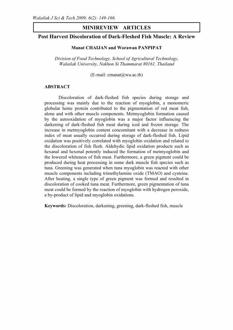
Darkening of dark-fleshed fish such as sardine (S. gibbosa) and mackerel (R. kanagurta) during iced storage is mainly caused by the formation of metmyoglobin [16]. Sohn and co-workers [17] suggested that the oxidation of myoglobin is the main cause in the development of the unpleasant color and undesirable odor during ice storage of fish muscle. The formation of metmyoglobin in bluefin tuna (Thunnus thynnus) increased with increasing storage time at 4 °C [18] (Figure 1). Furthermore, Chen [19] found that the metmyoglobin content in both horse mackerel (Trachurus japonicus) and chub mackerel (Scomber australasicus) increased as refrigerated or frozen storage time increased.
Figure 1 Changes in metmyoglobin content of fresh tuna meat during storage at 4 °C. Source: Modified from Chiou and co-workers. [18]
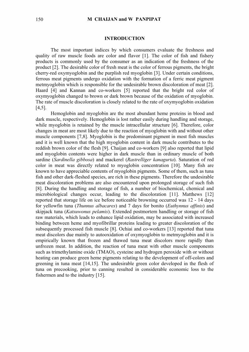
Chaijan and co-workers [16] reported that the redness index (a*/b* ratio) of
sardine (S. gibbosa) and mackerel (R. kanagurta) muscles decreased when the storage time increased (Figure 2), suggesting the darkening of meat most likely caused by the changes in pigments, mainly myoglobin. The decrease in redness index was associated
DARK-FLESHED FISH DISCOLORATION
152
-0.2
-0.1
0.0
0.1
0.2
0.3
0.4
0.5
0.6
0 3 6 9 12 15
storage time (day)
redn
ess i
ndex
sardine dark musclesardine ordinary musclemackerel dark musclemackerel ordinary muscle
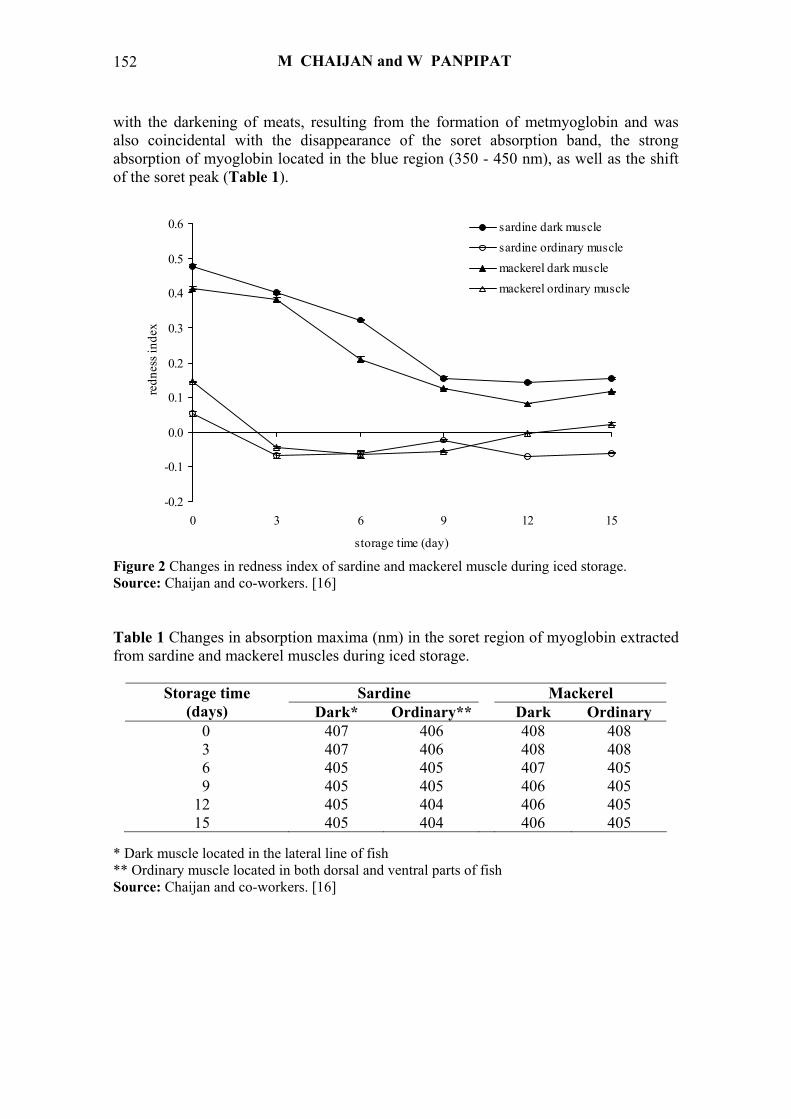
with the darkening of meats, resulting from the formation of metmyoglobin and was also coincidental with the disappearance of the soret absorption band, the strong absorption of myoglobin located in the blue region (350 - 450 nm), as well as the shift of the soret peak (Table 1).
Figure 2 Changes in redness index of sardine and mackerel muscle during iced storage. Source: Chaijan and co-workers. [16] Table 1 Changes in absorption maxima (nm) in the soret region of myoglobin extracted from sardine and mackerel muscles during iced storage.
Sardine Mackerel Storage time (days) Dark* Ordinary** Dark Ordinary
0 407 406 408 408 3 407 406 408 408 6 405 405 407 405 9 405 405 406 405
12 405 404 406 405 15 405 404 406 405
* Dark muscle located in the lateral line of fish ** Ordinary muscle located in both dorsal and ventral parts of fish Source: Chaijan and co-workers. [16]
M CHAIJAN and W PANPIPAT
153
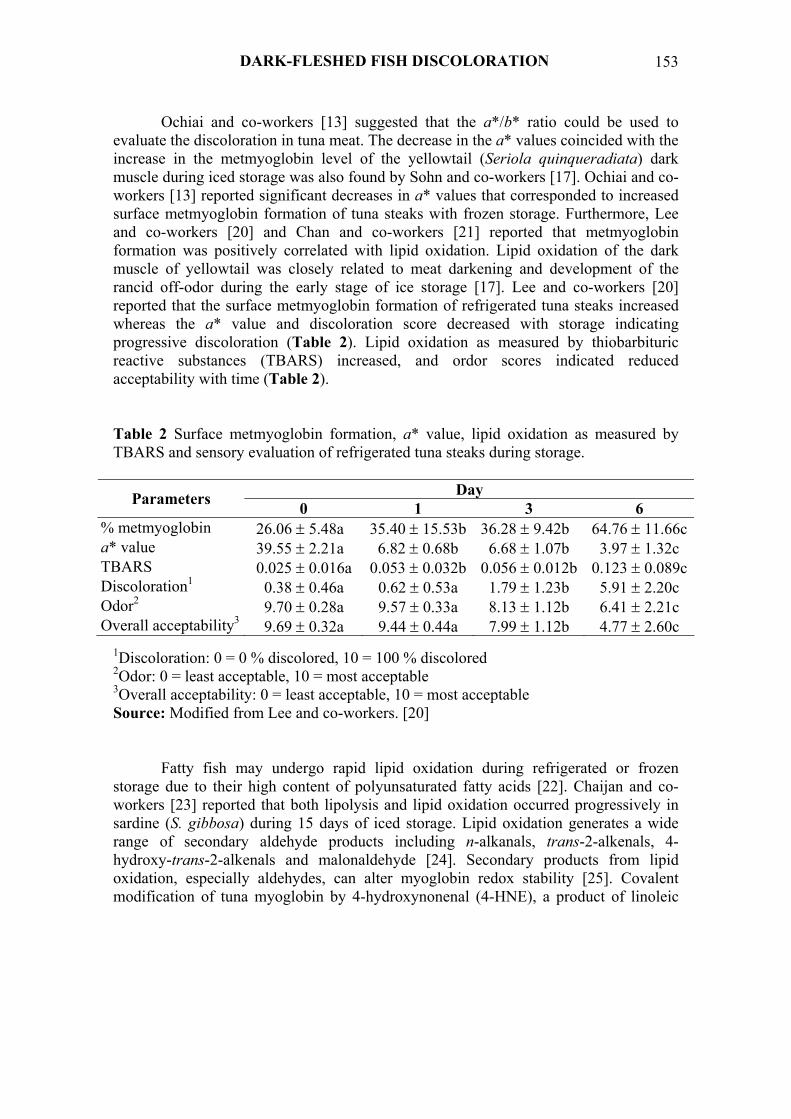
Ochiai and co-workers [13] suggested that the a*/b* ratio could be used to evaluate the discoloration in tuna meat. The decrease in the a* values coincided with the increase in the metmyoglobin level of the yellowtail (Seriola quinqueradiata) dark muscle during iced storage was also found by Sohn and co-workers [17]. Ochiai and co-workers [13] reported significant decreases in a* values that corresponded to increased surface metmyoglobin formation of tuna steaks with frozen storage. Furthermore, Lee and co-workers [20] and Chan and co-workers [21] reported that metmyoglobin formation was positively correlated with lipid oxidation. Lipid oxidation of the dark muscle of yellowtail was closely related to meat darkening and development of the rancid off-odor during the early stage of ice storage [17]. Lee and co-workers [20] reported that the surface metmyoglobin formation of refrigerated tuna steaks increased whereas the a* value and discoloration score decreased with storage indicating progressive discoloration (Table 2). Lipid oxidation as measured by thiobarbituric reactive substances (TBARS) increased, and ordor scores indicated reduced acceptability with time (Table 2). Table 2 Surface metmyoglobin formation, a* value, lipid oxidation as measured by TBARS and sensory evaluation of refrigerated tuna steaks during storage.
Day Parameters 0 1 3 6 % metmyoglobin 26.06 ± 5.48a 35.40 ± 15.53b 36.28 ± 9.42b 64.76 ± 11.66ca* value 39.55 ± 2.21a 6.82 ± 0.68b 6.68 ± 1.07b 3.97 ± 1.32c TBARS 0.025 ± 0.016a 0.053 ± 0.032b 0.056 ± 0.012b 0.123 ± 0.089cDiscoloration1 0.38 ± 0.46a 0.62 ± 0.53a 1.79 ± 1.23b 5.91 ± 2.20c Odor2 9.70 ± 0.28a 9.57 ± 0.33a 8.13 ± 1.12b 6.41 ± 2.21c Overall acceptability3 9.69 ± 0.32a 9.44 ± 0.44a 7.99 ± 1.12b 4.77 ± 2.60c
1Discoloration: 0 = 0 % discolored, 10 = 100 % discolored 2Odor: 0 = least acceptable, 10 = most acceptable 3Overall acceptability: 0 = least acceptable, 10 = most acceptable Source: Modified from Lee and co-workers. [20]
Fatty fish may undergo rapid lipid oxidation during refrigerated or frozen
storage due to their high content of polyunsaturated fatty acids [22]. Chaijan and co-workers [23] reported that both lipolysis and lipid oxidation occurred progressively in sardine (S. gibbosa) during 15 days of iced storage. Lipid oxidation generates a wide range of secondary aldehyde products including n-alkanals, trans-2-alkenals, 4-hydroxy-trans-2-alkenals and malonaldehyde [24]. Secondary products from lipid oxidation, especially aldehydes, can alter myoglobin redox stability [25]. Covalent modification of tuna myoglobin by 4-hydroxynonenal (4-HNE), a product of linoleic
DARK-FLESHED FISH DISCOLORATION
154
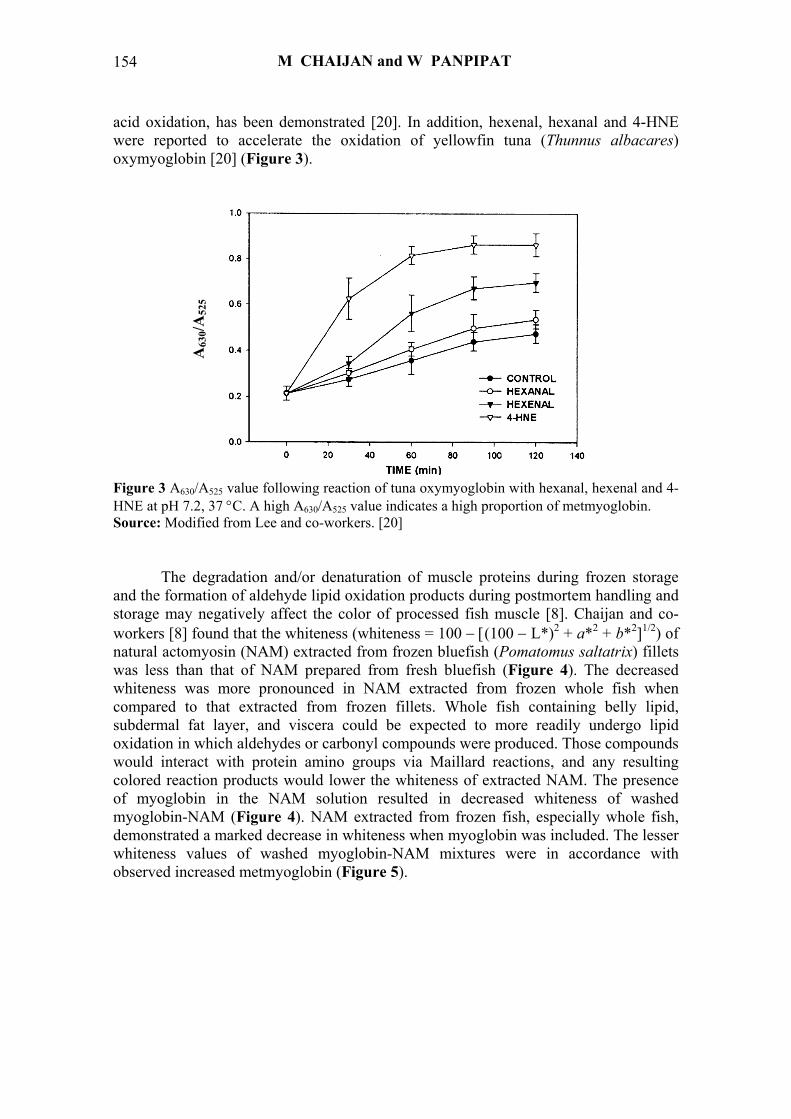
acid oxidation, has been demonstrated [20]. In addition, hexenal, hexanal and 4-HNE were reported to accelerate the oxidation of yellowfin tuna (Thunnus albacares) oxymyoglobin [20] (Figure 3).
Figure 3 A630/A525 value following reaction of tuna oxymyoglobin with hexanal, hexenal and 4-HNE at pH 7.2, 37 °C. A high A630/A525 value indicates a high proportion of metmyoglobin. Source: Modified from Lee and co-workers. [20]
The degradation and/or denaturation of muscle proteins during frozen storage
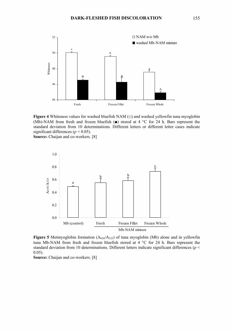
and the formation of aldehyde lipid oxidation products during postmortem handling and storage may negatively affect the color of processed fish muscle [8]. Chaijan and co-workers [8] found that the whiteness (whiteness = 100 − [(100 − L*)2 + a*2 + b*2]1/2) of natural actomyosin (NAM) extracted from frozen bluefish (Pomatomus saltatrix) fillets was less than that of NAM prepared from fresh bluefish (Figure 4). The decreased whiteness was more pronounced in NAM extracted from frozen whole fish when compared to that extracted from frozen fillets. Whole fish containing belly lipid, subdermal fat layer, and viscera could be expected to more readily undergo lipid oxidation in which aldehydes or carbonyl compounds were produced. Those compounds would interact with protein amino groups via Maillard reactions, and any resulting colored reaction products would lower the whiteness of extracted NAM. The presence of myoglobin in the NAM solution resulted in decreased whiteness of washed myoglobin-NAM (Figure 4). NAM extracted from frozen fish, especially whole fish, demonstrated a marked decrease in whiteness when myoglobin was included. The lesser whiteness values of washed myoglobin-NAM mixtures were in accordance with observed increased metmyoglobin (Figure 5).
A63
0/A52
5
M CHAIJAN and W PANPIPAT
155
ab b
c
0.0
0.2
0.4
0.6
0.8
1.0
Mb (control) Fresh Frozen Fillet Frozen Whole
Mb-NAM mixture
A63
0 /A
525
cb
a
B B
A
44
46
48
50
52
Fresh Frozen Fillet Frozen Whole
Whi
tene
ss
NAM w/o Mbwashed Mb-NAM mixture
Figure 4 Whiteness values for washed bluefish NAM (□) and washed yellowfin tuna myoglobin (Mb)-NAM from fresh and frozen bluefish (■) stored at 4 °C for 24 h. Bars represent the standard deviation from 10 determinations. Different letters or different letter cases indicate significant differences (p < 0.05). Source: Chaijan and co-workers. [8]
Figure 5 Metmyoglobin formation (A630/A525) of tuna myoglobin (Mb) alone and in yellowfin tuna Mb-NAM from fresh and frozen bluefish stored at 4 °C for 24 h. Bars represent the standard deviation from 10 determinations. Different letters indicate significant differences (p < 0.05). Source: Chaijan and co-workers. [8]
DARK-FLESHED FISH DISCOLORATION
156
a
bc
A
B
C
0.5
0.6
0.7
0.8
0.9
1.0
1.1
1.2
Control Hexanal Hexenal
A63
0/A
525
Mb w/o NAMMb-NAM mixture
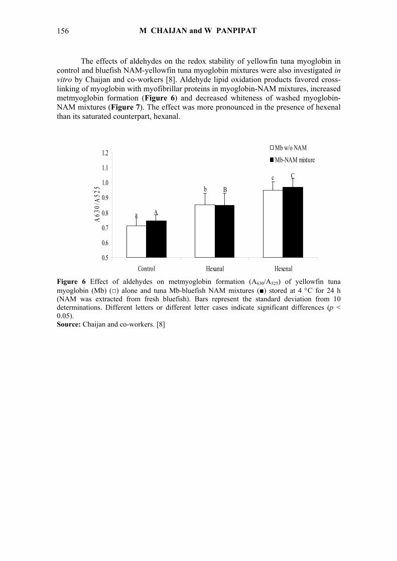
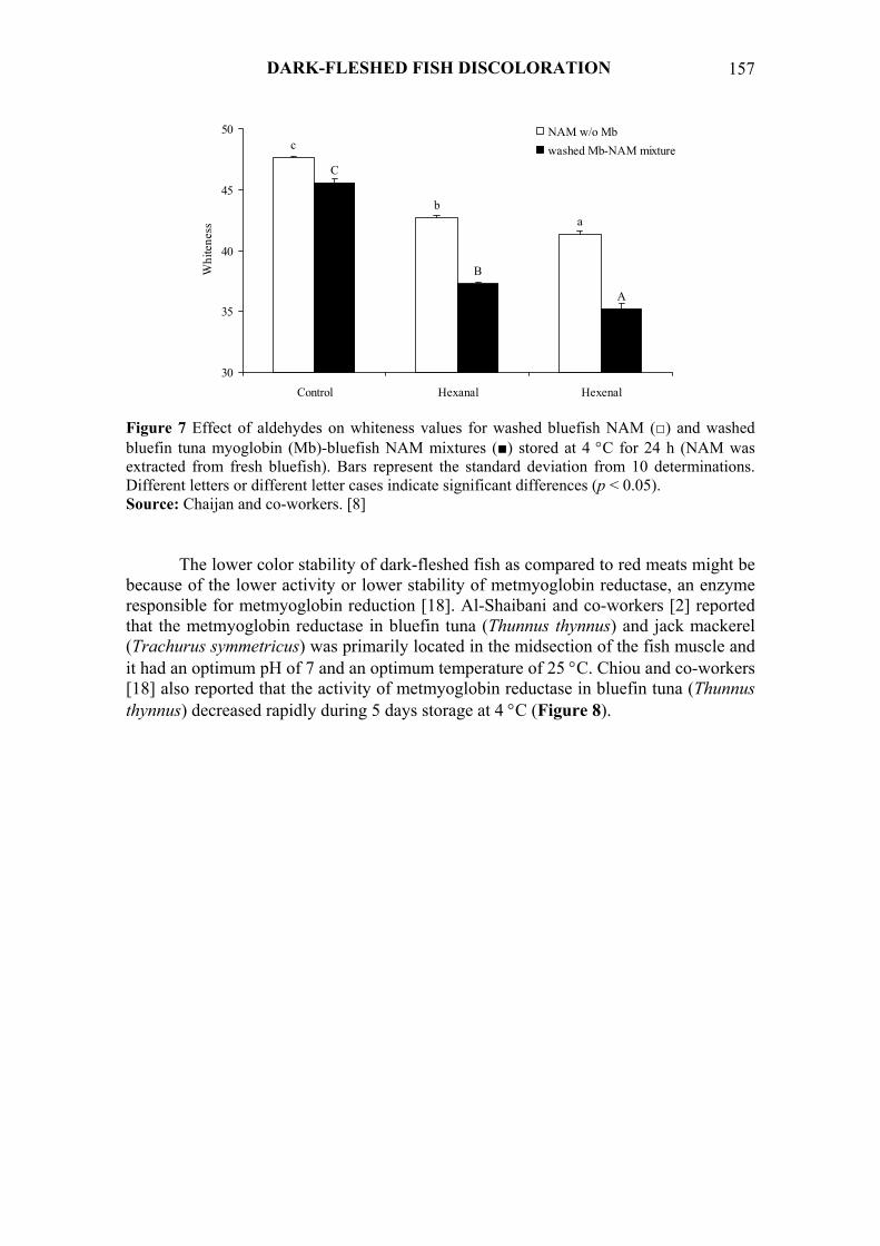
The effects of aldehydes on the redox stability of yellowfin tuna myoglobin in control and bluefish NAM-yellowfin tuna myoglobin mixtures were also investigated in vitro by Chaijan and co-workers [8]. Aldehyde lipid oxidation products favored cross-linking of myoglobin with myofibrillar proteins in myoglobin-NAM mixtures, increased metmyoglobin formation (Figure 6) and decreased whiteness of washed myoglobin-NAM mixtures (Figure 7). The effect was more pronounced in the presence of hexenal than its saturated counterpart, hexanal.
Figure 6 Effect of aldehydes on metmyoglobin formation (A630/A525) of yellowfin tuna myoglobin (Mb) (□) alone and tuna Mb-bluefish NAM mixtures (■) stored at 4 °C for 24 h (NAM was extracted from fresh bluefish). Bars represent the standard deviation from 10 determinations. Different letters or different letter cases indicate significant differences (p < 0.05). Source: Chaijan and co-workers. [8]
M CHAIJAN and W PANPIPAT
157
c
ba
C
B
A
30
35
40
45
50
Control Hexanal Hexenal
Whi
tene
ss
NAM w/o Mbwashed Mb-NAM mixture
Figure 7 Effect of aldehydes on whiteness values for washed bluefish NAM (□) and washed bluefin tuna myoglobin (Mb)-bluefish NAM mixtures (■) stored at 4 °C for 24 h (NAM was extracted from fresh bluefish). Bars represent the standard deviation from 10 determinations. Different letters or different letter cases indicate significant differences (p < 0.05). Source: Chaijan and co-workers. [8]
The lower color stability of dark-fleshed fish as compared to red meats might be
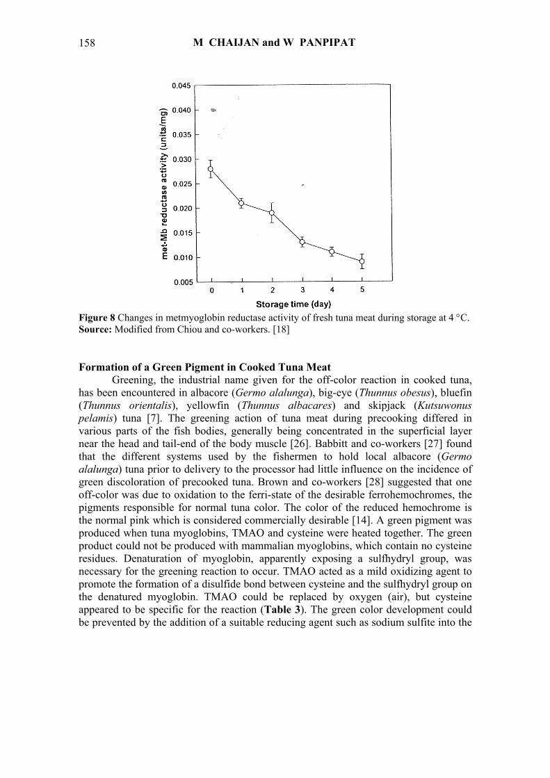
because of the lower activity or lower stability of metmyoglobin reductase, an enzyme responsible for metmyoglobin reduction [18]. Al-Shaibani and co-workers [2] reported that the metmyoglobin reductase in bluefin tuna (Thunnus thynnus) and jack mackerel (Trachurus symmetricus) was primarily located in the midsection of the fish muscle and it had an optimum pH of 7 and an optimum temperature of 25 °C. Chiou and co-workers [18] also reported that the activity of metmyoglobin reductase in bluefin tuna (Thunnus thynnus) decreased rapidly during 5 days storage at 4 °C (Figure 8).
DARK-FLESHED FISH DISCOLORATION
158
Figure 8 Changes in metmyoglobin reductase activity of fresh tuna meat during storage at 4 °C. Source: Modified from Chiou and co-workers. [18] Formation of a Green Pigment in Cooked Tuna Meat
Greening, the industrial name given for the off-color reaction in cooked tuna, has been encountered in albacore (Germo alalunga), big-eye (Thunnus obesus), bluefin (Thunnus orientalis), yellowfin (Thunnus albacares) and skipjack (Kutsuwonus pelamis) tuna [7]. The greening action of tuna meat during precooking differed in various parts of the fish bodies, generally being concentrated in the superficial layer near the head and tail-end of the body muscle [26]. Babbitt and co-workers [27] found that the different systems used by the fishermen to hold local albacore (Germo alalunga) tuna prior to delivery to the processor had little influence on the incidence of green discoloration of precooked tuna. Brown and co-workers [28] suggested that one off-color was due to oxidation to the ferri-state of the desirable ferrohemochromes, the pigments responsible for normal tuna color. The color of the reduced hemochrome is the normal pink which is considered commercially desirable [14]. A green pigment was produced when tuna myoglobins, TMAO and cysteine were heated together. The green product could not be produced with mammalian myoglobins, which contain no cysteine residues. Denaturation of myoglobin, apparently exposing a sulfhydryl group, was necessary for the greening reaction to occur. TMAO acted as a mild oxidizing agent to promote the formation of a disulfide bond between cysteine and the sulfhydryl group on the denatured myoglobin. TMAO could be replaced by oxygen (air), but cysteine appeared to be specific for the reaction (Table 3). The green color development could be prevented by the addition of a suitable reducing agent such as sodium sulfite into the
M CHAIJAN and W PANPIPAT
159
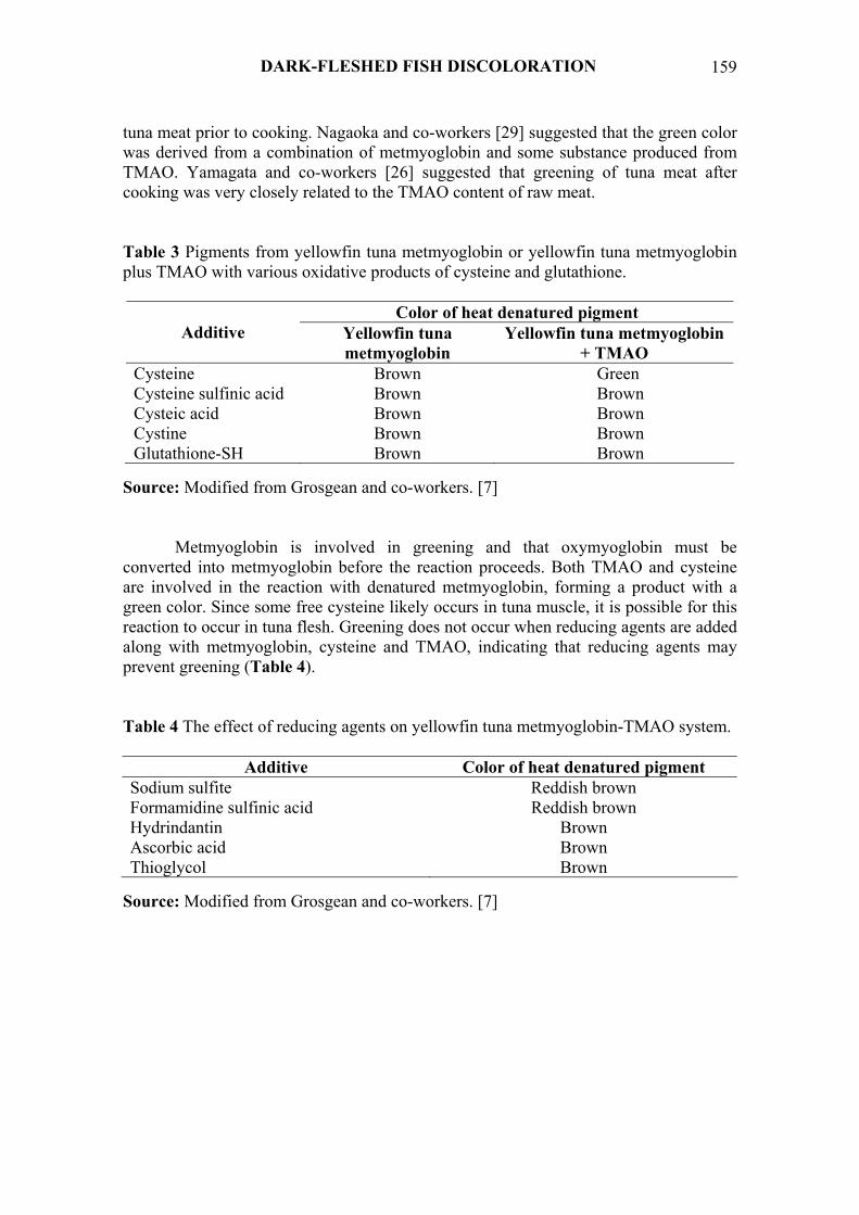
tuna meat prior to cooking. Nagaoka and co-workers [29] suggested that the green color was derived from a combination of metmyoglobin and some substance produced from TMAO. Yamagata and co-workers [26] suggested that greening of tuna meat after cooking was very closely related to the TMAO content of raw meat. Table 3 Pigments from yellowfin tuna metmyoglobin or yellowfin tuna metmyoglobin plus TMAO with various oxidative products of cysteine and glutathione.
Color of heat denatured pigment Additive Yellowfin tuna
metmyoglobin Yellowfin tuna metmyoglobin
+ TMAO Cysteine Brown Green Cysteine sulfinic acid Brown Brown Cysteic acid Brown Brown Cystine Brown Brown Glutathione-SH Brown Brown
Source: Modified from Grosgean and co-workers. [7]
Metmyoglobin is involved in greening and that oxymyoglobin must be converted into metmyoglobin before the reaction proceeds. Both TMAO and cysteine are involved in the reaction with denatured metmyoglobin, forming a product with a green color. Since some free cysteine likely occurs in tuna muscle, it is possible for this reaction to occur in tuna flesh. Greening does not occur when reducing agents are added along with metmyoglobin, cysteine and TMAO, indicating that reducing agents may prevent greening (Table 4).
Table 4 The effect of reducing agents on yellowfin tuna metmyoglobin-TMAO system.
Additive Color of heat denatured pigment Sodium sulfite Reddish brown Formamidine sulfinic acid Reddish brown Hydrindantin Brown Ascorbic acid Brown Thioglycol Brown
Source: Modified from Grosgean and co-workers. [7]
DARK-FLESHED FISH DISCOLORATION

160
The greening reaction involves the -SH group of the cysteine residue in the tuna myoglobin. No greening was produced when myoglobins such as sperm whale myoglobin and equine myoglobin, which do not contain a -SH group, were heat denatured in the presence of cysteine and TMAO (Table 5). Therefore, it appears that the greening reaction involves the -SH group of both free cysteine and the cysteine residue in the tuna myoglobin. TMAO functions as a mild oxidizing agent in the reaction of the cysteine -SH with the protein -SH to form a disulfide bond. Table 5 The participation of different myoglobins in the greening reaction.
Myoglobin Color produced on heat denaturation of different myoglobins in the presence of cysteine and TMAO
Yellowfin tuna Green Sperm whale Brown Equine Brown Albacore tuna Green Skipjack tuna Green Bluefin tuna Green Source: Modified from Grosgean and co-workers. [7]
Vaisey [30] found that TMAO at temperatures between 22 and 24 °C is readily reduced by cysteine in the presence of iron or hemoglobin as a catalyst. The major product of this reduction was trimethylamine (TMA) (and presumably cystine). This observation agreed with that of Nagaoka and co-workers [29] who found a positive correlation between the TMA content of cooked tuna, the TMAO content of raw tuna, and greening of cooked tuna. Vaisey [30] also found that glutathione could not replace cysteine as a reducing agent. The greening reaction appears to involve the reaction of free cysteine with the -SH group of heat denatured tuna myoglobin (under mild oxidizing conditions provided either by TMAO or air) to produce a disulfide bond and TMA with perhaps a catalytic influence by the iron (Fe3+) of metmyoglobin. Naughton and co-workers [14] reported that some degree of denaturation of the protein moiety of the heme pigment molecule took place even at low temperatures. Chanthai and co-workers [31] found that fish apomyoglobins showed endothermic peak-tops (Tm) at 36 - 38 °C (Figure 9). The overall mechanism for the greening caused by the reaction of denatured myoglobin with cysteine and TMAO may be summarized in Figure 10.
M CHAIJAN and W PANPIPAT

161
Figure 9 DSC thermograms of fish apomyoglobins (a) yellowtail, (b) bonito and (c) yellowfin tuna. Source: Modified from Chanthai and co-workers. [31]
Native metmyoglobin Denatured metmyoglobin-SH
+ Cysteine (R-SH) + TMAO + Fe3+
Denatured metmyoglobin-S-S-R + H2O Figure 10 Green pigment formation in cooked tuna meat. Source: Modified from Grosgean and co-workers. [7]
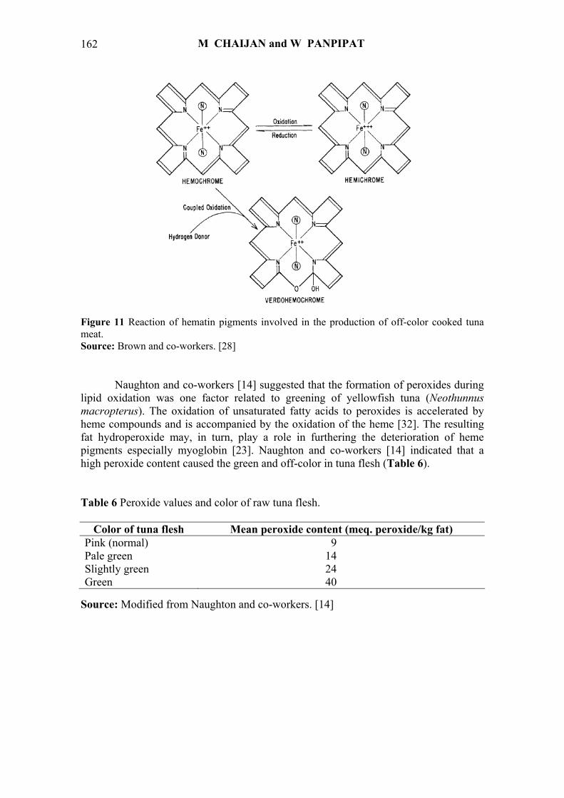
Furthermore, green pigmentation of tuna meat can be formed by the reaction of
myoglobin with hydrogen peroxide, a by-product of lipid and myoglobin oxidations [14]. Fox [15] reported that green discoloration in meat was attributed to an oxidative attack on the pyrrole ring of the heme pigments producing cholemyoglobin or verdohemochrome (Figure 11).
(Brown)
(Green)
(Brown)
DARK-FLESHED FISH DISCOLORATION
162
Figure 11 Reaction of hematin pigments involved in the production of off-color cooked tuna meat. Source: Brown and co-workers. [28]
Naughton and co-workers [14] suggested that the formation of peroxides during
lipid oxidation was one factor related to greening of yellowfish tuna (Neothunnus macropterus). The oxidation of unsaturated fatty acids to peroxides is accelerated by heme compounds and is accompanied by the oxidation of the heme [32]. The resulting fat hydroperoxide may, in turn, play a role in furthering the deterioration of heme pigments especially myoglobin [23]. Naughton and co-workers [14] indicated that a high peroxide content caused the green and off-color in tuna flesh (Table 6). Table 6 Peroxide values and color of raw tuna flesh.
Color of tuna flesh Mean peroxide content (meq. peroxide/kg fat) Pink (normal) 9 Pale green 14 Slightly green 24 Green 40
Source: Modified from Naughton and co-workers. [14]
M CHAIJAN and W PANPIPAT

163
CONCLUSION Post harvest pigmentation of dark-fleshed fish meat is mainly due to the
instability of myoglobin pigment. The reaction of myoglobin is highly variable and governed by a variety of factors. The autooxidation of myoglobin resulted in the formation of metmyoglobin and discoloration of stored fish flesh. The reaction of myoglobin and other muscle components such as TMAO and cysteine during heat processing causes the greening in some dark muscle fish such as tuna. The off-color of dark-fleshed fish during storage and processing might decrease the value of these fish from a commercial standpoint. Therefore, the control of these variables and suitable preservative methods currently offers the best means for maximizing dark-fleshed fish meat color stability.
164
REFERENCES [1] E Risvik. Sensory properties and preferences. Meat Sci. 1994; 36, 67-77. [2] KA Al-Shaibani, RJ Price and WD Brown. Enzymatic reduction of metmyoglobin
in fish. J. Food Sci. 1977; 42, 1156-8. [3] C Faustman and AL Phillips. Measurement of Discoloration in Fresh Meat. In: RE
Wrolstad (ed.). Current Protocols in Food Analytical Chemistry. 1st ed. Wiley & Sons, Inc., New York, 2001, p. F3.3.1-F3.3.13.
[4] NF Haard. Biochemistry and Chemistry of Color and Color Change in Seafoods. In: GJ Flick and RE Martin (eds.). Advance in Seafood Biochemistry. 1st ed. Technomic Publishing Co., Inc., USA, 1992, p. 312-9.
[5] G Kannan, B Kouakou and S Gelaye. Color changes reflecting myoglobin and lipid oxidation in chevon cuts during refrigerated display. Small Ruminant Res. 2001; 42, 67-75.
[6] DJ Livingston and WD Brown. The chemistry of myoglobin and its reactions. Food Technol. 1981; 25, 244-52.
[7] OK Grosjean, BF Cobb, B Mebine and WD Brown. Formation of a green pigment from tuna myoglobins. J. Food Sci. 1969; 34, 404-7.
[8] M Chaijan, S Benjakul, W Visessanguan, S Lee and C Faustman. The effect of freezing and aldehydes on the interaction between fish myoglobin and myofibrillar proteins. J. Agric. Food Chem. 2007; 55, 4562-8.
[9] M Chaijan, S Benjakul, W Visessanguan and C Faustman. Characteristics and gel properties of muscles from sardine (Sardinella gibbosa) and mackerel (Rastrelliger kanagurta) caught in Thailand. Food Res. Intern. 2004; 37, 1021-30.
[10] C Faustman, MC Yin and DB Nadeau. Color stability, lipid stability, and nutrient composition of red and white veal. J. Food Sci. 1992; 57, 302-4.
[11] R Pacheco-Aguilar, ME Lugo-Sanchez and MR Robles-Burgueno. Postmortem biochemical characteristic of Monterey sardine muscle stored at 0 °C. J. Food Sci. 2000; 65, 40-7.
[12] AD Matthews. Muscle colour deterioration in iced and frozen stored bonito, yellowfin and skipjack tuna caught in Seychelles waters. Food Technol. 1983; 18, 387-92.
[13] Y Ochiai, CJ Chow, S Watabe and K Hashimoto. Evaluation of tuna meat discoloration by Hunter color difference scale. Nippon Suisan Gakk. 1988; 54, 649-53.
[14] JJ Naughton, H Zeitlin and MM Frodyma. Spectral reflectance studies of the heme pigments in tuna fish flesh: some characteristics of the pigments and discoloration of tuna meat. J. Agric. Food Chem. 1958; 6, 933-8.
[15] JB Fox. The chemistry of meat pigments. J. Agric. Food Chem. 1966; 14, 207-10. [16] M Chaijan, S Benjakul, W Visessanguan and C Faustman. Changes of pigments
and color in sardine (Sardinella gibbosa) and mackerel (Rastrelliger kanagurta) muscle during iced storage. Food Chem. 2005; 93, 607-17.
DARK-FLESHED FISH DISCOLORATION

165
[17] JH Sohn, Y Taki, H Ushio, T Kohata, I Shioya and T Oshima. Lipid oxidations in ordinary and dark muscles of fish: influences on rancid off-ordor development and color darkening of yellowtail flesh during ice storage. J. Food Sci. 2005; 70, S490-6.
[18] TK Chiou, CY Pong, FP Nieh and ST Jiang. Effect of met-myoglobin reductase on the color stability of bluefin tuna during refrigerated storage. Fisheries Sci. 2001; 67, 694-702.
[19] HH Chen. Effect of cold storage on the stability of chub and horse mackerel myoglobins. J. Food Sci. 2003; 68, 1416-9.
[20] S Lee, ST Joo, AL Alderton, DW Hill and C Faustman. Oxymyoglobin and lipid oxidation in yellowfin tuna (Thunnus albacares) loins. J. Food Sci. 2003; 68, 1664-8.
[21] WKM Chan, C Faustman, M Yin and EA Decker. Lipid oxidation induced by oxymyoglobin and metmyoglobin with involvement of H2O2 and superoxide anion. Meat Sci. 1997; 46, 181-90.
[22] ME Apgar and HO Hultin. Lipid peroxidation in fish muscle microsomes in the frozen state. Cryobio. 1982; 19, 154-62.
[23] M Chaijan, S Benjakul, W Visessanguan and C Faustman. Changes of lipids in sardine (Sardinella gibbosa) muscle during iced storage. Food Chem. 2006; 99, 83-91.
[24] H Esterbauer, RJ Schaur and H Zollner. Chemistry and biochemistry of 4-hydroxynonenal, malonaldehyde and related aldehydes. Free Rad. Bio. Med. 1991; 11, 81-128.
[25] SJ Li and AJ King. Structural changes of rabbit myosin subfragment 1 altered by malonaldehyde, a byproduct of lipid oxidation. J. Agric. Food Chem. 1999; 47, 3124-9.
[26] M Yamagata, K Horimoto and C Nagaoka. Assessment of green tuna: determining trimethylamine oxide and its distribution in tuna muscles. J. Food Sci. 1969; 34, 156-9.
[27] JK Babbitt, DL Crawford and DK Low. Effect of handling and processing on discoloration of albacore tuna. J. Food Sci. 1977; 42, 557-8.
[28] WD Brown, AL Tappel and HS Olcott. The pigments of off-color cooked tuna meat. Food Res. 1958; 23, 262-8.
[29] C Nagaoka and N Suzuki. Detection of green-meat tuna before cooking. Food Technol. 1964; 18, 183-7.
[30] EB Vaisey. The non-enzymatic reduction of trimethylamine oxide to trimethylamine, dimethylamine and formaldehyde. Can. J. Biochem. Physiol. 1956; 34, 1085-9.
[31] S Chanthai, H Neida, M Ogawa, T Tamiya and T Tsuchiya. Studies on thermal denaturation of fish apomyoglobins using differential scanning calorimetry, circular dichroism, and tryptophan fluorescence. Fisheries Sci. 1996; 62, 927-32.
[32] JD Love. The role of heme iron in the oxidation of lipids in red meats. Food Technol. 1983; 12, 117-20.
M CHAIJAN and W PANPIPAT

166
บทคัดยอ มนัส ชัยจันทร
และ วรวรรณ พันพิพัฒน
การเปลี่ยนแปลงสีของกลามเน้ือปลาเน้ือดําระหวางการเก็บรักษาและการแปรรูป การเปลี่ยนแปลงสีของกลามเนื้อปลาเนื้อดําระหวางการเก็บรักษาและการแปรรูปมีสาเหตุมาจากปฏิกิริยา
ของไมโอโกลบินเปนสําคัญ โดยการเปลี่ยนแปลงของไมโอโกลบินสามารถเกิดขึ้นไดดวยตัวเองหรือเกิดปฏิกิริยากับองคประกอบอื่น ๆ ภายในกลามเนื้อ การเกิดเมทไมโอโกลบินอันเนื่องมาจากปฏิกิริยาออกซิเดชันดวยตัวเองของ ไมโอโกลบินเปนสาเหตุสําคัญของการเกิดสีคล้ําในปลาเนื้อดําระหวางการเก็บรักษาในน้ําแข็งและในสภาพแชเยือกแข็ง ซึ่งการเพิ่มขึ้นของปริมาณเมทไมโอโกลบินสอดคลองกับการลดลงของคาดัชนีสีแดงของกลามเนื้อปลา นอกจากนี้ การออกซิเดชันของไขมันยังมีความสัมพันธโดยตรงกับการออกซิเดชันของไมโอโกลบิน และมีสวนเกี่ยวของกับการเกิดสีคล้ําของกลามเนื้อปลาอีกดวย แอลดีไฮดซึ่งเปนผลิตภัณฑสําคัญที่เกิดขึ้นจากการออกซิเดชันของไขมัน เชน เฮกซานาล และ เฮกซีนาล สามารถสงเสริมใหกลามเนื้อปลาเกิดเมทไมโอโกลบินไดมากขึ้น และสงผลตอเนื่องใหคาความขาวของกลามเนื้อปลาลดต่ําลงได ยิ่งไปกวานั้นในระหวางการแปรรูปปลาเนื้อดําบางชนิดดวยความรอน เชน ปลาทูนา เปนตน อาจจะทําใหกลามเนื้อปลาดังกลาวเกิดสีเขียวขึ้นมาได โดยสีเขียวที่เกิดขึ้นนั้นมีสาเหตุมาจากอันตรกิริยาระหวางไมโอโกลบินกับองคประกอบอื่น ๆ ของกลามเนื้อปลาระหวางการใหความรอน ซึ่งไดแก ไตรเมทิลเอมีน และซีสเตอีน นอกจากนี้สีเขียวของกลามเนื้อปลาทูนา ยังเกิดขึ้นไดจากปฏิกิริยาระหวาง ไมโอโกลบินกับไฮโดรเจนเพอรออกไซดซึ่งเปนผลพลอยไดจากการออกซิเดชันของไขมันและไมโอโกลบินอีกดวย
สาขาวิชาเทคโนโลยีอาหาร สํานักวิชาเทคโนโลยีการเกษตร มหาวิทยาลัยวลัยลักษณ อําเภอทาศาลา จังหวัดนครศรีธรรมราช 80161
DARK-FLESHED FISH DISCOLORATION