mir-365,anovelnegativeregulatorofinterleukin-6gene ... ·...
TRANSCRIPT
miR-365, a Novel Negative Regulator of Interleukin-6 GeneExpression, Is Cooperatively Regulated by Sp1 and NF-�B*
Received for publication, October 28, 2010, and in revised form, April 9, 2011 Published, JBC Papers in Press, April 25, 2011, DOI 10.1074/jbc.M110.198630
Zheng Xu, Shao-Bo Xiao1, Peng Xu, Qian Xie, Lu Cao, Dang Wang, Rui Luo, Yao Zhong, Huan-Chun Chen,and Liu-Rong Fang2
From the Division of Animal Infectious Diseases, State Key Laboratory of Agricultural Microbiology, College of Veterinary Medicine,Huazhong Agricultural University, Wuhan 430070, China
Interleukin-6 (IL-6) is a pleiotropic cytokine that plays a cen-tral role inhost defense. IL-6 expression canbe regulated at botha transcriptional and a post-transcriptional level. We used acombination of bioinformatics and experimental techniques todemonstrate that the miR-365 is a direct negative regulator ofIL-6. Overexpression of miR-365 mimics decreased activity of aluciferase reporter containing the IL-6 3�-UTR and led torepression of IL-6 protein. In contrast, ectopic expression of amiR-365 inhibitor elevated IL-6 expression. The negative regu-lation of miR-365 was strictly dependent on a microRNA bind-ing element in the 3�-UTRof IL-6mRNA.Deletionmutant anal-ysis of the miR-365 promoter showed that two transcriptionfactors, Sp1 andNF-�B, are essential for the transcriptional reg-ulation of miR-365. We also demonstrate that the MAPK/ERKpathway contributes to the regulationofmiR-365. Furthermore,miR-365 exhibited a greater negative regulatory effect on IL-6than hsa-let-7a, a previously identified microRNA negativelyregulating IL-6. Taken together, our results show that miR-365is a novel negative regulator of IL-6.
MicroRNAs (miRNAs, miRs)3 are small RNAmolecules thatregulate gene expression at the post-transcriptional level (1). Inmammals, miRNAs are initially transcribed by RNA polymer-ase II, and the primary miRNA transcripts are sequentially cutby RNase III enzymes Drosha and Dicer (2, 3). The resulting�23-nucleotide double-stranded mature miRNA moleculesload into RNA-induced silencing complexes (4, 5), where theyact to repress mRNA translation or reduce mRNA stability byinteracting with miRNA recognition elements (MRE) within3�-untranslated region (UTR) of target genes (6). The specific-ity of miRNAs is thought to be primarily mediated by residues2–8 at the 5� end of miRNA, also known as the seed region (7).
Growing evidence exists indicating that miRNAs play animportant role in the regulation of inflammation. For example,miR-146a has been characterized as a negative regulator of theinflammatory response by targeting IL-1 receptor-associatedkinase 1 (IRAK1) andTNF receptor associated factor 6 (8). Xiaoet al. (9) reported that miR-155 is involved in negative regula-tion of Heliobacter pylori-induced inflammation, and miR-155was further identified as targeting myeloid differentiation pri-mary response gene 88 (MyD88) (10). miRNAs regulate inflam-mation not only by targeting adapter proteins involved in thesignaling pathway but also inflammatory mediators. Manyinterleukins have been found to be targets ofmiRNAs. Interleu-kin-10 (IL-10) has been identified as the target of hsa-miR-106a(11). Kaposi sarcoma-associated herpesvirus-encoded miR-K12-3 and miR-K12-7 up-regulate IL-10 and IL-6 levels inmurine macrophages and human myelomonocytic cells (12).miR-466l elevates IL-10 expression by preventing IL-10mRNAdegradation (13). IL-13, a cytokine essential for expression forallergic lung disease, was recently found to be regulated bymmu-let-7a (14). miR-346 indirectly regulates IL-18 release byindirectly inhibiting LPS-induced Bruton’s tyrosine kinaseexpression in LPS-activated rheumatoid fibroblast-like syno-viocytes (15).IL-6 is a pleiotropic cytokine involved in the regulation of the
immune response, hematopoiesis, and inflammation (16). Itwas previously considered to be a regulator of acute-phaseresponses and a lymphocyte-stimulatory factor (17–19). How-ever, recent advances have documented a series of IL-6 activi-ties that are critical for resolving innate immunity and promot-ing acquired immunity (20). IL-6 expression can be regulated atthe transcriptional and post-transcriptional level (21, 22), butwhether and how IL-6 is regulated at the post-transcriptionallevel by miRNA has so far been incompletely understood.In this study we establish that miR-365 is a novel miRNA
regulator of human IL-6. miR-365 regulates the expression ofIL-6 by classic interaction with the MRE in the 3�-UTR of IL-6and further represses mRNA translation, althoughmRNA deg-radation is not involved. We also investigated the transcrip-tional regulation mechanism of miR-365.
EXPERIMENTAL PROCEDURES
Prediction of Candidate miRNA Binding to IL-6 3�-UTR—The software programs Microcosm (23–25), miRanda (26, 27),and TargetScan (28–31) were used to predict potential bindingsites for miRNA in the IL-6 3�-UTR. Only those miRNA target
* This work was supported by the New Century Excellent Talent Project(NCET-07– 0347), the National Natural Sciences Foundation of China(30972189, 30871871), and the Program for Changjiang Scholars and Inno-vative Research Team in University (IRT0726).
1 To whom correspondence may be addressed: Laboratory of Animal Virol-ogy, College of Veterinary Medicine, Huazhong Agricultural University,Wuhan 430070, China. Tel.: 86-27-8728-6884; Fax: 86-27-8728-2608;E-mail: [email protected].
2 To whom correspondence may be addressed: Laboratory of Animal Virol-ogy, College of Veterinary Medicine, Huazhong Agricultural University,Wuhan 430070, China. Tel.: 86-27-8728-6884; Fax: 86-27-8728-2608;E-mail: [email protected].
3 The abbreviations used are: miRNAs, microRNA; MRE, miRNA recognitionelements; TSS, transcription start site.
THE JOURNAL OF BIOLOGICAL CHEMISTRY VOL. 286, NO. 24, pp. 21401–21412, June 17, 2011© 2011 by The American Society for Biochemistry and Molecular Biology, Inc. Printed in the U.S.A.
JUNE 17, 2011 • VOLUME 286 • NUMBER 24 JOURNAL OF BIOLOGICAL CHEMISTRY 21401
by guest on June 4, 2018http://w
ww
.jbc.org/D
ownloaded from

pairs detected by at least two of the three programs were usedfor further study.Cell Culture and Reagents—HEK293 and HeLa cells were
cultured in DMEM (Invitrogen) supplemented with 10% fetalbovine serum, 100 units/ml penicillin, and 100 �g/ml strepto-mycin at 37 °C and 5% CO2. miR-365 mimics and inhibitorswere obtained from GenePharma (Shanghai, China). Thesequence of mimics, inhibitors, or scrambled oligonucleotidesare as follows: miR-365mimics, 5�-UAAUGCCCCUAAAAAUCC UUAU-3� (forward) and 5�-AAG GAUUUUUAGGGGCAU UAU U-3� (reverse); mimics negative control, 5�-UUCUCC GAA CGU GUC ACG UTT-3� (forward) and 5�-ACGUGA CAC GUU CGG AGA ATT-3� (reverse); miR-365 inhib-itor, 5�-AUAAGGAUUUUUAGGGGCAUUA-3�; inhibitornegative control, 5�-CAGUACUUUUGUGUAGUACAA-3�.Pharmacological inhibitors of JNK (SP600125), p38 MAPkinase (SB203580 and SB202190), NF-�B (BAY11-7082), andERK (U0126) were obtained from Calbiochem-Merck. Phorbol12-myristate 13-acetate, poly(I:C), and poly(dAT:dAT) wereobtained from Sigma.Plasmid Constructs—To construct pMIR-IL-6 3�-UTR, the
3�-UTR of IL-6 was amplified from cDNA derived from HeLacells. The PCR product was digested with SacI and HindIII andcloned into the pMIR-REPORT luciferase reporter vector(Ambion). The luciferase reporter plasmids TSS-1348, TSS-1097, TSS-705, and TSS-336, which contains the1348-, 1097-,705-, and 336-bp proximal promoter sequences of miR-365,respectively, were constructed by PCR amplification using
genomic DNA of HeLa cells as templates and subsequent clon-ing into KpnI and BglII site of pGL3-basic (Promega, Madison,WI). To construct the miRNA expression vector, each miRNAprecursor sequence plus 3�- and 5�-flanking region (about 150bp) was amplified from genomic DNA of HeLa cells and clonedinto pSilencer 4.1-CMVneo. The Sp1 expression construct wascreated by amplifying the whole human Sp1 coding sequencefromHEK293 cells and then cloned into pEGFP-N1 at the XhoIand KpnI sites. The IL-6 3�-UTR reporter constructs lackingthe predicted miR-365 binding site or having a 3-bp substitu-tion in the MRE of IL-6 gene and miR-365 promoter reporterconstructs containing site-specific mutations for transcriptionfactor binding site NF-�B or Sp1were constructed by introduc-ing a point mutation or deletion with overlap-extension PCR(32). Primers used in this study are listed in Table 1. All DNAconstructs were verified by sequencing.Transient Transfections and Luciferase Assays—Transient
transfections were performed using Lipofectamine 2000 (Invit-rogen) according to themanufacturer’s protocol. For luciferaseassay, HEK293 cells were cultured in 24-well plates and co-transfected with 0.1 �g of an indicated reporter plasmid perwell and 0.05 �g pRL-TK (Promega) per well along withmiRNA expression vector (0.4 �g/well) or miRNAmimics (20,40, and 60 nM) or inhibitors (60, 80, and 100 nM). In someselected experiments, 1 �g/well poly(dAT:dAT), 1 �g/wellpoly(I:C), 1 �g/well phorbol 12-myristate 13-acetate, or 10hemagglutination units/well Sendai virus were transfected ortreated at 24 h after the initial co-transfection. Luciferase activ-
TABLE 1Primers used in this studyF, forward; R, reverse.
Primer Sequence 5�3 3�
IL6 3�-UTR intact F ATAGAGCTCCATGGGCACCTCAGATTGIL6 3�-UTR deletion F CGCGAGCTCGCATGGGCACCTCAGCTTCTTCTGGTCAGAAACCTGTIL6 3�-UTR Point Mut F CGCGAGCTCGCATGGGCACCTCAGATTGTTGTTGTTAATGACCAGTCCTIL6 3�-UTR R GCGAAGCTTGCTGAATTTTTTAAAATGCCTSS-336 F GGGGTACCCCGCTTGATAAAGCTTAATTGCATCTSS-705 F GGGGTACCCCAGGTCTAATTTTTATTATGCAAGTSS-1097 F GGGGTACCCCTTAAAATCACAGTGGAAACTGGTSS-1348 F GGGGTACCCCCTGCAGTCAGCGCAAGACCAACTGTSS-Universal R GAAGATCTTCAAAGAAAGAATGAATGTTAGCCNF-�B mut1 F TTTAAATTCCTAATTTAACCATTTCCCTTTGAGTCATTAGGAANF-�B mut1 R TTCCTAATGACTCAAAGGGAAATGGTTAAATTAGGAATTTAAANF-�B mut2 F GAGTCATTAACCATATCTCATAGGTCTAATTTTTATTATGNF-�B mut2 R CATAATAAAAATTAGACCTATGAGATATGGTTAATGACTCNF-�B mut3 F CAAACAATTACCAAATACCTTTCCAAGGAAAAATCAGGCACCNF-�B mut3 R GGTGCCTGATTTTTCCTTGGAAAGGTATTTGGTAATTGTTTGSp1 mut1 F GCAAGAAAAATATTTTTCAGAGTCAACAGGAGTATTCCCCSp1 mut1 R GGGGAATACTCCTGTTGACTCTGAAAAATATTTTTCTTGCSp1 mut2 F CGAGAGAACAACTAGTCCACCATTCGATCATGCCACGTAAGSp1 mut2 R CTTACGTGGCATGATCGAATGGTGGACTAGTTGTTCTCTCGSp1 mut3 F CATCCCTAACGTGACAGAACATGACTTAGCTCTCCAAGAAGSp1 mut3 R CTTCTTGGAGAGCTAAGTCATGTTCTGTCACGTTAGGGATGTSS-1097 mut F GGGGTACCCCTTAAAATCACAGTGGAAACTGGTTTGAATGTSS-1097 mut R GAAGATCTTCAAAGAAAGAATGAATGTTAGCCTAATTACAGhsa-miR-338–3p F ATAGGATCCGAGACAGACCCTGCTTCGhsa-miR-338–3p R GCGAAGCTTAAAAACCCCACATAAAACCCAThsa-miR-365 F ATAGGATCCTGAGGTCCCTTTCGTGhsa-miR-365 R GCGAAGCTTAAAAACAGCGGAAGAGTTTGGhsa-miR-548c F ATAGGATCCGTCCTAACTTATTTTGhsa-miR-548c R CGCAAGCTTAAAAAGTAACTCTTCACATCThsa-miR-548d F ATAGGATCCTTCATAGGCTCGAAAAhsa-miR-548d R GCGAAGCTTAAAAATACTCTGCCCCTGATGhsa-miR-548o F ATAGGATCCACCGACTACCACTTCThsa-miR-548o R CGCAAGCTTAAAAACTGGGATTTGCTCTTGHuman Sp1 CDS R GCCGGTACCGTGAAGCCATTGCCACTGATATTAATGGHuman Sp1 CDS F GCCCTCGAGCCACCATGAGCGACCAAGATC
miR-365 Negatively Regulates IL-6
21402 JOURNAL OF BIOLOGICAL CHEMISTRY VOLUME 286 • NUMBER 24 • JUNE 17, 2011
by guest on June 4, 2018http://w
ww
.jbc.org/D
ownloaded from
ity was analyzed 24 h later using the dual luciferase reporterassay (Promega). Renilla luciferase activity in the lysates wasused to normalize the firefly luciferase activity.Small RNA Interference (siRNA)—siRNA duplexes con-
sisting of 21 bp of oligonucleotides were purchased fromGenePharma (Shanghai, China). The sequences of siRNAduplexes for Sp1 and negative control (NC) were as follows:Sp1 siRNA, 5�-AUC ACU CCA UGG AUG AAA UGATT-3�(sense)/5�-UCA UUU CAU CCA UGG AGU GAUTT-3�(antisense); NC, 5�-UUC UCC GAA CGU GUC ACGUTT-3� (sense)/5�-ACG UGA CAC GUU CGG AGAATT-3� (antisense). Transfection of siRNA duplexes wasperformed using Lipofectamine 2000 (Invitrogen) accordingto the manufacturer’s instructions.Real-time PCR—HeLa cells were transfected with either
miR-365 mimics or inhibitors using Lipofectamine 2000. TotalRNA was extracted from the transfected cells with TRIzol(Invitrogen), and 0.4 �g of RNA was used to synthesize cDNAusing a first-strand cDNA synthesis kit (TOYOBO). Quantita-tive real-time PCR analysis was performed using the Lightcy-cler 480 (Roche Applied Science). The primers used for quan-titative real-time PCR are as follows: IL-6, 5�-AGG AGA CTTGCC TGG TGA AA-3� (forward)/5�-CAG GGG TGG TTATTG CAT CT-3� (reverse); GAPDH, 5�-GCA CCG TCA AGGCTG AGA AC-3� (forward)/5�-TGG TGA AGA CGC CAGTGG A-3� (reverse). Data were normalized according to thelevel of GAPDH expression in each sample. For miRNA real-
time PCR, a commercial Hirpin-itTM miRNAs qPCR Quantifi-cation kit (GenePharma) was used. Briefly, 2 �g of RNA wasused as the template and then reverse-transcribed by using amiR-365-specific RT-primer. The resulting cDNA was furtheramplified in the quantitative real-time PCR with a universalreverse primer and a specific forward primer. The PCR proce-dure included predenaturation at 95 °C for 2 min and then 40cycles of 94 °C for 10 s, 58 °C for 15 s, and 72 °C for 20 s followedby melting curve analysis.IL-6 ELISA—Human IL-6 expression was measured in the
supernatants of stimulated cells using a commercial human IL-6ELISA kit (Invitrogen) according tomanufacturer’s instructions.Statistical Analysis—Results obtained from three indepen-
dent experiments were expressed as the mean � S.D. For allstatistical analyses, two-tailed Student’s t test was used. A pvalue less than 0.05 was considered significant, and a p valueless than 0.01 was considered highly significant.
RESULTS
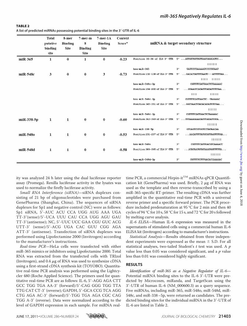
Identification of miR-365 as a Negative Regulator of IL-6—Potential miRNA binding sites to the IL-6 3�-UTR were pre-dicted by Microcosm, miRanda, and TargetScan using the3�-UTR of human IL-6 (NM_000600.3) as a query sequence.Five miRNAs, including miR-365, miR-548o, miR-548d, miR-548c, and miR-338–5p, were returned as candidates. The pre-dicted binding sites for the individual miRNA in the 3�-UTR ofIL-6 are listed in Table 2.
TABLE 2A list of predicted miRNAs possessing potential binding sites in the 3�-UTR of IL-6
miR-365 Negatively Regulates IL-6
JUNE 17, 2011 • VOLUME 286 • NUMBER 24 JOURNAL OF BIOLOGICAL CHEMISTRY 21403
by guest on June 4, 2018http://w
ww
.jbc.org/D
ownloaded from

To investigate the roles of these predicted miRNAs on IL-6expression, DNA constructs expressing the miRNA precursorswere co-transfected with pMIR-IL-6 3�-UTR. As shown in Fig.
1A, only transfection with miR-365 expression vector signifi-cantly down-regulated luciferase activity compared with trans-fection with an empty vector, indicating that miR-365 is apotential regulator of IL-6.To further evaluate the effect of mature miR-365 on IL-6
expression, chemosyntheticmiR-365mimic and inhibitor wereemployed. As shown in Fig. 1B, the miR-365 mimic also inhib-ited luciferase activity of pMIR-IL-6 3�-UTR in HEK293 cells.Conversely, the miR-365 inhibitor induced luciferase activity.In addition, the dose-dependent effects of the mimic and theinhibitor on inhibition and induction of luciferase activity,respectively, could be observed (Fig. 1B). To further validate themiR-365 inhibitory effect on IL-6 protein expression, HeLacells were transfectedwith themiR-365mimic (40 nM) or inhib-itor (100 nM), and IL-6 levels in the culture supernatant weremeasured by ELISA. As shown in Fig. 1C, treatment with themiR-365 mimic remarkably repressed IL-6 expression, whereasthe miR-365 inhibitor induced IL-6 expression. As a critical pro-inflammatory cytokine, IL-6 expression is highly inducible bysome immunostimulatory molecules such as poly(I:C) (dsRNAanalog) and poly(dAT:dAT) (dsDNA analog). Thus, we furtherevaluated the inhibitory effect of miR-365 on poly(I:C)- or poly-(dAT:dAT)-induced IL-6 expression. As shown in Fig. 1C, trans-fectionwithpoly(I:C)orpoly(dAT:dAT)significantly inducedIL-6expression, but overexpression of miR-365 remarkably blockedpoly(I:C)- or poly(dAT:dAT)-induced IL-6 expression.We further investigated whether miR-365 acts to affect the
stability of IL-6 mRNA. To this end, real-time PCR was per-formed by using total RNAprepared fromcells transfectedwiththe mimic or inhibitor oligonucleotides. As shown in Fig. 1D,neither miR-365mimic nor inhibitor affected the expression ofIL-6 mRNA. Thus, it can be concluded that miR-365 decreasesIL-6 expression by repressing its mRNA translation but doesnot affect its mRNA stability.Confirmation of the Response Element formiR-365 in the IL-6
3�-UTR—Post-transcriptional regulation of target genes bymiRNA is mediated by binding of miRNA to the 3�-UTR oftarget gene, whereas the specificity of the individual miRNAis determined by the seed region, which located in nucleo-tides 2–8 of 5� portion of the miRNA (7). Computationalanalysis suggested that there is a putative binding site formiR-365 in the 3�-UTR of the human IL-6 gene and thebinding site for the seed region of miR-365 is located at30–36 bp of IL-6 3�-UTR (Fig. 2A). The minimum freeenergy of hybridization between the target RNA and miR-365, as predicted by RNAhybrid (33), also supported the pos-sibility that miR-365 can bind at that site (Fig. 2B). Further-more, alignment of multiple IL-6 3�-UTRs showed that thebinding site for the seed region of miR-365 is highly con-served among different species, including human, mouse,rhesus, chimpanzee, armadillo, and cow (Fig. 2A).
To validate these bioinformatics predictions with experi-mental evidence, 3�-UTR reporter constructs lacking the pre-dictedmiR-365 binding site or having a 3-bp substitution in theMRE of IL-6 gene were generated and termed as pMIR-IL-63�-UTR deletion and point mutation, respectively (Fig. 2C).When the miR-365 mimic or inhibitor were co-transfectedwith these constructs, the miR-365 mimic was observed to
FIGURE 1. miR-365 is a negative regulator of IL-6. A, HEK293 cells wereco-transfected with the indicated miRNA expression construct (0.4 �g), pMIR-IL6 3�-UTR reporter plasmid (0.2 �g), and pRL-TK (0.05 �g). At 24 h post-transfection, cells were collected, and luciferase activity was measured. Lucif-erase activity of the empty vector was regarded as 1. *, p � 0.01, as comparedwith the empty vector group. B, mimics (20, 40, and 60 nM) or inhibitors (60,80, and 100 nM) of miR-365 were co-transfected with pMIR-IL-6 3�-UTRreporter plasmid into HEK293 cells. A scrambled mimic (60 nM) or inhibitor(100 nM) was used as control. Luciferase activity was measured at 24 h post-transfection. The luciferase activity of the scrambled mimic or inhibitor wasregarded as 1. C and D, HeLa cells were transfected with the indicated miRNAoligonucleotides. Poly(I:C) or poly(dAT:dAT) were transfected at 24 h after theinitial transfection. Culture supernatants were collected 24 h later, and IL-6was measured using a human IL-6 ELISA kit (C). Total RNA was extracted fromcells, and real-time PCR assay was performed with specific primers listed in“Experimental Procedures” (D).
miR-365 Negatively Regulates IL-6
21404 JOURNAL OF BIOLOGICAL CHEMISTRY VOLUME 286 • NUMBER 24 • JUNE 17, 2011
by guest on June 4, 2018http://w
ww
.jbc.org/D
ownloaded from

inhibit by 45% the luciferase activity of the reporter with anintact 3�-UTR, as compared with luciferase activity after treat-ment with scrambled mimics. However, overexpression of
miR-365 only inhibited 10 and 20% luciferase activity of thereporter pMIR-IL-6 3�-UTR deletion and point mutation,respectively (Fig. 2D). These results provided experimental
FIGURE 2. Identification of the binding site for miR-365 in the IL-6 3�-UTR. A, alignment of the IL-6 3�-UTR showed that the MRE (gray) was conserved amongdifferent species. B, shown is a schematic representation of the hybridization between miR-365 and IL-6 3�-UTR. Green and red letters indicate miR-365 and IL-6mRNA, respectively. C, shown is a schematic representation of mutant reporters of IL-6 3�-UTR. The frame and red letters indicate the deletion and the pointmutation, respectively. D, intact, point mutant, or deletion mutants pMIR-IL-6 3�-UTR and pMIR-REPORT were co-transfected with the indicated oligonucleo-tides into HEK293 cells. 24 h post-transfection, luciferase activities were measured. Results are presented as -fold induction over control groups. Bar graph dataare presented as the means � S.D. (n � 3); **, p � 0.01, as compared with the transfection with the scrambled oligonucleotides.
miR-365 Negatively Regulates IL-6
JUNE 17, 2011 • VOLUME 286 • NUMBER 24 JOURNAL OF BIOLOGICAL CHEMISTRY 21405
by guest on June 4, 2018http://w
ww
.jbc.org/D
ownloaded from

FIGURE 3. Transcriptional regulation of miR-365. A, shown is a schematic representation of the region encoding hsa-miR-365. TSS is represented as a blackbox. B, HEK293 cells were cultured in 24-well plates and transfected with miR-365 promoter reporter mutants containing various lengths of the miR-365promoter region. At 24 h post-transfection, cells were transfected or mock-transfected with poly(dAT:dAT) (1 �g/well). 24 h later luciferase activity wasmeasured, and luciferase activity of the mock-transfected empty vector pGL3 Basic group was regarded as 1. **, p � 0.01. C, HEK293 cells were transfected withTSS-1097-Luc reporter. At 24 h post-transfection, cells were infected with Sendai virus (10 hemagglutinin activity units/well) or incubated with phorbol12-myristate 13-acetate (PMA; 1 �g/well) or transfected with poly(I:C) (1 �g/well) or poly(dAT:dAT) (1 �g/well), respectively. Luciferase activities were mea-sured 24 h later. D, HEK293 cells were transfected with poly(I:C) or poly(dAT:dAT). Total RNA were extracted at 24 h post-transfection. The expression ofendogenous miR-365 was detected by miRNA real-time PCR. Results are presented as -fold induction over control groups.
miR-365 Negatively Regulates IL-6
21406 JOURNAL OF BIOLOGICAL CHEMISTRY VOLUME 286 • NUMBER 24 • JUNE 17, 2011
by guest on June 4, 2018http://w
ww
.jbc.org/D
ownloaded from
support to the prediction that the miR-365 response element islocated in the 3�-UTR of IL-6.Analysis of miR-365 Promoter Activity—To investigate the
transcriptional regulation of miR-365, the genomic informa-tion of miR-365, including the putative transcription start siteand genomic location, were obtained frommiRBase (23–25). Aschematic representation of the miR-365 promoter is shown inFig. 3A. For the deletion mutation assay, luciferase reporterswith different lengths of the miR-365 promoter were trans-fected intoHEK293 cells to determine the basal promoter activ-ity. As shown in Fig. 3B, the highest luciferase activity was pro-duced by reporter TSS-1097, with a 1.2-kb putative promoterregion, which led to a 10-fold induction in luciferase activitywith respect to pGL3-Basic, suggesting that TSS-1097 pos-sesses fully intact promoter activity. Transfection with TSS-1348, TSS-705, or TSS-336 reporter plasmids resulted in a lessbasal promoter activity. These results indicated that the regionfrom �1097 to �705 is required for intact promoter activityand the region from �1348 to �1097 may contain negativeregulatory element(s).We also investigated the responses of themiR-365 promoter to different inflammatory stimuli. As shownin Fig. 3C, luciferase activity of reporter TSS-1097 could beinduced by poly(dAT:dAT), poly(I:C), phorbol 12-myristate13-acetate, and Sendai virus. The highest induction was up to3-fold after stimulation with poly(dAT:dAT), whereas induc-tion by poly(dAT:dAT) was not observed in transfections withTSS-1348, TSS-705, orTSS-336 (Fig. 3B). In addition,we exam-ined whether endogenous miR-365 is inducible by stimulationwith poly(dAT:dAT) or poly(I:C). As shown in Fig. 3D, theexpression ofmiR-365 was significantly high in cells stimulatedwith poly(dAT:dAT) than with poly(I:C). The results were inagreement with that of luciferase activity of reporter TSS-1097 after stimulation with poly(dAT:dAT) and poly(I:C).Together, these results demonstrate that the region from�1097 to �705 is required for basal miR-365 promoteractivity and that miR-365 promoter activity can be signifi-cantly induced by poly(dAT:dAT).NF-�Band Sp1 Response Elements Are Essential for the Tran-
scriptional Activity of the miR-365 Promoter—To investigatethe transcription factors involved in the transcription of miR-365, a 1.1-kb region upstreamof the putative transcription startsite (TSS) of miR-365 was analyzed with prediction softwareAlibaba2.1. Sp1 and NF-�B binding sites were predicted to beseparately located in the promoter region. We used site-di-rected mutagenesis to abolish each site (Fig. 4A). Mutations ofeither the NF-�B or Sp1 site significantly inhibited the tran-scription activity of TSS-1097 induced by poly(dAT:dAT) (Fig.4B).We further investigated the involvement of Sp1 in the tran-scriptional regulation of miR-365 by using gain- and loss-of-function assays. Promoter activity of miR-365 was inhibited by45% in the presence of 80 nM Sp1 siRNA (Fig. 4C), whereasforced expression of Sp1 induced promoter activity by 3-fold(Fig. 4E). Mutation of any NF-�B or Sp1 binding sites in themiR-365 promoter resulted in similar responses to the induc-tion caused by Sp1. In contrast, the doublemutation (mutationsof NF-�B sites number 1 and 2 located in the region from�1097 to�705 bp) and Sp1-mut2 inhibited the transcriptional
increase induced by ectopic expression of Sp1 by 50 and 60%,respectively (Fig. 4E).The NF-�B signaling pathway has previously been impli-
cated in the regulation ofmanymiRNAs (8).We used gain- andloss-of-function assays to show that forced expression ofNF-�B subunit p65 induced a 12-fold up-regulation of themiR-365 promoter activity (Fig. 4E). However, interfering withNF-�B activity by pretreatment with 10 �M NF-�B specificinhibitor BAY11-7082 inhibited both basal promoter activityand transcriptional increase by poly(dAT:dAT) stimulation(Fig. 4D). These results indicated a role for NF-�B in the tran-scription regulation of miR-365. In addition, the site-directedmutation assays showed that mutations of any of the NF-�B orSp1 binding sites slightly inhibited the transcriptional increaseinduced by p65 overexpression, but the double mutation andSp1-mut2 mutants inhibited by at least 45 and 50%, respec-tively, the transcriptional increase induced by p65 overexpres-sion (Fig. 4E). These results indicate an important role for theNF-�B sites number 1 and 2 and Sp1 site number 2 in the tran-scription of miR-365.Previous studies have revealed an interaction between Sp1
and NF-�B in gene regulation processes through synergistic orantagonistic effects (34). To investigate the relationshipbetween Sp1 and NF-�B in the transcriptional regulation ofmiR-365, Sp1 and p65 expression constructs were co-trans-fected, and the luciferase activity of TSS-1097 was analyzed. Asshown in Fig. 4F, forced expression of both Sp1 and NF-�B p65up-regulated transcription of miR-365 to a greater degree (16-fold) than transfection with either one alone (12- or 13-fold,respectively). These results suggest a synergistic effect betweenSp1 and NF-�B on the transcription of miR-365.The Involvement of MAPK/ERK in miR-365 Transcription—
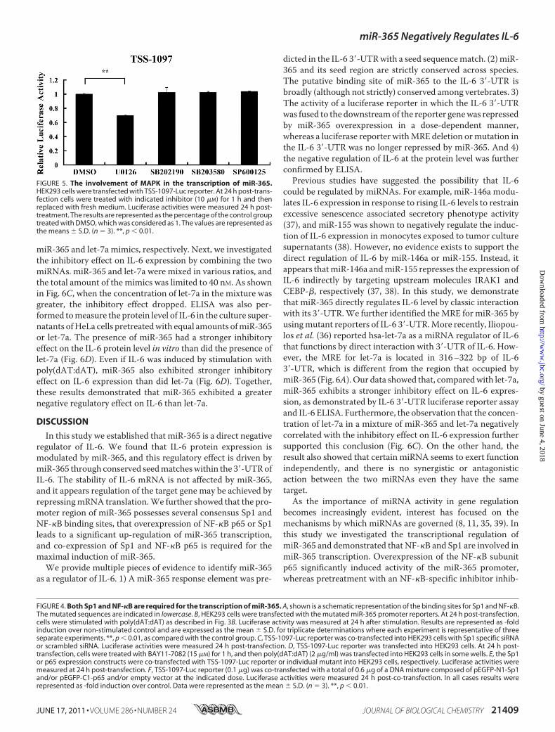
TheMAPKpathway is an important signaling pathway regulat-ing numerous biological processes includingmiRNA-mediatedeffects (35). To address the question as to whether MAPK sig-naling is involved in the transcription ofmiR-365, we employedspecific inhibitors of various intermediates of the pathway. Asshown in Fig. 5, pretreatment with the ERK inhibitor U0126inhibited miR-365 promoter activity by 30% with respect toDMSO pretreatment, whereas pretreatment with other inhibi-tors, including the p38 inhibitors SB202190 and SB203580 andthe JNK inhibitor SP600125, had no appreciable inhibitoryeffect on the transcription of miR-365. These results indicatedthatMAPK/ERKmay play a role in the transcription regulationof miR-365.miR-365 Has a Stronger Regulatory Effect on IL-6 in Vitro
ThanDoes Let-7a—Let-7a has recently been described as a reg-ulator of IL-6 by direct interaction with IL-6 3�-UTR (36).Let-7a and miR-365 bind to different regions of the IL-63�-UTR (Fig. 6A). To compare the inhibitory effect of the twomiRNAs on IL-6 expression, equal amounts of miR-365 orlet-7a mimics along with pMIR-IL-6 3�-UTR were transfectedinto HEK293 cells. As shown in Fig. 6B, when 40 nM mimicswere transfected, miR-365 inhibited by 50% the reporter activ-ity, whereas let-7a only inhibited by 30%, as compared withtransfection with the scrambled oligonucleotide. When theamount of miRNA mimics was raised to 80 nM, the reporteractivities were inhibited by 50 and 40% in transfections with
miR-365 Negatively Regulates IL-6
JUNE 17, 2011 • VOLUME 286 • NUMBER 24 JOURNAL OF BIOLOGICAL CHEMISTRY 21407
by guest on June 4, 2018http://w
ww
.jbc.org/D
ownloaded from

miR-365 Negatively Regulates IL-6
21408 JOURNAL OF BIOLOGICAL CHEMISTRY VOLUME 286 • NUMBER 24 • JUNE 17, 2011
by guest on June 4, 2018http://w
ww
.jbc.org/D
ownloaded from
miR-365 and let-7a mimics, respectively. Next, we investigatedthe inhibitory effect on IL-6 expression by combining the twomiRNAs. miR-365 and let-7a were mixed in various ratios, andthe total amount of the mimics was limited to 40 nM. As shownin Fig. 6C, when the concentration of let-7a in the mixture wasgreater, the inhibitory effect dropped. ELISA was also per-formed tomeasure the protein level of IL-6 in the culture super-natants ofHeLa cells pretreatedwith equal amounts ofmiR-365or let-7a. The presence of miR-365 had a stronger inhibitoryeffect on the IL-6 protein level in vitro than did the presence oflet-7a (Fig. 6D). Even if IL-6 was induced by stimulation withpoly(dAT:dAT), miR-365 also exhibited stronger inhibitoryeffect on IL-6 expression than did let-7a (Fig. 6D). Together,these results demonstrated that miR-365 exhibited a greaternegative regulatory effect on IL-6 than let-7a.
DISCUSSION
In this study we established that miR-365 is a direct negativeregulator of IL-6. We found that IL-6 protein expression ismodulated by miR-365, and this regulatory effect is driven bymiR-365 through conserved seedmatcheswithin the 3�-UTRofIL-6. The stability of IL-6 mRNA is not affected by miR-365,and it appears regulation of the target gene may be achieved byrepressingmRNA translation.We further showed that the pro-moter region of miR-365 possesses several consensus Sp1 andNF-�B binding sites, that overexpression of NF-�B p65 or Sp1leads to a significant up-regulation of miR-365 transcription,and co-expression of Sp1 and NF-�B p65 is required for themaximal induction of miR-365.We provide multiple pieces of evidence to identify miR-365
as a regulator of IL-6. 1) A miR-365 response element was pre-
dicted in the IL-6 3�-UTRwith a seed sequencematch. (2) miR-365 and its seed region are strictly conserved across species.The putative binding site of miR-365 to the IL-6 3�-UTR isbroadly (although not strictly) conserved among vertebrates. 3)The activity of a luciferase reporter in which the IL-6 3�-UTRwas fused to the downstreamof the reporter genewas repressedby miR-365 overexpression in a dose-dependent manner,whereas a luciferase reporter withMRE deletion ormutation inthe IL-6 3�-UTR was no longer repressed by miR-365. And 4)the negative regulation of IL-6 at the protein level was furtherconfirmed by ELISA.Previous studies have suggested the possibility that IL-6
could be regulated by miRNAs. For example, miR-146a modu-lates IL-6 expression in response to rising IL-6 levels to restrainexcessive senescence associated secretory phenotype activity(37), and miR-155 was shown to negatively regulate the induc-tion of IL-6 expression in monocytes exposed to tumor culturesupernatants (38). However, no evidence exists to support thedirect regulation of IL-6 by miR-146a or miR-155. Instead, itappears thatmiR-146a andmiR-155 represses the expression ofIL-6 indirectly by targeting upstream molecules IRAK1 andCEBP-�, respectively (37, 38). In this study, we demonstratethat miR-365 directly regulates IL-6 level by classic interactionwith its 3�-UTR.We further identified theMRE formiR-365 byusingmutant reporters of IL-6 3�-UTR.More recently, Iliopou-los et al. (36) reported hsa-let-7a as a miRNA regulator of IL-6that functions by direct interaction with 3�-UTR of IL-6. How-ever, the MRE for let-7a is located in 316–322 bp of IL-63�-UTR, which is different from the region that occupied bymiR-365 (Fig. 6A). Our data showed that, comparedwith let-7a,miR-365 exhibits a stronger inhibitory effect on IL-6 expres-sion, as demonstrated by IL-6 3�-UTR luciferase reporter assayand IL-6 ELISA. Furthermore, the observation that the concen-tration of let-7a in a mixture of miR-365 and let-7a negativelycorrelated with the inhibitory effect on IL-6 expression furthersupported this conclusion (Fig. 6C). On the other hand, theresult also showed that certain miRNA seems to exert functionindependently, and there is no synergistic or antagonisticaction between the two miRNAs even they have the sametarget.As the importance of miRNA activity in gene regulation
becomes increasingly evident, interest has focused on themechanisms by which miRNAs are governed (8, 11, 35, 39). Inthis study we investigated the transcriptional regulation ofmiR-365 and demonstrated that NF-�B and Sp1 are involved inmiR-365 transcription. Overexpression of the NF-�B subunitp65 significantly induced activity of the miR-365 promoter,whereas pretreatment with an NF-�B-specific inhibitor inhib-
FIGURE 4. Both Sp1 and NF-�B are required for the transcription of miR-365. A, shown is a schematic representation of the binding sites for Sp1 and NF-�B.The mutated sequences are indicated in lowercase. B, HEK293 cells were transfected with the mutated miR-365 promoter reporters. At 24 h post-transfection,cells were stimulated with poly(dAT:dAT) as described in Fig. 3B. Luciferase activity was measured at 24 h after stimulation. Results are represented as -foldinduction over non-stimulated control and are expressed as the mean � S.D. for triplicate determinations where each experiment is representative of threeseparate experiments. **, p � 0.01, as compared with the control group. C, TSS-1097-Luc reporter was co-transfected into HEK293 cells with Sp1 specific siRNAor scrambled siRNA. Luciferase activities were measured 24 h post-transfection. D, TSS-1097-Luc reporter was transfected into HEK293 cells. At 24 h post-transfection, cells were treated with BAY11-7082 (15 �M) for 1 h, and then poly(dAT:dAT) (2 �g/ml) was transfected into HEK293 cells in some wells. E, the Sp1or p65 expression constructs were co-transfected with TSS-1097-Luc reporter or individual mutant into HEK293 cells, respectively. Luciferase activities weremeasured at 24 h post-transfection. F, TSS-1097-Luc reporter (0.1 �g) was co-transfected with a total of 0.6 �g of a DNA mixture composed of pEGFP-N1-Sp1and/or pEGFP-C1-p65 and/or empty vector at the indicated dose. Luciferase activities were measured 24 h post-co-transfection. In all cases results wererepresented as -fold induction over control. Data were represented as the mean � S.D. (n � 3). **, p � 0.01.
FIGURE 5. The involvement of MAPK in the transcription of miR-365.HEK293 cells were transfected with TSS-1097-Luc reporter. At 24 h post-trans-fection cells were treated with indicated inhibitor (10 �M) for 1 h and thenreplaced with fresh medium. Luciferase activities were measured 24 h post-treatment. The results are represented as the percentage of the control grouptreated with DMSO, which was considered as 1. The values are represented asthe means � S.D. (n � 3). **, p � 0.01.
miR-365 Negatively Regulates IL-6
JUNE 17, 2011 • VOLUME 286 • NUMBER 24 JOURNAL OF BIOLOGICAL CHEMISTRY 21409
by guest on June 4, 2018http://w
ww
.jbc.org/D
ownloaded from

ited the transcription of miR-365. Furthermore, the NF-�Btranscription factor binding sites in the promoter of miR-365were required for the transcription of miR-365, collectively
FIGURE 6. Comparison of the effect of miR-365 and let-7a on IL6 expres-sion in vitro. A, shown is a schematic representation of the MREs for let-7aand miR-365. The MREs for let-7a and miR-365 were obtained from thereport by Iliopoulos et al. (36) and our study, respectively. CDS, codingsequence. B, pMIR-IL-6 3�-UTR was co-transfected with miR-365 or let-7ainto HEK293 cells. Luciferase activities were measured at 24 h post-trans-fection. NC, negative control. *, p � 0.05. C, pMIR-IL-6 3�-UTR was co-transfected with a total of 40 nM miRNA composed of individual or mixedmiR-365 and/or let-7a at the indicated ratios. Luciferase activities weremeasured 24 h post-co-transfection. D, HeLa cells were transfected with atotal of 60 nM miR-365 and/or let-7a mimics or scrambled oligonucleo-tides as indicated. Cells were transfected with poly(dAT:dAT) at 24 h afterthe initial transfection. Culture supernatants were collected 24 h later, andIL-6 was measured using a human IL-6 ELISA kit. Bar graph data are pre-sented as the means � S.D. (n � 3). *, p � 0.05; **, p � 0.01.
TA
BLE
3Li
sto
fpu
tati
veta
rget
gen
eso
fmiR
-365
Targetg
ene
Gen
ena
me
Con
served
sites
Poorlyco
nservedsites
Totalco
ntextsco
reAgg
regate
PCT
Total
8-Mer
7-Mer-m
87-Mer-1A
Total
8-Mer
7-Mer-m
87-Mer-1A
LPAR5
Lysoph
osph
atidicacid
receptor
51
10
02
11
0�0.93
0.11
NFIB
Nuclear
factor
I/B
21
10
10
01
�0.63
0.56
ZNF1
48Zinc
fingerp
rotein
148
21
10
10
01
�0.53
0.36
TMOD3
Tropo
mod
ulin
3(ubiqu
itous)
11
00
00
00
�0.53
0.24
SGK1
Serum/glucocorticoidregu
latedkina
se1
11
00
00
00
�0.52
0.42
USP
33Ubiqu
itin-specificpeptidase33
11
00
00
00
�0.48
0.53
ZNF6
44Zinc
fingerp
rotein
644
11
00
00
00
�0.47
0.47
USP
48Ubiqu
itin-specificpeptidase48
11
00
00
00
�0.47
0.51
ARR
DC3
Arrestin
domaincontaining
31
00
11
01
0�0.47
0.43
LOC28
3514
Similartosevenin
absentia2
11
00
00
00
�0.47
0.55
PIK3R
3Ph
osph
oino
sitid
e3-kina
se,regulatorysubu
nit3
(�)
10
10
11
00
�0.43
0.24
SGK3
Serum/glucocorticoid-regu
latedkina
sefamily,
mem
ber3
10
10
00
00
�0.29
0.24
MAPK
1IP1
LMito
gen-activ
ated
proteinkina
se1interacting
protein1-lik
e1
01
00
00
0�0.29
0.24
PRKAR2
AProteinkina
se,cAMP-depend
ent,regu
latory,type
II,�
10
10
00
00
�0.27
0.24
HSP
A8
Heatsho
ck70
-kDaprotein8
10
10
00
00
�0.26
0.24
miR-365 Negatively Regulates IL-6
21410 JOURNAL OF BIOLOGICAL CHEMISTRY VOLUME 286 • NUMBER 24 • JUNE 17, 2011
by guest on June 4, 2018http://w
ww
.jbc.org/D
ownloaded from

suggesting that NF-�B plays a critical role in the biological pro-cesses of miR-365. Indeed, in addition to miR-365, miR-146a(8), miR-155 (40), let-7i (41), miR-17–92, miR-125b-1, miR-21,miR-23b-27b-24–1, miR-30b, and miR-130a (42) have all alsobeen found to be under the control of NF-�B. Sp1 is a ubiqui-tously expressed transcription factor and is indispensable forthe development and survival of animals (43–45). Interestingly,Sp1 elements are often found in the enhancers or promoters ofNF-�B-regulated genes (46–49). Furthermore, the Sp1-NF-�Bcomplex has been shown to be involved in the regulation ofmiR-29b and mouse TLR-2 (50, 51). Considering that our datashow that both Sp1 and NF-�B contribute to the transcriptionof miR-365 and two NF-�B binding sites and one Sp1 bindingsite were found to be indispensable for miR-365 transcription,we speculated that cooperation between Sp1 and NF-�B in thetranscriptional regulation of miR-365 might exist. The syner-gistic increase in the miR-365 promoter activity induced byco-expression of Sp1 and NF-�B p65 partly supported ourhypothesis, but further studies are needed to test thesepossibilities.In addition to the NF-�B pathway, previous studies have
revealed that the MAPK signaling pathway also plays a regula-tory role in the generation of some miRNAs (35, 52, 53). In ourstudy, pretreatment with ERK inhibitor significantly inhibitedmiR-365 promoter activity, indicating a regulatory role for ERKin the transcription of miR-365. Although p38 and JNK havebeen demonstrated to regulate the expression of othermiRNAs, for example JNK regulatingmiR-155 and p38 regulat-ingmiR-34c (54, 55), we did not observe any appreciable role inthe regulation of miR-365 by p38 and JNK.A single miRNA can regulate several or even hundreds of
genes (1). With the exception of IL-6, bioinformatics analyseshave indicated other molecules, such as ubiquitin-specific pep-tidase 48 (USP48), heat shock 70-kDa protein 8 (HSPA8), zincfinger protein 148 (ZNF-148), and serum/glucocorticoid-regu-lated kinase 1 (SGK-1), are potential targets of miR-365 (Table3). Interestingly, a very close correlation between these targetsand NF-�B activation has been reported (56–59). These mole-cules regulate NF-�B by several mechanisms, including specificpromoter binding activity as well as phosphorylation and ubiq-uitination. For example, SGK-1 facilitates NF-�B activation byphosphorylation of IKK�, which in turn leads to the degrada-tion of I�B� (59). NF-�B has been shown to play amajor role inLPS-induced expression of IL-6 or other inflammatory cyto-kines (30). It is possible that, in addition to negative regulationof IL-6 expression by direct binding to the 3�-UTR of IL-6,miR-365 may also affect the activation of NF-�B and therebyindirectly inhibit IL-6 expression. Interestingly, we observedthat overexpression of miR-365 significantly attenuated NF-�Bactivation in HEK293 cells (data not shown). Certainly, addi-tional experiments should be performed to demonstrate theindirect regulation of IL-6 by miR-365.In conclusion, our studies identified miR-365 as a novel neg-
ative regulator of IL-6. In addition to direct negative regulation,miR-365 may regulate IL-6 expression indirectly. In vitro evi-dence indicated that miR-365 functions as a more powerfulregulator of IL-6 than let-7a. NF-�B, Sp1, and ERK are involvedin the expression of miR-365, suggesting that miR-365 is con-
trolled by a complex network to exert its function in an appro-priate manner.
REFERENCES1. Ambros, V. (2004) Nature 431, 350–3552. Lee, Y., Jeon, K., Lee, J. T., Kim, S., and Kim, V. N. (2002) EMBO J. 21,
4663–46703. Yi, R., Qin, Y., Macara, I. G., and Cullen, B. R. (2003) Genes Dev. 17,
3011–30164. Lee, Y., Ahn, C., Han, J., Choi, H., Kim, J., Yim, J., Lee, J., Provost, P.,
Rådmark, O., Kim, S., and Kim, V. N. (2003) Nature 425, 415–4195. Bartel, D. P. (2004) Cell 116, 281–2976. Kiriakidou, M., Nelson, P. T., Kouranov, A., Fitziev, P., Bouyioukos, C.,
Mourelatos, Z., and Hatzigeorgiou, A. (2004) Genes Dev. 18,1165–1178
7. Lewis, B. P., Shih, I. H., Jones-Rhoades, M. W., Bartel, D. P., and Burge,C. B. (2003) Cell 115, 787–798
8. Taganov, K. D., Boldin, M. P., Chang, K. J., and Baltimore, D. (2006) Proc.Natl. Acad. Sci. U.S.A. 103, 12481–12486
9. Xiao, B., Liu, Z., Li, B. S., Tang, B., Li,W., Guo,G., Shi, Y.,Wang, F.,Wu, Y.,Tong,W. D., Guo, H.,Mao, X. H., and Zou, Q.M. (2009) J. Infect. Dis. 200,916–925
10. Tang, B., Xiao, B., Liu, Z., Li, N., Zhu, E. D., Li, B. S., Xie, Q. H., Zhuang, Y.,Zou, Q. M., and Mao, X. H. (2010) FEBS Lett. 584, 1481–1486
11. Sharma, A., Kumar, M., Aich, J., Hariharan, M., Brahmachari, S. K.,Agrawal, A., and Ghosh, B. (2009) Proc. Natl. Acad. Sci. U.S.A. 106,5761–5766
12. Qin, Z., Kearney, P., Plaisance, K., and Parsons, C. H. (2010) J. Leukoc. Biol.87, 25–34
13. Ma, F., Liu, X., Li, D., Wang, P., Li, N., Lu, L., and Cao, X. (2010) J. Immu-nol. 184, 6053–6059
14. Polikepahad, S., Knight, J. M., Naghavi, A. O., Oplt, T., Creighton, C. J.,Shaw, C., Benham, A. L., Kim, J., Soibam, B., Harris, R. A., Coarfa, C.,Zariff, A.,Milosavljevic, A., Batts, L.M., Kheradmand, F., Gunaratne, P.H.,and Corry, D. B. (2010) J. Biol. Chem. 285, 30139–30149
15. Alsaleh, G., Suffert, G., Semaan, N., Juncker, T., Frenzel, L., Gottenberg,J. E., Sibilia, J., Pfeffer, S., and Wachsmann, D. (2009) J. Immunol. 182,5088–5097
16. Akira, S., Hirano, T., Taga, T., and Kishimoto, T. (1990) FASEB J. 4,2860–2867
17. Hirano, T., Akira, S., Taga, T., and Kishimoto, T. (1990) Immunol. Today11, 443–449
18. Kishimoto, T. (1989) Blood 74, 1–1019. Kishimoto, T. (2005) Annu. Rev. Immunol. 23, 1–2120. Jones, S. A. (2005) J. Immunol. 175, 3463–346821. Rambaldi, A., Bettoni, S., Rossi, V., Tini, M. L., Giudici, G., Rizzo, V.,
Bassan, R., Mantovani, A., Barbui, T., and Biondi, A. (1993) Br. J. Haema-tol. 83, 204–211
22. Clark, A. (2000) Arthritis Res. 2, 172–17423. Griffiths-Jones, S., Grocock, R. J., van Dongen, S., Bateman, A., and En-
right, A. J. (2006) Nucleic Acids Res. 34, D140–D14424. Griffiths-Jones, S. (2004) Nucleic Acids Res. 32, D109–D11125. Griffiths-Jones, S., Saini, H. K., van Dongen, S., and Enright, A. J. (2008)
Nucleic Acids Res. 36, D154–D15826. John, B., Enright, A. J., Aravin, A., Tuschl, T., Sander, C., and Marks, D. S.
(2004) PLoS Biol. 2, e36327. Betel, D., Wilson, M., Gabow, A., Marks, D. S., and Sander, C. (2008)
Nucleic Acids Res. 36, D149–D15328. Grimson, A., Farh, K. K., Johnston, W. K., Garrett-Engele, P., Lim, L. P.,
and Bartel, D. P. (2007)Mol. Cell 27, 91–10529. Friedman, R. C., Farh, K. K., Burge, C. B., and Bartel, D. P. (2009)Genome
Res. 19, 92–10530. Kim, B. H., Lee, K. H., Chung, E. Y., Chang, Y. S., Lee, H., Lee, C. K., Min,
K. R., and Kim, Y. (2006) Eur. J. Pharmacol. 543, 158–16531. Lewis, B. P., Burge, C. B., and Bartel, D. P. (2005) Cell 120, 15–2032. Higuchi, R., Krummel, B., and Saiki, R. K. (1988) Nucleic Acids Res. 16,
7351–7367
miR-365 Negatively Regulates IL-6
JUNE 17, 2011 • VOLUME 286 • NUMBER 24 JOURNAL OF BIOLOGICAL CHEMISTRY 21411
by guest on June 4, 2018http://w
ww
.jbc.org/D
ownloaded from
33. Rehmsmeier, M., Steffen, P., Hochsmann, M., and Giegerich, R. (2004)RNA 10, 1507–1517
34. Hirano, F., Tanaka, H., Hirano, Y., Hiramoto, M., Handa, H., Makino, I.,and Scheidereit, C. (1998)Mol. Cell. Biol. 18, 1266–1274
35. Paroo, Z., Ye, X., Chen, S., and Liu, Q. (2009) Cell 139, 112–12236. Iliopoulos, D., Hirsch, H. A., and Struhl, K. (2009) Cell 139, 693–70637. Bhaumik, D., Scott, G. K., Schokrpur, S., Patil, C. K., Orjalo, A. V., Rodier,
F., Lithgow, G. J., and Campisi, J. (2009) Aging 1, 402–41138. He, M., Xu, Z., Ding, T., Kuang, D. M., and Zheng, L. (2009) Cell. Mol.
Immunol. 6, 343–35239. Liu, G., Friggeri, A., Yang, Y., Park, Y. J., Tsuruta, Y., and Abraham, E.
(2009) Proc. Natl. Acad. Sci. U.S.A. 106, 15819–1582440. Gatto, G., Rossi, A., Rossi, D., Kroening, S., Bonatti, S., and Mallardo, M.
(2008) Nucleic Acids Res. 36, 6608–661941. O’Hara, S. P., Splinter, P. L., Gajdos, G. B., Trussoni, C. E., Fernandez-
Zapico, M. E., Chen, X. M., and LaRusso, N. F. (2010) J. Biol. Chem. 285,216–225
42. Zhou, R., Hu, G., Gong, A. Y., and Chen, X. M. (2010) Nucleic Acids Res.38, 3222–3232
43. Dynan, W. S., and Tjian, R. (1983) Cell 35, 79–8744. Narayan, V. A., Kriwacki, R. W., and Caradonna, J. P. (1997) J. Biol. Chem.
272, 7801–780945. Saffer, J. D., Jackson, S. P., and Annarella, M. B. (1991)Mol. Cell. Biol. 11,
2189–219946. Stade, B. G., Messer, G., Riethmuller, G., and Johnson, J. P. (1990) Immu-
nobiology 182, 79–8747. Ma, W., Lim, W., Gee, K., Aucoin, S., Nandan, D., Kozlowski, M., Diaz-
Mitoma, F., and Kumar, A. (2001) J. Biol. Chem. 276, 13664–1367448. Jones, K. A., Kadonaga, J. T., Luciw, P. A., and Tjian, R. (1986) Science 232,
755–75949. Perkins, N. D., Edwards, N. L., Duckett, C. S., Agranoff, A. B., Schmid,
R. M., and Nabel, G. J. (1993) EMBO J. 12, 3551–355850. Wang, T., Lafuse, W. P., and Zwilling, B. S. (2001) J. Immunol. 167,
6924–693251. Liu, S., Wu, L. C., Pang, J., Santhanam, R., Schwind, S., Wu, Y. Z., Hickey,
C. J., Yu, J., Becker, H., Maharry, K., Radmacher, M. D., Li, C., Whitman,S. P., Mishra, A., Stauffer, N., Eiring, A. M., Briesewitz, R., Baiocchi, R. A.,Chan, K. K., Paschka, P., Caligiuri, M. A., Byrd, J. C., Croce, C. M., Bloom-field, C. D., Perrotti, D., Garzon, R., and Marcucci, G. (2010) Cancer Cell17, 333–347
52. Cardinali, B., Castellani, L., Fasanaro, P., Basso, A., Alema, S., Martelli, F.,and Falcone, G. (2009) PLoS ONE 4, e7607
53. Zeng, Y., Sankala, H., Zhang, X., and Graves, P. R. (2008) Biochem. J. 413,429–436
54. O’Connell, R. M., Taganov, K. D., Boldin, M. P., Cheng, G., and Baltimore,D. (2007) Proc. Natl. Acad. Sci. U.S.A. 104, 1604–1609
55. Cannell, I. G., Kong, Y. W., Johnston, S. J., Chen, M. L., Collins, H. M.,Dobbyn, H. C., Elia, A., Kress, T. R., Dickens, M., Clemens, M. J., Heery,D. M., Gaestel, M., Eilers, M., Willis, A. E., and Bushell, M. (2010) Proc.Natl. Acad. Sci. U.S.A. 107, 5375–5380
56. Tzimas, C., Michailidou, G., Arsenakis, M., Kieff, E., Mosialos, G., andHatzivassiliou, E. G. (2006) Cell. Signal. 18, 83–92
57. Dokladny, K., Lobb, R., Wharton, W., Ma, T. Y., andMoseley, P. L. (2010)Cell Stress Chaperones 15, 153–163
58. Borghaei, R. C., Gorski, G., and Javadi, M. (2009) Biochem. Biophys. Res.Commun. 382, 269–273
59. Tai, D. J., Su, C. C., Ma, Y. L., and Lee, E. H. (2009) J. Biol. Chem. 284,4073–4089
miR-365 Negatively Regulates IL-6
21412 JOURNAL OF BIOLOGICAL CHEMISTRY VOLUME 286 • NUMBER 24 • JUNE 17, 2011
by guest on June 4, 2018http://w
ww
.jbc.org/D
ownloaded from

Zhong, Huan-Chun Chen and Liu-Rong FangZheng Xu, Shao-Bo Xiao, Peng Xu, Qian Xie, Lu Cao, Dang Wang, Rui Luo, Yao
BκCooperatively Regulated by Sp1 and NF-miR-365, a Novel Negative Regulator of Interleukin-6 Gene Expression, Is
doi: 10.1074/jbc.M110.198630 originally published online April 25, 20112011, 286:21401-21412.J. Biol. Chem.
10.1074/jbc.M110.198630Access the most updated version of this article at doi:
Alerts:
When a correction for this article is posted•
When this article is cited•
to choose from all of JBC's e-mail alertsClick here
http://www.jbc.org/content/286/24/21401.full.html#ref-list-1
This article cites 59 references, 26 of which can be accessed free at
by guest on June 4, 2018http://w
ww
.jbc.org/D
ownloaded from