mir-582-5p induces colorectal cancer cell proliferation … · manual. the expression level of...
TRANSCRIPT
RESEARCH Open Access
miR-582-5P induces colorectal cancer cellproliferation by targeting adenomatouspolyposis coliZhenbo Shu1, Libo Chen2 and Dayong Ding1*
Abstract
Background: microRNA (miRNAs) dysregulation is widely involved in cancer progression and contributed tosustained cell proliferation by directly targeting multiple targets. Therefore, better understand the underlyingmechanism of miRNA in carcinogenesis may improve diagnostic and therapeutic strategies for malignancy.
Methods: We assessed microRNA-582 (miR-582-5P) expression in colorectal cancer (CRC) specimens and cell linesby real-time PCR. Luciferase reporter assay was used to confirm the target associations. Colony formation assay andanchorage-independent growth assay were used to analyze the effect of miR-582-5P on cell proliferation. Adenomatouspolyposis coli (APC) gene and protein expression were examined using real-time PCR and western blotting, respectively.
Results: miR-582-5P was upregulated in the CRC specimens and cell lines and targeted the 3′ untranslated region of APCdirectly. miR-582-5P overexpression increased cyclin D1 and c-MYC expression, which subsequently induced CRC cellproliferation in an APC-dependent manner.
Conclusions: Our findings suggest that miR-582-5P plays an important role in the progression of CRC by inducingproliferation and may identify new targets for anti-cancer treatment.
Keywords: miR-582-5P, APC, Proliferation, Colorectal cancer
BackgroundColorectal cancer (CRC) is one of the most prevalentmalignant cancers and affects men and women almostequally. Currently, approximately 1.25 million people inChina are diagnosed with CRC, and the incidence ofCRC increases annually and it will continue to rise inthe next few years [1]. Furthermore, CRC is a majorcause of cancer-associated mortality, and more than600,000 patients will die from it every year worldwide[2]. In the USA and Europe, CRC is the second most fre-quent cancer that leads to death [3]. In recent years, sig-nificant advances have been made in targeted therapiesof CRC. However, better targeted drugs are required be-cause the effects of existing drugs are not satisfactory.The adenomatous polyposis coli (APC) gene located at
5q21–q22 encodes a tumor suppressor protein that acts
as an antagonist of the Wnt/β-catenin pathway, whichcontrols the CRC cell fate during the maintenance phaseof tumors in patients [4, 5]. It is also involved in otherprocesses, including cell cycle control and cell migration,adhesion, differentiation, apoptosis, and transcriptional ac-tivation [4]. Defects in this gene may cause familial aden-omatous polyposis, an autosomal dominant pre-malignantdisease that usually progresses to malignancy. APC muta-tion has been found in over 80 % of CRC cases and signifi-cantly less frequently in sporadic high microsatelliteinstability cancers than in low microsatellite instability ormicrosatellite instability cancers [6]. Furthermore, APCmutation-induced upregulation of the survivin/ABK cas-cade is associated with crypt cell maturation, expansion ofproliferative cell populations, and promotion of tumori-genesis [7].microRNAs (miRNAs) are a class of endogenous, small,
non-coding RNAs involved in multiple biological pro-cesses. They negatively regulate post-transcriptional geneexpression to act as tumor suppressors or oncogenes by
* Correspondence: [email protected] of Gastrointestinal Colorectal and Anal Surgery, China-JapanUnion Hospital, Jilin University, ChangChun 130033, ChinaFull list of author information is available at the end of the article
© 2016 The Author(s). Open Access This article is distributed under the terms of the Creative Commons Attribution 4.0International License (http://creativecommons.org/licenses/by/4.0/), which permits unrestricted use, distribution, andreproduction in any medium, provided you give appropriate credit to the original author(s) and the source, provide a link tothe Creative Commons license, and indicate if changes were made. The Creative Commons Public Domain Dedication waiver(http://creativecommons.org/publicdomain/zero/1.0/) applies to the data made available in this article, unless otherwise stated.
Shu et al. World Journal of Surgical Oncology (2016) 14:239 DOI 10.1186/s12957-016-0984-4

binding to the 3′ untranslated region (UTR) of a targetgene [8–11]. miRNAs have been widely proposed as po-tential targets for anti-cancer therapies because a numberof findings have indicated that some miRNAs, such asmiR-150 [12, 13], miR-153 [13], miR-561 [14], and miR-622 [15], are involved in the development of human CRC.Publicly available algorithms have indicated that miR-582-5P may directly target the 3′ UTR of APC. miR-582-5P re-duces the proliferation and invasion of human bladdercancer by suppressing the expression of target genes suchas protein geranylgeranyltransferase type I beta subunit(PGGT1B), leucine-rich repeat kinase 2 (LRRK2), and DIXdomain containing 1 (DIXDC1) [16]. However, the role ofmiR-582-5P in CRC progression has not been determined.In this study, we searched for the possible relationship be-tween miR-582-5P and APC and the role of miR-582-5Pin the development of CRC.
MethodsPatients and tissuesThe eight malignant CRC tissues and matched adjacentnoncancerous tissues used in this study were obtainedfrom patients who had undergone surgery at the China-Japan Union Hospital of Jilin University of the People’sRepublic of China. The CRC tissues and matched adja-cent noncancerous tissues were frozen and stored in li-quid nitrogen until used.
Cell cultureA normal colonic mucosal epithelial cell line (normalcontrol) was isolated and purified from the adjacentnoncancerous tissues obtained from the patients. Thehuman CRC cell lines HT29, SW403, SW480, COLO205,SW620, COLO320DM, and KM202L were purchasedfrom American Type Culture Collection (Manassas, VA,USA) and cultured in Dulbecco’s modified Eagle’s medium(Invitrogen, Carlsbad, CA, USA) supplemented with 10 %fetal bovine serum (Invitrogen) at 37 °C in a 5 % CO2 at-mosphere in a humidified incubator.
Plasmids and transfectionThe human APC 3′ UTR was PCR-amplified from gen-omic DNA from SW480 cells and cloned into pGL3 vectors(Promega, Madison, WI, USA). Transfection of miR-582-5P mimic, miR-582-5P inhibitor (miR-582-5P-in), negativecontrol (NC), NC inhibitor (NC-in) (RiboBio, Guangzhou,China), and plasmids was performed using Lipofecta-mine 2000 (Invitrogen) according to the manufac-turer’s instructions.
RNA extraction and real-time quantitative PCRTotal miRNA from cultured cells and cancer tissue sampleswas extracted using the mirVana miRNA Isolation Kit(Ambion, Austin, TX, USA) according to the manufacturer’s
manual. The expression level of miR-582-5P was performedusing miR-582-5P-specific primer and probe (TaqManMicroRNA Assay Kit; Applied Biosystems, Foster City, CA,USA) on an ABI 7900 system (Applied Biosystems). Theexpression of miR-582-5P was defined based on Ct, andrelative expression levels were calculated as 2−[(Ct of miR-582-
5p)− (Ct of U6)] after normalization with reference to thequantification of U6 small nuclear RNA expression. The fol-lowing primers (RiboBio, Guangzhou, China) were synthe-sized and used in this study: GAPDH forward: 5′-AATCTCCACTTTGCCACTG-3′, GAPDH reverse: 5′-CCTCGTCCCGTAGACAAAA-3′; cyclin D1 forward: 5′-AGGAGAACAAACAGATCA-3′, cyclin D1 reverse: 5′-TAGGACAGGAAGTTGTTG-3′; and c-MYC forward: 5′-TCAAGAGGTGCCACGTCTCC-3′, c-MYC reverse: 5′-TCTTGGCAGCAGGATAGTCCTT-3′.
Western blottingWestern blotting was performed according to a previouslyreported method [17]. The membranes were probed withpolyclonal mouse antibodies: anti-APC (ab15270; 1:1000;Abcam, Cambridge, UK), anti-cyclin D1 (1:1000; Cell Sig-naling Technology, Danvers, MA, USA), and anti-c-MYC(1:1000; Cell Signaling Technology). The membranes werestripped and re-probed with anti-α-tubulin mouse mono-clonal antibody (1:1000; Cell Signaling Technology) as theloading control.
Luciferase assayCells were seeded in 24-well plates and allowed to settlefor 24 h. PGL3-APC-luciferase plasmid or pGL3-Mut-luciferase plasmid (100 ng) was transfected into CRC cellsusing Lipofectamine 2000 according to the manufacturer’sinstructions. Luciferase and control signals were measured48 h after transfection using a Dual Luciferase ReporterAssay Kit (Promega) according to a protocol provided bythe manufacturer. Three independent experiments wereperformed, and the data are presented as the mean ± SD.
3-(4,5-Dimethyl-2-thiazolyl)-2, 5-diphenyl-2H-tetrazoliumbromide assayCells were seeded on 96-well plates and stained at the indi-cated time points with 100 μl sterile 3-(4,5-dimethyl-2-thiazolyl)-2, 5-diphenyl-2H-tetrazolium bromide (MTT)dye (0.5 mg/ml, Invitrogen) for 4 h at 37 °C, followed bythe removal of the culture medium and the addition of di-methyl sulfoxide (Sigma-Aldrich, St. Louis, MO, USA).The absorbance at 450 nm was measured using a micro-plate reader (Bio-Rad, La Jolla, CA, USA). Three inde-pendent repeat experiments were performed, and the dataare presented as the mean ± SD.
Shu et al. World Journal of Surgical Oncology (2016) 14:239 Page 2 of 7

Colony formation assayCells were seeded on a 6-well plate (1 × 10³ cells perwell) and cultured for 10 days. The colonies were stainedwith 1.0 % crystal violet for 5 min after a 15-min fixationwith 10 % formaldehyde. All experiments were per-formed in triplicates.
Anchorage-independent growth assayFive hundred cells were trypsinized and suspended in 2 mlcomplete medium plus 0.3 % agar (Sigma-Aldrich). Theagar-cell mixture was plated on top of a bottom layer con-taining 1 % complete medium agar mixture. After 10 days,viable colonies that were larger than 0.1 mm (diameter)were counted with an ocular micrometer (Xintu PhotonicsCo., Ltd, Fuzhou, China). The experiment was performedthree times independently for each cell line.
Statistical analysisStudent’s t test was used to evaluate the significant dif-ference between the two groups of data in all pertinentexperiments. P < 0.05 (Student’s t test) was consideredstatistically significant.
ResultsmiR-582-5P is upregulated in CRC cells and tumor tissuesTo investigate the function of miR-582-5P in the develop-ment of human CRC, we analyzed the expression of miR-582-5P in 218 CRC tumors and matched eight adjacentnoncancerous tissues with CRC tissues utilizing The Can-cer Genome Atlas (TCGA) dataset. As shown in Fig. 1a,miR-582-5P was significantly upregulated in the CRCtumor tissues (P < 0.05). Furthermore, miR-582-5P wassignificantly upregulated in the eight CRC tissue samplesas compared with the adjacent noncancerous colorectaltissues (Fig. 1b). Real-time PCR showed that miR-582-5Pexpression was markedly increased in the seven CRC celllines as compared with that in the normal colorectal epi-thelial cells (Fig. 1c). Collectively, our results show that
miR-582-5P is overexpressed in the CRC cell lines andtissues.
miR-582-5P overexpression induces cell proliferationTo investigate whether miR-582-5P plays a role in CRCdevelopment and progression, we transfected SW480cells with miR-582-5P mimic, miR-582-5P-in, NC, andNC-in RNA. As shown in Fig. 2a, the SW480 cells weresuccessfully transfected with miR-582-5P. The MTTassay was used to examine the effect of miR-582-5Poverexpression on the CRC cell proliferation and showedthat ectopic expression of miR-582-5P increased the cellgrowth rate significantly (Fig. 2b). Furthermore, miR-582-5P overexpression promoted SW480 cell proliferationin the colony formation assay and anchorage-independentgrowth assay (Fig. 2c, d). These results suggest thatmiR-582-5P upregulation increases CRC cell prolifer-ation in vitro.
Downregulation of miR-582-5P inhibits cell proliferationFigure 3a shows that SW480 cells were successfullytransfected with miR-582-5P-in. The MTT assay demon-strated that inhibiting miR-582-5P significantly reducedSW480 cell growth (Fig. 3b). Furthermore, the downregu-lation of miR-582-5P inhibited SW480 cell prolifera-tion in the colony formation assay and anchorage-independent growth assay (Fig. 3c, d). Consistent withthe findings depicted in Fig. 2, these results suggestthat miR-582-5P downregulation reduces CRC cellproliferation in vitro.
miR-582-5P targets APC in CRC cells directlyTo explore the molecular mechanism of miR-582-5P func-tion in CRC cells, we used publicly available algorithms(TargetScan, PicTar, miRanda) to predict miR-582-5P tar-gets in humans. The results indicated that APC was a po-tential target of miR-582-5P (Fig. 4a). As predicted, westernblotting showed that APC expression was decreased inmiR-582-5P-overexpressed SW480 cells and was increased
Fig. 1 miR-582-5P expression is upregulated in CRC. a miR-582-5P expression in 218 CRC tumors and eight matched adjacent noncancerous tissueswith CRC tissues based on the TCGA dataset. b Real-time PCR analysis of miR-582-5P expression in tumor tissues (T) and adjacent noncancerous tissues(ANT) from eight patients with CRC. c Real-time PCR analysis of miR-582-5P expression in human normal colonic mucosal epithelial cells and CRC cells.Mean miR-582-5P expression was normalized to U6 expression. Bars represent the mean ± SD of three independent experiments (*P < 0.05)
Shu et al. World Journal of Surgical Oncology (2016) 14:239 Page 3 of 7

in miR-582-5P-downregulated SW480 cells (Fig. 4b). Mean-while, the results of COLO205 were similar with that ofSW480 (Additional file 1: Figure S1).To examine whethermiR-582-5P-mediated APC downregulation occurred viathe 3′ UTR of APC, we subcloned the APC 3′ UTR frag-ment, containing a miR-582-5P binding site, into a pGL3luciferase reporter vector. miR-582-5P overexpression
reduced the luciferase reporter activity of the APC 3′UTR consistently and dose-dependently; miR-582-5P in-hibition had the opposite effect. However, APC 3′ UTRluciferase reporter activity was unaffected by point muta-tions in the miR-582-5P-binding seed region (Fig. 4c). Col-lectively, our results suggest that APC is a direct target ofmiR-582-5P.
Fig. 2 Ectopic miR-582-5P expression induces CRC cell proliferation. a Real-time PCR analysis confirming that SW480 cells were successfully transfectedwith miR-582-5P mimic or NC (negative control) RNA. b MTT assay analysis of the effects of ectopic miR-582-5P expression on CRC cell proliferation.c Ectopic overexpression of miR-582-5P promoted the colony formation ability of SW480 cells. Representative micrographs (left) and quantification(right) of crystal violet-stained cell colonies at 10 days after transfection. d Anchorage-independent growth assay demonstrating that ectopicoverexpression of miR-582-5P promoted SW480 cell tumorigenicity. Shown are representative micrographs (left) and quantification of colonies>0.1 mm (right). Bars represent the mean ± SD of three independent experiments (*P < 0.05)
Fig. 3 Downregulation of miR-582-5P inhibits CRC cell proliferation. a Real-time PCR analysis confirming that SW480 cells were successfully transfected withmiR-582-5P-in or NC (negative control) RNA. b MTT assay demonstrating that miR-582-5P downregulation reduced SW480 cell proliferation. c Downregulationof miR-582-5P reduced the colony formation ability of SW480 cells. Shown are representative micrographs (left) and quantification (right)of crystal violet-stained cell colonies at 10 days after transfection. d Anchorage-independent growth assay demonstrating that miR-582-5Pdownregulation reduced the tumorigenicity of SW480 cells. Shown are representative micrographs (left) and quantification of colonies >0.1 mm (right).Bars represent the mean ± SD of three independent experiments (*P < 0.05)
Shu et al. World Journal of Surgical Oncology (2016) 14:239 Page 4 of 7

Subsequently, real-time PCR demonstrated that miR-582-5P overexpression significantly increased the expres-sion of cyclin D1 and c-MYC mRNA in SW480 cells(Fig. 4d). Additionally, the western blotting results wereconsistent with the real-time PCR data (Fig. 4e). Thesefindings indicate that miR-582-5P may play an importantrole in regulating the proliferation of CRC cells.
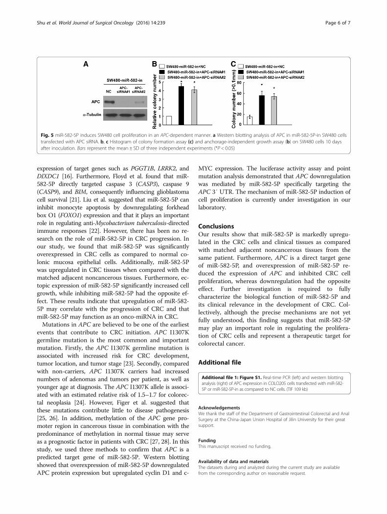
miR-582-5P induces SW480 cell proliferation in an APC-dependent mannerWe determined the effect of miR-582-5P repression inSW480 cells based on the hypothesis that APC repres-sion by miR-582-5P may lead to cell proliferation. Werepressed endogenous APC expression using an APC-specific small interfering RNA (siRNA) (Fig. 5a). Thecolony formation assay and anchorage-independentgrowth assay both showed that silencing APC in miR-582-5P-in-transfected cells increased cell proliferation
(Fig. 5b, c). Together, our results suggest that miR-582-5P may repress APC expression and further promoteCRC progression. However, the silenced APC expres-sion, and its effect on cell proliferation, is reversed inmiR-582-5P-in-transfected cells. These data show thatmiR-582-5P induces SW480 cell proliferation by repres-sing APC expression and that APC may play an import-ant role in miR-582-5P-mediated cell proliferation.
DiscussionmicroRNAs, a class of small regulatory RNA molecules thatnegatively regulate their mRNA targets in a sequence-specific manner, play important roles in multiple biologicalprocesses such as cell differentiation, proliferation, onco-genesis, angiogenesis, invasion, and metastasis and mayfunction as either tumor suppressors or oncogenes [18–20].Uchino et al. found that miR-582-5P reduced human blad-der cancer proliferation and invasion by suppressing the
Fig. 4 miR-582-5P targets the 3′ UTR of APC mRNA directly. a Schematic representation of mature miR-582-5P sequence and miR-582-5P targetsites of the 3′ UTR of APC mRNA (red) and the 3′ UTR of APC mutant mRNA containing three altered nucleotides (green) in the putative target site(APC-3′ UTR-mut). b Western blotting analysis of APC (300KD) expression in SW480 cells transfected with miR-582-5P or miR-582-5P-in as compared to NCcells. c Luciferase assay of pGL3-APC-3′ UTR reporter co-transfected with 10 or 50 nM miR-582-5P mimic, miR-582-5P-in, or miR-582-5P-mut (pGL3-APC-3′UTR) in SW480 cells. d, e Real-time PCR (d) and western blotting (e) analysis of cyclin D1 (cyclin D1, 34KD) and c-MYC (c-MYC, 53KD) mRNA expression inSW480 cells transfected with miR-582-5P mimic or miR-582-5P-in. Bars represent the mean ± SD of three independent experiments (*P< 0.05)
Shu et al. World Journal of Surgical Oncology (2016) 14:239 Page 5 of 7
expression of target genes such as PGGT1B, LRRK2, andDIXDC1 [16]. Furthermore, Floyd et al. found that miR-582-5P directly targeted caspase 3 (CASP3), caspase 9(CASP9), and BIM, consequently influencing glioblastomacell survival [21]. Liu et al. suggested that miR-582-5P caninhibit monocyte apoptosis by downregulating forkheadbox O1 (FOXO1) expression and that it plays an importantrole in regulating anti-Mycobacterium tuberculosis-directedimmune responses [22]. However, there has been no re-search on the role of miR-582-5P in CRC progression. Inour study, we found that miR-582-5P was significantlyoverexpressed in CRC cells as compared to normal co-lonic mucosa epithelial cells. Additionally, miR-582-5Pwas upregulated in CRC tissues when compared with thematched adjacent noncancerous tissues. Furthermore, ec-topic expression of miR-582-5P significantly increased cellgrowth, while inhibiting miR-582-5P had the opposite ef-fect. These results indicate that upregulation of miR-582-5P may correlate with the progression of CRC and thatmiR-582-5P may function as an onco-miRNA in CRC.Mutations in APC are believed to be one of the earliest
events that contribute to CRC initiation. APC I1307Kgermline mutation is the most common and importantmutation. Firstly, the APC I1307K germline mutation isassociated with increased risk for CRC development,tumor location, and tumor stage [23]. Secondly, comparedwith non-carriers, APC I1307K carriers had increasednumbers of adenomas and tumors per patient, as well asyounger age at diagnosis. The APC I1307K allele is associ-ated with an estimated relative risk of 1.5–1.7 for colorec-tal neoplasia [24]. However, Figer et al. suggested thatthese mutations contribute little to disease pathogenesis[25, 26]. In addition, methylation of the APC gene pro-moter region in cancerous tissue in combination with thepredominance of methylation in normal tissue may serveas a prognostic factor in patients with CRC [27, 28]. In thisstudy, we used three methods to confirm that APC is apredicted target gene of miR-582-5P. Western blottingshowed that overexpression of miR-582-5P downregulatedAPC protein expression but upregulated cyclin D1 and c-
MYC expression. The luciferase activity assay and pointmutation analysis demonstrated that APC downregulationwas mediated by miR-582-5P specifically targeting theAPC 3′ UTR. The mechanism of miR-582-5P induction ofcell proliferation is currently under investigation in ourlaboratory.
ConclusionsOur results show that miR-582-5P is markedly upregu-lated in the CRC cells and clinical tissues as comparedwith matched adjacent noncancerous tissues from thesame patient. Furthermore, APC is a direct target geneof miR-582-5P, and overexpression of miR-582-5P re-duced the expression of APC and inhibited CRC cellproliferation, whereas downregulation had the oppositeeffect. Further investigation is required to fullycharacterize the biological function of miR-582-5P andits clinical relevance in the development of CRC. Col-lectively, although the precise mechanisms are not yetfully understood, this finding suggests that miR-582-5Pmay play an important role in regulating the prolifera-tion of CRC cells and represent a therapeutic target forcolorectal cancer.
Additional file
Additional file 1: Figure S1. Real-time PCR (left) and western blottinganalysis (right) of APC expression in COLO205 cells transfected with miR-582-5P or miR-582-5P-in as compared to NC cells. (TIF 109 kb)
AcknowledgementsWe thank the staff of the Department of Gastrointestinal Colorectal and AnalSurgery at the China-Japan Union Hospital of Jilin University for their greatsupport.
FundingThis manuscript received no funding.
Availability of data and materialsThe datasets during and analyzed during the current study are availablefrom the corresponding author on reasonable request.
Fig. 5 miR-582-5P induces SW480 cell proliferation in an APC-dependent manner. a Western blotting analysis of APC in miR-582-5P-in SW480 cellstransfected with APC siRNA. b, c Histogram of colony formation assay (c) and anchorage-independent growth assay (b) on SW480 cells 10 daysafter inoculation. Bars represent the mean ± SD of three independent experiments (*P < 0.05)
Shu et al. World Journal of Surgical Oncology (2016) 14:239 Page 6 of 7
Authors’ contributionsZS and DD conceived the study and design. ZS and LC undertook theexperiment. LC performed the analysis and interpretation of data. ZS draftedthe manuscript. DD critically reviewed the manuscript. All authors read andapproved the final manuscript.
Competing interestsThe authors declare that they have no competing interests.
Consent for publicationNot applicable.
Ethics approval and consent to participateThe Institutional Ethical Board of Jilin University and the China-Japan UnionHospital approved the use of clinical materials for research purposes in thisstudy, and written informed consent was obtained from all patients. Thisresearch does not involve the use of any animal data or tissue.
Author details1Department of Gastrointestinal Colorectal and Anal Surgery, China-JapanUnion Hospital, Jilin University, ChangChun 130033, China. 2Department ofUltrasound, China-Japan Union Hospital, Jilin University, ChangChun 130033,China.
Received: 1 April 2016 Accepted: 17 August 2016
References1. Dai Z, Zheng RS, Zou XN, Zhang SW, Zeng HM, Li N, Chen WQ. Analysis and
prediction of colorectal cancer incidence trend in China. Zhonghua Yu FangYi Xue Za Zhi. 2012;46:598–603.
2. Ferlay J, Shin HR, Bray F, Forman D, Mathers C, Parkin DM. Estimates ofworldwide burden of cancer in 2008: GLOBOCAN 2008. Int J Cancer.2008;2010(127):2893–917.
3. Jemal A, Center MM, DeSantis C, Ward EM. Global patterns of cancerincidence and mortality rates and trends. Cancer Epidemiol Biomarkers Prev.2010;19:1893–907.
4. Christie M, Jorissen RN, Mouradov D, Sakthianandeswaren A, Li S, Day F, TsuiC, Lipton L, Desai J, Jones IT, et al. Different APC genotypes in proximal anddistal sporadic colorectal cancers suggest distinct WNT/beta-cateninsignalling thresholds for tumourigenesis. Oncogene. 2013;32:4675–82.
5. Scholer-Dahirel A, Schlabach MR, Loo A, Bagdasarian L, Meyer R, Guo R,Woolfenden S, Yu KK, Markovits J, Killary K, et al. Maintenance of adenomatouspolyposis coli (APC)-mutant colorectal cancer is dependent on Wnt/beta-catenin signaling. Proc Natl Acad Sci U S A. 2011;108:17135–40.
6. Jass JR, Barker M, Fraser L, Walsh MD, Whitehall VL, Gabrielli B, Young J,Leggett BA. APC mutation and tumour budding in colorectal cancer. J ClinPathol. 2003;56:69–73.
7. Zhang T, Fields JZ, Opdenaker L, Otevrel T, Masuda E, Palazzo JP, IsenbergGA, Goldstein SD, Brand M, Boman BM. Survivin-induced Aurora-B kinaseactivation: a mechanism by which APC mutations contribute to increasedmitoses during colon cancer development. Am J Pathol. 2010;177:2816–26.
8. Ambros V. The functions of animal microRNAs. Nature. 2004;431:350–5.9. Bartel DP. MicroRNAs: genomics, biogenesis, mechanism, and function. Cell.
2004;116:281–97.10. Lagos-Quintana M, Rauhut R, Lendeckel W, Tuschl T. Identification of novel
genes coding for small expressed RNAs. Science. 2001;294:853–8.11. Stamatopoulos B, Meuleman N, Haibe-Kains B, Saussoy P, Van Den Neste E,
Michaux L, Heimann P, Martiat P, Bron D, Lagneaux L. microRNA-29c andmicroRNA-223 down-regulation has in vivo significance in chronic lymphocyticleukemia and improves disease risk stratification. Blood. 2009;113:5237–45.
12. Minna E, Romeo P, De Cecco L, Dugo M, Cassinelli G, Pilotti S,Degl'Innocenti D, Lanzi C, Casalini P, Pierotti MA, et al. miR-199a-3p displaystumor suppressor functions in papillary thyroid carcinoma. Oncotarget.2014;5:2513–28.
13. Zhou X, Li L, Su J, Zhang G. Decreased miR-204 in H. pylori-associated gastriccancer promotes cancer cell proliferation and invasion by targeting SOX4.PLoS One. 2014;9:e101457.
14. Lee JC, Zhao JT, Clifton-Bligh RJ, Gill A, Gundara JS, Ip JC, Glover A, SywakMS, Delbridge LW, Robinson BG, Sidhu SB. MicroRNA-222 and microRNA-
146b are tissue and circulating biomarkers of recurrent papillary thyroidcancer. Cancer. 2013;119:4358–65.
15. Landi MT, Zhao Y, Rotunno M, Koshiol J, Liu H, Bergen AW, Rubagotti M,Goldstein AM, Linnoila I, Marincola FM, et al. MicroRNA expressiondifferentiates histology and predicts survival of lung cancer. Clin Cancer Res.2010;16:430–41.
16. Uchino K, Takeshita F, Takahashi RU, Kosaka N, Fujiwara K, Naruoka H,Sonoke S, Yano J, Sasaki H, Nozawa S, et al. Therapeutic effects ofmicroRNA-582-5p and -3p on the inhibition of bladder cancer progression.Mol Ther. 2013;21:610–9.
17. Patel BB, Li XM, Dixon MP, Blagoi EL, Nicolas E, Seeholzer SH, Cheng D, HeYA, Coudry RA, Howard SD, et al. APC +/- alters colonic fibroblast proteomein FAP. Oncotarget. 2011;2:197–208.
18. Gregory RI, Shiekhattar R. MicroRNA biogenesis and cancer. Cancer Res.2005;65:3509–12.
19. Calin GA, Croce CM. MicroRNA signatures in human cancers. Nat RevCancer. 2006;6:857–66.
20. Esquela-Kerscher A, Slack FJ. Oncomirs—microRNAs with a role in cancer.Nat Rev Cancer. 2006;6:259–69.
21. Floyd DH, Zhang Y, Dey BK, Kefas B, Breit H, Marks K, Dutta A, Herold-Mende C, Synowitz M, Glass R, et al. Novel anti-apoptotic microRNAs 582-5pand 363 promote human glioblastoma stem cell survival via directinhibition of caspase 3, caspase 9, and Bim. PLoS One. 2014;9:e96239.
22. Liu Y, Jiang J, Wang X, Zhai F, Cheng X. MiR-582-5P-5p is upregulated inpatients with active tuberculosis and inhibits apoptosis of monocytes bytargeting FOXO1. PLoS One. 2013;8:e78381.
23. Figer A, Shtoyerman-Chen R, Tamir A, Geva R, Irmin L, Flex D, Theodor L,Sulkes A, Sadetzki S, Bar-Meir S, Friedman E. Phenotypic characteristics ofcolo-rectal cancer in I1307K APC germline mutation carriers compared withsporadic cases. Br J Cancer. 2001;85:1368–71.
24. Gryfe R, Di Nicola N, Lal G, Gallinger S, Redston M. Inherited colorectalpolyposis and cancer risk of the APC I1307K polymorphism. Am J HumGenet. 1999;64:378–84.
25. Figer A, Irmin L, Geva R, Flex D, Sulkes A, Friedman E. Genetic analysis of theAPC gene regions involved in attenuated APC phenotype in Israeli patientswith early onset and familial colorectal cancer. Br J Cancer. 2001;85:523–6.
26. Feng M, Fang X, Yang Q, Ouyang G, Chen D, Ma X, Li H, Xie W. Associationbetween the APC gene D1822V variant and the genetic susceptibility ofcolorectal cancer. Oncol Lett. 2014;8:139–44.
27. Dimberg J, Hong TT, Skarstedt M, Lofgren S, Zar N, Matussek A. Analysis ofAPC and IGFBP7 promoter gene methylation in Swedish and Vietnamesecolorectal cancer patients. Oncol Lett. 2013;5:25–30.
28. Ding Z, Jiang T, Piao Y, Han T, Han Y, Xie X. Meta-analysis of the associationbetween APC promoter methylation and colorectal cancer. Onco TargetsTher. 2015;8:211–22.
• We accept pre-submission inquiries
• Our selector tool helps you to find the most relevant journal
• We provide round the clock customer support
• Convenient online submission
• Thorough peer review
• Inclusion in PubMed and all major indexing services
• Maximum visibility for your research
Submit your manuscript atwww.biomedcentral.com/submit
Submit your next manuscript to BioMed Central and we will help you at every step:
Shu et al. World Journal of Surgical Oncology (2016) 14:239 Page 7 of 7