mit science news in review
TRANSCRIPT


Introductory Letter2 From MURJ Staff
MIT Science News In Review3 Environment
World Science News In Review8 Environment
Reports14 Probing Molecular Mechanisms of Cell-cell in Co-cultures of
Hepatocytes and Fibroblasts Juhyun Oh
19 Investigation of Magnetic Resonance Imaging and Microscopy to Study Coral and Othere Marine Invertibrate Species
Anna Simon
27 UV Irradiation Experiments in Marine Microtube Prochlorococcus Brianne Holmbeck
31 Poverty-Environment Nexus: The Case of Grameen Bank Microcredit Farhana Khan
38 A Genome-wide Survey of miRNA Stability Dominic McDonald
44 Painting Blood Vessels with an Adhesive Polymer for Local Delivery of Therapeutics Swetha Kambhampati
46 Sourcemap- Visualizing the Global Supply Chains Mengjie Ding
47 3-Dimensional Tracking of Endothelial Glycocalyx Maryelise Cieslewicz
ournal
JM
UR
Ma
ss
ac
hu
se
tt
s I
ns
ti
tu
te
o
f T
ec
hn
ol
og
y
Un
de
rg
ra
du
at
e
Re
se
ar
ch
J
ou
rn
al
We are excited to introduce the eighteenth issue of the MIT Undergraduate Research Journal, which takes an in-depth look at the environmentally-focused research
already underway at MIT. As the rest of the world becomes even more aware of environmental issues, we feel it is especially important that MURJ also turn its attention to the environment. We hope that the articles in this issue will serve as a catalyst for motivating discussion and inspiring further investigation both at MIT and across the globe. This issue was originally planned for release in Fall 2008 and may contain dated materials that reflect the original publishing date. Due to technical difficulties, the issue is coming out about a year later than planned. We appologize to all of the contributers for the delay. Regular publishing will resume this year.
Sincerely,MURJ Staff
MIT UNDERGRADUATE RESEARCH JOURNALVolume 18, Winter 2010
Editors-In-Chief Omar AbudayyehPaul BaranayEvelyn WangHannah Zhou
EditorsConnie ChanAmy ChuongJosh CohenMengjie DingKevin FisherZiwei HaoMicca HechtYunxin (Joy) JiaoEva KlinmanPaul KominersIvana LuceroWilliam MorejonPriya RamaswamyBasant SagarJacinda ShellyNancy ShenEric TracFori WangMary WangGordon WintrobPhyllis YanGinger YangMimi Yen
ContributorsPaul BaranayMaryelise CieslewiczMengjie DingBrianne HolmbeckSwetha KambhampatiFarhana KhanDominic McDonaldJuhyun OhAnna SimonNaomi Stein
Layout ChiefIvana Lucero
Production advisorPublishing Services Bureau
Cover Art: Yunxin (Joy) Jiao
Massachusetts Institute of Technology77 Massachusetts Avenue Cambridge, MA 02139 PSB 10-01-0009
Science News in Review MURJ
MIT Science News In Review
Science News in Review Volume 18, Winter 2010 3
[ E n v i r o nme n t ]
Global Warming Stokes Hurricanes
In a recent paper published in the April issue of the Bulletin of the American Meteorology Society, scientists led by Professor Kerry Emanuel
from MIT’s Department of Earth, Atmospheric and Planetary Sciences describe their finding that global warming will lead to more devastating hur-ricanes in the North Atlantic. According to the paper, even though the overall number of storms worldwide is projected to go down, regions such as the Gulf Coast are likely to face more severe tropical cyclones.
Professor Emanuel’s study is an independent validation of his earlier research that predicted the same effect. This earlier study was published in Nature in 2005, just weeks before Hurricane Katrina. In that study, Professor Emanuel analyzed the last 30 years of experimental data dealing with hur-ricanes. In contrast, the new article shows the same outcome after adding finer scale details to the Global Circulation Models, computer simulations that form the basis of most climate change projections.
The new study also raises some unanswered questions. For instance, the model shows a consistent rise in the power of storms over a period of time, but by considerably less than the twofold rise that has actually been observed. According to Professor Emanuel, this either means that the increase over the last 25 years might have little to do with global warming or that there are gaps in the model regarding nature’s response to increased temperatures and CO2 levels.
Finally, the model also accounts for the period when the atmosphere would have stabilized at much higher CO2 concentrations, compared to the rapidly increasing levels at present.
The study was partly sponsored by the National Science Foundation. —B. Sagar
Source: "New MIT study validates hurricane prediction" http://web.mit.edu/newsoffice/2008/emanuel-paper-0417.html
Nanoparticles Photographed at Atomic Scales
A team of researchers led by Yang Shao-Horn from MIT, in collaboration with Professor Paulo Ferreira of the University of Texas at Austin and Dr.
Larry Allard of Oak Ridge National Laboratory, has captured images of atoms on the surface of fuel cell nanoparticles for the first time. The researchers published a paper in the September 24 online issue of the Journal of the American Chemical Society that identified specific atomic structures on the surface of platinum-cobalt nano-catalysts, using a new technique called aberration-corrected Scanning Transmission Electron Microscopy.
Nano-particles made from platinum and cobalt serve as catalysts in several key reactions in fuel cells. In fact, reactions in the presence of these nano-catalysts are four times faster than those containing platinum alone. While the reasons for this disparity are not clearly known, the team has proposed a novel theory that could explain why the Pt-Co nano-catalyst is so effective.
The researchers used platinum and cobalt nanoparticles that were treated with acid and sometimes also heated. Slightly different surface structures were observed in each case. For instance, the atoms subjected to high heat were found to be in a sandwich-like structure with a top layer of Pt, followed by a layer primarily made of Co and successive layers with atoms of both elements. The four fold activity spike can be traced back to the fact that
the surface Pt atoms are constrained by the Co atoms underneath, modifying inter-atomic distances.
According Shao-Horn, “the work bridges the gap between our under-standing of electro-catalysis in bulk materials and at the nano-scale.”
The study was funded by the Department of Energy and the National Science Foundation’s Materials Research Science and Engineering Center program. —B. SagarSource: "Team takes first atomic-scale compositional images of fuel-cell nanoparticles" http://web.mit.edu/newsoffice/2008/fuel-cell-1002.html
Storing Solar Energy Made Feasible
Since its conception, solar power has not been a feasible source of energy on a large scale. The process of storing solar energy for later use was
done by highly expensive electrolyzers, making the process impractical. Fortunately, Daniel Nocera, the Henry Dreyfus Professor of Energy at MIT, has recently developed an inexpensive and highly efficient method for solar energy storage, showing strong promises of unleashing a solar revolution.
The process involves using the sun’s energy to split water into hydrogen and oxygen gases. At any later point, the gases can be recombined inside a fuel cell, producing carbon-free electricity.
Nocera and Matthew Kanan, a post-doctoral fellow in Nocera’s lab, devel-oped a novel catalyst that consists of cobalt metal, phosphate, and an elec-trode all placed in water. When electric power contacts the electrode, the cobalt and phosphate produce a thin layer on the electrode and oxygen gas is simulta-neously created. To produce hydrogen gas, a second catalyst that can remove hydrogen from water, such as platinum, is used, allowing the system to emulate a water-splitting reaction that occurs during photosynthesis.
Left: Platinum-cobalt catalyst nanoparticles ‘sandwich’ structure of platinum and cobalt atoms near surface. Right: Cross-sectional model of the lower particle. (Image at left taken at Oak Ridge National Laboratory.)Credit: Electrochemical Energy Laboratory, MIT
Daniel G. Nocera, MIT Henry Dreyfus Professor of Energy, has developed a simple method to split water molecules and produce oxygen gas.Credit: Donna Coveney
phot
o: C
hrist
ophe
r H
artin
g

Science News in Review4 Volume 18, Winter 2010MURJ Science News in Review Volume 18, Winter 2010 5MURJ
Both catalysts are made of non-toxic natural materials and function properly at room temperature and in neutral pH water. Combined with the ease of system assembly, this new method is sure to open many doors to engineers seeking to implement feasible solar energy chemistry.
—E. TracSource: "'Major discovery' from MIT primed to unleash solar revolution" http://web.mit.edu/newsoffice/2008/oxygen-0731.html
The Latest Fad in Fish Farming
Commercial fish farming is notorious for polluting the waters near coastal zones. While some farmers turn to ocean-based farms to prevent this,
the towboats that haul enormous oceanic fish cages are inefficient and energy-intensive. A novel project developed by MIT Sea Grant’s Offshore Aquaculture Engineering Center could provide an easier way to move these operations into the high seas, thereby reducing water contamination.
Director Cliff Goudey’s system places large, electrically-powered propel-lers directly on the fish cage to free it from the constraints of a cumbersome boat. These 8-foot propellers are also attached to diesel generators and motor controllers on the surface. Recent trials using a 62-foot diameter cage in Puerto Rico indicate that this concept is feasible and as powerful as any boat-based method.
The increased mobility of the system may aid in the prevention of disease and contamination for the fish. Furthermore, the propellers also facilitate water circulation by replenishing the often-depleted concentrations of dis-solved oxygen within the cage, allowing larger and healthier crops of fish to be farmed.
Having posed the device to be technically sound, Goudey is now looking into its economic feasibility. The implications of this new technology are promising for both the Aquaculture industry and coastal environments.
—S. Lin
Source: "'MIT tests self propelled cage for fish farming" http://web.mit.edu/newsoffice/2008/aquaculture-0902.html
Cooking with the Power of the Sun
As part of the annual IDEAS competition, a team of students developed a portable solar cooker that will give Tibetans to a convenient way to cook
when working far from their homes.Two years ago when Scot Frank
of MIT and Caitlin Powers of Wellesley College visited Tibet, vil-lagers expressed their desire for an alternative to the current solar cook-ers. Current models use an arrange-ment of glass mirrors attached to blocks of concrete to concentrate sunlight and reach sufficient cook-ing temperatures.
The villagers’ discontent with the current technology revolved around a few significant issues with the design. Weight was a significant dis-advantage as the cookers required lifting by four people for transporta-tion. In addition, the cookers failed to distribute heat evenly and were unreliable in starting fires. These
problems inspired a team of students from MIT and Qinghai Normal University in Tibet’s Amdo region to solve this engineering problem.
The team, called SolSource Tibet, worked from the area’s nomadic tents and developed a lightweight device from yak-wool canvas sections that used bamboo ribs for support. Instead of using an array of mirrors, the team opted for reflective mylar. Because of the lightweight design, the cooker can easily be disassembled, transported, and reassembled later for use.
Due to the life-changing potential the device holds for Tibetans, imple-mentation of the project won the team a $3000 Yunus Innovation Challenge Award. The team, comprised of Frank, Powers, Orian Welling from Qinghai, and Brian Simpson, a seasoned researcher in clean energy, was accustomed to the area in Tibet and shared a common interest of developing a better solar cooker. Their goal was to find a functional model using the area’s natu-ral materials and the capabilities of local manufacturers.
Hoping to reach production by the summer of 2009, the team expects costs for each cooker to total $17. Eventually, they envision their project spreading to neighboring areas, including China, India, Nepal, Bhutan, and Pakistan.
In the meantime, the team plans to engage in various projects such as water and air-quality analysis and potential renewable energy techniques for the region. —O. AbudayyehSource: "'Harnessing the Tibeten sun" http://web.mit.edu/newsoffice/2008/itw-tibet-tt0604.html
Reusable Nanowire "Paper" Can Be Used for Oil Spill Clean Ups
MIT researchers in the Department of Material Science and Engineering and the Department of Electrical Engineering and Computer Science
have recently designed a paper towel which can absorb twenty times its weight in oil. The material, which is composed of potassium manganese oxide nanowires and can selectively absorb hydrophobic liquids such as oil, while remaining impervious to water. This paper may someday play an invaluable role in oil spill cleanups, water filtration, and water purification.
According to Associate Professor Francesco Stellaci, “Our material can be left in water a month or two, and when you take it out it’s still dry . . . but at the same time, if that water contains some hydrophobic contaminants, they will get absorbed.” Although similar materials presently exist, they are less efficient because they absorb some water along with the oil.
Furthermore, Assistant Professor Jin Kong claims that the nanowire paper towel could potentially be produced inexpensively due to the fabrica-tion process. In addition, the towel would also be reuseable. Because the nanowires which comprise the paper are stable at high temperatures, any
absorbed substances can be removed by heating the oil-filled paper to a temperature above the boiling point of oil. The oil would evaporate, and the nanowire paper could then be recycled for further use.
—A. ChuongSource: "'MIT develops a 'paper towel' for oil spills" http://web.mit.edu/newsoffice/2008/oil-paper-0530.html
Nobel Laureate Develops and Refines New Fertilizer Production Process
MIT’s Nobel laureate Richard Schrock has been trying to reduce the increasing cost of food production for the last 30 years. Fertilizer pro-
duction uses 2 percent of the world’s energy and has contributed to sharp rises in food cost. In 2003, Schrock developed the first catalytic production of ammonia, the main component of fertilizer, from nitrogen gas using metal molybdenum as the initiator.
Now, Schrock and his students are trying to refine the twelve-step reaction to compete with the current Haber-Bosch industrial process of fertilizer production. The Haber-Bosch “brute force” process annually yields 100 million tons of fertilizer, but requires large amounts of energy to combine nitrogen and hydrogen at very high temperatures (500 degrees Celsius and 300 atmo-spheres). In contrast, Schrock’s approach can occur at room temperature under atmospheric pressure, which uses significantly less energy. However, his current
process is painstakingly controlled at each step, and the catalyst can only perform the process a few times before it becomes ineffective. Schrok hopes to collaborate with chemical engineers to improve the synthesis. According to Schrock, his process is still “a long time off” from successfully revolution-izing the market. —P. RamaswamySource: "'Amid food price spike, Nobel laureate eyes fertilizer" http://web.mit.edu/newsoffice/2008/fertilizer-0813.html
Trees for Power
MIT researchers have been attempting to find an effec-
tive method of obtaining energy from an unlikely source: trees. The energy would be used to power a network of sensors to prevent the spread of forest fires. Currently, the U.S. Forest Service predicts and tracks fires using remote, automated weather sta-tions, which are expensive and sparsely distributed. Having additional sensors would save trees by providing better climatic data, but manually recharging or replacing these batteries would be inefficient and expensive.
It has long been known that trees produce small amounts of electricity based on the imbalance in pH between the tree and the soil it is grown in. The research team has been testing a number of theories as to where the voltage comes from and has developed a bioenergy harvester battery charger module.
The new system would use trees as a self-sustaining power supply. Enough electricity would be produced to allow wireless signal transmission
four times a day, or immediately if a fire occurs. The signals hop from one sensor to another until they reach a weather station that transmits the data via satellite to a forestry command center.
Testing would be conducted on four trees per acre. Currently, the team is fine-tuning the configuration of the wireless sensor network to use the minimum amount of power. —M. YenSource: "Preventing forest fires with tree power" http://web.mit.edu/newsoffice/2008/trees-0923.html
More Efficient Hurricane Evacuations Save Lives
When faced with an approaching hurricane, emergency managers have to make difficult
decisions that sometimes result in fatal and costly consequences. Michael Metzger, an MIT graduate student, has developed a computer model that could serve as a tool to help make early decisions concerning if and when to order evacuations, enabling people to be cleared out in stages.
Metzger developed this software based on data analysis from 50 years of hurricanes. His program provides a scientifically consistent framework to plan for an approaching hurricane. Although he used the best track models, there is still a large degree of uncertainty that further complicates the decision for emergency managers.
Emergency managers need a consistent, analytical framework that takes into consideration the sequence of complex decisions they face. For example, a poorly planned evacuation could cause roadway gridlock that would trap the evacuees. However, if too many precautionary evacuations were made, the residents would ignore future evacuation advisories that actually mat-ter.
An important innovation that has come out of Metzger’s software is the concept of stage evacuation where different categories of people, rather than different geographical locations determine the order of evacuation. This leads to a reduction in congestion on evacuation routes, which has been a significant problem in the past. The evacuation of different groups would be spaced out over two days, which could potentially save many lives.
This new methodology is being adopted by federal and state emergency officials who will use it along with existing procedures and compare results. Metzger, a PhD student in the Operations Research Center at MIT, has received a second-place award from the U.S. Department of Homeland Security, as well as an award from the National Science Foundation’s gradu-ate student conference for his work. His future plans over the next year are centered around refining the software further. —M. YenSource: "'Saving lives through smarter hurricane evacuations" http://web.mit.edu/newsoffice/2008/hurricanes-0828.html
Concentrating on Affordable Solar Energy
Quite commonly, sunlight is converted into energy by solar panels on roof-tops. Such technology, however, has long been regarded as impractical,
as panels are expensive and generate small amounts of energy per unit area. Fortunately, MIT researchers led by Marc Baldo, an associate professor of electrical engineering have developed cost-effective windows that absorb sunlight in a much more efficient manner, a breakthrough that could make solar energy technology affordable for the general public.
The revolutionary window involves a “solar concentrator,” through which the light absorbed by the window is concentrated at the edges. In other words, the expensive solar cells in rooftop solar panels only need to be
Tao Laoban cleaning a concrete solar cooker in Tibet.Credit: Scott Frank
MIT's new 'paper towel' for oil spills is seen here removing a layer of gaso-line (dyed blue) from water.Credit: Francesco Stellacci, MIT, and Nature Nanotechnology
Richard SchrockCredit: L. Barry Hetherington
The new MIT tree sensor system uses tree power to produce enough energy to send signals to a forest command center.Credit: Rebecca Macri
Sign showing a hurri-cane evacuation route in Florida.

Science News in Review6 Volume 18, Winter 2010MURJ Science News in Review Volume 18, Winter 2010 7MURJ
around the edges of a flat glass panel. The concentrated light increases the power obtained from each solar cell by over a factor of 40.
Borrowing optical tech-niques from lasers and organic, light-emitting diodes, the researchers created solar con-centrators by mixing two or more dyes that work together to absorb light across a range of wavelengths. The absorbed light is subsequently re-emit-
ted at a different wavelength and transferred across the pane to solar cells at the edges. The use of dyes is said to increase the energy production by tenfold.
To implement their solar technology, Mapel, Currie, and Goffri are start-ing a company called Covalent Solar. Placing first in the energy category ($20,000) and winning the Audience Judging Award ($10,000) at this year’s MIT $100K Entrepreneurship Competition, the MIT team is poised to intro-duce its cost-effective solar window to the market. Team members include Marc A. Baldo, Shalom Goffri, a postdoctoral associate in the Research Laboratory of Electronics, and Electrical Engineering and Computer Science graduate students Michael Currie, Jon Mapel, and Timothy Heidel.
—E. TracSource: "'MIT opens new 'window' on solar energy" http://web.mit.edu/newsoffice/2008/solarcells-0710.html
MIT Lecturer Showcases Energy Efficient Curtains
Sheila Kennedy, a visiting lecturer in architecture at MIT, spends her time designing new, practical applications for the emerging technology known
as solar textiles. Solar textiles are essentially flexible photovoltaic materi-als that absorb sunlight and covert the energy into usable electricity, much like the cells of a solar panel. Kennedy uses these solar textiles to design surfaces—such as roofs, walls, and curtains—that are both energy-efficient and aesthetically appealing.
Kennedy arrived at MIT earlier this year. She recalls being inspired by both President Susan Hockfield’s mission of making MIT the “energy univer-sity” and MIT’s interdisciplinary energy curriculum. As a visiting lecturer, Kennedy has now become part of this energy curriculum herself, teaching a new architecture course titled “Soft Space: Sustainable Strategies for Textile Construction” this past spring. Part of the class focused on having students create design proposals for a modern train station and open market in Porto, Portugal. Outside of MIT, Kennedy is the principal architect for the Boston architecture firm Kennedy & Violich Architecture, Ltd., and the design direc-tor for the company’s materials research division.
Beyond lecturing, Kennedy is using her time at MIT to advance her vision of energy-efficient architecture. According to Kennedy, “The boundaries between traditional walls and utilities are changing.” As one example of this paradigm shift, Kennedy points to one of her recent projects, “Soft House,” which uses solar textiles to transform ordinary household curtains into flex-ible energy-producing surfaces. Soft House’s curtains can move to follow the sun and generate up to 16,000 watt-hours of electricity—more than half the daily power needs of the average American household. The project was exhibited at the Vitra Design Museum in Essen, Germany. Moreover, a full-scale and completely functional prototype of Soft House was successfully developed.
However, the prototype also highlights the problem of under-performance—that is, the tendency of emerging technologies to be less efficient than dominant, mainstream technologies. While Kennedy believes that this a major challenge for energy innovators, she also points out that this roadblock can be overcome. For example, part of Soft House’s appeal is that it does not compete with the centralized power grid but rather helps to supplement existing power sources.
Fundamentally, Kennedy is excited to continue teaching, building prototypes, and developing hands-on projects at MIT. “Working prototypes are a very important demonstration tool for showing people that there are whole new ways to think about energy,” she says.
—P. BaranaySource: "'Getting wrapped up in solar textiles" http://web.mit.edu/newsoffice/2008/solar-textiles-0609.html
MIT Scientists Solve Century-Old Problem
Fluid mechanics, the study of fluid (both gases and liquids) movement, is an important field in engineering. It describes everything from blood
flow to geophysical convection. Thus, increasing efficiency and minimizing work of systems is a problem that engineers have long sought to solve and one that could be used to increase fuel efficiency. For example, consider air travelling around and over an object. Air flow is not smooth, but instead parts or seperates from the surface. The same applies to the water behind a moving boat. As water passes around the boat, it does not reconfigure into a smooth single stream but tends to be choppy. These conditions force the boat to do additional work.
In a paper published in the September 25 issue of the Journal of Fluid Mechanics and the September issue of Physics of Fluids, MIT visiting profes-sor in the Department of Mechanical Engineering, George Haller, as well as Thomas Peacok, the Atlantic Richfield Career Development Associate Professor in the same department, proposed a model of mathematical condi-tions that extend to unsteady three-dimensional flows. Their results built off Ludwig Prandtl’s work in 1904, which explained flow separation in steady
flows, constant aerodynamical conditions, and idealized two-dimensional systems. However, real-world models are quite unsteady and involve accel-eration as well as important three-dimensional considerations. Haller’s group, which also included Amit Surana (now at United Technologies), Oliver Grunberg (MIT student), and Gustaaf Jacobs (now at San Diego State University), provided theoretical and experimental results. Empirical data confirmed Prandtl’s 2-D theory and also validated Haller’s 3-D model. In the future, Haller’s approach will be applied to improve the efficiency of cars and planes. —C. OprescuSource: "'MIT solves 100-year-old engineering problem" http://web.mit.edu/newsoffice/2008/fluid-flow-0924.html
Travels to Africa Help Cities Explore Climate Change Solutions Online
MIT graduate stu-dents recently spent
three weeks in Durban, South Africa researching ways to help municipali-ties prepare for climate change. Their time was spent meeting with rep-resentatives from various communities preparing for climate change and
analyzing work that is already being accomplished. This analysis will become part of an online tool designed to aid cities in adapting to the effects of climate change.
The tool was originally conceived as a site-specific tool for Durban, but its creators are now designing it in a way that will help all cities. It will be organized around critical municipal functions, such as housing, health, and emergency and sanitation services. Users will be able to input information about their situation and the work they are doing, and receive as output both short and long-term adaptation measures. However, the primary focus will be on simple steps that can be implemented easily, since overarching plans with little visible connections to day-to-day life are unlikely to be imple-mented on a municipal level. For instance, in one of their field trips, the grad students found that simple solutions to offset decreasing water reservoir levels are already being implemented by workmen connecting pipes between cities. Ideas like this form a critical component of the new tool, which will continue to be developed as part of classes at MIT over the next academic year. —J. ShellySource: "'MIT students help cities plan for changing climate" http://web.mit.edu/newsoffice/2008/itw-southafrica-0722.html
The CarTel Project
Soon, many cars may contain smart car technology to tell assist drivers in cutting commuting time, signaling engine problems, and more. MIT’s
Professor Hari Balakrishnan and Assistant Professor Samuel Madden have developed the CarTel Project which aims to bring historical and real-time traffic conditions to drivers in order to ease up traffic and help pick smarter and safer routes.
This technology is currently being tested in 50 Boston area cars, includ-ing 40 taxis. As personal proof, Balakrishnan sites that his commute to MIT has reduced by 25 percent; the developed technology suggested an alterna-tive route that many current mapping websites had listed as longer. CarTel
technology, which can receive more than 600 data points a second, works using an onboard, cell-phone size computer and a visual web server. To make this venture possible, QuickWiFi, developed by Balakrishnan, Madden, and Ericksson (University of Illinois, Chicago), helps the onboard computers connect 35 times faster to WiFi networks than any other system.
—P. RamaswamySource: "'CarTel personalizes commutes by using WiFi to network cars" http://web.mit.edu/newsoffice/2008/car-sensors-tt1008.html
Smart Bikes
The bicycle community will be transforming in Copenhagen, Denmark by promoting urban sustainability and connecting the city’s cyclists, in time
for the 2009 U.N. Climate Change Conference. This SmartBiking project, developed by MIT researchers, allows bikers to be connected by sharing basic information, relative positioning and monitor personal information such as travel distance. The prototype for the bicycles was developed in collabora-tion with MIT Media Lab’s Smart Cities Group under Professor William J. Mitchell, the Alexander W Dreyfoos Professor of Architecture and Media Arts and Sciences. Similar to hybrid cars, this bicycle uses regenerative motor technology to utilize the energy created from breaking and releasing during cycling. The Smart Biking Project was developed by SENSEable City Laboratry, directed by Assaf Biderman, and the MIT Design Lab. Hopefully, this initiative could help reduce carbon usage and allow citizens in cities help produce a sustainable environment.
—P. RamaswamySource: "'MIT research brings 'smart bikes' to Denmark" http://web.mit.edu/newsoffice/2008/biking-1010.html
Marc Baldo, associate professor of electri-cal engineering and computer science and Shalom Goffri, postdoc in MIT’s Research Laboratory of Electronics hold examples of organic solar concentrators.Credit: Donna Coveney
Sheila KennedyCredit: MIT News
MIT extends its fluid seporation theory to 3D. Shown here is a simulation of fluid (green) separating from the surface of a spinning sphere.Credit: Amit Surana, Gustaaf Jacobs and George Haller, MIT
Tijs Van Maasakkers, MIT graduate stu-dent, examines one of the water lines near Durban, South Africa.Credit: Sai Balakrishnan
Rear wheel of smart bicycle being created in collaboration with the MIT Media Lab’s Smart Cities Group. Credit: Image © / Media Lab Smart Cities Group and SENSEable City Lab, MIT, 2008 (image by Michael Lin)

Science News in Review8 Volume 18, Winter 2010MURJ Science News in Review Volume 18, Winter 2010 9MURJ
[ E n v i r o nme n t ]
New Technology Capturing CO2 from Air Anywhere in the World
A team led by David Keith, a researcher from the University of Calgary in Canada, has developed a machine that is able to capture carbon dioxide
from the air. This method is currently the only way to capture the CO2 and emissions from transportation sources, such as vehicles and airplanes, which produce more than half of the emitted greenhouse gases on Earth. Keith’s innovative machine is also a useful complement to other current methods of reducing carbon dioxide emission such as biofuels and electric vehicles.
This new system is different from the carbon capture and storage method (CCS) presently used by Alberta and the federal government. CCS
involves installing equipment at coal-fired power plants to capture the CO2 that is pro-duced during the burning of coal. After being captured, the CO2 is stored in an under-ground geological reservoir. Not only is Keith’s technology more versatile than CCS, it can also be used anywhere in the world.
Keith’s machine can cap-ture one ton of CO2 at the cost of less than 100 kilowatt hours. It captures 20 tons of carbon dioxide per year on one single square meter, which is equivalent to the average amount a single per-son produces each year in North America. This suggests that for every unit of elec-tricity the machines uses, it will capture ten times the amount of carbon dioxide that the electricity would have produced.
Keith’s team developed a way to use the chemical methods employed by the pulp and paper industry to double the machine’s efficiency, enabling their new system to use the same amount of energy as the standard CO2 capture method. Though this method will be more expensive, it is entirely possible for a commercial plant to be built within years. —G. YangSource: "Global Warming Fix? Carbon Dioxide Captured Directly From Air With Simple Machine"
http://www.sciencedaily.com/releases/2008/09/080929123941.htm
Shrinking Radioactive Waste Isolation Time
Physicists at the Vienna University of Technology have discovered a process of dramatically reducing radioactive waste isolation time from
several million years to as little as 300 years. This discovery could lead to new solutions to the issue of nuclear waste disposal, a problem that the international nuclear industry faces.
Decreasing the isolation time for radioactive waste involves a process called transmutation to remove actinides from the waste. This consists of irradiating the actinides with fast neutrons to generate relatively short-lived nuclei, which rapidly disintegrate into stable isotopes. The Accelerator-Driven Systems (ADS) is one of several potential new facilities that would allow for the efficient transmutation of radioactive waste. This involves high-energy protons from a proton accelerator bombarding a spallation target located in the reactor core. “During the spallation, the atomic nuclei of the target (mainly lead) are broken with high-energy protons, while a large num-ber of neutrons are normally released, neutrons which are necessary for the stationary operation of the reactor. If the accelerator is turned off, the chain reaction ceases,” stated Professor Helmut Leeb, from the Atomic Institute of the Austrian Universities.
A prerequisite for this development was research involving neutron interactions and fission reactions. The European Organization for Nuclear Research (CERN), the world’s largest particle physics laboratory, conducted a multitude of radiative captures between 2002 and 2005. Furthermore, the construction of the Large Hadron Collider at CERN will allow physicists to study neutron radiative captures on iron and nickel. The results of CERN’s experiments could be insightful to both nuclear scientists and astrophysi-cists.
Meanwhile, scientists are also investigating reactors with thorium instead of uranium in the cores. “Thorium is a potential nuclear fuel, which may be incubated into a light uranium isotope, whose fission generates basically no actinide. Furthermore, thorium can be found approximately five times more often than uranium,” explains Leeb. While new reactors will have to be built to accommodate this new idea, a few countries have already begun conduct-ing experiments with thorium in reactor cores. —S. WuSource: “Deactivating Radioactive Waste In Hundreds, Not Millions, Of Years” http://www.sciencedaily.com/releases/2008/09/080922100148.htm
Graphene-Based Ultracapacitor Cells: A Promising Device for Energy Storage
In a recent breakthrough, Rod Ruoff and his team of graduate students at
the University of Texas in Austin have developed an improved way to store elec-trical energy through the use of graphene. Discovering improved methods for stor-age of electrical energy proves to be a key challenge in the drive for renewable energy, especially with the current defi-ciency in wind and solar power. Ruoff, a mechanical engineering professor, physi-cal chemist, and Cockrell Family Regents Chair in Engineering, along with his team, has demonstrated how ultracapacitor
World Science News In Reviewdevices can be produced using “graphene,” a one-atom thick, carbon-based material for storing electrical charge.
“Our interest derives from the exceptional properties of these atom-thick and electrically conductive graphene sheets, because in principle all of the surface of this new carbon material can be in contact with the electrolyte,” says Ruoff. The team prepared chemically modified graphene and used com-mon electrolytes to create graphene-based ultracapacitor cells. Combined with fuel cells, an ultracapacitor has higher power capability, longer life, a wider thermal operating range, and more flexible packaging for lower main-tenance compared to standard batteries. With this device, electrical charge can be both stored quickly on the graphene sheets and released likewise for rapid delivery of electricity. This process may also double the current store of electrical charge for commercial use. This technology promises to raise the efficiency and performance of many electrical appliances and has potential for drastically improving the transportation industry. —Z. HaoSource: “Breakthrough In Energy Storage” http://www.sciencedaily.com/releases/2008/09/080916143910.htm
Cost Effective Wastewater Treatment Strategies
Researchers at Clemson University have discovered a way to reduce the cost of reducing pollutants in runoff water by using commonly available
garden plants. A major problem with the use of soilless media in nurser-ies and greenhouses is the addition of plant nutrients, such as nitrogen and phosphorus, to runoff water. Issues arise when these nutrients in the runoff water then pollute surface and ground water. While constructed wetlands, which are “marshes” that are created to treat contaminated runoff water, have been partially successful in addressing this demand, wetland plants must be used, causing problems in regards to cost. However, Robert Polomski and colleagues at Clemson University have recently discovered that the cost of these constructed marshes can be reduced by using commer-cially available aquatic garden plants, which can remove the nitrogen and phosphorus from water at the same rate as they are added by nursery runoff. As Polomski states, “the results support the use of aquatic garden plants as aesthetic and economically viable alternatives to traditional wetland plants in constructed wetlands”. Scientists hope that these results may lead to the development and proliferation of new cost-effective means for wastewater treatment. —A. ChuongSource: "Commercial Aquatic Plants Offer Cost-effective Method For Treating Wastewater"
http://www.sciencedaily.com/releases/2008/09/080929104607.htm
Record Solar Cell Efficiency Achieved at 39.7%
The Fraunhofer Institute for Solar Energy Systems (ISE) has improved the efficiency of its existing solar cell, breaking the European record for
solar cell efficiency of 39.7%. This is the second time the Institute has set the record with their III-V semiconductor multi-junction solar cells, represent-ing a major step towards pushing solar cells into mainstream use.
The major design change that contributed to the increased efficiency of their solar cells was improving the contact structure of the solar cells. This way, using the same semiconductor structures, the ISE scientists were able to optimize the efficiency of converting sunlight into electricity. One crucial element to designing the solar cell is the metallization of the front side, where a network of thin wires on the front side of the cell conducts the cur-rent to the edge of the cell. Designing the cell’s front side is tricky because the wires must be large enough to efficiently transport current generated by sunlight. However, the bigger wires block sunlight from reaching the surface of the solar cell.
The Fraunhofer ISE has been devel-oping multi-junction solar cells with the highest efficiencies for ten years. During the last two years, the Fraunhofer ISE has been working on a program that focuses on the theoretical calculation of optimal contact structures, leading to the current cells with record efficien-cies. The solar cells being developed are called “metamorphic triple-junction solar cells” and have the highest theo-retical efficiency potential.
While the higher efficiencies of solar cells should shrink costs of generating solar electricity in the future, the multi-junction III-V solar cells still have large material and manufacturing costs, and are only used in concentrating photo-voltaic systems and in space.
—D. JuSource: "New European Record Efficiency For Solar Cells Achieved: 39.7%"
http://www.sciencedaily.com/releases/2008/09/080924085202.htm
Oil Seed Rape: A New Way of Biofuel Production and Soil Decontamination
Cleaning soil from contaminants using plants has been a widely used; however, it takes considerable time to grow the plants. Fortunately, as
announced at the Society for General Microbiology’s autumn meeting at Trinity College in Dublin, a new approach has been found at the Institute of Technology in Carlow, Ireland: oil seed rape.
The inoculation of the plants with the metal-resistant bacteria renders them capable of not only cleaning the soil but also germinating and growing better. The method is used in the Brassica family, including cabbages and Brussels sprouts, for the production of biodiesel. This will further enhance the biofuel production nationally and internationally and allow farmers in metal-contaminated lands to harvest their crops.
Until now, two types of successful metal resistant bacteria have been isolated, one colonizing the leaves and the other colonizing the roots of the brassicas. The preference of area of colonization probably allows the bacte-ria to differentially tolerate various metals. The Carlow team is working on extending their study to come up with other biofuel plants and metal-resis-tant bacteria. —E. HacisuleymanSource: "Oil Seed Rape Grown For Biofuel Can Help Clean Up Toxic Soils"
http://www.sciencedaily.com/releases/2008/09/080909204830.htm
Technology for Increased Fuel Efficiency
Many of America’s recent economic problems result from rising gas and diesel fuel prices. Luckily, Rongjia Tao, a physics professor at Temple
University, has now developed a device that may increase gas efficiency in internal combustion engines by up to 20%. The design itself is relatively simple, consisting of an electrically charged tube attached to a car’s fuel injector. Battery power is used to create an electric field that reduces the viscosity of the gas, thereby thinning the fuel that is injected into the engine. According to Tao, this will result in more efficient and cleaner combustion. The device has been tested with a diesel-powered car and the results have been promising: highway efficiency improved from 32mpg to 38mpg, and the device turned out a 12-15% gain for city driving efficiency. The patent
David Keith, University of Calgary climate change scientist stands by the machine he and his team are using to isolate carbon dioxide from the air.Credit: Image courtesy of University of Calgary, Sciencedaily.com
Single suspended sheet of gra-phene with individual carbon atoms visible in a honeycomb lattice.Credit: U.S. Department of Energy’s Lawrence Berkeley National Laboratory, Sciencedaily.com
Scientists report record solar efficiency.Credit: National Renewable Energy Lab, Sciencedaily.com

Science News in Review10 Volume 18, Winter 2010MURJ Science News in Review Volume 18, Winter 2010 11MURJ
for this technology has been licensed to Save The World Air, Inc., a company that focuses on reducing automobile emissions and is currently working on applying this technology to diesel-pow-ered trucks. This effort could save the trucking industry tens of billions of dol-lars, which would ultimately benefit the economy. Scientists at Temple University also believe that the device will reduce air pollution, and with further improve-ments, can be applied to gasoline, biodie-sel, and kerosene.
—K. LohSource: "Want Better Mileage? Simple Device Which Uses Electrical Field Could Boost Gas Efficiency Up To 20%"
http://www.sciencedaily.com/releases/2008/09/080925111836.htm
Predicting and Preventing Algal Blooms
The International Journal of Environment and Pollution recently published Dr. Senjie Lin’s method of using DNA to detect harmful algal
blooms. Algal blooms, a rapid increase in the algae of an aquatic system, are economically harmful to fisheries, recreational activities, and aquacul-ture sites. Tens of millions of dollars have already been lost in the United States alone. An associate professor of Molecular Ecology at the University of Connecticut, Lin explains that the impacts of algal bloom have increased dramatically in recent decades due to such factors as climate change and increasing pollution levels, ironically from aquaculture operations wastes themselves. Lin’s paper outlines how to use biological markers, like DNA or RNA, to detect algae without using sophisticated methods or equipment. A successful future requires a portable device that could easily board research vessels or fishing vessels to aid the aquatic industry. —P.
RamaswamySource: "DNA Tests Could Help Predict, Prevent Harmful Algal Blooms"
http://www.sciencedaily.com/releases/2008/09/080930144214.htm
Producing Energy from Biomass
In the chemical engineering field, it is often difficult to place solids in contact with gases to create particular interactions. Scientists from Carlos
III University of Madrid have recently analyzed this problem and developed a new solution: the fluidized bed. The fluidized bed is composed of a vertical cylinder with a perforated plate. Solid particles are placed inside with pres-surized air. In this way, the solid is suspended, acting like a liquid. This sys-tem then allows scientists to place biomass inside the fluidized bed and use it to produce energy. The energy can then be used in motors, gas turbines, and the pharmaceutical industry.
The scientists at Madrid improved the existing model by changing the original base of the fluidized bed to a rotational base that consists of holes that only cover up one percent of the total area. This leads to greater effi-ciency, since the rotating plate reduces agglomeration of the solid by preserv-ing a uniform fluidization. The researchers also hope to study how different rotational speeds and hole placement will affect the efficiency of the system in the future. —G. YangSource: "New Mechanism To Produce Energy From Biomass"
http://www.sciencedaily.com/releases/2008/09/080915083713.htm
An Effective Method for Converting Cellulose
In addition to being a food source in the future, biomass may soon be the main supply of fuels and carbon sources. A team of scientists, led by Tao
Zhang from China’s Dalian Institute of Chemical Physics and Jinggang G. Chen at the University of Delaware, has recently developed a catalyst that will allow the direct conversion of cellulose, the most abundant biomass on earth, into ethylene glycol, an important compound used in the chemical industry.
Currently, ethanol is mainly made from starch, which is broken down into simpler sugar components. However, there are several problems with using starch to make ethanol. Starch is a major food source, and its usage for fuel rather than food results in competition between industries. Cellulose has occasionally been used for this process, but since the existing process of con-verting cellulose to these molecules requires simple sugars, it is extremely expensive.
The tungsten carbide and nickel catalyst that the team created not only ameliorates these problems, but is also more efficient. 100% of the cellulose is converted and 61% of the resulting product is ethylene glycol, an amazing output percentage. The only problem that currently prevents the catalyst from being extensively used is the expensive metals needed to produce it.
—G. YangSource: "New More Efficient Ways To Use Biomass"
http://www.sciencedaily.com/releases/2008/09/080923104307.htm
Environmentally Safe Production of Gold Nanoparticles
Used in automobile sensors, cell phones, cancer treatments, hydro-
gen gas production, and blood sugar monitors, gold nanoparticles have an important role in science and society. Yet, producing these nanoparticles simultaneously yields environmentally unfriendly chemical byproducts. Kattesh Katti, professor of radiology and physics at the University of Missouri’s School of Medicine and College of Arts and Science, has developed a new method for producing gold nanoparticles that eliminates negative environmental impact.
Based on studies in energy generation and photochemical reactions in plants, Katti discovered that immersing gold salts in water and subsequently adding soybeans generates gold nanoparticles. The process involves water extracting two types of phytochemicals from soybeans. The first type of phytochemical reduces gold to nanoparticles and the second type stabilizes the nanoparticles and prevents them from merging with nearby particles. Such a procedure yields nanoparticles that are uniform in size by means of an environmentally friendly process.
For his groundbreaking discovery, Katti was recognized as one of the “25 most influential people in radiology” by RT Image magazine. Additionally, Katti and his team have founded Greennano Company, a firm that will concentrate on supplying gold nanoparticles for medical and technological needs worldwide. —E. TracSource: "Scientists Go Green With Gold, Distribute Environmentally Friendly Nanoparticles"
http://www.sciencedaily.com/releases/2008/09/080926194615.htm
Hybrid Material Advances Solar Power
Researchers at the Ohio State University and National Taiwan University have designed a
new material that promises to generate greater electric current than any existing solar cells. Solar cells work when light excites the atoms of a material and knocks electrons loose. The freed electrons are then captured by charge separation and used to feed an electric current. Most solar cells today can only absorb from a narrow range of frequencies, meaning they don’t use all the energy contained in sunlight, and only have free electrons for a brief instant, mak-ing charge separation difficult.
The new hybrid, made of conductive plastic, molybdenum, titanium, and other metals, addresses both problems. The material can absorb energy from every range of visible light, and the electrons generated also stay free for much longer. Current solar cells fluoresce when exposed to light and emits electron in the singlet state, which lasts about 12 picoseconds. The hybrid, however, phosphoresces, which is a longer-lasting effect. Besides singlets, it emits electrons in the triplet state which stay free up to 83 microseconds - 7 million times longer than present materials. The electrons stay free an even longer time, up to 200 microseconds, when depos-ited on a thin film to mimic an actual solar cell.
This new material is not yet ready for commer-cial development, but its special properties carry lots of potential. —L. WeiSource: "New Solar Energy Material Captures Every Color Of The Rainbow"
http://www.sciencedaily.com/eleases/2008/10/081016132836.htm
A New Look at Catalysts Brings Insight
Picture the chemistry of the future: reactions clearing the toxins from pollutants, powering
“greener” fuel refinement, even cleaning up after themselves by minimizing waste byproducts. These techniques seemed years away for scientists – until now.
All of these processes be highly dependent on catalysts, small molecules that work to speed up chemical reactions. The efficiency – and efficacy –
of a putative reaction is dependent on how well the mechanism of its catalyst is understood. Previously, scientists were able to visualize catalysts before and after a reaction took place, but their intermediate phase remained obscured. Recently, however, scientists from the Lawrence Berkeley National Laboratory – associated with the United States’ Department of Energy – have been able to visualize nanoparticle-sized catalysts changing their composi-tion while a reaction is actually taking place.
Gabor Somorjai and Miquel Salmeron, both working at the University of California Berkeley, began their research with the development of nanoscale compounds. These tiny molecules are composed of two common catalytic metals: either rhodium and palladium or platinum and palladium. These
Prototype of the fuel device. Credit: Image courtesy of Temple University, Sciencedaily.com
Gold nanoparticles.Credit: CSIRO, Sciencedaily.com
Rhodium-palladium nanoparticles (left) and platinum-palladium nanoparticles (right), change with their surroundings. This could be used to streamline catalytic reactions. Credit: Image courtesy of DOE/Lawrence Berkeley National Laboratory, Sciencedaily.com

Science News in Review12 Volume 18, Winter 2010MURJ Science News in Review Volume 18, Winter 2010 13MURJ
nanocatalysts were combined with dif-ferent gases, and the reactions observed in a specially-created spectroscopy instrument. Like most spectroscopy, it detects compounds by their spectral sig-nals. Uniquely, however, this machine is able to measure spectral signals under normal atmospheric conditions, not in a controlled vacuum. The researchers observed that the bimetallic catalysts segregated different metals to their sur-face, depending on which gases they were exposed to.
This research opens up new ave-nues in the field of catalyst design. Understanding how catalysts behave dur-ing a reaction enables scientists to create
new, smarter catalysts. More efficient catalysts mean products that can be synthesized more quickly, more cheaply, and with less waste. With this research, says Somorjai, “Now we can dream.” —A. SchwobSource: "Secret Lives Of Catalysts Revealed"
http://www.sciencedaily.com/releases/2008/10/081021185101.htm
The Future of High Efficiency Production Agriculture: Underground Sensors
Ratnesh Kumar, an Iowa State University professor of electrical and com-puter engineering, heads a team that is developing sensors that may one
day make farming more efficient. The goal is to build small devices, about 2” X 4” X ½”, that can perform tests on surrounding soil and relay the data wirelessly to a central computer.
Each sensor would gather information about moisture levels, tempera-ture, and nutrients like nitrogen. Once arranged in a grid, the sensors would provide the farmer with a map of data points and each the farmer could then maximize conditions for the greatest yield.
“If nutrients are in excess of what’s needed, it doesn’t help the yield,” Kumar pointed out. “Those resources just drain into the environment.”
Stuart Birrell, another member of the team and Iowa State associate pro-fessor of agricultural and biosystems engineering, said the grid of sensors will provide both researchers and farmers the high resolution, real time informa-tion they need to understand the dynamics of a large plot of land.
“A challenge of precision agriculture is collecting data at a high enough resolution that you can make good decisions,” Birrell said. “These sensors would provide very high resolution data for producers and researchers. They would give us another data layer to explain differences in yield and help us make management decisions.”
The research is funded by a $239,999 grant from the National Science Foundation and will continue over the next 3 years.
“The goal is to hopefully have these sensors in production agriculture,” Kumar remarked. “But first we need to develop them and answer more ques-tions about how cost-effective they could be.”
—M. McKiney Source: " Wireless Soil Sensors Designed To Improve Farming"
http://www.sciencedaily.com/releases/2008/10/081010135039.htm
Synthetic Fuel from Coal Gasification
If Italian scientists are to be believed, usage of coal gasification in the pro-duction of synthetic fuel for cars and trucks could soon be made feasible.
A study which outlines how the obstacles will be removed will appear in the November issue of ACS’ bi-monthly journal, Energy and Fuels.
Currently processes that convert coal into liquid fuels are not viable or environment friendly and result in very high emissions of carbon dioxide and several other pollutants.
Coal however, is an abundant resource and with world coal reserves being 25 percent greater than crude oil reserves, it is a dependable source of conventional energy. Furthermore, United States has sufficient coal to meet its energy requirements for centuries.
Laboratory simulations indicate that the coal gasification system devel-oped by Maria Sudiro and her colleagues is 70 percent more energy efficient, yields 40 percent more fuel and releases 32 percent less carbon dioxide. Their study concludes that “the new configuration can represent a valu-able alternative route to obtain Syngas for electric power generation and synthetic fuel production”. Syngas (from synthesis gas) is the name given to a gas mixture that contains varying amounts of carbon monoxide and hydro-gen. Examples of production methods include steam reforming of natural gas or liquid hydrocarbons to produce hydrogen, the gasification of coal and in some types of waste-to-energy gasification facilities. —A. VermaSource: "Squeezing More Synthetic Fuel From Abundant Supplies Of Coal"
http://www.sciencedaily.com/releases/2008/10/081020093404.htm
Hyperbranched polymers shown to possess immense potential
Materials scientists have recently been dedicating a great deal of energy into investigating an emerging class of designer molecules known as
hyperbranched polymers. These revolutionary molecules possess lengthy
Research-scale coal gasifier. Credit: Image courtesy of DOE/Pacific Northwest National Laboratory, Sciencedaily.com
and complex tree-and-branch structures, which possess a number of charac-teristics that promise to revolutionize not only the pharmaceutical industry but the field of materials science itself.
The European Science Foundation recently organized a workshop dedicated to discussing the emerging applications—and corresponding challenges—of hyperbranched polymers. According to the workshop’s organizers, the future success with hyperbranched polymers will necessitate uniting the theoretical and empirical aspects of the field. Currently, the field is divided into two distinct and separate subgroups: empirical research-ers experimenting with new compounds in the laboratory, and theoretical chemists simulating hyberbranched polymers on the computer. According to one of the workshop’s primary organizers, Dr. K. Karatasos, “these two communities do not interact at a desirable level.” This is principally due, he explained, to the lack of a common technical language between the two types of scientists, which hinders the efficient of exchange of information and results. As an alternative, Karatasos suggested that researchers with different backgrounds working together in a multidisciplinary environment would improve the flow of ideas.
Hyperbranched polymers have a variety of chemical properties that make them incredibly valuable to scientists. Because molecules with multiple branches stick together more strongly, hyperbranched polymers are already being used to develop more durable resins and wood coatings. Hyperbranched polymers also possess low viscosity, which makes them well-suited for inclusion in new inventions like flexible electronic displays.
Most excitingly, scientists have determined how to change the overall chemical character of hyperbranched polymers by manipulating the terminal side chains of the molecule. This enables scientists to create an incredibly variety of hyperbranched polymers, each finely tuned for a particular pur-pose. Potential medical applications for these polymers range from boosting the human immune system and tackling diseases caused by misfolded pro-teins, such as Alzheimer’s and Cruetzfeldt-Jacob disease.
—P. BaranaySource: "New Molecules With Many Branches Will Help Unleash Potential Of Nanotechnology"
http://www.sciencedaily.com/releases/2008/10/081024084740.htm

MURJMURJ Reports14 Volume 18, Winter 2010 MURJAuthor Name MURJReports Volume 18, Winter 2010 15
Probing Molecular Mechanisms of Cell-cell Interaction in Co-cul-tures of Hepatocytes and Fibroblasts
Juhyun Oh
AbstractDeveloping an artificial model of the liver may potentially augment or replace entire organ transplantation and will be of
great help for examining drugs for liver diseases. However, fabricating liver tissue in laboratory environment poses challenges due to the structural and functional complexity of the liver. When hepatocytes, the unit cells that compose most of the liver, are grown alone, they rapidly lose function and viability. In contrast, when grown with other supportive cells, they have been shown to maintain the liver functions of hepatocytes. In this study, we selected four potential mediator proteins for the induction of liver functions to find out why hepatocytes are stabilized when grown with other supportive cells. Supportive cells were manipu-lated to reduce the expression of each candidate mediator and their effect on hepatocyte function was observed. Reduced expression of two of the four candidate mediators, ceruloplasmin and MIP1-, caused decreased function of the hepatocytes. Our results demonstrate the significance of these two mediator proteins in the maintenance of hepatic function, permitting better understanding of the molecular mechanisms underlying interactions between hepatocytes and supportive cells.
IntroductionCell-cell interactions are central to the function of many organ systems, such as the gut, kidney, liver, testis, lung, and bone
marrow.[1] One of the common themes for such interactions is the heterotypic interaction of functional cells and non-functional cells in an organ with resultant modulation of cell growth, migration, differentiation, and function.[2]
In this study, we focused on the heterotypic interactions between primary hepatocytes, which were taken from rats and non-functional cells, and the mediators of these interactions. It has been reported that the morphology and functions of primary hepatocytes rapidly deteriorate when they are cultured alone. On the other hand, when primary hepatocytes are co-cultured with some other non-functional cell types, they maintain their morphology and functions for as long as two weeks. This hetero-typic interaction plays a fundamental role in development of liver function both in vivo and in vitro. These stably differentiated hepatocyte co-cultures have been used to study various aspects of liver physiology and pathophysiology such as lipid metabo-lism[6] and induction of the acute phase response.[7] This area of investigation has gained particular interest because of its relevance to both hepatic tissue engineering[8] and development of in vitro models for pharmaceutical drug screening.[9]
Although it has been reported that co-culture is useful for stabilizing hepatocytes functions, a complete picture of the molecu-lar mediators of the interaction in co-cultures of hepatocytes and non-functional cell is unavailable. To answer this question, one of our previous studies investigated all the genes of the fibroblast, a type of non-functional cell, and identified genes whose expression profiles correlated with functional responses of hepatocytes.[3, 10] As a result, a list of genes that encode for poten-tial mediators of hepatocyte-fibroblast interaction was compiled. Also, a previous study found out that the maintenance of the morphology of hepatocytes requires direct contact with non-functional cells for a limited time followed by a sustained soluble signal factors through mechanical control of cell composition and spatial organization in co-cultures.[4,6]
Based on the results from previous studies, we selected four candidate mediators that were most highly expressed in mouse fibroblasts: ceruloplasmin (CP), macrophage inflammatory protein-1gamma (MIP1- ), vascular endothelial growth factor (VEGF-D), and delta-like homolog1 (DLK-1). Ceruloplasmin is a secreted factor of fibroblasts as well as an enzyme synthesized in the liver. It contains eight copper atoms in its structure and carries 90% of copper in our plasma. Ceruloplasmin levels are known to drop significantly in patients with hepatic diseases due to reduced synthesizing capabilities.[11] MIP1- and VEGF-D are also secreted factors. MIP1- is known to be expressed in macrophage and myeloid, and VEFG-D is a growth factor.[11] Unlike other candidate mediators, DLK-1 is surface protein. We compared how secreted factors and surface proteins are differ-ent.
In the future, identification of critical molecules that mediate stabilization of the hepatic function will help the develop-ment of in vitro models for pharmaceutical drug screening by enabling us to establish pure hepatocyte cultures without needing
co-cultivation with non-functional cells.[12] Furthermore, a list of verified mediators will have implications in hepatic tissue engineering, stem cell biology, and pathophysiology of liver disease.
Materials and MethodsCell lines and cell culture techniques: Hepatocytes were
isolated from 2- to 3-month-old adult female Lewis rats weigh-ing 180-200g. Routinely, 200-300 million cells were isolated with 85-95% viability. Swiss-3T3 fibroblasts were purchased from the American Type Culture Collection.
Hepatocyte-Fibroblast Co-culture: Collagen-coated culture dishes were seeded with 1.25 x 10⁵ hepatocytes in 1 mL of hepatocyte medium. After 24 hours, 1.25 x 10⁵ fibroblasts were added in 1mL of fibroblast medium. The medium was replaced with hepatocyte medium 24 hours after fibroblast seeding and subsequently replaced daily. Media samples of co-culture were collected daily and stored at -20°C for subsequent analysis.
Urea Assay: Since urea secretion is one of the major func-tions of hepatocytes, urea content in collected media was measured by urea assay to examine functional stability of co-cultured cells.
Transfection and Short Interfering RNA: Pre-designed siRNA purchased from Dharmacon was used to selectively silence the targeted genes. Liposomes were added to form complex with siRNA so that siRNA could be endocyted into the cell.
Real-time Polymerase Chain Reaction: RNA was extracted and purified from siRNA transfected cells. For real-time PCR, purified RNA was reverse-transcribed into cDNA, with the iScript™ cDNA synthesis kit using the manufacturer’s proto-col. Real-time PCR was used to verify knock down of target genes.
ResultsHepatic Function in Co-cultivation of Hepatocytes and
Untreated Murine Fibroblasts. Pure hepatocytes (cultured without fibroblasts, used as negative control) rapidly deterio-rated, while hepatocytes in co-culture maintained differenti-ated functions for two weeks. In terms of morphology, the pure cultures deteriorated in 2-3 days. In contrast, co-cultured hepatocytes showed polygonal morphology with distinct multi-
nuclei and well-defined intercellular borders over two weeks (Figure 1).
Furthermore, hepatocytes differentiation was quantita-tively assessed by urea assay that measured urea secretion by hepatocytes per day. Primary hepatocytes were co-cultured with untreated fibroblasts and another set of hepatocytes were cultured alone at all other conditions being equal. Pure hepa-tocyte culture showed rapidly decreasing urea secretion, while co-culture maintained a certain level of urea secretion for two weeks (Figure 2). From these data, we could certify that fibro-blasts stabilize hepatic function.
Optimization of siRNA Transfection Conditions. We then used siRNA knockdown to evaluate the influence of specific mediator molecules. In order to maximize the knockdown of target genes, we examined three variables: the concentration of siRNA, the concentration of liposome, and the cell confluency during transfection. The tendency for the percentage of knock-down for each variable was compared. After knockdown of tar-get gene, RNA levels were measured by real-time PCR to verify and calculate the degree of reduction in gene expressions.
Knockdown for all four candidates was most effective at 100 nM of siRNA concentration, 10 ul of liposome in each 10 cm² well, and 85% of cell confluency. These optimized conditions were consistently used in this study. The knock-down percent-age of each target gene at optimized condition in the course of a week is shown in Table 1. For all candidates, the knockdown percentage was maximized at about 75-90 % on day 3 or 4 after treatment, and the gene expression level restored gradually from day 4 to 6.
Hepatic Function in Co-cultivation of Hepatocytes and siRNA Transfected Mouse Fibroblasts. The urea secretion of hepatocytes co-cultured with siRNA transfected fibroblasts was quantitatively measured by urea assay over the course of 14 days. Co-cultivation of hepatocytes with fibroblasts transfected with siRNA of CP and MIP1- resulted in a decrease in urea secretion up to approximately 50% (Figure 3).
Four negative controls were established: 1) hepatocytes with fibroblasts treated with transfection reagent only; 2) hepatocytes transfected with siRNA of lamin A, whose knock-down has been reported to have no effect on any cell func-tions; 3) hepatocytes co-culturued with untreated fibroblasts (labeled as “Untreated”); and 4) hepatocytes transfected
Table 1. siRNA kKnockdown of CP, MIP1- , VEGF-D and DLK-1 verified by real-time PCR. All data are average values of two repli-cates and are normalized to the expression of HPRT gene, a housekeeping gene that was used as a positive control. For negative control, RNA samples treated without reverse transcriptase were used. In this table, control indicates the co-culture of hepatocytes and fibroblasts without knock-down. Knockdown percentage was calculated from following formula: [1-1/2^(Cycle number of experimental sample – cycle number of control)]*100
Juhyun Oh

MURJ Author NameMURJ Reports16 Volume 18, Winter 2010 MURJAuthor Name MURJReports Volume 18, Winter 2010 17
with T-cadherin, whose function as a mediator protein for the cell-cell interaction in hepatocytes co-culture was recently reported.[12] As shown in the figures, “Untreated” control had the highest level of urea secretion of 60 ug/ml at maximum on day 7 of co-culture, while Liposome and Lamin A controls had relatively lower urea secretion of 40-50 ug/ml at average from day 5 through day 10.
Co-cultures of hepatocytes and fibroblasts with T-cad, CP and MIP1- knock-down had the lowest level of urea secretion of 25-30 ug/ml on day 7. Compared to T-cad knock-down co-culture, CP knock-down co-culture showed almost the same level of urea secretion over the two weeks, but MIP1- knock-down co-culture had higher urea secretion from day 7 to day 10. Co-culture with VEGF-D and DLK-1 knock-down fibroblast secreted about the same amount of urea as Liposome and Lamin A controls. These data indicate that hepatic functions are less stabilized in CP and MIP1- knockdown co-cultures than in control cultures. For each condition of co-culture, there were three replicates.
DiscussionPrevious studies on the role of cell-cell interactions in
modulating responses of complex tissues have been attempted using co-cultivation of two cell types in vitro. [21, 22, 23, 24, 25] In these co-cultures, as in vivo, the influence of one cell popula-tion on the other comes about as a result of a complex inter-play of factors. For further understanding of these complex processes, we focused on the molecular mechanism of the co-culture effect and concluded that two of the four candidates we selected were the mediators of induction of hepatic functions.
Specifically, the primary objective of this study was to vali-date whether the four candidates for mediator proteins, CP, MIP1- , VEGF-D, and Dlk-1, could induce hepatic functions in co-cultures of hepatocytes and mouse fibroblasts. We used siRNA to knock down the genes that encodes the candidate proteins in mouse fibroblasts and co-cultured the knocked-down fibroblasts with hepatocytes. Four different controls were included in the co-culture experiment (Liposome, Lamin A, Untreated, T-cad), and each of the controls confirmed that there was no non-specific effect that randomly caused a decrease hepatic function. In a recent study, T-cad was
reported to be an important mediator for the stabilization of hepatic functions. Knockdown of T-cad in fibroblasts caused decrease in albumin and urea secretion by 50%.[12] Decrease in function of CP and MIP1- knockdown co-culture was com-parable to that of T-cad knockdown co-culture. Untreated control showed a higher level of urea secretion compared to that of Liposome and Lamin A control, which indicated that the process of transfection might have a slight negative effect on the treated fibroblasts.
Among the four candidates, knock-down of candidate genes of CP and MIP1- caused a decrease in urea secretion of hepa-tocytes, which indicated that CP and MIP1- promote stabili-zation of hepatic functions. Knock-down of VEGF-D and DLK-1, however, did not show any decrease in urea secretion compared to Liposome and Lamin A control. Our findings are preliminary because more time is required to perform the appropriate num-ber of duplicate experiments. However, the initial indications are that CP and MIP1- are important signaling factors in hepatocyte-fibroblast co-culture, while VEGF-D and DLK-1 are not.
Our results are supported by findings from previous studies that have shown a drop in CP level in patients with hepatic diseases due to a defect in synthesizing capabilities of hepato-cytes.[27] Thus, it can be concluded that there is a close relationship between hepatic function and CP. Moreover, a reduced urea secretion in the knockdown co-culture confirmed the fact that secreted proteins play roles in communication between functional cells and neighboring nonfunctional cells.[28] CP and MIP1- are known to be secreted factors.
Differential induction of hepatic function by fibroblasts was measured by urea assay. Urea assay is useful to measure hepatic functions since urea synthesis is one of the representative functions of hepatocytes. Transformation of ammonia into urea is an example of the liver’s function of detoxifying foreign com-pounds in our body. Production of urea is related to the ability of hepatocytes to metabolize or detoxify other compounds such as drugs.
Besides measuring urea levels, we can also use several other methods to confirm our results. Albumin production is a critical function of hepatocytes. This can be assayed via an ELISA assay of sampled cultured media. Also, p450 enzyme
Figure 1. Morphology of hepatocytes under two different conditions. (Co-culture with fibroblasts are shown in left and pure hepatocytes are shown in right). Picture of co-culture was taken on day 4 of co-culture, and the arrow indicates the polygonal morphology of hepatocytes.
Juhyun Oh
activity is an important measure of drug metabolism pathways and can be assayed by measuring the rate of cleavage of specific target substrates. In present study, knockdown of targeted genes by siRNA was the only method used to see the influence of the candidate proteins on hepatocytes. For future study, however, functional blocking antibodies and over-expression of candidate genes can be alternative methods to evaluate the functional role of other promising candidate protein in co-cultures.
Recently, microfabrication approaches have lent insight into the mechanisms of hepatocyte-fibroblast interactions. In one report, micropatterning of hepatocytes and fibroblasts demonstrated that close proximity of hepatocytes to fibroblasts is important for maximum hepatic function. Also, the influence of cell number on tissue function was systematically examined, and it was demonstrated that fibroblast density modulates hepatocyte function in a dose-dependent manner [7]. Recently, a micromechanical substrate was used to isolate contact and soluble interactions, showing definitively that contact and soluble factors are both necessary; when either are removed, hepatocyte function decreased rapidly [4]. While these stud-ies highlighted the importance of both soluble and contact-mediated factors provided by fibroblasts to hepatocytes, these studies did not identify specific protein factors.
To expand present study, we can select more candidate proteins that can be experimentally validated as CP, MIP-1, VEGF-D, and DLK-1. Recently, several cadherins from hepa-tocytes-fibroblast junctions were identified as potential media-tors of liver-specific function in vitro.[26]. Also, a detailed
mechanism of how these proteins stabilize hepa-tocyte development and whether these proteins have synergetic effect when they coexist remain unknown.
In this study, we were able to see the influence of individual candidate proteins on hepatocyte function. CP and MIP-1g were implicated as critical intercellular signaling factors in the sta-bilization of hepatocyte function by co-cultured fibroblasts. Improved understanding of hepato-cyte-fibroblast co-culture is important for building accurate in vitro liver models and understanding of liver biology. This work contributes towards the goal of establishing a complete list of support fac-tors for maintaining hepatocyte phenotype in vitro without the need for a secondary supportive cell type in co-culture. Ultimately, we envision that this will enable highly functional and effective artificial liver therapies.[13]
References
1. Bhatia SN, Balis UJ, Yarmush ML, Toner M. Effect of cell-cell interactions in preservation of cellular pheno-type: cocultivation of hepatocytes and nonparenchymal cells. FASEB J 1999;13:1883–1900.
2. Guguen-Guillouzo, C.; Clement, B.; Baffet, G.; Beaumont, C.; Morel-Chany, E.; Glaise, D.; Guillouzo, A. Maintenance and reversibility of active albumin secretion by adult rat hepatocytes co-cultured with another liver epithelial cell type. Exp.Cell Res. 1983, 143, 47-54.
3. Khetani, S., Szulgit, G., Rio, J.D., Barlow, C. and Bhatia, S.N. Exploring interactions between hepatocytes and nonparenchymal cells using gene expression profiling. Hepatology 2004, 40, 545–554.
4. Hui E.E.; Bhatia, S.N. Micromechanical control of cell-cell interac-tions. Proc Natl Acad Sci U S A., 2007; 104(14), 5722-6
5. Cascio S, Zaret KS. Hepatocyte differentiation initiates during endo-dermal-mesenchymal interactions prior to liver formation. Development 1991;113:217–225.
6. Bhatia, SN. et al. Effect of cell– cell interactions in preservation of
cellular phenotype: cocultivation of hepatocytes and nonparenchymal cells. Biotechological Progress 1998; 14 : 378-387
7. Scoazec JY. Adhesion molecules in normal human liver. Hepatogastroenterology
1996;43:1103–1105.
8. Brieva TA, Moghe PV. Functional engineering of hepatocytes via heterocellular presentation of a homoadhesive molecule, E-cadherin. Biotechnol Bioeng 2001;76:295–302.
9. Pisu, P. et al. Copper regulated synthesis, secretion and degradation of ceruloplasmin in a mouse immortalized hepatocytic cell line. Cell Mol Biol (Noisy-le-grand). 2005; Suppl 51:OL859-67.
10. Chang HY, Chi JT, Dudoit S, Bondre C, van de Rijn M, Botstein D, et al. Diversity, topographic differentiation, and positional memory in human fibroblasts. Proc Natl Acad Sci U S A 2002;99:12877–12882.
11. Hellman, N., Gitlin, JD. Ceruloplasmin metabolism and function. Ann Rev Nutrit. 2002. 22, 439-458.
12. Chen, AA. et al. Quantum dots to monitor RNAi delivery and improve gene silencing. Nucleic Acids Research 2005; 33(22): 190.
Juhyun Oh

MURJ Author NameMURJ Reports18 Volume 18, Winter 2010 MURJMURJReports Volume 18, Winter 2010 19
13. Allen JW., Bhatia SN. Improving the next generation of bioartificial liver devices. Semin Cell Dev Biol. 2002 Dec;13 (6):447-54.
14. Simon-Assmann, P.; Bouziges, F.; Arnold, C.; Haffen, K.; Kedinger, M. Epithelial mesenchymal interactions in the production of basement membrane components in the gut. Development 1988, 102, 339.
15. Slavkin, H. C.; Snead, M. L.; Zeichner-David, M.; Jaskoll, TF., Smith, BT. Concepts of epithelial-mesenchymal interactions during develop-ment: tooth and lung organogenesis. J. Cell Biochem. 1984, 26, 117-125.
16. Sporn, M. B.; Roberts, A. B. The transforming growth factorbetas In Peptide Growth Factors and Their Receptors I; Sporn, M. B., Roberts, A. B., Eds.; Springer-Verlag: New York, 1990.
17. Donato, M. T.; Castell, J. V.; Gomez-Lechon, M. J. Cytochrome P450 activities in pure and co-cultured rat hepatocytes: effects of model inducers In Vitro Cell Dev. Biol. 30A (12), 1994; 825-832.
18. Macrophage inflammatory protein-1. Int J Biochem Cell Biol. 2004; 10:1882-6.
19. Shi, BM. et al. Angiogenesis effect on rat liver after administration of expression vector encoding vascular endothelial growth factor D. World J Gastroenterol 2003 ;9(2):312-5.
20. Huang, J. et al. Amino acid substitution polymorphisms of the DNA repair gene MGMT and the susceptibility to cervical carcinoma. Carcinogenesis. 2007; 6:1314-22.
21. Rheinwald, J.G.; Green, H. Serial Cultivation of Strains of Human Epidermal Keratinocytes: the Formation of Keratinizing Colonies from single cells. Cell 1975, 6, 331-343.
22. Howlett, A. R.; Hodges, G. M.; Rowlatt, C. Epithelial-stromal inter-actions in the adult bladder: urothelial growth, differentiation, and maturation on culture facsimiles of bladder stroma. Dev. Biol. 1986, 118, 403-415.
23. Nishida, M.; Sprinhorn, J. P.; Kelly, R. A.; Smith, T. W. Cellcell signal-ing between adult rat ventricular myocytes and cardiac microvascular endothelial cells in heterotypic primary culture. J. Clin. Invest. 1993, 91, 1934-1941.
24. Fillinger, M. F.; O’Connor, S. E.; Wagner, R. J.; Cronenvett, J. L. The effect of endothelial cell coculture on smooth muscle cell proliferation. J. Vasc. Surg. 1993, 17, 1058-1067.
25. Fuentealba, I.C. et al., Animal models of copper-associated liver dis-ease. Comp Hepatol. 2004, 2, 5.
26. Brieva TA, Moghe PV. Functional engineering of hepatocytes via heterocellular presentation of a homoadhesive molecule, E-cadherin. Biotechnol Bioeng 2001;76:295–302.
27. Utesch, D.; Molitor, E.; Platt, K. L.; Oesch, F. Differential stabilization of cytochrome p-450 isoenzymes in primary cultures of adult rat liver parenchymal cells. In Vitro Cell Dev. Biol. 1991, 27A, 858-863.
28. Rincon-Sanchez, A. R.; Hernandez, A.; Lopez, M. L.; Mendoz- Figueroa, T. Synthesis and secretion of lipids by long-term cultures of female rat hepatocytes. Biol. Cell. 1992, 131-138.
Figure 2. Urea secretion of pure hepatocytes and co-culture of hepatocytes with untreated fibroblasts. Primary hepatocytes were co-cultured with normal fibroblasts and another set of hepatocytes were cultured alone at all other conditions being equal. Pure hepatocytes culture showed rapidly decreasing urea secretion, while co-culture maintained certain level of urea secretion for two weeks. Three replicates were used in this experiment.
Figure 3. Urea secretion of co-culture of hepatocytes and fibroblasts knocked-down by siRNA for each target gene. Control indi-cates co-culture of hepatocytes and fibroblasts that were transfection without siRNA, and Untreated indicates co-culture of hepa-tocytes with untreated fibroblasts. The level of urea secretion for each day is the mean values of two repeated experiments, with three replicates in each experiment.
Juhyun Oh
Investigation of Magnetic Resonance Imaging and Microscopy to Study Coral and Other Marine Invertebrate Species
Anna J. SimonMentor: J. Michael Tyszka
AbstractCoral reefs, home to an estimated one million species, are threatened by increased ocean temperature and acidity. To under-
stand and mitigate these environmental changes, the physiology of coral polyps, which secrete calcium carbonate to form the structure of the reef, must be understood. Magnetic resonance (MR) is a potentially useful technique for studying coral physiol-ogy because it allows opaque organisms to be studied in vivo and can image dynamic flow and transport. To assess the practi-cality of using MR imaging (MRI) to study coral, three species of coral, as well as a marine snail test case and Aiptasia sp., an anemone commonly used as a model organism for coral, were imaged. To assess the practicality of using MR microscopy (MRM) and contrast agents, MRI of Aiptasia with exogenous contrast agents was performed, and MRM images of Aiptasia with exogenous and endogenous contrast agents were taken and evaluated. Although MRI and microscopy to study coral are potentially useful techniques, endogenous contrast agents may be necessary for sufficiently detailed images of coral and should be evaluated.
IntroductionCoral reefs, known as the “rainforests of the sea,” are one of the most productive and diverse ecosystems on earth, providing
habitat for an estimated one million species (coralreef.noaa.gov, 2007). Paradoxically, coral reefs occur in tropical waters with nutrient levels so low that the waters are nearly devoid of life beyond the coral reefs. Coral reefs are productive because of a mutualistic symbiosis between coral animals and endosymbiotic algae known as zooxanthellae. This allows for tight nutrient cycling and high rates of growth and primary productivity, as shown in Figure 1a. Individual coral animals form colonies, as shown in Figure 1b; these colonies take a variety of shapes, including branched, round, or fan like. As coral colonies grow, they secrete calcium carbonate, forming the structures that provide the physical basis of the coral reef, as shown in Figure 1c.
Unfortunately, coral reefs are seriously threatened by anthropogenic climate change. Increased ocean temperature has been causally linked to coral bleaching events, during which stressed coral polyps expel their zooxanthellae, causing retarded growth rates, decreased resistance to stress, and dramatically increased rates of mortality (Gates et al, 2007). Another threat to coral reefs is increasing ocean acidity, caused by increased concentration of atmospheric carbon dioxide and a shift in the global carbon equilibrium. Ocean pH has decreased from 8.179 to 8.104 since the industrial revolution, and is expected to be about 7.8-7.9 by 2100 (R.M Key, et al, 2004). Because calcium carbonate dissolves at lowered pH, increased ocean acidity is expected to decrease the rate of calcium carbonate deposition and reef growth, which could potentially dissolve existing reef structures. In order to understand how to mitigate these threats, increased knowledge about coral physiology under stressful conditions is needed. Although coral bleaching and growth are understood at a general level, details about how increased temperature and acidity impact coral physiology are not completely understood (Hoegh-Guldberg, 2007).
One way of studying coral physiology is through imaging. Currently, the principal methods for imaging coral are light micros-copy, histology, and scanning electron microscopy (SEM); for a variety of reasons, these methods are not ideal. Because corals are opaque, light microscopy cannot be used to study internal coral anatomy in vivo. SEM involves dehydrating and freezing a specimen, which cannot be done in vivo and distorts delicate anatomical features; similarly, histology involves cutting and stain-ing a section of a sample. MRI, which detects Nuclear Magnetic Resonance NMR signal from water using strong magnetic fields and radiofrequency pulses, avoids the problems with the other methods of imaging, allowing noninvasive, in vivo imaging of living samples. Additionally, MRI does not use potentially harmful ionizing radiation like CT and x-ray imaging.
However, there are several potential obstacles to using MRI to image coral. The ions in seawater dramatically increase its conductivity, dissipating the radiofrequency pulses and electric currents used to generate and receive signal in MRI. Although
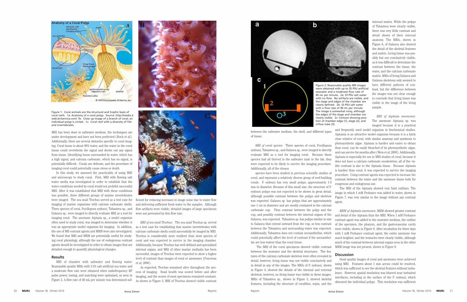
MURJ Author NameMURJ Reports20 Volume 18, Winter 2010 MURJAuthor Name MURJReports Volume 18, Winter 2010 21
MRI has been done in saltwater medium, the techniques are under development and have not been perfected (Boch et al). Additionally, there are several obstacles specific to coral imag-ing. Coral tissue is about 90% water, and the water in the coral tissue could overwhelm the signal and drown out any signal from tissue. Identifying tissue surrounded by water, which has a high signal, and calcium carbonate, which has no signal, is potentially difficult. Corals are delicate, and the procedure of imaging coral could potentially cause stress or death.
In this study, we assessed the practicality of using MRI and microscopy to study coral. First, MRI with flowing salt water media was investigated in order to establish that the water conditions needed by coral would not prohibit successful MRI. After it was established that MRI with these conditions was possible, three different groups of marine invertebrates were imaged. The sea snail Trochus served as a test case for imaging of marine organisms with calcium carbonate shells. Three species of coral, Pocillopora eydouxi, Tubastrea sp., and Galaxea sp., were imaged to directly evaluate MRI as a tool for imaging coral. The anemone Aiptasia sp., a model organism often used to study coral, was imaged to determine whether it was an appropriate model organism for imaging. In addition, the use of MR contrast agents and MRM were also investigated. We found that MRI and MRM are potentially useful for study-ing coral physiology, although the use of endogenous contrast agents should be investigated in order to obtain images that are detailed enough to quantify physiological changes.
ResultsMRI of chamber with saltwater and flowing water:
Reasonable quality MRIs with 3.5% salt artificial sea water and a moderate flow rate were obtained when radiofrequency RF pulse power, tuning, and matching were optimized, as seen in Figure 2. A flow rate of 40 mL per minute was determined suf-
ficient for reducing increases in image noise due to water flow and delivering sufficient fresh water to the samples. Although flow artifacts were visible, detailed images of large specimens were not prevented by this flow rate.
MRI of sea snail Trochus: The sea snail Trochus sp. served as a test case for establishing that marine invertebrates with calcium carbonate shells could successfully be imaged by MRI. Trochus is considerably more resilient than most species of coral and was expected to survive in the imaging chamber. Additionally, because Trochus has well-defined and specialized internal organs, and MRI of other marine mollusks has been successful, images of Trochus were expected to show a higher level of contrast than images of coral or anemones (Pouvreau et al, 2006).
As expected, Trochus remained alive throughout the pro-cess of imaging. Snail health was scored before and after imaging, and the scores of most specimens remained constant. As shown in Figure 3, MRI of Trochus showed visible contrast
Figure 1: Coral animals are the structural and trophic basis of coral reefs. 1a: Anatomy of a coral polyp. Source: http://media-2.web.britannica.com/ 1b: Close up image of a branch of coral; an individual polyp is circled. 1c: Coral reef with a diversity of fish and invertebrates.
Anna Simon
between the saltwater medium, the shell, and different types of tissue.
MRI of coral species: Three species of coral, Pocillopora
eydouxi, Tubastrea sp., and Galaxea sp., were imaged to directly evaluate MRI as a tool for imaging coral. Because these species had all thrived in the saltwater tank in the lab, they were expected to be likely to survive the imaging procedure. Additionally, all of the chosen
species have been studied in previous scientific studies of coral, and represent a relatively diverse group of reef-building corals. P. eydouxi has very small polyps, approximately 1-3 mm in diameter. Because of this small size, the structure of P. eydouxi polyps was not expected to be shown in great detail, although possible contrast between the polyps and skeleton was expected. Galaxea sp. has polyps that are approximately one 1 cm in diameter and are mostly contained in the calcium carbonate cup. Thus contrast between the polyp and the cup, and possibly contrast between the internal organs of the Galaxea, was expected. Tubastrea sp. has polyps similar in size to Galaxea that extend outward from the cup, so less contrast between the Tubastrea and surrounding water was expected. Additionally, Tubastrea does not contain zooxanthellae, which could potentially affect the level of contrast if the zooxanthel-lae are less watery than the coral tissue.
The MRI of the coral specimens showed visible contrast between the seawater and the skeletal structures. The fea-tures of the calcium carbonate skeleton were often revealed in detail; however, living tissue was not visible conclusively and in detail in any of the images. The MRIs of P. eydouxi, shown in Figure 4, showed the details of the internal and external skeleton; however, no living tissue was visible in these images. MRIs of Tubastrea sp., shown in Figure 5, showed skeletal features, including the structure of corallites, septa, and the
internal matrix. While the polyps of Tubastrea were clearly visible, there was very little contrast and detail shown of their internal anatomy. The MRIs, shown in Figure 6, of Galaxea also showed the detail of the skeletal features and matrix. Living tissue was pos-sibly but not conclusively visible, as it was difficult to determine the contrast between the tissue, the water, and the calcium carbonate matrix. MRIs of living Galaxea and Galaxea skeletons only seemed to have different patterns of con-trast, but the difference between the images was not clear enough to conclude that living tissue was visible in the image of the living sample.
MRI of Aiptasia anemones: The anemone Aiptasia sp. was imaged because it is a practical
and frequently used model organism in biochemical studies. Aiptaisia is an attractive model organism because it is a fairly close relative of coral, with similar anatomy and symbiosis to photosynthetic algae. Aiptasia is hardier and easier to obtain than coral, can be easily bleached of its photosynthetic algae, and can survive for months after (Weis et al, 2008). Additionally, Aiptasia is especially for use in MRI studies of coral, because it does not have a calcium carbonate exoskeleton; all of the vis-ible contrast is due to the Aiptasia tissue. Because Aiptasia is hardier than coral, it was expected to survive the imaging procedure. Using contrast agents was expected to increase the contrast between the water and the anemone tissue both for exogenous and endogenous use.
The MRI of the Aiptasia showed very faint outlines. The image in which 1 mM Prohance was added to water, shown in Figure 7, was very similar to the image without any contrast agent.
MRM of Aiptasia anemones: MRM showed greater contrast and detail of the Aiptasia than the MRI. When 1 mM Prohance contrast agent was added to the seawater medium, the outline of the specimen, the pharynx, and the gastrovascular cavity were visible, shown in Figure 8. After incubation for three days with 1 mM Prohance contrast agent, the entire anemone was much brighter, and the tentacles were clearly visible, although much of the contrast between internal organs seen in the other MRM image was not present, shown in Figure 9.
DiscussionGood quality images of coral and anemones were achieved
using MRI. Features about 1 mm across could be resolved, which was sufficient to see the skeletal features without turbu-lence. However, spatial resolution was blurred near turbulent interfaces, including at the surface of the P. eydouxi, which obscured the individual polyps. This resolution was sufficient
Figure 2: Reasonable quality MR images were obtained with up to 35 PSU artificial seawater and a moderate flow rate of 40 mL per minute. 2a: 33 PSU salt water with no flow. No artifacts are visible, and the stage and edges of the chamber are clearly defined. 2b: 33 PSU salt water with a flow rate of 40 mL per minute. The image is somewhat noisy, although the edges of the stage and chamber are clearly visible. 2c: Cartoon showing posi-tion of chamber edge (1), stage (2), and inflow tube (3).
Anna Simon

MURJ Author NameMURJ Reports22 Volume 18, Winter 2010 MURJAuthor Name MURJReports Volume 18, Winter 2010 23
to see anatomical details in Trochus, Galaxea, and Tubastrea. The imaging chamber allowed for imaging of samples about 5 cm across, 5 cm high, and 8 cm long; all of the desired samples easily fit or could be fragmented to fit into the chamber.
Much higher resolution was achieved using MRM; however, the available hardware limits the specimens that can be suc-cessfully imaged. Using MRM, features about 50 µm were resolved, such as the mesenterial filaments of the Aiptasia using exogenous contrast agents. Although the P. eydouxi pol-yps were too small to be meaningfully imaged with MRI, they could potentially be successfully imaged with MRM. However, in order to be imaged by MRM, a specimen must be relatively small and flat in order to be sufficiently close to the coil, which is an obstacle to using MRM for imaging much larger samples.
Spatial resolution in MRI and MRM was limited by the signal-to-noise (SN) ratio, which was lowered by the salt content of the artificial sea water. Although the salt content of the water did not prevent the acquisition of decent images, the level of noise was higher and the resolution was lower than what would have been expected from fresh water samples. A possible solution to this problem is to use fresh water model organisms to study marine organisms. While this approach is appropriate for some organisms, including snails, corals and anemones are intolerant of fresh water. Other approaches would be to further explore pulse sequences that would increase the signal to noise SN ratio or to use contrast agents to highlight certain tissues.
Even though the rate of water flow was relatively low (40 mm per minute or one chamber volume in 2.25 minutes), water flow patterns were visible, including at the interface between the coral and the water. This is important because exchange of water with the environment is a particularly important aspect of how coral and anemones interact with their environmen, and changes of the interaction with the environment can be a physiological response to changing environmental conditions.
MRI and MRM are particularly useful for studying marine organisms that are opaque, fragile, and have high water con-
tent. It is also useful for investigating organisms’ exchange of water with their surroundings. Initially, it was a concern that corals, widely regarded as delicate organisms, would not be able to survive MRI; however, coral, snails, and anemones all continued to thrive after MRI. The current methods for imaging coral and anemones, SEM and optical microscopy, both involve cutting or dehydrating the polyps, which often disturbs their delicate anatomy.
Water exchange is particularly important to the anatomy of snails, anemones, and corals. MRI and MRM allows for measurement and observations of water flow patterns. Even relatively slow flow patterns, such as the turbulence between the Galaxea coralites, shown in Figure 6, were visible. These patterns of flow and exchange with the environment might change in coral and anemones as a response to changing water conditions.
Although images sufficiently detailed to study physiologi-cal changes were not acquired, endogenous contrast agents may allow better resolution and visualization of important features. While coral tissue was visible in Tubastrea and pos-sibly Galaxea, the images were not clear or detailed enough to gain useful information about physiological changes. In the Aiptasia, exogenous contrast agents had essentially no effect on the amount of detail present in the image. This was most likely because even slight physical disruption or stress, such as moving the anemone dish from the tank to a table, causes the anemone to contract for a few minutes, as shown in Figure 10. This expels the water from its tissue, exchanging it with the environment, and essentially homogenizing the water every-where. All of the coral polyps in this study exhibited the same behavior, so exogenous contrast agents would most likely have little or no effect.
However, endogenous contrast agents were absorbed into Aiptasia tissue, allowing images of the tentacles to be acquired, as shown in Figure 9. This demonstrates that Prohance was dif-ferentially absorbed by different tissues in Aiptasia. Because of the anatomical similarities between Aiptasia and coral,
Figure 3: MR image of sea snail Trochus sp.. Visible contrast is apparent between the saltwater, snail shell, and different types of tissue within the snail. 3a: MR image of the snail. Notice the contrast within the body of the snail (different shades of gray in the area circled). 3b: Cartoon of MR image showing locations of snail shell (A), snail viscera (B), imaging platform (C), restraining net (D), turbulent water flow (E), and chamber edges (F).
Anna Simon
Prohance would likely be absorbed into certain coral tissues as well. Prohance, and other contrast agents, are absorbed at different rates into different tissues, so changing the time of exposure could potentially allow different tissues to be imaged in detail.
Methods:Animal husbandry: Eight species of coral, Montipora
sp., Turbinaria sp., Hydnophora sp., Pocillopora eyedouxa, Pocillopora damicornis, two varieties of Acropora sp., and Galaxea sp.; sea anemones Aiptasia sp.; and sea snail Trochus sp. were obtained from the Aquarium of the Pacific at Long Beach. The coral and Trochus were kept in a 14 gallon seawater X tank containing live rock and gravel substrate, with a 12 hr light-dark cycle. Water temperature was maintained at 26-28° C, salinity at 33-35 PSU, pH at 8.0-8.2, nitrate concentration less than 20 ppm, and nitrite and ammonia at undetectable levels. Water quality parameters were monitored 4-6 times per week, 10-30% of the tank water was replaced 2-4 times per week, and the tank was scrubbed and hydrovaced about once per week to maintain acceptable water quality.
Because of their tendency to sting other organisms, Aiptasia were kept in a separate, seven gallon tank; in order to be able to move them easily, the Aiptasia were each kept in separate Bakelite dishes. Water temperature was maintained at 24-26° C, salinity at 30-35 PSU, pH at 8.2-8.4, nitrate concentration at less than 50 ppm, and ammonia and nitrite concentration less than 1.0 ppm. Water quality parameters were measured 3-4 times per week, and 20-40% of the tank water was replaced once per week in order to maintain acceptable water quality.
Chamber construction: A holding chamber was constructed in order to hold the organism in a favorable environment during imaging. A cylindrical chamber was constructed out of Ultem 1000 X , with a diameter of approximately 70 mm and a length of approximately 180 mm. In order to create flow through the chamber, inflow and outflow tubes, approximately seven meters long were attached to an end of the chamber. The inflow tube
was attached to a Hagen Aqua Clear Power Head pump, in order to generate flow. A stage was placed near the middle of the chamber as a flat surface on which to hold the organism.
Saltwater and chamber MR imaging: The chamber was imaged with flowing and stationary fresh and salt water. This was done to establish the effects of flowing water and salt water on an image, as well as find artifacts from the chamber itself. Tuning, matching, and radiofrequency RF pulse power were optimized.
Coral MR imaging: Three species of coral, Galaxea sp., Tubastrea sp., and Pocillopora eyedouxi, were imaged in order to assess the practicality of using MR technology to image coral in a relatively diverse variety of samples. After a three week adjustment upon arriving at Caltech, the coral were frag-mented into smaller pieces with a hammer and dremel, accord-ing to suggestions by the staff at the Aquarium of the Pacific. This was done to obtain pieces small enough to fit in the imaging chamber. The water flowing through the chamber was freshly made before imaging, and had a salinity of 33-35 PSU, a temperature of 21-26°C, and a flow rate of 40 mm/minute. The samples were placed directly on the imaging chamber, and the imaging procedures lasted 1-3 hours. Tuning, matching, and RF pulse power were optimized. In order to assess the effect of the imaging procedure on the coral, the coral were visually inspected before and after imaging. The pulse sequences used included Rapid Acquision Relaxation Enhancement RARE , with 11.338 echo time, repetition time 4000.000 ms, and 10 averages.
Trochus MR imaging: Trochus was imaged in order to con-firm the equipment’s ability to successfully image marine inver-tebrates and explore marine snail anatomy and MRI of marine mollusks. After a three-week adjustment upon arriving at Caltech, individual snails were imaged. The snails were placed directly on the platform in the imaging chamber; in order to prevent motion while imaging, mesh was placed over the top of the snail and secured in place with rubber bands. The water flowing through the chamber was freshly made before imaging,
Figure 4: MR image of P. eyedouxi. Contrast is visible between calcium carbonate skeleton and water, and the verrucae, projec-tions on the surface of the coral, are visible, but no living tissue is visible in this image. 4a: MR image of P. eyedouxi. Notice the contrast between the skeleton (black) and the internal water (white), and the visible details of the skeleton in the circled area. However, no living tissue, which is located on the outside of the coral, is visible. 4b: Cartoon showing the location of internal cal-cium carbonate skeleton (A), outline of coral fragment (B), verruca projection (C), stage (D), and chamber edges (E).
Anna Simon
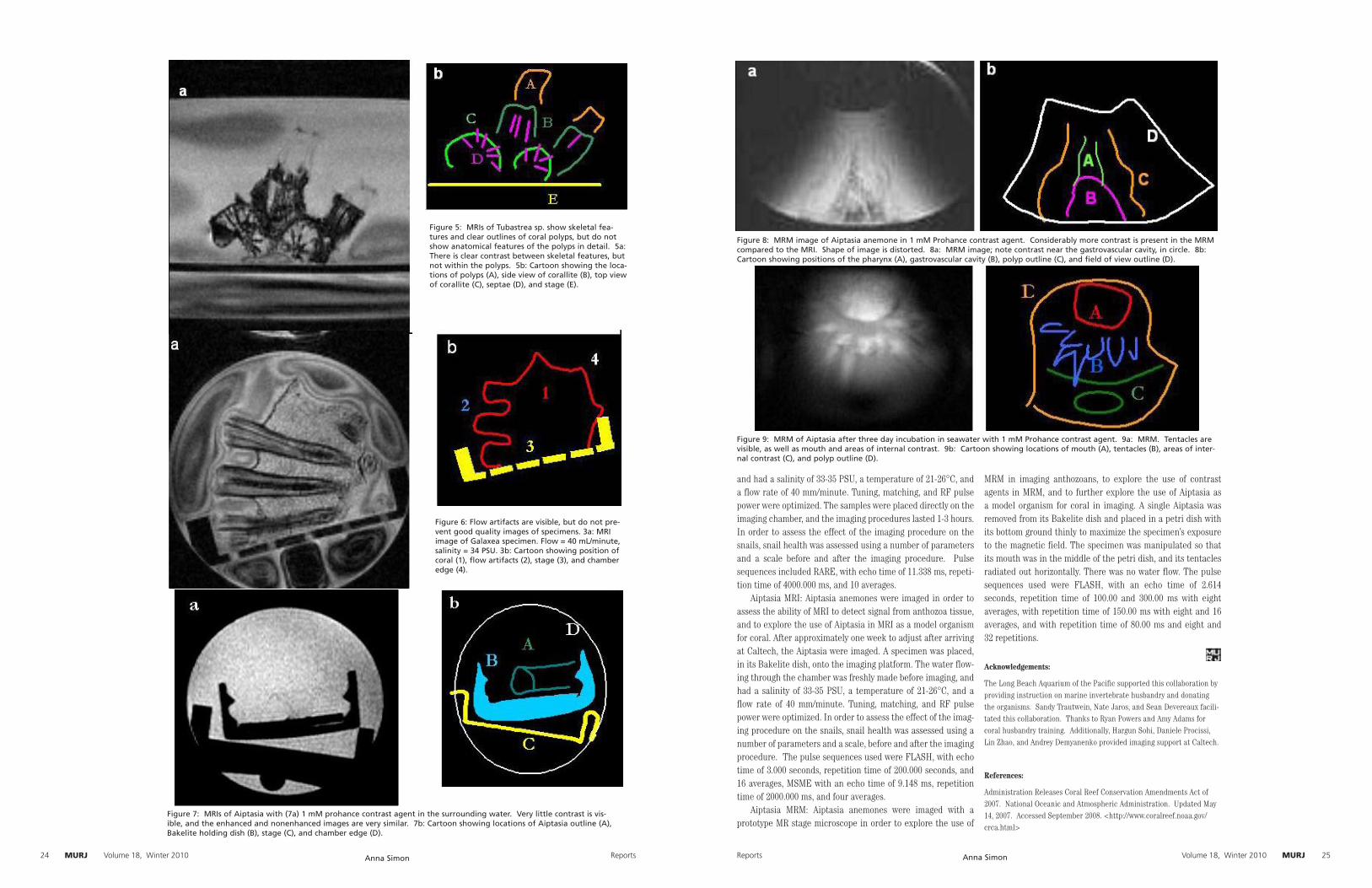
MURJ Author NameMURJ Reports24 Volume 18, Winter 2010 MURJAuthor Name MURJReports Volume 18, Winter 2010 25
Figure 5: MRIs of Tubastrea sp. show skeletal fea-tures and clear outlines of coral polyps, but do not show anatomical features of the polyps in detail. 5a: There is clear contrast between skeletal features, but not within the polyps. 5b: Cartoon showing the loca-tions of polyps (A), side view of corallite (B), top view of corallite (C), septae (D), and stage (E).
Figure 6: Flow artifacts are visible, but do not pre-vent good quality images of specimens. 3a: MRI image of Galaxea specimen. Flow = 40 mL/minute, salinity = 34 PSU. 3b: Cartoon showing position of coral (1), flow artifacts (2), stage (3), and chamber edge (4).
Figure 7: MRIs of Aiptasia with (7a) 1 mM prohance contrast agent in the surrounding water. Very little contrast is vis-ible, and the enhanced and nonenhanced images are very similar. 7b: Cartoon showing locations of Aiptasia outline (A), Bakelite holding dish (B), stage (C), and chamber edge (D).
Anna Simon
and had a salinity of 33-35 PSU, a temperature of 21-26°C, and a flow rate of 40 mm/minute. Tuning, matching, and RF pulse power were optimized. The samples were placed directly on the imaging chamber, and the imaging procedures lasted 1-3 hours. In order to assess the effect of the imaging procedure on the snails, snail health was assessed using a number of parameters and a scale before and after the imaging procedure. Pulse sequences included RARE, with echo time of 11.338 ms, repeti-tion time of 4000.000 ms, and 10 averages.
Aiptasia MRI: Aiptasia anemones were imaged in order to assess the ability of MRI to detect signal from anthozoa tissue, and to explore the use of Aiptasia in MRI as a model organism for coral. After approximately one week to adjust after arriving at Caltech, the Aiptasia were imaged. A specimen was placed, in its Bakelite dish, onto the imaging platform. The water flow-ing through the chamber was freshly made before imaging, and had a salinity of 33-35 PSU, a temperature of 21-26°C, and a flow rate of 40 mm/minute. Tuning, matching, and RF pulse power were optimized. In order to assess the effect of the imag-ing procedure on the snails, snail health was assessed using a number of parameters and a scale, before and after the imaging procedure. The pulse sequences used were FLASH, with echo time of 3.000 seconds, repetition time of 200.000 seconds, and 16 averages, MSME with an echo time of 9.148 ms, repetition time of 2000.000 ms, and four averages.
Aiptasia MRM: Aiptasia anemones were imaged with a prototype MR stage microscope in order to explore the use of
MRM in imaging anthozoans, to explore the use of contrast agents in MRM, and to further explore the use of Aiptasia as a model organism for coral in imaging. A single Aiptasia was removed from its Bakelite dish and placed in a petri dish with its bottom ground thinly to maximize the specimen’s exposure to the magnetic field. The specimen was manipulated so that its mouth was in the middle of the petri dish, and its tentacles radiated out horizontally. There was no water flow. The pulse sequences used were FLASH, with an echo time of 2.614 seconds, repetition time of 100.00 and 300.00 ms with eight averages, with repetition time of 150.00 ms with eight and 16 averages, and with repetition time of 80.00 ms and eight and 32 repetitions.
Acknowledgements:
The Long Beach Aquarium of the Pacific supported this collaboration by providing instruction on marine invertebrate husbandry and donating the organisms. Sandy Trautwein, Nate Jaros, and Sean Devereaux facili-tated this collaboration. Thanks to Ryan Powers and Amy Adams for coral husbandry training. Additionally, Hargun Sohi, Daniele Procissi, Lin Zhao, and Andrey Demyanenko provided imaging support at Caltech.
References:
Administration Releases Coral Reef Conservation Amendments Act of 2007. National Oceanic and Atmospheric Administration. Updated May 14, 2007. Accessed September 2008. <http://www.coralreef.noaa.gov/crca.html>
Figure 8: MRM image of Aiptasia anemone in 1 mM Prohance contrast agent. Considerably more contrast is present in the MRM compared to the MRI. Shape of image is distorted. 8a: MRM image; note contrast near the gastrovascular cavity, in circle. 8b: Cartoon showing positions of the pharynx (A), gastrovascular cavity (B), polyp outline (C), and field of view outline (D).
Figure 9: MRM of Aiptasia after three day incubation in seawater with 1 mM Prohance contrast agent. 9a: MRM. Tentacles are visible, as well as mouth and areas of internal contrast. 9b: Cartoon showing locations of mouth (A), tentacles (B), areas of inter-nal contrast (C), and polyp outline (D).
Anna Simon

MURJ Author NameMURJ Reports26 Volume 18, Winter 2010 MURJMURJReports Volume 18, Winter 2010 27
Belda-Baillie, C. A., B. K. Baillie, et al. (2002). “Specificity of a Model Cnidarian Dinoflagellate Symbiosis.” The Biological Bulletin 202: 74-85.
Berntson, E. A., S. C. France, et al. (1998). “Phylogenetic Relationships within the Class Anthozoa (Phylum Cnidaria) Based on Nuclear 18S rDNA Sequences.” Molecular Phylogenetics and Evolution 13(2): 417-433.
Black, N. A., R. Voellmy, et al. (1995). “Heat Shock Protein Induction in Montastraea faveolata and Aiptasia pallida Exposed to Elevated Temperatures.” The Biological Bulletin 188(3): 234-240.
Blackband, S. and M. Stoskopf (1990). “In vivo nuclear magnetic resonance imaging and spectroscopy of aquatic organisms.” Magnetic Resonance Imaging 8(2): 191-198.
Bock, C., M. Frederich, et al. (2001). “Simultaneous observations of haemolymph flow and ventilation in marine spider crabs at different temperatures: a flow weighted MRI study.” Magnetic Resonance Imaging 19: 1113-1124.
Bock, C., G. J. Lurman, et al. (2008). “Muscle bioenergetics of speeding fish: In vivo 31P-NMR studies in a 4.7 T MR scanner with an integrated swim tunnel.” Concepts in Magnetic Resonance Part B-Magnetic Resonance Engineering 33B(1): 62-73.
Bock, C., F. J. Sartoris, et al. (2002). “In vivo MR spectroscopy and MR imaging on non-anaesthetized marine fish: techniques and first results.” Magnetic resonance imaging 20: 165-172.
Bock, C., F. J. Sartoris, et al. (2001). “Temperature dependent pH regu-lation in stenothermal Antarctic and eurythermal temperate eelpout (Zoarcidae): an in vivo NMR study.” Polar Biology 24(11): 869-874.
Chen, C. A., D. M. Odorico, et al. (1995). “Systematic Relationships within the Anthozoa (Cnidaria: Anthozoa) Using the 5 -end of the 28S rDNA.” Molecular Phylogenetics and Evolution 4(2): 175-183.
Chen, M. C., Y. M. Cheng, et al. (2004). “Molecular cloning of Rab5 (ApRab5) in Aiptasia pulchella and its retention in phagosomes har-boring live zooxanthellae.” Biochemical and Biophysical Research Communications 324(3): 1024-1033.
Fautin, D. G. and S. L. Romano. (2001). “Anthozoa. Sea Anemones, Corals, Sea Pens.” The Tree of Life Web Project Retrieved July 9, 2008, 2008.
Gates, R. D., G. Baghdasarian, et al. (2004). “Temperature Stress Causes Host Cell Detachment in Symbiotic Cnidarians: Implications for Coral Bleaching.” The Biological Bulletin 182(2): 324-332.
Hoegh-Guldberg, O., P. J. Mumby, et al. (2007). “Coral reefs under rapid climate change and ocean acidification.” Science 5857(318): 1737-1742.
Kerr, A. M. (2005). “Molecular and morphological supertree of stony corals (Anthozoa: Scleractinia) using matrix representation parsimony.” Biological Reviews 80(80): 543-558.
Key, R.M.; Kozyr, A.; Sabine, C.L., et al. (2004). “A global ocean carbon climatology: Results from GLODAP”. Global Biogeochemical Cycles 18: GB4031
Lesser, M. P. and J. M. Shick (1989). “ Effects of irradiance and ultravio-let radiation on photoadaptation in the zooxanthellae of Aiptasia pallida: primary production, photoinhibition, and enzymic defenses against oxy-gen toxicity.” Marine Biology 102(2): 243-255.
Linden, A. V. d., M. Vanaudenhovea, et al. (1999). “Osmoregulation of the common carp (Cyprinus carpio) when exposed to an osmotic chal-lenge assessed in-vivo and non-invasively by diffusion- and T2-weighted magnetic resonance imaging.” Comparative Biochemistry and Physiology -Part A: Molecular & Integrative Physiology 124(4): 343-352.
Linden, A. V. d., M. Verhoye, et al. (2004). “The strengths of in vivo magnetic resonance imaging (MRI) to study environmental adaptational physiology in fish.” Magnetic resonance materials in physics biology and medicine 17: 236-248.
McCutcheon, A. L., G. S. Kamalikannangara, et al. (2004). “Preliminary analysis of pore distributions using NMR in natural coral and hydro-thermally prepared hydroxyapatite.” Journal of Materials Science 39: 5711-5717.
Muller-Parker, G., C. B. Cook, et al. (1990). “Feeding affects phosphate fluxes in the symbiotic sea anemone Aiptasia pallida.” Marine Ecology Progress Series 60: 283-290.
Neustadter, D. M., R. F. Drushel, et al. (2002). “Kinematics of the buccal mass during swallowing based on magnetic resonance imaging in intact, behaving Aplysia californica.” Journal of Experimental Biology 205(939-958).
Perez, S., C. Cook, et al. (2001). “The role of symbiotic dinoflagellates in the temperature-induced bleaching response of the subtropical sea anemone Aiptasia pallida.” Journal of Experimental Marine Biology and Ecology 256(1): 1-14.
Perez, S. and V. Weis (2006). “Nitric oxide and cnidarian bleaching: an eviction notice mediates breakdown of a symbiosis.” Journal of Experimental Biology 209: 2804-2810.
Pouveau, S., M. Rambeau, et al. (2006). “Investigation of marine bivalve morphology by in vivo MR imaging: First anatomical results of a promis-ing technique.” Aquaculture 259(1-4): 415-423.
Salceda, E., J. Pérez-Castells, et al. (2007). “CgNa, a type I toxin from the giant Caribbean sea anemone Condylactis gigantea shows structural similarities to both type I and II toxins, as well as distinctive structural and functional properties.” The Biochemical
Journal 406(1): 67-76.
Sawyer, S. J. and L. Muscatine (2001). “Cellular mechanisms underlying temperature-induced bleaching in the tropical sea anemone Aiptasia
pulchella.” The Journal of Experimental Biology 204: 3443-3456.
Weis, V. M., S. K. Davy, et al. (2008). “Cell biology in model sys-tems as the key to understanding corals.” Trends in Ecology and Evolution 23(7): 369-376.
William S. Clayton, J. (1984). “Pedal laceration by the anemone Aiptasia pallida.” Marine Ecology Progress Series 21: 75-80.
Won, J., B. Rho, et al. (2004). “ A phylogenetic study of the Anthozoa (phylum Cnidaria) based on morphological and molecu-lar characters.” Coral Reefs 20(1): 39-50
Figure 10: Aiptasia anemones expel water from their tissues when disturbed, decreasing in size and exchanging water with their envi-ronment. Green dishes in figures are the same size. 10a: Undisturbed Aiptasia anemone. 10b: Aiptasia after its dish was physically moved.
Anna Simon
UV Irradiation Experiments in Marine Microbe Prochlorococcus
Brianne Holmbeck
Ed. Marcia Osburne
Background InformationImagine one of the smallest cells possible, just one. Now paint it green and surround it with chlorophyll, just inside the cell
wall. Give it a tiny coil of DNA. Viola! Okay, so there is more to Prochlorococcus than that. This cyanobacterium is the most abundant photosynthetic marine
microbe found in the tropical and sub-tropical open oceans. It contains the smallest known genome of any oxygenic phototroph and yet manages to remain alive at depths ranging from the ocean’s surface to 100-200m below sea level. Prochlorococcus cells growing in the laboratory tend to replicate about once per day, mimicking those in the wild, which follow a circadian schedule. Prochlorococcus strains alone contribute up to 50% of the chlorophyll found in the tropical and sub-tropical regions of the open ocean and are responsible for up to 48% of the primary production, or the synthesis of organic compounds from inorganic ones, in these areas (Johnson et al., 2006). For these and other reasons, research in the Chisholm Laboratory at MIT focuses on using Prochlorococcus as a model system to study a range of topics, from genomics to widespread ocean ecology.
Prochlorococcus cells use two types of chlorophyll to recognize sunlight and convert it into energy, thanks to light-harvesting proteins in their antennae. The antennae and two forms of chlorophyll are extremely important to the cells’ survival at such great depths in the oceanic water column, where the light level is exceedingly low. The light level, or irradiance, is considered to be 100% at the surface of the ocean. At 100-200m deep, irradiance can be as low as 1%, and yet Prochlorococcus still manages to survive at even these great depths.
The species has been divided into phylogenetically distinct high-light-adapted and low-light-adapted ecotypes that gener-ally partition according to light availability in the environment (Pandhal, et al. 2007). Strain MED4 is a commonly studied high-light strain, whereas MIT9313 thrives in low light. Rocap et al. (2003) investigated many of the physical, physiological, and genomic differences between these two strains. MED4 has a smaller genome—1,657,990 base pairs encoding approximately 1,716 genes—making it the oxygenic phototroph with the smallest known sequenced genome. MIT9313, on the other hand, has 2,410,873 base pairs encoding 2,275 genes. The two strains share 1,352 genes, but MED4 has 364 genes that MIT9313 does not have, and 923 of the genes in MIT9313 do not appear in MED4.
Some, or perhaps even all, of the genes specific to each ecotype may represent the adaptation of these types to their ecologi-cal niche. For example, many of the MED4 genes absent from MIT9313 are connected to the use or absorption of light. MED4 has twenty-two potential high-light-inducible genes whereas MIT9313 has only nine. These ecotypes also possess different numbers and kinds of light-harvesting antenna proteins, which are encoded by pcb genes. Low-light ecotypes generally have a higher number of pcb genes, suggesting that this allows low-light strains to thrive as at lower depths in the water column by enhancing their ability to gather light. However, MIT9313 has only two pcb genes, suggesting that this hypothesis may not be true for all low-light strains (Ting et al. 2002).
In addition, a photolyase gene has been found in MED4 but not in MIT9313. Photolyase is a DNA repair protein that functions only in the presence of visible light (Sancar 1987). In Escherichia coli (E. coli) bacteria, UV light absorption creates cyclobutane pyrimidine dimers, which cause lesions in DNA (MacGregor 1999). Photolyase repairs double-stranded DNA wherever lesions resulting from pyrimidine dimers are present, using the energy from photons to reverse these lesions by splitting the pyrimidine dimer. E. coli also uses the SOS response system, which involves the recA and recF DNA repair pathways to repair DNA damage (“dark repair”). This system is error-prone, whereas the photolyase repair system is error-free. SOS genes have homologues in Prochlorococcus, but their role in DNA repair in this oceanic bacterium is not presently known.

MURJ Author NameMURJ Reports28 Volume 18, Winter 2010 MURJAuthor Name MURJReports Volume 18, Winter 2010 29
The Research and Laboratory WorkIn previous experiments with Prochlorococcus, my mentor
and I found that MED4 was more resistant to UV irradiation than the low-light strain MIT9313. We then became interested in how the two different ecotypes handle UV irradiation at the genomic and gene expression levels, given their different loca-tions in the water column. Traditional methods used to study these processes involve isolating mutants. However, obtaining and analyzing mutant strains of Prochlorococcus is exceedingly difficult, as robust genetic transfer systems for Prochlorococcus have not been developed as of yet. Preliminary conjugation experiments, using E. coli as a donor, (Tolonen et al., 2006) have been met with limited success; and transformation and transduction have not yet been achieved in a laboratory setting, despite evolutionary evidence for phages transferring genes across Prochlorococcus ecotypes, especially genes involved in Photosystem II (Lindell 2005). Finally, it is difficult to obtain colonies of Prochlorococcus on Petri plates and to isolate mutants using such an approach.
We therefore devised alternate means to obtain a mutant strain of MED4 that is resistant to UV irradiation (MED4uvr) by subjecting wild type MED4 (MED4wt) to multiple rounds of UV irradiation. Following UV irradiation of MED4wt, MED4uvr and MIT9313wt, the UV irradiated cells, along with their un-irradiated controls, were studied using microarray expression analysis and genetic comparisons to determine which genes are involved in Prochlorococcus irradiation sensitivity and to com-pare the new information with previous research concerning UV irradiation repair pathways in other organisms. We also ana-lyzed the differences in gene expression between the high-light and low-light ecotypes in order to better understand the adap-tations leading to niche differentiation in Prochlorococcus. Subsequently, a time-course analysis of the expression of relevant repair genes was carried out in MED4uvr and MED4wt using qPCR analysis of specific repair genes.
The DiscoveryMultiple rounds of UV mutagenesis led to the isolation of
a mutant strain (MED4uvr) that is more resistant to UV irra-diation than the MED4wt parent. The MED4uvr mutant was able to survive at the same dose of UV irradiation that killed the parent strain, MED4wt. Following irradiation, growth of the resistant mutant was initially stimulated by the UV light for a short period of time, after which the cells settled into a normal growth rate. We found that the mutant strain was more resistant to UV light in both the UV-C and UV-B ranges, but not in the UV-A range. UV-C and UV-B are both known to damage DNA by causing pyrimidine dimer formation, whereas UV-A does not damage DNA by this mechanism.
Interestingly, we also found that the high-light MED4wt strain was more resistant to UV irradiation than the low-light MIT9313 strain. We hypothesize that this difference is likely due to an evolutionary niche adaptation: UV light is hardly detectable at the depth at which MIT9313 lives (Steglich et al. 2006), whereas MED4 lives much closer to the surface of the ocean.
Microarray expression analysis of the MED4uvr mutant of Prochlorococcus revealed that three genes were significantly upregulated as compared with MED4wt. These genes appeared
to be constitutively expressed, as they displayed equally high levels of expression with and without thirty seconds of UV irradiation. Two of the three upregulated genes—one encod-ing a photolyase (phrB) and the other encoding a NUDIX hydrolase (mutT)—have been shown bioinformatically to form an operon. Both of these gene products appear to have a prominent impact on UV sensitivity and irradiation recov-ery. Photolyase works only in the presence of visible light to repair pyrimidine dimers in DNA, which can result from UV irradiation-induced damage. The enzyme uses photons from visible light as an energy source. Interestingly, the photolyase gene, phrB, is found only in MED4, not in MIT9313, supporting the idea that each of the two strains is evolutionarily adapted to its particular niche in the ocean. In fact, most of the low-light strains of Prochlorococcus do not contain a photolyase gene, whereas every high-light strain isolated thus far does contain this gene, further validating the idea of global niche adaptation.
NUDIX hydrolases, found in both the high-light and the low-light strains of Prochlorococcus, are also involved in DNA repair. This type of enzyme hydrolyzes a specific aberrant form of dGTP, which is most often formed by ionizing radiation such as UV and thereby inhibits the incorporation of the aberrant form into DNA. Previous experiments have shown that the nudix hydrolase gene was slightly upregulated in MED4wt after the cells were shifted from the dark to blue, white, or high levels of light (Steglich et al. 2006). In addition, both of these genes were downregulated in response to nitrogen starvation (Tolonen et al. 2006), which may reflect an overall downshift in DNA synthesis when nitrogen is limiting.
In order to determine the genetic lesion responsible for the phenotype in the MED4uvr mutant, we sequenced the entire genome of this strain and compared it with the sequence of the wild type parent strain. Remarkably, despite the many doses of UV irradiation that the mutant was subjected to, we found that the mutant strain had only a single mutation: a single-base deletion in the non-coding region just upstream of the nudix hydrolase-photolyase operon. We hypothesize that this mutation results in altered regulation of the expression of this operon, causing the genes to be overexpressed.
The SOS genes of E. coli and many other organisms encode a cellular response to DNA damage, such as that caused by UV irradiation. Many of these genes are present in Prochlorococcus genomes. In a time-course experiment to examine the expression of this gene, we found that the RecA protein was significantly upregulated within an hour after UV irradiation of both the MED4wt and MED4uvr strains. RecA is the primary SOS DNA repair protein used in E. coli. However, as mentioned above, SOS repair of DNA damage is error-prone. The photolyase protein may be a more effective alternative in Prochlorococcus for two reasons: 1) it is energized by visible light, which is in abundant supply for Prochlorococcus (but not for E. coli) and 2) it is error-free and helps to maintain genetic stability for the organism.
ImplicationsFuture work on this project will involve molecular biologi-
cal studies on the effects of different doses of UV irradiation at a wavelengths experienced by cells in the open ocean. Additionally, selecting for a UV-resistant mutant of a low-light
Brianne Holmbeck Brianne Holmbeck
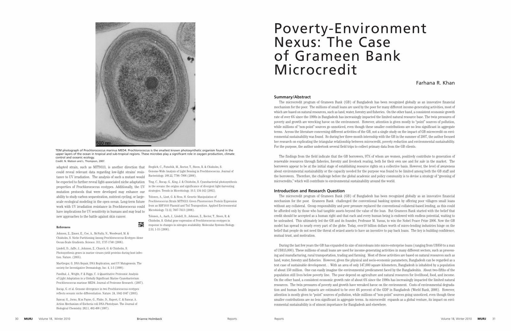
MURJ Author NameMURJ Reports30 Volume 18, Winter 2010 MURJMURJReports Volume 18, Winter 2010 31
adapted strain, such as MIT9313, is another direction that could reveal relevant data regarding low-light strains’ resis-tance to UV irradiation. The analysis of such a mutant would be expected to further reveal light-associated niche adaptation properties of Prochlorococcus ecotypes. Additionally, the UV mutation protocols that were developed may enhance our ability to study carbon sequestration, nutrient cycling, or large-scale ecological modeling in the open ocean. Long-term future work with UV irradiation resistance in Prochlorococcus could have implications for UV sensitivity in humans and may lead to new approaches to the battle against skin cancer.
References
Johnson, Z., Zinser, E., Coe, A., McNulty, N., Woodward, M. & Chisholm, S. Niche Partitioning Among Prochlorococcus Ecotypes Alone Ocean-Scale Gradients. Science. 311, 1737-1740 (2006).
Lindell, D., Jaffe, J., Johnson, Z., Church, G. & Chisholm, S. Photosynthesis genes in marine viruses yield proteins during host infec-tion. Nature. (2005).
MacGregor, G. DNA Repair, DNA Replication, and UV Mutagenesis. The society for Investigative Dermatology, Inc. 4, 1-5 (1999).
Pandhal, J., Wright, P. & Biggs, C. A Quantitative Proteomic Analysis of Light Adaptation in a Globally Significant Marine Cyanobacterium Prochlorococcus marinue MED4. Journal of Proteome Research. (2007).
Rocap, G. et al. Genome divergence in two Prochlorococcus ecotypes reflects oceanic niche differentiation. Nature. 24, 1042-1047 (2003).
Sancar, G., Jorns, M.m Payne, G., Fluke, D., Rupert, C. & Sancar, A. Action Mechanism of Escheria coli DNA Photolyase. The Journal of Biological Chemistry. 262.1, 492-498 (1987).
Steglich, C., Futschik, M., Rector, T., Steen, R. & Chisholm, S. Genome-Wide Analysis of Light Sensing in Prochlorococcus. Journal of Bacteriology. 188.22, 7796–7806 (2006).
Ting, C., Rocap, G., King, J. & Chisholm, S. Cyanobacterial photosynthesis in the oceans: the origins and significance of divergent light-harvesting strategies. Trends in Microbiology. 10.3, 134-142 (2002).
Tolonen, A., Liszt, G. & Hess, H. Genetic Manipulation of Prochlorococcus Strain MIT9313: Green Fluorescence Protein Expression from an RSF1010 Plasmid and Tn5 Transposition. Applied Environmental Microbiology. 72.12, 7607-7613 (2006).
Tolonen, A., Aach, J., Lindell, D., Johnson, Z., Rector, T., Steen, R. & Chisholm, S. Global gene expression of Prochlorococcus ecotypes in response to changes in nitrogen availability. Molecular Systems Biology. 2.53, 1-15 (2006).
Poverty-Environment Nexus: The Case of Grameen Bank Microcredit
Farhana R. Khan
Summary/AbstractThe microcredit program of Grameen Bank (GB) of Bangladesh has been recognized globally as an innovative financial
mechanism for the poor. The millions of small loans are used by the poor for many different income-generating activities, most of which are based on natural resources, such as land, water, forestry and fisheries. On the other hand, a consistent economic growth rate of over 6% since the 1990s in Bangladesh has increasingly impacted the limited natural resource base. The twin pressures of poverty and growth are wrecking havoc on the environment. However, attention is given mostly to “point” sources of pollution, while millions of “non-point” sources go unnoticed, even though these smaller contributions are no less significant in aggregate terms. Across the literature concerning different activities of the GB, not a single study on the impact of GB micrecredit on envi-ronmental sustainability was found. So during her three-month internship with the GB in the summer of 2007, the author focused her research on explicating the triangular relationship between microcredit, poverty reduction and environmental sustainability. For the purpose, the author undertook several field trips to collect primary data from the GB clients.
The findings from the field indicate that the GB borrowers, 97% of whom are women, positively contribute to generation of renewable resources through fisheries, forestry and livestock rearing, both for their own use and for sale in the market. The borrowers appear to be at the initial stage of establishing resource rights on a collective basis. However, the level of awareness about environmental sustainability or the capacity needed for the purpose was found to be limited among both the GB staff and the borrowers. Therefore, the challenge before the global academic and policy community is to devise a strategy of “greening of microcredits,” which will contribute to environmental sustainability around the world.
Introduction and Research QuestionThe microcredit program of Grameen Bank (GB) of Bangladesh has been recognized globally as an innovative financial
mechanism for the poor. Grameen Bank challenged the conventional banking system by offering poor villagers small loans without any collateral. Group responsibility and peer pressure replaced the conventional collateral-based lending, as this could be afforded only by those who had tangible assets beyond the value of the loan. But Grameen Bank started with the belief that credit should be accepted as a human right and that each and every human being is endowed with endless potential, waiting to be unleashed. This ultimately led the GB and its founder, Professor M. Yunus, to win the Nobel Peace Prize 2006. Now the GB model has spread to nearly every part of the globe. Today, over10 billion dollars worth of micro-lending industries hinge on the belief that people do not need the threat of seized assets to have an incentive to pay back loans. The key is building confidence, mutual trust, and motivation.
During the last few years the GB has expanded its size of microloans into micro-enterprise loans (ranging from US$50 to a max of US$15,000). These millions of small loans are used for income-generating activities in many different sectors, such as process-ing and manufacturing, rural transportation, trading and farming. Most of these activities are based on natural resources such as land, water, forestry and fisheries. However, given the physical and socio-economic parameters, Bangladesh can be regarded as a test case of sustainable development. . With an area of only 147,000 square kilometers, Bangladesh is inhabited by a population of about 150 million. One can easily imagine the environmental predicament faced by the Bangladeshis. About two-fifths of the population still lives below poverty line. The poor depend on agriculture and natural resources for livelihood, food, and income. On the other hand, a consistent economic growth rate of about 6% since the 1990s has increasingly impacted the limited natural resources. The twin pressures of poverty and growth have wreaked havoc on the environment. Costs of environmental degrada-tion and human health impacts are estimated to be over 4% percent of the GDP in Bangladesh (World Bank, 2006). However, attention is mostly given to “point” sources of pollution, while millions of “non-point” sources going unnoticed, even though these smaller contributions are no less significant in aggregate terms. As microcredit expands as a global venture, its impact on envi-ronmental sustainability is of utmost importance for Bangladesh and elsewhere.
TEM photograph of Prochlorococcus marinus MED4. Prochlorococcus is the smallest known photosynthetic organism found in the upper layers of the ocean in tropical and sub-tropical regions. These microbes play a significant role in oxygen production, climate control and oceanic ecology. Credit: N. Watson and L. Thompson, 2007.
Brianne Holmbeck

MURJ Author NameMURJ Reports32 Volume 18, Winter 2010 MURJAuthor Name MURJReports Volume 18, Winter 2010 33
As an intern with the GB from June to August 2007, this author started learning first hand about the GB and how it works. Across the literature concerning different activities of the GB, not a single comprehensive study on the impact of Grameen Bank microcredit on environmental sustainability in Bangladesh was found. Virtually no research has been done on the triangular relationship between microcredit, poverty reduc-tion, and environmental sustainability. So it seemed obvious to ask: how are the millions of activities carried out using micro-loans and a growing number of micro-enterprise loans affecting the environment? Although the microcredit program is aimed at poverty alleviation, is it promoting or hindering environmen-tal protection? What is the approach of the GB to this issue? Can environmental sustainability be enhanced through the use of micro credits, i.e., can there be a “greening of microcredits”? This research paper focuses on explicating this relationship in the context of Grameen Bank.
MethodologyThis research is based on both primary and secondary
sources of information. Published literature on poverty-environment relationship and on GB activities serves as the secondary sources of information. However, no study has been found as yet on the environmental impact of microcredit use by Grameen borrowers in different types of economic activi-ties. Therefore, as a part of internship, the author undertook several field trips to collect primary data from the Grameen clients. This intern discussed the issues of how microcredits are used by the poor, in what activities they invest, how they do it when their activities are related to natural resources, and other related questions. The field visits applied the methods of individual and group discussions and completing structured questionnaires. Given the time and resource constraints, the qualitative method has been used, as the study was meant as an exploratory one. It is hoped that this research will lay the ground for further studies that will be larger in scale and more in-depth than the present one
Poverty-Environment LinkageSustainable development (SD) has been defined by the
World Commission on Environment and Development (WCED) in its report, Our Common Future, as “development that meets the needs of the present without compromising the needs of the future generations” (WCED, 1987). Yet this definition sounds vague, as the needs of the present generation widely differ among countries and groups of people and the needs of the future generations are unknown. Therefore, the WCED put focus on satisfaction of the needs of the world’s poor, based on a less material and less energy-intensive economic growth (WCED Report, 1987). As far as future generations are con-cerned, the current generation is supposed to leave the world behind in a state at least as good as the one inherited from the previous generation. Therefore, the value of this definition is that it looks into the intra- and inter-generational equity. As the concept of SD is difficult to operationalize, the notion of environmental sustainability has come into common use. The use of natural resources for development without degradation is the very foundation of SD.
Over the years, the notion of poverty has been expanded to encompass both the financial and non-financial dimensions of deprivation. These dimensions include the lack of income and other material goods, lack of access to basic social services, and lack of personal security, rights and voice (World Bank, 2002).
There are different perspectives on poverty-environment relationship. The WCED report argues that poverty is the root of environmental degradation in the developing coun-tries, because the poor are forced to earn their livelihood from exploiting natural resources in their immediate vicinity. Poverty, by its very nature, causes resource depletion and deg-radation, which then further exacerbates poverty in a vicious cycle. Sometimes, poor communities continue their exploita-tion of natural resources such as wood, despite the fact that this exacerbates the situation in the long term because they have no other choices. The majority of owner-operators and workers in the microenterprise sector rank among the poor or near poor (Remenyi, 1998; Pollack & Orlando, 2000). But the poor cannot afford to foul the environment, because they depend on environmental resources for their livelihoods, their health is more greatly affected by pollution, and they are more vulnerable to environmental hazards (World Bank, 2002) than other classes. Lack of access to resources has been singled out as one of the most important factors that sustain poverty. Therefore, economic growth and equitable distribution are the key to environmental sustainability. What is needed for trans-forming the poor into agents of environmental protection is to increase the choices and opportunities for livelihood available to the poor. Poverty reduction and environmental sustain-ability can come together if the strategy involves meeting basic needs like education, health, and shelter; productive employ-ment in the natural resource sectors; enhancing the capacity of the poor for management and control of natural resources; expansion of awareness and access to environmentally-sound and locally appropriate technology; use of renewable energy resources; and population control. Policy efforts should focus on enhancing the assets of the poor, and improving the quality of growth.
The quality of economic growth matters to the poor. Ignoring the environmental soundness of growth can undermine growth itself and its effectiveness in reducing poverty (World Bank, 2002). Thus there is a direct link between millennium develop-ment goal number one, the eradication of extreme poverty, and number seven, environmental sustainability.
GB’s Activity ProfileAccording to a GB survey conducted in 2007, 58 percent of
the families of Grameen borrowers have crossed the poverty line. As of May 2007, GB works in 94% of villages in Bangladesh with 7.21 million borrowers, of which 97 percent are women. The total amount of loans disbursed by GB since its inception is about US$6.7 billion. The loan recovery rate is 98.48 percent, much higher than loans by conventional banks (GB, 2007).
The GB has branched out its activities in diverse areas, such as the Grameen Fisheries and Livestock Foundation (GFLF), Grameen Krishi (Agriculture) Foundation, Grameen Shakti (Power), Grameen Phone, etc. The Grameen Fisheries and Livestock Foundation has initiated activities such as a) Community Fisheries Development, b) Community Livestock
Farhana Khan Farhana Khan

MURJ Author NameMURJ Reports34 Volume 18, Winter 2010 MURJAuthor Name MURJReports Volume 18, Winter 2010 35
and Dairy Development, c) Community Farming and Social Afforestation, and d) Training and Manpower Development. The Grameen Agriculture Foundation supplies poor farmers with operating capital, production advices, and marketing assistance. The Bank has given over 6 million packets of veg-etable seeds and almost 3 million saplings to encourage home gardening and conservation. It has made over 2 million loans for poultry, livestock, and fish production. The Grameen Shakti introduced, through microloans, over one million solar home systems (SHS) in the remote, off-grid areas of Bangladesh. The Grameen organizations take lease of government-owned resources and its clients manage them on a resource-sharing basis. Common resource management by the GFLF allows a holistic livelihood approach to natural resource management, thereby integrating the environment into the life of the com-munity. Initiatives for participation by the poor, usually guided by self-interest, become spontaneous.
Table 1 shows that over the five-year period, the highest number of microloans has been invested in the livestock and fisheries sector, followed by trading. The latter also involves trading over natural resources, such as vegetables and fruits. However, almost half of the microloans (46%) have been directly invested in the fisheries and livestock and agriculture and forestry Sector. The sector of processing and manufactur-ing (using 15.7% of all loans) also involves the use of environ-mental resources.
However, Table 2 shows that shop-keeping ranks the high-est in investment of micro-enterprise activities. These loans are given up to a maximum of about US$15,000. Those micro-borrowers, who do very well initially with microloans, can afford
to get these micro-enterprise loans. The second category of investment is trading (17.9%), closely followed by livestock and fisheries (17.6%). The data show that a significant por-tion of Grameen Bank’s investment goes directly to activities that involve the use of natural resources. The indirect impact is much higher. So this huge number of small-scale activities, particularly the poultry and livestock rearing, generates large amounts of pollution.
Grameen Bank’s environmental practices appear in the conditions of lending, which are manifested in the 16 decisions that the Grameen Bank encourages its borrowers to respect. Members pledge the following: “….we will keep our children and the environment clean, we will drink safe and clean drinking water, we shall grow vegetables all the year round, we will build and use pit latrines (and) during the plantation season, we will plant as many seedlings as possible.” But out of the set of ten principles that the GB applies to evaluate the borrowers’ performance, few of them are related to the environment.. These principles have to do with the quality of drinking water and its sources, sanitary practices and the level of income derived from homestead gardening. However, through discussions with officials at the Bank’s branch offices, this author found that the Grameen Bank authorities have done little follow-up to directly monitor the implementation of these pledges.
There are a small but growing number of microfinance organizations in the world concerned with producing “green” products or technologies. The Grameen Bank embarked on a renewable energy initiative through a sister, not-for-profit company called “Grameen Shakti” (Grameen Energy), which
Bar chart constructed from Grameen Bank’s Annual Reports over the years, 2002-2006. Shows the cumulative amount of micro-loans and microenterprise loans, in Bangladeshi taka (millions) disbursed by the Grameen Bank to different sectors of activities (livestock and fisheries, processing and manufacturing, etc.)
Farhana Khan
provides renewable energy sources to micro-entrepreneurs and people in villages in Bangladesh who have no or little access to electricity.
Findings from field visitsDuring the field visits to rural areas, the author met
many Grameen beneficiaries, observed their activities, and discussed issues related to their microinvestments and their consequences. The idea behind the GB—that the poor need a “hand-up, not a hand-out”—is worth pursuing more vigor-ously. A small infusion of money has made a lot of difference for poor families in Bangladesh. Overall, it was found that the borrowers from Grameen positively contribute to generation of renewable resources through fisheries and forestry, both for their own use and also for sale in the market. The following specific observations are worth mentioning:
1. The author’s encounter with the GB clients dismissed the stereo-type of poor village women as shy, frightened, nonplussed, timorous, subjugated creatures. They proved to be bold, confident and eager to share their success stories. Looking at their multifarious income-generating activities, this author was convinced that there was enormous potential of women as better handlers of resources. Their involvement in planting and management of homestead forest provides shade, prevents deforestation and desertification, conserves water resources, controls soil erosion, and helps to reduce the greenhouse effect. Researchers point to gender dimension of deforestation and that, women suffer more from deforestation and their eco conditions deteriorate. It has been found by researchers that women and forests are closely related (Shiva, 1998). WED (women, environment and development) dis-course attracted worldwide attention. It encompasses a wide variety of fields including forestry, agriculture, water. Poor women interact with the environment on a daily basis. Their knowledge and behavior can play an important role in protect-ing species and preserving biodiversity.
2. The dependence on wood-based fuel or on animal waste and agricultural residues by the Grameen borrowers for cook-ing greatly contribute to environmental and soil degradation. Use of cow dung as a fuel has been increasing due to the high cost of firewood. Farmers who have their own supply of cow dung apply that as manure on the land over time. Though small farms were found to be better in conservation of soil health (Quasem and Roy, 2002), these famers still need alternative sources of cooking fuel. Otherwise, the quantity of organic matter in soil will continue to decrease. Provision of off-farm employment is also very important, as it will ease off the biotic pressure on limited natural resources.
3. It was found that the borrowers have good general aware-ness regarding various environmental issues. However, there is little follow-up regarding the pledges of borrowers contained in the 16 GB decisions which borrowers undertake as a require-ment of acquiring loans.
4. The GB restricts itself strictly to rural areas. Despite the fact that its head office is in the city of Dhaka, it does not
Farhana Khan
MURJ Author NameMURJ Reports36 Volume 18, Winter 2010 MURJAuthor Name MURJReports Volume 18, Winter 2010 37
have any urban program for the poor of Dhaka. A study by Joe Remenyi et al. found no evidence that Grameen Bank loans or services were being accessed via relatives in rural areas or otherwise to finance Urban Agriculture or sharecropping in partnership with urban residents. The study shows that there is significant potential in Urban Agriculture for productive and profitable employment of the poor, achieving both poverty reduction and environmental improvement at the same time.
5. However, the microinvestments need to be followed up in the context of environmental sustainability. For the purpose, the GB may establish a specific Environment Division and develop staff skills for undertaking environmental assessments and monitoring of micro activities of the micro enterprises of the borrowers. Greater sensitization of Grameen Bank bor-rowers and staff is needed, with regard to the environment. Training and skill development in environmental issues is necessary.
6. Looking at Grameen’s borrowers, it was found that they are at the initial stage of establishing resource rights. The Grameen Fisheries and Livestock Foundation are taking initial steps towards establishing the new perspective of poverty-environment nexus. Livestock and poultry rearing, and reclaim-ing derelict ponds aim to address the different dimensions of poverty, e.g.: lack of access to natural resources. However, the process of empowerment of the poor by linking them up to natural resources is still in development.
Recommendations for Further ResearchAs this research is a result of a just three-month internship
with the GB, the author could not go into much detail of the important issues raised in the paper. Therefore, a comprehen-sive program of academic as well as action research should be undertaken to explicate the relationship between microcredit, poverty reduction and environmental sustainability. In other words, devising a strategy of “greening of microcredit” is the challenge before the global community. Such a research program is suggested to include, among others, the following sub-themes:
1. Develop an appropriate set of locale-specific indicators of poverty-environment relationship. These indicators should include the current state of poverty-environment impact and how poverty reduction in turn will change this impact on the environment.
2. The city of Dhaka is projected to be the fourth largest metropolis of the world by 2025. Obviously, the current slum population of about four million is likely to more than double, further challenging the carrying capacity of the city. This requires a serious study on finding out ways of GB’s microcredit involvement with the slum poor in activities, such as urban and peri-urban agriculture, waste management and composting, environment-friendly microenterprises, etc.
3. It can be argued that addressing the gender dimension in development holds the key to achieving the Millennium Development Goals; so further research should address the gender dimension of environmental and poverty issues, which are intertwined with the Millennium Development Goals.
4. Another interesting issue for research could be the ways and means of strengthening and scaling up of the nascent resource rights of the GB borrowers.
5. Capacity building at individual and institutional levels of the GB and the borrowers for management of environmental sustainability is another important area for research.
This author aims to pursue further research on some of these issues as an undergraduate and graduate student.
References
Pallen, D. 1997. Environmental Sourcebook for Micro finance Institutions, Ottawa, Canada. Canadian International Development Agency Asia branch
Chambers, Robert, and Gordon Conway. 1992. Sustainable Rural Livelihoods: Practical Concepts for the 21stCentury, Institute of Development Studies. Sussex: University of England.
Lal, Abhishek; Hall, Joan. 1999. How Microfinance Institutions and their Clients Can Have a Positive Impact on the Environment
People’s Report on the Bangladesh Environment. 2004-2005. SEMP, MoEF, UNDP
Grameen Bank Annual Reports. 2002-2006.
Christen, Robert Peck, et al. Maximising the Outreach of Microenterprise Finance: An Analysis of Successful Microfinance Programs. USAID Program and Operations Assessment Report No. 10. Washington, D.C.: U.S. Agency for International Development.
Pallen, Dean. 1996. Environmental Assessment Manual for Community Development Projects. Hull, Quebec: Canadian International Development Agency, Asia Branch.
Skillicorn, Paul, William Spira, and William Journey. 1993. Duckweed Aquaculture. Washington, D.C.: World Bank.
Smit, J., and J. Nasr. 1992. “Urban Agriculture for Sustainable Cities: Using Wastes and Idle Land and Water Bodies as Resource.” Environment and Urbanisation. Vol. 4, No. 2, pp.141-152. London: International Institute for Environment and Development.
World Bank. 1991. Environmental Assessment Sourcebook: Vol. 1. Policies, Procedures and Cross Cultural Issues. Technical Paper No. 139. Washington, D.C.: Environment Department, World Bank.
Hossain, Zakir, PRPA Research Report #154: Microcredit for Crisis Coping: The Grameen Bank Experience in the 1998 Floods
Yunus, Muhammad. 1998. Banker to the Poor.
Koziell, I. and J. Saunders. 2001. Living off biodiversity: Exploring liveli-hood and biodiversity. Issues in natureal resources management. IIED, London.
Kaushal, K.K. and J.C. Kala. Nurturing Joint Forest Management through Microfinance: A Case from India Journal of Microcredit, Marriott School, BYU Vol 7, No. 2, Winter 2005.
Remenyi, J. 1998. Microfinance: A Panacea for Poverty. Microfinance and Development 1491
Wessner, M. 2002. Microenterprise growth and Environmental Protection. Microenterprise Development Review 4(2):1-8
World Commission on Environment and Development. 1987. Our Common Future. New York & Oxford.
Farhana Khan
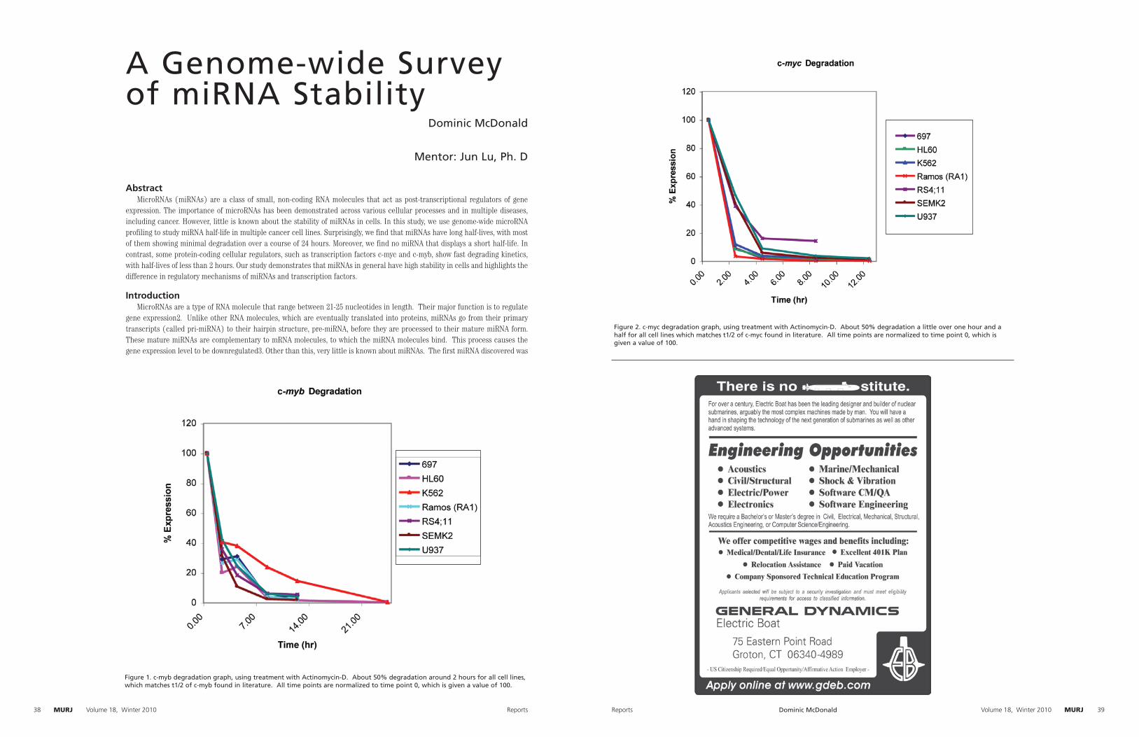
MURJMURJ Reports38 Volume 18, Winter 2010 MURJAuthor Name MURJReports Volume 18, Winter 2010 39
A Genome-wide Survey of miRNA Stability
Dominic McDonald
Mentor: Jun Lu, Ph. D
AbstractMicroRNAs (miRNAs) are a class of small, non-coding RNA molecules that act as post-transcriptional regulators of gene
expression. The importance of microRNAs has been demonstrated across various cellular processes and in multiple diseases, including cancer. However, little is known about the stability of miRNAs in cells. In this study, we use genome-wide microRNA profiling to study miRNA half-life in multiple cancer cell lines. Surprisingly, we find that miRNAs have long half-lives, with most of them showing minimal degradation over a course of 24 hours. Moreover, we find no miRNA that displays a short half-life. In contrast, some protein-coding cellular regulators, such as transcription factors c-myc and c-myb, show fast degrading kinetics, with half-lives of less than 2 hours. Our study demonstrates that miRNAs in general have high stability in cells and highlights the difference in regulatory mechanisms of miRNAs and transcription factors.
IntroductionMicroRNAs are a type of RNA molecule that range between 21-25 nucleotides in length. Their major function is to regulate
gene expression2. Unlike other RNA molecules, which are eventually translated into proteins, miRNAs go from their primary transcripts (called pri-miRNA) to their hairpin structure, pre-miRNA, before they are processed to their mature miRNA form. These mature miRNAs are complementary to mRNA molecules, to which the miRNA molecules bind. This process causes the gene expression level to be downregulated3. Other than this, very little is known about miRNAs. The first miRNA discovered was
Figure 1. c-myb degradation graph, using treatment with Actinomycin-D. About 50% degradation around 2 hours for all cell lines, which matches t1/2 of c-myb found in literature. All time points are normalized to time point 0, which is given a value of 100.
Figure 2. c-myc degradation graph, using treatment with Actinomycin-D. About 50% degradation a little over one hour and a half for all cell lines which matches t1/2 of c-myc found in literature. All time points are normalized to time point 0, which is given a value of 100.
Dominic McDonald
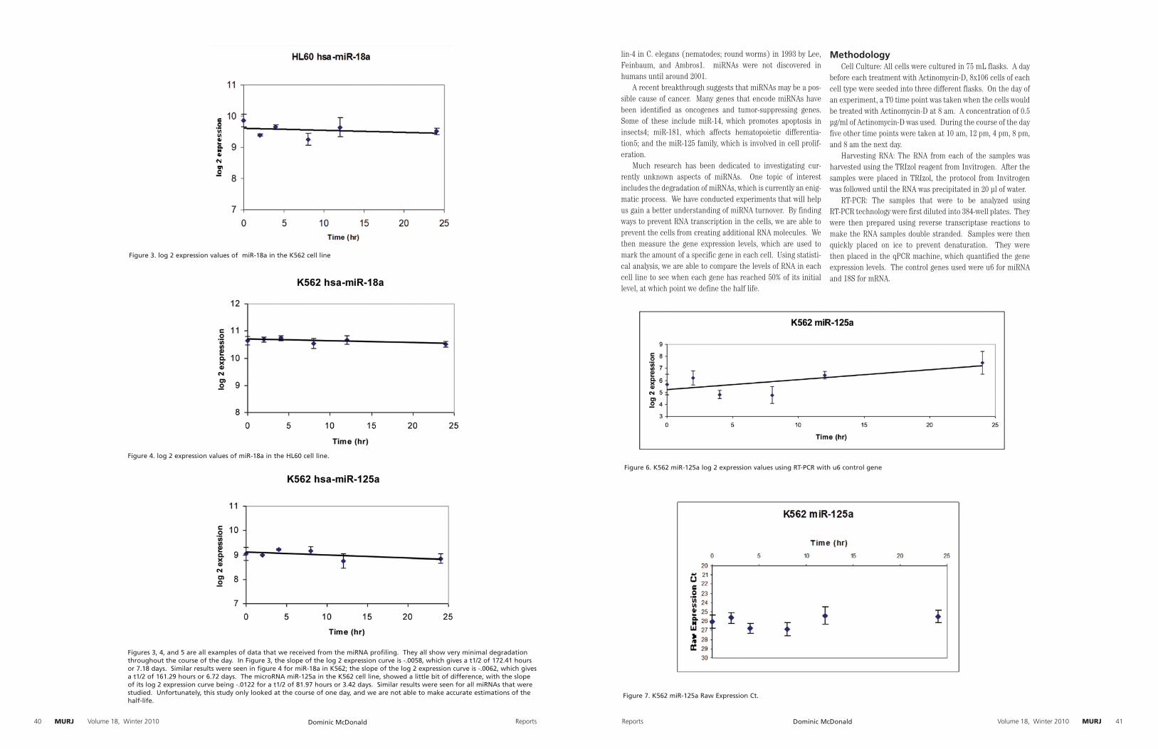
MURJ Author NameMURJ Reports40 Volume 18, Winter 2010 MURJAuthor Name MURJReports Volume 18, Winter 2010 41
lin-4 in C. elegans (nematodes; round worms) in 1993 by Lee, Feinbaum, and Ambros1. miRNAs were not discovered in humans until around 2001.
A recent breakthrough suggests that miRNAs may be a pos-sible cause of cancer. Many genes that encode miRNAs have been identified as oncogenes and tumor-suppressing genes. Some of these include miR-14, which promotes apoptosis in insects4; miR-181, which affects hematopoietic differentia-tion5; and the miR-125 family, which is involved in cell prolif-eration.
Much research has been dedicated to investigating cur-rently unknown aspects of miRNAs. One topic of interest includes the degradation of miRNAs, which is currently an enig-matic process. We have conducted experiments that will help us gain a better understanding of miRNA turnover. By finding ways to prevent RNA transcription in the cells, we are able to prevent the cells from creating additional RNA molecules. We then measure the gene expression levels, which are used to mark the amount of a specific gene in each cell. Using statisti-cal analysis, we are able to compare the levels of RNA in each cell line to see when each gene has reached 50% of its initial level, at which point we define the half life.
MethodologyCell Culture: All cells were cultured in 75 mL flasks. A day
before each treatment with Actinomycin-D, 8x10⁶ cells of each cell type were seeded into three different flasks. On the day of an experiment, a T0 time point was taken when the cells would be treated with Actinomycin-D at 8 am. A concentration of 0.5 µg/ml of Actinomycin-D was used. During the course of the day five other time points were taken at 10 am, 12 pm, 4 pm, 8 pm, and 8 am the next day.
Harvesting RNA: The RNA from each of the samples was harvested using the TRIzol reagent from Invitrogen. After the samples were placed in TRIzol, the protocol from Invitrogen was followed until the RNA was precipitated in 20 µl of water.
RT-PCR: The samples that were to be analyzed using RT-PCR technology were first diluted into 384-well plates. They were then prepared using reverse transcriptase reactions to make the RNA samples double stranded. Samples were then quickly placed on ice to prevent denaturation. They were then placed in the qPCR machine, which quantified the gene expression levels. The control genes used were u6 for miRNA and 18S for mRNA.
Figure 3. log 2 expression values of miR-18a in the K562 cell line
Figure 4. log 2 expression values of miR-18a in the HL60 cell line.
Figures 3, 4, and 5 are all examples of data that we received from the miRNA profiling. They all show very minimal degradation throughout the course of the day. In Figure 3, the slope of the log 2 expression curve is -.0058, which gives a t1/2 of 172.41 hours or 7.18 days. Similar results were seen in figure 4 for miR-18a in K562; the slope of the log 2 expression curve is -.0062, which gives a t1/2 of 161.29 hours or 6.72 days. The microRNA miR-125a in the K562 cell line, showed a little bit of difference, with the slope of its log 2 expression curve being -.0122 for a t1/2 of 81.97 hours or 3.42 days. Similar results were seen for all miRNAs that were studied. Unfortunately, this study only looked at the course of one day, and we are not able to make accurate estimations of the half-life.
Figure 6. K562 miR-125a log 2 expression values using RT-PCR with u6 control gene
Figure 7. K562 miR-125a Raw Expression Ct.
Dominic McDonald Dominic McDonald

MURJ Author NameMURJ Reports42 Volume 18, Winter 2010 MURJAuthor Name MURJReports Volume 18, Winter 2010 43
miRNA Profiling: Samples to undergo miRNA profiling were also diluted into 384-well plates. The miRNAs of interest would be coupled to different probes. The samples then ran through the Luminex NextGen machine to measure the gene expression levels of the miRNAs. This method was developed at the Broad Institute of MIT and Harvard by Jun Lu, Ph.D.7
Data Analysis: After the raw data (Ct) values are received from the RT-PCR machine, the value ∆Ct is calculated as the Ct value for the control gene (u6 for miRNA and 18S rRNA for mRNA). Plotting the ∆Ct value against time produces a linear trend proportional to the % Expression value. An example of this is seen in Figures 3, 4, and 5. The inverse of the slope of the linear trend is the t1/2 (half life) of the gene of interest.
To find the percent expression value, first take the value of 2∆Ct at all time points. The data is then normalized to time point 0, and time point 0 is given a value of 100.
ResultsBefore we began the experiment, we had to ensure the
Actinomycin-D and our procedures worked as anticipated. To do this, we chose two genes, c-myc and c-myb, which are endog-enous genes to the cancer cell lines, to analyze. These genes have a half-life that has been reported through literature as very short—around 2 hours for both c-myc8 and c-myb9. As you can see in Figures 1 and 2, we were able to confirm this half-life.
After analyzing the data, we found that there was minimal degradation over 24 hours of most of the microRNAs in any of the cell lines. In the K562 and HL60 cell line, miR-18a shows very little degradation in both cell lines throughout the course of one day, as shown in Figures 3 and 4.
In the K562 cell line, miR-125a also shows very little deg-radation (Figure 5). This trend was seen among all of the miRNAs in both the HL60 and K562 cell lines.
To validate the accuracy of data received from the Luminex machine, we created a new batch of K562 time points and instead used the RT-PCR machine to measure the expression levels of miR-125a. This experiment yielded very interesting results as at first glance, it would seem as if the RNA content of miR-125a actually increased in the cell (Figure 6), which does not follow logically if Actinomycin D is inhibiting transcription. Closer investigation of the data (Figures 7 and 8), however, reveals that the RNA content of miR-125a actually stays approximately the same while the control gene, u6, is actually degrading in the cell—giving the impression that miR-125a is actually increasing.
With the data gained from the Luminex technology as well as the RT-PCR validation with K562 miR-125a, we can confidently say that miRNAs degrade very slowly. Most miRNA had a half life that was longer than 24 hours. We observed no fast-degrading miRNA either, as there were no miRNA with a half-life under 8 hours.
DiscusionThis study raises an interesting question about why nature
would have created such a stable class of cellular regulators. Many regulators have very short half lives as most genes need to be shut on and off very quickly to regulate cellular activities.
The experiment only covers the short time period of a day, which does not provide enough data to accurately estimate the half life of any of the miRNAs, which is longer than a day. More experiments are needed to learn more about the degradation of miRNAs after one day. One possible experiment involves an inducible system. This allows us to force the cells to produce RNA molecules and then silence the promoter so that no more RNA molecules are produced. The RNA content can then be analyzed at any time, and the cells will not die. This method does have some drawbacks, however, as it is very hard to
Figure 6 shows the RT-PCR validation step, which strangely shows the content of miR-125a increasing in the K562 cell line. After looking at the raw data, it is shown that miR-125a is actually staying constant throughout the course of the day, figure 7, while the control gene u6 is degrading, figure 8; therefore giving the impression that miR-125a is increasing in content within the cell.
Figure 8. K562 u6 Raw Expression Ct.
distinguish between the gene of interest and the endogenous genes. Also, the cells will continue to split and therefore the growth rate—which is difficult to measure—has to be taken into account as well.
Acknowledgements
I would like to thank my mentor Jun Lu, Ph.D., for the mentorship and guidance he has given me throughout my stay at the Broad Institute. I would also like to thank Hao Zhang and Judy Wang, for their help with the experiments, as well as the time spent getting me acquainted with the laboratory. I would like to thank Jinyan Du, Ph.D., for providing me with the cells necessary to complete this experiment. I would like to thank my project investigator, Todd Golub, M.D., for allowing me to participate in his laboratory. Lastly, I would like to thank Shawna Young; Bruce Birren, Ph.D.; Lucia Vielma; and Maura Silverstein for their guid-ance throughout the Summer Research Program in Genomics here at the Broad Institute.
References
1 Lee, R.C., Feinbaum, R.L., Ambros, V., “The C. elegans Heterochronic Gene lin-4 Encodes Small RNAs with Antisense Complementarity to lin-14”, Cell, Vol. 75 (Dec. 3, 1993), pp 843-854
2 Grosshans, H., Slack, F.J., “Micro-RNAs: Small is Plentiful”, The Journal of Cell Biology Vol. 156, No.1 (Jan 7, 2002), pp 17-21.
3 Bartel, D.P., “MicroRNAs Genomics, Biogenesis, Mechanism, and Function”, Cell Vol. 116, Issue 2 (Jan 23, 2004), pp 281-297.
4 Xu, P., Vernooy, S.Y., Guo, M., Hay, B.A., “The Drosphila MicroRNA Mir-14 Suppresses Cell Death and is Required for Normal Fat Metabolism”, Current Biology, Vol. 13, Issue 9 (April 29, 2003), pp 790-795
5 Naguibneva, I., Ameyar-Zazoua, M., Polesskaya, A., Ait-Si-Ali, S., Groisman, R., Souidi, M., Cuvellier, S., Harel-Bellan, A., “The microRNA miR-181 targets the homeobox protein Hox-A11 during mammalian myo-blast differentiation”, Nature Cell Biology, (Feb. 19, 2006), pp 278-284.
6 Fraschini, A., Bottone, M.G., Scovassi, A.I., Denegri, M., Risueno, M.C., Testillano, P.S., Martin, T.E., Biggiogera, M., Pellicciari, C., “Changes in extranucleolar transcription during actinomycin D-induced apoptosis”, Histol Histopathol (Jan 2005), pp 107-117.
7 Lu, J., Getz, G., Miska, E.A., Alvarez-Saavedra, E., Lamb, J., Peck, D., Sweet-Cordero, A., Ebert, B.L., Mak, R.H., Ferrando, A.A., Downing, J.R., Jacks, T., Horvitz, H.R., Golub, T.R., “MicroRNA expression profiles clas-sify human cancers”, Nature 435 (June 9, 2005), pp 834-838.
8 Rabbitts, T.H., Van Straaten, P., Rabbitts, P.H., Watson, J., “Discussion on the Metabolism of c-myc mRNA and Protein”, Proceedings of the Royal Society of London. Series B, Biological Sciences, Vol. 226, No. 1242, Oncogenes: Their role in Normal and Malignant Growth (Oct.22, 1985), pp 79-82
9 Luscher, B., Eisenman, R.N., “c-myc and c-myb Protein Degradation: Effect of Metabolic Inihibitors and Heat Shock”, Molecular and Cellular Biology, Vol. 8 No. 6 (June 1988), pp 2504-2512
Dominic McDonaldDominic McDonald

MURJMURJ Reports44 Volume 18, Winter 2010 MURJAuthor Name MURJReports Volume 18, Winter 2010 45
Painting Blood Vessels with An Adhesive Polymer for Local Delivery of TherapeuticsUROP ReportFall 2008 - PresentLanger LabPrincipal Investigator: Robert LangerDirect Supervisor: Christian Kastrup
Swetha Kambhampati, Class of 2010
Major: Biology
Delivering therapeutic agents to specific regions of the vasculature is an important problem in the medical sciences. Small molecules, larger biological molecules, nanoparticles, and microparticles have all been chemically modified to deliver these agents to specific cells or tissues. In addition to molecular targeting, implantable devices and materials, such as drug-eluting stents, have been developed to deliver agents to specific tissues. , Yet these current delivery techniques still possess many limita-tions. Molecular targeting requires a suitable receptor with a high selectivity for the target, as well as the ability to be coupled to other agents. Implantable devices are often not compatible with the complex environment of the vasculature.
Our current study proposes a technique to overcome these limitations by incorporating the therapeutic agent into an adhesive or “sticky” polymer that can be painted and formed in situ. There are many examples in which polymers have been used to coat blood vessels. “Super-glue” type polymers are currently used to embolize and clog diseased blood vessels, and a photo-curable hydrogel has been used to coat portions of mouse arterial walls. , While these examples demonstrate the feasibility of coating vessels, they are very invasive and lack a high degree of control that our polymer possesses.
The adhesive polymer we are testing is a four-arm polyethyleneglycol (PEG) molecule coupled to four 3,4-dihydroxy-L-phe-nylalanine (DOPA) moieties which render the polymer “sticky.” PEG is considered to be relatively bioorthogonal and is already used clinically in vivo. This DOPA-conjugated polymer mimics the polymers secreted by marine mussels which stick to a wide range of wet materials. The polymerization of this adhesive polymer occurs in a pH-dependent manner and hence can be tightly controlled. Monomers are soluble under slightly acidic conditions, but when injected into the vasculature at physiological pH, polymerization will occur rapidly. We expect the polymer to be capable of trapping particles and large biomolecules that contain agents and therapeutics of interest in its framework. We are using fluorescent proteins, nanospheres, and microspheres to deter-mine the range of particle sizes that can be incorporated into the polymer.
The biocompatibility of this polymer is being executed at many levels, from cell culture toxicity studies to inflammatory responses in animal models. We are currently testing the toxicity of the polymer coated on HeLa and human umbilical vein endo-thelial cells (HUVEC) using the WST-1 Proliferation Assay and live/dead staining with fluorescent probes. Preliminary results are promising. Next, we are testing the degree of local inflammation generated by the polymer when injected subcutaneously in mice.
Detailed in vitro and in vivo experiments will be performed to determine the ability of the polymer to coat and remain localized in the target region. Endothelial cells are being grown on the walls of microfluidic channels to mimic blood vessels. Preliminary results indicate that healthy confluent monolayers of endothelial cells can be obtained in the proposed microfluidic device. The adhesive polymer is being delivered to the walls of the device by a micro-balloon catheter and the effect of different shear rates on accumulation, stable deposition, and localization of the polymer, as well as the optimum catheter design, is being analyzed. In collaboration with the Nahrendorf Lab in the Center for Molecular Imaging Research at Massachusetts General Hospital (MGH), mouse models will be used to assess the capabilities of the adhesive polymer in vivo.
Preliminary results indicate that vascular painting has great promise of delivering therapeutic agents to localized targets in a highly controllable and non-invasive fashion. Vascular painting will be ideal for delivering a variety of molecules, such as siRNA contained in nanoparticles for knockdown of matrix metalloprotease (MMP) activity in atheroclerostic plaques; tissue factors in
lipid vesicles to initiate coagulation and embolize vasculature feeding tumors, and microorganisms, such as Bacillus cereus, to colonize vasculature around tumors.
References
1. Langer, R. Biomaterials in drug delivery and tissue engineering: One laboratory’s experience. Accounts Chem. Res. 33, 94-101(2000)
2. LaVan, D.A., McGuire, T. & Langer, R. Small-scale systems for in vivo drug delivery. Nature Biotechnology 21, 1184-1191 (2003)
3. West, J.L. & Hubbell, J.A. Separation of the arterial wall from blood contact using hydrogel barriers reduces intimal thickening after balloon injury in the rat: The roles of medial and luminal factors in arterial heal-ing. Proc. Natl. Acad. Sci. U.S.A 93, 13188-13193 (1996).
4. Jayaraman, M.V. et al. Neurologic complications of arteriovenous mal-formation embolization using liquid embolic agents. Am. J. Neuroradiol. 29, 242-246 (2008)
5. Lee, H., Dellatore, S.M., Miller, W.M. & Messersmith, P.B. Mussel-inspired surface chemistry for multifunctional coatings. Science 318, 426-430 (2007)
6. Agrawal, N. et al. Bacteriolytic therapy can generate a potent immune response against experimental tumors. Proc. Natl. Acad. Sci. U. S. A. 101, 15172-15177 (2004)
7. Zhao, M. et al. Monotherapy with a tumor-targeting mutant of Salmonella typhimurium cures orthotopic metastatic mouse models of human prostate cancer. Proc. Natl. Acad. Sci. U. S. A. 104, 10170-10174
Swetha Kamhampati
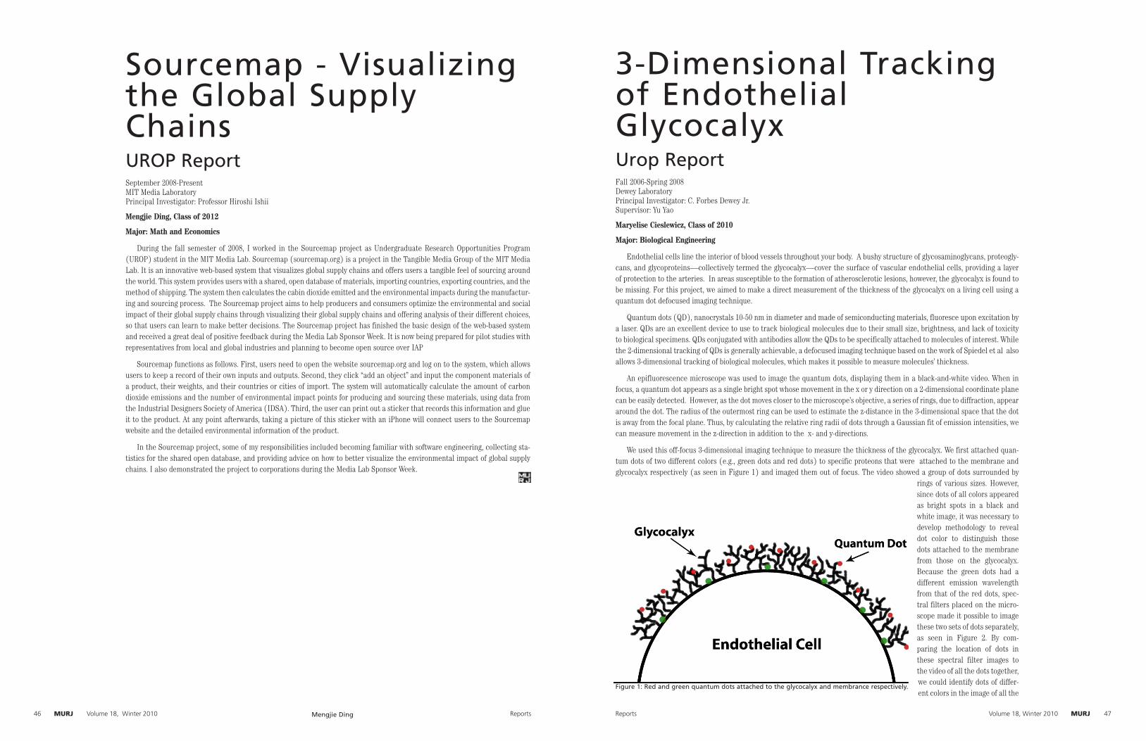
MURJMURJ Reports46 Volume 18, Winter 2010 MURJMURJReports Volume 18, Winter 2010 47
Sourcemap - Visualizing the Global Supply ChainsUROP ReportSeptember 2008-PresentMIT Media LaboratoryPrincipal Investigator: Professor Hiroshi Ishii
Mengjie Ding, Class of 2012
Major: Math and Economics
During the fall semester of 2008, I worked in the Sourcemap project as Undergraduate Research Opportunities Program (UROP) student in the MIT Media Lab. Sourcemap (sourcemap.org) is a project in the Tangible Media Group of the MIT Media Lab. It is an innovative web-based system that visualizes global supply chains and offers users a tangible feel of sourcing around the world. This system provides users with a shared, open database of materials, importing countries, exporting countries, and the method of shipping. The system then calculates the cabin dioxide emitted and the environmental impacts during the manufactur-ing and sourcing process. The Sourcemap project aims to help producers and consumers optimize the environmental and social impact of their global supply chains through visualizing their global supply chains and offering analysis of their different choices, so that users can learn to make better decisions. The Sourcemap project has finished the basic design of the web-based system and received a great deal of positive feedback during the Media Lab Sponsor Week. It is now being prepared for pilot studies with representatives from local and global industries and planning to become open source over IAP
Sourcemap functions as follows. First, users need to open the website sourcemap.org and log on to the system, which allows users to keep a record of their own inputs and outputs. Second, they click “add an object” and input the component materials of a product, their weights, and their countries or cities of import. The system will automatically calculate the amount of carbon dioxide emissions and the number of environmental impact points for producing and sourcing these materials, using data from the Industrial Designers Society of America (IDSA). Third, the user can print out a sticker that records this information and glue it to the product. At any point afterwards, taking a picture of this sticker with an iPhone will connect users to the Sourcemap website and the detailed environmental information of the product.
In the Sourcemap project, some of my responsibilities included becoming familiar with software engineering, collecting sta-tistics for the shared open database, and providing advice on how to better visualize the environmental impact of global supply chains. I also demonstrated the project to corporations during the Media Lab Sponsor Week.
3-Dimensional Tracking of Endothelial GlycocalyxUrop ReportFall 2006-Spring 2008Dewey LaboratoryPrincipal Investigator: C. Forbes Dewey Jr.Supervisor: Yu Yao
Maryelise Cieslewicz, Class of 2010
Major: Biological Engineering
Endothelial cells line the interior of blood vessels throughout your body. A bushy structure of glycosaminoglycans, proteogly-cans, and glycoproteins—collectively termed the glycocalyx—cover the surface of vascular endothelial cells, providing a layer of protection to the arteries. In areas susceptible to the formation of atherosclerotic lesions, however, the glycocalyx is found to be missing. For this project, we aimed to make a direct measurement of the thickness of the glycocalyx on a living cell using a quantum dot defocused imaging technique.
Quantum dots (QD), nanocrystals 10-50 nm in diameter and made of semiconducting materials, fluoresce upon excitation by a laser. QDs are an excellent device to use to track biological molecules due to their small size, brightness, and lack of toxicity to biological specimens. QDs conjugated with antibodies allow the QDs to be specifically attached to molecules of interest. While the 2-dimensional tracking of QDs is generally achievable, a defocused imaging technique based on the work of Spiedel et al also allows 3-dimensional tracking of biological molecules, which makes it possible to measure molecules’ thickness.
An epifluorescence microscope was used to image the quantum dots, displaying them in a black-and-white video. When in focus, a quantum dot appears as a single bright spot whose movement in the x or y direction on a 2-dimensional coordinate plane can be easily detected. However, as the dot moves closer to the microscope’s objective, a series of rings, due to diffraction, appear around the dot. The radius of the outermost ring can be used to estimate the z-distance in the 3-dimensional space that the dot is away from the focal plane. Thus, by calculating the relative ring radii of dots through a Gaussian fit of emission intensities, we can measure movement in the z-direction in addition to the x- and y-directions.
We used this off-focus 3-dimensional imaging technique to measure the thickness of the glycocalyx. We first attached quan-tum dots of two different colors (e.g., green dots and red dots) to specific proteons that were attached to the membrane and glycocalyx respectively (as seen in Figure 1) and imaged them out of focus. The video showed a group of dots surrounded by
rings of various sizes. However, since dots of all colors appeared as bright spots in a black and white image, it was necessary to develop methodology to reveal dot color to distinguish those dots attached to the membrane from those on the glycocalyx. Because the green dots had a different emission wavelength from that of the red dots, spec-tral filters placed on the micro-scope made it possible to image these two sets of dots separately, as seen in Figure 2. By com-paring the location of dots in these spectral filter images to the video of all the dots together, we could identify dots of differ-ent colors in the image of all the
Figure 1: Red and green quantum dots attached to the glycocalyx and membrance respectively.
Mengjie Ding
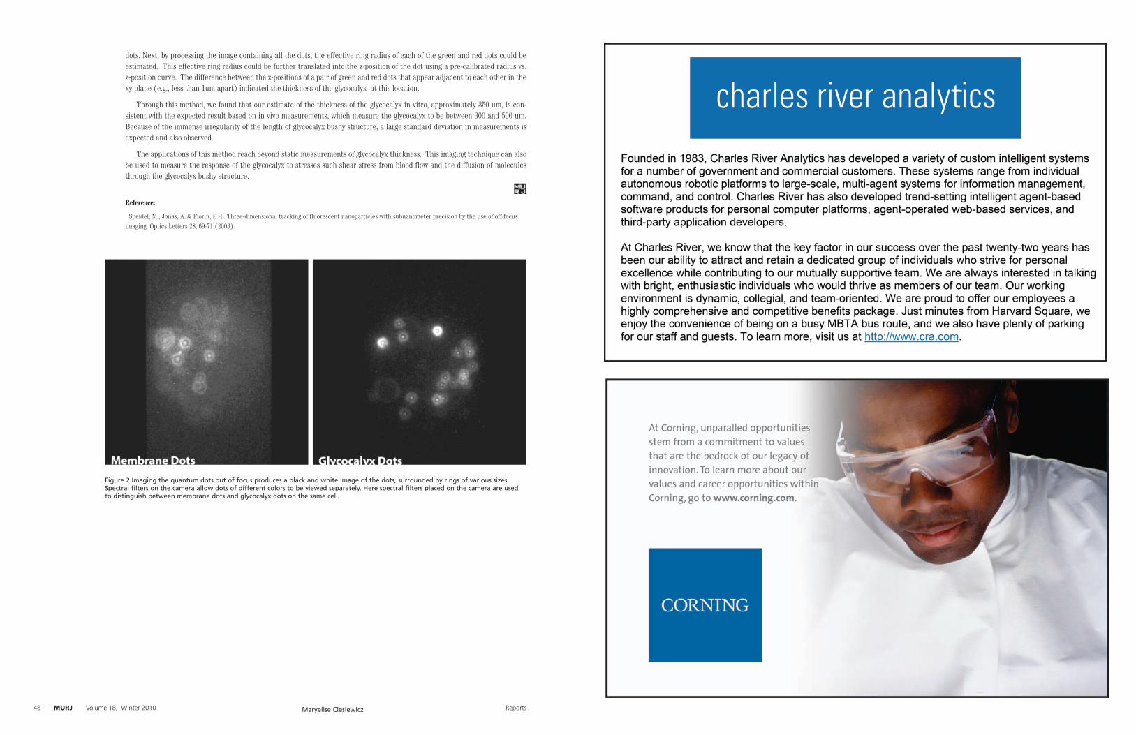
MURJ Author NameMURJ Reports48 Volume 18, Winter 2010
dots. Next, by processing the image containing all the dots, the effective ring radius of each of the green and red dots could be estimated. This effective ring radius could be further translated into the z-position of the dot using a pre-calibrated radius vs. z-position curve. The difference between the z-positions of a pair of green and red dots that appear adjacent to each other in the xy plane (e.g., less than 1um apart) indicated the thickness of the glycocalyx at this location.
Through this method, we found that our estimate of the thickness of the glycocalyx in vitro, approximately 350 um, is con-sistent with the expected result based on in vivo measurements, which measure the glycocalyx to be between 300 and 500 um. Because of the immense irregularity of the length of glycocalyx bushy structure, a large standard deviation in measurements is expected and also observed.
The applications of this method reach beyond static measurements of glycocalyx thickness. This imaging technique can also be used to measure the response of the glycocalyx to stresses such shear stress from blood flow and the diffusion of molecules through the glycocalyx bushy structure.
Reference:
Speidel, M., Jonas, A. & Florin, E.-L. Three-dimensional tracking of fluorescent nanoparticles with subnanometer precision by the use of off-focus imaging. Optics Letters 28, 69-71 (2003).
Figure 2 Imaging the quantum dots out of focus produces a black and white image of the dots, surrounded by rings of various sizes. Spectral filters on the camera allow dots of different colors to be viewed separately. Here spectral filters placed on the camera are used to distinguish between membrane dots and glycocalyx dots on the same cell.
Maryelise Cieslewicz

50 Volume 18, Winter 2010MURJ Volume 18, Winter 2010 51MURJ

52 Volume 18, Winter 2010MURJ