mitochondria as a therapeutic target in heart...
TRANSCRIPT

Journal of the American College of Cardiology Vol. 61, No. 6, 2013© 2013 by the American College of Cardiology Foundation ISSN 0735-1097/$36.00
Downloa
STATE-OF-THE-ART PAPER
Mitochondria as aTherapeutic Target in Heart Failure
Marina Bayeva, PHD,* Mihai Gheorghiade, MD,† Hossein Ardehali, MD, PHD*
Chicago, Illinois
Heart failure is a pressing public health problem with no curative treatment currently available. The existing ther-apies provide symptomatic relief, but are unable to reverse molecular changes that occur in cardiomyocytes. Themechanisms of heart failure are complex and multiple, but mitochondrial dysfunction appears to be a criticalfactor in the development of this disease. Thus, it is important to focus research efforts on targeting mitochon-drial dysfunction in the failing heart to revive the myocardium and its contractile function. This review highlightsthe 3 promising areas for the development of heart failure therapies, including mitochondrial biogenesis, mito-chondrial oxidative stress, and mitochondrial iron handling. Moreover, the translational potential of compoundstargeting these pathways is discussed. (J Am Coll Cardiol 2013;61:599–610) © 2013 by the American Collegeof Cardiology Foundation
Published by Elsevier Inc. http://dx.doi.org/10.1016/j.jacc.2012.08.1021
The 20th century has witnessed a dramatic improvement inpatients’ survival after adverse cardiovascular events. How-ever, heart disease still remains the number one cause ofdeath in the industrialized world, affecting �27 millionpeople in the United States alone. With $40 billion inannual costs and 1 of every 5 patients dying within 1 year ofdiagnosis (1), HF has become a major public health prob-lem. Although significant progress has been made in theoutpatient management of chronic heart failure (HF), post-discharge mortality and rehospitalization rates within 60 to 90days can be as high as 15% and 30%, respectively (2).
The primary goal of treating HF patients is restoration ofcardiac function. Recent studies show that heart functioncan be successfully recovered in patients with HF, even afterstructural alterations have occurred. Thus, failing myocar-dium is “viable but dysfunctional” rather than irreversibly
From the *Feinberg Cardiovascular Research Institute, Northwestern UniversitySchool of Medicine, Chicago, Illinois; and the †Center for Cardiovascular Innova-tion, Northwestern University School of Medicine, Chicago, Illinois. This work wassupported, in part, by grants from the National Institutes of Health K02 HL107448,R01 HL104181, and 1P01 HL108795 (to Dr. Ardehali). Dr. Gheorghiade is aconsultant for Abbott Laboratories, Astellas, AstraZeneca, Bayer Schering PharmaAG, Cardiorentis Ltd., CorThera, Cytokinetics, CytoPherx, Inc., DebioPharm S.A.,Errekappa Terapeutici, GlaxoSmithKline, Ikaria, Intersection Medical, Inc., Johnson& Johnson, Medtronic, Merck, Novartis Pharma AG, Ono Pharmaceuticals USA,Otsuka Pharmaceuticals, Palatin Technologies, Pericor Therapeutics, Protein DesignLaboratories, sanofi-aventis, Sigma Tau, Solvay Pharmaceuticals, Sticares InterACT,Takeda Pharmaceuticals North America, Inc., and Trevena Therapeutics; and hasreceived significant (�$10,000) support from Bayer Schering Pharma AG, Debio-Pharm S.A., Medtronic, Novartis Pharma AG, Otsuka Pharmaceuticals, Sigma Tau,Solvay Pharmaceuticals, Sticares InterACT, and Takeda Pharmaceuticals NorthAmerica, Inc. Dr. Ardehali is a consultamt for Cubist Pharma, Novartis, and theGerson Lehman Group, and is a speaker for Merck. Dr. Bayeva has reported that shehas no relationships relevant to the contents of this paper to disclose.
Manuscript received June 22, 2012; revised manuscript received August 13, 2012,accepted August 21, 2012.
ded From: http://content.onlinejacc.org/ on 11/30/2014
damaged, independent of the presence or absence of coro-nary artery disease. This finding opens up an avenue forrational design of treatments that target the cardiomyocyteitself, not the indirect pathways that suppress neurohor-monal axis or induce vasodilation.
The mechanisms underlying the development of HF aremultiple, complex, and not well understood. Althoughvirtually all aspects of myocyte physiology are altered in HF,the past decade of research provided convincing evidencethat mitochondrial dysfunction may be an important eventin the development of hypertrophy and HF. First, geneticmutations that disrupt mitochondrial function are associ-ated with cardiac dysfunction in mice (3) and humans (4,5).Second, currently available therapies for HF, such asangiotensin-converting enzyme inhibitors and angiotensinreceptor II (ATII) blockers significantly improve survival inischemic and nonischemic HF, and their administrationalso correlates with improved mitochondrial function (6,7).Finally, it is important to note that cardiomyocytes in thefailing heart remain viable, although metabolically stunned,and their function can potentially be rescued (8). Thus,therapeutic efforts should venture beyond symptomatic re-lief and focus on reviving the dormant myocardium bytargeting the underlying molecular defects in HF. In thisreview, we discuss the changes that occur in the mitochon-dria of failing myocardium, followed by an overview of thepertinent therapeutic targets and approaches that can po-tentially reverse these changes and preserve cardiac health.Of note, we refrain from discussing the changes in glucoseand fatty acid utilization, as these topics have recently beenreviewed in detail elsewhere (8). Instead, we focus on
mitochondrial biogenesis, production of reactive oxygen species
tFfoS
itrm
(Emm
sstcnf
hhsyi
b(cttsnHadm
600 Bayeva et al. JACC Vol. 61, No. 6, 2013Mitochondria and Heart Failure February 12 2013:599–610
Downloa
(ROS) and maintenance of cel-lular iron homeostasis as promis-ing novel therapies for HF.
Mitochondrial Biogenesis
Pathophysiology. One of the waysto augment energy production in thesetting of increased contractile de-mand is to stimulate production ofnew mitochondria, termed mito-chondrial biogenesis. Mitochondriacontain �16.5 kilobase of circulardouble-stranded DNA that en-codes 13 protein components ofthe electron transport chain andneeds to be replicated before thedivision. In addition, as many as1000 nuclear-encoded proteinsmust be imported into the newlyformed mitochondria to make afully functional organelle (9).Thus, generation of new mito-chondria requires a coordinatedtranscription of mitochondrial andnuclear genomes orchestrated byperoxisome proliferator–activatedreceptor gamma coactivator 1�(PGC1�) (10). PGC1�, a nuclear-encoded protein, is induced inthe states of enhanced energydemand, such as increased car-diac workload, high adenosinediphosphate/adenosine triphos-phate (ATP) ratio, cold, exercise,and fasting (for review, see refer-ences 11 and 12). High PGC1�activity is associated with in-creased mitochondrial content,as exemplified by cardiac-specificPGC1� transgenic mice, whichexhibit uncontrolled mitochon-drial proliferation and increase inmarkers of mitochondrial bio-genesis (13,14). PGC1� stimu-lates mitochondrial proliferation
hrough its interaction with several transcription factors.irst, PGC1� binds to and coactivates nuclear respiratory
actors (NRFs) 1 and 2, which in turn promote transcriptionf nuclear-encoded genes targeted to mitochondria (15).econd, PGC1� activates estrogen-related nuclear orphan
receptors ERR� and �, which induce expression of genesnvolved in glucose and fatty acid uptake, energy produc-ion, and ATP transport (16,17). Finally, PGC1� promoteseplication of mitochondrial genome through NRF1/2-
Abbreviationsand Acronyms
AMPK � adenosinemonophosphate kinase
ATII � angiotensinreceptor II
ATP � adenosinetriphosphate
cGMP � cyclic guanosinemonophosphate
DFP � deferiprone
eNOS � endothelial nitricoxide synthase
Fe/S � iron/sulfur
FRDA � Friedrich’s ataxia
HF � heart failure
I/R � ischemia/reperfusion
MI � myocardial infarction
MnSOD � manganesesuperoxide dismutase
mtDNA � mitochondrialDNA
NO � nitric oxide
Nox � nicotinamideadenine dinucleotidephosphate (reduced form)oxidase
NRF � nuclear respiratoryfactor
PDE � phosphodiesterase
PDE5I � phosphodiesterasetype 5 inhibitor
PGC1� � peroxisomeproliferator–activatedreceptor gammacoactivator 1�
ROS � reactive oxygenspecies
SOD � superoxidedismutase
SS � Szeto-Schiller
Tfam � transcriptionfactor A
TPP � triphenylphosphonium
ediated induction of mitochondrial transcription factor A
ded From: http://content.onlinejacc.org/ on 11/30/2014
Tfam) (12). Cardiac-specific deletion of NRF1 (18),RR� (19), and Tfam (20) are all associated with decreaseditochondrial content or function, confirming their role initochondrial biogenesis.Studies of rodents (21–23), dogs (24), and humans (25)
uggest that disruption of mitochondrial biogenesis repre-ents an early event in the pathophysiology of HF, theimely reversal of which is cardioprotective. Grossly, mito-hondrial content and mitochondrial DNA (mtDNA) copyumber are significantly reduced in rodent and humanailing myocardium, and downregulation of the PGC1�
pathway has been observed in various models of HF in miceand rats (21,22,26,27). However, the role of PGC1� in
uman HF remains controversial, and contradictory resultsave also been reported (28–30). Because PGC1� is exten-ively regulated on the post-translational level by phosphor-lation (31), acetylation (32), and protein stabilization (33),t is not clear whether PGC1� activity is reduced in the
failing hearts and whether the reduction in mitochondrialnumber in HF in humans is due to deregulation of PGC1�signaling.
A defect in mtDNA replication was proposed as analternative mechanism for the reduction in mitochondrialbiogenesis (30,34). Importantly, changes in mtDNA repli-cation machinery represented a very early event detected inhypertrophied hearts that have not yet transitioned to failure(30). The actual trigger for reducing mtDNA replication ina setting of increased workload is unknown, and it would beof interest to replicate these studies in animal models and/orHF patients.Therapeutic strategies. Despite the controversy about therole of PGC1� in human HF, boosting mitochondrialiogenesis in failing myocardium appears to be beneficial35). In fact, the angiotensin-converting enzyme inhibitoraptopril was shown to increase mitochondrial content inhe hearts of dogs after coronary ligation (36), suggestinghat some of its beneficial effects may be due to thetimulation of mitochondrial biogenesis. Although currentlyo drugs that specifically target mitochondrial biogenesis inF are available, acceleration of this process through
denosine monophosphate–activated kinase (AMPK), en-othelial nitric oxide synthase (eNOS), and other pathwaysay represent a promising therapeutic approach (Fig. 1).
ADENOSINE MONOPHOSPHATE–ACTIVATED KINASE.
AMPK exhibits very low baseline activity in the heart, butis upregulated in response to a variety of stressors (37,38).Importantly, activation of AMPK is thought to be themechanism driving the increase in mitochondrial biogenesisin exercise-induced adaptive hypertrophy of the athlete’sheart (39), which, unlike the pathological hypertrophydiscussed earlier, does not lead to HF. AMPK inducesPGC1� function (39,40), activates NRF1, Tfam (41), andERR� (28), establishing this kinase as an important mod-
ulator of mitochondrial biogenesis.
mMt
AhdUsiAacto
AadtcSah
601JACC Vol. 61, No. 6, 2013 Bayeva et al.February 12 2013:599–610 Mitochondria and Heart Failure
Downloa
The activity of AMPK is increased in failing hearts (42);however, pharmacological activation of this pathway ap-pears to exert additional cardiac protection. For example,metformin, a commonly used antidiabetic drug that acti-vates AMPK signaling, reduced infarct size and preservedcardiac function in a long-term post-myocardial infarction(MI) rat model in the absence of diabetes (43) and exhibitedcardioprotective properties in a rapid ventricular pacingcanine model (44). Gundewar et al. (45) found increasedPGC1� levels and preservation of heart function in wild-type metformin-treated mice subjected to MI or ischemia/reperfusion (I/R), but not in a AMPK knockout mousemodel (45). Although metformin was also shown to reducegluconeogenesis through direct inhibition of mitochondrialcomplex I, the total cellular ATP content in the livers ofmetformin-treated rats remained unchanged, consistent withthe maintenance of the cumulative mitochondrial bioenergeticcapacity possibly due to increased biogenesis (46).
Accumulating evidence of the cardioprotective propertiesof AMPK in the setting of hypertrophy and cardiac failurecalls for the development of pharmacological agonists ofAMPK in the heart. Several clinically available compoundshave been shown to activate AMPK, including metformin,5-aminoimidazole-4-carboxamide-1-�-D-ribofuranosyl 5=-
onophosphate (AICAR), thiazolidinediones, and statins.oreover, the ATII receptor blocker telmisartan was shown
o increase phosphorylated AMPK� levels in culturedmyotybes (7), suggesting that the beneficial effects of this
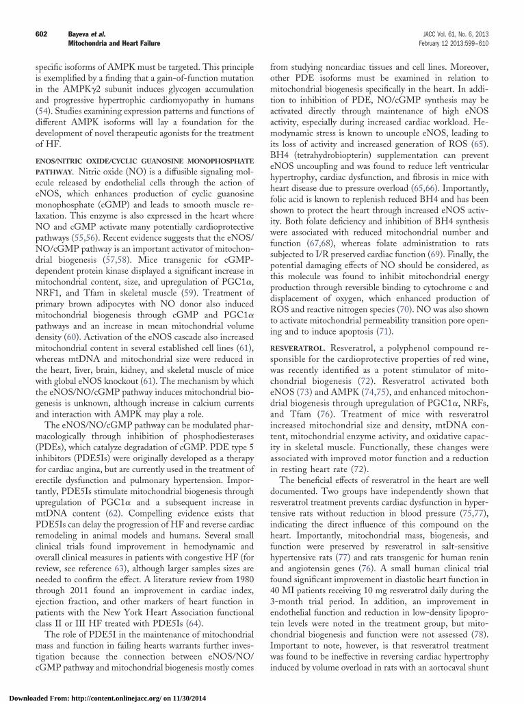
Figure 1 Mitochondrial Biogenesis
Mitochondrial biogenesis impairment is an early event in the development of hearbe enhanced therapeutically with the use of adenosine monophosphate kinase (AMpathway (including phosphodiesteraes type 5 inhibitors), or resveratrol. All of thesreceptor gamma coactivator 1� (PGC1�), nuclear respiratory factor (NRF)1/2, andin the heart. ATP � adenosine triphosphate.
drug may partially be due to stimulation of mitochondrial T
ded From: http://content.onlinejacc.org/ on 11/30/2014
biogenesis. However, the effect of current HF therapies onthe AMPK pathway is likely indirect via an increase in theadenosine monophosphate/ATP ratio, inhibition of mito-chondrial respiration, or other cellular and systemic effects(47). Compounds targeting AMPK itself are currently indevelopment. Of note, compound A769662 by AbbottLaboratories (Abbott Park, Illinois) is a specific allostericactivator of AMPK complexes on the � subunit (48).
769662 reduced infarct size in rats fed low-fat andigh-fat diets (49), providing proof-of-principle that airect activation of AMPK is beneficial to the heart.nfortunately, the compound also inhibited 26S protea-
ome and caused cell-cycle arrest through an AMPK-ndependent mechanism, limiting its clinical prospects (50).nother small molecule, PT1, appears to activate AMPK�1
nd AMPK�2 isoforms by removing autoinhibition in theatalytic subunits of the kinase (51). Thus, PT1 potentiallyargets a vast array of AMPK complexes, although its effectsn the heart remain to be studied.Several points must be considered when developing an
MPK agonist for the treatment of HF. First, AMPKctivation may only be suitable for treatment of HF due toefined etiologies. Although AMPK activation was protec-ive in the mouse models of MI and I/R, it failed to preserveardiac function in rats with chronic volume overload (52).econd, AMPK is ubiquitously expressed and regulates anrray of processes, and the subunit composition of AMPKeterodimeric complex differs widely among tissues (53).
e, and reversal of this process is cardioprotective. Mitochondrial biogenesis cangonists, stimulants of nitric oxide/cyclic guanosine monophosphate (NO/cGMP)oaches stimulate nuclear-encoded proteins peroxisome proliferator–activatedription factor A (Tfam), which, in turn, facilitate production of new mitochondria
t failurPK) a
e apprtransc
hus, to maximize clinical benefits and minimize toxicity,
a(ddo
Npm
mPrcorntepc
mtc
fomtaamiBehhfsiwfsptpdRti
aitiai
drtihfhaf43etcIw
602 Bayeva et al. JACC Vol. 61, No. 6, 2013Mitochondria and Heart Failure February 12 2013:599–610
Downloa
specific isoforms of AMPK must be targeted. This principleis exemplified by a finding that a gain-of-function mutationin the AMPK�2 subunit induces glycogen accumulationnd progressive hypertrophic cardiomyopathy in humans54). Studies examining expression patterns and functions ofifferent AMPK isoforms will lay a foundation for theevelopment of novel therapeutic agonists for the treatmentf HF.
ENOS/NITRIC OXIDE/CYCLIC GUANOSINE MONOPHOSPHATE
PATHWAY. Nitric oxide (NO) is a diffusible signaling mol-ecule released by endothelial cells through the action ofeNOS, which enhances production of cyclic guanosinemonophosphate (cGMP) and leads to smooth muscle re-laxation. This enzyme is also expressed in the heart whereNO and cGMP activate many potentially cardioprotectivepathways (55,56). Recent evidence suggests that the eNOS/NO/cGMP pathway is an important activator of mitochon-drial biogenesis (57,58). Mice transgenic for cGMP-dependent protein kinase displayed a significant increase inmitochondrial content, size, and upregulation of PGC1�,
RF1, and Tfam in skeletal muscle (59). Treatment ofrimary brown adipocytes with NO donor also induceditochondrial biogenesis through cGMP and PGC1�
pathways and an increase in mean mitochondrial volumedensity (60). Activation of the eNOS cascade also increasedmitochondrial content in several established cell lines (61),whereas mtDNA and mitochondrial size were reduced inthe heart, liver, brain, kidney, and skeletal muscle of micewith global eNOS knockout (61). The mechanism by whichthe eNOS/NO/cGMP pathway induces mitochondrial bio-genesis is unknown, although increase in calcium currentsand interaction with AMPK may play a role.
The eNOS/NO/cGMP pathway can be modulated phar-macologically through inhibition of phosphodiesterases(PDEs), which catalyze degradation of cGMP. PDE type 5inhibitors (PDE5Is) were originally developed as a therapyfor cardiac angina, but are currently used in the treatment oferectile dysfunction and pulmonary hypertension. Impor-tantly, PDE5Is stimulate mitochondrial biogenesis throughupregulation of PGC1� and a subsequent increase in
tDNA content (62). Compelling evidence exists thatDE5Is can delay the progression of HF and reverse cardiac
emodeling in animal models and humans. Several smalllinical trials found improvement in hemodynamic andverall clinical measures in patients with congestive HF (foreview, see reference 63), although larger samples sizes areeeded to confirm the effect. A literature review from 1980hrough 2011 found an improvement in cardiac index,jection fraction, and other markers of heart function inatients with the New York Heart Association functionallass II or III HF treated with PDE5Is (64).
The role of PDE5I in the maintenance of mitochondrialass and function in failing hearts warrants further inves-
igation because the connection between eNOS/NO/
GMP pathway and mitochondrial biogenesis mostly comes ided From: http://content.onlinejacc.org/ on 11/30/2014
rom studying noncardiac tissues and cell lines. Moreover,ther PDE isoforms must be examined in relation toitochondrial biogenesis specifically in the heart. In addi-
ion to inhibition of PDE, NO/cGMP synthesis may bectivated directly through maintenance of high eNOSctivity, especially during increased cardiac workload. He-odynamic stress is known to uncouple eNOS, leading to
ts loss of activity and increased generation of ROS (65).H4 (tetrahydrobiopterin) supplementation can preventNOS uncoupling and was found to reduce left ventricularypertrophy, cardiac dysfunction, and fibrosis in mice witheart disease due to pressure overload (65,66). Importantly,olic acid is known to replenish reduced BH4 and has beenhown to protect the heart through increased eNOS activ-ty. Both folate deficiency and inhibition of BH4 synthesisere associated with reduced mitochondrial number and
unction (67,68), whereas folate administration to ratsubjected to I/R preserved cardiac function (69). Finally, theotential damaging effects of NO should be considered, ashis molecule was found to inhibit mitochondrial energyroduction through reversible binding to cytochrome c andisplacement of oxygen, which enhanced production ofOS and reactive nitrogen species (70). NO was also shown
o activate mitochondrial permeability transition pore open-ng and to induce apoptosis (71).
RESVERATROL. Resveratrol, a polyphenol compound re-sponsible for the cardioprotective properties of red wine,was recently identified as a potent stimulator of mito-chondrial biogenesis (72). Resveratrol activated botheNOS (73) and AMPK (74,75), and enhanced mitochon-drial biogenesis through upregulation of PGC1�, NRFs,nd Tfam (76). Treatment of mice with resveratrolncreased mitochondrial size and density, mtDNA con-ent, mitochondrial enzyme activity, and oxidative capac-ty in skeletal muscle. Functionally, these changes weressociated with improved motor function and a reductionn resting heart rate (72).
The beneficial effects of resveratrol in the heart are wellocumented. Two groups have independently shown thatesveratrol treatment prevents cardiac dysfunction in hyper-ensive rats without reduction in blood pressure (75,77),ndicating the direct influence of this compound on theeart. Importantly, mitochondrial mass, biogenesis, andunction were preserved by resveratrol in salt-sensitiveypertensive rats (77) and rats transgenic for human reninnd angiotensin genes (76). A small human clinical trialound significant improvement in diastolic heart function in0 MI patients receiving 10 mg resveratrol daily during the-month trial period. In addition, an improvement inndothelial function and reduction in low-density lipopro-ein levels were noted in the treatment group, but mito-hondrial biogenesis and function were not assessed (78).mportant to note, however, is that resveratrol treatmentas found to be ineffective in reversing cardiac hypertrophy
nduced by volume overload in rats with an aortocaval shunt
fiimcomgn
M
Phsrte(imp
fdb
ftNtmaiadtada
Hlccmiephmt
603JACC Vol. 61, No. 6, 2013 Bayeva et al.February 12 2013:599–610 Mitochondria and Heart Failure
Downloa
(79). Thus, the beneficial effects of this compound may belimited to select clinical scenarios, such as hypertensive andpost-MI patients.
No significant toxicity of this compound was noted inhealthy human volunteers in phase I study during the4-week trial period (80). Unfortunately, resveratrol has ashort half-life of 8 to 14 min and is extensively metabolizedin the body (81). Thus, an effective human dose cannot beeasily extrapolated from animal studies. Development ofmore potent analogs with longer half-lives may help toovercome these limitations.
OTHER STRATEGIES. The cardioprotective effects of estrogenare well documented in various animal models. Moreover,epidemiological studies reveal a reduced risk of cardiovasculardisease in premenopausal, but not post-menopausal, womencompared with men. Estrogen-like compounds were shownto stimulate mitochondrial biogenesis through induction ofNRF1 expression and increase in mtDNA content (82).Unfortunately, estrogen replacement therapy not only failedto reduce, but actually increased, the number of cardiacevents in post-menopausal women (83). The likely reasonsfor that are discussed by Mendelsohn et al. (84). Thus,although the estrogen pathway represents a promisingtherapeutic target, more research is needed to understand itsrisks, benefits, and target patient population.
The approaches discussed here are aimed at the produc-tion of new mitochondria via induction of PGC1�, NRFs,and Tfam signaling. However, it is also important to notethat mitochondria are dynamic organelles that constantlyundergo fusion and fission. Although the significance ofthese processes in HF is not well understood, both fusionand fission are essential for maintenance of mitochondrialfunction (for review, see references 85 and 86). Constantexchange of mitochondrial metabolites, proteins, DNA, andCa2� throughout the mitochondrial network by fusion/
ssion may protect these organelles in the setting of annsult. On the other hand, selective elimination of damaged
itochondria by the process of autophagy is also critical inardiac physiology, as disruption of autophagy in a pressure-verloaded heart facilitates its transition into HF. Thus,itochondrial biogenesis should be re-examined and tar-
eted in a broader context of preserving mitochondrialumber, network organization, size, and quality.
itochondrial Oxidative Stress
athophysiology. Generation of ROS is significantly en-anced in the failing myocardium, as has been unequivocallyhown by studies of animal models and human patients (foreview, see references 87 and 88). The majority of ROS inhe heart appear to come from uncoupling of mitochondriallectron transport chain at the level of complexes I and III89), although the view of mitochondria as a major source ofntracellular ROS has been challenged (90). The activities of
itochondrial electron transport chain complexes are sup-
ressed in HF, and disruption of mitochondrial bioenergeticded From: http://content.onlinejacc.org/ on 11/30/2014
unction was found to increase ROS and oxidative DNAamage (91), providing a possible pathophysiological linketween mitochondrial dysfunction and ROS (88,92).Nicotinamide adenine dinucleotide phosphate (reduced
orm) oxidase (Nox) is another important source of ROS inhe heart. Five isoforms of Nox have been described, withox4 being the most abundant in cardiomyocytes, endo-
helial cells, and fibroblasts. Nox4 localizes primarily to theitochondria, does not require cytosolic subunits for its
ctivation, and is implicated in enhanced ROS productionn pressure overload and aging models of HF (93–95). Noxctivity is induced by pathways that are also active inysfunctional myocardium, including ATII stimulation,umor necrosis factor-�, and mechanical stretch (88). Noxctivity is high in human failing hearts (96), whereas geneticeletion of this enzyme protects against cardiac dysfunctionnd remodeling in the MI mouse model (97).
The exact contribution of ROS to the development ofF is complex and remains a subject of intense debate. One
ikely mechanism is physical damage of cellular and mito-hondrial structures, such as sarcomeric and excitation-ontraction coupling proteins, which would impair theechanical properties of the heart (98). Because the major-
ty of ROS in HF comes from mitochondria, these organ-lles are the primary target of oxidative damage. mtDNA isarticularly sensitive to ROS due to the lack of protectiveistones and less efficient DNA repair, and mutations intDNA-encoded genes are known to cause cardiomyopa-
hy. Moreover, reduction of PGC1� in failing hearts canexacerbate oxidative stress and mitochondrial damage, asthis protein was found to maintain mitochondrial, but notcytosolic, antioxidant defenses (99). In addition to damag-ing cellular components, ROS regulate several signalingcascades, including the known hypertrophic pathways suchas protein kinase C, mitogen-activated protein kinase, JunN-terminal kinase, and Ras (100). Finally, ROS facilitatethe remodeling of the extracellular matrix by inducingmatrix metalloproteinases through direct post-translationalmodifications or indirectly through the nuclear factor �Bpathway (101). Given that ROS affect virtually all aspects ofcardiomyocyte physiology, they represent an importanttherapeutic target for treating HF. In support of this claim,cardioprotective therapies such as angiotensin-convertingenzyme inhibitors and ATII receptor blockers were shownto possess antioxidant properties, although it is not knownwhether they target mitochondrial ROS directly or indi-rectly (102,103).Therapeutic strategies. Several trials have assessed theefficacy of antioxidants in the treatment of HF, but theresults were disappointing. Long-term supplementationwith �-tocopherol (vitamin E) was actually associated withan increased risk of the development of HF (104). Evidencefrom animal studies suggests that preferential inhibition ofROS inside the mitochondria, rather than global antioxidanttreatment, may be cardioprotective. Overexpression of the
mitochondria-specific antioxidant peroxiredoxin-3 pro-
tlpmSia(wshmtfsorb
Ms
risvtpmniTIprbpgoclft
mwtd
604 Bayeva et al. JACC Vol. 61, No. 6, 2013Mitochondria and Heart Failure February 12 2013:599–610
Downloa
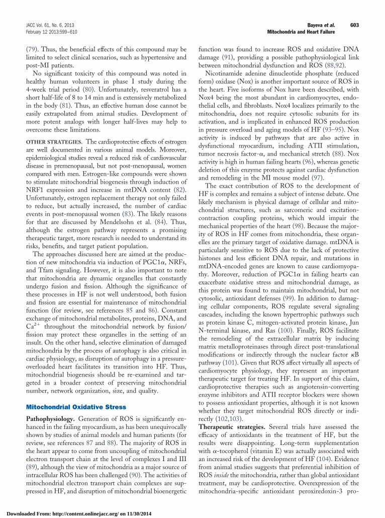
tected the heart against failure and remodeling in the mousemodel of MI (105). Similarly, overexpression ofmitochondria-targeted catalase attenuated hypertrophy inpressure-overload (106) and hypertensive (107) mousemodels. Thus, scavenging ROS within the mitochondriamay protect the heart against the development of HF andmake it more resistant to stressful stimuli. Several ap-proaches for targeting antioxidant compounds to the mito-chondria are being explored and hold promise (Fig. 2).
MITOQ. The best characterized mitochondria-targeted an-ioxidant to date is MitoQ, a quinol ROS scavenging moietyinked to triphenylphosphonium (TPP), a lipophilic com-ound that easily crosses membranes and accumulates in theitochondrial matrix as a function of membrane potential.
cavenging of ROS is achieved through oxidation of MitoQnto its quinone form, which is then recycled back into thective quinol by the action of mitochondrial complex II108). MitoQ is bioavailable orally with no toxicity detectedhen administered to mice at an �20-mg/kg dose. Tracer
tudies found the compound to be rapidly taken up into theeart, liver, brain, kidney, and muscle, with highest accu-ulation in the heart and liver (109). Long-term adminis-
ration of MitoQ had no effect on plasma glucose, insulin,ree fatty acid, or cholesterol levels, but was associated withignificantly reduced triglycerides. Affymetrix chip analysisf the heart and liver tissue of mice receiving MitoQevealed no significant differences in gene expression profileetween the treatment and control groups (110). Thus,
Figure 2 Targeting Mitochondrial ROS Production
Mitochondrial electron transport chain (ETC) complexes and nicotinamide adenineamounts of reactive oxygen species (ROS) in failing hearts. Moreover, mitochondrifailure (HF). Although nonspecific antioxidants, such as vitamin E, show no benefitapproaches to targeting antioxidant compounds to mitochondria, including triphenmanganese superoxide dismutase/catalase mimetics, should be explored in the d
ded From: http://content.onlinejacc.org/ on 11/30/2014
itoQ is a safe, orally available small molecule that does notignificantly alter baseline physiology.
In WT mice, MitoQ does not lead to a significanteduction in oxidative stress at baseline. However, admin-stration of MitoQ to rats for 2 weeks reduced oxidativetress and protected the heart against I/R injury using an exivo Langendorff setup. These effects were specifically dueo the inhibition of ROS inside the mitochondria, as norotection was observed in control groups receiving eitherethyl-TPP, which can enter the mitochondria but does
ot scavenge ROS, or short-chain antioxidant quinol, whichs impermeable to the mitochondrial membrane (111).hese findings were later confirmed in a mouse model of
/R and in an established cardiac cell line (112). MitoQ alsoreserved cardiac function in a spontaneously hypertensiveat model of HF. However, this favorable outcome may alsoe attributed to the reduction in blood pressure and im-rovement in endothelial function observed in the MitoQroup (113). Finally, MitoQ was found to be protective inther models of mitochondrial oxidative stress, includingardiac damage by doxorubicin (114), liver damage byipopolysaccharide (115), and protection of substantia nigrarom 1-methyl-4-phenyl-1,2,3,6-tetrahydropryridine (mi-ochondrial permeability transition pore) toxicity (116).
Two human trials assessed MitoQ’s efficacy in the treat-ent of Parkinson’s disease (PROTECT) and in patientsith chronic hepatitis C infection. Although the results of
he PROTECT trial were negative, it provided a wealth ofata on the safety of the drug administered orally for as long
eotide phosphate (reduced form) oxidase 4 (Nox4) enzyme generate excessivevery sensitive to oxidative stress, and their function is severely impaired in heart, targeting of ROS-scavenging molecules to mitochondria is protective. Variousphonium conjugation (MitoQ), Szeto-Schiller peptides, and synthesis of novelment of HF treatments.
dinucla arein HF
ylphosevelop

eo
dsmpsadooMabtmfbHrd
stahpsEbttcdmp
mIiviocmIstsiwitp(i
sdafsEotivnfObom
605JACC Vol. 61, No. 6, 2013 Bayeva et al.February 12 2013:599–610 Mitochondria and Heart Failure
Downloa
as 1 year (117). On the other hand, the patients receiving 40and 80 mg MitoQ in the chronic hepatitis C trial showedsignificant improvement in hepatic function (118). Impor-tantly, no severe side effects of the MitoQ regimen werereported in either of the trials.
Despite the significant therapeutic potential of MitoQand other TPP-conjugated antioxidants, there are limita-tions. The uptake of these compounds is governed by themitochondrial membrane potential, which may be severelydisrupted in failing hearts. Moreover, accumulation ofcationic TPP in the matrix can potentially depolarizemitochondria, leading to unwanted side effects.
SZETO-SCHILLER PEPTIDES. Unlike TPP conjugates, thesmall (�10 amino acids) Szeto-Schiller (SS) peptides selec-tively accumulate in the mitochondrial matrix independentof membrane potential. SS peptides are rapidly taken up bythe mitochondria, with 1,000- to 5,000-fold accumulationin this organelle compared with the cytosolic compartment(119). Multiple variants of SS molecules have been synthe-sized to date, and the tyrosine-containing SS-02 and SS-31peptides hold therapeutic promise due to their antioxidantproperties (120). These products reduced mitochondrialROS production in cells treated with mitochondrial com-plex I (121) and II and III (119) inhibitors. SS compoundswere also shown to be protective in vivo.
Administration of SS-31 before ischemia and beforereperfusion reduced MI size, lipid peroxidation indexes,and increased ATP content in the rat heart (122).Recently, SS-31, but not the nontargeted antioxidantN-acetylcysteine, was shown to protect the heart fromcardiomyopathy due to angiotensin II administration orG�q overexpression in mice (123), providing the firstvidence of SS-31 effectiveness in a more chronic modelf cardiac dysfunction.Although peptides are typically considered poor candi-
ates for drug development due to the issues of solubility,tability, rapid clearance, and inability to cross cellularembranes, studies have revealed excellent pharmacokinetic
roperties of the SS peptides. These molecules are wateroluble due to the 3� net charge and are stable in anqueous solution at 37°C for 6 months. They can beelivered via the intravenous, intraperitoneal, or subcutane-us route and are rapidly distributed to highly perfusedrgans, including the heart, kidney, lung, and brain (120).oreover, enzymatic degradation of the SS peptides is low,
nd they remain stable even after 2 h of incubation in wholelood (124). Finally, the toxicity of SS compounds is low atherapeutic doses, with no side effects observed after 5onths of daily treatments in mice (125). Given these
avorable pharmacokinetic properties, SS peptides appear toe good candidates for further testing in the treatment ofF. However, additional animal and human studies are
equired to validate these compounds as therapeutic candi-
ates. pded From: http://content.onlinejacc.org/ on 11/30/2014
MANGANESE SUPEROXIDE DISMUTASE/CATALASE MIMETICS.
Superoxide dismutases (SODs) are metal-containing anti-oxidant enzymes that catalyze the conversion of superoxideradical to hydrogen peroxide and O2. The mitochondria-pecific manganese SOD (MnSOD or SOD2) is located inhe matrix, and its overexpression was shown to protectgainst HF (126). Several inorganic MnSOD mimeticsave been synthesized, and many of these compounds exertrotection in conditions associated with oxidative stress (forreview, see reference 127). Salen derivatives, such asUK-8 and EUK-134, possess antioxidant properties ofoth MnSOD and catalases and appear to be effective inhe heart. Although no studies have assessed the ability ofhese molecules to penetrate mitochondria directly, theirhemical properties (small, lipophilic, water soluble) andocumented ability to reduce mitochondrial ROS andaintain activities of mitochondrial enzymes (128) sup-
ort this assumption.Both EUK-8 and EUK-134 were found to protectitochondria and the heart against various oxidative insults.
n an early study by Pucheu et al. (129), EUK-8 protectedron-overloaded rat hearts from I/R injury, maintained leftentricular diastolic pressure, and preserved mitochondrialntegrity. EUK-8 treatment also prevented the developmentf cardiomyopathy and maintained contractility and ATPontent in the heart/muscle-specific MnSOD2 knockoutice, in which HF develops as early as 4 weeks after birth.
mportantly, ROS generation by isolated mitochondria wasignificantly reduced by EUK-8 in these mice, suggestinghe ability of this molecule to offset mitochondrial oxidativetress (130). Finally, EUK-8 ameliorated pressure overload-nduced cardiac dysfunction in wild-type mice and miceith the deletion of mitochondrial antioxidant, apoptosis-
nducing factor (131). EUK-134, a more lipophilic deriva-ive of EUK-8, exhibited similar protective properties inulmonary arterial hypertension–induced HF (132) and I/R133) and reduced apoptosis in norepinephrine-stimulatedsolated adult rat ventricular myocytes (134).
New SOD/catalase mimetics are being developed andtudied, thus opening up an exciting new therapeuticirection. However, most evidence of the mitoprotectivend cardioprotective properties of these molecules comerom genetic models of increased mitochondrial oxidativetress. The mechanism of cardiac protection conferred byUK-8/EUK-134 and related compounds must be thor-ughly investigated. In particular, cardioprotective proper-ies of these molecules may be independent of their antiox-dant effects, as EUK-8 was reported to possess significantasodilatory properties, which may increase oxygen andutrient delivery to the heart and indirectly improve heartunction (135).
ther strategies. As our understanding of the chemistryehind mitochondrial targeting is increasing, rational designf novel therapies holds promise. A number of antioxidantoieties have been conjugated to TPP and appear to confer
rotection as well as control the degree of antioxidation and
pg
606 Bayeva et al. JACC Vol. 61, No. 6, 2013Mitochondria and Heart Failure February 12 2013:599–610
Downloa
the duration of the effect (108). In addition to synthesizingnew molecules, it is important to understand various signal-ing pathways that regulate mitochondrial antioxidantdefenses. A recent report by Lu et al. (99) foundantioxidant enzyme levels to be significantly decreased inPGC1� knockout mice with pressure-overload hypertro-
hy, suggesting that enhancement of mitochondrial bio-enesis and PGC1� expression by the strategies dis-
cussed earlier may have a positive effect on an antioxidantprofile as well.
Mitochondrial Iron Homeostasis
Pathophysiology. Although the role of mitochondrial ironin HF has not been explicitly studied, indirect evidencepoints toward potential therapeutic implications of alteringmitochondrial iron homeostasis in the diseased heart. Iron isessential for maintenance of cellular viability and functionthrough its role in oxidative phosphorylation, antioxidantenzyme activities, ribosome biogenesis, oxygen storage anddelivery, and more (136). Mitochondria are the key sites ofcellular iron processing where synthesis of iron/sulfur (Fe/S)clusters and heme takes place, but are also the place whereROS are generated (137). Being a reactive metal, free ironcatalyzes production of highly toxic hydroxyl radicals fromless reactive species such as hydrogen peroxide and super-oxide anion via the Fenton reaction (138).
Although many studies examined changes in systemiciron homeostasis in animals and humans with HF, very littlework has been done to characterize the intrinsic defects iniron regulatory pathways of failing cardiomyocytes. In par-
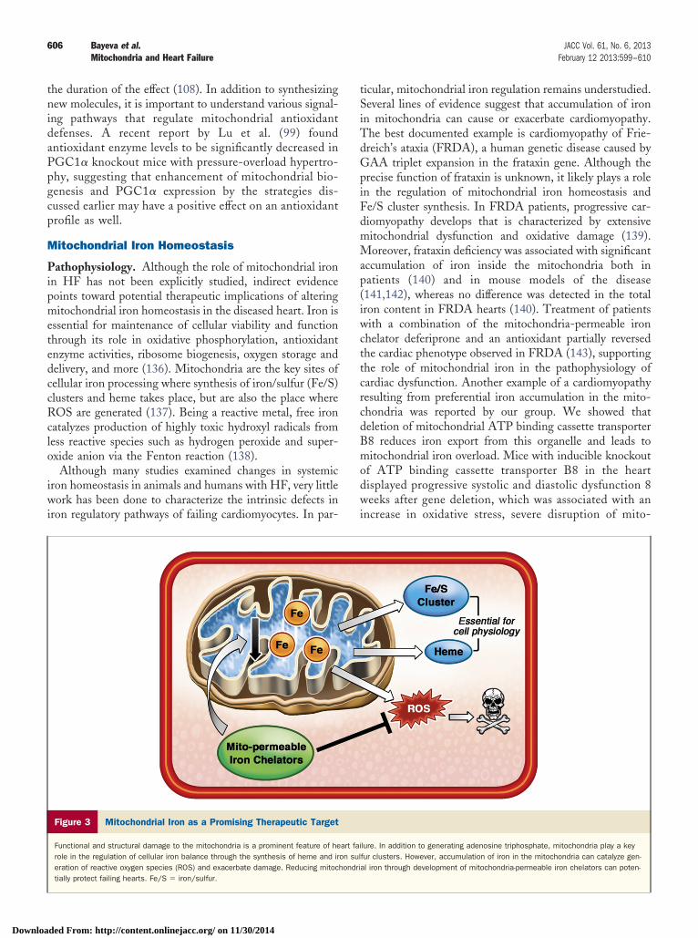
Figure 3 Mitochondrial Iron as a Promising Therapeutic Target
Functional and structural damage to the mitochondria is a prominent feature of herole in the regulation of cellular iron balance through the synthesis of heme and ireration of reactive oxygen species (ROS) and exacerbate damage. Reducing mitoctially protect failing hearts. Fe/S � iron/sulfur.
ded From: http://content.onlinejacc.org/ on 11/30/2014
ticular, mitochondrial iron regulation remains understudied.Several lines of evidence suggest that accumulation of ironin mitochondria can cause or exacerbate cardiomyopathy.The best documented example is cardiomyopathy of Frie-dreich’s ataxia (FRDA), a human genetic disease caused byGAA triplet expansion in the frataxin gene. Although theprecise function of frataxin is unknown, it likely plays a rolein the regulation of mitochondrial iron homeostasis andFe/S cluster synthesis. In FRDA patients, progressive car-diomyopathy develops that is characterized by extensivemitochondrial dysfunction and oxidative damage (139).Moreover, frataxin deficiency was associated with significantaccumulation of iron inside the mitochondria both inpatients (140) and in mouse models of the disease(141,142), whereas no difference was detected in the totaliron content in FRDA hearts (140). Treatment of patientswith a combination of the mitochondria-permeable ironchelator deferiprone and an antioxidant partially reversedthe cardiac phenotype observed in FRDA (143), supportingthe role of mitochondrial iron in the pathophysiology ofcardiac dysfunction. Another example of a cardiomyopathyresulting from preferential iron accumulation in the mito-chondria was reported by our group. We showed thatdeletion of mitochondrial ATP binding cassette transporterB8 reduces iron export from this organelle and leads tomitochondrial iron overload. Mice with inducible knockoutof ATP binding cassette transporter B8 in the heartdisplayed progressive systolic and diastolic dysfunction 8weeks after gene deletion, which was associated with anincrease in oxidative stress, severe disruption of mito-
lure. In addition to generating adenosine triphosphate, mitochondria play a keyfur clusters. However, accumulation of iron in the mitochondria can catalyze gen-al iron through development of mitochondria-permeable iron chelators can poten-
art faion sulhondri

SwhdaaTitudoTtmtertagcdmmrvtafiFncetaanorrtm2t
C
MnppapyirIsb
607JACC Vol. 61, No. 6, 2013 Bayeva et al.February 12 2013:599–610 Mitochondria and Heart Failure
Downloa
chondrial architecture, and the presence of mitochondrialiron aggregates (144).
Whether iron accumulates in the mitochondria offailing hearts due to common etiologies such as hyper-tension and MI has not been examined directly. Onestudy used electron paramagnetic resonance spectroscopyto analyze the iron status of the failing hearts in themouse model of cardiac-specific overexpression of G�q.
ignificant accumulation of iron inside cardiomyocytesas noted, although the mitochondria-specific iron poolas not been examined. However, activities of mitochon-rial Fe/S cluster proteins were reduced in failing hearts,nd the defect in complex III activity was specificallyttributed to the lack of a Fe/S cluster center (145).hese findings suggest deregulation of mitochondrial
ron processing in at least 1 model of chronic HF. Thus,he defects in mitochondrial iron handling may contrib-te to the development of HF through an ROS-ependent mechanism and through potential disruptionf Fe/S cluster biogenesis.herapeutic strategies. Extensive characterization of mi-
ochondrial iron homeostasis in various models of HFust be performed to target these pathways in the
ranslational studies. Reducing mitochondrial iron mayxert cardioprotection through inhibition of hydroxyladical formation and alleviation of oxidative stress (Fig. 3). Theherapy must be precisely targeted to the mitochondria,s iron homeostasis is often disrupted in HF patients andlobal iron deficiency is common (146). Instead ofhelating iron on a systemic level and exacerbating ironeficiency, the drug must remove free iron from theitochondria and donate it to other cellular compart-ents. Such redistribution of iron within a cell was
eported for deferiprone (DFP), an orally available re-erse siderophore iron chelator. DFP was shown to enterhe cells, reduce mitochondrial free iron levels, exit cellss an iron chelate, and transfer chelated iron to apotrans-errin in the blood, thus potentially increasing systemicron availability (147). Oral administration of DFP toRDA patients for 6 months significantly improvedeurological symptoms and reduced iron accumulation inerebellar dentate nuclei, with no hematological sideffects noted (148). Moreover, DFP and idebenonereatment led to a partial reversal of FRDA cardiomyop-thy in human patients (143). In addition to DFP,nalogs of hydrophobic iron chelator pyridoxal isonicoti-oyl hydrazone, such as 2-pyridylcarboxaldehyde isonic-tinoyl hydrazone were shown to selectively removeadioactive isotopes of iron from the mitochondria ofabbit reticulocytes (149) and were proposed as a poten-ial therapy for FRDA and other diseases associated withitochondrial iron overload (150). The effects of
-pyridylcarboxaldehyde isonicotinoyl hydrazone on sys-
emic iron homeostasis have not yet been examined.ded From: http://content.onlinejacc.org/ on 11/30/2014
onclusions
itochondria are taking the center stage in our search forovel cardioprotective therapies, as their dysfunction ap-ears early and invariably in the development of hypertro-hy and HF. Maintenance of mitochondrial biogenesisgainst cardiac insults and reduction in mitochondrial ROSroduction are the 2 promising directions that may soonield effective treatments. Moreover, exploration and target-ng of other vital mitochondrial processes in HF, includingegulation of iron homeostasis, should be actively pursued.mportantly, our basic research and translational effortshould focus on targeting the intrinsic processes of viable,ut dysfunctional, cardiomyocytes.
Reprint requests and correspondence: Dr. Hossein Ardehali,Feinberg Cardiovascular Research Institute, Northwestern Univer-sity Medical Center, Tarry 14-733, 303 East Chicago Avenue,Chicago, Illinois 60611. E-mail: [email protected].
REFERENCES
1. Lloyd-Jones D, Adams RJ, Brown TM, et al. Heart disease andstroke statistics–2010 update: a report from the American HeartAssociation. Circulation 2010;121:e46–215.
2. Gheorghiade M, Peterson ED. Improving postdischarge outcomes inpatients hospitalized for acute heart failure syndromes. JAMA 2011;305:2456–7.
3. Dai DF, Chen T, Wanagat J, et al. Age-dependent cardiomyopathyin mitochondrial mutator mice is attenuated by overexpression ofcatalase targeted to mitochondria. Aging Cell 2010;9:536–44.
4. Cizkova A, Stranecky V, Mayr JA, et al. TMEM70 mutations causeisolated ATP synthase deficiency and neonatal mitochondrial en-cephalocardiomyopathy. Nat Genet 2008;40:1288–90.
5. Mayr JA, Haack TB, Graf E, et al. Lack of the mitochondrial proteinacylglycerol kinase causes Sengers syndrome. Am J Hum Genet2012;90:314–20.
6. Sanbe A, Tanonaka K, Kobayasi R, Takeo S. Effects of long-termtherapy with ACE inhibitors, captopril, enalapril and trandolapril, onmyocardial energy metabolism in rats with heart failure followingmyocardial infarction. J Mol Cell Cardiol 1995;27:2209–222.
7. Feng X, Luo Z, Ma L, et al. Angiotensin II receptor blockertelmisartan enhances running endurance of skeletal muscle throughactivation of the PPAR-delta/AMPK pathway. J Cell Mol Med2011;15:1572–81.
8. Ardehali H, Sabbah HN, Burke MA, et al. Targeting myocardialsubstrate metabolism in heart failure: potential for new therapies. EurJ Heart Fail 2012;14:120–9.
9. Garesse R, Vallejo CG. Animal mitochondrial biogenesis and func-tion: a regulatory cross-talk between two genomes. Gene 2001;263:1–16.
10. Puigserver P, Wu Z, Park CW, et al. A cold-inducible coactivator ofnuclear receptors linked to adaptive thermogenesis. Cell 1998;92:829–39.
11. Ventura-Clapier R, Garnier A, Veksler V. Transcriptional control ofmitochondrial biogenesis: the central role of PGC-1alpha. Cardio-vasc Res 2008;79:208–17.
12. Kelly DP, Scarpulla RC. Transcriptional regulatory circuits controllingmitochondrial biogenesis and function. Genes Dev 2004;18:357–68.
13. Lehman JJ, Barger PM, Kovacs A, et al. Peroxisome proliferator-activated receptor gamma coactivator-1 promotes cardiac mitochon-drial biogenesis. J Clin Invest 2000;106:847–56.
14. Russell LK, Mansfield CM, Lehman JJ, et al. Cardiac-specificinduction of the transcriptional coactivator peroxisome proliferator-activated receptor gamma coactivator-1alpha promotes mitochondrialbiogenesis and reversible cardiomyopathy in a developmental stage-
dependent manner. Circ Res 2004;94:525–33.
608 Bayeva et al. JACC Vol. 61, No. 6, 2013Mitochondria and Heart Failure February 12 2013:599–610
Downloa
15. Wu Z, Puigserver P, Andersson U, et al. Mechanisms controllingmitochondrial biogenesis and respiration through the thermogeniccoactivator PGC-1. Cell 1999;98:115–24.
16. Dufour CR, Wilson BJ, Huss JM, et al. Genome-wide orchestrationof cardiac functions by the orphan nuclear receptors ERRalpha andgamma. Cell Metab 2007;5:345–56.
17. Huss JM, Torra IP, Staels B, et al. Estrogen-related receptor alphadirects peroxisome proliferator-activated receptor alpha signaling inthe transcriptional control of energy metabolism in cardiac andskeletal muscle. Mol Cell Biol 2004;24:9079–91.
18. Huo L, Scarpulla RC. Mitochondrial DNA instability and peri-implantation lethality associated with targeted disruption of nuclearrespiratory factor 1 in mice. Mol Cell Biol 2001;21:644–54.
19. Huss JM, Imahashi K, Dufour CR, et al. The nuclear receptorERRalpha is required for the bioenergetic and functional adaptationto cardiac pressure overload. Cell Metab 2007;6:25–37.
20. Larsson NG, Wang J, Wilhelmsson H, et al. Mitochondrial tran-scription factor A is necessary for mtDNA maintenance and embryo-genesis in mice. Nat Genet 1998;18:231–6.
21. Garnier A, Fortin D, Delomenie C, et al. Depressed mitochondrialtranscription factors and oxidative capacity in rat failing cardiac andskeletal muscles. J Physiol 2003;551:491–501.
22. Witt H, Schubert C, Jaekel J, et al. Sex-specific pathways in earlycardiac response to pressure overload in mice. J Mol Med (Berl)2008;86:1013–24.
23. Faerber G, Barreto-Perreia F, Schoepe M, et al. Induction of heartfailure by minimally invasive aortic constriction in mice: reducedperoxisome proliferator-activated receptor gamma coactivator levelsand mitochondrial dysfunction. J Thorac Cardiovasc Surg 2011;141:492–500, 500.e491.
24. Marin-Garcia J, Goldenthal MJ, Damle S, et al. Regional distribu-tion of mitochondrial dysfunction and apoptotic remodeling inpacing-induced heart failure. J Card Fail 2009;15:700–8.
25. Sebastiani M, Giordano C, Nediani C, et al. Induction of mitochon-drial biogenesis is a maladaptive mechanism in mitochondrial cardio-myopathies. J Am Coll Cardiol 2007;50:1362–9.
26. Sun CK, Chang LT, Sheu JJ, et al. Losartan preserves integrity ofcardiac gap junctions and PGC-1 alpha gene expression and preventscellular apoptosis in remote area of left ventricular myocardiumfollowing acute myocardial infarction. Int Heart J 2007;48:533–46.
27. Watson PA, Reusch JE, McCune SA, et al. Restoration of CREBfunction is linked to completion and stabilization of adaptive cardiachypertrophy in response to exercise. Am J Physiol Heart Circ Physiol2007;293:H246–59.
28. Hu X, Xu X, Lu Z, et al. AMP activated protein kinase-alpha2regulates expression of estrogen-related receptor-alpha, a metabolictranscription factor related to heart failure development. Hyperten-sion 2011;58:696–703.
29. Sihag S, Cresci S, Li AY, et al. PGC-1alpha and ERRalpha targetgene downregulation is a signature of the failing human heart. J MolCell Cardiol 2009;46:201–12.
30. Karamanlidis G, Bautista-Hernandez V, Fynn-Thompson F, et al.Impaired mitochondrial biogenesis precedes heart failure in rightventricular hypertrophy in congenital heart disease. Circ Heart Fail2011;4:707–13.
31. Barger PM, Browning AC, Garner AN, Kelly DP. p38 mitogen-activated protein kinase activates peroxisome proliferator-activatedreceptor alpha: a potential role in the cardiac metabolic stressresponse. J Biol Chem 2001;276:44495–501.
32. Rodgers JT, Lerin C, Haas W, et al. Nutrient control of glucosehomeostasis through a complex of PGC-1alpha and SIRT1. Nature2005;434:113–8.
33. Puigserver P, Rhee J, Lin J, et al. Cytokine stimulation of energyexpenditure through p38 MAP kinase activation of PPARgammacoactivator-1. Mol Cell 2001;8:971–82.
34. Karamanlidis G, Nascimben L, Couper GS, et al. Defective DNAreplication impairs mitochondrial biogenesis in human failing hearts.Circ Res 2010;106:1541–8.
35. Ikeuchi M, Matsusaka H, Kang D, et al. Overexpression of mito-chondrial transcription factor a ameliorates mitochondrial deficien-cies and cardiac failure after myocardial infarction. Circulation2005;112:683–90.
36. Yanagishita T, Tomita M, Itoh S, et al. Protective effect of captopril
on ischemic myocardium. Jpn Circ J 1997;61:161–9.ded From: http://content.onlinejacc.org/ on 11/30/2014
37. Li J, Coven DL, Miller EJ, et al. Activation of AMPK alpha- andgamma-isoform complexes in the intact ischemic rat heart. Am JPhysiol Heart Circ Physiol 2006;291:H1927–34.
38. Tian R, Musi N, D’Agostino J, et al. Increased adenosinemonophosphate-activated protein kinase activity in rat hearts withpressure-overload hypertrophy. Circulation 2001;104:1664–9.
39. Gibala MJ, McGee SL, Garnham AP, et al. Brief intense intervalexercise activates AMPK and p38 MAPK signaling and increases theexpression of PGC-1alpha in human skeletal muscle. J Appl Physiol2009;106:929–34.
40. Jager S, Handschin C, St-Pierre J, Spiegelman BM. AMP-activatedprotein kinase (AMPK) action in skeletal muscle via direct phos-phorylation of PGC-1alpha. Proc Natl Acad Sci U S A 2007;104:12017–22.
41. Kukidome D, Nishikawa T, Sonoda K, et al. Activation of AMP-activated protein kinase reduces hyperglycemia-induced mitochon-drial reactive oxygen species production and promotes mitochondrialbiogenesis in human umbilical vein endothelial cells. Diabetes 2006;55:120–7.
42. Kim M, Shen M, Ngoy S, et al. AMPK isoform expression in thenormal and failing hearts. J Mol Cell Cardiol 2012;52:1066–73.
43. Yin M, van der Horst IC, van Melle JP, et al. Metformin improvescardiac function in a nondiabetic rat model of post-MI heart failure.Am J Physiol Heart Circ Physiol 2011;301:H459–68.
44. Sasaki H, Asanuma H, Fujita M, et al. Metformin prevents progres-sion of heart failure in dogs: role of AMP-activated protein kinase.Circulation 2009;119:2568–77.
45. Gundewar S, Calvert JW, Jha S, et al. Activation of AMP-activatedprotein kinase by metformin improves left ventricular function andsurvival in heart failure. Circ Res 2009;104:403–411.
46. Owen MR, Doran E, Halestrap AP. Evidence that metformin exertsits anti-diabetic effects through inhibition of complex 1 of themitochondrial respiratory chain. Biochem J 2000;3483:607–14.
47. Zhou G, Myers R, Li Y, Chen Y, et al. Role of AMP-activatedprotein kinase in mechanism of metformin action. J Clin Invest2001;108:1167–74.
48. Sanders MJ, Ali ZS, Hegarty BD, et al. Defining the mechanism ofactivation of AMP-activated protein kinase by the small moleculeA-769662, a member of the thienopyridone family. J Biol Chem2007;282:32539–48.
49. Song T, Lv LY, Xu J, et al. Diet-induced obesity suppressessevoflurane preconditioning against myocardial ischemia-reperfusioninjury: role of AMP-activated protein kinase pathway. Exp Biol Med(Maywood) 2011;236:1427–36.
50. Moreno D, Knecht E, Viollet B, Sanz P. A769662, a novel activatorof AMP-activated protein kinase, inhibits non-proteolytic compo-nents of the 26S proteasome by an AMPK-independent mechanism.FEBS Lett 2008;582:2650–4.
51. Pang T, Zhang ZS, Gu M, et al. Small molecule antagonizesautoinhibition and activates AMP-activated protein kinase in cells.J Biol Chem 2008;283:16051–60.
52. Benes J, Kazdova L, Drahota Z, et al. Effect of metformin therapy oncardiac function and survival in a volume-overload model of heartfailure in rats. Clin Sci (Lond) 2011;121:29–41.
53. Kim M, Tian R. Targeting AMPK for cardiac protection: opportu-nities and challenges. J Mol Cell Cardiol 2011;51:548–53.
54. Blair E, Redwood C, Ashrafian H, et al. Mutations in the gamma(2)subunit of AMP-activated protein kinase cause familial hypertrophiccardiomyopathy: evidence for the central role of energy compromisein disease pathogenesis. Hum Mol Genet 2001;10:1215–20.
55. Balligand JL, Feron O, Dessy C. eNOS activation by physical forces:from short-term regulation of contraction to chronic remodeling ofcardiovascular tissues. Physiol Rev 2009;89:481–534.
56. Manoury B, Montiel V, Balligand JL. Nitric oxide synthase inpost-ischaemic remodelling: new pathways and mechanisms. Cardio-vasc Res 2012;94:304–15.
57. Clementi E, Nisoli E. Nitric oxide and mitochondrial biogenesis: akey to long-term regulation of cellular metabolism. Comp BiochemPhysiol A Mol Integr Physiol 2005;142:102–10.
58. Brown GC. Nitric oxide and mitochondria. Front Biosci 2007;12:1024–33.
59. Miyashita K, Itoh H, Tsujimoto H, et al. Natriuretic peptides/
cGMP/cGMP-dependent protein kinase cascades promote muscle
1
1
1
1
1
1
609JACC Vol. 61, No. 6, 2013 Bayeva et al.February 12 2013:599–610 Mitochondria and Heart Failure
Downloa
mitochondrial biogenesis and prevent obesity. Diabetes 2009;58:2880–92.
60. Nisoli E, Clementi E, Tonello C, et al. Effects of nitric oxide onproliferation and differentiation of rat brown adipocytes in primarycultures. Br J Pharmacol 1998;125:888–94.
61. Nisoli E, Clementi E, Paolucci C, et al. Mitochondrial biogenesis inmammals: the role of endogenous nitric oxide. Science 2003;299:896–9.
62. De Toni L, Strapazzon G, Gianesello L, et al. Effects of type5-phosphodiesterase inhibition on energy metabolism and mitochon-drial biogenesis in human adipose tissue ex vivo. J Endocrinol Invest2011;34:738–41.
63. Schwartz BG, Levine LA, Comstock G, et al. Cardiac uses ofphosphodiesterase-5 inhibitors. J Am Coll Cardiol 2012;59:9–15.
64. Cvelich RG, Roberts SC, Brown JN. Phosphodiesterase type 5inhibitors as adjunctive therapy in the management of systolic heartfailure. Ann Pharmacother 2011;45:1551–8.
65. Moens AL, Takimoto E, Tocchetti CG, et al. Reversal of cardiachypertrophy and fibrosis from pressure overload by tetrahydrobiop-terin: efficacy of recoupling nitric oxide synthase as a therapeuticstrategy. Circulation 2008;117:2626–36.
66. Moens AL, Ketner EA, Takimoto E, et al. Bi-modal dose-dependent cardiac response to tetrahydrobiopterin in pressure-overload induced hypertrophy and heart failure. J Mol Cell Cardiol2011;51:564–9.
67. Chou YF, Yu CC, Huang RF. Changes in mitochondrial DNAdeletion, content, and biogenesis in folate-deficient tissues of youngrats depend on mitochondrial folate and oxidative DNA injuries. JNutr 2007;137:2036–42.
68. Ceylan-Isik AF, Guo KK, Carlson EC, et al. Metallothioneinabrogates GTP cyclohydrolase I inhibition-induced cardiac contrac-tile and morphological defects: role of mitochondrial biogenesis.Hypertension 2009;53:1023–31.
69. Moens AL, Champion HC, Claeys MJ, et al. High-dose folic acidpretreatment blunts cardiac dysfunction during ischemia coupled tomaintenance of high-energy phosphates and reduces postreperfusioninjury. Circulation 2008;117:1810–9.
70. Brown GC, Cooper CE. Nanomolar concentrations of nitric oxidereversibly inhibit synaptosomal respiration by competing with oxygenat cytochrome oxidase. FEBS Lett 1994;356:295–8.
71. Vieira HL, Belzacq AS, Haouzi D, et al. The adenine nucleotidetranslocator: a target of nitric oxide, peroxynitrite, and4-hydroxynonenal. Oncogene 2001;20:4305–16.
72. Lagouge M, Argmann C, Gerhart-Hines Z, et al. Resveratrol improvesmitochondrial function and protects against metabolic disease by acti-vating SIRT1 and PGC-1alpha. Cell 2006;127:1109–22.
73. Takahashi S, Nakashima Y. Repeated and long-term treatment withphysiological concentrations of resveratrol promotes NO productionin vascular endothelial cells. Br J Nutr 2012;107:774–80.
74. Zang M, Xu S, Maitland-Toolan KA, et al. Polyphenols stimulateAMP-activated protein kinase, lower lipids, and inhibit acceleratedatherosclerosis in diabetic LDL receptor-deficient mice. Diabetes2006;55:2180–91.
75. Thandapilly SJ. Wojciechowski P, Behbahani J, et al. Resveratrolprevents the development of pathological cardiac hypertrophy andcontractile dysfunction in the SHR without lowering blood pressure.Am J Hypertens 2010;23:192–6.
76. Biala A, Tauriainen E, Siltanen A, et al. Resveratrol inducesmitochondrial biogenesis and ameliorates Ang II-induced cardiacremodeling in transgenic rats harboring human renin and angio-tensinogen genes. Blood Press 2010;19:196–205.
77. Rimbaud S, Ruiz M, Piquereau J, et al. Resveratrol improves survival,hemodynamics and energetics in a rat model of hypertension leadingto heart failure. PLoS One 2011;6:e26391.
78. Magyar K, Halmosi R, Palfi A, et al. Cardioprotection by resveratrol:A human clinical trial in patients with stable coronary artery disease.Clin Hemorheol Microcirc 2012;50:179–187.
79. Wojciechowski P, Juric D, Louis XL, et al. Resveratrol arrests andregresses the development of pressure overload- but not volumeoverload-induced cardiac hypertrophy in rats. J Nutr 2010;140:962–68.
80. Patel KR, Scott E, Brown VA, et al. Clinical trials of resveratrol. Ann
N Y Acad Sci 2011;1215:161–9.ded From: http://content.onlinejacc.org/ on 11/30/2014
81. Wang H, Yang YJ, Qian HY, et al. Resveratrol in cardiovasculardisease: what is known from current research? Heart Fail Rev2012;17:437–48.
82. Mattingly KA, Ivanova MM, Riggs KA, et al. Estradiol stimulatestranscription of nuclear respiratory factor-1 and increases mitochon-drial biogenesis. Mol Endocrinol 2008;22:609–22.
83. Anderson GL, Limacher M, Assaf AR, et al. Effects of conjugatedequine estrogen in postmenopausal women with hysterectomy: theWomen’s Health Initiative randomized controlled trial. JAMA 2004;291:1701–12.
84. Mendelsohn ME, Karas RH. HRT and the young at heart. N EnglJ Med 2007;356:2639–41.
85. Iglewski M, Hill JA, Lavandero, Rothermel BA. Mitochondrialfission and autophagy in the normal and diseased heart. CurrHypertens Rep 2010;12:418–25.
86. Palaniyandi SS, Qi X, Yogalingam G, et al. Regulation of mitochon-drial processes: a target for heart failure. Drug Discov Today DisMech 2010;7:e95–102.
87. Tsutsui H, Kinugawa S, Matsushima, S. Oxidative stress and heartfailure. Am J Physiol Heart Circ Physiol 2011;301:H2181–90.
88. Tsutsui H, Kinugawa S, Matsushima S. Oxidative stress and mito-chondrial DNA damage in heart failure. Circ J 2008;72 SupplA:A31–7.
89. Ide T, Tsutsui H, Kinugawa S, et al. Mitochondrial electrontransport complex I is a potential source of oxygen free radicals in thefailing myocardium. Circ Res 1999;85:357–63.
90. Brown GC, Borutaite V. There is no evidence that mitochondria arethe main source of reactive oxygen species in mammalian cells.Mitochondrion 2012;12:1–4.
91. Sung HJ, Ma W, Wang PY, et al. Mitochondrial respiration protectsagainst oxygen-associated DNA damage. Nat Commun 2010;1:5.
92. Ide T, Tsutsui H, Hayashidani S, et al. Mitochondrial DNA damageand dysfunction associated with oxidative stress in failing hearts aftermyocardial infarction. Circ Res 2001;88:529–35.
93. Lambeth JD. NOX enzymes and the biology of reactive oxygen. NatRev Immunol 2004;4:181–9.
94. Ago T, Kuroda J, Pain J, et al. Upregulation of Nox4 by hypertrophicstimuli promotes apoptosis and mitochondrial dysfunction in cardiacmyocytes. Circ Res 2010;106:1253–64.
95. Kuroda J, Ago T, Matsushima S, et al. NADPH oxidase 4 (Nox4) isa major source of oxidative stress in the failing heart. Proc Natl AcadSci U S A 2010;107:15565–70.
96. Heymes C, Bendall JK, Ratajczak P, et al. Increased myocardialNADPH oxidase activity in human heart failure. J Am Coll Cardiol2003;41:2164–71.
97. Doerries C, Grote K, Hilfiker-Kleiner D, et al. Critical role of theNAD(P)H oxidase subunit p47phox for left ventricular remodeling/dysfunction and survival after myocardial infarction. Circ Res 2007;100:894–903.
98. Bayeva M, Ardehali H. Mitochondrial dysfunction and oxidative dam-age to sarcomeric proteins. Curr Hypertens Rep 2010;12:426–32.
99. Lu Z, Xu X, Hu X, et al. PGC-1 alpha regulates expression ofmyocardial mitochondrial antioxidants and myocardial oxidativestress after chronic systolic overload. Antioxid Redox Signal 2010;13:1011–22.
00. Kwon SH, Pimentel DR, Remondino A, et al. H(2)O(2) regulatescardiac myocyte phenotype via concentration-dependent activation ofdistinct kinase pathways. J Mol Cell Cardiol 2003;35:615–21.
01. Siwik DA, Colucci WS. Regulation of matrix metalloproteinases bycytokines and reactive oxygen/nitrogen species in the myocardium.Heart Fail Rev 2004;9:43–51.
02. Yamazaki T, Tanimoto M, Gohda T, et al. Combination effects ofenalapril and losartan on lipid peroxidation in the kidneys ofKK-Ay/Ta mice. Nephron Exp Nephrol 2009;113:e66–76.
03. Goyal BR, Mehta AA. Beneficial role of spironolactone, telmisartanand their combination on isoproterenol-induced cardiac hypertrophy.Acta Cardiol 2012;67:203–11.
04. Marchioli R, Levantesi G, Macchia A, et al. Vitamin E increases therisk of developing heart failure after myocardial infarction: Resultsfrom the GISSI-Prevenzione trial. J Cardiovasc Med (Hagerstown)2006;7:347–50.
05. Matsushima S, Ide T, Yamato M, et al. Overexpression of mitochon-drial peroxiredoxin-3 prevents left ventricular remodeling and failure
after myocardial infarction in mice. Circulation 2006;113:1779–86.
610 Bayeva et al. JACC Vol. 61, No. 6, 2013Mitochondria and Heart Failure February 12 2013:599–610
Downloa
106. Dai DF, Hsieh EJ, Liu Y, et al. Mitochondrial proteome remodellingin pressure overload-induced heart failure: the role of mitochondrialoxidative stress. Cardiovasc Res 2012;93:79–88.
107. Dai DF, Johnson SC, Villarin JJ, et al. Mitochondrial oxidative stressmediates angiotensin II-induced cardiac hypertrophy and Galphaqoverexpression-induced heart failure. Circ Res 2011;108:837–46.
108. Murphy MP, Smith RA. Targeting antioxidants to mitochondria byconjugation to lipophilic cations. Annu Rev Pharmacol Toxicol2007;47:629–56.
109. Smith RA, Porteous CM, Gane AM, Murphy MP. Delivery ofbioactive molecules to mitochondria in vivo. Proc Natl Acad Sci U SA 2003;100:5407–12.
110. Rodriguez-Cuenca S, Cocheme HM, Logan A, et al. Consequencesof long-term oral administration of the mitochondria-targeted anti-oxidant MitoQ to wild-type mice. Free Radic Biol Med 2010;48:161–72.
111. Adlam VJ, Harrison JC, Porteous CM, et al. Targeting an antioxi-dant to mitochondria decreases cardiac ischemia-reperfusion injury.FASEB J 2005;19:1088–95.
112. Neuzil J, Widen C, Gellert N, et al. Mitochondria transmit apoptosissignalling in cardiomyocyte-like cells and isolated hearts exposed toexperimental ischemia-reperfusion injury. Redox Rep 2007;12:148–62.
113. Graham D, Huynh NN, Hamilton CA, et al. Mitochondria-targetedantioxidant MitoQ10 improves endothelial function and attenuatescardiac hypertrophy. Hypertension 2009;54:322–8.
114. Chandran K, Aggarwal D, Migrino RQ, et al. Doxorubicin inacti-vates myocardial cytochrome c oxidase in rats: cardioprotection byMito-Q. Biophys J 2009;96:1388–98.
115. Lowes DA, Thottakam BM, Webster NR, et al. The mitochondria-targeted antioxidant MitoQ protects against organ damage in alipopolysaccharide-peptidoglycan model of sepsis. Free Radic BiolMed 2008;45:1559–65.
116. Yang L, Zhao K, Calingasan NY, et al. Mitochondria targeted peptidesprotect against 1-methyl-4-phenyl-1,2,3,6-tetrahydropyridine neurotox-icity. Antioxid Redox Signal 2009;11:2095–104.
117. Snow BJ, Rolfe FL, Lockhart MM, et al. A double-blind, placebo-controlled study to assess the mitochondria-targeted antioxidantMitoQ as a disease-modifying therapy in Parkinson’s disease. MovDisord 2010;25:1670–4.
118. Gane EJ, Weilert F, Orr DW, et al. The mitochondria-targetedanti-oxidant mitoquinone decreases liver damage in a phase II studyof hepatitis C patients. Liver Int 2010;30:1019–26.
119. Zhao K, Zhao GM, Wu D, et al. Cell-permeable peptide antioxi-dants targeted to inner mitochondrial membrane inhibit mitochon-drial swelling, oxidative cell death, and reperfusion injury. J BiolChem 2004;279:34682–90.
120. Szeto HH. Mitochondria-targeted cytoprotective peptides forischemia-reperfusion injury. Antioxid Redox Signal 2008;10:601–19.
121. Cassarino DS, Parks JK, Parker WD Jr., Bennett JP Jr. The parkinso-nian neurotoxin MPP� opens the mitochondrial permeability transitionpore and releases cytochrome c in isolated mitochondria via an oxidativemechanism. Biochim Biophys Acta 1999;1453:49–62.
122. Cho J, Won K, Wu D, et al. Potent mitochondria-targeted peptidesreduce myocardial infarction in rats. Coron Artery Dis 2007;18:215–20.
123. Dai DF, Chen T, Szeto H, et al. Mitochondrial targeted antioxidantPeptide ameliorates hypertensive cardiomyopathy. J Am Coll Cardiol2011;58:73–82.
124. Szeto HH, Lovelace JL, Fridland G, et al. In vivo pharmacokineticsof selective mu-opioid peptide agonists. J Pharmacol Exp Ther2001;298:57–61.
125. Petri S, Kiaei M, Damiano M, et al. Cell-permeable peptideantioxidants as a novel therapeutic approach in a mouse model ofamyotrophic lateral sclerosis. J Neurochem 2006;98:1141–8.
126. Omar BA, McCord JM. Interstitial equilibration of superoxidedismutase correlates with its protective effect in the isolated rabbitheart. J Mol Cell Cardiol 1991;23:149–59.
127. Iranzo O. Manganese complexes displaying superoxide dismutaseactivity: a balance between different factors. Bioorg Chem 2011;39:73–87.
128. Melov S, Doctrow SR, Schneider JA, et al. Lifespan extension andrescue of spongiform encephalopathy in superoxide dismutase 2
nullizygous mice treated with superoxide dismutase-catalase mimet-ics. J Neurosci 2001;21:8348–53.ded From: http://content.onlinejacc.org/ on 11/30/2014
129. Pucheu S, Boucher F, Sulpice T, et al. EUK-8 a synthetic catalyticscavenger of reactive oxygen species protects isolated iron-overloadedrat heart from functional and structural damage induced by ischemia/reperfusion. Cardiovasc Drugs Ther 1996;10:331–9.
130. Kawakami S, Matsuda A, Sunagawa T, et al. Antioxidant, EUK-8,prevents murine dilated cardiomyopathy. Circ J 2009;73:2125–34.
131. van Empel VP, Bertrand AT, van Oort RJ, et al. EUK-8, asuperoxide dismutase and catalase mimetic, reduces cardiac oxidativestress and ameliorates pressure overload-induced heart failure in theharlequin mouse mutant. J Am Coll Cardiol 2006;48:824–32.
132. Redout EM, van der Toorn A, Zuidwijk MJ, et al. Antioxidanttreatment attenuates pulmonary arterial hypertension-induced heartfailure. Am J Physiol Heart Circ Physiol 2010;298:H1038–47.
133. Xu Y, Liu B, Zweier JL, He G. Formation of hydrogen peroxide andreduction of peroxynitrite via dismutation of superoxide at reperfu-sion enhances myocardial blood flow and oxygen consumption inpostischemic mouse heart. J Pharmacol Exp Ther 2008;327:402–10.
134. Remondino A, Kwon SH, Communal C, et al. Beta-adrenergicreceptor-stimulated apoptosis in cardiac myocytes is mediated byreactive oxygen species/c-Jun NH2-terminal kinase-dependent acti-vation of the mitochondrial pathway. Circ Res 2003;92:136–8.
135. Barandier C, Boucher F, Malfroy B, de Leiris J. Vasodilatory effectsof a salen-manganese complex with potent oxyradical scavengeractivities. J Vasc Res 1997;34:49–57.
136. Anderson GJ, Vulpe CD. Mammalian iron transport. Cell Mol LifeSci 2009;66:3241–61.
137. Richardson DR, Lane DJ, Becker EM, et al. Mitochondrial irontrafficking and the integration of iron metabolism between themitochondrion and cytosol. Proc Natl Acad Sci U S A 2010;107:10775–82.
138. Eaton JW, Qian M. Molecular bases of cellular iron toxicity. FreeRadic Biol Med 2002;32:833–40.
139. Payne RM. The heart in Friedreich’s ataxia: basic findings andclinical implications. Prog Pediatr Cardiol 2011;31:103–9.
140. Michael S, Petrocine SV, Qian J, et al. Iron and iron-responsiveproteins in the cardiomyopathy of Friedreich’s ataxia. Cerebellum2006;5:257–67.
141. Whitnall M, Suryo Rahmanto Y, Sutak R, et al. The MCK mouseheart model of Friedreich’s ataxia: Alterations in iron-regulatedproteins and cardiac hypertrophy are limited by iron chelation. ProcNatl Acad Sci U S A 2008;105:9757–62.
142. Puccio H, Simon D, Cossee M, et al. Mouse models for Friedreichataxia exhibit cardiomyopathy, sensory nerve defect and Fe-S enzymedeficiency followed by intramitochondrial iron deposits. Nat Genet2001;27:181–6.
143. Velasco-Sanchez D, Aracil A, Montero R, Mas A, et al. Combinedtherapy with idebenone and deferiprone in patients with Friedreich’sataxia. Cerebellum 2011;10:1–8.
144. Ichikawa Y, Bayeva M, Ghanefar M, et al. Disruption of ATP-binding cassette B8 in mice leads to cardiomyopathy through adecrease in mitochondrial iron export. Proc Natl Acad Sci U S A2012;109:4152–7.
145. Elas M, Bielanska J, Pustelny K, et al. Detection of mitochondrialdysfunction by EPR technique in mouse model of dilated cardiomy-opathy. Free Radic Biol Med 2008;45:321–8.
146. van Veldhuisen DJ, Anker SD, Ponikowski P, Macdougall IC.Anemia and iron deficiency in heart failure: mechanisms and thera-peutic approaches. Nat Rev Cardiol 2011;8:485–93.
147. Sohn YS, Breuer W, Munnich A, Cabantchik ZI. Redistribution ofaccumulated cell iron: a modality of chelation with therapeuticimplications. Blood 2008;111:1690–9.
148. Boddaert N, Le Quan Sang KH, Rotig A, et al. Selective ironchelation in Friedreich ataxia: biologic and clinical implications.Blood 2007;110:401–8.
149. Richardson DR, Mouralian C, Ponka P, Becker E. Development ofpotential iron chelators for the treatment of Friedreich’s ataxia:ligands that mobilize mitochondrial iron. Biochim Biophys Acta2001;1536:133–40.
150. Richardson DR. Friedreich’s ataxia: iron chelators that target themitochondrion as a therapeutic strategy? Expert opinion on investi-gational drugs 2003;12:235–45.
Key Words: cardiomyocytes y heart failure y mitochondria.