mitochondrial damage by nitric oxide is potentiated by dopamine in pc12 cells
TRANSCRIPT

Mitochondrial damage by nitric oxide is potentiated by dopamine
in PC12 cells
Fernando Antunesa,b,*, Derick Hana, Daniel Rettoria, Enrique Cadenasa
aDepartment of Molecular Pharmacology and Toxicology, School of Pharmacy, University of Southern California, Los Angeles, CA 90089-9121, USAbCentro de Estudos de Bioquımica e Fisiologia at University of Lisbon and Grupo de Bioquımica e Biologia Teoricas
at Instituto Bento da Rocha Cabral, Portugal
Received 10 June 2002; received in revised form 7 August 2002; accepted 26 September 2002
Abstract
Mitochondrial damage in PC12 cells, a model for dopaminergic cells, was examined in terms of the contribution of oxidative stress, nitric
oxide (SNO), and dopamine to impairment of mitochondrial respiratory control (RC). A kinetic analysis suggested that the oxidative
deamination of dopamine catalyzed by monoamine oxidase (MAO) was not a significant source of hydrogen peroxide, because of constrains
imposed by the low cytosolic level of dopamine. SNO induced irreversible damage of mitochondrial complex I in PC12 cells: this damage
followed a sigmoid response on SNO concentration with a well-defined threshold level. Dopamine did not elicit damage of mitochondria in
PC12 cells; however, the amine potentiated the effects of SNO at or near the threshold level, thus leading to irreversible impairment of
mitochondrial respiration. This synergism between SNO and dopamine was not observed at SNO concentrations below the threshold level.
Depletion of dopamine from the storage vesicles by reserpine protected mitochondria from SNO damage. Dopamine oxidation by SNO
increased with pH, and occurred at modest levels at pH 5.5. In spite of this, calculations showed that the oxidation of dopamine in the storage
vesicles (pH 5.5) was higher than that in the cytosol (pH 7.4), due to the higher dopamine concentration in the storage vesicles (millimolar
range) compared to that in the cytosol (micromolar range). It is suggested that storage vesicles may be the cellular sites where the potential for
dopamine oxidation by SNO is higher.
These data provide further support to the hypothesis that dopamine renders dopaminergic cells more susceptible to the mitochondrial
damaging effects of SNO. In the early stages of Parkinson’s disease, SNO production increases until reaching a point near the threshold level
that induces neuronal damage. Dopamine stored in dopaminergic cells may cause these cells to be more susceptible to the deleterious effects
of SNO, which involve irreversible impairment of mitochondrial respiration.
D 2002 Elsevier Science B.V. All rights reserved.
Keywords: Nitric oxide; Mitochondrion; Storage vesicle; Complex I; Reserpine; Synergism
1. Introduction
Parkinson’s disease is characterized by a progressive loss
of muscular coordination caused by a lower rate of produc-
tion of dopamine as a result of the damage of dopaminergic
nigrostriatal neurons. Although the mechanism underlying
the selective damage to dopaminergic neurons remains to be
elucidated, the specific mitochondrial dysfunction in dopa-
minergic neurons inherent in Parkinson’s disease is widely
recognized [1]. A current hypothesis purports that dopamine
itself renders dopaminergic neurons more susceptible to
damage, a view supported by, on the one hand, the oxidative
stress produced by H2O2 generated during the oxidative
deamination of dopamine catalyzed by monoamine oxidase
(MAO), located on the mitochondrial outer membrane [2,3]
and, on the other hand, the electrophilic character of quinones
derived from dopamine oxidation and involved in damage of
cellular components [4], among them mitochondria [5,6].
An important recent discovery is that nitric oxide (SNO)
production increases during the progress of Parkinson’s
disease as a result of inflammation-like processes and that
this species plays a key role in the damage of dopaminergic
0005-2728/02/$ - see front matter D 2002 Elsevier Science B.V. All rights reserved.
PII: S0005 -2728 (02 )00365 -1
Abbreviations:MAO, monoamine oxidase; iNOS, inducible nitric oxide
synthase; RC, respiratory control
* Corresponding author. Departamento de Quımica e Bioquımica,
Centro de Estudos de Bioquımica e Fisiologia, Universidade de Lisboa, Ed.
C8, Campo Grande, 1749-016 Lisboa, Portugal. Tel.: +351-21-750-0916;
fax: +351-21-750-0961.
E-mail address: [email protected] (F. Antunes).
www.bba-direct.com
Biochimica et Biophysica Acta 1556 (2002) 233–238
neurons: for example, mutant mice lacking inducible nitric
oxide synthase (iNOS) were more resistant to MPTP-
induced dopaminergic neurodegeneration [7,8].
Taking together these observations, the aforementioned
hypothesis may be broadened by considering that dopamine
renders dopaminergic cells more susceptible to the mitochon-
drial damaging effects of SNO; this hypothesis is supported
by two features of SNO biological reactivity: first, it oxidizes
dopamine in aerobic conditions [9] and, second, it regulates
mitochondrial functions in a gradient-dependent manner: at
low concentrations SNO binds to cytochrome oxidase [10],
whereas at higher concentrations it inhibits electron transfer
at the bc1 segment [10,11] and oxidizes ubiquinol [12,13],
two effects associated with H2O2 production.
Although, the interaction between SNO—or SNO-derived
species—and dopamine has been investigated in vitro
[9,14–16], a possible synergism between dopamine andSNO that causes cellular damage has not been addressed.
In this study, a synergism between SNO and dopamine
leading to mitochondrial damage in PC12 cells is reported.
Undifferentiated PC12 cells produce dopamine [17] and are
a good model to study dopamine-related metabolism [18].
In addition, simple mathematical calculations were applied
to integrate the knowledge available on this issue and
analyze the validity of alternative mechanisms by which
dopamine could render dopaminergic neurons more suscep-
tible to oxidative and/or nitrosative damage.
2. Materials and methods
2.1. Chemicals and biochemicals
SNO gas was from Praxair (Danbury, CT, USA). Dop-
amine and digitonin were from Fluka (Buchs, Switzerland).
H2O2, aurothioglucose, reserpine, diethylamine/SNO com-
plex, Cu,Zn-superoxide dismutase (from bovine red blood
cells), malate, glutamate, ADP, horseradish peroxidase type
VI, p-hydroxyphenylacetic acid, succinate, antimycin A,
and tyramine were from Sigma Chemical Co. (St. Louis,
MO, USA).
2.2. Cell culture
PC12 cells from ATCC were cultured in complete
medium (RPMI-1640 medium supplemented with 10%
horse serum, 5% fetal calf serum, L-glutamine, and anti-
biotics).
Cells were incubated at 37 jC in humidified air with 5%
CO2 and kept in logarithmic phase by routine passage.
2.3. Incubation conditions
Cells (1.25 mg of protein) were incubated with the SNO
donor (diethylamine/SNO complex) for 30 min at 37 jC(SNO release rate at t0 was 3 AM s� 1). The half-life of the
donor was f 2.1 min; hence, every 2.1 min, SNO release
was decreased by 50%. Incubations were carried out in the
presence or absence of exogenous dopamine (1 mM). Cells
were spun down, collected, and used for mitochondrial
respiration measurements.
2.4. Biochemical measurements
H2O2 production by PC12 cells was measured by
monitoring fluorescence originating from p-hydroxypheny-
lacetate oxidation by horseradish peroxidase compound I
[19]. Fluorescence measurements (kex = 320 nm; kem = 400
nm) were performed in a Perkin-Elmer LS-5 spectrofluor-
ometer equipped with a thermal-controlled and magnetic
stirring sample compartment. For all measurements, PC12
cells (2.5 mg of protein) were incubated at 37 jC in
respiration buffer (0.07 M sucrose, 0.23 M mannitol, 30
mM Tris HCl, 4 mM MgCl2, 5 mM KH2PO4, 1 mM
EDTA, and 0.5% bovine serum albumin, pH 7.4) contain-
ing 0.01% digitonin, 40 AM aurothioglucose, and 0.015
mg/ml superoxide dismutase.
2.5. Mitochondrial damage
Complex I-driven respiration was measured in respira-
tion buffer in the presence of malate/glutamate (20 mM)
(state 4) and malate/glutamate plus ADP (0.125 mM) (state
3) at room temperature in digitonin-permeabilized cells
(1.25 mg of protein). Mitochondrial damage was expressed
as inhibition of the respiratory control (RC) calculated as
(RCcontrol�RCsample)/RCcontrol� 1). The RCcontrol obtained
was 2.36F 0.37 (n = 8). This RC cannot be compared
directly with the classical values obtained with isolated
mitochondria because in permeabilized cells there is
endogenous ADP present before the addition of exogenous
ADP, and so the state 4 measured is not a ‘‘true’’ state 4.
Because RC showed some variation from day to day, we
reported inhibition of RC as percentage of control.
3. Results and discussion
The potential synergistic effects of dopamine and SNO
on mitochondrial function were examined with a widely
used model for dopaminergic cells, PC12 cells [17,18].
The assessment of mitochondrial functions described
below was performed on PC12 cells treated with a low
dose of digitonin (0.01%) that selectively disrupts plasma
membranes without affecting mitochondrial membranes
[20–22]. Hence, this experimental approach may be
viewed as assessing mitochondrial functions in situ. The
model was used to (a) quantify mitochondrial damage by
dopamine and/or SNO, (b) ascertain a synergism arising
from exposure of cells to both agents, and (c) establish the
implications of this process for dopaminergic neurons in
vivo.
F. Antunes et al. / Biochimica et Biophysica Acta 1556 (2002) 233–238234
3.1. Oxidative stress exerted by dopamine
A possible mechanism by which dopamine damages cells
is through its oxidative deamination catalyzed by MAO
present in the outer mitochondrial membrane leading to
production of H2O2. This view was examined in PC12
cells—endowed with MAO-A activity [23]—in which the
production of H2O2 during dopamine metabolism and by the
respiratory chain was measured (Table 1). At variance with
measurements in isolated brain mitochondria, the rate of
H2O2 production ascribed to MAO catalysis was lower than
that observed during oxidation of succinate and ascribed to
the respiratory chain. In agreement with these results,
mitochondria were not damaged when PC12 cells were
incubated with dopamine (see below).
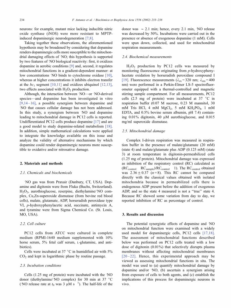
Experiments carried out with isolated brain mitochon-
dria [3] yielded H2O2 production rates, during oxidative
deamination of amines, f 50-fold higher than those aris-
ing from the electron-transport chain. These findings are at
variance with the contribution of MAO to H2O2 produc-
tion in permeabilized PC12 cells (Table 1). Dopamine
concentration in storage vesicles of peripheral sympathetic
neurons can be as high as 50 mM [2]. However, cytosolic
levels of dopamine are expected to be in the low micro-
molar range. Assuming a cytosolic concentration of dop-
amine of 1 AM [25] and the kinetic data obtained with
isolated brain mitochondria (Fig. 1 legend) [3], it can be
estimated that the production of H2O2 derived from MAO
catalysis is f 40-fold lower than that from the respiratory
chain (Fig. 1). Moreover, it can be argued that H2O2
production by the isolated brain mitochondria respiratory
chain is an overestimation over the physiological levels,
for these measurements were performed in the presence of
antimycin A [3]. Yet, these values may bear physiological
significance when considering that SNO exerts an antimy-
cin A-like effect on the respiratory chain by inhibiting
electron transfer at the bc1 segment and, thus, eliciting
H2O2 production [10].
It may be concluded from experimental (Table 1) and
calculated (Fig. 1) data that the rate of production of H2O2
via MAO catalysis is lower than that originating from the
electron-transfer chain, a situation that could be also
expected to occur in dopaminergic neurons in vivo, taking
into consideration the kinetic data available for brain
MAO.
3.2. Mitochondrial damage induced by SNO and its
potentiation by dopamine
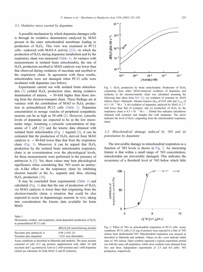
The irreversible damage to mitochondrial respiration as a
function of SNO levels is shown in Fig. 2. An interesting
feature is that within a small range of SNO concentration,
mitochondria are irreversibly damaged. This indicates the
occurrence of a threshold level of SNO below which little
Table 1
Monoamine oxidase- and respiratory chain-dependent production of H2O2
in permeabilized PC12 cells
d[H2O2]/dt (nmol/min/mg protein)
Succinate plus antimycin A 0.09F 0.01 (3)
Tyramine plus dopamine < 0.01 (not detectable)
Assay conditions as described in Materials and methods. The assay mixture
consisted of cells (2.5 mg protein) supplemented with either 10 mM
succinate and 1 Ag antimycin A/ml or 2 mM tyramine and 1 mM dopamine
(which are substrates for both MAO A and B isoforms).
Fig. 1. H2O2 production by brain mitochondria. Production of H2O2
originating from either MAO-catalyzed oxidation of dopamine and
tyramine or the electron-transfer chain was calculated assuming the
following data taken from Ref. [3]: (a) oxidation of tyramine by MAO
follows Henri–Michaelis–Menten kinetics (KM of 0.65 mM and Vmax of
4.5� 10� 5 M s� 1); (b) oxidation of dopamine catalyzed by MAO is 2.7-
fold lower than that of tyramine; and (c) production of H2O2 by the
respiratory chain is 9.5� 10� 7 M s� 1. Dotted line indicates calculations
obtained with tyramine and straight line with dopamine. The arrow
indicates the level of H2O2 originating from the mitochondrial respiratory
chain.
Fig. 2. Effect of SNO on mitochondrial respiration in PC12 cells. Assay
conditions: PC12 cells (1.25 mg of protein) were exposed to a flux of SNO
release from diethylamine/SNO. Mitochondrial respiration was assayed as
described in Materials and methods. Values on the x-axis indicate initial
rates of SNO release. Open symbols represent a typical experiment carried
out with the same cell population, while close symbols were obtained from
five and three independent experiments at 2.5 and 4.0 AM/s SNO
production, respectively.
F. Antunes et al. / Biochimica et Biophysica Acta 1556 (2002) 233–238 235

damage is exerted, but once this threshold is reached,
mitochondria offer little resistance to further increases inSNO concentration. Mitochondrial damage by SNO may
involve the reported inactivation of complex I and protein
nitrosation [24].
Approximately 15% inhibition of mitochondrial RC was
observed with SNO delivery rates below or at the threshold
level (2.5 AM� s� 1; see Fig. 2) (Table 2). This effect was
dramatically amplified by dopamine (f 70% inhibition)
(Table 2). The amine by itself elicited negligible respiratory
damage. The synergistic effect observed with dopamine andSNO was slightly affected by Cu,Zn-superoxide dismutase
(not shown), thus suggesting that the contribution of per-
oxynitrite (arising from the reaction of superoxide anion
(O2S�), formed during dopamine oxidation, and SNO) to
mitochondrial damage was not significant. The prevalent
mechanism underlying the interaction of dopamine withSNO was described [9] as depending largely on the concen-
tration of the latter: at high SNO concentrations, dopamine
undergoes nitrosation with subsequent nitration, whereas at
low SNO concentrations, dopaminochrome is formed via
two o-semiquinone intermediates and with ensuing forma-
tion of hydroxyl radical.
No synergistic effect was observed when PC12 cells
were preincubated with dopamine for 30 min, followed by
removal of extracellular dopamine and incubation with SNO
(not shown). Likewise, no damage was observed when a
mixture of dopamine and SNO (incubated for 30 min) was
added to PC12 cells. These observations suggest that (a)
dopamine does not induce cell changes that render them
more susceptible to SNO damage and (b) a stable, long-lived
product from the reaction between SNO and dopamine was
not involved in mitochondrial damage.
It may be surmised that if dopamine renders mitochon-
dria more susceptible to SNO damage, cells depleted of
dopamine should be more resistant to the deleterious effects
of SNO. Accordingly, treatment of PC12 cells with reserpine,
a compound that depletes the cellular dopamine storage
vesicles (including PC12 cells [17]), rendered cells more
resistant to damage elicited by SNO (Table 2). Although
long-term (18 h) treatment of cell with reserpine may elicit
cellular changes besides dopamine depletion, these results
concur with the view that dopamine causes mitochondria in
PC12 cells to be more susceptible to damage by SNO.
3.3. SNO-mediated oxidation of dopamine in cytosol and
storage vesicles
As mentioned above, dopamine, synthesized in the
cytosol, is present at the micromolar range in this compart-
ment [25] and is rapidly stored in vesicles at very high
concentrations (millimolar range) [26]. The low pH of the
vesicles (5.5) is considered to protect dopamine against
autoxidation and, as a corollary, the oxidative damage
mediated by dopamine is assumed to involve cytosolic or
extracellular dopamine. SNO oxidizes dopamine at pH 5.5,
albeit at a rate substantially lower than at pH 7.4 (Fig. 3).
The dependence of dopamine oxidation on pH follows the
general tenet that deprotonation is a requisite for electron
transfer: for each unit of pH increased, the rate of oxidation
of dopamine increases 10-fold. However, the calculated data
in Table 3 indicate that the rate of dopamine oxidation in the
storage vesicles is f 17-fold higher than that in the cytosol
because the concentration factor overcomes the pH con-
straints.
These calculations ought to be interpreted as semiquan-
titative estimations at the order of magnitude level; never-
theless, they suggest that storage vesicles may be the
cellular sites where the potential for dopamine oxidation is
higher. Biomembranes do not constitute a barrier for SNO
diffusion and, actually, they promote the diffusion of this
species [27]; therefore, SNO has free access to the interior of
the storage vesicles. The results in Table 2 also support the
interaction between SNO and dopamine in storage vesicles:
reserpine, a compound that releases dopamine from storage
vesicles, followed by its secretion in the extracellular milieu
Table 2
Potentiation of SNO-mediated damage by dopamine
Conditions Inhibition of respiratory
control (RCcontrol�RCsample)/
(RCcontrol� 1) (%)
+Dopamine 0.9F 0.5 (n= 3)
+NO (2.5 AM s� 1) 15.0F 5.7 (n= 5)
+NO (2.5 AM s� 1) + dopamine 70.9F 8.9 (n= 4)
+NO (4.0 AM s� 1) 90.1F 7.5 (n= 3)
+NO (4.0 AM s� 1) + reserpine 48.4F 8.5 (n= 3)
Assay conditions: PC12 were exposed for 30 min at 37 jC to exogenous
dopamine (1 mM) or SNO (initial flux rate either 2.5 or 4.0 AM s� 1) or SNO
plus dopamine. Reserpine concentration was 1 AM. Cells were collected
and mitochondrial damage assessed as described in Materials and methods.
A flux rate of SNO of 2.5 AM s� 1 is at or near the threshold level; that of 4.0
AM s� 1 is above the threshold level and elicits maximal mitochondrial
damage (see Fig. 1). Absolute RCcontrol was 2.36F 0.37 (n= 8).
Fig. 3. pH dependence of dopamine oxidation by SNO. The assay mixture
consisted of 1 mM dopamine in 0.1 M phosphate buffer, pH 5.7–7.4. The
reaction was initiated upon addition of 70 AM SNO, delivered as an SNO-
saturated solution kept in anaerobiosis. Dopamine oxidation was followed
at 480 nm.
F. Antunes et al. / Biochimica et Biophysica Acta 1556 (2002) 233–238236
protected against mitochondrial damage induced by SNO.
Reserpine also causes a temporal increase of the cytosolic
levels of dopamine, as observed by the feed-back inhibition
of tyrosine hydroxylase [25].
4. Concluding remarks
The low production of H2O2 during MAO-catalyzed
oxidative deamination of dopamine (with respect to H2O2
generated upon oxidation of respiratory chain substrates;
Table 1, Fig. 1), the lack of a direct effect of dopamine on
mitochondrial respiration (Table 2), and the potentiation of
the deleterious effects of SNO by the amine (Table 2) need
be assessed in terms of (a) the MAO content and activity in
dopaminergic neurons and the concentration of cytosolic
dopamine accessible to this enzyme on the outer mitochon-
drial membrane, and (b) the cellular site for the interaction
between dopamine and SNO with implications for mitochon-
drial function.
Although PC12 cells are widely used in neurobiological
and neurochemical studies, they do not have a neuronal
origin, and extrapolation of the results to dopaminergic
neurons deserves caution. Oxidative deamination of dopa-
mine and consequent H2O2 production is the domain of
outer mitochondrial membrane MAO, the activity levels of
which are a matter of controversy in dopaminergic neurons.
A recent histochemical study showed only low levels of
MAO in these cells [28] and that PC12 cells contain MAO-
A activity [23].
In early stages of the onset of MPTP-induced Parkinson’s
model, preceding dopaminergic neurodegeneration, there is
an increase in SNO production through iNOS in glial cells
[7]. According to the data shown here, cells are expected to
withstand some increase in SNO concentration, but once a
threshold levels is reached, cells will have a very limited
resistance to further SNO increases that cause irreversible
damage to mitochondria. Due to the strong synergism
between SNO and dopamine, the storage of dopamine in
dopaminergic neurons causes this threshold to be lower in
these cells and, accordingly, they are selectively harmed.
The potentiation of SNO-mediated mitochondrial damage by
dopamine was observed in a narrow critical region of SNO
concentration near the threshold level that induces mito-
chondrial damage: for SNO concentrations lower than the
threshold level, no synergistic effects were observed (not
shown), whereas at high SNO concentrations, the damage
observed in the absence of dopamine was already maximal.
It may be expected that in dopaminergic neurons in vivo the
concentration of SNO with pathological significance in the
development of Parkinson’s disease is the one near the
threshold level that induces cellular damage. Therefore,
the occurrence of a synergism between SNO and dopamine
for the concentration window of SNO near the threshold
level found to induce mitochondrial damage is highly
significant. Although data in Table 3 suggest that storage
vesicles may be the cellular sites where the potential for
dopamine oxidation is higher, alternative pathways cannot
be ruled out: the interaction between SNO and dopamine
may occur extracellularly and yield a reactive, long-lived
product, which is either endowed with a high permeability
constant and able to cross biomembranes and reach mito-
chondrial targets or capable of interacting with the plasma
membrane and thus trigger a cascade that damages mito-
chondria.
Acknowledgements
FA acknowledges grant BPD/11778/97 from PRAXIS
XXI/FCT. This research was supported by NIH grant 1RO1-
ES11342.
References
[1] S. Kosel, G. Hofhaus, A. Maassen, P. Vieregge, M.B. Graeber, Biol.
Chem. 380 (1999) 865–870.
[2] G. Cohen, R. Farooqui, N. Kesler, Proc. Natl. Acad. Sci. U. S. A. 94
(1997) 4890–4894.
[3] N. Hauptmann, J. Grimsby, J.C. Shih, E. Cadenas, Arch. Biochem.
Biophys. 335 (1996) 295–304.
[4] O. Terland, T. Flatmark, A. Tangeras, M. Gronberg, J. Mol. Cell.
Cardiol. 29 (1997) 1731–1738.
[5] S.B. Berman, T.G. Hastings, J. Neurochem. 73 (1999) 1127–1137.
[6] A.H. Schapira, M. Gu, J.W. Taanman, S.J. Tabrizi, T. Seaton, M.
Cleeter, J.M. Cooper, Ann. Neurol. 44 (1998) S89–S98.
[7] G.T. Liberatore, V. Jackson-Lewis, S. Vukosavic, A.S. Mandir, M.
Vila, W.G. McAuliffe, V.L. Dawson, T.M. Dawson, S. Przedborski,
Nat. Med. 5 (1999) 1403–1409.
[8] T. Dehmer, J. Lindenau, S. Haid, J. Dichgans, J.B. Schulz, J. Neuro-
chem. 74 (2000) 2213–2216.
[9] D. Rettori, Y. Tang, L.C. Dias, E. Cadenas, Free Radic. Biol. Med. 33
(2002) 685–690.
[10] J.J. Poderoso, M.C. Carreras, C. Lisdero, N. Riobo, F. Schopfer, A.
Boveris, Arch. Biochem. Biophys. 328 (1996) 85–92.
[11] M.W.J. Cleeter, J.M. Cooper, V.M. Darley-Usmar, S. Moncada,
A.H.V. Schapira, FEBS Lett. 345 (1994) 50–54.
[12] J.J. Poderoso, C. Lisdero, F. Schopfer, N. Riobo, M.C. Carreras, E.
Cadenas, A. Boveris, J. Biol. Chem. 274 (1999) 37709–37716.
[13] J.J. Poderoso, M.C. Carreras, F. Schopfer, C. Lisdero, N. Riobo, C.
Giulivi, A.D. Boveris, A. Boveris, E. Cadenas, Free Radic. Biol.
Med. 26 (1999) 925–935.
Table 3
Relative rates of dopamine oxidation in cytosol and storage vesicles
Cellular site pH [Dopamine]
(mM)a[Dopamine�]
(AM)bRate of
oxidationc
Storage vesicles 5.5 1.25 0.50 17
Cytosol 7.4 0.001 0.03 1
a Total concentration of dopamine (sum of protonated and anionic
forms).b The concentration of the anionic form of dopamine was calculated
from [Dopamine]=([Dopamine]totalKa/[H+])/(1 +Ka/[H
+]) and assuming a
pKa of 8.9.c The relative rate of oxidation is given by the ratio of the concentration
of dopamine anion in storage vesicles over that in cytosol.
F. Antunes et al. / Biochimica et Biophysica Acta 1556 (2002) 233–238 237

[14] M.J. LaVoie, T.G. Hastings, J. Neurochem. 73 (1999) 2546–2554.
[15] A.J. Nappi, E. Vass, J. Biol. Chem. 276 (2001) 11214–11222.
[16] A. Palumbo, A. Napolitano, P. Barone, M. D’Ischia, Chem. Res.
Toxicol. 12 (1999) 1213–1222.
[17] D. Schubert, F.G. Klier, Proc. Natl. Acad. Sci. U. S. A. 74 (1977)
5184–5188.
[18] B. Kittner, M. Brautigam, H. Herken, Arch. Int. Pharmacodyn. Ther.
286 (1987) 181–194.
[19] P.A. Hyslop, L.A. Sklar, Anal. Biochem. 141 (1984) 280–286.
[20] M.M. Fukami, T. Flatmark, Biochim. Biophys. Acta 889 (1986) 91.
[21] D. Han, E. Williams, E. Cadenas, Biochem. J. 353 (1-15-2001) 411.
[22] A.J. Kowaltowski, A.E. Vercesi, G. Fiskum, Cell Death Differ. 7
(2000) 903.
[23] M.B. Youdim, E. Heldman, H.B. Pollard, P. Fleming, E. McHugh,
Neuroscience 19 (1986) 1311–1318.
[24] N.A. Riobo, E. Clementi, M. Melani, A. Boveris, E. Cadenas, S.
Moncada, J.J. Poderoso, Biochem. J. 359 (2001) 139–145.
[25] M. Brautigam, B. Kittner, H. Herken, Arzneim.-Forsch. 35 (1985)
277–284.
[26] D. Njus, P.M. Kelley, G.J. Harnadek, Biochim. Biophys. Acta 853
(1986) 237–265.
[27] W.K. Subczynski, M. Lomnicka, J.S. Hyde, Free Radic. Res. 24
(1996) 343–349.
[28] T. Hida, Y. Hasegawa, R. Arai, Brain Res. 842 (1999) 491–495.
F. Antunes et al. / Biochimica et Biophysica Acta 1556 (2002) 233–238238