mitochondrial dna analysis - · pdf filemitochondrial dna analysis ... vntrs can be used to...
TRANSCRIPT

Mitochondrial DNA Analysis Used for samples that cannot be analyzed using RFLP or STR
Uses DNA extracted from mitochondrion rather than nuclear DNA
Mitochondrial DNA degrades at a much slower rate than nuclear DNA
Allows analysis of older biological samples, such as hair and bones
Not as precise as STR
Extremely expensive and time consuming

bsapp.com
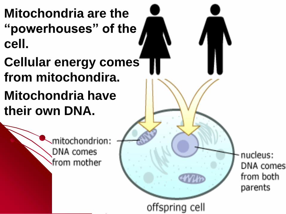
Oocyte Maturation Mitochondria are the
“powerhouses” of the
cell.
Cellular energy comes
from mitochondira.
Mitochondria have
their own DNA.

Regular DNA - from all of your ancestors.
Mitochondrial DNA is inherited from your
mother ONLY
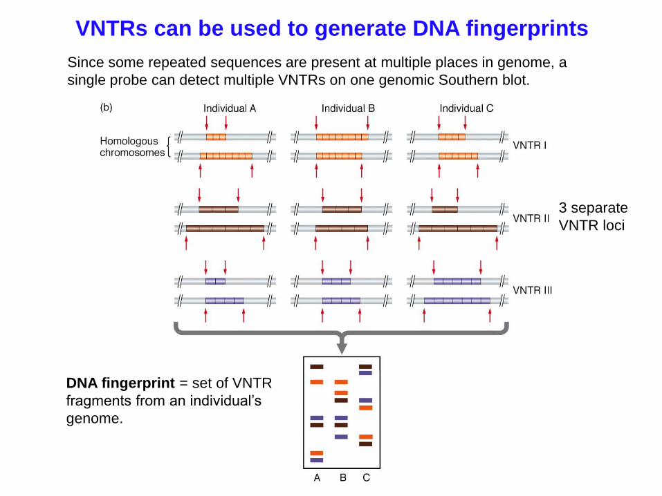
VNTRs can be used to generate DNA fingerprints
Since some repeated sequences are present at multiple places in genome, a
single probe can detect multiple VNTRs on one genomic Southern blot.
DNA fingerprint = set of VNTR
fragments from an individual’s
genome.
3 separate
VNTR loci
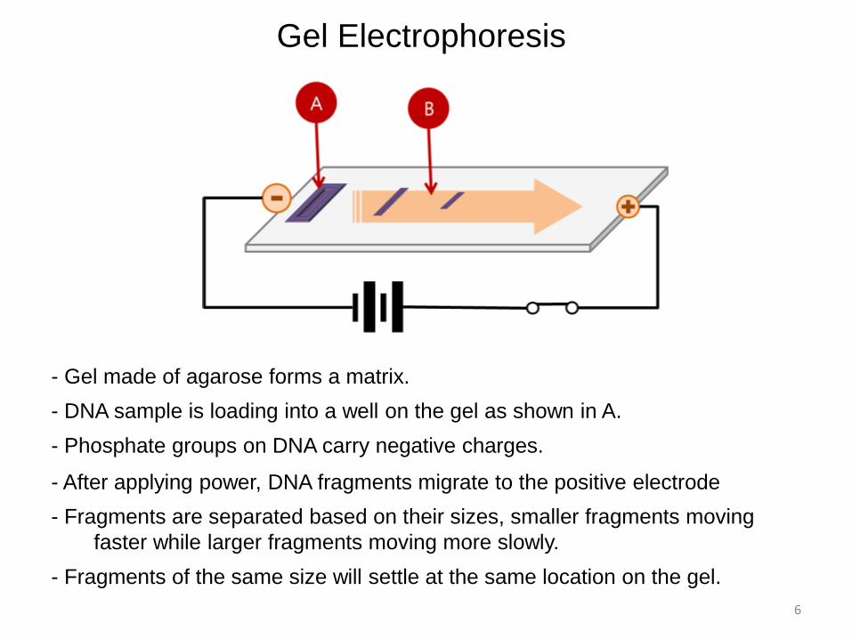
Gel Electrophoresis
- Gel made of agarose forms a matrix.
- Phosphate groups on DNA carry negative charges.
- DNA sample is loading into a well on the gel as shown in A.
- Fragments of the same size will settle at the same location on the gel.
- After applying power, DNA fragments migrate to the positive electrode
- Fragments are separated based on their sizes, smaller fragments moving
faster while larger fragments moving more slowly.
6

DNA Structure
Phosphate groups give
DNA its negative
charge
7

• DNA bands do not have colors.
• Ethidium bromide (a chemical) inserts itself between DNA bases.
• When a gel stained with ethidium bromide is put on a UV table, DNA
bands glow orange light.
• Photography of the gel produces pictures in which DNA bands are
white on a dark background.
• The brighter the DNA bands, the more molecules there are in the
bands.
Sizing
ladders
(standard)
8
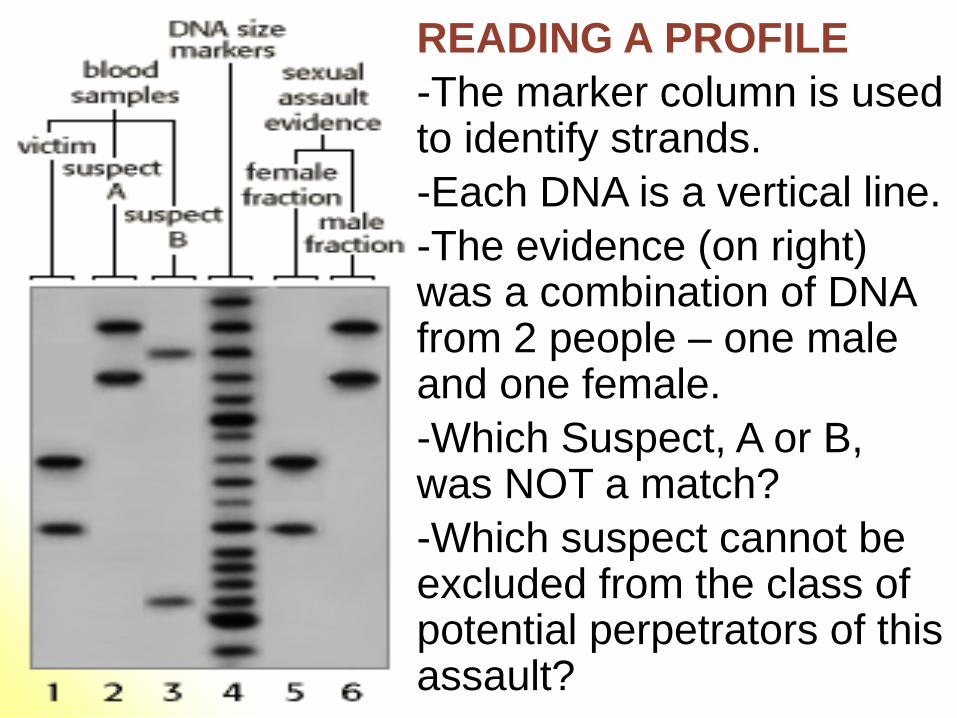
READING A PROFILE
-The marker column is used to identify strands.
-Each DNA is a vertical line.
-The evidence (on right) was a combination of DNA from 2 people – one male and one female.
-Which Suspect, A or B, was NOT a match?
-Which suspect cannot be excluded from the class of potential perpetrators of this assault?
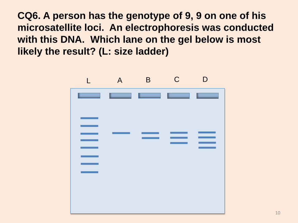
CQ6. A person has the genotype of 9, 9 on one of his
microsatellite loci. An electrophoresis was conducted
with this DNA. Which lane on the gel below is most
likely the result? (L: size ladder)
L A B C D
10

Q – which individual matched the forensic sample?

• STR Data is frequently presented as markers with repeats.
• There are 5 different Loci in the Profile above
• 1- Name one Locus that is homozygous (same marker from
both parents). How many repeats at that Locus?
• 2 - Name one locus that is heterozygous (different markers
from each parent). How many repeats at that Locus?

The following are the results of an analysis on two microsatellite loci
from a mother, a father and their child.
A
B
C
194
198 286
290
290 190 198
190 194
282
286
Locus 1 Locus 2
13
• Q – Why can’t person A be the
child of the other two? (Hint: look
at Locus 2)
• Q – Why can’t person B be the
child of the other two? (Hint:
remember what a homozygous
genotype means)
• Q – Which person could be the
child of the other two?
• Q – from this profile, can you tell
which is the mother and which is
the father?
A
B
C
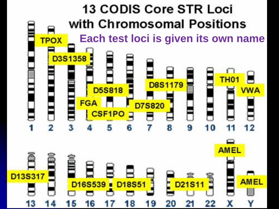
Each test loci is given its own name
D2S1338 FGA CSF1PO
D7S820
D13S317
D16S539 D18S51
D21S11
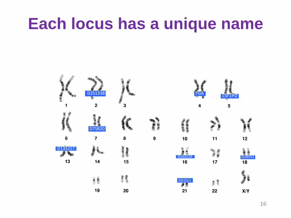
16
Each locus has a unique name

If the repeat section falls within a gene, it has the name of the gene. For example, vWA occurs on the von Willebrand Factor. CSF1P0 is named for the CSF-1 receptor gene. Other loci are named for the chromosome they are on, and the order in which the marker was discovered
How do they get those funny names for CODIS test loci?
• Q – which
chromosome
is D7S820
on?
• Which
chromosome
is D8S1179
on?
• D21S11?
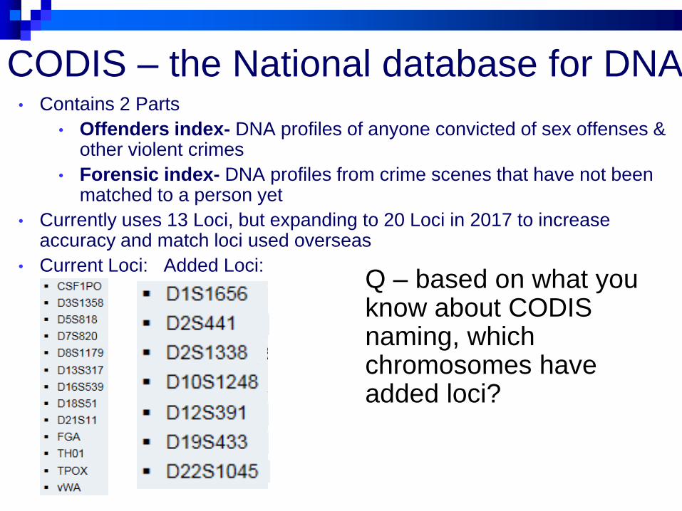
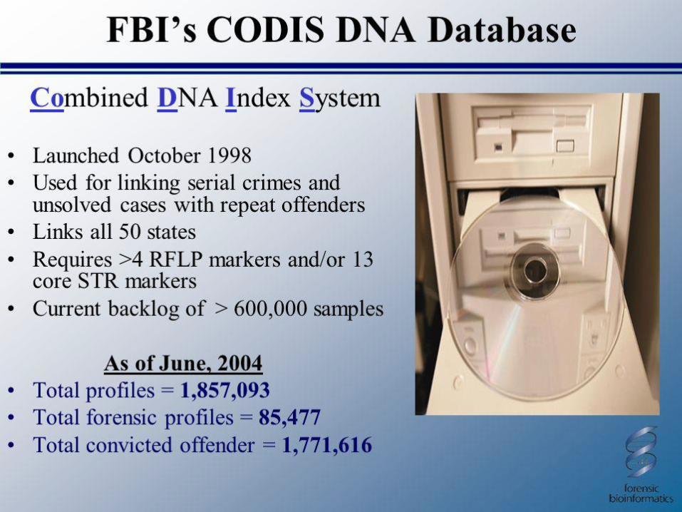
CODIS – the National database for DNA • Contains 2 Parts
• Offenders index- DNA profiles of anyone convicted of sex offenses & other violent crimes
• Forensic index- DNA profiles from crime scenes that have not been matched to a person yet
• Currently uses 13 Loci, but expanding to 20 Loci in 2017 to increase accuracy and match loci used overseas
• Current Loci: Added Loci: Q – based on what you know about CODIS naming, which chromosomes have added loci?

How can DNA evidence be
contaminated?
Sneezing or coughing over evidence
Person touches their mouth, nose or other
part of the face and then touches the area
that may contain the DNA to be tested.
Scene personnel can deposit hairs, fibers,
or trace material from their clothing
Wind can carry in contaminants

DNA Strands
Building block of genetic makeup
A complete copy of an individuals
entire genome exist in nearly
every cell
Each persons genome is made up
of billions of base pairs
Most base pairs are “junk” DNA bsapp.com

• DNA strands
come from the
nucleus or the
mitochondria
Only one-tenth
of a percent of
DNA differs from
one human to
the next
bsapp.com