mitochondrial dna variation of leber's hereditary optic
TRANSCRIPT

Cells 2019, 8, 1574; doi:10.3390/cells8121574 www.mdpi.com/journal/cells
Article
Mitochondrial DNA Variation of Leber's Hereditary Optic Neuropathy in Western Siberia Elena Starikovskaya 1,*, Sofia Shalaurova 1, Stanislav Dryomov 1, Azhar Nazhmidenova 1, Natalia Volodko 2, Igor Bychkov 3, Ilia Mazunin 4,* and Rem Sukernik 1
1 Laboratory of Human Molecular Genetics, Institute of Molecular and Cellular Biology, SBRAS, Novosibirsk, Russia; [email protected]
2 University of Alberta, Edmonton, AB, Canada; [email protected] 3 Novosibirsk branch of S.N. Fedorov NMRC "MNTK Eye Microsurgery", Novosibirsk, Russia; [email protected] 4 Center of Life Sciences, Skolkovo Institute of Science and Technology, Skolkovo, Russia; [email protected] *Correspondence: [email protected] (E.S.); [email protected] (I.M.)
Received: 23 October 2019; Accepted: 02 December 2019; Published: 4 December 2019
Abstract: Our data first represent the variety of Leber’s hereditary optic neuropathy (LHON) mutations in Western Siberia. LHON is a disorder caused by pathogenic mutations in mitochondrial DNA (mtDNA), inherited maternally and presents mainly in young adults, predominantly males. Clinically, LHON manifests itself as painless central vision loss, resulting in early onset of disability. The epidemiology of LHON has not been fully investigated yet. In this study, we report 44 genetically unrelated families with LHON manifestation. We performed whole mtDNA genome sequencing and provided genealogical and molecular genetic data on mutations and haplogroup background of LHON patients. Known “primary” pathogenic mtDNA mutations (MITOMAP) were found in 32 families: m.11778G>A represents 53.10% (17/32), m.3460G>A—21.90% (7/32), m.14484T>C–18.75% (6/32), and rare m.10663T>C and m.3635G>A represent 6.25% (2/32). We describe potentially pathogenic m.4659G>A in one subject without known pathogenic mutations, and potentially pathogenic m.6261G>A, m.8412T>C, m.8551T>C, m.9444C>T, m.9921G>A, and m.15077G>A in families with known pathogenic mutations confirmed. We suppose these mutations could contribute to the pathogenesis of optic neuropathy development. Our results indicate that haplogroup affiliation and mutational spectrum of the Western Siberian LHON cohort substantially deviate from those of European populations.
Keywords: LHON; Siberian population; ancient mutation; specific genetic background
1. Introduction
Leber’s hereditary optic neuropathy (LHON) is a form of hereditary disorder caused by pathogenic mutations in mitochondrial DNA. These mutations are non-synonymous, and affect genes coding for different subunits of complex I of the mitochondrial respiratory chain. The occurrence of such kinds of mutations in mtDNA subunits leads to dysfunction of the electron transport, increased reactive oxygen species production, and defective ATP synthesis [1–3]. Retinal ganglion cells are highly susceptible to death during LHON progression, because of their high sensibility to disrupted ATP production and oxidative stress [4]. Therefore, LHON is usually painless, acute, or subacute, with central visual loss in one or both eyes, and results in early onset of disability. The onset of Leber’s hereditary optic neuropathy is relatively rare in childhood and has better visual prognosis. The peak age of onset of visual loss among LHON carriers is 20–30 years old [5]. In some cases, LHON patients have been reported as having additional neurologic, cardiac, and endocrine disorders [6–8]. Leber's disease is inherited maternally and manifests itself in the young, predominantly affecting males—50% of male but only 10% of female subjects experience vision loss.

Cells 2019, 8, 1574 2 of 15
Interestingly, this sex predilection cannot be explained by the principles of mitochondrial inheritance [4].
At the present time, a number of mtDNA point mutations have been described, but the most prevalent are m.3460G>A, m.11778G>A, and m.14484T>C, accounting for about 90% of cases of LHON worldwide. The prevalence of each mutation varies among different populations, but the average is 69–92% for m.11778G>A, and 3–19% for m.14484T>C and m.3460G>A [9–14]. However, there are significant deviations from the average in some populations, for example, among French Canadians 87% of cases are due to m.14484T>C as a result of a founder effect [15]. Moreover, phenotypic expression of these primary mutations has been found to vary in different populations and different pedigrees. This incomplete penetrance suggests that other factors, such as mtDNA haplogroup background, nuclear genetic background, and environmental factors, may influence the modulation of phenotypic expression and severity of the disease [16–18].
Consequently, the worldwide prevalence of LHON varies in different populations and is unknown for the majority of them. This prevalence is estimated to range between 1:30,000 and 1:50,000 [19,20]. The epidemiology of LHON has not been fully investigated in Russian Federation, and our previous studies included the description of isolated cases [21–23]. Hence, in the present study, we report 44 genetically unrelated LHON families, performed whole mtDNA sequencing, and provide molecular genetic data on mutations and the haplogroup background of LHON patients in the Western Siberian population.
2. Materials and Methods
2.1. Subjects
This study was approved by the Ethics Committee IRB 00001360 affiliated with Vector State Research Center of Virology and Biotechnology (SRC VB Vector), Novosibirsk, Russian Federation. The total number of subjects in the study is 168 individuals from 44 unrelated families (85 affected and 83 healthy carriers), including 17 cases from our previous studies [21–23]. The clinical follow-up of LHON patients has been carried out by the Novosibirsk Branch of Federal Eye Microsurgery Department since 1997, conducted by one of the authors. The clinical diagnosis was based on a combination of symptoms and signs: painless acute or subacute central vision loss; fundus changes; and visual field abnormality, such as pseudopapilledema, optic nerve atrophy, and central or centrocecal scotoma. All the individuals made an informed decision to take part in the study and provided written consent. Family history was taken in each case to identify maternal inheritance of symptoms. The complete mtDNA genomes were sequenced for the family’s probands, and for the other individuals the certain mutations were confirmed by sequencing of associated mtDNA regions.
2.2. MtDNA Analysis
Whole peripheral blood samples were collected from the donors in 10 mL Vacutech EDTA tubes. Total DNA was extracted from a buffy-coat layer using the SileksMagNA-G Blood DNA Isolation kit, according to the manufacturer's protocols. The complete sequencing procedure entailed PCR amplification of 22 overlapping mtDNA templates [24], which were sequenced in both directions with BigDye 3.1 terminator chemistry (PE Applied Biosystems, Foster City, CA, United States). The trace files were analyzed with Sequencher (version 4.5 GeneCode Corporation) software. To perform capillary electrophoresis on an ABI Prism 3130XL DNA Analyzer, we used core facilities of the “Genomika” Sequencing Center (SBRAS, Novosibirsk, Russian Federation). Variants were scored relative to the Reconstructed Sapiens Reference Sequence, RSRS [25]. MtDNA haplotypes were identified following the nomenclature suggested by the PhyloTree Build 17 [26]. Forty-two mitochondrial genomes obtained through this study were deposited in GenBank with accession numbers MN413201–MN413242. Two genomes, EU807741.1 and EU807742.1, had been deposited to the GenBank earlier [21].

Cells 2019, 8, 1574 3 of 15
2.3. Penetrance Analysis
We determined penetrance as the proportion of affected individuals from all maternally related family members using family pedigrees [19]. Values for both men and women were calculated separately.
2.4. Analysis of Pathogenicity for Non-Synonymous Mutations
To make sure that the revealed non-synonymous mtDNA mutations were not sequencing errors or to not point out to general population polymorphisms, and in turn to find out the disease-associated polymorphisms among those published earlier, we used several databases: MITOMAP [27]; mtDB (Human Mitochondrial Genome Database, containing 2704 human mitochondrial genomes) [28]; and HmtDB (Human Mitochondrial DataBase), which contains 32922 human mitochondrial genomes [29]. To assess the possible pathogenicity of these mutations, and to predict whether a protein sequence variation affects protein function, we used the following web applications: MutPred 1.2 [30], MutPred 2 [31], PolyPhen–2 [32], PROVEAN (Protein Variation Effect Analyzer) [33], and SIFT (Sorting Intolerant from Tolerant) [34]. All the sources are provided in the public domain.
3. Results
From 44 LHON families, 32 harbored a primary mutation; the results are shown in Table 1. Among families with a primary mutation, the m.11778G>A represented 53.10% (17/32), m.3460G>A was 21.90% (7/32), and m.14484T>C represented18,75% (6/32). Rare m.10663T>C and m.3635G>A represented 6.25% (2/32) of the families.
Table 1. Summary data for examined Leber’s hereditary optic neuropathy (LHON) and LHON-like (without primary mutations) families. Age of onset of visual loss vary between families/patients; the peak age of onset is ~20–30 years old. Published previously by *[23]; **[22]; ***[21].
No. Family
Name Ethnicity in
Maternal Line
Number of
Examined
Individuals
(Affected/
Healthy)
Family
History of
Visual Loss
Primary LHON
Mutation
(MITOMAP)
mtDNA
Haplogroup
1 L18* Altaian 4 (2/2) No m.3460G>A D4p
2 L24* Tuvinian 19 (7/12) Yes m.3460G>A C5d1
3 L25*** Russian 5 (3/2) Yes m.3460G>A D5a2a2
4 L41 German 2 (1/1) No m.3460G>A H40a
5 L57 -/- 1 (1/0) Yes m.3460G>A V1a1
6 L58 -/- 2 (1/1) Yes m.3460G>A J1c3
7 L61 -/- 4 (2/2) Yes m.3460G>A H1b1
8 L30*** -/- 19 (11/8) Yes m.3635G>A J2b1c1
9 L2** -/- 6 (2/4) Yes m.10663T>C J1c4
10 L1* Russian 9 (1/8) Yes m.11778G>A T2b

Cells 2019, 8, 1574 4 of 15
No. Family
Name Ethnicity in
Maternal Line
Number of
Examined
Individuals
(Affected/
Healthy)
Family
History of
Visual Loss
Primary LHON
Mutation
(MITOMAP)
mtDNA
Haplogroup
11 L3* -/- 4 (1/3) Yes m.11778G>A T2d1b1
12 L5* -/- 1 (1/0) Yes m.11778G>A J1c2i
13 L12* -/- 2 (1/1) No m.11778G>A J2b1a1
14 L14* -/- 5 (2/3) No m.11778G>A T2b28
15 L23* Azerbaijani 1 (1/0) Unknown m.11778G>A J2b1
16 L26* -/- 13 (8/5) Yes m.11778G>A J1c7a
17 L27*** -/- 11 (2/9) Yes m.11778G>A H2a5b
18 L28*** -/- 9 (2/7) Yes m.11778G>A T2b8
19 L38 Ukrainian 2 (2/0) Yes m.11778G>A J1c2c2a
20 L39 Unknown 2 (1/1) No m.11778G>A V
21 L42 Belarusian 2 (1/1) Yes m.11778G>A H1c
22 L43 Russian 3 (1/2) No m.11778G>A H
23 L49 Unknown 1 (1/0) No m.11778G>A K1c
24 L52 Russian 3 (1/2) No m.11778G>A J1c2
25 L53 Unknown 3 (1/2) Unknown m.11778G>A H1b2
26 L60 Russian 2 (1/1) No m.11778G>A H1b2
27 L10* -/- 8 (3/5) Yes m.14484T>C M9a1a1c1a
28 L17* -/- 8 (4/4) Yes m.14484T>C J1c2c1
29 L32 Unknown 1 (1/0) Unknown m.14484T>C V
30 L40 Albanian 1 (1/0) Yes m.14484T>C H
31 L47 -/- 1 (1/0) No m.14484T>C J1c5a1
32 L50 Unknown 1 (1/0) Unknown m.14484T>C U5a2b1c
33 L6 -/- 2 (2/0) No - U4a1d
34 L8 Unknown 1 (1/0) No - U2e1
35 L9 Russian 2 (2/0) Yes - U5a1b1c1

Cells 2019, 8, 1574 5 of 15
No. Family
Name Ethnicity in
Maternal Line
Number of
Examined
Individuals
(Affected/
Healthy)
Family
History of
Visual Loss
Primary LHON
Mutation
(MITOMAP)
mtDNA
Haplogroup
36 L20 -/- 3 (2/1) Yes - U4b1b1
37 L31*** -/- 3 (2\1) Yes - U3b1b
38 L45 -/- 2 (1/1) No - U5a2e
39 L46 -/- 2 (1/1) No - H13a1d
40 L51 Unknown 1 (1/0) Unknown - U2c1b
41 L54 Russian 2 (1/1) No - U4a2a
42 L56 -/- 2 (1/1) No - J1c1b1
43 L59 Ukrainian 2 (1/1) No - V7a
44 L62 -/- 1 (1/0) Yes - H
According to the family pedigrees, only 50% (22/44) of cases had a family history of vision loss in the maternal lineage in more than one generation, among which m.11778G>A represented 36% (8/22), the m.14484T>C was 14% (3/22), and m.3460G>A covered 23% (5/22), as well as those without primary mutations (LHON-like cases), which represented 18% (4/22). Rare mutation cases (m.10663T>C and m.3635G>A) were family–inherited. In total, 39% (17/44) of cases were sporadic, among which 13 were cases with only one affected person diagnosed, and four were cases with two affected persons in one generation. Among the sporadic cases, m.11778G>A represented 41% (7/17), m.3460G>A was 12% (2/17), m.14484T>C was 6% (1/17), and the LHON-like cases represented 41% (7/17) of the total.
Summary information on penetrance is shown in Table 2. The penetrance is highly variable between separate families, even with the same primary mutation. The average penetrance among men was 32% (6–100%) and 12% among women (0–58%); these correlate with data previously published [4]. However, there are some families with higher penetrance among females than among males: L24, L26, and L28.
Table 2. Summary information about penetrance.
m.11778G>A (n = 15) m.14484T>C (n = 4) m.3460G>A (n= 7) Average
Males 34% 46% 15% 32%
Females 12% 12% 10% 12%
In 12 families with clear-cut LHON phenotypes, no pathogenic mtDNA mutations were found. Analysis of the mtDNA revealed non-synonymous mutations: m.14002A>G, m.4766A>G, and m.13105A>G, which have not been noted as associated with LHON or other diseases in the MITOMAP database. All pathogenicity prediction tools indicated low probability that the amino acid substitutions are disease-associated for these mutations. Mutation m.4659G>A has been previously reported as being associated with Parkinson’s disease [35], as well as in an Australian

Cells 2019, 8, 1574 6 of 15
LHON pedigree that was heteroplasmic for the m.14484T>C [36]. Polyphen-2 predicted the pathogenicity for this mutation as benign and MutPed 2 showed low probability score, but MutPred 1.2, PROVEAN, and SIFT determined this mutation as deleterious. The results are shown in Table 3.
In several families with primary mutations (L01, L03, L12, L28, L30, L40, L43, and L50) we found out additional, non-synonymous mutations (Table 4). Mutations m.8875T>C, m.14582A>G, m.8400T>C, and m.4639T>C were neutral, and mutation m.9444C>T had a high probability of being pathogenic, according to data from all the pathogenicity prediction tools; for other mutations, we observed divergence of prediction results. Since prediction results for primary pathogenic mutations diverged too (see Table 3), novel non-synonymous nucleotide change was considered potentially pathogenic if it had extremely low frequency in the general population, and if it was predicted by at least three algorithms to have an effect on protein function. For mutations m.6261G>A and m.15468C>T, only PolyPhen2 predicted pathogenicity as probably damaging and possible damaging, respectively. However, mutation m.6261G>A had already been reported by Abu-Amero [37] in patients with optic neuropathy, and also as a somatic mutation associated with prostate cancer. Interestingly, the family (L01) with m.6261G>A and m.11778G>A has the same haplogroup, T2, as the case reported by Abu-Amero. Other mutations (m.9921G>A, m.8551T>C, m.8412T>C, m.15077G>A) were predicted as pathogenic by at least by three algorithms, but the first three of them have not been noted as associated with diseases in the MITOMAP database, and mutation m.15077G>A was reported as being associated with maternally-inherited isolated deafness [38].
Table 3. Non-synonymous mutations revealed in LHON-like cases. Known primary mutations (m.3460G>A, m.3635G>, m.10663T>C, m.11778G>A, and m.14484T>C) are placed in bold to demonstrate distinction between different prediction algorithms and frequencies in general population for pathogenic mutations. A MutPred 1.2 score > 0.75 and a Mutpred 2 score > 0.50 would suggest pathogenicity.
Mutation
Protein-C
oding
Region of
mtDNA
Amino
Acid
Substitutio
n
PolyPhen –
2 score
MutPred
1.2/2
Score
(cutoff
0.75/0.50)
PROVEA
N/SIFT
Pathogenic
ity
Prediction
Frequency
in General
Population
(as per
mtDB)
Frequency
in General
Population
(as per
HmtDB)
Family
m.14002A>G ND5 T556A 0.002
(benign) 0.387/0.059
Neutral/To
lerated 0.0037 0.00289 L45
m.4766A>G ND2 M99I 0.001
(benign) 0.571/0.225
Neutral/
Tolerated 0 0.00009 L46
m.4659G>A ND2 A64T 0.029
(benign) 0.790/0.256
Deleterious
/
Damaging 0.0011 0.00161
L51
m.13105A>G ND5 I257V 0.001
(benign) 0.198/0.032
Neutral/To
lerated 0.0612 0
m.3460G>A ND1 A52T 1.000
(probably
damaging) 0.789/0.418
Neutral/
Damaging 0.0097 0.00058 -
m.3635G>A ND1 S110N 0.999
(probably 0.873/0.493
Deleterious
/ 0 0.00027 -

Cells 2019, 8, 1574 7 of 15
damaging) Damaging
m.10663T>C ND4L V65A 0.946
(probably
damaging) 0.604/0.694
Deleterious
/Damaging 0 0.00003 -
m.11778G>A ND4 R340H 0.999
(probably
damaging) 0.919/0.494
Deleterious
/Damaging 0.0097 0.0034 -
m.14484T>C ND6 M64V 0.993
(probably
damaging) 0.618/0.787
Neutral/Da
maging 0.0026 0.00146 -
Table 4. Additional, non-synonymous mutations revealed in LHON cases. All these mutations still have no the status of “primary LHON mutations”. A MutPred 1.2 score > 0.75 and a Mutpred 2 score > 0.50 would suggest pathogenicity.
Mutation
Protein-Co
ding
Region of
mtDNA
Amino
Acid
Substitutio
n
PolyPhen–
2 score
MutPred
1.2/2
Score
(cutoff
0,75/0,50)
PROVEAN
/SIFT
Prediction
Frequency
in General
Population
(mtDB)
Frequency
in General
Population
(HmtDB)
Family
m.6261G>A CO1 A120T 0.998
(probably
damaging) 0.491/0.324
Neutral/
Tolerated 0.0048 0.00553 L01
m.8875T>C ATP6 F117L 0 (benign) 0.251/0.429 Neutral/
Tolerated 0.0007 0.001276 L03
m.9921G>A CO3 A239T 0.009
(benign) 0.543/0.624
Deleterious
/Damaging 0.0011 0.00082 L12
m.15468C>
T CYB T241M
0.890
(possible
damaging) 0.245/0.079
Neutral/
Tolerated 0.0004 0.00043 L28
m.8551T>C ATP6 F9L 0.976
(probably
damaging) 0.676/0.418
Deleterious
/ Damaging 0.0007 0 L30
m.14582A>
G ND6 V31A
0.003
(benign) 0.245/0.181
Neutral/Tol
erated 0.0086 0.00571 L40
m.8400T>C ATP8 M12T 0 (benign) 0.504/0.118 Neutral/Tol
erated 0.0011 0.00052 L43

Cells 2019, 8, 1574 8 of 15
m.9444C>T CO3 R80W 0.999
(probably
damaging) 0.875/0.586
Deleterious
/Damaging 0 0
m.4639T>C ND2 I57T 0.001
(benign) 0.297/0.047
Neutral/
Tolerated 0.0082 0.00395
L50 m.8412T>C ATP8 M16T
0.711
(possible
damaging) 0.677/0.542
Deleterious
/Tolerated 0 0.00039
m.15077G>
A CYB E111K
0.992
(probably
damaging) 0.684/0.331
Deleterious
/ Damaging
(low
confidence)
0.0007 0.00213
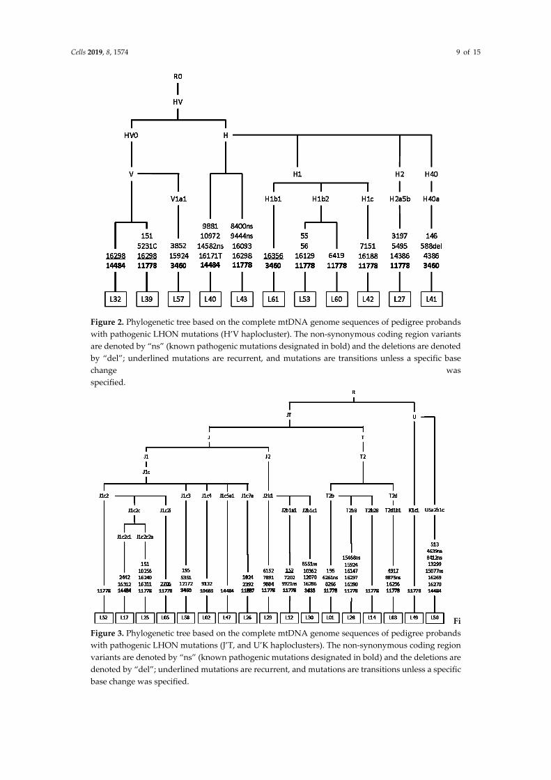
Phylogenetic analysis illustrates that Siberian carriers of pathogenic LHON mutations are unrelated and belong to different maternal lines. In rare cases (4/44), m.3460G>A and m.14484T>C belong to East Eurasian M8, M9, and D haploclusters (Figure 1). The classic (m.3460G>A, m.11778G>A, m.14484T>C) and rare LHON-causing mutations occur mostly in the mtDNA background of West Eurasian haploclusters H’V (Figure 2), J’T and U’K (Figure 3).
Figure 1. Phylogenetic tree based on the complete mtDNA genome sequences of pedigree probands with pathogenic LHON mutations (M8, M9, and D haploclusters). The non-synonymous coding region variants are denoted by “ns” (known pathogenic mutations designated in bold) and the deletions are denoted by “del”; underlined mutations are recurrent, and mutations are transitions unless a specific base change was specified.
Cells 2019, 8, 1574 9 of 15
Figure 2. Phylogenetic tree based on the complete mtDNA genome sequences of pedigree probands with pathogenic LHON mutations (H’V haplocluster). The non-synonymous coding region variants are denoted by “ns” (known pathogenic mutations designated in bold) and the deletions are denoted by “del”; underlined mutations are recurrent, and mutations are transitions unless a specific base change was specified.
FiFigure 3. Phylogenetic tree based on the complete mtDNA genome sequences of pedigree probands with pathogenic LHON mutations (J’T, and U’K haploclusters). The non-synonymous coding region variants are denoted by “ns” (known pathogenic mutations designated in bold) and the deletions are denoted by “del”; underlined mutations are recurrent, and mutations are transitions unless a specific base change was specified.

Cells 2019, 8, 1574 10 of 15
4. Discussion
4.1. Leber’s Hereditary Optic Neuropathy Primary Mutations
Regarding our preliminary data, the frequencies of primary mutations are different from frequencies reported for Europe and Asia. The most prevalent m.11778G>A (~55%) is less common in Western Siberia than in Europe, at ~69% [9]; as well as in China and Japan, at ~90% [10,13]. The prevalence of m.3460G>A (~24%) is twice as much as in Europe, but m.14484T>C (~14%) do not deviate from those of other European populations [9].
In 12 families with LHON-like manifestation, no known pathogenic mtDNA mutation was found. However, there are other elaborations in which the clinical diagnosis cannot be confirmed by molecular genetic analysis [37,39–41]. Definitively, mtDNA mutation-caused LHON is clinically indistinguishable from the other forms of optic neuropathy, such as a dominant optic neuropathy (DOA), especially when it is sporadic. Compared to LHON, DOA visual loss is detected between ages 4 and 6 in the majority of patients, and 58–84% of patients with DOA report visual impairment by age 11 [42]. In our 11 cases, the ages of onset were between 13–36 years. We will be studying these cases for the mutation spectrum of common pathogenic genes for DOA in future.
4.2. Penetrance
We suppose that relatively reduced incidence of LHON in Western Siberia is associated with incomplete penetrance and diagnostic difficulties of atypical (e.g. late-onset) and combined (e.g. multiple sclerosis) forms of LHON. Patients with LHON could have also been wrongly diagnosed as suffering from toxic amblyopia, tobacco–alcohol amblyopia, or optic neuritis [41]. On the other hand, the problem is that patients do not know their family history. Molecular testing for LHON is not routinely performed in patients with optic atrophy in Russian Federation. Identification and registration of unaffected carriers plays an important role for prevention of disease manifestation. For example, there is strong evidence now that smoking is associated with an increased risk of visual failure among LHON carriers—93% penetrance of vision loss in male smokers versus a 66% penetrance in male non-smokers [17]. Our observations highlight the importance of molecular genetic examination for unaffected carriers. Since the proportion of sporadic cases is high: 39% according to our data and 40% according to published data [4]. The presence of pathogenic mutations should be tested not only for probands, but for all relatives in the maternal line.
4.3. Potentially Pathogenic Mutation m.4659G>A
We found m.4659G>A in one subject without any known primary mutations (L51). This sequence change is located at codon 64 in the functional domain of the ND2 gene and changes an alanine—a hydrophobic amino acid—into threonine—a neutral amino acid. Mutation m.4659G>A has been reported as being associated with LHON in an Australian pedigree that also had heteroplasmic mutations m.14484T>C and m.5460G>A [36]. This family had 10 maternally-related descendants, five of whom had vision loss. Unfortunately, our patient does not know his family history, and we could not confirm maternal inheritance for this mutation. However, m.4659G>A has very low frequency in the general population (0.0011–0.00161), and has a high probability of being pathogenic (Table 3).
4.4. Additional Non-Synonymous Mutations Revealed in LHON Cases
The phenomenon of co-existence of two pathogenic mutations in one family has already been described. The first case included m.14484T>C, m.4659G>A, and m.5460G>A in an Australian LHON pedigree, described above [36]. In the second case, a Polish family harboring two primary LHON mutations m.3460G>A and m.11778G>A occurred in a haplogroup H background [43]. In the third case, a family harbored two primary LHON mutations, m.11778G>A and m.14484T>C, and both mutations had a synergistic pathogenic effect on protein function, as well as a higher degree of heteroplasmy of the m.14484T>C, correlated with an earlier age at onset [44]. Finally, the fourth

Cells 2019, 8, 1574 11 of 15
example is a unique double-mutant ND4L with two concurrent mutations (m.10609T>C and m.10663T>C) in an Arab pedigree from Kuwait [45].
We reported mutations m.6261G>A, m.8412T>C, m.8551T>C, m.9444C>T, m.9921G>A, and m.15077G>A, which could be potentially pathogenic because of their low frequency in the general population, and have a high probability of being pathogenic according to data from different prediction tools. Two of them, m.6261G>A and m.15077G>A, have already been reported in subjects with optic neuropathy and maternally-inherited isolated deafness, respectively. However, we suppose that additional non-synonymous mutations could either have a synergistically pathogenic or a protective effect. To demonstrate the full significance of novel mutations, a respiratory chain assay would need to be performed. An example is the study by [38], where cybrids with m.15077G>A showed normal activities for mitochondrial electron chain enzymatic complexes.
4.5. Haplogroup Analysis
Our LHON cohort from the Western Siberia region is represented predominantly by West Eurasian haplogroups and includes several East Eurasian haplogroups, namely C5d1 (L24), D4p (L18), D5a2a2 (L25), and M9a1a1c1a (L10).
Rare LHON mutations m.10663T>C and m.3635G>A were found in Russian families from Kazakhstan (the first) and the Novosibirsk region (the second), associated with the European haplogroups J1c4 and J2b1c1, respectively [21,22]. Mutation m.10663T>C was also reported in the background of the haplogroups J1c2c, L2a1, L3’4, and L3f1b [37,45–47], and mutation m.3635G>A was reported in haplogroups R11a, D4g2b, M7b4, F1a, B5b, and M7b [48,49]. The presence of the same pathogenic mutations on the background of various mitochondrial haplogroups confirms that pathogenic LHON mutations arise de novo, independently from the mtDNA or ethnic backgrounds.
It is known that the clinical impact of mDNA mutations may be modulated by mitochondrial haplogroup background. For example, Hudson et al. performed a multicenter study of 3613 subjects from 159 different families, and showed that the risk of visual failure is greater when m.11778G>A or m.14484T>C mutations are present in specific subgroups of haplogroup J; the same as the m.3460G>A mutation is present in haplogroup K, and the risk of visual failure is significantly lower when m.11778G>A occurs in haplogroup H [50]. Romero et al. supposed that haplogroup D has a protective effect in carriers of LHON mutations. His hypothesis was based on the fact that there was a markedly decreased frequency of haplogroup D in Chilean subjects with LHON, as haplogroup D is one of the most common in the Chilean population [12]. Also, other experimental research serves as proof that cybrids and fibroblasts bearing LHON mutations have different response to neurotoxic agents, depending on haplogroup background [51].
It has been suggested that at the end of the last glaciation, phylogenetically more ancient mutations could have provided their carriers with adaptive advantages during the human population expansion. Today, those mutations contribute to the saving and expression of weakly pathogenic LHON mutations, which appear randomly in different region-specific genetic backgrounds [52,53]. The theory could be tested by further searching of pathogenic, LHON-causing mutations in relation to specific mtDNA backgrounds (phylogenetically ancient set of mutations).
New data collected from future studies regarding mtDNA variations of LHON in Western Siberia might be used to develop a LHON system registry in the Russian Federation. We intend to conduct consecutive experimental research, including the parameters of the pathogenicity of each novel substitution.
5. Limitations of the Study
The main limitation of the data presented is the absence of physiological tests as proof with respect to the pathogenicity of novel mtDNA substitutions. Additional tests should be done, such as oxygen consumption, ATP and ROS measuring, and electron microscopy study (for example, [54]). In addition, development of the newest editing systems [55] could give us more reliable instruments to test pathogenicity.

Cells 2019, 8, 1574 12 of 15
Author Contributions: conceptualization, E.S. and R.S.; data curation, Y.B., E.S., S.S., S.D., A.N., N.V., I.M., and R.S.; analysis and interpretation of data, E.S. S.S., S.D., A.N., and R.S., writing—review and editing, E.S., S.S., S.D., A.N., I.M., and R.S.
Funding: The research was supported by the Russian Science Foundation [No. 17-75-20015] and the Russian Foundation of Basic Research [No. 19-29-04101].
Conflicts of Interest: The authors declare no conflict of interest.
References
1. Baracca, A.; Solaini, G.; Sgarbi, G.; Lenaz, G.; Baruzzi, A.; Schapira, A.H.; Martinuzzi, A.; Carelli, V. Severe impairment of complex I-driven adenosine triphosphate synthesis in leber hereditary optic neuropathy cybrids. Arch. Neurol. 2005, 62, 730–736. doi: 10.1001/archneur.62.5.730.
2. Lin, C.S.; Sharpley, M.S.; Fan, W.; Waymire, K.G.; Sadun, A.A.; Carelli, V.; Ross-Cisneros, F.N.; Baciu, P.; Sung, E.; McManus, M.J.; et al. Mouse mtDNA mutant model of Leber hereditary optic neuropathy. Proc Natl. Acad. Sci. USA 2012, 109, 20065–20070. doi: 10.1073/pnas.1217113109.
3. Kim, U.S.; Jurkute, N.; Yu-Wai-Man, P. Leber Hereditary Optic Neuropathy-Light at the End of the Tunnel? Asia Pac. J. Opthalmol. (Phila), 2018, 7, 242–245. doi: 10.22608/APO.2018293.
4. Meyerson, C.; Van Stavern, G.; McClelland, C. Leber hereditary optic neuropathy: Current perspectives. Clin. Ophthalmol. 2015, 9, 1165–1176. doi: 10.2147/OPTH.S62021.
5. Majander, A.; Bowman, R.; Poulton, J.; Antcliff, R.J.; Reddy, M.A.; Michaelides, M.; Webster, A.R.; Chinnery, P.F.; Votruba, M.; Moore, A.T.; et al. Childhood-onset Leber hereditary optic neuropathy. Br. J. Ophthalmol. 2017, 101, 1505–1509. doi: 10.1136/bjophthalmol-2016-310072.
6. Finsterer, J.; Zarrouk-Mahjoub, S. Leber's hereditary optic neuropathy is multiorgan not mono-organ. Clin. Ophthalmol. 2016, 10, 2187–2190. doi: 10.2147/OPTH.S120197.
7. Haas, R.H. Mitochondrial Dysfunction in Aging and Diseases of Aging. Biology 2019, 8, 48. doi: 10.3390/biology8020048.
8. Barcelos, I.P.d.; Troxell, R.M.; Graves, J.S. Mitochondrial dysfunction and multiple sclerosis. Biology 2019, 8, 37. doi: 10.3390/biology8020037.
9. Mackey, D.A.; Oostra, R.J.; Rosenberg, T.; Nikoskelainen, E.; Bronte-Stewart, J.; Poulton, J.; Harding, A.E.; Govan, G.; Bolhuis, P.A.; Norby, S. Primary pathogenic mtDNA mutations in multigeneration pedigrees with Leber hereditary optic neuropathy. Am. J. Hum. Genet. 1996, 59, 481–485.
10. Mashima, Y.; Yamada, K.; Wakakura, M.; Kigasawa, K.; Kudoh, J.; Shimizu, N.; Oguchi, Y. Spectrum of pathogenic mitochondrial DNA mutations and clinical features in Japanese families with Leber's hereditary optic neuropathy. Curr. Eye Res. 1998, 17, 403–408. doi: 10.1080/02713689808951221.
11. Kumar, M.; Kaur, P.; Kumar, M.; Saxena, R.; Sharma, P.; Dada, R. Clinical characterization and mitochondrial DNA sequence variations in Leber hereditary optic neuropathy. Mol. Vis. 2012, 18, 2687–2699.
12. Romero, P.; Fernandez, V.; Slabaugh, M.; Seleme, N.; Reyes, N.; Gallardo, P.; Herrera, L.; Pena, L.; Pezo, P.; Moraga, M. Pan-American mDNA haplogroups in Chilean patients with Leber's hereditary optic neuropathy. Mol. Vis. 2014, 20, 334–340.
13. Jiang, P.; Liang, M.; Zhang, J.; Gao, Y.; He, Z.; Yu, H.; Zhao, F.; Ji, Y.; Liu, X.; Zhang, M.; et al. Prevalence of Mitochondrial ND4 Mutations in 1281 Han Chinese Subjects With Leber's Hereditary Optic Neuropathy. Invest Ophthalmol. Vis. Sci. 2015, 56, 4778–4788, doi: 10.1167/iovs.14-16158.
14. Khan, N.A.; Govindaraj, P.; Soumittra, N.; Sharma, S.; Srilekha, S.; Ambika, S.; Vanniarajan, A.; Meena, A.K.; Uppin, M.S.; Sundaram, C.; et al. Leber’s hereditary optic neuropathy–specific mutation m.11778G>A exists on diverse mitochondrial haplogroups in India. Invest. Ophthalmol. Vis. Sci. 2017, 58, 3923–3930. doi: 10.1167/iovs.16-20695.
15. Laberge, A.M.; Jomphe, M.; Houde, L.; Vezina, H.; Tremblay, M.; Desjardins, B.; Labuda, D.; St-Hilaire, M.; Macmillan, C.; Shoubridge, E.A.; et al. A "Fille du Roy" introduced the T14484C Leber hereditary optic neuropathy mutation in French Canadians. Am. J. Hum. Genet. 2005, 77, 313–317. doi: 10.1086/432491.
16. Jacobi, F.K.; Leo-Kottler, B.; Mittelviefhaus, K.; Zrenner, E.; Meyer, J.; Pusch, C.M.; Wissinger, B. Segregation patterns and heteroplasmy prevalence in Leber's hereditary optic neuropathy. Inves.t Ophthalmol. Vis. Sci. 2001, 42, 1208–1214.
17. Kirkman, M.A.; Yu-Wai-Man, P.; Korsten, A.; Leonhardt, M.; Dimitriadis, K.; De Coo, I.F.; Klopstock, T.; Chinnery, P.F Gene-environment interactions in Leber hereditary optic neuropathy. Brain 2009, 132, 2317–2326. doi: 10.1093/brain/awp158.

Cells 2019, 8, 1574 13 of 15
18. Istikharah, R.; Tun, A.W.; Kaewsutthi, S.; Aryal, P.; Kunhapan, B.; Katanyoo, W.; Chuenkongkaew, W.; Lertrit, P. Identification of the variants in PARL, the nuclear modifier gene, responsible for the expression of LHON patients in Thailand. Exp. Eye Res. 2013, 116, 55–57. doi: 10.1016/j.exer.2013.08.007.
19. Puomila, A.; Hamalainen, P.; Kivioja, S.; Savontaus, M.L.; Koivumaki, S.; Huoponen, K.; Nikoskelainen, E. Epidemiology and penetrance of Leber hereditary optic neuropathy in Finland. Eur. J. Hum. Genet. 2007, 15, 1079–1089. doi: 10.1038/sj.ejhg.5201828.
20. Mascialino, B.; Leinonen, M.; Meier, T. Meta-analysis of the prevalence of Leber hereditary optic neuropathy mtDNA mutations in Europe. Eur. J. Ophthalmol. 2012, 22, 461–465. doi: 10.5301/ejo.5000055.
21. Brown, M.D.; Zhadanov, S.; Allen, J.C.; Hosseini, S.; Newman, N.J.; Atamonov, V.V.; Mikhailovskaya, I.E.; Sukernik, R.I.; Wallace, D.C. Novel mtDNA mutations and oxidative phosphorylation dysfunction in Russian LHON families. Hum. Genet. 2001, 109, 33–39. doi: 10.1007/s004390100538.
22. Brown, M.D.; Starikovskaya, E.; Derbeneva, O.; Hosseini, S.; Allen, J.C.; Mikhailovskaya, I.E.; Sukernik, R.I.; Wallace, D.C. The role of mtDNA background in disease expression: A new primary LHON mutation associated with Western Eurasian haplogroup J. Hum. Genet. 2002, 110, 130–138. doi: 10.1007/s00439-001-0660-8.
23. Volod'ko, N.V.; L’Vova, M.; Starikovskaia, E.B.; Derbeneva, O.A.; Bychkov, I.; Mikhailovskaia, I.E.; Pogozheva, I.V.; Fedotov, F.F.; Soyan, G.V.; Procaccio, V.; et al. Spectrum of pathogenic mtDNA mutations in Leber hereditary optic neuropathy families from Siberia. Genetika 2006, 42, 89–97.
24. Nochez, Y.; Arsene, S.; Gueguen, N.; Chevrollier, A.; Ferre, M.; Guillet, V.; Desquiret, V.; Toutain, A.; Bonneau, D.; Procaccio, V.; et al. Acute and late-onset optic atrophy due to a novel OPA1 mutation leading to a mitochondrial coupling defect. Mol. Vis. 2009, 15, 598–608.
25. Behar, D.M.; van Oven, M.; Rosset, S.; Metspalu, M.; Loogväli, E.L.; Silva, N.M.; Kivisild, T.; Torroni, A.; Villems, R. A "Copernican" reassessment of the human mitochondrial DNA tree from its root. Am. J. Hum. Genet. 2012, 90, 675–684. doi: 10.1016/j.ajhg.2012.03.002.
26. van Oven, M.; Kayser, M. Updated comprehensive phylogenetic tree of global human mitochondrial DNA variation. Hum. Mutat. 2009, 30, E386–E394. doi: 10.1002/humu.20921.
27. Brandon, M.C.; Lott, M.T.; Nguyen, K.C.; Spolim, S.; Navathe, S.B.; Baldi, P.;Wallace, D.C. MITOMAP: A human mitochondrial genome database--2004 update. Nucleic Acids Res. 2005, 33(Database issue): D611-613. doi: 10.1093/nar/gki079.
28. Ingman, M.; Gyllensten, U. mtDB: Human Mitochondrial Genome Database, a resource for population genetics and medical sciences. Nucleic Acids Res. 2006, 34(Database issue): D749-751. doi: 10.1093/nar/gkj010.
29. Attimonelli, M.; Accetturo, M.; Santamaria, M.; Lascaro, D.; Scioscia, G.; Pappada, G.; Russo, L.; Zanchetta, L.; Tommaseo-Ponzetta, M. HmtDB, a human mitochondrial genomic resource based on variability studies supporting population genetics and biomedical research. BMC Bioinformatics 2005, doi: 10.1186/1471-2105-6-S4-S4.
30. Li, B.; Krishnan, V.G.; Mort, M.E.; Xin, F.; Kamati, K.K.; Cooper, D.N.; Mooney, S.D.; Radivojac, P. Automated inference of molecular mechanisms of disease from amino acid substitutions. Bioinformatics 2009, 25, 2744–2750. doi: 10.1093/bioinformatics/btp528.
31. Pejaver, V.; Urresti, J.; Lugo-Martinez, J.; Pagel, K.A.; Lin, G.N.; Nam, H.; Mort, M.; Cooper, D.N.; Sebat, J.; Iakoucheva, L.M.; et al. MutPred2: Inferring the molecular and phenotypic impact of amino acid variants. bioRxiv 134981; doi: https://doi.org/10.1101/134981.
32. Adzhubei, I.A.; Schmidt, S.; Peshkin, L.; Ramensky, V.E.; Gerasimova, A.; Bork, P.; Kondrashov, A.S.; Sunyaev, S.R. A method and server for predicting damaging missense mutations. Nat. Methods 2010, 7, 248–249. doi: 10.1038/nmeth0410-248.
33. Choi, Y.; Sims, G.E.; Murphy, S.; Miller, J.R.; Chan, A.P. Predicting the functional effect of amino acid substitutions and indels. PLoS One, 2012, 7, e46688. doi: 10.1371/journal.pone.0046688.
34. Kumar, P.; Henikoff, S.; Ng, P.C. Predicting the effects of coding non-synonymous variants on protein function using the SIFT algorithm. Nat. Protoc. 2009, 4, 1073–1081. doi: 10.1038/nprot.2009.86.
35. Khusnutdinova, E.; Gilyazova, I.; Ruiz-Pesini, E.; Derbeneva, O.; Khusainova, R.; Khidiyatova, I.; Magzhanov, R.; Wallace, D.C. A mitochondrial etiology of neurodegenerative diseases: Evidence from Parkinson's disease. Ann. NY Acad. Sci. 2008, 1147, 1–20. doi: 10.1196/annals.1427.001.
36. Mackey, D.; Howell, N. A variant of Leber hereditary optic neuropathy characterized by recovery of vision and by an unusual mitochondrial genetic etiology. Am. J. Hum. Genet. 1992, 51, 1218–1228.
37. Abu-Amero, K.K.; Bosley, T.M. Mitochondrial abnormalities in patients with LHON-like optic neuropathies. Invest. Ophthalmol. Vis. Sci. 2006, 47, 4211–4220. doi: 10.1167/iovs.06-0295.

Cells 2019, 8, 1574 14 of 15
38. Gutierrez Cortes, N.; Pertuiset, C.; Dumon, E.; Borlin, M.; Hebert-Chatelain, E.; Pierron, D.; Feldmann, D.; Jonard, L.; Marlin, S.; Letellier, T.; et al. Novel mitochondrial DNA mutations responsible for maternally inherited nonsyndromic hearing loss. Hum. Mutat. 2012, 33, 681–689. doi: 10.1002/humu.22023.
39. Howell, N.; Oostra, R.J.; Bolhuis, P.A.; Spruijt, L.; Clarke, L.A.; Mackey, D.A.; Preston, G.; Herrnstadt, C. Sequence analysis of the mitochondrial genomes from Dutch pedigrees with Leber hereditary optic neuropathy. Am. J. Hum. Genet. 2003, 72, 1460–1469, doi: 10.1086/375537.
40. Aitullina, A.; Baumane, K.; Zalite, S.; Ranka, R.; Zole, E.; Pole, I.; Sepetiene, S.; Laganovska, G.; Baumanis, V.; Pliss, L. Point mutations associated with Leber hereditary optic neuropathy in a Latvian population. Mol. Vis. 2013, 19, 2343–2351.
41. Rosenberg, T.; Norby, S.; Schwartz, M.; Saillard, J.; Magalhaes, P.J.; Leroy, D.; Kann, E.C.; Duno, M. Prevalence and Genetics of Leber Hereditary Optic Neuropathy in the Danish Population. Invest. Ophthalmol. Vis. Sci. 2016, 57, 1370–1375. doi: 10.1167/iovs.15-18306.
42. Fraser, J.A.; Biousse, V.; Newman, N.J. The neuro-ophthalmology of mitochondrial disease. Surv. Ophthalmol. 2010, 55, 299–334. doi: 10.1016/j.survophthal.2009.10.002.
43. Tonska, K.; Kurzawa, M.; Ambroziak, A.M.; Korwin-Rujna, M.; Szaflik, J.P.; Grabowska, E., Szaflik, J.; Bartnik, E. A family with 3460G>A and 11778G>A mutations and haplogroup analysis of Polish Leber hereditary optic neuropathy patients. Mitochondrion 2008, 5–6, 383-388, doi: 10.1016/j.mito.2008.08.002.
44. Catarino, C.B.; Ahting, U.; Gusic, M.; Iuso, A.; Repp, B.; Peters, K.; Biskup, S.; von Livonius, B.; Prokisch, H.; Klopstock, T. Characterization of a Leber's hereditary optic neuropathy (LHON) family harboring two primary LHON mutations m.11778G>A and m.14484T>C of the mitochondrial DNA. Mitochondrion 2016, 36, 15–20. doi: 10.1016/j.mito.2016.10.002.
45. Behbehani, R.; Melhem, M.; Alghanim, G.; Behbehani, K.; Alsmadi, O. ND4L gene concurrent 10609T>C and 10663T>C mutations are associated with Leber's hereditary optic neuropathy in a large pedigree from Kuwait. Br. J. Ophthalmol. 2014, 98, 826–831. doi: 10.1136/bjophthalmol-2013-304140.
46. Achilli, A.; Iommarini, L.; Olivieri, A.; Pala, M.; Hooshiar Kashani, B.; Reynier, P.; La Morgia, C.; Valentino, L.M.; Liguori, R.; Pizza, F.; et al. Rare primary mitochondrial DNA mutations and probable synergistic variants in Leber's hereditary optic neuropathy. PLoS One 2012, 7, e42242. doi: 10.1371/journal.pone.0042242.
47. Al-Kharashi, M.; Al-Kharashi, A.; Al-Obailan, M.; Kondkar, A.A.; Abu-Amero, K.K. Co-existence of m.10663T>C Mutation with Haplogroup L3f1b Background in a Patient with LHON. Can. J. Neurol. Sci. 2016, 43, 332–333. doi: 10.1017/cjn.2015.345.
48. Yang, J.; Zhu, Y.; Tong, Y.; Chen, L.; Liu, L.; Zhang, Z.; Wang, X.; Huang, D.; Qiu, W.; Zhuang, S.; et al. Confirmation of the mitochondrial ND1 gene mutation G3635A as a primary LHON mutation. Biochem. Biophys. Res. Commun. 2006, 386, 50–54. doi: 10.1016/j.bbrc.2009.05.127.
49. Bi, R.; Zhang, A.M.; Jia, X.; Zhang, Q.; Yao, Y.G. Complete mitochondrial DNA genome sequence variation of Chinese families with mutation m.3635G>A and Leber hereditary optic neuropathy. Mol. Vis. 2012. 18, 3087-3094.
50. Hudson, G.; Carelli, V.; Spruijt, L.; Gerards, M.; Mowbray, C.; Achilli, A.; Pyle, A.; Elson, J.; Howell, N.; La Morgia, C.; et al. Clinical expression of Leber hereditary optic neuropathy is affected by the mitochondrial DNA-haplogroup background. Am. J. Hum. Genet. 2007, 81, 228–233. doi: 10.1086/519394.
51. Ghelli, A.; Porcelli, A.M.; Zanna, C.; Vidoni, S.; Mattioli, S.; Barbieri, A.; Iommarini, L.; Pala, M.; Achilli, A.; Torroni, A.; et al. The background of mitochondrial DNA haplogroup J increases the sensitivity of Leber's hereditary optic neuropathy cells to 2,5-hexanedione toxicity. PLoS One 2009, 4, e7922, doi: 10.1371/journal.pone.0007922.
52. Wallace, D.C. The mitochondrial genome in human adaptive radiation and disease: On road to therapeutics and performance enhancement. Gene 2005, 354, 169–180. doi: 10.1016/j.gene.2005.05.001.
53. Wallace, D.C. Mitochondrial DNA Variation in Human Radiation and Disease, Cell 2015, 163, 33–38. doi: 10.1016/j.cell.2015.08.067.

Cells 2019, 8, 1574 15 of 15
54. Lui, Z.; Song, Y.; Li, D.; He, X.; Li, S.; Wu, B.; Wang, W.; Gu, S.; Zhu, X.; Wang, X.; Zhou, Q.; et al.; Yan, Q. The novel mitochondrial 16S rRNA 2336T>C mutation is associated with hypertrophic cardiomyopathy. J. Med. Genet. 2014, 51, 176–184. doi: 10.1136/jmedgenet-2013-101818.
55. Verechshagina, N.; Nikitchina, N.; Yamada, Y.; Harashima, H.; Tanaka, M.; Orishchenko, K., Mazunin, I. Future of human mitochondrial DNA editing technologies. Mitochondrial DNA A DNA Mapp. Seq. Anal. 2019, 30, 214–221. doi: 10.1080/24701394.2018.1472773.
© 2019 by the authors. Licensee MDPI, Basel, Switzerland. This article is an open access article distributed under the terms and conditions of the Creative Commons Attribution (CC BY) license (http://creativecommons.org/licenses/by/4.0/).