mitochondrial metabolism in hematopoietic stem cells...
TRANSCRIPT

Article
Mitochondrial metabolism in hematopoietic stemcells requires functional FOXO3Pauline Rimmelé1,†, Raymond Liang1,2,†, Carolina L Bigarella1,†, Fatih Kocabas3, Jingjing Xie3,
Madhavika N Serasinghe4, Jerry Chipuk4,5, Hesham Sadek3,6, Cheng Cheng Zhang6 &
Saghi Ghaffari1,2,5,7,8,*
Abstract
Hematopoietic stem cells (HSC) are primarily dormant but havethe potential to become highly active on demand to reconstituteblood. This requires a swift metabolic switch from glycolysis tomitochondrial oxidative phosphorylation. Maintenance of lowlevels of reactive oxygen species (ROS), a by-product of mitochon-drial metabolism, is also necessary for sustaining HSC dormancy.Little is known about mechanisms that integrate energy metabo-lism with hematopoietic stem cell homeostasis. Here, we identifythe transcription factor FOXO3 as a new regulator of metabolicadaptation of HSC. ROS are elevated in Foxo3�/� HSC that aredefective in their activity. We show that Foxo3�/� HSC areimpaired in mitochondrial metabolism independent of ROS levels.These defects are associated with altered expression of mitochon-drial/metabolic genes in Foxo3�/� hematopoietic stem and progen-itor cells (HSPC). We further show that defects of Foxo3�/� HSClong-term repopulation activity are independent of ROS or mTORsignaling. Our results point to FOXO3 as a potential node thatcouples mitochondrial metabolism with HSC homeostasis. Thesefindings have critical implications for mechanisms that promotemalignant transformation and aging of blood stem and progenitorcells.
Keywords FOXO3; HSC; metabolism; mitochondria; ROS
Subject Categories Metabolism; Stem Cells
DOI 10.15252/embr.201439704 | Received 9 October 2014 | Revised 11 June
2015 | Accepted 15 June 2015 | Published online 24 July 2015
EMBO Reports (2015) 16: 1164–1176
Introduction
Like all stem cells, hematopoietic stem cells (HSC) are characterized
primarily by self-renewal and multipotency [1]. Blood stem cells in
adults are largely dormant in the bone marrow (BM) hypoxic niches
and divide extremely rarely [2–4]. Despite quiescence, however, a
single HSC has the potential to reconstitute the entire hematopoiesis
in response to damage or loss. This implies that blood stem cells
have the ability to be efficiently activated and rapidly divide to
regenerate hematopoietic tissue in a relatively short period of time.
This intrinsic potential of HSC to reconstitute all blood cells in a
mouse in which bone marrow is ablated is an exquisite measure of
their activity [5]. The quiescence of blood stem cells and their self-
renewal are highly coupled [1], making the tight balance between
quiescence and proliferation of HSC key to the maintenance of the
HSC pool throughout adult life. HSC must therefore be poised with
plasticity to adapt metabolically to either quiescence or the highly
active state.
HSC dormancy is maintained by low metabolic activity supplied
by glycolytic metabolites in hypoxic niches [6,7]. In agreement with
this, dormant HSC exhibit very low levels of reactive oxygen species
(ROS) that are intimately tied to cellular metabolic activity [7–9].
Although oxygen radicals are mostly known for their deleterious
properties, they also serve as signaling messengers that variably
influence cell fate [10,11]. Dormant HSC are acutely sensitive to
oxidative stress, a cellular state instigated by an imbalance between
the generation and the detoxification of ROS [12–17]. In many cases,
unbalanced accumulation of ROS mediates deficiencies of HSC func-
tion [12,18,19]; however, the impact of ROS on HSC activity when
mitochondrial function is defective is less clear [20–26].
Mitochondria are the major site of ATP production through
oxidative phosphorylation and constitute the metabolic center of the
cell. During the process of oxidative phosphorylation, ROS are
1 Department of Developmental & Regenerative Biology, Icahn School of Medicine at Mount Sinai, New York, NY, USA2 Developmental and Stem Cell Biology Multidisciplinary Training Area, Icahn School of Medicine at Mount Sinai, New York, NY, USA3 Division of Cardiology, UT Southwestern Medical Center, Dallas, TX, USA4 Department of Oncological Sciences, Icahn School of Medicine at Mount Sinai, New York, NY, USA5 Tisch Cancer Institute, Icahn School of Medicine at Mount Sinai, New York, NY, USA6 Department of Physiology, UT Southwestern Medical Center, Dallas, TX, USA7 Division of Hematology, Oncology, Department of Medicine, Icahn School of Medicine at Mount Sinai, New York, NY, USA8 Black Family Stem Cell Institute, Icahn School of Medicine at Mount Sinai, New York, NY, USA
*Corresponding author. Tel: +1 212 659 8271; Fax: +1 212 803 6740; E-mail: [email protected]†These authors contributed equally to this work
EMBO reports Vol 16 | No 9 | 2015 ª 2015 The Authors1164
Published online: July 24, 2015

produced as the by-product of mitochondrial respiration [11]. HSC
contain relatively few and inactive mitochondria [7,27] consistent
with their low levels of ROS. Recent evidence suggests that mito-
chondria have a key function in the maintenance of HSC quiescence
and their potency to rapidly switch from dormancy to a metaboli-
cally active state [20,22–24,27–31] (and reviewed in [32]). mTOR
signaling in particular has been implicated in HSC mitochondrial
biogenesis [20]. Despite this importance, relatively little is known
about the mechanisms that control mitochondria or the metabolic
adaptation in HSC.
Among potential candidates that may regulate stem cell metabo-
lism are homeostatic FOXO proteins [33]. Transcription factors
FOXO (FOXO1, FOXO3, FOXO4, FOXO6 in mammals) are critical
regulators of oxidative stress [34–39]. In addition, FOXOs are key
regulators of some fundamental biological processes including cell
cycle, apoptosis, and metabolism that by integrating various signals
insure tissue homeostasis [30,33]. Notably, FOXOs are among the
very few transcription factors that are essential for the maintenance
of pluripotency/multipotency in several types of stem cells including
adult hematopoietic and neural, and embryonic pluripotent stem cells
[14–16,40–44] as well as both mouse and human leukemic stem
cells [45–47]. FOXO3 is the principal FOXO required to maintain
normal adult hematopoietic and leukemic stem cells [15,16,45–48].
In addition, genetic variation within FOXO3 gene is associated with
human longevity [49]. These functions combined with other FOXO3
attributes including its key role in communicating mitochondrial–
nuclear signals [50,51] and its potential function in HSC aging
[15,17,48] make FOXO3 a suitable candidate for regulating HSC
metabolism. Consistent with a potential metabolic function in HSC,
FOXO3 is critical for the regulation of oxidative stress in HSC and
hematopoietic progenitors; loss of FOXO3 results in elevated ROS
associated with defective HSC activity [15–17], as well as ROS-
mediated myeloproliferation in mice [41]. Whether FOXO3 is impli-
cated in the mitochondrial regulation of HSC remains unexplored.
Here, we show that FOXO3 is critical for the regulation of mito-
chondrial respiration in HSC. We further show that the deficiency
of Foxo3�/� HSC activity as measured by long-term competitive
repopulation is not predominantly mediated by the enhanced levels
of ROS or mTOR activation. In addition, we provide evidence that
activation of mTOR signaling pathway mediates the abnormal
mitochondrial function in the less primitive subset of Foxo3 mutant
HSPC. Our combined results suggest that elevation of ROS is not
solely due to the reduced expression of antioxidant enzymes [34]
in Foxo3�/� HSC in vivo [14–16], rather elevated ROS is associated
with, and may indicate, an underlying unhealthy mitochondrial
state [52] in Foxo3�/� HSC. These findings are likely to have
important implications for mechanisms that control hematopoietic
stem cell homeostasis and aging as well as leukemic stem cell
activity.
Results
Loss of FOXO3 represses mitochondrial metabolism in HSC
To address whether FOXO3 regulation of HSC metabolism is
restricted to controlling ROS levels or is also implicated in a more
global control of energy homeostasis, we investigated the status of
mitochondrial function. ROS including mitochondrial superoxide
are increased in Foxo3 mutant Lin�Sca-1+cKit+ (LSK) cells, a popu-
lation enriched for hematopoietic stem and progenitor cells (HSPC)
that comprise < 0.05% of bone marrow (Fig EV1A and B) [15,16].
To further address mitochondrial function, we measured the levels
of ATP (adenosine triphosphate) that is generated mainly through
glycolysis and oxidative phosphorylation in hematopoietic stem
cells [7,32]. Blood stem cells are accessed and isolated by flow
cytometry using a combination of cell surface markers to deplete
mature cells (Lin�, lineage negative), and enrich for a highly pure
population of primitive cells. In our studies, we have used long-term
HSC (LT-HSC) (CD34�Flk2�LSK or CD150+CD48�LSK) that are
highly quiescent, constitute < 0.01% of total BM, and have the abil-
ity to reconstitute blood in a lethally irradiated mouse for at least
4 months [53]. With lineage specification, HSC generate progenitors
with more restricted activity and lineage potential. Short-term HSC
(ST-HSC) with more limited reconstitution capacity which does not
surpass 2 months generate multipotent primitive hematopoietic
progenitors (MPP) isolated in Lin�cKit+Sca1� (c-Kit+) cells. These
progenitor cells have also been included in our experiments.
Wild-type and Foxo3�/� LT-HSC were freshly isolated from the
bone marrow and subjected to ATP bioluminescence assay [7]. To
our surprise, ATP was depleted by almost 50% in Foxo3 mutant
LT-HSC as compared to controls (Fig 1A). Oxygen consumption that
is a major indicator of oxidative phosphorylation was also markedly
reduced (almost by 50%) in Foxo3 mutant HSC as analyzed by an
Oxygen Biosensor (Fig 1B). Lower rates of mitochondrial respiration
may reflect lower energy requirements. That is unlikely since Foxo3
mutant HSC in contrast to their wild-type counterparts have exited
the quiescence state and are likely subject to higher energy demand
[15,16]. Alternatively, lower respiration rates may indicate that
despite loss of quiescence, Foxo3 mutant HSC increase glycolysis for
energy production instead of increasing oxidative phosphorylation.
In agreement with this, using gas chromatography–mass spectrome-
try we found increased 13C lactate production in the Foxo3 mutant
HSC, suggesting the glycolytic flux was enhanced in these cells
(Fig 1C). Collectively, these results indicated (Fig 1A–C) a shift in
the ATP production from oxidative phosphorylation in mitochondria
to glycolysis in the cytosol of Foxo3 mutant HSC. Glycolysis is a rela-
tively inefficient means for generating ATP [54]. Nonetheless, the
increased glycolysis associated with ATP depletion by half and
impaired mitochondrial respiration in Foxo3 mutant HSC suggests
that oxidative phosphorylation is compromised. These results were
highly unexpected as HSC use glycolysis as their main source of
energy [7,9,28,55]. Mutations that cause HSC loss of quiescence
associated with increased ROS as observed in Foxo3�/� HSC are
often associated with decreased glycolysis and increased oxidative
phosphorylation that is the major alternative source of energy to
glycolysis in HSC [18–20].
Mitochondrial mass was increased in primitive hematopoietic
stem cell compartment of Foxo3�/� mice including Foxo3�/� LSK
cells and LT-HSC (although in these latter cells the increase did not
reach significance in the limit of the number of replicates used) as
compared to controls according to the MitoTracker Green probe
that measures mitochondrial mass independently of membrane
potential (Fig 1D). Notably however, the mitochondrial mass was
not significantly modulated in Foxo3�/� c-Kit+ cells enriched for
hematopoietic progenitors. As the mitochondrial proton gradient
ª 2015 The Authors EMBO reports Vol 16 | No 9 | 2015
Pauline Rimmelé et al HSC mitochondrial respiration requires FOXO3 EMBO reports
1165
Published online: July 24, 2015

A B C
D G
E
F
Figure 1. Mitochondrial dysfunction in Foxo3�/� HSC.
A–C Mitochondrial parameters as ATP level (A), oxygen consumption (B), and lactate production (C) were measured in WT and Foxo3�/� LT-HSC (LSKCD34�Flk2�)(n = 10 mice in each group), and experiments were performed in triplicate.
D–F Mitochondrial mass (D) and membrane potential (E, F) were measured in freshly isolated primitive hematopoietic stem and progenitor cells. (D) One representativeFACS plot of the mitochondrial mass measured by the geometric mean fluorescence intensity of MitoTracker Green of 2 (LT-HSC, LSKCD48+CD150�) or 3 (LSK andc-Kit+) independent experiments (n = 3 mice per genotype) is shown: LT-HSC, P = 0.324; LSK, P = 0.021; and c-Kit+, P = 0.092. (E) One representative FACS plot ofthe mitochondria membrane potential measured by the geometric mean fluorescence intensity of 1,10 ,3,3,30 ,30-hexamethylindodicarbo-cyanine iodide [DiIC1(5)] of2 (LT-HSC, LSKCD48+CD150�) or 3 (LSK and c-Kit+) independent experiments (n = 3 mice per genotype) is shown: LT-HSC, P = 0.046; LSK, P = 0.042; and c-Kit+,P = 0.478. (F) Mitochondria membrane potential was also measured using 5,50 ,6,60-tetrachloro-1,10 ,3,30-tetraethylbenzimidazolyl-carbocyanine iodide (JC-1) probeby flow cytometry. Monomeric JC-1 has a green fluorescent emission spectrum while its aggregated form has a red fluorescent emission spectrum. As JC-1 probeaccumulates, its aggregates and shifts fluorescent color. Representative FACS plots of JC-1 red and green fluorescence and relative DΨm measured by the red/greenfluorescence ratio are shown for each population (n = 3 mice per genotype for LT-HSC and n = 6 mice per genotype for LSK and c-Kit+ cells).
G Histogram (top) and quantification (bottom) of TMRE fluorescence intensity comparing mitochondrial membrane potential between WT and Foxo3�/� LT-HSC.TMRE florescence normalized to WT TMRE levels in LT-HSC (n = 3 mice per group).
Data information: All data are expressed as mean � SEM (Student’s t-test, *P < 0.05).
EMBO reports Vol 16 | No 9 | 2015 ª 2015 The Authors
EMBO reports HSC mitochondrial respiration requires FOXO3 Pauline Rimmelé et al
1166
Published online: July 24, 2015
generated by the respiratory chain drives ATP synthesis [56], and
given the reduced mitochondrial respiration and ATP levels in Foxo3
mutant HSC, we suspected the mitochondrial membrane potential
would be decreased. Unexpectedly however, the mitochondrial
membrane potential was increased in Foxo3�/� LT-HSC and LSK
according to DilC1(5) (Fig 1E), a probe that is actively transported
into mitochondria in a mitochondrial membrane potential (DΨm)-
dependent manner. To confirm these results, we used JC-1 probe
combined with flow cytometry. As anticipated [7], mitochondrial
membrane potential was increased with differentiation and matura-
tion of wild-type HSC (Fig 1F, compare LT-HSC, to LSK to c-Kit+
cells). However, the conversion of green to red fluorescence was
increased in Foxo3�/� LT-HSC (1.5-fold), and LSK (fourfold) as
compared to wild-type controls independently of any other mito-
chondrial parameter indicating enhanced mitochondrial membrane
potential (Fig 1F). Increased mitochondrial membrane potential was
specific to Foxo3�/� primitive hematopoietic stem cell compartment
as it was not detected in Foxo3�/� c-Kit+ cells (Fig 1F). Treatment
with CCCP (carbonyl cyanide 3-chlorophenylhydrazone), an inhibi-
tor of oxidative phosphorylation (Fig EV1C and D), reduced signifi-
cantly both DiIC1(5) and JC-1 signals confirming their specificity.
To further validate these measurements and exclude any potential
aberration [57], we also used TMRE (tetramethylrhodamine, ethyl
ester) to label active mitochondria. TMRE is a positively charged
dye that readily accumulates in active mitochondria in live cells
[57]. As anticipated, TMRE loading of mitochondria detected
increased and decreased mitochondrial membrane potential after
oligomycin inhibition of ATP synthase and CCCP inhibition of
oxidative phosphorylation, respectively (Fig EV1E). Using TMRE,
we further confirmed that mitochondrial membrane potential is
significantly increased in Foxo3�/� LT-HSC relative to controls
(Fig 1G).
These results were highly unanticipated as increased mitochon-
drial membrane potential in HSC is often associated with enhanced
oxidative phosphorylation [7]. Loss of quiescence [15,16] associated
with increased mitochondrial membrane potential, decreased ATP,
and reduced mitochondrial respiration (Figs 1 and EV1) despite
high levels of ROS underscore an abnormality of Foxo3�/� HSC
mitochondrial function. Consistent with this contention, mitochon-
dria was hyper-fragmented and mitochondrial morphology compro-
mised in Foxo3�/� HSPC, suggesting that mitochondrial dynamics
[58] might be altered (Fig EV2). Collectively, these findings
(Figs 1A–G, EV1 and EV2) suggest that mitochondrial function is
defective in Foxo3�/� HSC compartment. Increased glycolysis
associated with enhanced mitochondrial membrane potential might
indicate compensatory mechanisms responding to ATP depletion in
Foxo3�/� HSC.
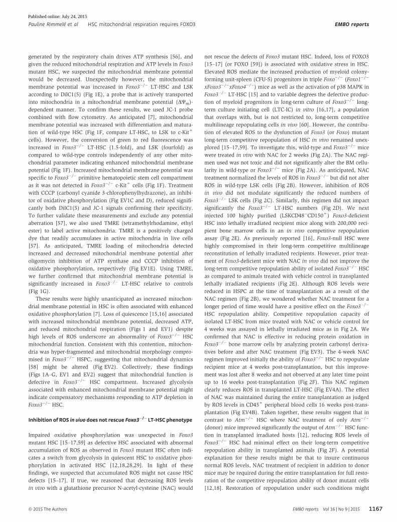
Inhibition of ROS in vivo does not rescue Foxo3�/� LT-HSCphenotype
Impaired oxidative phosphorylation was unexpected in Foxo3
mutant HSC [15–17,59] as defective HSC associated with abnormal
accumulation of ROS as observed in Foxo3 mutant HSC often indi-
cates a switch from glycolysis in quiescent HSC to oxidative phos-
phorylation in activated HSC [12,18,28,29]. In light of these
findings, we suspected that accumulated ROS might not cause HSC
defects [15–17]. If true, we reasoned that decreasing ROS levels
in vivo with a glutathione precursor N-acetyl-cysteine (NAC) would
not rescue the defects of Foxo3 mutant HSC. Indeed, loss of FOXO3
[15–17] (or FOXO [59]) is associated with oxidative stress in HSC.
Elevated ROS mediate the increased production of myeloid colony-
forming unit-spleen (CFU-S) progenitors in triple Foxo�/� (Foxo1�/�
xFoxo3�/�xFoxo4�/�) mice as well as the activation of p38 MAPK in
Foxo3�/� LT-HSC [15] and to variable degrees the defective produc-
tion of myeloid progenitors in long-term culture of Foxo3�/� long-
term culture initiating cell (LTC-IC) in vitro [16,17], a population
that overlaps with, but is not restricted to, long-term competitive
multilineage repopulating cells in vivo [60]. However, the contribu-
tion of elevated ROS to the dysfunction of Foxo3 (or Foxo) mutant
long-term competitive repopulation of HSC in vivo remained unex-
plored [15–17,59]. To investigate this, wild-type and Foxo3�/� mice
were treated in vivo with NAC for 2 weeks (Fig 2A). The NAC regi-
men used was not toxic and did not significantly alter the BM cellu-
larity in wild-type or Foxo3�/� mice (Fig 2A). As anticipated, NAC
treatment normalized the levels of ROS in Foxo3�/� but did not alter
ROS in wild-type LSK cells (Fig 2B). However, inhibition of ROS
in vivo did not modulate significantly the reduced numbers of
Foxo3�/� LSK cells (Fig 2C). Similarly, this regimen did not impact
significantly the Foxo3�/� LT-HSC numbers (Fig 2D). We next
injected 100 highly purified (LSKCD48�CD150+) Foxo3-deficient
HSC into lethally irradiated recipient mice along with 200,000 reci-
pient bone marrow cells in an in vivo competitive repopulation
assay (Fig 2E). As previously reported [16], Foxo3-null HSC were
highly compromised in their long-term competitive multilineage
reconstitution of lethally irradiated recipients. However, prior treat-
ment of Foxo3-deficient mice with NAC in vivo did not improve the
long-term competitive repopulation ability of isolated Foxo3�/� HSC
as compared to animals treated with vehicle control in transplanted
lethally irradiated recipients (Fig 2E). Although ROS levels were
reduced in HSPC at the time of transplantation as a result of the
NAC regimen (Fig 2B), we wondered whether NAC treatment for a
longer period of time would have a positive effect on the Foxo3�/�
HSC repopulation ability. Competitive repopulation capacity of
isolated LT-HSC from mice treated with NAC or vehicle control for
4 weeks was assayed in lethally irradiated mice as in Fig 2A. We
confirmed that NAC is effective in reducing protein oxidation in
Foxo3�/� bone marrow cells by analyzing protein carbonyl deriva-
tives before and after NAC treatment (Fig EV3). The 4-week NAC
regimen improved initially the ability of Foxo3�/� HSC to repopulate
recipient mice at 4 weeks post-transplantation, but this improve-
ment was lost after 8 weeks and not observed at any later time point
up to 16 weeks post-transplantation (Fig 2F). This NAC regimen
clearly reduces ROS in transplanted LT-HSC (Fig EV4A). The effect
of NAC was maintained during the entire transplantation as judged
by ROS levels in CD45+ peripheral blood cells 16 weeks post-trans-
plantation (Fig EV4B). Taken together, these results suggest that in
contrast to Atm�/� HSC where NAC treatment of only Atm�/�
(donor) mice improved significantly the output of Atm�/� HSC func-
tion in transplanted irradiated hosts [12], reducing ROS levels of
Foxo3�/� HSC had minimal effect on their long-term competitive
repopulation ability in transplanted animals (Fig 2F). A potential
explanation for these results might be that to insure continuous
normal ROS levels, NAC treatment of recipient in addition to donor
mice may be required during the entire transplantation for full resto-
ration of the competitive repopulation ability of donor mutant cells
[12,18]. Restoration of repopulation under such conditions might
ª 2015 The Authors EMBO reports Vol 16 | No 9 | 2015
Pauline Rimmelé et al HSC mitochondrial respiration requires FOXO3 EMBO reports
1167
Published online: July 24, 2015
indicate potential additional effects of NAC on bone marrow micro-
environment. Regardless, NAC treatment of both pre- and post-
transplanted Foxo3�/� HSC did not improve significantly their
repopulation ability beyond 4 weeks post-transplantation (Fig 2F).
Similarly, the positive effect of post-transplant NAC treatment of
Foxo3�/� HSC recipients did not last beyond 8 weeks (Fig 2F).
Thus, unlike Atm�/� [12] and Meis1�/� HSC [18] where reducing
elevated ROS rescued their long-term repopulation abnormalities,
normalizing ROS levels in vivo may not be sufficient to overcome
Foxo3�/� HSC long-term repopulation defects.
A
B C
D E
F
Figure 2. Inhibition of ROS does not rescue Foxo3�/� LT-HSC phenotype.
A WT and Foxo3�/� mice were injected intraperitoneally daily with 100 mg/kg body weight of N-acetyl-L-cysteine (NAC) or vehicle control for 2 weeks (left panel). Totalnumber of bone marrow (BM) cells isolated from one femur and one tibia (n = 6 mice per genotype) (right panel).
B ROS levels were measured by flow cytometry in WT and Foxo3�/� LSK cells isolated after 2 weeks. Fold change of geometric mean normalized with WT of DCFfluorescence (n = 6 mice per genotype).
C Total number of LSK cells in the BM (n = 6 mice per genotype).D Total number of LSKCD48�CD150+ (LT-HSC) cells in the BM (n = 6 mice per genotype).E Contribution of LT-HSC (CD45.1) from (A) to the peripheral blood (PB) of recipient mice (CD45.2) in a long-term competitive repopulation assay (n = 5 in each group).F Contribution of LSKCD48�CD150+ (LT-HSC) (CD45.1) isolated from NAC- or vehicle control-treated mice for 4 weeks to long-term competitive repopulation of lethally
irradiated mice treated with NAC or vehicle control during post-transplantation (P.T.). One of two experiments is shown.
Data information: Data are expressed as mean � SEM (n = 5 mice transplanted per group); NS, not significant; *P < 0.05, Student’s t-test.
EMBO reports Vol 16 | No 9 | 2015 ª 2015 The Authors
EMBO reports HSC mitochondrial respiration requires FOXO3 Pauline Rimmelé et al
1168
Published online: July 24, 2015

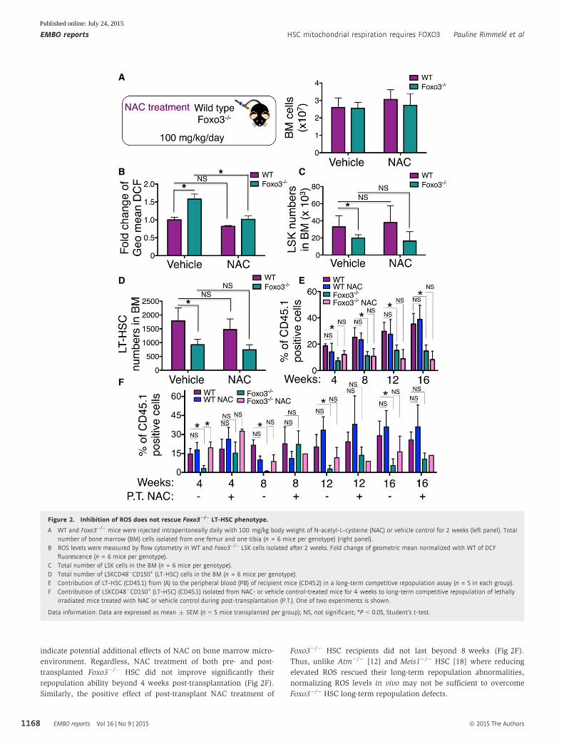
Reducing ROS levels does not improve Foxo3�/� HSCmitochondrial dysfunction in vivo
As a chronic increase in ROS might impair mitochondrial function
[61], we evaluated whether Foxo3�/� HSC mitochondrial dysfunc-
tion was at least partially due to increased ROS. The in vivo NAC
treatment did not revert the abnormal increased mitochondrial
membrane potential in Foxo3�/� LSK cells as measured by both
DiIC1(5) (Fig 3A) and JC-1 (Fig 3B) probes. In agreement with these
results, TMRE levels of Foxo3�/� LT-HSC or LSK cells (Figs 3C and
EV4C) despite reduced ROS levels (Fig EV4A and D) were not
reduced in response to NAC treatment, suggesting that the increased
mitochondrial membrane potential of Foxo3�/� HSC was not due to
oxidative stress. Altogether, these results indicate that loss of
FOXO3 impairs mitochondrial function independent of ROS in HSC.
In agreement with this, many genes implicated in the regulation of
mitochondrial function, electron transfer chain, and/or metabolism,
particularly glycolysis, were significantly deregulated in Foxo3�/�
HSPC (Fig 4). Among these genes, impaired expression of a master
regulator of mitochondrial biogenesis peroxisome proliferator-acti-
vated receptor-c coactivator (Pgc) 1 was notable. Some of these
genes that are potential direct targets of FOXO3 including isocitrate
dehydrogenase 1 (Idh1) and Idh2 genes [62,63] were specifically
modulated in Foxo3�/� HSPC (LSK cells), but not in more commit-
ted Foxo3�/� c-Kit+ hematopoietic progenitor cells. One of the most
remarkable impacts among the genes surveyed was on Atpif1,
the inhibitor of mitochondrial F1F0-ATPase that limits the ATP
depletion. Atpif1 expression was reduced by over 60% (Fig 4A).
Reduced expression of ATPif1 maintains mitochondrial membrane
potential to protect cells with severe deficiencies in electron transfer
chain from apoptosis [64]. It is noteworthy in this context that mito-
chondrial membrane potential is elevated in Foxo3�/� HSPC
(Fig 1E–G), and despite high ROS, these cells do not exhibit
increased apoptosis [15].
Thus, loss of Foxo3�/� HSC long-term competitive repopulation
is associated with impaired mitochondrial metabolism, but not
mediated by ROS. These combined findings raise the possibility that
compromised Foxo3�/� HSC mitochondria may be implicated in
defects of Foxo3�/� LT-HSC activity.
Inhibition of mTOR signaling improves ROS levels but does notameliorate Foxo3�/� LT-HSC function in vivo
The mammalian target of rapamycin (mTOR) is critical for the regu-
lation of mitochondrial biogenesis including in HSC [20,65,66].
A B
C
Figure 3. Reducing ROS levels does not improve mitochondrialdysfunction in Foxo3�/� HSPC.
A Mice were treated with NAC or vehicle control, and mitochondrialmembrane potential was measured in freshly isolated cells after 2 weeks.Fold change of geometric mean of DilC1(5) fluorescence measured in LSKcells (n = 3 mice per genotype) normalized with vehicle-treated (WT).
B Relative DΨm measured in LSK cells by the red/green fluorescence ratio ofJC-1 probe (n = 6 mice per genotype).
C TMRE quantification of mitochondrial membrane potential in LT-HSCisolated from mice treated with NAC or vehicle control (n = 3 mice).
Data information: All data are expressed as mean � SEM; NS, not significant;*P < 0.05, Student’s t-test. Controls are the same as in Fig 1G.
A
B
Figure 4. Alteration of mitochondrial and metabolic gene expression inFoxo3�/� HSPC.
A qRT–PCR analysis of genes whose products are implicated in mitochondrialmetabolism. Results are relative to WT set to one in each population.
B qRT–PCR analysis of glycolytic genes. Results are relative to WT set to onein each population.
Data information: Results are from three independent experiments each basedon three replicates of one cDNA generated from a pool of cells isolated fromthree mice. All data are expressed as mean � SEM (Student’s t-test, *P < 0.05).
ª 2015 The Authors EMBO reports Vol 16 | No 9 | 2015
Pauline Rimmelé et al HSC mitochondrial respiration requires FOXO3 EMBO reports
1169
Published online: July 24, 2015
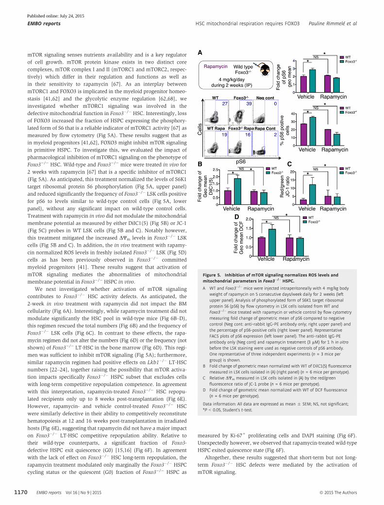
mTOR signaling senses nutrients availability and is a key regulator
of cell growth. mTOR protein kinase exists in two distinct core
complexes, mTOR complex I and II (mTORC1 and mTORC2, respec-
tively) which differ in their regulation and functions as well as
in their sensitivity to rapamycin [67]. As an interplay between
mTORC1 and FOXO3 is implicated in the myeloid progenitor homeo-
stasis [41,62] and the glycolytic enzyme regulation [62,68], we
investigated whether mTORC1 signaling was involved in the
defective mitochondrial function in Foxo3�/� HSC. Interestingly, loss
of FOXO3 increased the fraction of HSPC expressing the phosphory-
lated form of S6 that is a reliable indicator of mTORC1 activity [67] as
measured by flow cytometry (Fig 5A). These results suggest that as
in myeloid progenitors [41,62], FOXO3 might inhibit mTOR signaling
in primitive HSPC. To investigate this, we evaluated the impact of
pharmacological inhibition of mTORC1 signaling on the phenotype of
Foxo3�/� HSC. Wild-type and Foxo3�/� mice were treated in vivo for
2 weeks with rapamycin [67] that is a specific inhibitor of mTORC1
(Fig 5A). As anticipated, this treatment normalized the levels of S6K1
target ribosomal protein S6 phosphorylation (Fig 5A, upper panel)
and reduced significantly the frequency of Foxo3�/� LSK cells positive
for pS6 to levels similar to wild-type control cells (Fig 5A, lower
panel), without any significant impact on wild-type control cells.
Treatment with rapamycin in vivo did not modulate the mitochondrial
membrane potential as measured by either DiIC1(5) (Fig 5B) or JC-1
(Fig 5C) probes in WT LSK cells (Fig 5B and C). Notably however,
this treatment mitigated the increased DΨm levels in Foxo3�/� LSK
cells (Fig 5B and C). In addition, the in vivo treatment with rapamy-
cin normalized ROS levels in freshly isolated Foxo3�/� LSK (Fig 5D)
cells as has been previously observed in Foxo3�/� committed
myeloid progenitors [41]. These results suggest that activation of
mTOR signaling mediates the abnormalities of mitochondrial
membrane potential in Foxo3�/� HSPC in vivo.
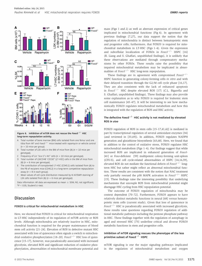
We next investigated whether activation of mTOR signaling
contributes to Foxo3�/� HSC activity defects. As anticipated, the
2-week in vivo treatment with rapamycin did not impact the BM
cellularity (Fig 6A). Interestingly, while rapamycin treatment did not
modulate significantly the HSC pool in wild-type mice (Fig 6B–D),
this regimen rescued the total numbers (Fig 6B) and the frequency of
Foxo3�/� LSK cells (Fig 6C). In contrast to these effects, the rapa-
mycin regimen did not alter the numbers (Fig 6D) or the frequency (not
shown) of Foxo3�/� LT-HSC in the bone marrow (Fig 6D). This regi-
men was sufficient to inhibit mTOR signaling (Fig 5A); furthermore,
similar rapamycin regimen had positive effects on Lkb1�/� LT-HSC
numbers [22–24], together raising the possibility that mTOR activa-
tion impacts specifically Foxo3�/� HSPC subset that excludes cells
with long-term competitive repopulation competence. In agreement
with this interpretation, rapamycin-treated Foxo3�/� HSC repopu-
lated recipients only up to 8 weeks post-transplantation (Fig 6E).
However, rapamycin- and vehicle control-treated Foxo3�/� HSC
were similarly defective in their ability to competitively reconstitute
hematopoiesis at 12 and 16 weeks post-transplantation in irradiated
hosts (Fig 6E), suggesting that rapamycin did not have a major impact
on Foxo3�/� LT-HSC competitive repopulation ability. Relative to
their wild-type counterparts, a significant fraction of Foxo3-
defective HSPC exit quiescence (G0) [15,16] (Fig 6F). In agreement
with the lack of effect on Foxo3�/� HSC long-term repopulation, the
rapamycin treatment modulated only marginally the Foxo3�/� HSPC
cycling status or the quiescent (G0) fraction of Foxo3�/� HSPC as
measured by Ki-67+ proliferating cells and DAPI staining (Fig 6F).
Unexpectedly however, we observed that rapamycin-treated wild-type
HSPC exited quiescence state (Fig 6F).
Altogether, these results suggested that short-term but not long-
term Foxo3�/� HSC defects were mediated by the activation of
mTOR signaling.
A
B C
D
Figure 5. Inhibition of mTOR signaling normalizes ROS levels andmitochondrial parameters in Foxo3�/� HSPC.
A WT and Foxo3�/� mice were injected intraperitoneally with 4 mg/kg bodyweight of rapamycin on 5 consecutive days/week daily for 2 weeks (leftupper panel). Analysis of phosphorylated form of S6K1 target ribosomalprotein S6 (pS6) by flow cytometry in LSK cells isolated from WT andFoxo3�/� mice treated with rapamycin or vehicle control by flow cytometrymeasuring fold change of geometric mean of pS6 compared to negativecontrol (Neg cont: anti-rabbit IgG-PE antibody only; right upper panel) andthe percentage of pS6-positive cells (right lower panel). RepresentativeFACS plots of pS6 expression (left lower panel). The anti-rabbit IgG-PEantibody only (Neg cont) and rapamycin treatment (3 lM) for 1 h in vitrobefore the LSK staining were used as negative controls of pS6 antibody.One representative of three independent experiments (n = 3 mice pergroup) is shown.
B Fold change of geometric mean normalized with WT of DilC1(5) fluorescencemeasured in LSK cells isolated in (A) (right panel) (n = 6mice per genotype).
C Relative DΨm measured in LSK cells isolated in (A) by the red/greenfluorescence ratio of JC-1 probe (n = 6 mice per genotype).
D Fold change of geometric mean normalized with WT of DCF fluorescence(n = 6 mice per genotype).
Data information: All data are expressed as mean � SEM; NS, not significant;*P < 0.05, Student’s t-test.
EMBO reports Vol 16 | No 9 | 2015 ª 2015 The Authors
EMBO reports HSC mitochondrial respiration requires FOXO3 Pauline Rimmelé et al
1170
Published online: July 24, 2015
Discussion
FOXO3 is critical for mitochondrial metabolism in HSC
Here, we showed that FOXO3 is critical for mitochondrial respiration
in LT-HSC independently of its regulation of mTOR activity or ROS
levels. Although mitochondria are relatively inactive in HSC, mito-
chondrial function is required for the proper maintenance of blood
stem cell activity [21–24]. Elevation of ROS in defective mutant HSC
associated with loss of quiescence often signals a switch to mitochon-
drial oxidative phosphorylation [18–20]. Foxo3�/� HSC loss of quies-
cence [15–17], however, was paradoxically associated with increased
glycolysis, elevated ROS and significant reduction of oxidative phos-
phorylation, abnormalities of mitochondrial membrane potential and
mass (Figs 1 and 2) as well as aberrant expression of critical genes
implicated in mitochondrial functions (Fig 4). In agreement with
previous findings [7,27], our data support the notion that the
regulation of mitochondria is distinct between hematopoietic stem
and progenitor cells; furthermore, that FOXO3 is required for mito-
chondrial metabolism in LT-HSC (Figs 1–4). Given the expression
and subcellular localization of FOXOs in Foxo3�/� HSPC [16]
(R. Liang and S. Ghaffari, unpublished findings), it is unlikely that
these observations are mediated through compensatory mecha-
nisms by other FOXOs. These results raise the possibility that
impaired mitochondrial metabolism may be implicated in abnor-
malities of Foxo3�/� HSC long-term repopulation.
These findings are in agreement with compromised Foxo3�/�
HSPC function in generating colony-forming cells in vitro and with
their delayed transition through the G2/M cell cycle phase [16,17].
They are also consistent with the lack of enhanced apoptosis
in Foxo3�/� HSC despite elevated ROS [17] (C.L. Bigarella and
S. Ghaffari, unpublished findings). These findings may also provide
some explanation as to why FOXO3 is required for leukemic stem
cell maintenance [45–47]. It will be interesting to see how mecha-
nistically FOXO3 regulates mitochondrial metabolism and how this
is integrated with the regulation of ROS and HSC activity.
The defective Foxo3�/� HSC activity is not mediated by elevatedROS in vivo
FOXO3 regulation of ROS in stem cells [15–17,41,42] is mediated in
part by transcriptional regulation of several antioxidant enzymes [34]
(and reviewed in [33,69]). In addition, FOXO3 regulates NADPH
generation and glutathione biosynthesis [63,68]. Here, we found that
in addition to the control of oxidative stress, FOXO3 regulates HSC
mitochondrial metabolism (Figs 1–4). Our findings suggest that while
increased ROS are implicated in enhanced generation of Foxo3-
[41] or Foxo-deficient [59] multipotent colony-forming unit-spleen
(CFU-S), and cell cycle-related abnormalities of HSPC [14,16,59],
elevated ROS do not mediate the functional defects of Foxo3�/� long-
term HSC but rather might reflect an abnormal mitochondrial func-
tion. These results are consistent with the notion that NAC treatment
only partially rescued the p38 MAPK activation in Foxo3�/� HSPC
[15]. These findings raise the interesting possibility that underlying
mechanisms that uncouple ROS from mitochondrial potential might
disengage HSC cycling from HSC repopulation potential.
The outcome of FOXO3 regulation of mitochondria may be
context dependent [70–72]. Furthermore, FOXO3 appears to have
relatively distinct metabolic functions in neural [68] versus hemato-
poietic stem cells (current study). Given that loss of quiescence in
Foxo3�/� HSC is paradoxically associated with increased glycolysis,
these results raise questions regarding FOXO3 regulation of addi-
tional metabolic pathways including the pentose phosphate pathway
in HSC. These findings together with the regulation of autophagy in
aged and stressed HSC [73] underline critical and diverse FOXO3
metabolic functions in stem and progenitor cells.
Inhibition of mTOR signaling rescues the phenotype of the lessprimitive subset of Foxo3�/� HSPC
mTOR signaling is one the major signaling pathways implicated
in the regulation of mitochondrial metabolism and oxygen
A B
C D
E F
Figure 6. Inhibition of mTOR does not rescue the Foxo3�/� HSClong-term repopulation activity.
A Total number of bone marrow (BM) cells isolated from one femur and onetibia from WT and Foxo3�/� mice treated with rapamycin or vehicle control(n = 10 mice per group).
B Total number of LSK cells in the BM of mice from (A) (n = 10 mice pergenotype).
C Frequency of Lin�Sca-1+c-Kit+ cells (n = 10 mice per genotype).D Total number of LSKCD48�CD150+ (LT-HSC) cells in the BM of mice from
(A) (n = 6 mice per genotype).E The contribution of transplanted LT-HSC (CD45.1) cells isolated from (A) to
the PB of recipient mice (CD45.2) in a long-term competitive repopulationassay (n = 8 in each group).
F Mean values of cell cycle distribution measured by Ki-67/DAPI staining ofLSK cells isolated from (A) (n = 6 mice per genotype).
Data information: All data are expressed as mean � SEM; NS, not significant;*P < 0.05, Student’s t-test.
ª 2015 The Authors EMBO reports Vol 16 | No 9 | 2015
Pauline Rimmelé et al HSC mitochondrial respiration requires FOXO3 EMBO reports
1171
Published online: July 24, 2015
consumption in somatic and embryonic stem cells [20,65,74,75].
However, the activation of mTOR signaling in Foxo3�/� HSPC seems
to mediate the defects of Foxo3�/� ST-HSC, but not LT-HSC. Rapa-
mycin treatment rescued the numbers of Foxo3�/� LSK cells and
normalized the mitochondrial membrane potential and ROS in
Foxo3�/� LSK cells (Fig 5). In addition, rapamycin rescued the
competitive repopulation ability of Foxo3�/� HSC up to 8 weeks,
suggesting that mTOR mediates the Foxo3�/� ST-HSC defects
(Fig 6). In contrast, while this 2-week period of treatment was
clearly sufficient to reduce pS6 downstream of mTOR signaling and
rescue Tsc1 and Lkb1 mutant LT-HSC numbers [20,22–24], rapa-
mycin did not rescue the number of Foxo3�/� LT-HSC in treated mice
or their competitive repopulating potential in lethally irradiated
transplanted hosts (Figs 5 and 6). These findings combined with
our previous results [41] indicate that mTOR activation mediates
defects of Foxo3�/� myeloid and multipotential progenitors, but not
Foxo3�/� LT-HSC abnormalities. These results were not too surpris-
ing after all as the phenotype of Foxo3�/� and Tsc1�/� LT-HSC in
which mTOR is constitutively activated is quite distinct [20]. Consis-
tent with PTEN-independent regulation of FOXO3 in HSC [48,76],
these results (Figs 5 and 6) further delineate differences of mTOR
activation in HSC on a background of PTEN versus FOXO3 loss of
function [76]. In addition, they suggest that mTOR activation in
Foxo3�/� ST-HSC might result in their defective function leading to
proliferation of downstream hematopoietic progenitors [41]. As both
rapamycin and NAC treatments normalized ROS but not the compet-
itive repopulation of Foxo3�/� LT-HSC, our results support the
notion that elevated ROS do not mediate the defective competitive
repopulation of Foxo3�/� LT-HSC. However, despite similarities of
Foxo�/� and Foxo3�/� HSC [15–17,41,59], our results do not rule
out the potential oxidative stress mediation of in vivo Foxo�/� HSC
competitive repopulation defects [59].
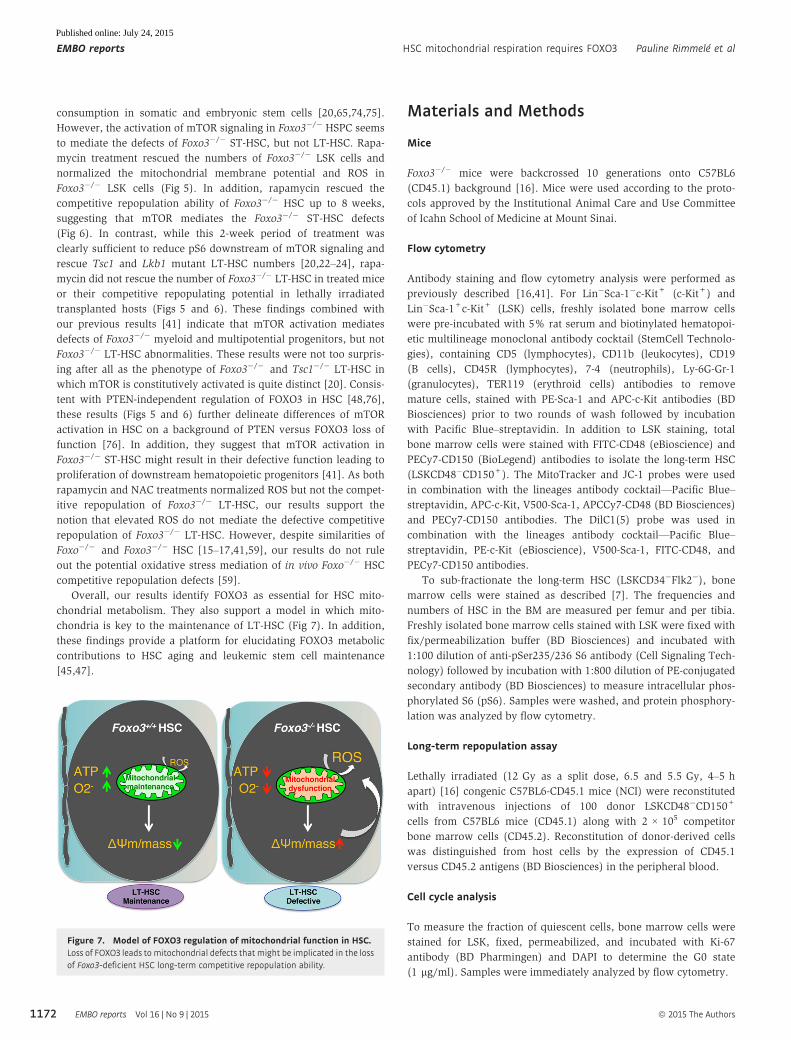
Overall, our results identify FOXO3 as essential for HSC mito-
chondrial metabolism. They also support a model in which mito-
chondria is key to the maintenance of LT-HSC (Fig 7). In addition,
these findings provide a platform for elucidating FOXO3 metabolic
contributions to HSC aging and leukemic stem cell maintenance
[45,47].
Materials and Methods
Mice
Foxo3�/� mice were backcrossed 10 generations onto C57BL6
(CD45.1) background [16]. Mice were used according to the proto-
cols approved by the Institutional Animal Care and Use Committee
of Icahn School of Medicine at Mount Sinai.
Flow cytometry
Antibody staining and flow cytometry analysis were performed as
previously described [16,41]. For Lin�Sca-1�c-Kit+ (c-Kit+) and
Lin�Sca-1+c-Kit+ (LSK) cells, freshly isolated bone marrow cells
were pre-incubated with 5% rat serum and biotinylated hematopoi-
etic multilineage monoclonal antibody cocktail (StemCell Technolo-
gies), containing CD5 (lymphocytes), CD11b (leukocytes), CD19
(B cells), CD45R (lymphocytes), 7-4 (neutrophils), Ly-6G-Gr-1
(granulocytes), TER119 (erythroid cells) antibodies to remove
mature cells, stained with PE-Sca-1 and APC-c-Kit antibodies (BD
Biosciences) prior to two rounds of wash followed by incubation
with Pacific Blue–streptavidin. In addition to LSK staining, total
bone marrow cells were stained with FITC-CD48 (eBioscience) and
PECy7-CD150 (BioLegend) antibodies to isolate the long-term HSC
(LSKCD48�CD150+). The MitoTracker and JC-1 probes were used
in combination with the lineages antibody cocktail—Pacific Blue–
streptavidin, APC-c-Kit, V500-Sca-1, APCCy7-CD48 (BD Biosciences)
and PECy7-CD150 antibodies. The DilC1(5) probe was used in
combination with the lineages antibody cocktail—Pacific Blue–
streptavidin, PE-c-Kit (eBioscience), V500-Sca-1, FITC-CD48, and
PECy7-CD150 antibodies.
To sub-fractionate the long-term HSC (LSKCD34�Flk2�), bone
marrow cells were stained as described [7]. The frequencies and
numbers of HSC in the BM are measured per femur and per tibia.
Freshly isolated bone marrow cells stained with LSK were fixed with
fix/permeabilization buffer (BD Biosciences) and incubated with
1:100 dilution of anti-pSer235/236 S6 antibody (Cell Signaling Tech-
nology) followed by incubation with 1:800 dilution of PE-conjugated
secondary antibody (BD Biosciences) to measure intracellular phos-
phorylated S6 (pS6). Samples were washed, and protein phosphory-
lation was analyzed by flow cytometry.
Long-term repopulation assay
Lethally irradiated (12 Gy as a split dose, 6.5 and 5.5 Gy, 4–5 h
apart) [16] congenic C57BL6-CD45.1 mice (NCI) were reconstituted
with intravenous injections of 100 donor LSKCD48�CD150+
cells from C57BL6 mice (CD45.1) along with 2 × 10⁵ competitor
bone marrow cells (CD45.2). Reconstitution of donor-derived cells
was distinguished from host cells by the expression of CD45.1
versus CD45.2 antigens (BD Biosciences) in the peripheral blood.
Cell cycle analysis
To measure the fraction of quiescent cells, bone marrow cells were
stained for LSK, fixed, permeabilized, and incubated with Ki-67
antibody (BD Pharmingen) and DAPI to determine the G0 state
(1 lg/ml). Samples were immediately analyzed by flow cytometry.
Figure 7. Model of FOXO3 regulation of mitochondrial function in HSC.Loss of FOXO3 leads to mitochondrial defects that might be implicated in the lossof Foxo3-deficient HSC long-term competitive repopulation ability.
EMBO reports Vol 16 | No 9 | 2015 ª 2015 The Authors
EMBO reports HSC mitochondrial respiration requires FOXO3 Pauline Rimmelé et al
1172
Published online: July 24, 2015
NAC treatment
WT and Foxo3�/� mice were injected intraperitoneally with
100 mg/kg body weight of N-acetyl-L-cysteine (NAC; Sigma, MO) in
phosphate-buffered saline (PBS) solution (pH 7.4) daily, as
previously described [16,41]. For long-term treatment, NAC was
alternatively injected every day and added to the water every other
week.
Rapamycin treatment
WT and Foxo3�/� mice were injected intraperitoneally with
4 mg/kg body weight of rapamycin (Enzo Life Sciences, NY) in
PBS with 5% Tween-80, 5% PEG400 and 4% ethanol during
five consecutive days/week for 2 weeks, as previously described
[41].
Measurement of intracellular ROS
ROS was measured using CM-H2DCFDA (Molecular Probes) as
previously described [16,41]. To measure the concentration of
superoxide anion, 106 cells stained first for LSK were resuspended
in PBS 2% FBS, loaded with 5 lM mitoSOX Red (M36008; Mole-
cular Probes) and incubated in the dark for 30 min at 37°C. The
fluorescent product was measured immediately by flow cytometry.
Measurement of mitochondrial mass and membrane potential
To measure the mitochondrial mass, 3 × 10⁶ cells stained first to
gate c-Kit+, LSK, LT-HSC were resuspended in PBS 2% FBS and
loaded with 20 nM MitoTracker Green (M7514; Molecular Probes),
in the dark for 30 min at 37°C. To measure the mitochondrial
membrane potential, cells were loaded with 2 lM JC-1 probe
(M34152; Molecular Probes) or 25 nM DilC1(5) (M34151; Molecular
Probes) in accordance with the manufacturer’s recommendations
and incubated in the dark for 20 min or 25 min, respectively, at
37°C. To test the specificity, cells were incubated with 50 mM CCCP
(carbonyl cyanide 3-chlorophenylhydrazone), a DΨm inhibitor for
20 min at 37°C. For each probe, the fluorescent product was
measured immediately by flow cytometry.
Metabolic assays
Oxygen consumption was measured with the BD Oxygen Biosensor
System in accordance with the manufacturer’s recommendations, as
previously described [7]. ATP levels were quantified with ATP
Bioluminescence Assay Kit HS II (Roche) in accordance with the
manufacturer’s recommendations, as previously described [7].
Lactate production was measured with gas chromatography–mass
spectrometry, as previously described [7].
Protein oxidation detection
Cell lysate proteins were treated as previously described [39],
reacted with 2,4-dinitrophenylhydrazine to derivatize carbonyl
groups (OxyBlot Protein Oxidation Detection kit; Chemicon Interna-
tional) to dinitrophenylhydrazone (DNP) and resolved by 12%
SDS–PAGE and detected by rabbit anti-DNP antibody.
Real-time quantitative RT–PCR
Total RNA was isolated using RNeasy MicroPlus kit (Qiagen). First-
strand cDNA was synthesized using SuperScriptII (Invitrogen).
cDNA obtained from 300 cells was used per well for RT–PCR
performed using SYBR Green JumpStart Taq ReadyMix (Takara) in
triplicates, using the primers indicated in the figures and ABI Prism
7900 HT Cycler (Applied Biosystems, see Primer sequences in
Appendix Table S1). All the results were normalized to b-actinRNA levels.
Statistical analysis
Unpaired two-tailed Student’s t-test was used for all experiments.
P-value < 0.05 was considered significant.
Expanded View for this article is available online:
http://embor.embopress.org
AcknowledgementsWe thank Dr. Safak Yalcin for her initial work on Foxo3 mutant mitochondrial
phenotype and The Flow Cytometry Shared Research Facility at Icahn School
of Medicine at Mount Sinai. R.L. was partially supported by NIH T32 GM08553-
13 and T32 HD075735 and C.LB. partially supported by a Roche TCRC -Young
Investigator. This work was supported in part by the National Institutes of
Health grants RO1 DK077174, RO1 RHL116365A (Co-PI), a Myeloproliferative
Neoplasm Foundation (MPN) award and an Irma Hirschl/Weill-Caulier Trust
Research award to S.G.
Author contributionsPR, RL, CLB, FK and JX designed experiments, performed experiments, and
analyzed data. PR and RL wrote the paper; HS and CCZ designed experiments
and analyzed data; MNS and JC provided valuable tools; JC edited the paper
and SG conceived the project, designed experiments, analyzed data, and wrote
the paper.
Conflict of interestThe authors declare that they have no conflict of interest.
References
1. Orford KW, Scadden DT (2008) Deconstructing stem cell self-renewal:
genetic insights into cell-cycle regulation. Nat Rev Genet 9: 115 – 128
2. Wilson A, Laurenti E, Oser G, der van Wath RC, Blanco-Bose W, Jaworski
M, Offner S, Dunant CF, Eshkind L, Bockamp E et al (2008) Hematopoi-
etic stem cells reversibly switch from dormancy to self-renewal during
homeostasis and repair. Cell 135: 1118 – 1129
3. Foudi A, Hochedlinger K, Van Buren D, Schindler JW, Jaenisch R, Carey V,
Hock H (2009) Analysis of histone 2B-GFP retention reveals slowly
cycling hematopoietic stem cells. Nat Biotechnol 27: 84 – 90
4. Qiu J, Papatsenko D, Niu X, Schaniel C, Moore K (2014) Divisional history
and hematopoietic stem cell function during homeostasis. Stem Cell Rep
2: 473 – 490
5. Sauvageau G, Iscove NN, Humphries RK (2004) In vitro and in vivo
expansion of hematopoietic stem cells. Oncogene 23: 7223 – 7232
6. Unwin RD, Smith DL, Blinco D, Wilson CL, Miller CJ, Evans CA, Jaworska
E, Baldwin SA, Barnes K, Pierce A et al (2006) Quantitative proteomics
ª 2015 The Authors EMBO reports Vol 16 | No 9 | 2015
Pauline Rimmelé et al HSC mitochondrial respiration requires FOXO3 EMBO reports
1173
Published online: July 24, 2015
reveals posttranslational control as a regulatory factor in primary hemato-
poietic stem cells. Blood 107: 4687 – 4694
7. Simsek T, Kocabas F, Zheng J, Deberardinis RJ, Mahmoud AI, Olson EN,
Schneider JW, Zhang CC, Sadek HA (2010) The distinct metabolic profile
of hematopoietic stem cells reflects their location in a hypoxic niche.
Cell Stem Cell 7: 380 – 390
8. Jang YY, Sharkis SJ (2007) A low level of reactive oxygen species selects
for primitive hematopoietic stem cells that may reside in the low-
oxygenic niche. Blood 110: 3056 – 3063
9. Wang YH, Israelsen WJ, Lee D, Yu VW, Jeanson NT, Clish CB, Cantley LC,
Vander Heiden MG, Scadden DT (2014) Cell-state-specific metabolic
dependency in hematopoiesis and leukemogenesis. Cell 158: 1309 – 1323
10. Bigarella CL, Liang R, Ghaffari S (2014) Stem cells and the impact of
ROS signaling. Development 141: 4206 – 4218
11. Finkel T (2012) Signal transduction by mitochondrial oxidants. J Biol
Chem 287: 4434 –4440
12. Ito K, Hirao A, Arai F, Matsuoka S, Takubo K, Hamaguchi I, Nomiyama K,
Hosokawa K, Sakurada K, Nakagata N et al (2004) Regulation of oxida-
tive stress by ATM is required for self-renewal of haematopoietic stem
cells. Nature 431: 997 – 1002
13. Ito K, Hirao A, Arai F, Takubo K, Matsuoka S, Miyamoto K, Ohmura M,
Naka K, Hosokawa K, Ikeda Y (2006) Reactive oxygen species act through
p38 MAPK to limit the lifespan of hematopoietic stem cells. Nat Med 12:
446 – 451
14. Tothova Z, Kollipara R, Huntly BJ, Lee BH, Castrillon DH, Cullen DE,
McDowell EP, Lazo-Kallanian S, Williams IR, Sears C et al (2007) FoxOs
are critical mediators of hematopoietic stem cell resistance to
physiologic oxidative stress. Cell 128: 325 – 339
15. Miyamoto K, Araki KY, Naka K, Arai F, Takubo K, Yamazaki S, Matsuoka
S, Miyamoto T, Ito K, Ohmura M et al (2007) Foxo3a is essential for
maintenance of the hematopoietic stem cell pool. Cell Stem Cell 1:
101 – 112
16. Yalcin S, Zhang X, Luciano JP, Mungamuri SK, Marinkovic D, Vercherat C,
Sarkar A, Grisotto M, Taneja R, Ghaffari S et al (2008) Foxo3 is essential
for the regulation of ataxia telangiectasia mutated and oxidative stress-
mediated homeostasis of hematopoietic stem cells. J Biol Chem 283:
25692 – 25705
17. Miyamoto K, Miyamoto T, Kato R, Yoshimura A, Motoyama N, Suda T
(2008) FoxO3a regulates hematopoietic homeostasis through a negative
feedback pathway in conditions of stress or aging. Blood 112:
4485 – 4493
18. Kocabas F, Zheng J, Thet S, Copeland NG, Jenkins NA, DeBerardinis RJ,
Zhang C, Sadek HA (2012) Meis1 regulates the metabolic phenotype and
oxidant defense of hematopoietic stem cells. Blood 120: 4963 – 4972
19. Zheng J, Lu Z, Kocabas F, Bottcher RT, Costell M, Kang X, Liu X, Deberar-
dinis RJ, Wang Q, Chen GQ et al (2014) Profilin 1 is essential for reten-
tion and metabolism of mouse hematopoietic stem cells in bone
marrow. Blood 123: 992 – 1001
20. Chen C, Liu Y, Liu R, Ikenoue T, Guan KL, Zheng P (2008) TSC-mTOR
maintains quiescence and function of hematopoietic stem cells by
repressing mitochondrial biogenesis and reactive oxygen species. J Exp
Med 205: 2397 – 2408
21. Yu WM, Liu X, Shen J, Jovanovic O, Pohl EE, Gerson SL, Finkel T,
Broxmeyer HE, Qu CK (2013) Metabolic regulation by the mitochondrial
phosphatase PTPMT1 is required for hematopoietic stem cell differentia-
tion. Cell Stem Cell 12: 62 – 74
22. Gan B, Hu J, Jiang S, Liu Y, Sahin E, Zhuang L, Fletcher-Sananikone E,
Colla S, Wang YA, Chin L et al (2010) Lkb1 regulates quiescence and
metabolic homeostasis of haematopoietic stem cells. Nature 468:
701 – 704
23. Gurumurthy S, Xie SZ, Alagesan B, Kim J, Yusuf RZ, Saez B, Tzatsos A,
Ozsolak F, Milos P, Ferrari F et al (2010) The Lkb1 metabolic sensor
maintains haematopoietic stem cell survival. Nature 468: 659 – 663
24. Nakada D, Saunders TL, Morrison SJ (2010) Lkb1 regulates cell cycle
and energy metabolism in haematopoietic stem cells. Nature 468:
653 – 658
25. Mantel C, Messina-Graham S, Moh A, Cooper S, Hangoc G, Fu XY,
Broxmeyer HE (2012) Mouse hematopoietic cell-targeted STAT3 deletion:
stem/progenitor cell defects, mitochondrial dysfunction, ROS overpro-
duction, and a rapid aging-like phenotype. Blood 120: 2589 – 2599
26. Chen Y, Yu M, Dai X, Zogg M, Wen R, Weiler H, Wang D (2011) Critical
role for Gimap5 in the survival of mouse hematopoietic stem and
progenitor cells. J Exp Med 208: 923 – 935
27. Norddahl GL, Pronk CJ, Wahlestedt M, Sten G, Nygren JM, Ugale A,
Sigvardsson M, Bryder D (2011) Accumulating mitochondrial DNA muta-
tions drive premature hematopoietic aging phenotypes distinct from
physiological stem cell aging. Cell Stem Cell 8: 499 – 510
28. Takubo K, Goda N, Yamada W, Iriuchishima H, Ikeda E, Kubota Y, Shima
H, Johnson RS, Hirao A, Suematsu M et al (2010) Regulation of the HIF-
1alpha level is essential for hematopoietic stem cells. Cell Stem Cell 7:
391 – 402
29. Takubo K, Nagamatsu G, Kobayashi CI, Nakamura-Ishizu A, Kobayashi H,
Ikeda E, Goda N, Rahimi Y, Johnson RS, Soga T et al (2013) Regulation of
glycolysis by pdk functions as a metabolic checkpoint for cell cycle
quiescence in hematopoietic stem cells. Cell Stem Cell 12: 49 – 61
30. Zhang X, Rielland M, Yalcin S, Ghaffari S (2011) Regulation and function
of FoxO transcription factors in normal and cancer stem cells: what
have we learned? Curr Drug Targets 12: 1267 – 1283
31. Ito K, Carracedo A, Weiss D, Arai F, Ala U, Avigan DE, Schafer ZT, Evans
RM, Suda T, Lee CH et al (2012) A PML-PPAR-delta pathway for fatty
acid oxidation regulates hematopoietic stem cell maintenance. Nat Med
18: 1350 – 1358
32. Zhang CC, Sadek HA (2014) Hypoxia and metabolic properties of hemato-
poietic stem cells. Antioxid Redox Signal 20: 1891 – 1901
33. Eijkelenboom A, Burgering BM (2013) FOXOs: signalling integrators for
homeostasis maintenance. Nat Rev Mol Cell Biol 14: 83 – 97
34. Kops GJ, Dansen TB, Polderman PE, Saarloos I, Wirtz KW, Coffer PJ,
Huang TT, Bos JL, Medema RH, Burgering BM (2002) Forkhead
transcription factor FOXO3a protects quiescent cells from oxidative
stress. Nature 419: 316 – 321
35. Essers MA, de Vries-Smits LM, Barker N, Polderman PE, Burgering BM,
Korswagen HC (2005) Functional interaction between beta-catenin and
FOXO in oxidative stress signaling. Science 308: 1181 – 1184
36. Essers MA, Weijzen S, de Vries-Smits AM, Saarloos I, de Ruiter ND, Bos JL,
Burgering BM (2004) FOXO transcription factor activation by oxidative
stress mediated by the small GTPase Ral and JNK. EMBO J 23: 4802 – 4812
37. Furukawa-Hibi Y, Yoshida-Araki K, Ohta T, Ikeda K, Motoyama N (2002)
FOXO forkhead transcription factors induce G(2)-M checkpoint in
response to oxidative stress. J Biol Chem 277: 26729 – 26732
38. Dansen TB, Smits LM, van Triest MH, de Keizer PL, van Leenen D,
Koerkamp MG, Szypowska A, Meppelink A, Brenkman AB, Yodoi J et al
(2009) Redox-sensitive cysteines bridge p300/CBP-mediated acetylation
and FoxO4 activity. Nat Chem Biol 5: 664 – 672
39. Marinkovic D, Zhang X, Yalcin S, Luciano JP, Brugnara C, Huber T,
Ghaffari S (2007) Foxo3 is required for the regulation of oxidative stress
in erythropoiesis. J Clin Invest 117: 2133 – 2144
EMBO reports Vol 16 | No 9 | 2015 ª 2015 The Authors
EMBO reports HSC mitochondrial respiration requires FOXO3 Pauline Rimmelé et al
1174
Published online: July 24, 2015

40. Castrillon DH, Miao L, Kollipara R, Horner JW, DePinho RA (2003)
Suppression of ovarian follicle activation in mice by the transcription
factor Foxo3a. Science 301: 215 – 218
41. Yalcin S, Marinkovic D, Mungamuri SK, Zhang X, Tong W, Sellers R,
Ghaffari S (2010) ROS-mediated amplification of AKT/mTOR signalling
pathway leads to myeloproliferative syndrome in Foxo3(�/�) mice. EMBO
J 29: 4118 – 4131
42. Renault VM, Rafalski VA, Morgan AA, Salih DA, Brett JO, Webb AE, Villeda
SA, Thekkat PU, Guillerey C, Denko NC et al (2009) FoxO3 regulates
neural stem cell homeostasis. Cell Stem Cell 5: 527 – 539
43. Paik JH, Ding Z, Narurkar R, Ramkissoon S, Muller F, Kamoun WS, Chae
SS, Zheng H, Ying H, Mahoney J et al (2009) FoxOs cooperatively regu-
late diverse pathways governing neural stem cell homeostasis. Cell Stem
Cell 5: 540 – 553
44. Zhang X, Yalcin S, Lee DF, Yeh TY, Lee SM, Su J, Mungamuri SK, Rimmele
P, Kennedy M, Sellers R et al (2011) FOXO1 is an essential regulator of
pluripotency in human embryonic stem cells. Nat Cell Biol 13:
1092 – 1099
45. Naka K, Hoshii T, Muraguchi T, Tadokoro Y, Ooshio T, Kondo Y, Nakao S,
Motoyama N, Hirao A (2010) TGF-beta-FOXO signalling maintains
leukaemia-initiating cells in chronic myeloid leukaemia. Nature 463:
676 – 680
46. Helgason GV, Young GA, Holyoake TL (2010) Targeting chronic myeloid
leukemia stem cells. Curr Hematol Malig Rep 5: 81 – 87
47. Sykes SM, Lane SW, Bullinger L, Kalaitzidis D, Yusuf R, Saez B, Ferraro F,
Mercier F, Singh H, Brumme KM et al (2011) AKT/FOXO signaling
enforces reversible differentiation blockade in myeloid leukemias. Cell
146: 697 – 708
48. Rimmele P, Bigarella CL, Liang R, Izac B, Dieguez-Gonzalez R, Barbet G,
Donovan M, Brugnara C, Blander JM, Sinclair DA et al (2014) Aging-like
phenotype and defective lineage specification in SIRT1-deleted hemato-
poietic stem and progenitor cells. Stem Cell Rep 3: 44 – 59
49. Willcox BJ, Donlon TA, He Q, Chen R, Grove JS, Yano K, Masaki KH,
Willcox DC, Rodriguez B, Curb JD (2008) FOXO3A genotype is strongly
associated with human longevity. Proc Natl Acad Sci USA 105:
13987 – 13992
50. Mouchiroud L, Houtkooper RH, Moullan N, Katsyuba E, Ryu D, Canto C,
Mottis A, Jo YS, Viswanathan M, Schoonjans K et al (2013) The NAD(+)/
sirtuin pathway modulates longevity through activation of mitochon-
drial UPR and FOXO signaling. Cell 154: 430 – 441
51. Zhang P, Judy M, Lee SJ, Kenyon C (2013) Direct and indirect gene
regulation by a life-extending FOXO protein in C. elegans: roles for GATA
factors and lipid gene regulators. Cell Metab 17: 85 – 100
52. Kowaltowski AJ, de Souza-Pinto NC, Castilho RF, Vercesi AE (2009)
Mitochondria and reactive oxygen species. Free Radic Biol Med 47:
333 – 343
53. Kiel MJ, Yilmaz OH, Iwashita T, Yilmaz OH, Terhorst C, Morrison SJ
(2005) SLAM family receptors distinguish hematopoietic stem and
progenitor cells and reveal endothelial niches for stem cells. Cell 121:
1109 – 1121
54. Vander Heiden MG, Locasale JW, Swanson KD, Sharfi H, Heffron GJ,
Amador-Noguez D, Christofk HR, Wagner G, Rabinowitz JD, Asara JM
et al (2010) Evidence for an alternative glycolytic pathway in rapidly
proliferating cells. Science 329: 1492 – 1499
55. Miharada K, Karlsson G, Rehn M, Rorby E, Siva K, Cammenga J, Karlsson
S (2011) Cripto regulates hematopoietic stem cells as a hypoxic-niche-
related factor through cell surface receptor GRP78. Cell Stem Cell 9:
330 – 344
56. Chen LB (1988) Mitochondrial membrane potential in living cells. Annu
Rev Cell Biol 4: 155 – 181
57. Duchen MR (2004) Mitochondria in health and disease: perspectives on
a new mitochondrial biology. Mol Aspects Med 25: 365 – 451
58. Roy M, Reddy PH, Iijima M, Sesaki H (2015) Mitochondrial division and
fusion in metabolism. Curr Opin Cell Biol 33: 111 – 118
59. Tothova Z, Gilliland DG (2007) FoxO transcription factors and stem cell
homeostasis: insights from the hematopoietic system. Cell Stem Cell 1:
140 – 152
60. Lemieux ME, Rebel VI, Lansdorp PM, Eaves CJ (1995) Characterization
and purification of a primitive hematopoietic cell type in adult mouse
marrow capable of lymphomyeloid differentiation in long-term marrow
“switch” cultures. Blood 86: 1339 – 1347
61. Shibanuma M, Inoue A, Ushida K, Uchida T, Ishikawa F, Mori K, Nose K
(2011) Importance of mitochondrial dysfunction in oxidative stress
response: a comparative study of gene expression profiles. Free Radic
Res 45: 672 – 680
62. Zhang X, Camprecios G, Rimmele P, Liang R, Yalcin S, Mungamuri SK,
Barminko J, D’Escamard V, Baron MH, Brugnara C et al (2014) FOXO3-
mTOR metabolic cooperation in the regulation of erythroid cell matura-
tion and homeostasis. Am J Hematol 89: 954 – 963
63. Charitou P, Rodriguez-Colman M, Gerrits J, van Triest M, Groot Koerk-
amp M, Hornsveld M, Holstege F, Verhoeven-Duif NM, Burgering BM
(2015) FOXOs support the metabolic requirements of normal and tumor
cells by promoting IDH1 expression. EMBO Rep 16: 456 – 466
64. Chen WW, Birsoy K, Mihaylova MM, Snitkin H, Stasinski I, Yucel B,
Bayraktar EC, Carette JE, Clish CB, Brummelkamp TR et al (2014)
Inhibition of ATPIF1 ameliorates severe mitochondrial respiratory chain
dysfunction in mammalian cells. Cell Rep 7: 27 – 34
65. Schieke SM, Phillips D, McCoy JP Jr, Aponte AM, Shen RF, Balaban RS,
Finkel T (2006) The mammalian target of rapamycin (mTOR) pathway
regulates mitochondrial oxygen consumption and oxidative capacity.
J Biol Chem 281: 27643 – 27652
66. Chen C, Liu Y, Zheng P (2009) The axis of mTOR-mitochondria-ROS and
stemness of the hematopoietic stem cells. Cell Cycle 8: 1158 – 1160
67. Zoncu R, Efeyan A, Sabatini DM (2011) mTOR: from growth signal
integration to cancer, diabetes and ageing. Nat Rev Mol Cell Biol 12:
21 – 35
68. Yeo H, Lyssiotis CA, Zhang Y, Ying H, Asara JM, Cantley LC, Paik JH
(2013) FoxO3 coordinates metabolic pathways to maintain redox
balance in neural stem cells. EMBO J 32: 2589 – 2602
69. Liang R, Ghaffari S (2014) Stem cells, redox signaling, and stem cell
aging. Antioxid Redox Signal 20: 1902 – 1916
70. Olmos Y, Valle I, Borniquel S, Tierrez A, Soria E, Lamas S, Monsalve M
(2009) Mutual dependence of Foxo3a and PGC-1alpha in the induction
of oxidative stress genes. J Biol Chem 284: 14476 – 14484
71. Jensen KS, Binderup T, Jensen KT, Therkelsen I, Borup R, Nilsson E,
Multhaupt H, Bouchard C, Quistorff B, Kjaer A et al (2011) FoxO3A
promotes metabolic adaptation to hypoxia by antagonizing Myc func-
tion. EMBO J 30: 4554 – 4570
72. Ferber EC, Peck B, Delpuech O, Bell GP, East P, Schulze A (2012) FOXO3a
regulates reactive oxygen metabolism by inhibiting mitochondrial gene
expression. Cell Death Differ 19: 968 – 979
73. Warr MR, Binnewies M, Flach J, Reynaud D, Garg T, Malhotra R,
Debnath J, Passegue E (2013) FOXO3A directs a protective autophagy
program in haematopoietic stem cells. Nature 494: 323 – 327
74. Schieke SM, Ma M, Cao L, McCoy JP Jr, Liu C, Hensel NF, Barrett AJ,
Boehm M, Finkel T (2008) Mitochondrial metabolism modulates
ª 2015 The Authors EMBO reports Vol 16 | No 9 | 2015
Pauline Rimmelé et al HSC mitochondrial respiration requires FOXO3 EMBO reports
1175
Published online: July 24, 2015

differentiation and teratoma formation capacity in mouse embryonic
stem cells. J Biol Chem 283: 28506 – 28512
75. Schieke SM, McCoy JP Jr, Finkel T (2008) Coordination of mitochondrial
bioenergetics with G1 phase cell cycle progression. Cell Cycle 7: 1782 – 1787
76. Lee JY, Nakada D, Yilmaz OH, Tothova Z, Joseph NM, Lim MS, Gilliland
DG, Morrison SJ (2010) mTOR activation induces tumor suppressors that
inhibit leukemogenesis and deplete hematopoietic stem cells after Pten
deletion. Cell Stem Cell 7: 593 – 605
77. Dagda RK, Cherra SJ III, Kulich SM, Tandon A, Park D, Chu CT (2009)
Loss of PINK1 function promotes mitophagy through effects on
oxidative stress and mitochondrial fission. J Biol Chem 284:
13843 – 13855
EMBO reports Vol 16 | No 9 | 2015 ª 2015 The Authors
EMBO reports HSC mitochondrial respiration requires FOXO3 Pauline Rimmelé et al
1176
Published online: July 24, 2015