mitochondrial quality control as a therapeutic...
TRANSCRIPT

1521-0081/68/1/20–48$25.00 http://dx.doi.org/10.1124/pr.115.011502PHARMACOLOGICAL REVIEWS Pharmacol Rev 68:20–48, January 2016Copyright © 2015 by The American Society for Pharmacology and Experimental Therapeutics
ASSOCIATE EDITOR: MARK P. MATTSON
Mitochondrial Quality Control as a Therapeutic TargetHagir B. Suliman and Claude A. Piantadosi
Departments of Medicine (C.A.P.), Anesthesiology (H.B.S.), Duke Cancer Institute (H.B.S.), and Pathology (C.A.P.), Duke University MedicalCenter, Durham North Carolina
Abstract . . . . . . . . . . . . . . . . . . . . . . . . . . . . . . . . . . . . . . . . . . . . . . . . . . . . . . . . . . . . . . . . . . . . . . . . . . . . . . . . . . . . . 20I. Introduction and Background . . . . . . . . . . . . . . . . . . . . . . . . . . . . . . . . . . . . . . . . . . . . . . . . . . . . . . . . . . . . . . . . 21II. Mitochondrial Biogenesis and Mitochondrial Turnover. . . . . . . . . . . . . . . . . . . . . . . . . . . . . . . . . . . . . . . . 23III. Mitochondrial Disorders and Diseases. . . . . . . . . . . . . . . . . . . . . . . . . . . . . . . . . . . . . . . . . . . . . . . . . . . . . . . . 25
A. Metabolic Disorders . . . . . . . . . . . . . . . . . . . . . . . . . . . . . . . . . . . . . . . . . . . . . . . . . . . . . . . . . . . . . . . . . . . . . . 26B. Mitochondrial Quality Control and Inflammation . . . . . . . . . . . . . . . . . . . . . . . . . . . . . . . . . . . . . . . . . 27
1. Importance of Innate Immunity. . . . . . . . . . . . . . . . . . . . . . . . . . . . . . . . . . . . . . . . . . . . . . . . . . . . . . . 272. Role of the Inflammasome.. . . . . . . . . . . . . . . . . . . . . . . . . . . . . . . . . . . . . . . . . . . . . . . . . . . . . . . . . . . . 27
C. Mitochondrial Dysfunction Caused by Therapeutic Agents . . . . . . . . . . . . . . . . . . . . . . . . . . . . . . . . 29IV. Induction of Mitochondrial Biogenesis by the Metabolic Gases . . . . . . . . . . . . . . . . . . . . . . . . . . . . . . . . 29
A. Nitric Oxide . . . . . . . . . . . . . . . . . . . . . . . . . . . . . . . . . . . . . . . . . . . . . . . . . . . . . . . . . . . . . . . . . . . . . . . . . . . . . 29B. The Heme Oxygenase-1/Carbon Monoxide System . . . . . . . . . . . . . . . . . . . . . . . . . . . . . . . . . . . . . . . . 31C. Hydrogen Sulfide . . . . . . . . . . . . . . . . . . . . . . . . . . . . . . . . . . . . . . . . . . . . . . . . . . . . . . . . . . . . . . . . . . . . . . . . 32
V. Induction of Mitochondrial Biogenesis by Hormones, Drugs, and Natural Products . . . . . . . . . . . . 33A. Estrogens, Erythropoietin, and Thyroid Hormone . . . . . . . . . . . . . . . . . . . . . . . . . . . . . . . . . . . . . . . . . 33
1. Estrogens. . . . . . . . . . . . . . . . . . . . . . . . . . . . . . . . . . . . . . . . . . . . . . . . . . . . . . . . . . . . . . . . . . . . . . . . . . . . . 332. Erythropoietin. . . . . . . . . . . . . . . . . . . . . . . . . . . . . . . . . . . . . . . . . . . . . . . . . . . . . . . . . . . . . . . . . . . . . . . . 343. Thyroid Hormone. . . . . . . . . . . . . . . . . . . . . . . . . . . . . . . . . . . . . . . . . . . . . . . . . . . . . . . . . . . . . . . . . . . . . 34
B. Pharmacological Agents . . . . . . . . . . . . . . . . . . . . . . . . . . . . . . . . . . . . . . . . . . . . . . . . . . . . . . . . . . . . . . . . . . 341. Peroxisome Proliferator-Activated Receptor Activation by Bezafibrate. . . . . . . . . . . . . . . . . . 342. b2-Adrenergic Receptor Agonists.. . . . . . . . . . . . . . . . . . . . . . . . . . . . . . . . . . . . . . . . . . . . . . . . . . . . . . 353. Metformin. . . . . . . . . . . . . . . . . . . . . . . . . . . . . . . . . . . . . . . . . . . . . . . . . . . . . . . . . . . . . . . . . . . . . . . . . . . . 354. 5-Aminoimidazole-4-Carboxamide Ribonucleotide. . . . . . . . . . . . . . . . . . . . . . . . . . . . . . . . . . . . . . 35
C. Polyphenols and Other Natural Products . . . . . . . . . . . . . . . . . . . . . . . . . . . . . . . . . . . . . . . . . . . . . . . . . 361. Resveratrol. . . . . . . . . . . . . . . . . . . . . . . . . . . . . . . . . . . . . . . . . . . . . . . . . . . . . . . . . . . . . . . . . . . . . . . . . . . 362. Quercetin. . . . . . . . . . . . . . . . . . . . . . . . . . . . . . . . . . . . . . . . . . . . . . . . . . . . . . . . . . . . . . . . . . . . . . . . . . . . . 373. Hydroxytyrosol.. . . . . . . . . . . . . . . . . . . . . . . . . . . . . . . . . . . . . . . . . . . . . . . . . . . . . . . . . . . . . . . . . . . . . . . 384. Triterpenoids. . . . . . . . . . . . . . . . . . . . . . . . . . . . . . . . . . . . . . . . . . . . . . . . . . . . . . . . . . . . . . . . . . . . . . . . . 385. Thiazolidinedione Drugs.. . . . . . . . . . . . . . . . . . . . . . . . . . . . . . . . . . . . . . . . . . . . . . . . . . . . . . . . . . . . . . 39
D. Other Compounds that Protect Mitochondria through Mitochondrial Biogenesis . . . . . . . . . . . 391. Mitochondrial Nutrients.. . . . . . . . . . . . . . . . . . . . . . . . . . . . . . . . . . . . . . . . . . . . . . . . . . . . . . . . . . . . . . 392. Mitochondrial-Targeted Reactive Oxygen Species Scavengers.. . . . . . . . . . . . . . . . . . . . . . . . . . 39
VI. Targeting Mitochondrial Dynamics and Mitophagy . . . . . . . . . . . . . . . . . . . . . . . . . . . . . . . . . . . . . . . . . . . 40VII. Conclusions . . . . . . . . . . . . . . . . . . . . . . . . . . . . . . . . . . . . . . . . . . . . . . . . . . . . . . . . . . . . . . . . . . . . . . . . . . . . . . . . . 42
References. . . . . . . . . . . . . . . . . . . . . . . . . . . . . . . . . . . . . . . . . . . . . . . . . . . . . . . . . . . . . . . . . . . . . . . . . . . . . . . . . . . 42
Abstract——In addition to oxidative phosphorylation(OXPHOS), mitochondria perform other functions suchas heme biosynthesis and oxygen sensing and mediatecalcium homeostasis, cell growth, and cell death. Theyparticipate in cell communication and regulation of
inflammation and are important considerations in ag-ing, drug toxicity, and pathogenesis. The cell’s capacityto maintain its mitochondria involves intramitochon-drial processes, such as heme and protein turnover, andthose involving entire organelles, such as fusion, fission,
Address correspondence to: Dr. Piantadosi, Department of Medicine, Duke University Medical Center, 200 Trent Drive, Durham, NC27710. E-mail: [email protected]
dx.doi.org/10.1124/pr.115.011502.
20
by guest on August 21, 2018
Dow
nloaded from
selective mitochondrial macroautophagy (mitophagy),and mitochondrial biogenesis. The integration of theseprocesses exemplifies mitochondrial quality control(QC), which is also important in cellular disordersranging from primary mitochondrial genetic diseasesto those that involve mitochondria secondarily, such asneurodegenerative, cardiovascular, inflammatory, andmetabolic syndromes. Consequently, mitochondrial bi-ology represents a potentially useful, but relatively un-exploited area of therapeutic innovation. In patientswith genetic OXPHOS disorders, the largest group ofinborn errors of metabolism, effective therapies, apartfrom symptomatic and nutritional measures, arelargely lacking. Moreover, the genetic and biochemicalheterogeneity of these states is remarkably similar tothose of certain acquired diseases characterized by
metabolic and oxidative stress and displaying widevariability. This biologic variability reflects cell-specific and repair processes that complicate rationalpharmacological approaches to both primary andsecondary mitochondrial disorders. However, emergingconcepts of mitochondrial turnover and dynamics alongwith new mitochondrial disease models are providingopportunities todevelopandevaluatemitochondrialQC-based therapies. The goals of such therapies extendbeyond amelioration of energy insufficiency and tissueloss and entail cell repair, cell replacement, and theprevention of fibrosis. This review summarizes currentconcepts of mitochondria as disease elements andoutlines novel strategies to address mitochondrialdysfunction through the stimulation of mitochondrialbiogenesis and quality control.
I. Introduction and Background
Eukaryotic mitochondria were once considered staticbean-shaped organelles that generate ATP, but are nowrecognized as an expansive network of organelles thatundergoes dimensional and structural changes to meetthe specific energy needs of the cell (Fig. 1). Theirinvolvement in hememetabolism, calcium homeostasis,inflammation, cell proliferation, and apoptosis givesthem wide-ranging roles in cell survival (Smith et al.,2012). The cell’smitochondrial mass is closely regulatedby complex intracellular and extracellular signalingpathways that respond to energy demand and isadjusted through the inducible process of mitochondrialbiogenesis (Scarpulla, 2008; Piantadosi and Suliman,2012a,b; Scarpulla et al., 2012).Mitochondrial biogenesis is defined as the set of
molecular instructions by which cells replace or in-crease their mitochondria through the proliferation ofpre-existing organelles (Scarpulla, 2011; Piantadosiand Suliman, 2012a). It involves close cooperationbetween nuclear and mitochondrial genomes that wasoriginally characterized as part of the process of organ-elle expansion during mitosis, where the doubling ofmitochondrial volume imparts each daughter cell with aroughly equivalent complement of mitochondria. Thisprocess is fundamental to growth and development, isregulated by specific hormonal or paracrine signals, andin adult tissues is induced in response to increasedenergy requirements, for instance, in cardiac and skele-tal muscle during exercise training (Holloszy, 2008;
Joseph et al., 2012). It is also induced by calorie re-striction as well as loss of mitochondrial functionalreserve due to damage to the organelles by a range ofpathologic events (Piantadosi and Suliman, 2012b).
To maintain its mitochondria and conserve aerobicenergy reserve, the cell must integrate three processesnear simultaneously: the identification of irreparablydamaged mitochondria; their targeted eliminationthrough selective mitochondrial autophagy (mitophagy);and their efficient replacement through mitochondri-al biogenesis. If this cycle is compromised, the cellbecomes susceptible not just to loss of energy regulation,but to calcium dysregulation, disruption of heme bio-synthesis, oxidative damage from excessive generationof reactive oxygen species (ROS) by dysfunctional mito-chondria and intrinsic apoptosis (Murphy and Smith,2007). Thus, like other high-fidelity subcellular processes,for example, the proofreading of nascent protein folding inthe endoplasmic reticulum (Kopito, 1997), mitochondriaare engaged in an assiduous quality control (QC) system.
Under most circumstances, mitochondria are the mainendogenous producers of ROS in the cell but also consti-tute a major antioxidant defense. Both facets encompasscentral prosurvival functions involving antiapoptotic andanti-inflammatory pathways that limit tissue loss andhold fibrotic mechanisms in check. The antioxidant role isrelated both to inducible mitochondrial ROS-scavengingsystems and to the fact that cytochrome c oxidase fullyreduces molecular O2 to water. The antiapoptoticeffect derives from both the calcium storage function andthe expression of antiapoptotic mitochondrial proteins,
ABBREVIATIONS: AD, Alzheimer’s disease; AICAR, 5-aminoimidazole-4-carboxamide ribonucleotide; AMPK, AMP kinase; ARE,antioxidant response element; CoQ10, coenzyme Q10; CORM, CO-releasing molecule; COX, cytochrome c oxidase; CREB1, cAMP responseelement-binding protein; DM2, type 2 diabetes; DMD, Duchenne’s muscular dystrophy; Drp1, Dynamin-related protein 1; E2, 17b-estradiol;EPO, erythropoietin; EpoR, erythropoietin receptor; ER, endoplasmic reticulum; ERRa, estrogen response receptor alpha; ETT, electrontransport chain; FAO, fatty acid oxidation; FoxO, Forkhead box O; GC, guanylate cyclase; H, heavy; HD, Huntington’s disease; HO, hemeoxygenase; HSD10, hydroxysteroid (17-b) dehydrogenase 10; IL, interleukin; IR, ischemia-reperfusion; L, light; MDV, mitochondrial-derivedvesicle; MELAS, mitochondrial encephalomyopathy, lactic acidosis, and stroke-like episodes; mHtt, mutant Huntingtin; mtDNA,mitochondrial DNA; mTOR, mammalian target of rapamycin; NASH, nonalcoholic steatohepatitis; NEMP, nuclear-encoded mitochondrialprotein; NF-kB, nuclear factor kB; NLR, Nuclear oligomerization domain (NOD)-like receptor; NO, nitric oxide; NOS, nitric oxide synthase;NR, nuclear receptor; NRF, nuclear respiratory factor; OXPHOS, oxidative phosphorylation; PD, Parkinson’s disease; PGC, peroxisomeproliferator-activated receptor g coactivator; PPAR, peroxisome proliferator-activated receptors; QC, quality control; ROS, reactive oxygenspecies; SIRT1, sirtuin-1; SOD, superoxide dismutase; TLR, Toll-like receptor; TPP, triphenylphosphonium; TR, thyroid receptor.
Mitochondrial Quality Control 21

whereas counter-inflammatory mechanisms are relatedmainly to regulation of inflammasome assembly. Each ofthese aspects is also coupled to the genetic programmingfor mitochondrial QC.Despite extensive work on the molecular regulation of
mitochondrial turnover in health and disease, rationalmitochondrial-based strategies are only now being for-mulated for conditions that involve mitochondrial dys-function and have proven refractory to conventionaltherapeutics. The conditions that damage mitochondriaentail a range of injuries including ischemia-reperfusion,systemic inflammatory states, cardiovascular disease,degenerative diseases of themusculoskeletal and centralnervous systems, aging, and toxic injuries to solid organssuch as the liver and kidney.Many of the signaling pathways that maintain energy
homeostasis also support the resolution of mitochondrialdamage. In addition to ATP production, mitochondriacoordinate numerous metabolic and anaplerotic reac-tions through the Krebs cycle and fatty acid metabolism(Chan, 2007). The capacity to regenerate mitochondria,however, is challenging because of the mitochondrialgenome, as each organelle harbors multiple copies of thecircular ;16.6-kb double-stranded DNA molecule (mito-chondrial DNA, mtDNA) that encode for 13 electrontransport chain proteins, 2 rRNA of the mitochondrialribosome, and 22 tRNA required for the translation ofprotein in the matrix (Bonawitz et al., 2006). The remain-ing ;80 OXPHOS subunits and 1,000+ other mitochon-drial proteins are encoded by nuclear genes, the mRNAtranslated by cytosolic ribosomes, and the protein prod-ucts imported via specializedmitochondrial targeting andtranslocation mechanisms (Becker et al., 2012). Thisbigenomic origination means that mutations in eithermitochondrial or nuclear genes produce mitochondrialdysfunction and contribute to heritable metabolic disor-ders, cancer, neurodegenerative disease, and to age-related disorders (Wallace, 2005; Shutt and Shadel, 2010).The genetic organization of mammalian mtDNA is
highly conserved (Shadel and Clayton, 1997). Genes are
present on both DNA strands, designated by relativeweight per nucleotide as the heavy (H)- and light (L)-strands. The H-strand encodes two rRNAs, 12 mRNAs,and 14 tRNAs, whereas the L-strand encodes 1 mRNAand 8 tRNAs (Fig. 2). The noncoding control regionor D-loop encompasses cis-acting regulatory elements
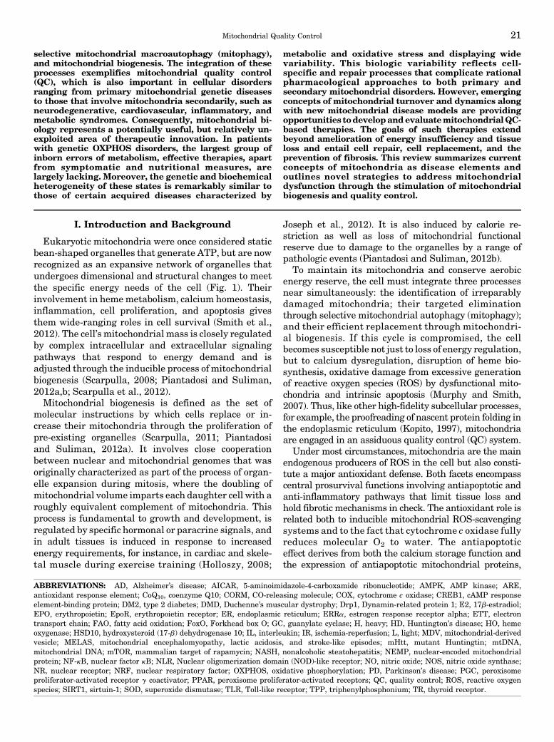
Fig. 1. Normal mitochondrial structure in the heart. (Left) Representative micrographs of section of the mouse left ventricle showingimmunofluorescence staining for TCA cycle enzyme citrate synthase. The mitochondria appear as tiny green, bead-like structures (Alexa 488, greenfluorescence). Nuclei are stained blue (DAPI; 49,6-diamidino-2-phenylindole). Scale bar = 10 mm. (Middle) Isolated rat cardiomyocytes showing theintracellular mitochondrial network by confocal fluorescence microscopy stained with MitoTracker red. The cytoskeleton protein F-actin is green andthe cell nucleus is blue (DAPI; 49,6-diamidino-2-phenylindole). 600�. Scale bar = 0.4 mm. (Right) Electron micrograph of normal mouse heartmitochondria. Cardiomyocyte exhibits abundant electron-dense mitochondria (m) surrounded by smooth endoplasmic reticulum (SER). N = cellnucleus. Scale bar = 0.2 mm.
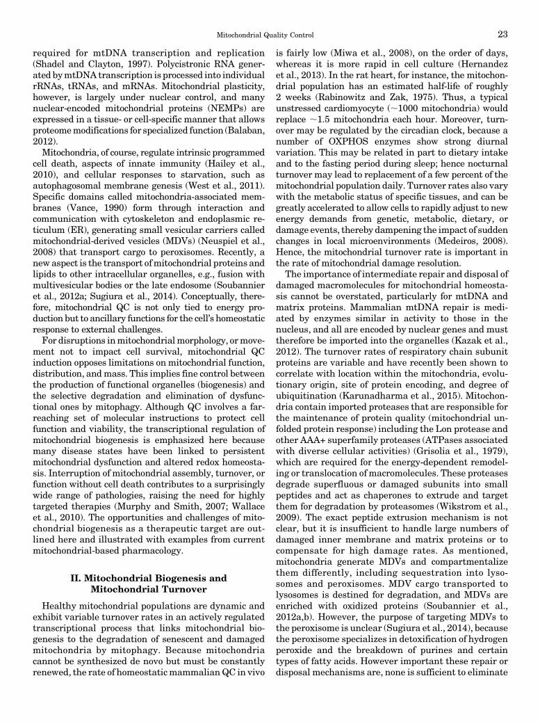
Fig. 2. Schematic of the major steps of mtDNA replication and transcrip-tion. The double-stranded mtDNA molecule is replicated in a strand asynchronous manner and L-strand replication starts at its origin ofreplication (OL) after the nascent H-strand has completed about two-thirdsof the circle proceeding in the opposite direction. H-strand replication isinitiated using a short L-strand mRNA transcript synthesized by mitochon-drial RNA polymerase (POLRMT). POLRMT accesses its DNA template bybinding of mitochondrial transcription factor A, B1, and B2 (Tfam, TFB1M,and TFB2M) at the mtDNA duplex. Precursor RNA primer is cleaved byprocessing endonuclease (RNase) at the H-strand origin of replication (OH).Bases are added to free 39-termini by mitochondrial DNA polymerase gamma(Polg). Twinkle is required for preparation of single-stranded templates forPolg activity. H-strand transcription is initiated from two promoter sites,HSP1 and HSP2. HSP1 transcripts are terminated at the terminationsequence (TERM) within the tRNA-Leu (UUR) gene where MTERF1 binds.HSP2 generate near full-length polycistronic transcripts that are processedinto individual RNAs. L-strand transcription is initiated from a singlepromoter site LSP, which generates near full-length polycistronic messages.The replication origins of both the H- and L-strands are indicated (OH andOL, respectively). CSB, conserved sequence block; HSP, heavy-strandpromoter; LS, light-strand promoter; MTERF1, mitochondrial terminationfactor 1; MtSSB, mitochondrial single-stranded binding protein.
22 Suliman and Piantadosi
required for mtDNA transcription and replication(Shadel and Clayton, 1997). Polycistronic RNA gener-ated bymtDNA transcription is processed into individualrRNAs, tRNAs, and mRNAs. Mitochondrial plasticity,however, is largely under nuclear control, and manynuclear-encoded mitochondrial proteins (NEMPs) areexpressed in a tissue- or cell-specific manner that allowsproteomemodifications for specialized function (Balaban,2012).Mitochondria, of course, regulate intrinsic programmed
cell death, aspects of innate immunity (Hailey et al.,2010), and cellular responses to starvation, such asautophagosomal membrane genesis (West et al., 2011).Specific domains called mitochondria-associated mem-branes (Vance, 1990) form through interaction andcommunication with cytoskeleton and endoplasmic re-ticulum (ER), generating small vesicular carriers calledmitochondrial-derived vesicles (MDVs) (Neuspiel et al.,2008) that transport cargo to peroxisomes. Recently, anewaspect is the transport ofmitochondrial proteins andlipids to other intracellular organelles, e.g., fusion withmultivesicular bodies or the late endosome (Soubannieret al., 2012a; Sugiura et al., 2014). Conceptually, there-fore, mitochondrial QC is not only tied to energy pro-duction but to ancillary functions for the cell’s homeostaticresponse to external challenges.For disruptions inmitochondrialmorphology, ormove-
ment not to impact cell survival, mitochondrial QCinduction opposes limitations on mitochondrial function,distribution, andmass. This implies fine control betweenthe production of functional organelles (biogenesis) andthe selective degradation and elimination of dysfunc-tional ones by mitophagy. Although QC involves a far-reaching set of molecular instructions to protect cellfunction and viability, the transcriptional regulation ofmitochondrial biogenesis is emphasized here becausemany disease states have been linked to persistentmitochondrial dysfunction and altered redox homeosta-sis. Interruption of mitochondrial assembly, turnover, orfunction without cell death contributes to a surprisinglywide range of pathologies, raising the need for highlytargeted therapies (Murphy and Smith, 2007; Wallaceet al., 2010). The opportunities and challenges of mito-chondrial biogenesis as a therapeutic target are out-lined here and illustrated with examples from currentmitochondrial-based pharmacology.
II. Mitochondrial Biogenesis andMitochondrial Turnover
Healthy mitochondrial populations are dynamic andexhibit variable turnover rates in an actively regulatedtranscriptional process that links mitochondrial bio-genesis to the degradation of senescent and damagedmitochondria by mitophagy. Because mitochondriacannot be synthesized de novo but must be constantlyrenewed, the rate of homeostaticmammalianQC in vivo
is fairly low (Miwa et al., 2008), on the order of days,whereas it is more rapid in cell culture (Hernandezet al., 2013). In the rat heart, for instance, the mitochon-drial population has an estimated half-life of roughly2 weeks (Rabinowitz and Zak, 1975). Thus, a typicalunstressed cardiomyocyte (;1000 mitochondria) wouldreplace ;1.5 mitochondria each hour. Moreover, turn-over may be regulated by the circadian clock, because anumber of OXPHOS enzymes show strong diurnalvariation. This may be related in part to dietary intakeand to the fasting period during sleep; hence nocturnalturnover may lead to replacement of a few percent of themitochondrial population daily. Turnover rates also varywith the metabolic status of specific tissues, and can begreatly accelerated to allow cells to rapidly adjust to newenergy demands from genetic, metabolic, dietary, ordamage events, thereby dampening the impact of suddenchanges in local microenvironments (Medeiros, 2008).Hence, the mitochondrial turnover rate is important inthe rate of mitochondrial damage resolution.
The importance of intermediate repair and disposal ofdamaged macromolecules for mitochondrial homeosta-sis cannot be overstated, particularly for mtDNA andmatrix proteins. Mammalian mtDNA repair is medi-ated by enzymes similar in activity to those in thenucleus, and all are encoded by nuclear genes and musttherefore be imported into the organelles (Kazak et al.,2012). The turnover rates of respiratory chain subunitproteins are variable and have recently been shown tocorrelate with location within the mitochondria, evolu-tionary origin, site of protein encoding, and degree ofubiquitination (Karunadharma et al., 2015). Mitochon-dria contain imported proteases that are responsible forthe maintenance of protein quality (mitochondrial un-folded protein response) including the Lon protease andother AAA+ superfamily proteases (ATPases associatedwith diverse cellular activities) (Grisolia et al., 1979),which are required for the energy-dependent remodel-ing or translocation of macromolecules. These proteasesdegrade superfluous or damaged subunits into smallpeptides and act as chaperones to extrude and targetthem for degradation by proteasomes (Wikstrom et al.,2009). The exact peptide extrusion mechanism is notclear, but it is insufficient to handle large numbers ofdamaged inner membrane and matrix proteins or tocompensate for high damage rates. As mentioned,mitochondria generate MDVs and compartmentalizethem differently, including sequestration into lyso-somes and peroxisomes. MDV cargo transported tolysosomes is destined for degradation, and MDVs areenriched with oxidized proteins (Soubannier et al.,2012a,b). However, the purpose of targeting MDVs tothe peroxisome is unclear (Sugiura et al., 2014), becausethe peroxisome specializes in detoxification of hydrogenperoxide and the breakdown of purines and certaintypes of fatty acids. However important these repair ordisposal mechanisms are, none is sufficient to eliminate
Mitochondrial Quality Control 23
whole mitochondria (Sugiura et al., 2014). Those situ-ations require mitophagy in conjunction with theactivation of mitochondrial biogenesis. Although ourunderstanding of this area is evolving rapidly, mitophagyis not a singular event (Lemasters, 2014), and it isnot clear how the cell determines when to halt repairand eliminate the organelle.For many years, our understanding of the regulation
of mitochondrial biogenesis was limited to events re-lated to cell division and to increased work. The uniquechallenge for the cell is the rapid synchronization ofmtDNA replication and mitochondrial-encoded geneexpression in conjunction with the upregulation of manygenes for NEMPs and associated subsets of nonmito-chondrial genes. Subsequently, it becameknown that theprogram is activated by changes in substrate availability(e.g., starvation) (Moreno-Loshuertos et al., 2006, 2011)and by certain paracrine effectors and hormones(Scarpulla, 2011) necessary for the cell to maintain itsfunctional mitochondrial mass (Onyango et al., 2010).The process is also essential to recovery from oxidativestress, inflammation, and mitochondrial drug toxicity(Suliman et al., 2007a). In each case, signal specificity ismaintained through a network of nuclear DNA-bindingtranscription factors and coregulators upregulated bydiverse physiologic conditions and by tissue or cell-specific events that respond to changes inmitochondrialfunction and/or mass. The response to cell damage isespecially challenging because the precise communica-tion between nuclear and mitochondrial genomes maybecome difficult to maintain, but is critical to supportfunction and avoid apoptosis or necrosis as well as toadjust mitochondrial phenotype to changes in microen-vironment or disease pathogenesis.The data on mitochondrial dysfunction in acquired
diseases has at times seemed contradictory becauseeach situation is different and quite complex. Forinstance, during acute experimental ischemia, inflam-mation, and/or oxidative stress, various studies havereported impaired (Crouser et al., 2004; Gellerich et al.,1999), unchanged (Mela-Riker et al., 1992; Taylor et al.,1995), or improved (Taylor et al., 1998; Lu et al., 2003)mitochondrial function. Such discrepancies have manycauses, such as type of cell injury, duration and severityof injury, the cell type and function, and the experimen-tal situation. There are also uncontrolled variables inthe repair responses that promote or impede the resolu-tion of mitochondrial dysfunction (Reynolds et al., 2009).These complexities have made it harder to deviserational mitochondrial-based therapies and to confirmthe mode of action of the intervention and its impact oncell damage and resolution.Nonetheless, there is a clear set of mitochondria-
related events that occur after acute sterile or infectiousdamage to previously healthy cells and tissues. Cellsthat exhibit mitochondrial damage, but are able tosurvive, show the induction ofmitochondrial biogenesis,
activation of autophagy, and eventual repopulationwith functional mitochondria (Chang et al., 2015). Thissequence of events is now well appreciated morpholog-ically for major disturbances in oxygen delivery totissues, for instance after ischemia-reperfusion or se-vere hypoxia-reoxygenation in the heart and the brain(Gutsaeva et al., 2008; Ahuja et al., 2010). Mitochon-drial QC is activated by mechanisms involving bothenergy limitation and redox pathways, which, as dis-cussed later, remain under active investigation.
The early response to oxidative or electrophilic stressinvolve activation of the transcription factor nuclearfactor (erythoid-derived 2)-like2 (NFE2L2 or Nrf2),which binds to antioxidant response (ARE) elementsin the promoter regions of target genes (Wakabayashiet al., 2004), coordinates antioxidant and antiapoptoticgene upregulation (Alam et al., 2004), and inducesmitochondrial biogenesis (Piantadosi et al., 2008; Athaleet al., 2012; MacGarvey et al., 2012). In a chemical stressmodel of renal ischemia-reperfusion, increased mito-chondrial biogenesis has been shown to accelerate therecovery of OXPHOS through p38 and epidermal growthfactor receptor activation of PGC-1a (Rasbach andSchnellmann, 2007). Those investigators also found thatstimulation of the 5-hydroxytryptamine receptor acti-vated mitochondrial biogenesis and suggested that5-hydroxytryptamine agonists as a treatment of mito-chondrial and cell injury (Rasbach et al., 2010). Becausedamage by ongoing inflammatory processes is oftenperiodic, mitochondria are frequently damaged not justby the original insult but by subsequent oxidative andnitrosative stress driven by cytokine production. Insevere sepsis, for instance, ROS and nitric oxide (NO)overproduction leads to mtDNA damage, but NO alsoactivates mitochondrial biogenesis through complexcrosstalk between a mitochondrion and the nucleus thatyields a net increase in the synthesis of new organelles(Piantadosi et al., 2007; Medeiros, 2008; Ahuja et al.,2010; Seo et al., 2010).
The transcriptional activation of nuclear and mito-chondrial gene expression leads to a nascent proteinsupply in appropriate quantities at appropriate timesfor an expandingmitochondrial subpopulation. Becausethe bulk of the proteins are NEMPs, and mtDNA-encoded mRNAs are involved only in the synthesis ofa dozen OXPHOS proteins, nuclear transcription fac-tors play a predominant role in mitochondrial biogen-esis. The mitochondrial genome, however, must betranscribed and replicated during mitochondrial bio-genesis so that the new population remains at fulltranscriptional capacity.
A relatively small set of DNA-binding transcriptionalregulators are essential for mitochondrial biogenesisincluding nuclear respiratory factors 1 and 2 (NRF-1and NRF-2), cAMP response element-binding protein(CREB1), estrogen response receptor alpha (ERRa), andthe nuclear coactivators peroxisome proliferator-activated
24 Suliman and Piantadosi

receptor g coactivator 1-a, 1-b (PGC-1a, b), and PPRC(Peroxisome proliferator-activated receptor gammacoactivator-related protein 1). PGC-1a interacts, for exam-ple, with NRF-1 and 2, ERRa, and CREB1 to regulate thetranscription of genes involved in mitochondrial bio-genesis and energy metabolism (Wu et al., 2006).These coactivators provide a direct link betweenphysiologic events and the regulation of mitochondrialbiogenesis and can be induced pathologically as well aspharmacologically.Several other nuclear transcription factors are linked
to the expression of respiratory chain subunits such asYY1 and myocyte enhancer factor-2, and YY1 is animportant component of nutrient sensing by the mam-malian target of rapamycin (mTOR) pathway. Inhibi-tion of mTOR decreases ERRa and NRF-1/2 expressionand prevents the interaction between YY1 and PGC-1a.The oncoprotein Myc also regulates nuclear-encodedmitochondrial programming largely by an interactionwith NRF-1 (Morrish et al., 2003) and by increasingPGC-1b expression(Morrish and Hockenbery, 2014).Although mitochondria are often damaged by ROS,
now recognized as both agents of damage and architectsof senescence, some ROS, particularly H2O2, act asredox messengers for NEMP expression. Indeed, mito-chondrial H2O2 production is an important trigger formitochondrial proliferation, particularly during acuteinflammation where severe mitochondrial damage inorgans like the liver, heart, and kidneys must be cleared(Suliman and Piantadosi, 2014). Hence, mitochondria-targeted antioxidant compounds may limit damage butalso interfere with the redox sensing of damage that isnot prevented by the drug.The prompt restoration of a highly functional pop-
ulation of mitochondria is fundamental to the recoveryof injured cells and tissues, particularly those withdelicate bioenergetic equilibria such as cardiomyocytes,hepatocytes, renal tubular cells, and neurons. More-over, proliferating cells depend on mitochondrial bio-genesis to provide daughter cells with an adequatesupply of mitochondria for the required aerobic capac-ity. These concepts have brought mitochondrial bio-genesis to the forefront as a therapeutic target fordiseases characterized by gradual cell loss that to datehave had no effective therapies. The presence of cyclicalcell damage leads to the need for additional energywhile exposing the proliferating mitochondria to re-current bouts of damage.Only a handful of small molecules are known today to
induce mitochondrial biogenesis without first causingexplicit mitochondrial damage. Therefore, the pharma-cological manipulation of mitochondrial biogenesis us-ing noninjurious agents is a priority for injuries anddiseases already characterized by mitochondrial dys-function (Funk et al., 2010). Mitochondrial pharmacol-ogy is thus an exciting area with many promising newdirections, and in covering a number of approaches to
inducemitochondrial biogenesis, including natural prod-ucts, we have highlighted novel concepts and targets fortherapy of mitochondrial genetic disorders, chronic de-generative diseases, and life span extension.
III. Mitochondrial Disorders and Diseases
Mitochondrial diseases are conveniently divided intoprimary and secondary disorders. Primary disordersare characterized by heritable mitochondrial genedefects such as base substitution or insertion-deletionmutations in mtDNA coding regions or genes forNEMPs, whereas the secondary disorders are due tonongenetic events including mitochondrial toxins, is-chemia, infections, or sterile inflammation. The mostcommon inherited disorders, Friedreich’s ataxia;Leber’s hereditary optic neuropathy; mitochondrialencephalomyopathy, lactic acidosis, and stroke-likeepisodes (MELAS); myoclonic epilepsy with ragged-redfibers; and Leigh Syndrome show variable clinical profilesand susceptibility to stressors. In addition to defects inenergy conservation, they share, to various degrees,oxidative stress as a pathogenic feature due to endog-enous ROS production or to a decrease in mitochondrialanti-oxidant protection or both.
The frequency of primary mitochondrial disordersranges up to 1 in 5,000 (Schaefer et al., 2004), and theorganelle a logical pharmacological target in theseindividuals. Many primary disorders manifest in earlychildhood and therapeutic options are limited. Concep-tually, however, phenotypic commonalities in mtDNAloss-of-function mutations help to inform the patho-genic impact of secondary or acquired disorders. Ac-quired mitochondrial loss-of-function has been linkedwith varying levels of evidence to Parkinson’s disease(PD), Alzheimer’s disease (AD), amyotrophic lateralsclerosis (Lin and Beal, 2006; Trushina and McMurray,2007), schizophrenia, and autism (Manji et al., 2012;Rossignol and Frye, 2012). Secondary mitochondrialdefects are best exemplified by severe, persistent in-flammatory states but are also represented inmetabolicdisorders (Nasrallah and Horvath, 2014) and somecancers (Wallace, 2012). For example, mutations inthe mitochondrial intermembrane form of superoxidedismutase SOD1, which prevents superoxide damage,is linked to the progression of amyotrophic lateralsclerosis; (Vehvilainen et al., 2014) NADH dehydroge-nase 4 toLeber’s hereditary optic neuropathy (Kornmann,2013); PARKIN to the familial form of PD (Schmidt et al.,2010); and TCA cycle enzymes to oncogenesis (Schaeferet al., 2004).
The induction of mitochondrial biogenesis has alsobeen promoted as a strategy for the treatment ofOXPHOS disorders in mouse models (Smith et al.,2012). Hypothetically, boosting mitochondrial qualityand mass should increase remaining OXPHOS activityunless the defective protein acts as a dominant-negative.
Mitochondrial Quality Control 25

Otherwise, the approach should enhance the cellularATP synthetic capacity and presumably improve cellularenergy deficits and enhance repair mechanisms. Al-though optimal and specific agents are not yet available,there is promising proof-of-principle evidence. For in-stance, the mTOR signaling pathway integrates manyprocesses that are involved in the promotion of cellgrowth (Laplante and Sabatini, 2009). Specific inhibitionof themTORpathwaywith rapamycin enhances survivaland attenuates disease progression in a mouse model ofLeigh syndrome. These mice, which are deficient inmitochondrial Complex I subunit Ndufs4 [NADH de-hydrogenase (ubiquinone) Fe-S protein 4] display re-duced neurologic symptoms, neuroinflammation, andbrain lesions after rapamycin treatment. Although anexact rescue mechanism was not identified, the meta-bolic shift toward amino acid catabolism was postulatedto have alleviated a buildup of glycolytic intermediates(Johnson et al., 2013).
A. Metabolic Disorders
Mitochondrial dysfunction in primary metabolic or-gans such as the liver has a large impact on systemicmetabolic homeostasis (Grattagliano et al., 2012). Stud-ies on patients with obesity, diabetes, nonalcoholicsteatohepatitis (NASH) or nonalcoholic fatty liver dis-ease consistently display functional and structuralabnormalities in hepatocyte mitochondria, such asOXPHOS impairment or mega-mitochondria (Begricheet al., 2006). Increased and decreased oxidation isreported as features of hepatocyte steatosis and insulinresistance (Fabbrini et al., 2010; Pessayre, 2007; Zhanget al., 2007). A decrease in oxidative activity in thehepatocyte induces diacylglycerol accumulation andsteatosis with concurrent PKC activation and inhibitionof insulin signaling (Zhang et al., 2007). Insulin-resistant patients show an increase in hepatic oxidationthat is correlated with an increase in ROS production(Pessayre and Fromenty, 2005; Fabbrini et al., 2010).Persistently high hepatic carbon oxidation rates in
insulin resistance could be an adaptive mechanism tolimit free fatty acid toxicity or to provide large quanti-ties of reduced NADH. This effect may be decoupledfrom actual energy requirements and the ROS pro-duction reflective of respiratory chain damage or dys-function (Vial et al., 2010). On the other hand,mitochondrial ROS production during OXPHOS facili-tates insulin signaling through insulin receptor oxida-tion and phosphatase inhibition, for instance PTP1Band phosphatase and tensin homolog (PTEN) (Chenget al., 2010). As such, ROS productionwould be expectedto promote the inflammation by some of the cellularmechanisms discussed earlier.Insulin resistance is characteristic of type 2 diabetes
(DM2) and the inability of insulin to inhibit glucoseoutput from the liver and promote glucose uptake bymuscle (Saltiel and Kahn, 2001; Hribal et al., 2002). In
mouse models, hepatic PGC-1a expression is elevatedwith the onset of DM2, where it contributes to consti-tutive gluconeogenesis and fatty acid oxidation (FAO)through coactivation of nuclear receptors HNF-4 andperoxisome proliferator-activated receptors (PPAR)-a,respectively (Herzig et al., 2001; Rhee et al., 2003).Insulin suppresses gluconeogenesis stimulated by Fork-head box O (FOXO) 1/PGC-1a, whereas a mutant,insulin-insensitive FOXO1 allele completely reversesthis suppression in hepatocytes and in transgenic mice.Recent findings support a role for PGC-1a in linkingnutrient deprivation to mitochondrial oxidant produc-tion through coactivation of ERRa to enhance sirtuin-1(SIRT1) expression (Kong et al., 2010; Giralt et al.,2011).
SIRT3 is s nicotinamide adenine dinucleotide (NAD)-dependent protein deacetylase localized to mitochon-dria that allows tissue-specific optimization of ATPlevels and metabolic enzyme activities (Verdin et al.,2010). SIRT3 opposes oxidative stress by precipitating aseries of reactions beginning with activation of isoci-trate dehydrogenase 2 and culminating in detoxifica-tion of peroxides by glutathione peroxidase (Someyaet al., 2010). It deacetylates SOD2, leading to increasedactivity and enhanced oxidant scavenging in the matrix(Qiu et al., 2010; Tao et al., 2010). Another key factor isthe high expression of PPARa in the liver, heart, andsmall intestine (Kersten, 2014). Acting as a nutrientsensor, PPARa promotes the adaptation of fatty acidcatabolism, lipogenesis, and ketone body synthetic ratesin response to feeding and starvation to stimulatehepatic fatty acid oxidation and fasting ketogenesis(Hashimoto et al., 2000; Sugden et al., 2002; Patsouriset al., 2004). An increase in PPARa leads to target geneactivation in fatty acid oxidation (e.g., Cpt1a, Cpt2,Acadvl, Hadha) and ketogenesis (Hmgcs2, Hmgcl,Acat1). Furthermore, PPARa negatively regulatesproinflammatory and nuclear factor (NF)-kB and AP-1signaling in systemic inflammation, atherosclerosis,and NASH (Ip et al., 2003; Gervois et al., 2004).
Mitochondrial dysfunction and body energy balanceare integral to metabolic disorders such as obesity andmetabolic syndrome (Joseph et al., 2012; Szendroediet al., 2012). In these disorders, there is renewedinterest in the role of mitochondria-rich brown fat,which efficiently dissipates energy through nonshiver-ing thermogenesis as a result of high levels of theuncoupling protein 1 (Wu et al., 2013). Attention hasalso been focused on the role of beige fat, which can begenerated from white adipose tissue by thermogenicpathways in which mitochondrial biogenesis has a keyrole (Schulz et al., 2013). Although strategies to targetmitochondria in the treatment of nonalcoholic fattyliver disease and NASH are still in the early stages,the opposite induction of mitochondrial biogenesis is alogical target for treatment of obesity and metabolicsyndrome (Szendroedi et al., 2012; Wu et al., 2013).
26 Suliman and Piantadosi
Pharmacological stimulation of mitochondrial biogene-sis seems to induce certain positive effects of calorierestriction and exercise on such generic metabolicdisorders.
B. Mitochondrial Quality Control and Inflammation
1. Importance of Innate Immunity. The regulationof mitochondrial QC provides an important duel anti-inflammatory function: the control of inflammasomeassembly and the expression of counter-inflammatorygenes. This is made possible through connections be-tween nuclear QC network programming and the in-nate immune system in tissueswhere host defensemustbe coordinated with repair mechanisms necessary tocontrol damage from circulatory failure, pro-oxidants,xenobiotics, and pathogens. The connections betweenmitochondria, inflammation, and metabolism are ofgreat interest, because dysfunctionality is associatedwith chronic inflammatory diseases, cancer, and fibro-sis. The presence of infection is normally sensed bypattern recognition receptors, which, upon activation,trigger an acute inflammatory response and initiatedefense and repair programs (Dostert et al., 2008;Schroder and Tschopp, 2010). The offender is seques-tered and eliminated while tissue repair is initiated,which occurs over the course of a few days. When thesource cannot be effectively removed, progressive celldamage and cell death leads to tissue loss and failure toheal. Such cycles of tissue destruction and healing leadto chronic inflammation and fibrosis and are character-istic of many degenerative diseases, including DM2,arthritis, inflammatory bowel disease, and neurodegen-erative conditions. Under such circumstances, the mi-tochondrial mechanisms discussed here are activelyinvolved (Tschopp, 2011).The activation ofmitochondrial biogenesis by the host
immune response was first demonstrated more than adecade ago when lipopolysaccharide was found tostimulate hepatic and cardiac mitochondrial biogenesisin rats (Suliman et al., 2003b, 2004). Subsequently, theresolution of extensive cytokine-induced mtDNA dam-age after heat-inactivated Escherichia coli exposurewas found to be due to compensatory mitochondrialbiogenesis initiated in part by activation of toll-likereceptor 4 (TLR4) and downstream signaling viaCREB1 and NF-kB-dependent induction of NRF-1(Suliman et al., 2005, 2007b). In murine peritonitis,depressed oxidative metabolism is eventually restoredby mitochondrial biogenesis, thus providing a way toaffect sepsis outcome (Haden et al., 2007). Indeed,sepsis-induced mitochondrial dysfunction is linked tomultiple organ dysfunction syndrome, whereas the rateof resolution and restoration of organ function maydepend on the reestablishment of mitochondrial num-ber and function (Singer, 2007). The failure to maintainand/or restore mitochondrial mass not only impairstissue repair but may foster immune suppression through
prolonged upregulation of counter-inflammatorymediatorgenes suchas interleukin (IL)-10 (SulimanandPiantadosi,2014). Clinical data also support that activation ofmitochondrial biogenesis in skeletal muscle influencessurvival in critical illness (Carre et al., 2010). Accord-ingly, innovative strategies to promote and protectmitochondrial biogenesis may lead to new ways toprevent organ failure in severe systemic inflammatorystates (Piantadosi et al., 2007).
2. Role of the Inflammasome. The full expression ofhost inflammation after immune cell exposure to path-ogens or to diverse pathogen- and danger-associatedmolecular patterns or environmental triggers requiresassembly of multiprotein inflammasome complexes.Inflammasomes generally contain a Nuclear oligomer-ization domain (NOD)-like receptor (NLR) molecule,NLRP1 (NOD-, leucine rich repeat-, and pyrin domain-containing 1), NLRP3, or others (Latz et al., 2013). Atypical sensor complex consists of three protein compo-nents, for example NLRP3 (NALP3), ASC (PYCARD),and caspase-1. The NLRP3 complex activates caspase-1(and caspase-11 in mice), leading to processing andrelease of IL-1b and IL-18.
Inflammasomes are also mitochondrial stress moni-tors and regulators of intrinsic apoptosis (Latz et al.,2013). Mitochondrial stress is sensed by the NLRP3inflammasome through ROS production (Zhou et al.,2011) and/or the release of oxidized mitochondrial DNA(Shimada et al., 2012). Two lines of evidence favormitochondria as the main source of ROS in NLRP3inflammasome activation and hence as a signal-integrating organelle for innate immunity (Schroder andTschopp, 2010;Nakahira et al., 2011; Zhou et al., 2011). Inmacrophages, inflammasome activation is impaired bymtDNA depletion or by inactivation of the outer mem-brane voltage-dependent anion channel (Fabbrini et al.,2010). Also, respiratory chain inhibition activates theNLRP3 inflammasome (Nakahira et al., 2011).
Disturbances in energy homeostasis lead to mito-chondrial relocation via the microtubule system, whichfacilitates NLRP3 interaction with the ASC adaptorprotein (Misawa et al., 2013). Optimal NLRP3 activa-tion requires a Retinoic acid-inducible gene-I-like re-ceptor adaptor molecule called MAVS (mitochondrialantiviral signaling protein) localized to the outer mito-chondrial membrane (Subramanian et al., 2013). An-other mechanism that increases IL-1b output inactivated macrophages is through increases in the TCAcycle intermediate succinate, which stabilizes hypoxia-inducible factor-1a and allows sustained IL-1b transcrip-tion (Tannahill et al., 2013). The NLRP3 inflammasomeis inhibited by the removal of damaged or dysfunctionalmitochondria by mitophagy. The cell eliminates highROS-generating mitochondria to avoid autodestructiveinflammation (Nakahira et al., 2011). Inhibition ofmitophagy leads to retention of ROS-generating mito-chondria and inflammasome activation (Nakahira et al.,
Mitochondrial Quality Control 27

2011; Zhou et al., 2011). Mitochondria removed bymitophagy are replaced through mitochondrial biogene-sis, thus avoiding persistent inflammasome activationthat would lead to energy failure and cell death.In overwhelming infections, immune dysregulation
may lead to tissue damage in which surviving cellsundergo cycles of mitochondrial damage and exhibitaccelerated mitochondrial turnover, particularly forhighly active tissues. Mitochondrial damage is drivenby toll-like (TLR) receptor-dependent production ofearly-phase inflammatory mediators, such as tumornecrosis factor alpha and nitric oxide (NO) (Schulze-Osthoff et al., 1992). Activated macrophages andKupffer cells generate certain interleukins that alsocause mitochondrial damage (Schulze-Osthoff et al.,1992). Damaged mitochondria generate higher levels ofROS and may release calcium and intrinsic apoptosisproteins (Taylor et al., 1995; Kantrow et al., 1997).Excessive oxidant production can compromise mito-chondrial function and structure through the directchemical oxidation of proteins, lipids, and DNA. Forexample, the NO-superoxide reaction generates thepowerful oxidant peroxynitrite (ONOO2) (Kuroseet al., 1993), and in the presence of transition metalssuch as iron, H2O2 and superoxide generate the hy-droxyl radical (×OH) by the Fenton reaction (Halliwell,1989).Transcription factors that initiate the host defense,
including NF-kB, are also activated by oxidative stress(Koay et al., 2001). Hence, with the transcriptionalactivity of PGC-1a, NRF-1 upregulates many antioxi-dant defenses (Piantadosi and Suliman, 2006; St-Pierreet al., 2006). PGC-1a also promotes the induction forROS-detoxifying enzymes such as mitochondrial super-oxide dismutase (SOD2) and glutathione peroxidase-1.And in neuronal cells, CREB1 is an important activatorof the Pargc1a (PGC-1a) gene after oxidant exposure(St-Pierre et al., 2006). In macrophages, PGC-1b over-expression inhibits canonical NF-kB-dependent cytokineproduction, which mitigates the acute inflammatoryresponse (Vats et al., 2006). In murine Staphylococcusaureus peritonitis, early TLR-mediated events includethe rapid upregulation of hepatic PGC-1a and PGC-1b inwild-type mice, whereas both genes are deregulated inTLR22/2 mice and increased in TLR42/2 mice. Mean-while, PRC is upregulated in all three strains (Sweeneyet al., 2010).An inability to regulate inflammasome activity cre-
ates overlapping cycles of inflammatory tissue damagethat may partly conceal organized mitochondrial QCprocesses. In mice, sublethal damage by lipopolysac-charide transiently depletes hepatic mtDNA contentand impairs mitochondrial transcription (Sulimanet al., 2003a). Compensation involves Akt/PKB phos-phorylation of NRF-1, increasing its nuclear translocationand subsequent expression of Tfam and other mitochon-drial transcriptome proteins, which after importation,
allow mtDNA copy number to be restored (Sulimanet al., 2003b). Initial mtDNA content depletion is relatedto TLR4 and NF-kB activation, and genetic ablation ofTLR4 reduces, but does not eliminate this damage;however, recovery of mtDNA copy number is delayed inTLR4 null mice (Suliman et al., 2005).
This mtDNA damage leads to upregulation of repairenzymes such as the base excision glycosylase, OGG1,in part through NRF-1 and NRF-2 binding to promoterelements (Bartz et al., 2011). Notably, mtDNA copynumber can be protected by increasing SOD2 in mito-chondria, by inhibiting NOS activity, or by scavengingONOO2 (Choumar et al., 2011). In true infection, orafter exposure to surrogate damage-associated molecu-lar patterns, there is morphologic evidence of mitophagyand mitochondrial biogenesis, which in survivors restoremitochondrial mass within days (Suliman et al., 2003a;Haden et al., 2007; Watanabe et al., 2009).
If these processes cannot prevent energy compromise,impending energy failure, specifically ATP depletion,activates a key pathway regulated by the serine/threonine kinase AMPK (Bergeron et al., 2001). AMPKstimulates glucose and lipid catabolism and blocksenergy-utilizing pathways such as protein and fattyacid biosynthesis. AMPK promotes mitochondrial bio-genesis (Bergeron et al., 2001; Zong et al., 2002; Reznickand Shulman, 2006), NO production (Morrow et al.,2003), and autophagy (Kim et al., 2011) and opposesinflammation by interfering with NF-kB-dependentcytokine expression (Bai et al., 2010). The mechanismof NF-kB antagonism is not clear, but may indirectlyinvolve PGC-1a, Forkhead box O (FoxO)-type tran-scription factors, and/or SIRT1 (Canto et al., 2009;Barroso et al., 2011; Salminen et al., 2011). Conversely,loss of AMPK activity is associated with increasedinflammation.
However, the transcriptional activity of relevantnuclear receptors (NRs), particularly estrogen-relatedreceptor ERRa, is blocked by repressors such as RIP140and others (L’Horset et al., 1996); RIP140 thus sup-presses metabolic gene expression and mitochondrialbiogenesis (Powelka et al., 2006). In macrophages,RIP140 is actually a cytokine gene coactivator, andRIP140 deficiency inhibits the inflammatory response(Zschiedrich et al., 2008). RIP140 interacts with theRelA subunit of NF-kBand the histone acetylase CREB-binding protein and cooperates with CREB-bindingprotein coactivator complex on RelA-regulated pro-moters. RIP140 modulation of inflammatory gene ex-pression is thus a good example of cell-specificintegration of control pathways for metabolism andinflammation (Zschiedrich et al., 2008). Accordingly,newer findings on the trainedmonocyte response, whichimproves resistance to infection, indicates a require-ment for a switch from respiration to glycolysis in-volving genes of the mTOR-HIF-1 a pathway (Borden,2014).
28 Suliman and Piantadosi

C. Mitochondrial Dysfunction Caused byTherapeutic Agents
Well over 300 mitochondria-targeted small moleculeshave been annotated in DrugBank (Wishart et al., 2008)with a number of recent additions, suggesting that thisis an area of growing therapeutic interest. However,drugs are also a frequent cause of mitochondrialdysfunction (Wallace, 2008), and a surprising numberof them are taken up bymitochondria and inhibit one ormore mitochondrial functions directly or lead to in-creased ROS generation and the damage produces aclinical phenotype. Thus, mitochondrial dysfunction isoften implicated in drug-induced toxicity (Dykens andWill, 2007), yet there is no requirement to screen drugsfor mitochondrial toxicity before Food and Drug Admin-istration approval. This issue has been recognized forsome time in patients with genetic mitochondrialdiseases, particularly for agents that increase mito-chondrial ROS production. Hence, molecules thatsupress mitochondrial ROS production have been anarea of strong research interest for a number of years,particularly for CNS applications.An example is the triaminopyridine flupirtine, a
nonopioid analgesic with mitochondria-dependent an-tioxidant and free radical scavenging activity withefficacy toward ischemic neuronal damage, apoptosis,and age-associated brain disorders (Schluter et al.,2000). Instructively, the commercially available statins(e.g., atorvastatin and simvastatin), which lower cho-lesterol levels by inhibition of 3-hydroxy-3-methylglutarylcoenzyme A reductase, have been implicated in neuro-protection against PD, AD, traumatic brain injury, andsecondary progressive multiple sclerosis (Kumar et al.,2012; Malkki, 2014). In some instances, neuroprotectionhas been associated with the lipophilicity of the drug(Haag et al., 2009; Huang et al., 2015), but directmitochondrial protection is not established either ex-perimentally or clinically, and the well-known adverseeffects of statins on mitochondria, such as inhibition ofCoenzyme Q synthesis, is an important caveat withthese agents (Golomb and Evans, 2008). The 3-hydroxy-3-methylglutaryl coenzyme A reductase inhibitorscause a myopathy that has been attributed in part to areduction in CoQ10 levels (Potgieter et al., 2013).Other notable examples of mitochondrial drug toxic-
ity include cardiac and skeletal myopathy associatedclinically with doxorubicin, cisplatin, and nucleosidereverse-transcriptase inhibitors such as zidovudine(Galluzzi et al., 2014). Doxorubicin is notable becauseit prevents expression of compensatory mitochondrialbiogenesis (Suliman et al., 2007a; Finsterer andOhnsorge, 2013). Certain antibiotics (e.g., tetracyclineand aminoglycosides) inhibit mtDNA translation and pro-tein synthesis. Anticonvulsants, such as valproate andbarbiturates, inhibit respiration, and the common over-the-counter analgesic acetaminophen (paracetamol)
can inhibit mitochondrial oxygen consumption as dis-cussed in Than et al. (2014).
Mitochondrial toxicity has led to withdrawal of anumber of previously approved drugs, such as troglita-zone, mainly due to hepatotoxicity. A majority of drugswith U.S. Food and Drug Administration black boxwarnings have been shown to be associated with mito-chondrial toxicity (Dykens and Will, 2007). This has ledthe pharmaceutical industry to begin to screen formitochondrial toxicity earlier to identify compoundsthat may too toxic to bring into clinical testing.
IV. Induction of Mitochondrial Biogenesis by theMetabolic Gases
The discovery that the metabolic gases induce mito-chondrial biogenesis by specific cell signaling pathwayshas raised the possibility of capitalizing pharmacolog-ically on them to hasten the resolution of tissue injuryduring and after episodes of extensive mitochondrialdamage. Moreover, many cells activate the productionof these gases during periods of severe cell stresses thatare associated with mitochondrial damage.
A. Nitric Oxide
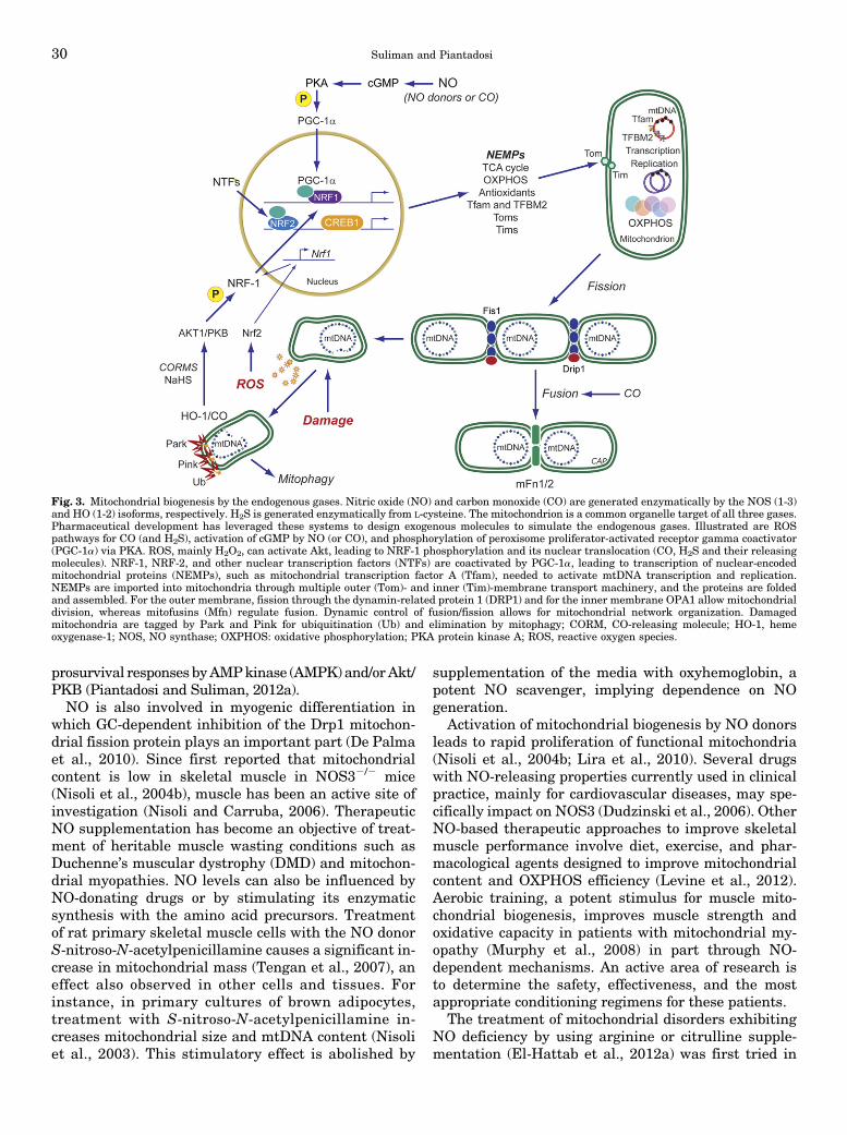
The potent paracrine vasodilator gas, nitric oxide(NO), has numerous effects on mitochondrial biology.NO is produced catalytically from L-arginine in anNADPH-dependent reaction by the three isoforms ofnitric oxide synthase (NOS), and NO generated invascular endothelium (by NOS3) transcriptionally acti-vates mitochondrial biogenesis (Nisoli et al., 2004a).Correspondingly, the inducible NOS (NOS2) augmentsmitochondrial biogenesis during periods of inflamma-tion (Reynolds et al., 2009; Suliman et al., 2010).Physiologic NO activates mitochondrial biogenesis viasoluble guanylate cyclase (GC) and cyclic GMP-dependent signals (Nisoli et al., 2003; Gao, 2010); cGMPproduction upregulates PGC-1a and NRF-1 to initiatethe program (Nisoli et al., 2003). The NO-dependentsignaling mechanisms are not fully defined, but in-creased PGC-1a, CREB1, and NRF-1 expression arerequired for mitochondrial proliferation (Fig. 3) (Nisoliet al., 2004b; Lira et al., 2010).
NO-dependent vasodilation improves oxygen andcarbon substrate availability for cell respiration andmetabolism, but despite this vasodilation, excessive NOproduction during certain other pathologic conditionsdirectly inhibits respiration through blockade of mito-chondrial Complexes I (NADH dehydrogenase) andIV (cytochrome c oxidase, COX), markedly reducingOXPHOS (Antunes et al., 2004; Brown and Borutaite,2007). NO reacts with superoxide and generates peroxy-nitrite (ONOO2), which not only diminishes vasodilationbut leads to macromolecular damage to mitochondria(Pacher et al., 2007; Poderoso, 2009). This damage mayenhance mitochondrial ROS production and stimulate
Mitochondrial Quality Control 29
prosurvival responses byAMPkinase (AMPK) and/orAkt/PKB (Piantadosi and Suliman, 2012a).NO is also involved in myogenic differentiation in
which GC-dependent inhibition of the Drp1 mitochon-drial fission protein plays an important part (De Palmaet al., 2010). Since first reported that mitochondrialcontent is low in skeletal muscle in NOS32/2 mice(Nisoli et al., 2004b), muscle has been an active site ofinvestigation (Nisoli and Carruba, 2006). TherapeuticNO supplementation has become an objective of treat-ment of heritable muscle wasting conditions such asDuchenne’s muscular dystrophy (DMD) and mitochon-drial myopathies. NO levels can also be influenced byNO-donating drugs or by stimulating its enzymaticsynthesis with the amino acid precursors. Treatmentof rat primary skeletal muscle cells with the NO donorS-nitroso-N-acetylpenicillamine causes a significant in-crease in mitochondrial mass (Tengan et al., 2007), aneffect also observed in other cells and tissues. Forinstance, in primary cultures of brown adipocytes,treatment with S-nitroso-N-acetylpenicillamine in-creases mitochondrial size and mtDNA content (Nisoliet al., 2003). This stimulatory effect is abolished by
supplementation of the media with oxyhemoglobin, apotent NO scavenger, implying dependence on NOgeneration.
Activation of mitochondrial biogenesis by NO donorsleads to rapid proliferation of functional mitochondria(Nisoli et al., 2004b; Lira et al., 2010). Several drugswith NO-releasing properties currently used in clinicalpractice, mainly for cardiovascular diseases, may spe-cifically impact on NOS3 (Dudzinski et al., 2006). OtherNO-based therapeutic approaches to improve skeletalmuscle performance involve diet, exercise, and phar-macological agents designed to improve mitochondrialcontent and OXPHOS efficiency (Levine et al., 2012).Aerobic training, a potent stimulus for muscle mito-chondrial biogenesis, improves muscle strength andoxidative capacity in patients with mitochondrial my-opathy (Murphy et al., 2008) in part through NO-dependent mechanisms. An active area of research isto determine the safety, effectiveness, and the mostappropriate conditioning regimens for these patients.
The treatment of mitochondrial disorders exhibitingNO deficiency by using arginine or citrulline supple-mentation (El-Hattab et al., 2012a) was first tried in
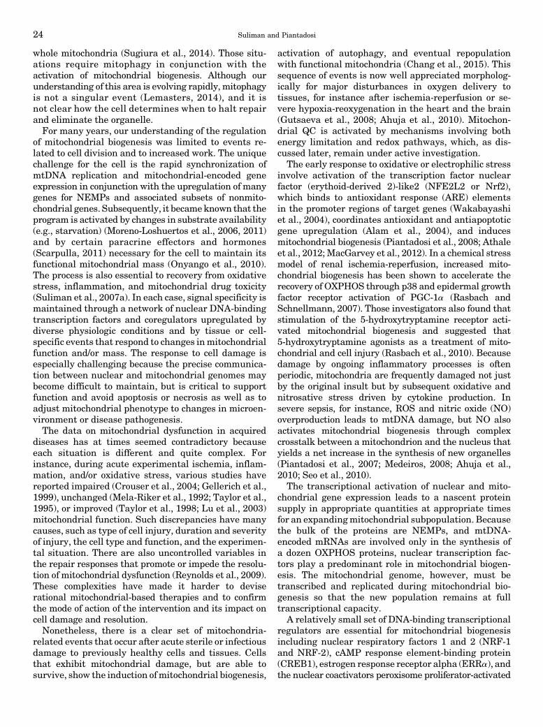
Fig. 3. Mitochondrial biogenesis by the endogenous gases. Nitric oxide (NO) and carbon monoxide (CO) are generated enzymatically by the NOS (1-3)and HO (1-2) isoforms, respectively. H2S is generated enzymatically from L-cysteine. The mitochondrion is a common organelle target of all three gases.Pharmaceutical development has leveraged these systems to design exogenous molecules to simulate the endogenous gases. Illustrated are ROSpathways for CO (and H2S), activation of cGMP by NO (or CO), and phosphorylation of peroxisome proliferator-activated receptor gamma coactivator(PGC-1a) via PKA. ROS, mainly H2O2, can activate Akt, leading to NRF-1 phosphorylation and its nuclear translocation (CO, H2S and their releasingmolecules). NRF-1, NRF-2, and other nuclear transcription factors (NTFs) are coactivated by PGC-1a, leading to transcription of nuclear-encodedmitochondrial proteins (NEMPs), such as mitochondrial transcription factor A (Tfam), needed to activate mtDNA transcription and replication.NEMPs are imported into mitochondria through multiple outer (Tom)- and inner (Tim)-membrane transport machinery, and the proteins are foldedand assembled. For the outer membrane, fission through the dynamin-related protein 1 (DRP1) and for the inner membrane OPA1 allow mitochondrialdivision, whereas mitofusins (Mfn) regulate fusion. Dynamic control of fusion/fission allows for mitochondrial network organization. Damagedmitochondria are tagged by Park and Pink for ubiquitination (Ub) and elimination by mitophagy; CORM, CO-releasing molecule; HO-1, hemeoxygenase-1; NOS, NO synthase; OXPHOS: oxidative phosphorylation; PKA protein kinase A; ROS, reactive oxygen species.
30 Suliman and Piantadosi

MELAS (Koga et al., 2005; El-Hattab et al., 2012b),because data from these patients had indicated lowlevels of serum arginine (Koga et al., 2005) andcitrulline (Naini et al., 2005) accompanied by endothe-lial dysfunction (Koga et al., 2006). The rationale was toimprove the low NO production rates observed in somemitochondrial diseases and not necessarily to directlyincrease mitochondrial biogenesis. Some encouragingresults were reported using arginine and citrulline totreat relative NO deficiency in MELAS patients (Kogaet al., 2005; El-Hattab et al., 2012a; Tengan et al., 2012),although mitochondrial proliferation in an effort tocompensate for the disease has been proposed as anunderlying cause of impaired NO synthesis by thesepatients in the first place (Tengan et al., 2012).On the basis of these findings and the NO deficiency
observed inmuscle fibers of patients withmitochondrialmyopathies, it seems reasonable that NO precursorsupplementation could have broader applicability inmitochondrial diseases that exhibit muscle weakness(El-Hattab et al., 2012a). The precursor strategy hasbeen tested in the Mdx mouse model of DMD anddelayed disease progression was observed (De Luca,2012; De Palma and Clementi, 2012). Clinically, thesuccess of NO therapy depends on whether the patientcan increase the proliferation rate of mitochondria withfunctional OXPHOS systems, a limitation in hereditarymtDNA disorders with low heteroplasmy.Other drugs such as HCT 1025, a nonsteroidal anti-
inflammatory derivative of flurbiprofen that releasesNO, seems to slow disease progression inmice with limbgirdle and Duchenne muscular dystrophies (Brunelliet al., 2007). HCT 1026 combined with arterially de-livered donor stem cells offered improved efficacy. Thisalso demonstrated better functional gain than L-argi-nine combined with deflazacort in mdx dystrophic mice(Archer et al., 2006). Another study reported that theNO-releasing drug molsidomine has therapeutic effectsin dystrophic mice (Buono et al., 2012). The drugincreased muscle fiber regeneration and enhancedmuscle function by increasing satellite cell prolifera-tion. These studies raise new prospects for the treat-ment of both primary and secondary mitochondrialdysfunction, because several NO-based drugs alreadytested for safety in humans are available for study inclinical trials.NO is also cardioprotective during ischemia-
reperfusion (I/R) injury. By using a mitochondrial-selective S-nitrosating agent, reversible mitochondrialS-nitrosation (S-nitrosylation) of Complex I subunitsconstitutes a major mechanism of NO protection duringI/R and is a potential therapeutic target for the preventionof injury after myocardial and possibly cerebral infarction(Chouchani et al., 2013). In the heart and brain, progres-sion of I/R injury is associated with mitochondrial dys-function manifested by ATP depletion, calcium-inducedopening of themitochondrial permeability transition pore,
and exacerbation of mitochondrial ROS release (Cadenaset al., 2010). Several studies have investigated acutenitrite treatment, a modest NO donor under hypoxic/acidic conditions, and one has shown that nitrite therapyinitiated 24 hours after I/R improves tissue and vascularregeneration, and functional recovery (Kapil et al., 2014).
B. The Heme Oxygenase-1/Carbon Monoxide System
Endogenous carbon monoxide (CO) produced by theheme oxygenases (Hmox, HO-1 and -2) acts as amessenger gas that exerts control over mitochondrialQC through its impact on the regulation of mitochon-drial biogenesis and presumably mitophagy. Prior tothis understanding, there was controversy over theincontrovertible toxicity of the gas versus observationssupporting low dose CO as a cell protectant operatingthrough general anti-inflammatory and antiapoptoticproperties (Ryter et al., 2006; Suliman and Piantadosi,2014). Indeed, many protective effects of HO-1 induc-tion by ischemia, hypoxia, oxidative stress, metals, andinflammation are not simply due to heme clearance andin many cases can be recapitulated by CO administra-tion (Maines, 2005; Vieira et al., 2008; Biermann et al.,2010; Lee et al., 2014).
The targets of CO binding in tissue are largelyrestricted to reduced transition metals, such as enzy-matic copper and iron centers in hemoglobin, myoglo-bin, and cytochrome c oxidase, thereby influencingenzymatic function and redox state (Piantadosi, 2008;Motterlini and Otterbein, 2010). Its affinity for hemeproteins is responsible for its potent asphyxia effect.Like NO, CO activates guanylate cyclase but binds witha far lower affinity, whereas its effects on mitochondriaare mediated through direct binding to reduced cyto-chrome c oxidase or indirectly to nearby heme proteinsand enzymes of cytochrome P450 systems. An impor-tant effect is an increase in mitochondrial ROS produc-tion that leads to further HO-1 induction (Piantadosi,2008). Under such circumstances, CO administration orthe use of CO-releasing molecules (CORMs) generates apositive feedback loop that contributes to upregulationof the HO-1/CO pathway and its induction of mitochon-drial biogenesis (Fig. 3).
Abundant preclinical evidence has demonstratedbeneficial effects of CO administered as authentic gasor as CORMs in experimental models of cardiovasculardisease, stroke, sepsis, organ transplant rejection, andvarious acute injuries in the lungs, heart, kidneys, andliver (Ryter et al., 2006; Bauer and Pannen, 2009;Motterlini and Otterbein, 2010). Early studies foundCOmodulates local inflammatory responses by promotinganti-inflammatory cytokine production and opposing ap-optosis, and a number of these effects appear to be linkedto the transcriptional network ofmitochondrial biogenesis(Piantadosi et al., 2011). Moreover, the induction ofmitochondrial biogenesis leads directly to counter-inflammatory cytokine induction such as interleukin-10
Mitochondrial Quality Control 31
(IL-10), IL-1 receptor antagonist (Piantadosi et al., 2011),and suppressor of cytokine synthesis 3 (Athale et al.,2012).In resting humans breathing air, CO has a half-life of
4–5 hours and is eliminated unaltered almost exclu-sively through the lungs (Coburn, 2012) with limitedoxidation to CO2 bymitochondria. Moreover, the criticalsafe upper limit of carboxyhemoglobin [HbCO] innormal individuals is not known, but it is certainlylower in patients with cardiovascular and cerebrovas-cular disease (Hampson et al., 2012).CO gas is relatively nonreactive chemically, straight-
forward to produce, and inexpensive. Like NO, which isin clinical use, it is readily administered by inhalation(Motterlini and Otterbein, 2010). By giving inhaled COas discrete pulses, tissue CO levels will rise transientlyand follow PO2-dependent elimination kinetics. Theease of administration has led to clinical trials focusedon the anti-inflammatory effects of CO and set an upperlimit of exposure at 8% HbCO, the equivalent ofsmoking about two packs of cigarettes per day. Thefirst randomized, placebo-controlled Phase I trial toevaluate the clinical safety and pharmacokinetics ofinhaled CO in healthy humans has been completed(Motterlini and Otterbein, 2010). Low concentrations ofthe gas were found to be safe in dose-escalation studiesin healthy humans and deemed acceptable for Phase IItesting in emphysema and fibrotic lung disease. InhaledCO is also undergoing evaluation for safety and poten-tial efficacy in patients with advanced pulmonaryarterial hypertension. In humans, it has also beenshown that low-dose inhaled CO improves muscle mito-chondrial density but also regulates myoglobin content,localization of the insulin-regulated glucose transporter(GLUT4), and muscle capillarity (Pecorella et al., 2015).The application of CO releasing molecules (CORMs)
is a useful alternative to inhaled CO because they can beadministered via systemic routes and in a tissue-specific manner, thus avoiding generalized pulmonarydelivery systems and allowing judicious targeting oforgans such as the brain. CORMs also showmany of theprotective features of authentic CO gas, e.g., suppress-ing the inflammatory response in glial cells (Bani-Haniet al., 2006), and may therefore offer neuroprotection.The apparent inconsistency of neuroprotection in theliterature has caused confusion but may in part reflectthe choice of CORM and route of administration.CORMs all possess a backbone carrier moiety embod-
ied most commonly as organometallic carbonyl com-plexes that must be stringently characterized from ametabolic and toxicological standpoint. Some agentssuch as the boranocarbonates avoid metal toxicity butgenerally release CO rapidly. Several CORMs havebeen synthesized for therapeutic agents for deliveringcontrolled amounts of CO to tissues and organs(Motterlini et al., 2002). CORM-1 is soluble in water anddecomposes rapidly, but under physiologic conditions
CO is released with slower kinetics (half-life of 21 min)(Motterlini et al., 2003). CORM-2 is soluble in DMSOand olive oil and releases CO by photo-dissociation(Motterlini et al., 2002). CORM-3 is a water-solubleruthenium-based agent with a half-life of 1 min (Clarket al., 2003). CORM-3 is effective when given beforeneuronal injury but not if given shortly afterward(Yabluchanskiy et al., 2012). A molybdenum-based,water-soluble CORM (ALF186) that releases CO in adose- and oxygen-dependent manner is protective invivo in models of acute inflammation (Sheikh et al.,2011) and in the regulation of vasomotor tone (Szabó,2007;Marazioti et al., 2011). ALF186 inhibits I/R-inducedneuronal cell death via soluble GC activation and maybe useful for acute ischemic insults to the retina and thebrain. More recent work has shown utility of water-soluble synthetic carbonyl complexes of rhenium, whichrelease CO and ReO4; the latter is excreted by thekidneys (Zobi et al., 2013). All of these agents relaxblood vessels and generally lower blood pressure via GCand potassium channel activation, and some are postu-lated to improve vascular function. In addition, COreleased from CORMs inhibits NF-kΒ-mediated inflam-matory gene expression and upregulates adaptive geneexpression for oxidative stress. Thus, these compoundsare being proposed as novel agents for the therapy of anarray of vascular, inflammatory, and oxidative stress-related disorders.
C. Hydrogen Sulfide
A substantial body of evidence implicates H2S asa signaling molecule involved in multiple physiologic andcellular processes that protect organ function againstdamage (Szabó, 2007; Wagner et al., 2009), including I/Rinjury (Zhang et al., 2006; Tamizhselvi et al., 2008). Itis made endogenously by the conversion of L-cysteinethrough two pyridoxal-50 phosphate-dependent en-zymes, cystathionine b-synthase and cystathionineg-lyase. It has anti-inflammatory properties, such asinhibiting NF-kB-dependent NOS2 expression, but mayalso inhibit HO-1 expression; nonetheless, its preciserole as an endogenous suppressor of inflammation isstill under investigation. Marked proinflammatoryeffects reported for H2S (Collin et al., 2005; Li et al.,2005) stand in contrast with the anti-inflammatoryeffects observed in different studies (Elrod et al., 2007;Wagner et al., 2011).
H2S is a well-known inhibitor of OXPHOS via cyto-chrome c oxidase (COX) blockade, a toxic property ofmany low molecular weight sulfides (Cooper andBrown, 2008). On the other hand, low-dose H2S hasbeen reported to preserve mitochondrial function in theheart muscle of rodents after I/R injury (Elrod et al.,2007; Calvert et al., 2010). Like CO, low level H2Sadministration or exposure of cells to H2S-releasingcompounds increases the phosphorylation of proteinserine/threonine kinase B (Akt) and enhances the
32 Suliman and Piantadosi
nuclear localization of NRF-1 and -2 to increase mito-chondrial biogenesis, attenuate apoptosis, and increaseendogenous antioxidant levels (Calvert et al., 2010).The impact of the gas on mitophagy and overall mito-chondrial QC in injury and disease states however is notyet clear.
V. Induction of Mitochondrial Biogenesis byHormones, Drugs, and Natural Products
A. Estrogens, Erythropoietin, and Thyroid Hormone
Certain hormones modulate metabolism and mito-chondrial function through the actions of the nuclearreceptor (NR) superfamily of proteins. Upon ligandbinding, these receptors enter the nucleus and bindhormone-responsive elements in the promoter regionsof target genes, including TFAM, TFB1M, and TFB2M(Psarra and Sekeris, 2011). In various tissues, steroid(type I) and nonsteroid (type II) NRs influence mito-chondrial biogenesis and OXPHOS components (Weberet al., 2002). Some NRs, such as the glucocorticoidreceptor (GR), estrogen receptor, and thyroid receptor(TR) are also found inmitochondria where certain directtranscriptional effects can occur.1. Estrogens. The loss of the main circulating estro-
gen 17b-estradiol (E2) due to either natural or surgicalmenopause leads to a prompt reduction in body meta-bolic rate. Other manifestations may include muscleweakness, fatigue, reduced exercise capacity, andweight gain. Some of these signs and symptoms are
clearly associated with changes in aerobic energymetabolism.
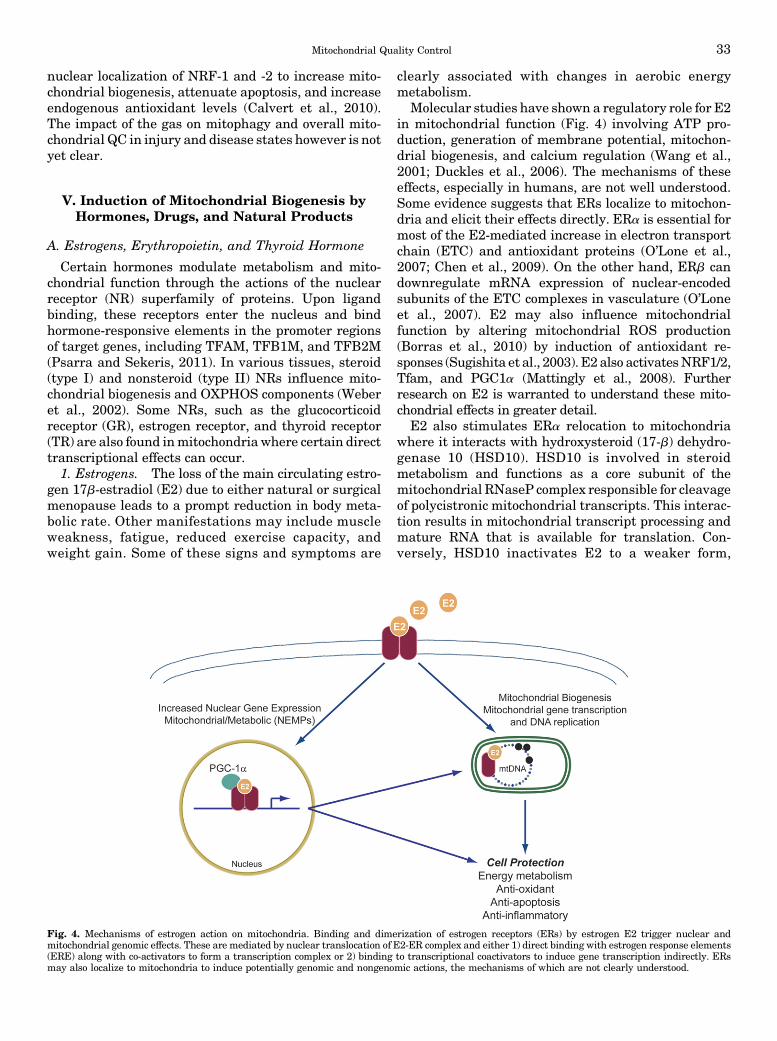
Molecular studies have shown a regulatory role for E2in mitochondrial function (Fig. 4) involving ATP pro-duction, generation of membrane potential, mitochon-drial biogenesis, and calcium regulation (Wang et al.,2001; Duckles et al., 2006). The mechanisms of theseeffects, especially in humans, are not well understood.Some evidence suggests that ERs localize to mitochon-dria and elicit their effects directly. ERa is essential formost of the E2-mediated increase in electron transportchain (ETC) and antioxidant proteins (O’Lone et al.,2007; Chen et al., 2009). On the other hand, ERb candownregulate mRNA expression of nuclear-encodedsubunits of the ETC complexes in vasculature (O’Loneet al., 2007). E2 may also influence mitochondrialfunction by altering mitochondrial ROS production(Borras et al., 2010) by induction of antioxidant re-sponses (Sugishita et al., 2003). E2 also activatesNRF1/2,Tfam, and PGC1a (Mattingly et al., 2008). Furtherresearch on E2 is warranted to understand these mito-chondrial effects in greater detail.
E2 also stimulates ERa relocation to mitochondriawhere it interacts with hydroxysteroid (17-b) dehydro-genase 10 (HSD10). HSD10 is involved in steroidmetabolism and functions as a core subunit of themitochondrial RNaseP complex responsible for cleavageof polycistronic mitochondrial transcripts. This interac-tion results in mitochondrial transcript processing andmature RNA that is available for translation. Con-versely, HSD10 inactivates E2 to a weaker form,
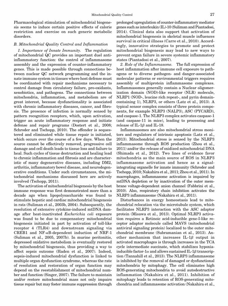
Fig. 4. Mechanisms of estrogen action on mitochondria. Binding and dimerization of estrogen receptors (ERs) by estrogen E2 trigger nuclear andmitochondrial genomic effects. These are mediated by nuclear translocation of E2-ER complex and either 1) direct binding with estrogen response elements(ERE) along with co-activators to form a transcription complex or 2) binding to transcriptional coactivators to induce gene transcription indirectly. ERsmay also localize to mitochondria to induce potentially genomic and nongenomic actions, the mechanisms of which are not clearly understood.
Mitochondrial Quality Control 33

estrone, the significance of which requires furtherinvestigation (Sanchez et al., 2015).Impaired E2 signaling and subsequent mitochondrial
dysfunction may also be involved in insulin resistance.Mitochondrial dysfunction is associated with reduced orpartial FAO that can activate stress kinases that inhibitinsulin signaling (Koves et al., 2008; Zhang et al., 2010).The expression of the adipokine, adiponectin, and itsreceptor, AR1, is induced by estrogen in conjunctionwith mitochondrial biogenesis (Capllonch-Amer et al.,2014), and adiponectin may improve insulin sensitivity.Estrogen replacement therapy to protect postmeno-
pausal women from diabetes and other metabolicdisorders and osteoporosis is offset by the increasedcancer risk by E2 and ER agonists. Bazedoxifene withconjugated E2 is a combination of selective tissue E2complexes and receptor modulator that provides tissue-specific benefits of E2 in menopausal osteoporosis inwomen (Archer, 2010) and improves cardiovasculardisease risk and metabolic syndrome with greatereffects in endometrium and breast in animal models(Barrera et al., 2014; Kim et al., 2014). Althoughselective tissue E2 complexes are still in trials, newer,more innovative, efficient, and tissue-specific E2 re-ceptor agonists are being investigated. An E2 conju-gated preparation with glucagon-like peptide-1 is moreactive than either hormone alone in the reversal ofobesity, hyperglycemia, and dyslipidemia in mice andprevents reproductive endocrine toxicity and oncoge-nicity (Finan et al., 2012). It is not yet clear whichpatients benefit and which agents would minimize therisks of E2-related therapy.2. Erythropoietin. EPO regulates erythrocyte mass
in response to hypoxia and anemia, but EPO activity isdetectable in some non-erythroid cells that expressfunctional EPO receptor (EpoR). Protective effects ofEPO have been demonstrated in various nonerythroidtissues and experimental models of I/R injury andattributed to nonhematopoietic metabolic effects, in-hibition of apoptosis, or to stimulation of angiogenesis(Chatterjee, 2005). EPO stimulates cardiac mitochon-drial proliferation through mitochondrial biogenesismediated by NO and NRF-1 and PGC-1a (Carrawayet al., 2010). Clinically, EPO reverses cardiac remodel-ing, improves cardiac function, and enhances exercisetolerance and quality of life of a subset of heart patientsby protective effects beyond the correction of anemia(Bergmann et al., 2011). These findings highlight thepossibility that EPO-mediated protection may dependon modulatory effects on bioenergetics. EPO can stim-ulate proliferation of cultured myoblasts through bind-ing to EpoR to expand the progenitor population duringdifferentiation, and it may have a role in muscle repair(Jia et al., 2009;Ogilvie et al., 2000). EPO/EpoR signalingalso improves glucose tolerance and protects againstdietary obesity in association with skeletal muscleadaptation (Hojman et al., 2009; Teng et al., 2011).
The association of EPO treatment in mice withincreased mitochondrial biogenesis and PGC-1a activa-tion in myocytes raises the possibility that EPO maycontribute to skeletal muscle plasticity and the balancebetween slow and fast-twitch fibers. Furthermore,recombinant protein increases OXPHOS in humanskeletal muscle (Plenge et al., 2012). The enhancementand recovery of cellular functions through stimulationof hemoglobin production and mitochondrial activity innonhematopoietic cells by EPO-like drugs might be atestable therapeutic strategy for certain ischemic ormitochondrial diseases in carefully selected patientpopulations, although the procoagulant effects of EPOadministration have raised a legitimate caution.
3. Thyroid Hormone. The ability of thyroid hormone(T3 and T4) and thyroid hormone receptors (TRs) toregulate oxygen uptake and energy utilization is relatedto the thyroid system’s multiple effects on mitochondri-al function. TH targets several regulatory pathways forgene expression that may contribute to mitochondrialbiogenesis (Weitzel and Iwen, 2011). TH binds tonuclear-localized TR belonging to a ligand-dependenttranscription factor family that modulates nuclear geneexpression via binding to a thyroid response or TREmotif. TH also directly affectsmitochondria by binding amitochondrial-localized TR. However, intermediate fac-tors are also expressed (perhaps via TREs) that enter thenucleus and regulate other TH-target genes. PGC-1aand PGC-1b and NRF-1, NRF-2 have been postulatedto serve as these intermediate factors. There is evidenceof rapid PGC-1a induction by TH both at themRNA andprotein levels [mediated by a TRE in the gene promoter(Wulf et al., 2008)], but its impact on mitochondrialproliferation seems modest and the therapeutic oppor-tunities limited.
B. Pharmacological Agents
The pharmacological activation of mitochondrial bio-genesis is potentially achievable by a broad range ofcompounds including select natural products and phar-maceutical agents (Fig. 5) (Andreux et al., 2013). Someof these agents may operate through physiologic path-ways, whereas others may cause mild mitochondrialdamage that then leads to the induction of physiologicQC mechanisms. These distinctions are made belowwhen possible.
1. Peroxisome Proliferator-Activated Receptor Activa-tion by Bezafibrate. The PPAR (a, b/d, g) agonistbezafibrate is commonly used to treat dyslipidemias(Tenenbaum et al., 2005). Although fibrates have longbeen linked to fatty acid oxidation (FAO) and toperoxisome proliferation, later studies reveal thatPPARs via PGC-1a also regulate other aspects of mito-chondrial function (Hondares et al., 2011). Bezafibratestimulates heart and skeletal muscle PGC-1a expressionin mice in a PPARd-dependent manner (Hondareset al., 2007). Moreover, bezafibrate treatment or PGC-1a
34 Suliman and Piantadosi
overexpression upregulates respiratory capacity andOXPHOS function in fibroblasts and myoblasts ofOXPHOS-deficient patients (Srivastava et al., 2009).Dietary bezafibrate at a concentration of 0.5% (w/w)induced mitochondrial biogenesis in skeletal muscle ofCox10 knockoutmice. Bezafibrate stimulatesPpar andPgc-1 gene expression and the induction of mtDNA andmitochondrial proteins in muscle. Importantly, bezafi-brate treatment in Cox10 knockout mice delays theonset ofmyopathy and prolongs survival. In the Surf12/2model of mild COX deficiency, bezafibrate does notreplicate the effect of PGC-1a overexpression (Viscomiet al., 2011). Instead, the drug induces both PPARaand d and causes FAO upregulation in skeletal muscle.It also caused weight loss and severe hepatomegaly,indicating drug toxicity that could be due to PPARaactivation (Hays et al., 2005).Bezafibrate showed benefit in Deletor mice, a model
of adult-onset mitochondrial myopathy from accumula-tion of mtDNA deletions as a result of transgenicexpression of mutated Twinkle helicase (Wallaceet al., 2010; Yatsuga and Suomalainen, 2012). The drugcaused significant induction of hepatic liver FAO,hepatomegaly, and decreases in adipose tissue in thismodel (Yatsuga and Suomalainen, 2012). In anotherstudy, the bezafibrate effect in Deletor mice was limitedto delayed accumulation of COX-negative muscle fibersand a reduced mtDNA deletion-load with no change inphenotype and no induction of mitochondrial biogene-sis. The treatment strategy differed in that the drugwasstarted at the onset of symptoms at 1 year of age insteadof at 5 weeks (Viscomi et al., 2011).In a mouse model of AD (P301S transgenic mice)
bezafibrate decreased disease progression by diminish-ing locomotor and stress-related abnormalities(Dumont et al., 2012; Noe et al., 2013). A modestincrease in mitochondrial proliferation was noted, butmild increases in mtDNA content and Sirt1 and Tfamgene expression was more apparent in brown fat withincreased expression of NRF1 and PGC-1a. Small dif-ferences between mouse and human PPARa orthologsalso seem to influence hepatocyte proliferation byPPARa activators such as fenofibrate, which does notinduce hepatomegaly in PPARa-humanized mice as inwild-type mice (Yang et al., 2008). Bezafibrate is rela-tively safe in humans, although rhabdomyolysis andkidney failure have been reported (Wu et al., 2009). Apromising finding is that long-term bezafibrate treat-ment of patients with the mitochondrial FAO disorderCPT2 deficiency leads to significant clinical improve-ment (Bonnefont et al., 2009, 2010).2. b2-Adrenergic Receptor Agonists. b2-AR receptor
agonists are airway smooth muscle relaxants andbronchodilators approved for the treatment of asthmaand other airways diseases. These agonists are reportedto modulate oxidative metabolism, energy expendi-ture, lipolysis, glucose transport, and glucose oxidation
(Pearen et al., 2008). Mice treated with formoterol, ab2-AR agonist, showed a fivefold induction of PGC-1amRNA in skeletal muscle. The b2-AR has been suc-cessfully targeted for drug discovery using a ligand-based approach, resulting in the creation of multiplereceptor-specific drugs (Wishart et al., 2006; Kolb et al.,2009). However, the therapeutic potential of establishedand novel b2-AR agonists for the induction of mitochon-drial biogenesis and as counter-inflammatory agents hasnot been well explored.
3. Metformin. This insulin-sensitizing biguanide is afront-line oral hypoglycemic treatment of DM2 (Halimi,2006). Metformin lowers blood glucose mainly viahepatic glucose output and insulin-stimulated glucoseuptake in skeletal muscle and adipocytes (Larsen et al.,2011). The molecular mechanisms involved are notcompletely clear. Although inhibition of mitochondrialComplex I has been reported in cells, isolated mitochon-dria, and muscle homogenate (Brunmair et al., 2004);metformin-induced activation of AMPK has been shownin rodent muscle (Zhou et al., 2001) and in humanskeletal muscle from patients with DM2 (Musi et al.,2002). Numerous studies have documented a role forAMPK in regulating mitochondrial function and bio-genesis, and it too leads to anti-inflammatory effects(O’Neill et al., 2011). On the other hand, effects ofmetformin on mitochondrial function by decreasingROS production have been postulated (Hou et al., 2010).Long-term metformin treatment may increase PGC-1aexpression inmuscle together with increased activity andprotein expression of certain mitochondrial marker en-zymes (Jager et al., 2007).
4. 5-Aminoimidazole-4-Carboxamide Ribonucleotide.5-Aminoimidazole-4-carboxamide ribonucleotide (AICAR)has been found to increased mitochondrial biogenesisand cell ATP content while decreasing ROS production.The effect is mediated via AMPK and direct regulationof energy expenditure bymodulating SIRT1 deacetylaseactivity, resulting in deacetylation of downstream tar-gets including PGC-1a and Forkhead box O1 and 3transcription factors (Canto et al., 2009). The drug doeshave significant toxicity, and the use of AICAR as acardiac therapeutic is limited by bradycardia andhypoglycemia (Young et al., 2005). AICAR requiresphosphorylation inside the cell, and the phosphorylatedderivative known as ZMP acts as an AMP mimetic(Merrill et al., 1997). Unlike other presumed AMPKagonists, AICAR promotes AMPK phosphorylation andactivation while not disturbing the energy status of thecell, i.e., the AMP/ATP ratio remains the same.
AICAR is orally effective to increase enduranceperformance in sedentarymice by increasing expressionof oxidative metabolism genes (Narkar et al., 2008).This is thought to happen via direct phosphorylation/activation of PGC-1a by activated AMPK and subse-quent induction of mitochondrial biogenesis (Jageret al., 2007). Unfortunately, AICAR penetrates the
Mitochondrial Quality Control 35
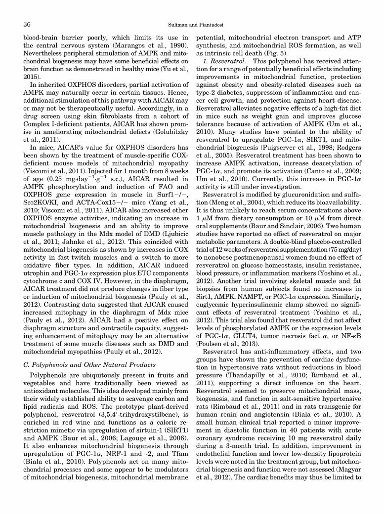
blood-brain barrier poorly, which limits its use inthe central nervous system (Marangos et al., 1990).Nevertheless peripheral stimulation of AMPK and mito-chondrial biogenesis may have some beneficial effects onbrain function as demonstrated in healthy mice (Yu et al.,2015).In inherited OXPHOS disorders, partial activation of
AMPK may naturally occur in certain tissues. Hence,additional stimulation of this pathwaywith AICARmayor may not be therapeutically useful. Accordingly, in adrug screen using skin fibroblasts from a cohort ofComplex I-deficient patients, AICAR has shown prom-ise in ameliorating mitochondrial defects (Golubitzkyet al., 2011).In mice, AICAR’s value for OXPHOS disorders has
been shown by the treatment of muscle-specific COX-deficient mouse models of mitochondrial myopathy(Viscomi et al., 2011). Injected for 1 month from 8 weeksof age (0.25 mg×day21×g21 s.c.), AICAR resulted inAMPK phosphorylation and induction of FAO andOXPHOS gene expression in muscle in Surf12/2,Sco2KO/KI, and ACTA-Cox152/2 mice (Yang et al.,2010; Viscomi et al., 2011). AICAR also increased otherOXPHOS enzyme activities, indicating an increase inmitochondrial biogenesis and an ability to improvemuscle pathology in the Mdx model of DMD (Ljubicicet al., 2011; Jahnke et al., 2012). This coincided withmitochondrial biogenesis as shown by increases in COXactivity in fast-twitch muscles and a switch to moreoxidative fiber types. In addition, AICAR inducedutrophin and PGC-1a expression plus ETC componentscytochrome c and COX IV. However, in the diaphragm,AICAR treatment did not produce changes in fiber typeor induction of mitochondrial biogenesis (Pauly et al.,2012). Contrasting data suggested that AICAR causedincreased mitophagy in the diaphragm of Mdx mice(Pauly et al., 2012). AICAR had a positive effect ondiaphragm structure and contractile capacity, suggest-ing enhancement of mitophagy may be an alternativetreatment of some muscle diseases such as DMD andmitochondrial myopathies (Pauly et al., 2012).
C. Polyphenols and Other Natural Products
Polyphenols are ubiquitously present in fruits andvegetables and have traditionally been viewed asantioxidantmolecules. This idea developedmainly fromtheir widely established ability to scavenge carbon andlipid radicals and ROS. The prototype plant-derivedpolyphenol, resveratrol (3,5,49-trihydroxystilbene), isenriched in red wine and functions as a caloric re-striction mimetic via upregulation of sirtuin-1 (SIRT1)and AMPK (Baur et al., 2006; Lagouge et al., 2006).It also enhances mitochondrial biogenesis throughupregulation of PGC-1a, NRF-1 and -2, and Tfam(Biala et al., 2010). Polyphenols act on many mito-chondrial processes and some appear to be modulatorsof mitochondrial biogenesis, mitochondrial membrane
potential, mitochondrial electron transport and ATPsynthesis, and mitochondrial ROS formation, as wellas intrinsic cell death (Fig. 5).
1. Resveratrol. This polyphenol has received atten-tion for a range of potentially beneficial effects includingimprovements in mitochondrial function, protectionagainst obesity and obesity-related diseases such astype-2 diabetes, suppression of inflammation and can-cer cell growth, and protection against heart disease.Resveratrol alleviates negative effects of a high-fat dietin mice such as weight gain and improves glucosetolerance because of activation of AMPK (Um et al.,2010). Many studies have pointed to the ability ofresveratrol to upregulate PGC-1a, SIRT1, and mito-chondrial biogenesis (Puigserver et al., 1998; Rodgerset al., 2005). Resveratrol treatment has been shown toincrease AMPK activation, increase deacetylation ofPGC-1a, and promote its activation (Canto et al., 2009;Um et al., 2010). Currently, this increase in PGC-1aactivity is still under investigation.
Resveratrol is modified by glucuronidation and sulfa-tion (Meng et al., 2004), which reduce its bioavailability.It is thus unlikely to reach serum concentrations above1 mM from dietary consumption or 10 mM from directoral supplements (Baur and Sinclair, 2006). Two humanstudies have reported no effect of resveratrol on majormetabolic parameters. A double-blind placebo-controlledtrial of 12weeksof resveratrol supplementation (75mg/day)to nonobese postmenopausal women found no effect ofresveratrol on glucose homeostasis, insulin resistance,blood pressure, or inflammation markers (Yoshino et al.,2012). Another trial involving skeletal muscle and fatbiopsies from human subjects found no increases inSirt1, AMPK, NAMPT, or PGC-1a expression. Similarly,euglycemic hyperinsulinemic clamp showed no signifi-cant effects of resveratrol treatment (Yoshino et al.,2012). This trial also found that resveratrol did not affectlevels of phosphorylated AMPK or the expression levelsof PGC-1a, GLUT4, tumor necrosis fact a, or NF-kB(Poulsen et al., 2013).
Resveratrol has anti-inflammatory effects, and twogroups have shown the prevention of cardiac dysfunc-tion in hypertensive rats without reductions in bloodpressure (Thandapilly et al., 2010; Rimbaud et al.,2011), supporting a direct influence on the heart.Resveratrol seemed to preserve mitochondrial mass,biogenesis, and function in salt-sensitive hypertensiverats (Rimbaud et al., 2011) and in rats transgenic forhuman renin and angiotensin (Biala et al., 2010). Asmall human clinical trial reported a minor improve-ment in diastolic function in 40 patients with acutecoronary syndrome receiving 10 mg resveratrol dailyduring a 3-month trial. In addition, improvement inendothelial function and lower low-density lipoproteinlevels were noted in the treatment group, but mitochon-drial biogenesis and function were not assessed (Magyaret al., 2012). The cardiac benefits may thus be limited to
36 Suliman and Piantadosi
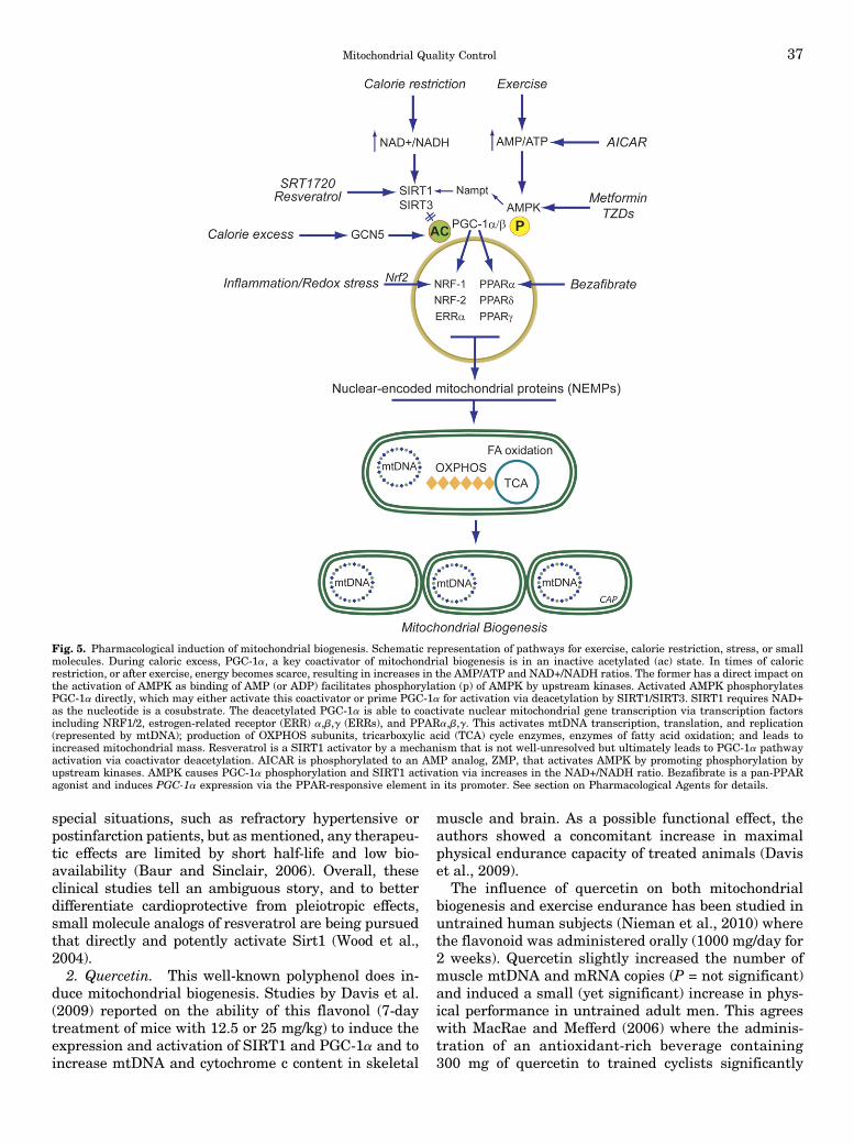
special situations, such as refractory hypertensive orpostinfarction patients, but as mentioned, any therapeu-tic effects are limited by short half-life and low bio-availability (Baur and Sinclair, 2006). Overall, theseclinical studies tell an ambiguous story, and to betterdifferentiate cardioprotective from pleiotropic effects,small molecule analogs of resveratrol are being pursuedthat directly and potently activate Sirt1 (Wood et al.,2004).2. Quercetin. This well-known polyphenol does in-
duce mitochondrial biogenesis. Studies by Davis et al.(2009) reported on the ability of this flavonol (7-daytreatment of mice with 12.5 or 25 mg/kg) to induce theexpression and activation of SIRT1 and PGC-1a and toincrease mtDNA and cytochrome c content in skeletal
muscle and brain. As a possible functional effect, theauthors showed a concomitant increase in maximalphysical endurance capacity of treated animals (Daviset al., 2009).
The influence of quercetin on both mitochondrialbiogenesis and exercise endurance has been studied inuntrained human subjects (Nieman et al., 2010) wherethe flavonoid was administered orally (1000 mg/day for2 weeks). Quercetin slightly increased the number ofmuscle mtDNA and mRNA copies (P = not significant)and induced a small (yet significant) increase in phys-ical performance in untrained adult men. This agreeswith MacRae and Mefferd (2006) where the adminis-tration of an antioxidant-rich beverage containing300 mg of quercetin to trained cyclists significantly
Fig. 5. Pharmacological induction of mitochondrial biogenesis. Schematic representation of pathways for exercise, calorie restriction, stress, or smallmolecules. During caloric excess, PGC-1a, a key coactivator of mitochondrial biogenesis is in an inactive acetylated (ac) state. In times of caloricrestriction, or after exercise, energy becomes scarce, resulting in increases in the AMP/ATP and NAD+/NADH ratios. The former has a direct impact onthe activation of AMPK as binding of AMP (or ADP) facilitates phosphorylation (p) of AMPK by upstream kinases. Activated AMPK phosphorylatesPGC-1a directly, which may either activate this coactivator or prime PGC-1a for activation via deacetylation by SIRT1/SIRT3. SIRT1 requires NAD+as the nucleotide is a cosubstrate. The deacetylated PGC-1a is able to coactivate nuclear mitochondrial gene transcription via transcription factorsincluding NRF1/2, estrogen-related receptor (ERR) a,b,g (ERRs), and PPARa,b,g. This activates mtDNA transcription, translation, and replication(represented by mtDNA); production of OXPHOS subunits, tricarboxylic acid (TCA) cycle enzymes, enzymes of fatty acid oxidation; and leads toincreased mitochondrial mass. Resveratrol is a SIRT1 activator by a mechanism that is not well-unresolved but ultimately leads to PGC-1a pathwayactivation via coactivator deacetylation. AICAR is phosphorylated to an AMP analog, ZMP, that activates AMPK by promoting phosphorylation byupstream kinases. AMPK causes PGC-1a phosphorylation and SIRT1 activation via increases in the NAD+/NADH ratio. Bezafibrate is a pan-PPARagonist and induces PGC-1a expression via the PPAR-responsive element in its promoter. See section on Pharmacological Agents for details.
Mitochondrial Quality Control 37

increased their physical performance after 6 weeks (noeffect was seen in controls given a quercetin-freeantioxidant-rich beverage). However, no mitochondrialbiogenesis markers were reported.3. Hydroxytyrosol. Another polyphenol linked to
mitochondrial biogenesis is hydroxytyrosol, a phenolicalcohol present in olives (and in extra virgin olive oils).In vitro, hydroxytyrosol (1–10 mM) activates PGC-1athrough SIRT1 deacetylation and induces mitochondri-al biogenesis in 3T3-L1 murine adipocytes and inARPE-19 human retinal pigment epithelial cells (Zhuet al., 2010). The administration of hydroxytyrosol torats (25 mg/kg daily during 8 weeks) by the sameinvestigators stimulated PGC-1a activity and the ex-pression of mitochondrial biogenesis markers in skele-tal muscle of both treadmill-exercised and sedentaryanimals, and increased their endurance (Feng et al.,2011). The authors noted that in contrast to the mito-chondrial biogenesis-inducing effect of moderate exer-cise, extreme exercise was associated with lower PGC-1alevels. This effect was prevented by hydroxytyrosoladministration to the animals (Feng et al., 2011).Other dietary polyphenols have been implicated in
the induction of mitochondrial biogenesis. For instance,isoflavones (genistein, daidzein, and formononetin) inrabbit renal proximal tubular cells (Rasbach andSchnellmann, 2008), flavones (baicalein, its 7-O-glucu-ronide baicalin, andwogonin) in L6 skeletalmuscle cells(Im et al., 2012), and flavan-3-ol epigallocatechin-3-gallate in skin fibroblasts from Down’s syndromepatients (Valenti et al., 2013) all activate the SIRT1/PGC-1a pathway. In contrast to the SIRT1-inducingeffect of genistein (Rasbach and Schnellmann, 2008),this isoflavone also reduces SIRT1 mRNA and proteinexpression and induces SIRT1 nuclear exclusion intothe cytosol of the human prostate cancer cell linesLNCaP and PC-3 (Kikuno et al., 2008). However,PGC-1a levels or acetylation were not measured in thisstudy. The SIRT1-mediated actions of genistein couldrelate to differences in basal levels or activity of SIRT-1or to mechanisms that modulate its expression innormal compared with cancer cells.Greater consistency of SIRT-1-inducing effects of
polyphenols is seen by comparing in vivo studies. Inhumans (Taub et al., 2012), the administration of anepicatechin-rich cocoa (total epicatechin was 100 mg/dayduring 3 months) to patients with type 2 diabetesand heart failure stimulates mitochondrial biogenesisin skeletal muscle biopsies, as expressed by an in-crement in SIRT1-dependent activation of PGC-1aand as an elevation in mitofilin, porin, and mitochon-drial complexes I and V protein levels. The long-termeffect of curcumin, a dietary polyphenol derived fromturmeric has been investigated on various markersof mitochondrial biogenesis. For instance, curcumin(5-month dietary supplementation at 0.05%) upregulatesPGC-1a protein expression in the brain of senescence-
accelerated mouse-prone 8, a fast-aging mice strain,improving MMP and ATP levels and restoring mitochon-drial fusion (Eckert et al., 2013). Dietary curcumin(3-month dietary supplementation at 0.2%) increasesthe expression of TFAMand PGC-1a in association witha greater expression of mitochondrial respiratory com-plexes, especially Complex IV, and an increase in ATPlevels in the brains of apolipoprotein E3-targeted genereplacement mice. Comparatively, although ineffectiveonmitochondrial biogenesis, curcumin did increase ATPlevels in high Alzheimer-risk apolipoprotein E4 mice(Chin et al., 2014). Hence, polyphenols belonging to manysubclasses (stilbenes, flavonols, phenolic alcohols, isofla-vones, flavones, and flava-3-ols), are all potential dietaryregulators of mitochondrial QC, but most of the regula-tory pathways are not clear.
4. Triterpenoids. TPs are Nrf2 transcription factor-activating compounds. Normally Nrf2 is activated bycellular oxidative stress or by electrophiles, but fortherapeutic purposes readily activated by syntheticTP analogs of olenolic acid. These inhibit oxidativestress and inflammation and are protective in somecancer models (Liby et al., 2007). For decades, manyplant-derived chemicals, or phytochemicals, have beenknown to have chemopreventive activities, and manyare Nrf2 inducers (Lau et al., 2008). The most potentalso induce HO-1 and include sulforaphanes fromcruciferous vegetables (Kensler et al., 2000), curcumin(Balogun et al., 2003), caffeic acid phenethyl ester(Balogun et al., 2003), and wasabi (Morimitsu et al.,2002). A novel Nrf2-activating, synthetic TP, 2-Cyano-3,12-Dioxooleana-1,9-Dien-28-Oic acid (CDDO) methyla-mide, was found to augment the expression of genesinvolved inmitochondria biogenesis, glutathione synthesis,and the antioxidant defenses (Yang et al., 2009). Theseauthors also found neuroprotective effects of CDDO meth-ylamide in 3-nitropropionic acid rat and MPTP (1-methyl-4-phenyl-1,2,3,6-tetrahydropyridine)mousemodels (Yanget al., 2009). Other intriguing neuroprotective agents thatinduce Hmox1 expression by this mechanism include theelectrophilic neurite outgrowth-promoting prostaglandincompounds (Satoh et al., 2000).
The cell’s redox state is protected by the induction ofthe antioxidant response, which responds physiologi-cally to a variety of stimuli and entails the upregulationof genes containing the antioxidant response element(ARE) (Hayes et al., 2010; Magesh et al., 2012). The bestknown inducer of the antioxidant response is a sulfor-aphane, an isothiocyanatediscovered inBrassica oleraceavar. italica (Zhang et al., 1992). It binds irreversiblyto the Nrf2 docking protein, Keap1, that blocks itsactivity (Kensler et al., 2013). Inactivation of Keap1permits Nrf2 to enter the nucleus and bind to promoterAREs (Cheng et al., 2011;Dinkova-Kostova andAbramov,2015), responsible for antioxidant gene expression aswell as the expression of the p62 mitophagy protein(Dinkova-Kostova and Abramov, 2015). Another protein,
38 Suliman and Piantadosi

the p62-mediated mitophagy inducer is reported to in-duce the antioxidant response specifically by inhibit-ing Keap1 (East et al., 2014). This protein inducesmitophagy via p62 upregulation with a beneficial effecton mitochondrial homeostasis and no major toxicity.Moreover, the antioxidant response also inducesneuroprotection in the mitochondrial-damage drivenMPTP mouse model (Jazwa et al., 2011; Lavich et al.,2015). The pharmacological exploitation of mitochon-drial QC through the ARE is an attractive prospect, buta core pharmacological approach is moving slowlybecause long-term activation of some of these tran-scriptional elements is tumorigenic.A notable mitophagy inducer is the ATP analog
kinetin triphosphate, which increases mitophagy bymeans of PINK1 (Hertz et al., 2013), and increasessurvival of dopaminergic neurons in culture. Anothermitochondrial targeting approach is drugs that inhibitthe mitochondrial PTP such as cyclosporine (Tedescoand Haragsim, 2012). This and related calcineurininhibitors are potent immunosuppressive agents buthave side effect profiles that include hepatic, renal, andheart failure and the potential to impact mitochondrialQC, which is unexplored.5. Thiazolidinedione Drugs. Thiazolidinediones ac-
tivate the PPAR (peroxisome proliferator-activatedreceptors) group of nuclear receptors with high affinityfor PPARg, also a cognate target of PGC-1a. Theendogenous ligands (mainly free fatty acids and eicos-anoids) allow the receptor to bind to DNA in complexwith the retinoid X receptor to increase the transcrip-tion of some genes while decreasing the transcription ofothers. The main effect of this differential gene expres-sion is an increase in free fatty acid storage in adipo-cytes, thereby decreasing the circulating fatty acid pool.The cells then become more dependent on carbohydrateoxidation, i.e., glucose, for free energy. Thiazolidine-diones are approved only for the treatment of DM2,and although their modulation of PGC-1a is wellknown, thiazolidinedione’s effects on mitochondrialbiogenesis are controversial. In the heart, for in-stance, mitochondrial biogenesis has been implicatedin thiazolidinedione-mediated postinfarction cardiacremodeling, and the drug reduces infarct size inseveral species (Liu et al., 2004; Yue et al., 2005),although these findings have also been disputed(Lygate et al., 2003; Xu et al., 2005). Drug toxicityhas been an issue in clinical studies.
D. Other Compounds that Protect Mitochondriathrough Mitochondrial Biogenesis
1. Mitochondrial Nutrients. Mitochondrial nutri-ents protect the organelle from oxidative damage andmaintain or improve aerobic metabolism. Some arebeing employed to correct relative or absolute deficien-cies of critical metabolites or to increase the levels ofcritical substrates and enzymes resulting in enhanced
cellular metabolic efficiency. Such nutrients can alsoperform other functions including limiting mitochon-drial ROS production and oxidative stress and reducingacute inflammation. Key micronutrients include iron-chelating agents; enzymatic and nonenzymatic free-radical scavengers, such as a-tocopherol (Liu and Ames,2005); antioxidant enhancers, such as coenzyme Q,lipoic acid, glutathione, and a-tocopherol; and en-hancers of metabolism and mechanisms that repair ordegrade mitochondria. Some may increase mitochon-drial biogenesis, such as lipoic acid and acetyl-L-carni-tine, creatine, pyruvate, and choline (Shen et al., 2008a,b),or protect mitochondrial enzymes and/or stimulatematrix enzyme activity by elevating substrate andcofactor levels, such as B vitamins (Liu and Ames,2005). It is important to reiterate that potent ROSscavengers may not only decrease mitochondrial dam-age but interfere with redox signals involved in mito-chondrial QC.
2. Mitochondrial-Targeted Reactive Oxygen SpeciesScavengers. The persistence of damagedmitochondriaas a source of ROS production in aerobic cells and hasled to many attempts to target them with smallmolecule antioxidant drugs. The best known endoge-nous molecule with specific mitochondrial oxidant/antioxidant properties is probably the neurohormonemelatonin (Luchetti et al., 2010), but the prototypicalsynthetic mitochondria-targeted antioxidant is MitoQ,a hydroquinone ROS-scavenging moiety linked to tri-phenylphosphonium (TPP), a lipophilic compound thatcrosses membranes and accumulates in the matrix as afunction of membrane potential. MitoQ thus mimics therole of endogenous coenzyme Q10 (CoQ10), and alsoaugments the antioxidant capacity of CoQ in a mito-chondrial membrane potential-dependent manner.
ROS scavenging is achieved by oxidation ofMitoQ to itsquinone, which is then recycled to the active hydroqui-none by mitochondrial Complex II (Murphy and Smith,2007). MitoQ is orally bioavailable with no detectabletoxicity when administered to mice at;20 mg/kg. Tracerstudies find the compound to be taken up rapidly by theheart, liver, brain, kidney, and muscle, with highestaccumulation in heart and liver (Smith et al., 2003).Long-term administration of MitoQ had no effect onplasma glucose, insulin, free fatty acid, or cholesterollevels but is associated with reduced triglyceride levels.After receiving MitoQ, heart and liver tissues of miceshowed no significant treatment-related differences ingene expression profiles by microarray analysis(Rodriguez-Cuenca et al., 2010). MitoQ thus appears tobe a relatively safe, orally available small molecule thatdoes not significantly alter basal physiology.
In humans, MitoQ has been studied in Parkinson’sdisease (PROTECT) and in patients with chronic hep-atitis C infection. The PROTECT trial was negative, butit provided good data on oral drug safety for as long asone year (Snow et al., 2010). Major side effects of the
Mitochondrial Quality Control 39

MitoQ regimen were not seen in either trial. In thechronic hepatitis C trial, patients receiving 40 and 80mgMitoQ showed significant improvement in liver func-tion (Gane et al., 2010). Despite the therapeuticpotential of MitoQ and other TPP-conjugated antiox-idants, they have significant limitations. The uptake ofthese compounds is governed by the membrane poten-tial, which may be close to collapse in the cells of failingorgans, such as the heart. Moreover, the accumulationof cationic TPP in the matrix may depolarize mitochon-dria, leading to adverse effects. Thus, this strategyrequires further investigation.Mitochondrial targeting with peptide mimetics or
lipophilic cationic agents (MitoQ, SkQ1) or manganesesuperoxide (SOD2) mimetics (Pucheu et al., 1996; vanEmpel et al., 2006; Kawakami et al., 2009; Iranzo, 2011)may also offer therapy for mitochondrial antioxidantprotection. However, strategies to direct these com-pounds to the organ or cell type of interest at the appositetime are lacking, and their impact on the redox-regulation of mitochondrial biogenesis is unknown.The deployment of mitochondrial protective agents is
ensnared by the bewildering array of molecular targetsfrom which to choose and the difficulty of delivering acandidate to the desiredmitochondrial compartment andtarget (Szeto and Schiller, 2011). There are few methodsfor noninvasively assessing mitochondrial function, al-though newer spectroscopic approaches are being de-veloped for monitoring electron transport and OXPHOSin experimental animals and humans (Marcinek andConley, 2014). Attempts to improve the delivery of CoQ10
into the mitochondrial matrix have been made byconjugating it to the triphenylphosphonium ion (TPP+)(Smith et al., 2012). MitoTEMPO has also been usedexperimentally to minimize sepsis-induced acute kidneyinjury by increasing mitochondrial superoxide scaveng-ing and preserving capillary blood flow (Sims et al., 2014).Targeted peptides are a promising therapeutic ap-
proach for treating other causes of mitochondrial dys-function. Rather thoroughly investigated is SS-31(Szeto and Birk, 2014), which concentrates in the innermitochondrial membrane by binding selectively tocardiolipin, causing a conformational change that opti-mizes mitochondrial electron transport and ATP syn-thesis while inhibiting cardiolipin peroxidation (Birket al., 2014). The latter property enables SS-31 to inhibitMPT pore opening in myocardial ischemia and reduceinfarct size (Kloner et al., 2012). The therapeuticpotential of SS-31 also has been demonstrated in aginganimals and in diseases associated with mitochondrialdysfunction (Szeto, 2014).
VI. Targeting Mitochondrial Dynamicsand Mitophagy
As mitophagy is often either activated or blocked bypathologic processes, pharmacological approaches to
these pathways are receiving considerable interest(Rubinsztein et al., 2012) and highlight the transla-tional potential of this area of research. During anenergy crisis, for instance, induction of mitochondrialbiogenesis by AMPK may also directly activateautophagy/mitophagy, e.g., via phosphorylation of theULK1 mitophagy regulator (Egan et al., 2011), whichtargets the degradation/recycling of dysfunctional anddamaged mitochondria.
Certain compounds have been identified as strongmodulators of autophagy/mitophagy including sirolimus(rapamycin), an mTORC1 inhibitor that blocks auto-phagy. The relationship between ROS productionand elimination must be delicately balanced to appro-priately stimulate mitophagy (Frank et al., 2012).Overwhelming cellular ROS production is not onlydirectly injurious but also disrupts redox signal locali-zation, while ROS over-scavenging may interrupt redoxcommunication. This apparent paradoxmay explain theunexpectedly weak effects of antioxidant therapies(Benfeito et al., 2013), especially for chronic degenera-tive diseases (Singh et al., 2011). Still, highly localizablemitochondrial antioxidant compounds hold promise formitochondrial protection without interference with re-dox signaling. For instance, targeted antioxidants maylimit oxidative damage by protecting lipid-richmitochon-drial membranes while allowing redox signals to stimu-late the replacement of badly damaged mitochondria.
There also appear to be redox-controlled geneticmechanisms involved in mitochondrial biogenesis thatare involved in the expression of mitophagy proteins(Chang et al., 2015). Using argument by analogy,certain antioxidants may influence mitophagy in accor-dance with the redox signaling pathways outlinedabove. This could also be true of mitochondrial dynam-ics, and although one study has reported that CoQ10
supplementation in DM2 decreased renal mitochondrialfragmentation and mitochondrial size (Persson et al.,2012), it is not clear if the compound prevented alter-ations inmorphology and function by strictly antioxidanteffects or also by modulation of mitophagy.
The expanding recognition that mitochondrial dy-namics influences function has led to other therapeuticperspectives(Andreux et al., 2013). Mitochondrial massand morphology are defined by an exquisite balanceamong fission/fusion, biogenesis, and mitophagy (Fig.6). Thus, modifications in one or more each of theseprocesses may be required for the treatment of differentmitochondrial disorders (Andreux et al., 2013). Thereare genetic disorders of mitochondrial dynamics in-cluding defects of mitochondrial fusion caused bymutations in MFN2 or OPA1 in Charcot-Marie-Toothtype 2A and autosomal dominant optic atrophy, re-spectively (Zuchner et al., 2004), andmitochondrial fissioncaused by mutations in DRP1 and MFF (Shamseldinet al., 2012). The discovery of specific inhibitors of mito-chondrial fusion (M-hydrazone) and fission (MDIVI-1
40 Suliman and Piantadosi

and P110) hint at potential therapeutic applications forthese disorders (Qi et al., 2013), but rigorous pre-clinical studies are needed to investigate efficacy andpossible adverse effects. Such novel agents may alsohelp regulate mitophagy in mitochondrial disordersnot primarily caused by fission/fusion defects (Andreuxet al., 2013).One devastating autosomal dominant disease that
displays increased mitochondrial fragmentation,Huntington’s disease (HD), is a relentless and fatal neuro-degenerative condition characterized by progressive choreaand cognitive impairments. The mutant Huntingtin(mHtt) interacts with Dynamin-related protein 1(Drp1), causing excessive mitochondrial fission, lead-ing to abnormal mitochondrial dynamics and neuronaldamage in HD-affected neurons. In neurons thatexpress mHtt, an imbalance between fission and fusionleads to aberrant mitochondrial structure and functionand ultimately neuronal damage. By using mice thatexpress full-length human Htt gene with 97 CAA andCAG mixed repeats (BACHD mice), significant in-creases in mRNA levels of Drp1, Fis1, and CyclophilinD and decreased levels of Mfn1 and Mfn2 in 2-month-old BACHD compared with age-matched wild-typemice support abnormal mitochondrial dynamics as anearly event in HD progression(Shirendeb et al., 2012).It has also been determined that S-nitrosylation ofDrp1 contributes to HD pathogenesis (Haun et al.,2013), because mHtt protein triggers a significantincrease in nitric oxide (NO) production in primaryneurons. Consistent with this result, increased levelsof S-nitrosylation in Drp1 were seen in the striatum ofan HD transgenic mouse as well as in the neurons frompatients with HD.
Progress is being made in developing therapeuticmolecules that can prevent excessive mitochondrialfission and still maintain fission-fusion balance, mito-chondrial function, and neuronal activity in diseaseswhere mitochondrial oxidative stress and bioenergeticdysfunction is involved. Several groups have screenedchemical libraries and uncovered three novel molecules:Mdivi-1 (Cassidy-Stone et al., 2008), Dynasore (Maciaet al., 2006), and P110 (Qi et al., 2013). Among these,Mdivi-1 has been extensively investigated in experimen-tal rodent models of epilepsy (Qiu et al., 2013; Xie et al.,2013), ischemia (Park et al., 2011; Zhang et al., 2013),oxygen-glucose deprivation (Wappler et al., 2013), rhab-domyolysis (Tang et al., 2013), and conditions involv-ing endosome aggregation and vesicle fusion duringexocytosis (Chlystun et al., 2013). In all of these states,Mdivi-1 exhibited beneficial effects on cells and tissuesby restricting mitochondrial fission and maintainingnormal fission-fusion balance and cell function. How-ever, this is not the only role of Mdivi-1, which alsoindependently suppresses the activity of acetylcholine-activated potassium channels in murine cardiomyo-cytes in a concentration-dependent manner (So et al.,2012),
The Dynasore molecule interferes with the GTPaseactivities of Dynamin 1, Dynamin 2, and Drp1 but doesnot interfere with fusion. The protective role ofDynasore in ischemia and/or I/R injury in mice hasbeen studied by Gao et al. (2013) who observedthat mice pretreated with Dynasore exhibited lessischemia and/or reperfusion-induced cardiac im-pairment, suggesting that Dynasore protects cardi-omyocytes from excessive mitochondrial fission andoxidative stress.
Fig. 6. Regulation of mitochondrial quality control (QC) via mitochondrial biogenesis and mitophagy. Mitophagy, in conjunction with mitochondrialbiogenesis, regulates changes in mitochondrial number required to meet metabolic demand. Activated AMPK triggers ULK1-dependent mitophagy andsimultaneously triggers the biogenesis of new mitochondria via effects on PGC-1a-dependent transcription. Conversely, mTOR represses ULK1-dependent mitophagy when nutrients are plentiful. In addition, AMPK can be activated by ROS-mediated opening of calcium release-activated calcium(CRAC) channels, leading to cytosolic calcium flux that activate the AMPK upstream kinase CaMKKb. These processes controlled by AMPK andmTOR determine the net effect of replacing defective mitochondria with new functional mitochondria. AMPK: AMP-activated protein kinase; mTOR:mammalian target of rapamycin; PGC-1a: PPARg co-activator 1-alpha; ULK1: the mammalian Atg1 homologs, uncoordinated family member (unc)-51:like kinase 1; ERK2: the extracellular signal-regulated protein kinase 2.
Mitochondrial Quality Control 41

VII. Conclusions
The enhancement of mitochondrial QC through thepharmacological induction of mitochondrial biogenesis,mitophagy, and/or the dynamic control of fission/fusionbalance is emerging as a strategy for the prevention ortreatment of primary and secondary mitochondrialdisorders. Efforts to develop these strategies have high-lighted the complexity of mitochondrial targeting, thelimitations of the tools available today, and the manyuncertainties of mitochondrial QC regulation. Nonethe-less, the biologic elimination of unsalvageable mitochon-dria prevents excessive intracellular oxidative stress andthe pathologic accumulation of oxidized macromoleculesthat may propagate damage and interfere with redoxsignaling mechanisms. It should also be kept in mindthat induction of mitochondrial biogenesis in some casesmay actually lead to increased numbers of dysfunctionalmitochondria. For example, patients with heritableOXPHOS disorders have residual activity because thecomplete loss of OXPHOS is incompatible with life;hence, an increased mass of even low efficiency mito-chondria may increase ATP synthesis rates above thethreshold of functional sustainability. Moreover, noagent can rescue every cell or tissue, but a measurableimpact on a subset of cells and conditions depending onthe type of mitochondrial defect and the pathologicphenotype could be beneficial to tissue repair.Apart from OXPHOS defects, mitochondrial dysfunc-
tion commonly increases endogenous ROS generation,and although ROS are necessary in cellular communica-tion, excessive oxidation of constitutive macromoleculescompromises cell viability. OXPHOS disorders also donot always show damaging mitochondrial ROS produc-tion, and mitochondrial-targeted antioxidants may thusbe of little value and may interfere with redox commu-nication in some situations. Many times, however, mito-chondrial ROS production is substantially increased andmitochondrial antioxidants may be more useful than theinduction of biogenesis, which could increase the numberofROSgenerating sites in the cell. On the other hand, there-establishment of a healthy population of workingmitochondria through mitochondrial biogenesis mayactually ameliorate ROS production by both close regula-tion of mitochondrial ROS release and by the concurrentantioxidant response, for example the SIRT3-dependentPGC-1a-mediated antioxidant response (Bell andGuarente, 2011). Finally, the natural molecular correspon-dence between mitochondrial biogenesis and mitophagy,for instance mediated by AMPK during an impendingenergy crisis, may provide opportunities for targetedprotection against persistentmitochondrial dysfunctionthrough overall QC mechanisms. Scientific efforts haveprepared this area for innovative molecular pharmaco-logical approaches and clinical protocols that addresspromising mitochondrial QC targets, but further eluci-dation of novel pathways that protect the cell through
mitochondrial QC may offer unique opportunities fordisease therapy where ongoing mitochondrial damageis less obvious as an important factor.
Authorship Contributions: Drafted and edited the manuscript: Sulimanand Piantadosi.
ReferencesAhuja P, Zhao P, Angelis E, Ruan H, Korge P, Olson A, Wang Y, Jin ES, Jeffrey FM,and Portman M, et al. (2010) Myc controls transcriptional regulation of cardiacmetabolism and mitochondrial biogenesis in response to pathological stress inmice. J Clin Invest 120:1494–1505.
Alam J, Igarashi K, Immenschuh S, Shibahara S, and Tyrrell RM (2004) Regulationof heme oxygenase-1 gene transcription: recent advances and highlights from theInternational Conference (Uppsala, 2003) on Heme Oxygenase. Antioxid RedoxSignal 6:924–933.
Andreux PA, Houtkooper RH, and Auwerx J (2013) Pharmacological approaches torestore mitochondrial function. Nat Rev Drug Discov 12:465–483.
Antunes F, Boveris A, and Cadenas E (2004) On the mechanism and biology ofcytochrome oxidase inhibition by nitric oxide. Proc Natl Acad Sci USA 101:16774–16779.
Archer DF (2010) Tissue-selective estrogen complexes: a promising option for thecomprehensive management of menopausal symptoms. Drugs Aging 27:533–544.
Archer JD, Vargas CC, and Anderson JE (2006) Persistent and improved functionalgain in mdx dystrophic mice after treatment with L-arginine and deflazacort.FASEB J 20:738–740.
Athale J, Ulrich A, MacGarvey NC, Bartz RR, Welty-Wolf KE, Suliman HB,and Piantadosi CA (2012) Nrf2 promotes alveolar mitochondrial biogenesis andresolution of lung injury in Staphylococcus aureus pneumonia in mice. Free RadicBiol Med 53:1584–1594.
Bai A, Ma AG, Yong M, Weiss CR, Ma Y, Guan Q, Bernstein CN, and Peng Z (2010)AMPK agonist downregulates innate and adaptive immune responses inTNBS-induced murine acute and relapsing colitis. Biochem Pharmacol 80:1708–1717.
Balaban RS (2012) Perspectives on: SGP symposium on mitochondrial physiologyand medicine: metabolic homeostasis of the heart. J Gen Physiol 139:407–414.
Balogun E, Hoque M, Gong P, Killeen E, Green CJ, Foresti R, Alam J, andMotterlini R(2003) Curcumin activates the haem oxygenase-1 gene via regulation of Nrf2 and theantioxidant-responsive element. Biochem J 371:887–895.
Bani-Hani MG, Greenstein D, Mann BE, Green CJ, and Motterlini R (2006) Modu-lation of thrombin-induced neuroinflammation in BV-2 microglia by carbonmonoxide-releasing molecule 3. J Pharmacol Exp Ther 318:1315–1322.
Barrera J, Chambliss KL, Ahmed M, Tanigaki K, Thompson B, McDonald JG, Mineo C,and Shaul PW (2014) Bazedoxifene and conjugated estrogen prevent diet-inducedobesity, hepatic steatosis, and type 2 diabetes in mice without impacting thereproductive tract. Am J Physiol Endocrinol Metab 307:E345–E354.
Barroso E, Eyre E, Palomer X, and Vázquez-Carrera M (2011) The peroxisomeproliferator-activated receptor b/d (PPARb/d) agonist GW501516 prevents TNF-a-induced NF-kB activation in human HaCaT cells by reducing p65 acetylationthrough AMPK and SIRT1. Biochem Pharmacol 81:534–543.
Bartz RR, Suliman HB, Fu P, Welty-Wolf K, Carraway MS, MacGarvey NC, WithersCM, Sweeney TE, and Piantadosi CA (2011) Staphylococcus aureus sepsis andmitochondrial accrual of the 8-oxoguanine DNA glycosylase DNA repair enzyme inmice. Am J Respir Crit Care Med 183:226–233.
Bauer I and Pannen BH (2009) Bench-to-bedside review: Carbon monoxide–frommitochondrial poisoning to therapeutic use. Crit Care 13:220.
Baur JA, Pearson KJ, Price NL, Jamieson HA, Lerin C, Kalra A, Prabhu VV, AllardJS, Lopez-Lluch G, and Lewis K, et al. (2006) Resveratrol improves health andsurvival of mice on a high-calorie diet. Nature 444:337–342.
Baur JA and Sinclair DA (2006) Therapeutic potential of resveratrol: the in vivoevidence. Nat Rev Drug Discov 5:493–506.
Becker T, Böttinger L, and Pfanner N (2012) Mitochondrial protein import: fromtransport pathways to an integrated network. Trends Biochem Sci 37:85–91.
Begriche K, Igoudjil A, Pessayre D, and Fromenty B (2006) Mitochondrial dysfunc-tion in NASH: causes, consequences and possible means to prevent it. Mitochon-drion 6:1–28.
Bell EL and Guarente L (2011) The SirT3 divining rod points to oxidative stress. MolCell 42:561–568.
Benfeito S, Oliveira C, Soares P, Fernandes C, Silva T, Teixeira J, and Borges F(2013) Antioxidant therapy: still in search of the ‘magic bullet’. Mitochondrion 13:427–435.
Bergeron R, Ren JM, Cadman KS, Moore IK, Perret P, Pypaert M, Young LH,Semenkovich CF, and Shulman GI (2001) Chronic activation of AMP kinase resultsin NRF-1 activation and mitochondrial biogenesis. Am J Physiol Endocrinol Metab281:E1340–E1346.
Bergmann MW, Haufe S, von Knobelsdorff-Brenkenhoff F, Mehling H, Wassmuth R,Münch I, Busjahn A, Schulz-Menger J, Jordan J, and Luft FC, et al. (2011) A pilotstudy of chronic, low-dose epoetin-beta following percutaneous coronary in-tervention suggests safety, feasibility, and efficacy in patients with symptomaticischaemic heart failure. Eur J Heart Fail 13:560–568.
Biala A, Tauriainen E, Siltanen A, Shi J, Merasto S, Louhelainen M, Martonen E,Finckenberg P, Muller DN, and Mervaala E (2010) Resveratrol induces mito-chondrial biogenesis and ameliorates Ang II-induced cardiac remodeling intransgenic rats harboring human renin and angiotensinogen genes. Blood Press 19:196–205.
42 Suliman and Piantadosi
Biermann J, Lagrèze WA, Dimitriu C, Stoykow C, and Goebel U (2010) Pre-conditioning with inhalative carbon monoxide protects rat retinal ganglion cellsfrom ischemia/reperfusion injury. Invest Ophthalmol Vis Sci 51:3784–3791.
Birk AV, Chao WM, Bracken C, Warren JD, and Szeto HH (2014) Targeting mito-chondrial cardiolipin and the cytochrome c/cardiolipin complex to promote electrontransport and optimize mitochondrial ATP synthesis. Br J Pharmacol 171:2017–2028.
Bonawitz ND, Clayton DA, and Shadel GS (2006) Initiation and beyond: multiplefunctions of the human mitochondrial transcription machinery. Mol Cell 24:813–825.
Bonnefont JP, Bastin J, Behin A, and Djouadi F (2009) Bezafibrate for an inbornmitochondrial beta-oxidation defect. N Engl J Med 360:838–840.
Bonnefont JP, Bastin J, Laforêt P, Aubey F, Mogenet A, Romano S, Ricquier D,Gobin-Limballe S, Vassault A, and Behin A, et al. (2010) Long-term follow-up ofbezafibrate treatment in patients with the myopathic form of carnitine palmi-toyltransferase 2 deficiency. Clin Pharmacol Ther 88:101–108.
Bordon Y (2014) Macrophages: innate memory training. Nat Rev Immunol 14:713–713.
Borrás C, Gambini J, López-Grueso R, Pallardó FV, and Viña J (2010) Direct anti-oxidant and protective effect of estradiol on isolated mitochondria. Biochim Bio-phys Acta 1802:205–211.
Brown GC and Borutaite V (2007) Nitric oxide and mitochondrial respiration in theheart. Cardiovasc Res 75:283–290.
Brunelli S, Sciorati C, D’Antona G, Innocenzi A, Covarello D, Galvez BG, Perrotta C,Monopoli A, Sanvito F, and Bottinelli R, et al. (2007) Nitric oxide release combinedwith nonsteroidal antiinflammatory activity prevents muscular dystrophy pa-thology and enhances stem cell therapy. Proc Natl Acad Sci USA 104:264–269.
Brunmair B, Staniek K, Gras F, Scharf N, Althaym A, Clara R, Roden M, Gnaiger E,Nohl H, and Waldhäusl W, et al. (2004) Thiazolidinediones, like metformin, inhibitrespiratory complex I: a common mechanism contributing to their antidiabeticactions? Diabetes 53:1052–1059.
Buono R, Vantaggiato C, Pisa V, Azzoni E, Bassi MT, Brunelli S, Sciorati C,and Clementi E (2012) Nitric oxide sustains long-term skeletal muscle re-generation by regulating fate of satellite cells via signaling pathways requiringVangl2 and cyclic GMP. Stem Cells 30:197–209.
Cadenas S, Aragonés J, and Landázuri MO (2010) Mitochondrial reprogrammingthrough cardiac oxygen sensors in ischaemic heart disease. Cardiovasc Res 88:219–228.
Calvert JW, Elston M, Nicholson CK, Gundewar S, Jha S, Elrod JW, Ramachandran A,and Lefer DJ (2010) Genetic and pharmacologic hydrogen sulfide therapy attenuatesischemia-induced heart failure in mice. Circulation 122:11–19.
Cantó C, Gerhart-Hines Z, Feige JN, Lagouge M, Noriega L, Milne JC, Elliott PJ,Puigserver P, and Auwerx J (2009) AMPK regulates energy expenditure by mod-ulating NAD+ metabolism and SIRT1 activity. Nature 458:1056–1060.
Capllonch-Amer G, Sbert-Roig M, Galmés-Pascual BM, Proenza AM, Lladó I, Gianotti M,and García-Palmer FJ (2014) Estradiol stimulates mitochondrial biogenesis and adi-ponectin expression in skeletal muscle. J Endocrinol 221:391–403.
Carraway MS, Suliman HB, Jones WS, Chen CW, Babiker A, and Piantadosi CA(2010) Erythropoietin activates mitochondrial biogenesis and couples red cell massto mitochondrial mass in the heart. Circ Res 106:1722–1730.
Carré JE, Orban JC, Re L, Felsmann K, Iffert W, Bauer M, Suliman HB, Piantadosi CA,Mayhew TM, and Breen P, et al. (2010) Survival in critical illness is associated withearly activation of mitochondrial biogenesis. Am J Respir Crit Care Med 182:745–751.
Cassidy-Stone A, Chipuk JE, Ingerman E, Song C, Yoo C, Kuwana T, Kurth MJ,Shaw JT, Hinshaw JE, and Green DR, et al. (2008) Chemical inhibition of themitochondrial division dynamin reveals its role in Bax/Bak-dependent mitochon-drial outer membrane permeabilization. Dev Cell 14:193–204.
Chan DC (2007) Mitochondrial dynamics in disease. N Engl J Med 356:1707–1709.Chang AL, Ulrich A, Suliman HB, and Piantadosi CA (2015) Redox regulation ofmitophagy in the lung during murine Staphylococcus aureus sepsis. Free RadicBiol Med 78:179–189.
Chatterjee PK (2005) Pleiotropic renal actions of erythropoietin. Lancet 365:1890–1892.
Chen JQ, Cammarata PR, Baines CP, and Yager JD (2009) Regulation of mito-chondrial respiratory chain biogenesis by estrogens/estrogen receptors and physi-ological, pathological and pharmacological implications. Biochim Biophys Acta1793:1540–1570.
Cheng X, Siow RC, and Mann GE (2011) Impaired redox signaling and antioxidantgene expression in endothelial cells in diabetes: a role for mitochondria and thenuclear factor-E2-related factor 2-Kelch-like ECH-associated protein 1 defensepathway. Antioxid Redox Signal 14:469–487.
Cheng Y, Zhou M, Tung CH, Ji M, and Zhang F (2010) Studies on two types of PTP1Binhibitors for the treatment of type 2 diabetes: Hologram QSAR for OBA and BBBanalogues. Bioorg Med Chem Lett 20:3329–3337.
Chin D, Hagl S, Hoehn A, Huebbe P, Pallauf K, Grune T, Frank J, Eckert GP,and Rimbach G (2014) Adenosine triphosphate concentrations are higher in thebrain of APOE3- compared to APOE4-targeted replacement mice and can bemodulated by curcumin. Genes Nutr 9:397.
Chlystun M, Campanella M, Law AL, Duchen MR, Fatimathas L, Levine TP, Gerke V,andMoss SE (2013) Regulation of mitochondrial morphogenesis by annexin A6. PLoSOne 8:e53774.
Chouchani ET, Methner C, Nadtochiy SM, Logan A, Pell VR, Ding S, James AM,Cochemé HM, Reinhold J, and Lilley KS, et al. (2013) Cardioprotection by S-nitrosationof a cysteine switch on mitochondrial complex I. Nat Med 19:753–759.
Choumar A, Tarhuni A, Lettéron P, Reyl-Desmars F, Dauhoo N, Damasse J, Vadrot N,Nahon P, Moreau R, and Pessayre D, et al. (2011) Lipopolysaccharide-induced mi-tochondrial DNA depletion. Antioxid Redox Signal 15:2837–2854.
Clark JE, Naughton P, Shurey S, Green CJ, Johnson TR, Mann BE, Foresti R,and Motterlini R (2003) Cardioprotective actions by a water-soluble carbonmonoxide-releasing molecule. Circ Res 93:e2–e8.
Coburn RF (2012) The measurement of endogenous carbon monoxide production. JAppl Physiol (1985) 112:1949–1955.
Collin M, Anuar FB, Murch O, Bhatia M, Moore PK, and Thiemermann C (2005)Inhibition of endogenous hydrogen sulfide formation reduces the organ injurycaused by endotoxemia. Br J Pharmacol 146:498–505.
Cooper CE and Brown GC (2008) The inhibition of mitochondrial cytochrome oxidase bythe gases carbon monoxide, nitric oxide, hydrogen cyanide and hydrogen sulfide:chemical mechanism and physiological significance. J Bioenerg Biomembr 40:533–539.
Crouser ED, Julian MW, Huff JE, Joshi MS, Bauer JA, Gadd ME, Wewers MD,and Pfeiffer DR (2004) Abnormal permeability of inner and outer mitochondrialmembranes contributes independently to mitochondrial dysfunction in the liverduring acute endotoxemia. Crit Care Med 32:478–488.
Davis JM, Murphy EA, Carmichael MD, and Davis B (2009) Quercetin increasesbrain and muscle mitochondrial biogenesis and exercise tolerance. Am J PhysiolRegul Integr Comp Physiol 296:R1071–R1077.
De Luca A(2012) Pre-clinical drug tests in the mdx mouse as a model of dystrophi-nopathies: an overview. Acta Myol 31:40–47.
De Palma C and Clementi E (2012) Nitric oxide in myogenesis and therapeuticmuscle repair. Mol Neurobiol 46:682–692.
De Palma C, Falcone S, Pisoni S, Cipolat S, Panzeri C, Pambianco S, Pisconti A,Allevi R, Bassi MT, and Cossu G, et al. (2010) Nitric oxide inhibition of Drp1-mediated mitochondrial fission is critical for myogenic differentiation. Cell DeathDiffer 17:1684–1696.
Dinkova-Kostova AT and Abramov AY (2015) The emerging role of Nrf2 in mito-chondrial function. Free Radic Biol Med; doi: 10.1016/j.freeradbiomed.2015.04.036.
Dostert C, Pétrilli V, Van Bruggen R, Steele C, Mossman BT, and Tschopp J (2008)Innate immune activation through Nalp3 inflammasome sensing of asbestos andsilica. Science 320:674–677.
Duckles SP, Krause DN, Stirone C, and Procaccio V (2006) Estrogen and mitochon-dria: a new paradigm for vascular protection? Mol Interv 6:26–35.
Dudzinski DM, Igarashi J, Greif D, and Michel T (2006) The regulation and pharma-cology of endothelial nitric oxide synthase. Annu Rev Pharmacol Toxicol 46:235–276.
Dumont M, Stack C, Elipenahli C, Jainuddin S, Gerges M, Starkova N, Calingasan NY,Yang L, Tampellini D, and Starkov AA, et al. (2012) Bezafibrate administration improvesbehavioral deficits and tau pathology in P301S mice. Hum Mol Genet 21:5091–5105.
Dykens JA and Will Y (2007) The significance of mitochondrial toxicity testing indrug development. Drug Discov Today 12:777–785.
East DA, Fagiani F, Crosby J, Georgakopoulos ND, Bertrand H, Schaap M, Fowkes A,Wells G, and Campanella M (2014) PMI: a DCm independent pharmacological reg-ulator of mitophagy. Chem Biol 21:1585–1596.
Eckert GP, Schiborr C, Hagl S, Abdel-Kader R, Müller WE, Rimbach G, and Frank J(2013) Curcumin prevents mitochondrial dysfunction in the brain of thesenescence-accelerated mouse-prone 8. Neurochem Int 62:595–602.
Egan DF, Shackelford DB, Mihaylova MM, Gelino S, Kohnz RA, Mair W, VasquezDS, Joshi A, Gwinn DM, and Taylor R, et al. (2011) Phosphorylation of ULK1(hATG1) by AMP-activated protein kinase connects energy sensing to mitophagy.Science 331:456–461.
El-Hattab AW, Emrick LT, Craigen WJ, and Scaglia F (2012a) Citrulline and argi-nine utility in treating nitric oxide deficiency in mitochondrial disorders.Mol GenetMetab 107:247–252.
El-Hattab AW, Hsu JW, Emrick LT, Wong LJ, Craigen WJ, Jahoor F, and Scaglia F(2012b) Restoration of impaired nitric oxide production in MELAS syndrome withcitrulline and arginine supplementation. Mol Genet Metab 105:607–614.
Elrod JW, Calvert JW, Morrison J, Doeller JE, Kraus DW, Tao L, Jiao X, Scalia R,Kiss L, and Szabo C, et al. (2007) Hydrogen sulfide attenuates myocardialischemia-reperfusion injury by preservation of mitochondrial function. Proc NatlAcad Sci USA 104:15560–15565.
Fabbrini E, Sullivan S, and Klein S (2010) Obesity and nonalcoholic fatty liver dis-ease: biochemical, metabolic, and clinical implications. Hepatology 51:679–689.
Feng Z, Bai L, Yan J, Li Y, Shen W, Wang Y, Wertz K, Weber P, Zhang Y, and Chen Y,et al. (2011) Mitochondrial dynamic remodeling in strenuous exercise-induced muscleand mitochondrial dysfunction: regulatory effects of hydroxytyrosol. Free Radic BiolMed 50:1437–1446.
Finan B, Yang B, Ottaway N, Stemmer K, Müller TD, Yi CX, Habegger K, SchrieverSC, García-Cáceres C, and Kabra DG, et al. (2012) Targeted estrogen deliveryreverses the metabolic syndrome. Nat Med 18:1847–1856.
Finsterer J and Ohnsorge P (2013) Influence of mitochondrion-toxic agents on thecardiovascular system. Regul Toxicol Pharmacol 67:434–445.
Frank M, Duvezin-Caubet S, Koob S, Occhipinti A, Jagasia R, Petcherski A, RuonalaMO, Priault M, Salin B, and Reichert AS (2012) Mitophagy is triggered by mildoxidative stress in a mitochondrial fission dependent manner. Biochim BiophysActa 1823:2297–2310.
Funk JA, Odejinmi S, and Schnellmann RG (2010) SRT1720 induces mitochondrialbiogenesis and rescues mitochondrial function after oxidant injury in renal proxi-mal tubule cells. J Pharmacol Exp Ther 333:593–601.
Galluzzi L, Vitale I, Michels J, Brenner C, Szabadkai G, Harel-Bellan A, Castedo M,and Kroemer G (2014) Systems biology of cisplatin resistance: past, present andfuture. Cell Death Dis 5:e1257.
Gane EJ, Weilert F, Orr DW, Keogh GF, Gibson M, Lockhart MM, Frampton CM,Taylor KM, Smith RA, and Murphy MP (2010) The mitochondria-targeted anti-oxidant mitoquinone decreases liver damage in a phase II study of hepatitis Cpatients. Liver Int 30:1019–1026.
Gao D, Zhang L, Dhillon R, Hong TT, Shaw RM, and Zhu J (2013) Dynasore protectsmitochondria and improves cardiac lusitropy in Langendorff perfused mouse heart.PLoS One 8:e60967.
Gao Y (2010) The multiple actions of NO. Pflugers Arch 459:829–839.Gellerich FN, Trumbeckaite S, Hertel K, Zierz S, Müller-Werdan U, Werdan K, Redl H,and Schlag G (1999) Impaired energy metabolism in hearts of septic baboons: di-minished activities of Complex I and Complex II of the mitochondrial respiratorychain. Shock 11:336–341.
Mitochondrial Quality Control 43

Gervois P, Kleemann R, Pilon A, Percevault F, Koenig W, Staels B, and Kooistra T(2004) Global suppression of IL-6-induced acute phase response gene expressionafter chronic in vivo treatment with the peroxisome proliferator-activated receptor-alpha activator fenofibrate. J Biol Chem 279:16154–16160.
Giralt A, Hondares E, Villena JA, Ribas F, Díaz-Delfín J, Giralt M, Iglesias R,and Villarroya F (2011) Peroxisome proliferator-activated receptor-gammacoactivator-1alpha controls transcription of the Sirt3 gene, an essential compo-nent of the thermogenic brown adipocyte phenotype. J Biol Chem 286:16958–16966.
Golomb BA and Evans MA (2008) Statin adverse effects: a review of the literatureand evidence for a mitochondrial mechanism. Am J Cardiovasc Drugs 8:373–418.
Golubitzky A, Dan P, Weissman S, Link G, Wikstrom JD, and Saada A (2011)Screening for active small molecules in mitochondrial complex I deficient patient’sfibroblasts, reveals AICAR as the most beneficial compound. PLoS One 6:e26883.
Grattagliano I, de Bari O, Bernardo TC, Oliveira PJ, Wang DQ, and Portincasa P(2012) Role of mitochondria in nonalcoholic fatty liver disease–from origin topropagation. Clin Biochem 45:610–618.
Grisolía S, Knecht E, Hernández-Yago J, and Wallace R (1979) Turnover and deg-radation of mitochondria and their proteins. Ciba Found Symp 75:167–188.
Gutsaeva DR, Carraway MS, Suliman HB, Demchenko IT, Shitara H, Yonekawa H,and Piantadosi CA (2008) Transient hypoxia stimulates mitochondrial biogenesisin brain subcortex by a neuronal nitric oxide synthase-dependent mechanism. JNeurosci 28:2015–2024.
Haag MD, Hofman A, Koudstaal PJ, Stricker BH, and Breteler MM (2009) Statinsare associated with a reduced risk of Alzheimer disease regardless of lipophilicity.The Rotterdam Study. J Neurol Neurosurg Psychiatry 80:13–17.
Haden DW, SulimanHB, CarrawayMS, Welty-Wolf KE, Ali AS, Shitara H, YonekawaH,and Piantadosi CA (2007) Mitochondrial biogenesis restores oxidative metabolismduring Staphylococcus aureus sepsis. Am J Respir Crit Care Med 176:768–777.
Hailey DW, Rambold AS, Satpute-Krishnan P, Mitra K, Sougrat R, Kim PK,and Lippincott-Schwartz J (2010) Mitochondria supply membranes for autopha-gosome biogenesis during starvation. Cell 141:656–667.
Halimi S (2006) Metformin: 50 years old, fit as a fiddle, and indispensable for itspivotal role in type 2 diabetes management. Diabetes Metab 32:555–556.
Halliwell B (1989) Free radicals, reactive oxygen species and human disease: acritical evaluation with special reference to atherosclerosis. Br J Exp Pathol 70:737–757.
Hampson NB, Piantadosi CA, Thom SR, and Weaver LK (2012) Practice recom-mendations in the diagnosis, management, and prevention of carbon monoxidepoisoning. Am J Respir Crit Care Med 186:1095–1101.
Hashimoto T, Cook WS, Qi C, Yeldandi AV, Reddy JK, and Rao MS (2000) Defect inperoxisome proliferator-activated receptor alpha-inducible fatty acid oxidationdetermines the severity of hepatic steatosis in response to fasting. J Biol Chem275:28918–28928.
Haun F, Nakamura T, Shiu AD, Cho DH, Tsunemi T, Holland EA, La Spada AR,and Lipton SA (2013) S-nitrosylation of dynamin-related protein 1 mediates mu-tant huntingtin-induced mitochondrial fragmentation and neuronal injury inHuntington’s disease. Antioxid Redox Signal 19:1173–1184.
Hayes JD, McMahon M, Chowdhry S, and Dinkova-Kostova AT (2010) Cancer che-moprevention mechanisms mediated through the Keap1-Nrf2 pathway. AntioxidRedox Signal 13:1713–1748.
Hays T, Rusyn I, Burns AM, Kennett MJ, Ward JM, Gonzalez FJ, and Peters JM(2005) Role of peroxisome proliferator-activated receptor-alpha (PPARalpha) inbezafibrate-induced hepatocarcinogenesis and cholestasis. Carcinogenesis 26:219–227.
Hernandez G, Thornton C, Stotland A, Lui D, Sin J, Ramil J, Magee N, Andres A,Quarato G, and Carreira RS, et al. (2013) MitoTimer: a novel tool for monitoringmitochondrial turnover. Autophagy 9:1852–1861.
Hertz NT, Berthet A, Sos ML, Thorn KS, Burlingame AL, Nakamura K, and ShokatKM (2013) A neo-substrate that amplifies catalytic activity of parkinson’s-disease-related kinase PINK1. Cell 154:737–747.
Herzig S, Long F, Jhala US, Hedrick S, Quinn R, Bauer A, Rudolph D, Schutz G,Yoon C, and Puigserver P, et al. (2001) CREB regulates hepatic gluconeogenesisthrough the coactivator PGC-1. Nature 413:179–183.
Hojman P, Brolin C, Gissel H, Brandt C, Zerahn B, Pedersen BK, and Gehl J (2009)Erythropoietin over-expression protects against diet-induced obesity in micethrough increased fat oxidation in muscles. PLoS One 4:e5894.
Holloszy JO (2008) Regulation by exercise of skeletal muscle content of mitochondriaand GLUT4. J Physiol Pharmacol 59 (Suppl 7):5–18.
Hondares E, Pineda-Torra I, Iglesias R, Staels B, Villarroya F, and Giralt M (2007)PPARdelta, but not PPARalpha, activates PGC-1alpha gene transcription inmuscle. Biochem Biophys Res Commun 354:1021–1027.
Hondares E, Rosell M, Díaz-Delfín J, Olmos Y, Monsalve M, Iglesias R, Villarroya F,and Giralt M (2011) Peroxisome proliferator-activated receptor a (PPARa) inducesPPARg coactivator 1a (PGC-1a) gene expression and contributes to thermogenicactivation of brown fat: involvement of PRDM16. J Biol Chem 286:43112–43122.
Hou X, Song J, Li XN, Zhang L, Wang X, Chen L, and Shen YH (2010) Metforminreduces intracellular reactive oxygen species levels by upregulating expression ofthe antioxidant thioredoxin via the AMPK-FOXO3 pathway. Biochem Biophys ResCommun 396:199–205.
Hribal ML, Oriente F, and Accili D (2002) Mouse models of insulin resistance. Am JPhysiol Endocrinol Metab 282:E977–E981.
Huang X, Alonso A, Guo X, Umbach DM, Lichtenstein ML, Ballantyne CM, MailmanRB, Mosley TH, and ChenH (2015) Statins, plasma cholesterol, and risk of Parkinson’sdisease: a prospective study. Mov Disord 30:552–559.
Im AR, Kim YH, Uddin MR, Lee HW, Chae SW, Kim YH, Jung WS, Kang BJ, MunCS, and Lee MY (2012) Scutellaria baicalensis extracts and flavonoids protect ratl6 cells from antimycin a-induced mitochondrial dysfunction. Evid Based Com-plement Alternat Med 2012:517965.
Ip E, Farrell GC, Robertson G, Hall P, Kirsch R, and Leclercq I (2003) Central role ofPPARalpha-dependent hepatic lipid turnover in dietary steatohepatitis in mice.Hepatology 38:123–132.
Iranzo O (2011) Manganese complexes displaying superoxide dismutase activity: abalance between different factors. Bioorg Chem 39:73–87.
Jäger S, Handschin C, St-Pierre J, and Spiegelman BM (2007) AMP-activated proteinkinase (AMPK) action in skeletal muscle via direct phosphorylation of PGC-1alpha.Proc Natl Acad Sci USA 104:12017–12022.
Jahnke VE, Van DerMeulen JH, Johnston HK, Ghimbovschi S, Partridge T, Hoffman EP,and Nagaraju K (2012) Metabolic remodeling agents show beneficial effects in thedystrophin-deficient mdx mouse model. Skelet Muscle 2:16.
Jazwa A, Rojo AI, Innamorato NG, Hesse M, Fernández-Ruiz J, and Cuadrado A(2011) Pharmacological targeting of the transcription factor Nrf2 at the basalganglia provides disease modifying therapy for experimental parkinsonism. Anti-oxid Redox Signal 14:2347–2360.
Jia Y, Warin R, Yu X, Epstein R, and Noguchi CT (2009) Erythropoietin signalingpromotes transplanted progenitor cell survival. FASEB J 23:3089–3099.
Johnson SC, Yanos ME, Kayser EB, Quintana A, Sangesland M, Castanza A, Uhde L,Hui J, Wall VZ, and Gagnidze A, et al. (2013) mTOR inhibition alleviates mito-chondrial disease in a mouse model of Leigh syndrome. Science 342:1524–1528.
Joseph AM, Joanisse DR, Baillot RG, and Hood DA (2012) Mitochondrial dysregu-lation in the pathogenesis of diabetes: potential for mitochondrial biogenesis-mediated interventions. Exp Diabetes Res 2012:642038.
Kantrow SP, Taylor DE, Carraway MS, and Piantadosi CA (1997) Oxidative me-tabolism in rat hepatocytes and mitochondria during sepsis. Arch Biochem Biophys345:278–288.
Kapil V, Weitzberg E, Lundberg JO, and Ahluwalia A (2014) Clinical evidencedemonstrating the utility of inorganic nitrate in cardiovascular health. Nitric Oxide38:45–57.
Karunadharma PP, Basisty N, Chiao YA, Dai DF, Drake R, Levy N, Koh WJ, EmondMJ, Kruse S, and Marcinek D, et al. (2015) Respiratory chain protein turnoverrates in mice are highly heterogeneous but strikingly conserved across tissues,ages, and treatments. FASEB J 29:3582–3592.
Kawakami S, Matsuda A, Sunagawa T, Noda Y, Kaneko T, Tahara S, Hiraumi Y,Adachi S, Matsui H, and Ando K, et al. (2009) Antioxidant, EUK-8, prevents mu-rine dilated cardiomyopathy. Circ J 73:2125–2134.
Kazak L, Reyes A, and Holt IJ (2012) Minimizing the damage: repair pathways keepmitochondrial DNA intact. Nat Rev Mol Cell Biol 13:659–671.
Kensler TW, Curphey TJ, Maxiutenko Y, and Roebuck BD (2000) Chemoprotectionby organosulfur inducers of phase 2 enzymes: dithiolethiones and dithiins. DrugMetabol Drug Interact 17:3–22.
Kensler TW, Egner PA, Agyeman AS, Visvanathan K, Groopman JD, Chen JG, ChenTY, Fahey JW, and Talalay P (2013) Keap1-nrf2 signaling: a target for cancerprevention by sulforaphane. Top Curr Chem 329:163–177.
Kersten S (2014) Integrated physiology and systems biology of PPARa. Mol Metab 3:354–371.
Kikuno N, Shiina H, Urakami S, Kawamoto K, Hirata H, Tanaka Y, Majid S, Igawa M,and Dahiya R (2008) Genistein mediated histone acetylation and demethylation ac-tivates tumor suppressor genes in prostate cancer cells. Int J Cancer 123:552–560.
Kim J, Kundu M, Viollet B, and Guan KL (2011) AMPK and mTOR regulate auto-phagy through direct phosphorylation of Ulk1. Nat Cell Biol 13:132–141.
Kim JH, Meyers MS, Khuder SS, Abdallah SL, Muturi HT, Russo L, Tate CR,Hevener AL, Najjar SM, and Leloup C, et al. (2014) Tissue-selective estrogencomplexes with bazedoxifene prevent metabolic dysfunction in female mice. MolMetab 3:177–190.
Kloner RA, Hale SL, Dai W, Gorman RC, Shuto T, Koomalsingh KJ, Gorman JH 3rd,Sloan RC, Frasier CR, and Watson CA, et al. (2012) Reduction of ischemia/reperfusion injury with bendavia, a mitochondria-targeting cytoprotective Peptide.J Am Heart Assoc 1:e001644.
Koay MA, Christman JW, Segal BH, Venkatakrishnan A, Blackwell TR, Holland SM,and Blackwell TS (2001) Impaired pulmonary NF-kappaB activation in response tolipopolysaccharide in NADPH oxidase-deficient mice. Infect Immun 69:5991–5996.
Koga Y, Akita Y, Junko N, Yatsuga S, Povalko N, Fukiyama R, Ishii M,and Matsuishi T (2006) Endothelial dysfunction in MELAS improved by l-argininesupplementation. Neurology 66:1766–1769.
Koga Y, Akita Y, Nishioka J, Yatsuga S, Povalko N, Tanabe Y, Fujimoto S,and Matsuishi T (2005) L-arginine improves the symptoms of strokelike episodes inMELAS. Neurology 64:710–712.
Kolb P, Rosenbaum DM, Irwin JJ, Fung JJ, Kobilka BK, and Shoichet BK (2009)Structure-based discovery of beta2-adrenergic receptor ligands. Proc Natl Acad SciUSA 106:6843–6848.
Kong X, Wang R, Xue Y, Liu X, Zhang H, Chen Y, Fang F, and Chang Y (2010) Sirtuin3, a new target of PGC-1alpha, plays an important role in the suppression of ROSand mitochondrial biogenesis. PLoS One 5:e11707.
Kopito RR (1997) ER quality control: the cytoplasmic connection. Cell 88:427–430.Kornmann B (2013) The molecular hug between the ER and the mitochondria. CurrOpin Cell Biol 25:443–448.
Koves TR, Ussher JR, Noland RC, Slentz D, Mosedale M, Ilkayeva O, Bain J, Stevens R,Dyck JR, and Newgard CB, et al. (2008) Mitochondrial overload and incompletefatty acid oxidation contribute to skeletal muscle insulin resistance. Cell Metab7:45–56.
Kumar A, Sharma N, Gupta A, Kalonia H, and Mishra J (2012) Neuroprotectivepotential of atorvastatin and simvastatin (HMG-CoA reductase inhibitors) against6-hydroxydopamine (6-OHDA) induced Parkinson-like symptoms. Brain Res 1471:13–22.
Kurose I, Miura S, Fukumura D, Yonei Y, Saito H, Tada S, Suematsu M,and Tsuchiya M (1993) Nitric oxide mediates Kupffer cell-induced reduction ofmitochondrial energization in hepatoma cells: a comparison with oxidative burst.Cancer Res 53:2676–2682.
44 Suliman and Piantadosi

LagougeM, Argmann C, Gerhart-Hines Z, Meziane H, Lerin C, Daussin F, Messadeq N,Milne J, Lambert P, and Elliott P, et al. (2006) Resveratrol improves mitochondrialfunction and protects against metabolic disease by activating SIRT1 and PGC-1alpha. Cell 127:1109–1122.
Laplante M and Sabatini DM (2009) mTOR signaling at a glance. J Cell Sci 122:3589–3594.
Larsen S, Stride N, Hey-Mogensen M, Hansen CN, Andersen JL, Madsbad S, Worm D,Helge JW, and Dela F (2011) Increased mitochondrial substrate sensitivity in skeletalmuscle of patients with type 2 diabetes. Diabetologia 54:1427–1436.
Latz E, Xiao TS, and Stutz A (2013) Activation and regulation of the inflammasomes.Nat Rev Immunol 13:397–411.
Lau A, Villeneuve NF, Sun Z, Wong PK, and Zhang DD (2008) Dual roles of Nrf2 incancer. Pharmacol Res 58:262–270.
Lavich IC, de Freitas BS, Kist LW, Falavigna L, Dargél VA, Köbe LM, Aguzzoli C,Piffero B, Florian PZ, and Bogo MR, et al. (2015) Sulforaphane rescues memorydysfunction and synaptic and mitochondrial alterations induced by brain iron ac-cumulation. Neuroscience 301:542–552.
Lee S, Lee SJ, Coronata AA, Fredenburgh LE, Chung SW, Perrella MA, Nakahira K,Ryter SW, and Choi AM (2014) Carbon monoxide confers protection in sepsis byenhancing beclin 1-dependent autophagy and phagocytosis. Antioxid Redox Signal20:432–442.
Lemasters JJ (2014) Variants of mitochondrial autophagy: Types 1 and 2 mitophagyand micromitophagy (Type 3). Redox Biol 2:749–754.
Levine AB, Punihaole D, and Levine TB (2012) Characterization of the role of nitricoxide and its clinical applications. Cardiology 122:55–68.
L’Horset F, Dauvois S, Heery DM, Cavaillès V, and Parker MG (1996) RIP-140 in-teracts with multiple nuclear receptors by means of two distinct sites.Mol Cell Biol16:6029–6036.
Li L, Bhatia M, Zhu YZ, Zhu YC, Ramnath RD, Wang ZJ, Anuar FB, Whiteman M,Salto-Tellez M, and Moore PK (2005) Hydrogen sulfide is a novel mediator oflipopolysaccharide-induced inflammation in the mouse. FASEB J 19:1196–1198.
Liby KT, Yore MM, and Sporn MB (2007) Triterpenoids and rexinoids as multi-functional agents for the prevention and treatment of cancer. Nat Rev Cancer 7:357–369.
Lin MT and Beal MF (2006) Mitochondrial dysfunction and oxidative stress in neu-rodegenerative diseases. Nature 443:787–795.
Lira VA, Brown DL, Lira AK, Kavazis AN, Soltow QA, Zeanah EH, and Criswell DS(2010) Nitric oxide and AMPK cooperatively regulate PGC-1 in skeletal musclecells. J Physiol 588:3551–3566.
Liu HR, Tao L, Gao E, Lopez BL, Christopher TA, Willette RN, Ohlstein EH, Yue TL,and Ma XL (2004) Anti-apoptotic effects of rosiglitazone in hypercholesterolemic rab-bits subjected to myocardial ischemia and reperfusion. Cardiovasc Res 62:135–144.
Liu J and Ames BN (2005) Reducing mitochondrial decay with mitochondrial nu-trients to delay and treat cognitive dysfunction, Alzheimer’s disease, and Parkinson’sdisease. Nutr Neurosci 8:67–89.
Ljubicic V, Miura P, Burt M, Boudreault L, Khogali S, Lunde JA, Renaud JM,and Jasmin BJ (2011) Chronic AMPK activation evokes the slow, oxidative myo-genic program and triggers beneficial adaptations in mdx mouse skeletal muscle.Hum Mol Genet 20:3478–3493.
Lu SM, Song SM, Liu JC, Yang HM, Li P, and Wang ZG (2003) Changes of protontransportation across the inner mitochondrial membrane and H(+)-ATPase inendotoxic shock rats. Chin J Traumatol 6:292–296.
Luchetti F, Canonico B, Betti M, Arcangeletti M, Pilolli F, Piroddi M, Canesi L, Papa S,and Galli F (2010) Melatonin signaling and cell protection function. FASEB J 24:3603–3624.
Lygate CA, Hulbert K, Monfared M, Cole MA, Clarke K, and Neubauer S (2003) ThePPARgamma-activator rosiglitazone does not alter remodeling but increasesmortality in rats post-myocardial infarction. Cardiovasc Res 58:632–637.
MacGarvey NC, Suliman HB, Bartz RR, Fu P, Withers CM, Welty-Wolf KE,and Piantadosi CA (2012) Activation of mitochondrial biogenesis by hemeoxygenase-1-mediated NF-E2-related factor-2 induction rescues mice from lethalStaphylococcus aureus sepsis. Am J Respir Crit Care Med 185:851–861.
Macia E, Ehrlich M, Massol R, Boucrot E, Brunner C, and Kirchhausen T (2006)Dynasore, a cell-permeable inhibitor of dynamin. Dev Cell 10:839–850.
MacRae HS and Mefferd KM (2006) Dietary antioxidant supplementation combinedwith quercetin improves cycling time trial performance. Int J Sport Nutr ExercMetab 16:405–419.
Magesh S, Chen Y, and Hu L (2012) Small molecule modulators of Keap1-Nrf2-AREpathway as potential preventive and therapeutic agents. Med Res Rev 32:687–726.
Magyar K, Halmosi R, Palfi A, Feher G, Czopf L, Fulop A, Battyany I, Sumegi B, Toth K,and Szabados E (2012) Cardioprotection by resveratrol: A human clinical trial inpatients with stable coronary artery disease. Clin Hemorheol Microcirc 50:179–187.
Maines MD (2005) The heme oxygenase system: update 2005. Antioxid Redox Signal7:1761–1766.
Malkki H (2014) Multiple sclerosis: could simvastatin slow down secondary pro-gressive MS? Nat Rev Neurol 10:241.
Manji H, Kato T, Di Prospero NA, Ness S, Beal MF, Krams M, and Chen G (2012)Impaired mitochondrial function in psychiatric disorders. Nat Rev Neurosci 13:293–307.
Marangos PJ, Loftus T, Wiesner J, Lowe T, Rossi E, Browne CE, and Gruber HE(1990) Adenosinergic modulation of homocysteine-induced seizures in mice. Epi-lepsia 31:239–246.
Marazioti A, Bucci M, Coletta C, Vellecco V, Baskaran P, Szabó C, Cirino G, MarquesAR, Guerreiro B, and Gonçalves AM, et al. (2011) Inhibition of nitric oxide-stimulated vasorelaxation by carbon monoxide-releasing molecules. ArteriosclerThromb Vasc Biol 31:2570–2576.
Marcinek DJ and Conley KE (2014) In vivo metabolic spectroscopy identifies deficitsin mitochondrial quality and capacity in aging skeletal muscle. Clin PharmacolTher 96:669–671.
Mattingly KA, Ivanova MM, Riggs KA, Wickramasinghe NS, Barch MJ, and KlingeCM (2008) Estradiol stimulates transcription of nuclear respiratory factor-1 andincreases mitochondrial biogenesis. Mol Endocrinol 22:609–622.
Medeiros DM (2008) Assessing mitochondria biogenesis. Methods 46:288–294.Mela-Riker L, Bartos D, Vlessis AA, Widener L, Muller P, and Trunkey DD (1992)Chronic hyperdynamic sepsis in the rat. II. Characterization of liver and muscleenergy metabolism. Circ Shock 36:83–92.
Meng X, Maliakal P, Lu H, Lee MJ, and Yang CS (2004) Urinary and plasma levels ofresveratrol and quercetin in humans, mice, and rats after ingestion of pure com-pounds and grape juice. J Agric Food Chem 52:935–942.
Merrill GF, Kurth EJ, Hardie DG, and Winder WW (1997) AICA riboside increasesAMP-activated protein kinase, fatty acid oxidation, and glucose uptake in ratmuscle. Am J Physiol 273:E1107–E1112.
Misawa T, Takahama M, Kozaki T, Lee H, Zou J, Saitoh T, and Akira S (2013)Microtubule-driven spatial arrangement of mitochondria promotes activation ofthe NLRP3 inflammasome. Nat Immunol 14:454–460.
Miwa S, Lawless C, and von Zglinicki T (2008) Mitochondrial turnover in liver is fastin vivo and is accelerated by dietary restriction: application of a simple dynamicmodel. Aging Cell 7:920–923.
Moreno-Loshuertos R, Acín-Pérez R, Fernández-Silva P, Movilla N, Pérez-Martos A,Rodriguez de Cordoba S, Gallardo ME, and Enríquez JA (2006) Differences inreactive oxygen species production explain the phenotypes associated with commonmouse mitochondrial DNA variants. Nat Genet 38:1261–1268.
Moreno-Loshuertos R, Ferrín G, Acín-Pérez R, Gallardo ME, Viscomi C, Pérez-Martos A,Zeviani M, Fernández-Silva P, and Enríquez JA (2011) Evolution meets disease:penetrance and functional epistasis of mitochondrial tRNA mutations. PLoS Genet 7:e1001379.
Morimitsu Y, Nakagawa Y, Hayashi K, Fujii H, Kumagai T, Nakamura Y, Osawa T,Horio F, Itoh K, and Iida K, et al. (2002) A sulforaphane analogue that potentlyactivates the Nrf2-dependent detoxification pathway. J Biol Chem 277:3456–3463.
Morrish F, Giedt C, and Hockenbery D (2003) c-MYC apoptotic function is mediatedby NRF-1 target genes. Genes Dev 17:240–255.
Morrish F and Hockenbery D (2014) MYC and mitochondrial biogenesis. Cold SpringHarb Perspect Med 4: doi: 10.1101/cshperspect.a014225.
Morrow VA, Foufelle F, Connell JM, Petrie JR, Gould GW, and Salt IP (2003) Directactivation of AMP-activated protein kinase stimulates nitric-oxide synthesis inhuman aortic endothelial cells. J Biol Chem 278:31629–31639.
Motterlini R, Clark JE, Foresti R, Sarathchandra P, Mann BE, and Green CJ (2002)Carbon monoxide-releasing molecules: characterization of biochemical and vascu-lar activities. Circ Res 90:E17–E24.
Motterlini R, Mann BE, Johnson TR, Clark JE, Foresti R, and Green CJ (2003)Bioactivity and pharmacological actions of carbon monoxide-releasing molecules.Curr Pharm Des 9:2525–2539.
Motterlini R and Otterbein LE (2010) The therapeutic potential of carbon monoxide.Nat Rev Drug Discov 9:728–743.
Murphy JL, Blakely EL, Schaefer AM, He L, Wyrick P, Haller RG, Taylor RW,Turnbull DM, and Taivassalo T (2008) Resistance training in patients with single,large-scale deletions of mitochondrial DNA. Brain 131:2832–2840.
Murphy MP and Smith RA (2007) Targeting antioxidants to mitochondria by con-jugation to lipophilic cations. Annu Rev Pharmacol Toxicol 47:629–656.
Musi N, Hirshman MF, Nygren J, Svanfeldt M, Bavenholm P, Rooyackers O, Zhou G,Williamson JM, Ljunqvist O, and Efendic S, et al. (2002) Metformin increasesAMP-activated protein kinase activity in skeletal muscle of subjects with type 2diabetes. Diabetes 51:2074–2081.
Naini A, Kaufmann P, Shanske S, Engelstad K, De Vivo DC, and Schon EA (2005)Hypocitrullinemia in patients with MELAS: an insight into the “MELAS paradox”.J Neurol Sci 229-230:187–193.
Nakahira K, Haspel JA, Rathinam VA, Lee SJ, Dolinay T, Lam HC, Englert JA,Rabinovitch M, Cernadas M, and Kim HP, et al. (2011) Autophagy proteins regu-late innate immune responses by inhibiting the release of mitochondrial DNAmediated by the NALP3 inflammasome. Nat Immunol 12:222–230.
Narkar VA, Downes M, Yu RT, Embler E, Wang YX, Banayo E, Mihaylova MM,Nelson MC, Zou Y, and Juguilon H, et al. (2008) AMPK and PPARdelta agonistsare exercise mimetics. Cell 134:405–415.
Nasrallah CM and Horvath TL (2014) Mitochondrial dynamics in the central regu-lation of metabolism. Nat Rev Endocrinol 10:650–658.
Neuspiel M, Schauss AC, Braschi E, Zunino R, Rippstein P, Rachubinski RA, Andrade-Navarro MA, and McBride HM (2008) Cargo-selected transport from the mitochon-dria to peroxisomes is mediated by vesicular carriers. Curr Biol 18:102–108.
Nieman DC, Williams AS, Shanely RA, Jin F, McAnulty SR, Triplett NT, Austin MD,and Henson DA (2010) Quercetin’s influence on exercise performance and musclemitochondrial biogenesis. Med Sci Sports Exerc 42:338–345.
Nisoli E and Carruba MO (2006) Nitric oxide and mitochondrial biogenesis. J Cell Sci119:2855–2862.
Nisoli E, Clementi E, Moncada S, and Carruba MO (2004a) Mitochondrial biogenesisas a cellular signaling framework. Biochem Pharmacol 67:1–15.
Nisoli E, Clementi E, Paolucci C, Cozzi V, Tonello C, Sciorati C, Bracale R, Valerio A,Francolini M, and Moncada S, et al. (2003) Mitochondrial biogenesis in mammals:the role of endogenous nitric oxide. Science 299:896–899.
Nisoli E, Falcone S, Tonello C, Cozzi V, Palomba L, Fiorani M, Pisconti A, Brunelli S,Cardile A, and Francolini M, et al. (2004b) Mitochondrial biogenesis by NO yields func-tionally active mitochondria in mammals. Proc Natl Acad Sci USA 101:16507–16512.
Noe N, Dillon L, Lellek V, Diaz F, Hida A, Moraes CT, and Wenz T (2013) Bezafibrateimproves mitochondrial function in the CNS of a mouse model of mitochondrialencephalopathy. Mitochondrion 13:417–426.
O’Lone R, Knorr K, Jaffe IZ, Schaffer ME, Martini PG, Karas RH, Bienkowska J,Mendelsohn ME, and Hansen U (2007) Estrogen receptors alpha and beta mediatedistinct pathways of vascular gene expression, including genes involved in mito-chondrial electron transport and generation of reactive oxygen species. MolEndocrinol 21:1281–1296.
Mitochondrial Quality Control 45
O’Neill HM, Maarbjerg SJ, Crane JD, Jeppesen J, Jørgensen SB, Schertzer JD,Shyroka O, Kiens B, van Denderen BJ, and Tarnopolsky MA, et al. (2011) AMP-activated protein kinase (AMPK) beta1beta2 muscle null mice reveal an essentialrole for AMPK in maintaining mitochondrial content and glucose uptake duringexercise. Proc Natl Acad Sci USA 108:16092–16097.
Ogilvie M, Yu X, Nicolas-Metral V, Pulido SM, Liu C, Ruegg UT, and Noguchi CT(2000) Erythropoietin stimulates proliferation and interferes with differentiation ofmyoblasts. J Biol Chem 275:39754–39761.
Onyango IG, Lu J, Rodova M, Lezi E, Crafter AB, and Swerdlow RH (2010) Regu-lation of neuron mitochondrial biogenesis and relevance to brain health. BiochimBiophys Acta 1802:228–234.
Pacher P, Beckman JS, and Liaudet L (2007) Nitric oxide and peroxynitrite in healthand disease. Physiol Rev 87:315–424.
Park SW, Kim KY, Lindsey JD, Dai Y, Heo H, Nguyen DH, Ellisman MH, WeinrebRN, and Ju WK (2011) A selective inhibitor of drp1, mdivi-1, increases retinalganglion cell survival in acute ischemic mouse retina. Invest Ophthalmol Vis Sci52:2837–2843.
Patsouris D, Mandard S, Voshol PJ, Escher P, Tan NS, Havekes LM, Koenig W,MärzW,Tafuri S, and Wahli W, et al. (2004) PPARalpha governs glycerol metabolism. J ClinInvest 114:94–103.
Pauly M, Daussin F, Burelle Y, Li T, Godin R, Fauconnier J, Koechlin-Ramonatxo C,Hugon G, Lacampagne A, and Coisy-Quivy M, et al. (2012) AMPK activationstimulates autophagy and ameliorates muscular dystrophy in the mdx mouse di-aphragm. Am J Pathol 181:583–592.
Pearen MA, Myers SA, Raichur S, Ryall JG, Lynch GS, and Muscat GE (2008) Theorphan nuclear receptor, NOR-1, a target of beta-adrenergic signaling, regulatesgene expression that controls oxidative metabolism in skeletal muscle. Endocri-nology 149:2853–2865.
Pecorella SR, Potter JV, Cherry AD, Peacher DF,Welty-Wolf KE, Moon RE, Piantadosi CA,and Suliman HB (2015) HO-1/CO regulation of mitochondrial-capillary densityrelationships in human skeletal muscle. Am J Physiol Lung Cell Mol Physiol309:L857–L871.
Persson MF, Franzén S, Catrina SB, Dallner G, Hansell P, Brismar K, and Palm F(2012) Coenzyme Q10 prevents GDP-sensitive mitochondrial uncoupling, glomer-ular hyperfiltration and proteinuria in kidneys from db/db mice as a model of type2 diabetes. Diabetologia 55:1535–1543.
Pessayre D (2007) Role of mitochondria in non-alcoholic fatty liver disease. J Gas-troenterol Hepatol 22 (Suppl 1):S20–S27.
Pessayre D and Fromenty B (2005) NASH: a mitochondrial disease. J Hepatol 42:928–940.
Piantadosi CA (2008) Carbon monoxide, reactive oxygen signaling, and oxidativestress. Free Radic Biol Med 45:562–569.
Piantadosi CA, Carraway MS, Babiker A, and Suliman HB (2008) Heme oxygenase-1regulates cardiac mitochondrial biogenesis via Nrf2-mediated transcriptionalcontrol of nuclear respiratory factor-1. Circ Res 103:1232–1240.
Piantadosi CA, Carraway MS, Haden DW, and Suliman HB (2007) Protecting thepermeability pore and mitochondrial biogenesis. Novartis Found Symp 280:266–276, discussion 276–280.
Piantadosi CA and Suliman HB (2006) Mitochondrial transcription factor A in-duction by redox activation of nuclear respiratory factor 1. J Biol Chem 281:324–333.
Piantadosi CA and Suliman HB (2012a) Redox regulation of mitochondrial bio-genesis. Free Radic Biol Med 53:2043–2053.
Piantadosi CA and Suliman HB (2012b) Transcriptional control of mitochondrialbiogenesis and its interface with inflammatory processes. Biochim Biophys Acta1820:532–541.
Piantadosi CA, Withers CM, Bartz RR, MacGarvey NC, Fu P, Sweeney TE, Welty-Wolf KE,and Suliman HB (2011) Heme oxygenase-1 couples activation of mitochondrial biogenesis toanti-inflammatory cytokine expression. J Biol Chem 286:16374–16385.
Plenge U, Belhage B, Guadalupe-Grau A, Andersen PR, Lundby C, Dela F, Stride N,Pott FC, Helge JW, and Boushel R (2012) Erythropoietin treatment enhancesmuscle mitochondrial capacity in humans. Front Physiol 3:50.
Poderoso JJ (2009) The formation of peroxynitrite in the applied physiology of mi-tochondrial nitric oxide. Arch Biochem Biophys 484:214–220.
Potgieter M, Pretorius E, and Pepper MS (2013) Primary and secondary coenzymeQ10 deficiency: the role of therapeutic supplementation. Nutr Rev 71:180–188.
Poulsen MM, Vestergaard PF, Clasen BF, Radko Y, Christensen LP, Stødkilde-Jørgensen H, Møller N, Jessen N, Pedersen SB, and Jørgensen JO (2013) High-doseresveratrol supplementation in obese men: an investigator-initiated, randomized,placebo-controlled clinical trial of substrate metabolism, insulin sensitivity, and bodycomposition. Diabetes 62:1186–1195.
Powelka AM, Seth A, Virbasius JV, Kiskinis E, Nicoloro SM, Guilherme A, Tang X,Straubhaar J, Cherniack AD, and Parker MG, et al. (2006) Suppression of oxida-tive metabolism and mitochondrial biogenesis by the transcriptional corepressorRIP140 in mouse adipocytes. J Clin Invest 116:125–136.
Psarra AM and Sekeris CE (2011) Glucocorticoids induce mitochondrial gene tran-scription in HepG2 cells: role of the mitochondrial glucocorticoid receptor. BiochimBiophys Acta 1813:1814–1821.
Pucheu S, Boucher F, Sulpice T, Tresallet N, Bonhomme Y, Malfroy B, and de Leiris J(1996) EUK-8 a synthetic catalytic scavenger of reactive oxygen species protectsisolated iron-overloaded rat heart from functional and structural damage induced byischemia/reperfusion. Cardiovasc Drugs Ther 10:331–339.
Puigserver P, Wu Z, Park CW, Graves R, Wright M, and Spiegelman BM (1998) Acold-inducible coactivator of nuclear receptors linked to adaptive thermogenesis.Cell 92:829–839.
Qi X, Qvit N, Su YC, and Mochly-Rosen D (2013) A novel Drp1 inhibitor diminishesaberrant mitochondrial fission and neurotoxicity. J Cell Sci 126:789–802.
Qiu X, Brown K, Hirschey MD, Verdin E, and Chen D (2010) Calorie restriction reducesoxidative stress by SIRT3-mediated SOD2 activation. Cell Metab 12:662–667.
Qiu X, Cao L, Yang X, Zhao X, Liu X, Han Y, Xue Y, Jiang H, and Chi Z (2013) Role ofmitochondrial fission in neuronal injury in pilocarpine-induced epileptic rats.Neuroscience 245:157–165.
Rabinowitz M and Zak R (1975) Mitochondria and cardiac hypertrophy. Circ Res 36:367–376.
Rasbach KA, Funk JA, Jayavelu T, Green PT, and Schnellmann RG (2010)5-hydroxytryptamine receptor stimulation of mitochondrial biogenesis. J Phar-macol Exp Ther 332:632–639.
Rasbach KA and Schnellmann RG (2007) Signaling of mitochondrial biogenesis fol-lowing oxidant injury. J Biol Chem 282:2355–2362.
Rasbach KA and Schnellmann RG (2008) Isoflavones promote mitochondrial bio-genesis. J Pharmacol Exp Ther 325:536–543.
Reynolds CM, Suliman HB, Hollingsworth JW, Welty-Wolf KE, Carraway MS,and Piantadosi CA (2009) Nitric oxide synthase-2 induction optimizes cardiacmitochondrial biogenesis after endotoxemia. Free Radic Biol Med 46:564–572.
Reznick RM and Shulman GI (2006) The role of AMP-activated protein kinase inmitochondrial biogenesis. J Physiol 574:33–39.
Rhee J, Inoue Y, Yoon JC, Puigserver P, Fan M, Gonzalez FJ, and Spiegelman BM(2003) Regulation of hepatic fasting response by PPARgamma coactivator-1alpha(PGC-1): requirement for hepatocyte nuclear factor 4alpha in gluconeogenesis. ProcNatl Acad Sci USA 100:4012–4017.
Rimbaud S, Ruiz M, Piquereau J, Mateo P, Fortin D, Veksler V, Garnier A,and Ventura-Clapier R (2011) Resveratrol improves survival, hemodynamics andenergetics in a rat model of hypertension leading to heart failure. PLoS One 6:e26391.
Rodgers JT, Lerin C, Haas W, Gygi SP, Spiegelman BM, and Puigserver P (2005)Nutrient control of glucose homeostasis through a complex of PGC-1alpha andSIRT1. Nature 434:113–118.
Rodriguez-Cuenca S, Cochemé HM, Logan A, Abakumova I, Prime TA, Rose C,Vidal-Puig A, Smith AC, Rubinsztein DC, and Fearnley IM, et al. (2010)Consequences of long-term oral administration of the mitochondria-targetedantioxidant MitoQ to wild-type mice. Free Radic Biol Med 48:161–172.
Rossignol DA and Frye RE (2012) Mitochondrial dysfunction in autism spectrumdisorders: a systematic review and meta-analysis. Mol Psychiatry 17:290–314.
Rubinsztein DC, Codogno P, and Levine B (2012) Autophagy modulation as a po-tential therapeutic target for diverse diseases. Nat Rev Drug Discov 11:709–730.
Ryter SW, Alam J, and Choi AM (2006) Heme oxygenase-1/carbon monoxide: frombasic science to therapeutic applications. Physiol Rev 86:583–650.
Salminen A, Hyttinen JM, and Kaarniranta K (2011) AMP-activated protein kinaseinhibits NF-kB signaling and inflammation: impact on healthspan and lifespan. JMol Med (Berl) 89:667–676.
Saltiel AR and Kahn CR (2001) Insulin signalling and the regulation of glucose andlipid metabolism. Nature 414:799–806.
Sanchez MI, Shearwood AM, Chia T, Davies SM, Rackham O, and Filipovska A(2015) Estrogen-mediated regulation of mitochondrial gene expression. MolEndocrinol 29:14–27.
Satoh T, Furuta K, Tomokiyo K, Nakatsuka D, Tanikawa M, Nakanishi M, Miura M,Tanaka S, Koike T, and Hatanaka H, et al. (2000) Facilitatory roles of novelcompounds designed from cyclopentenone prostaglandins on neurite outgrowth-promoting activities of nerve growth factor. J Neurochem 75:1092–1102.
Scarpulla RC (2008) Transcriptional paradigms in mammalian mitochondrial bio-genesis and function. Physiol Rev 88:611–638.
Scarpulla RC (2011) Metabolic control of mitochondrial biogenesis through the PGC-1family regulatory network. Biochim Biophys Acta 1813:1269–1278.
Scarpulla RC, Vega RB, and Kelly DP (2012) Transcriptional integration of mito-chondrial biogenesis. Trends Endocrinol Metab 23:459–466.
Schaefer AM, Taylor RW, Turnbull DM, and Chinnery PF (2004) The epidemiology ofmitochondrial disorders–past, present and future. Biochim Biophys Acta 1659:115–120.
Schlüter T, Struy H, and Schönfeld P (2000) Protection of mitochondrial integrityfrom oxidative stress by the triaminopyridine derivative flupirtine. FEBS Lett 481:42–46.
Schmidt O, Pfanner N, and Meisinger C (2010) Mitochondrial protein import: fromproteomics to functional mechanisms. Nat Rev Mol Cell Biol 11:655–667.
Schroder K and Tschopp J (2010) The inflammasomes. Cell 140:821–832.Schulz TJ, Huang P, Huang TL, Xue R, McDougall LE, Townsend KL, Cypess AM,Mishina Y, Gussoni E, and Tseng YH (2013) Brown-fat paucity due to impairedBMP signalling induces compensatory browning of white fat. Nature 495:379–383.
Schulze-Osthoff K, Bakker AC, Vanhaesebroeck B, Beyaert R, Jacob WA, and Fiers W(1992) Cytotoxic activity of tumor necrosis factor is mediated by early damage ofmitochondrial functions. Evidence for the involvement of mitochondrial radical gen-eration. J Biol Chem 267:5317–5323.
Seo AY, Joseph AM, Dutta D, Hwang JC, Aris JP, and Leeuwenburgh C (2010) Newinsights into the role of mitochondria in aging: mitochondrial dynamics and more.J Cell Sci 123:2533–2542.
Shadel GS and Clayton DA (1997) Mitochondrial DNA maintenance in vertebrates.Annu Rev Biochem 66:409–435.
Shamseldin HE, Alshammari M, Al-Sheddi T, Salih MA, Alkhalidi H, Kentab A,Repetto GM, Hashem M, and Alkuraya FS (2012) Genomic analysis of mitochon-drial diseases in a consanguineous population reveals novel candidate diseasegenes. J Med Genet 49:234–241.
Sheikh SZ, Hegazi RA, Kobayashi T, Onyiah JC, Russo SM, Matsuoka K, SepulvedaAR, Li F, Otterbein LE, and Plevy SE (2011) An anti-inflammatory role for carbonmonoxide and heme oxygenase-1 in chronic Th2-mediated murine colitis. JImmunol 186:5506–5513.
Shen W, Hao J, Tian C, Ren J, Yang L, Li X, Luo C, Cotma CW, and Liu J (2008a) Acombination of nutriments improves mitochondrial biogenesis and function inskeletal muscle of type 2 diabetic Goto-Kakizaki rats. PLoS One 3:e2328.
Shen W, Liu K, Tian C, Yang L, Li X, Ren J, Packer L, Cotman CW, and Liu J (2008b)R-alpha-lipoic acid and acetyl-L-carnitine complementarily promote mitochondrialbiogenesis in murine 3T3-L1 adipocytes. Diabetologia 51:165–174.
46 Suliman and Piantadosi

Shimada K, Crother TR, Karlin J, Dagvadorj J, Chiba N, Chen S, Ramanujan VK,Wolf AJ, Vergnes L, and Ojcius DM, et al. (2012) Oxidized mitochondrial DNAactivates the NLRP3 inflammasome during apoptosis. Immunity 36:401–414.
Shirendeb UP, Calkins MJ, Manczak M, Anekonda V, Dufour B, McBride JL, Mao P,and Reddy PH (2012) Mutant huntingtin’s interaction with mitochondrial proteinDrp1 impairs mitochondrial biogenesis and causes defective axonal transport andsynaptic degeneration in Huntington’s disease. Hum Mol Genet 21:406–420.
Shutt TE and Shadel GS (2010) A compendium of human mitochondrial gene ex-pression machinery with links to disease. Environ Mol Mutagen 51:360–379.
Sims CR, MacMillan-Crow LA, and Mayeux PR (2014) Targeting mitochondrial ox-idants may facilitate recovery of renal function during infant sepsis. Clin Phar-macol Ther 96:662–664.
Singer M (2007) Mitochondrial function in sepsis: acute phase versus multiple organfailure. Crit Care Med 35(9, Suppl)S441–S448.
Singh DK, Winocour P, and Farrington K (2011) Oxidative stress in early diabeticnephropathy: fueling the fire. Nat Rev Endocrinol 7:176–184.
Smith RA, Hartley RC, Cochemé HM, and Murphy MP (2012) Mitochondrial phar-macology. Trends Pharmacol Sci 33:341–352.
Smith RA, Porteous CM, Gane AM, and Murphy MP (2003) Delivery of bioactivemolecules to mitochondria in vivo. Proc Natl Acad Sci USA 100:5407–5412.
Snow BJ, Rolfe FL, Lockhart MM, Frampton CM, O’Sullivan JD, Fung V, Smith RA,Murphy MP, and Taylor KM; Protect Study Group (2010) A double-blind, placebo-controlled study to assess the mitochondria-targeted antioxidant MitoQ as adisease-modifying therapy in Parkinson’s disease. Mov Disord 25:1670–1674.
So EC, Hsing CH, Liang CH, and Wu SN (2012) The actions of mdivi-1, an inhibitor ofmitochondrial fission, on rapidly activating delayed-rectifier K⁺ current andmembrane potential in HL-1 murine atrial cardiomyocytes. Eur J Pharmacol 683:1–9.
Someya S, Yu W, Hallows WC, Xu J, Vann JM, Leeuwenburgh C, Tanokura M, DenuJM, and Prolla TA (2010) Sirt3 mediates reduction of oxidative damage and pre-vention of age-related hearing loss under caloric restriction. Cell 143:802–812.
Soubannier V, McLelland GL, Zunino R, Braschi E, Rippstein P, Fon EA,and McBride HM (2012a) A vesicular transport pathway shuttles cargo from mi-tochondria to lysosomes. Curr Biol 22:135–141.
Soubannier V, Rippstein P, Kaufman BA, Shoubridge EA, and McBride HM (2012b)Reconstitution of mitochondria derived vesicle formation demonstrates selectiveenrichment of oxidized cargo. PLoS One 7:e52830.
Srivastava S, Diaz F, Iommarini L, Aure K, Lombes A, and Moraes CT (2009) PGC-1alpha/beta induced expression partially compensates for respiratory chain de-fects in cells from patients with mitochondrial disorders. Hum Mol Genet 18:1805–1812.
St-Pierre J, Drori S, Uldry M, Silvaggi JM, Rhee J, Jäger S, Handschin C, Zheng K,Lin J, and Yang W, et al. (2006) Suppression of reactive oxygen species and neu-rodegeneration by the PGC-1 transcriptional coactivators. Cell 127:397–408.
Subramanian N, Natarajan K, Clatworthy MR, Wang Z, and Germain RN (2013) Theadaptor MAVS promotes NLRP3 mitochondrial localization and inflammasomeactivation. Cell 153:348–361.
Sugden MC, Bulmer K, Gibbons GF, Knight BL, and Holness MJ (2002) Peroxisome-proliferator-activated receptor-alpha (PPARalpha) deficiency leads to dysregula-tion of hepatic lipid and carbohydrate metabolism by fatty acids and insulin.Biochem J 364:361–368.
Sugishita K, Li F, Su Z, and Barry WH (2003) Anti-oxidant effects of estrogen reduce[Ca2+]i during metabolic inhibition. J Mol Cell Cardiol 35:331–336.
Sugiura A, McLelland GL, Fon EA, and McBride HM (2014) A new pathway formitochondrial quality control: mitochondrial-derived vesicles. EMBO J 33:2142–2156.
Suliman HB, Babiker A, Withers CM, Sweeney TE, Carraway MS, Tatro LG, BartzRR, Welty-Wolf KE, and Piantadosi CA (2010) Nitric oxide synthase-2 regulatesmitochondrial Hsp60 chaperone function during bacterial peritonitis in mice. FreeRadic Biol Med 48:736–746.
Suliman HB, Carraway MS, Ali AS, Reynolds CM, Welty-Wolf KE, and PiantadosiCA (2007a) The CO/HO system reverses inhibition of mitochondrial biogenesis andprevents murine doxorubicin cardiomyopathy. J Clin Invest 117:3730–3741.
Suliman HB, Carraway MS, and Piantadosi CA (2003a) Postlipopolysaccharide oxi-dative damage of mitochondrial DNA. Am J Respir Crit Care Med 167:570–579.
Suliman HB, Carraway MS, Tatro LG, and Piantadosi CA (2007b) A new activatingrole for CO in cardiac mitochondrial biogenesis. J Cell Sci 120:299–308.
Suliman HB, Carraway MS, Welty-Wolf KE, Whorton AR, and Piantadosi CA (2003b)Lipopolysaccharide stimulates mitochondrial biogenesis via activation of nuclearrespiratory factor-1. J Biol Chem 278:41510–41518.
Suliman HB and Piantadosi CA (2014) Mitochondrial biogenesis: regulation by en-dogenous gases during inflammation and organ stress. Curr Pharm Des 20:5653–5662.
Suliman HB, Welty-Wolf KE, Carraway M, Tatro L, and Piantadosi CA (2004) Li-popolysaccharide induces oxidative cardiac mitochondrial damage and biogenesis.Cardiovasc Res 64:279–288.
Suliman HB, Welty-Wolf KE, Carraway MS, Schwartz DA, Hollingsworth JW,and Piantadosi CA (2005) Toll-like receptor 4 mediates mitochondrial DNA dam-age and biogenic responses after heat-inactivated E. coli. FASEB J 19:1531–1533.
Sweeney TE, Suliman HB, Hollingsworth JW, and Piantadosi CA (2010) Differentialregulation of the PGC family of genes in a mouse model of Staphylococcus aureussepsis. PLoS One 5:e11606.
Szabó C (2007) Hydrogen sulphide and its therapeutic potential.Nat Rev Drug Discov6:917–935.
Szendroedi J, Phielix E, and Roden M (2012) The role of mitochondria in insulinresistance and type 2 diabetes mellitus. Nat Rev Endocrinol 8:92–103.
Szeto HH (2014) First-in-class cardiolipin-protective compound as a therapeuticagent to restore mitochondrial bioenergetics. Br J Pharmacol 171:2029–2050.
Szeto HH and Birk AV (2014) Serendipity and the discovery of novel compounds thatrestore mitochondrial plasticity. Clin Pharmacol Ther 96:672–683.
Szeto HH and Schiller PW (2011) Novel therapies targeting inner mitochondrialmembrane–from discovery to clinical development. Pharm Res 28:2669–2679.
Tamizhselvi R, Moore PK, and Bhatia M (2008) Inhibition of hydrogen sulfide syn-thesis attenuates chemokine production and protects mice against acute pancre-atitis and associated lung injury. Pancreas 36:e24–e31.
Tang WX, Wu WH, Qiu HY, Bo H, and Huang SM (2013) Amelioration ofrhabdomyolysis-induced renal mitochondrial injury and apoptosis through sup-pression of Drp-1 translocation. J Nephrol 26:1073–1082.
Tannahill GM, Curtis AM, Adamik J, Palsson-McDermott EM, McGettrick AF, Goel G,Frezza C, Bernard NJ, Kelly B, and Foley NH, et al. (2013) Succinate is an in-flammatory signal that induces IL-1b through HIF-1a. Nature 496:238–242.
Tao R, ColemanMC, Pennington JD, Ozden O, Park SH, Jiang H, Kim HS, Flynn CR,Hill S, and Hayes McDonald W, et al. (2010) Sirt3-mediated deacetylation of evo-lutionarily conserved lysine 122 regulates MnSOD activity in response to stress.Mol Cell 40:893–904.
Taub PR, Ramirez-Sanchez I, Ciaraldi TP, Perkins G, Murphy AN, Naviaux R,Hogan M, Maisel AS, Henry RR, and Ceballos G, et al. (2012) Alterations inskeletal muscle indicators of mitochondrial structure and biogenesis in patientswith type 2 diabetes and heart failure: effects of epicatechin rich cocoa. ClinTransl Sci 5:43–47.
Taylor DE, Ghio AJ, and Piantadosi CA (1995) Reactive oxygen species produced byliver mitochondria of rats in sepsis. Arch Biochem Biophys 316:70–76.
Taylor DE, Kantrow SP, and Piantadosi CA (1998) Mitochondrial respiration aftersepsis and prolonged hypoxia. Am J Physiol 275:L139–L144.
Tedesco D and Haragsim L (2012) Cyclosporine: a review. J Transplant 2012:230386.Tenenbaum A, Motro M, and Fisman EZ (2005) Dual and pan-peroxisomeproliferator-activated receptors (PPAR) co-agonism: the bezafibrate lessons.Cardiovasc Diabetol 4:14.
Teng R, Gavrilova O, Suzuki N, Chanturiya T, Schimel D, Hugendubler L, Mammen S,Yver DR, Cushman SW, and Mueller E, et al. (2011) Disrupted erythropoietin sig-nalling promotes obesity and alters hypothalamus proopiomelanocortin production.Nat Commun 2:520.
Tengan CH, Kiyomoto BH, Godinho RO, Gamba J, Neves AC, Schmidt B, OliveiraAS, and Gabbai AA (2007) The role of nitric oxide in muscle fibers with oxidativephosphorylation defects. Biochem Biophys Res Commun 359:771–777.
Tengan CH, Rodrigues GS, and Godinho RO (2012) Nitric oxide in skeletal muscle:role on mitochondrial biogenesis and function. Int J Mol Sci 13:17160–17184.
Than TA, Win S, and Kaplowitz N (2014) In vitro assays of mitochondrial function/dysfunction. Clin Pharmacol Ther 96:665–668.
Thandapilly SJ, Wojciechowski P, Behbahani J, Louis XL, Yu L, Juric D, Kopilas MA,Anderson HD, and Netticadan T (2010) Resveratrol prevents the development ofpathological cardiac hypertrophy and contractile dysfunction in the SHR withoutlowering blood pressure. Am J Hypertens 23:192–196.
Trushina E and McMurray CT (2007) Oxidative stress and mitochondrial dysfunctionin neurodegenerative diseases. Neuroscience 145:1233–1248.
Tschopp J (2011) Mitochondria: Sovereign of inflammation? Eur J Immunol 41:1196–1202.
Um JH, Park SJ, Kang H, Yang S, Foretz M, McBurney MW, Kim MK, Viollet B,and Chung JH (2010) AMP-activated protein kinase-deficient mice are resistant tothe metabolic effects of resveratrol. Diabetes 59:554–563.
Valenti D, De Rasmo D, Signorile A, Rossi L, de Bari L, Scala I, Granese B, Papa S,and Vacca RA (2013) Epigallocatechin-3-gallate prevents oxidative phosphoryla-tion deficit and promotes mitochondrial biogenesis in human cells from subjectswith Down’s syndrome. Biochim Biophys Acta 1832:542–552.
Vance JE (1990) Phospholipid synthesis in a membrane fraction associated withmitochondria. J Biol Chem 265:7248–7256.
van Empel VP, Bertrand AT, van Oort RJ, van der Nagel R, Engelen M, van RijenHV, Doevendans PA, Crijns HJ, Ackerman SL, and Sluiter W, et al. (2006) EUK-8,a superoxide dismutase and catalase mimetic, reduces cardiac oxidative stress andameliorates pressure overload-induced heart failure in the harlequin mouse mu-tant. J Am Coll Cardiol 48:824–832.
Vats D, Mukundan L, Odegaard JI, Zhang L, Smith KL, Morel CR, Wagner RA,Greaves DR, Murray PJ, and Chawla A (2006) Oxidative metabolism and PGC-1beta attenuate macrophage-mediated inflammation. Cell Metab 4:13–24.
Vehviläinen P, Koistinaho J, and Gundars G (2014) Mechanisms of mutant SOD1induced mitochondrial toxicity in amyotrophic lateral sclerosis. Front Cell Neurosci8:126.
Verdin E, Hirschey MD, Finley LW, and Haigis MC (2010) Sirtuin regulation of mito-chondria: energy production, apoptosis, and signaling. Trends Biochem Sci 35:669–675.
Vial G, Dubouchaud H, and Leverve XM (2010) Liver mitochondria and insulin re-sistance. Acta Biochim Pol 57:389–392.
Vieira HL, Queiroga CS, and Alves PM (2008) Pre-conditioning induced by carbonmonoxide provides neuronal protection against apoptosis. J Neurochem 107:375–384.
Viscomi C, Bottani E, Civiletto G, Cerutti R, Moggio M, Fagiolari G, Schon EA,Lamperti C, and Zeviani M (2011) In vivo correction of COX deficiency by activa-tion of the AMPK/PGC-1a axis. Cell Metab 14:80–90.
Wagner F, Asfar P, Calzia E, Radermacher P, and Szabó C (2009) Bench-to-bedsidereview: Hydrogen sulfide–the third gaseous transmitter: applications for criticalcare. Crit Care 13:213.
Wagner F, Wagner K, Weber S, Stahl B, Knöferl MW, Huber-Lang M, Seitz DH,Asfar P, Calzia E, and Senftleben U, et al. (2011) Inflammatory effects of hypo-thermia and inhaled H2S during resuscitated, hyperdynamic murine septic shock.Shock 35:396–402.
Wakabayashi N, Dinkova-Kostova AT, Holtzclaw WD, Kang MI, Kobayashi A,Yamamoto M, Kensler TW, and Talalay P (2004) Protection against electrophileand oxidant stress by induction of the phase 2 response: fate of cysteines of theKeap1 sensor modified by inducers. Proc Natl Acad Sci USA 101:2040–2045.
Wallace DC (2005) A mitochondrial paradigm of metabolic and degenerative diseases,aging, and cancer: a dawn for evolutionary medicine. Annu Rev Genet 39:359–407.
Mitochondrial Quality Control 47

Wallace DC (2012) Mitochondria and cancer. Nat Rev Cancer 12:685–698.Wallace DC, Fan W, and Procaccio V (2010) Mitochondrial energetics and thera-peutics. Annu Rev Pathol 5:297–348.
Wallace KB (2008) Mitochondrial off targets of drug therapy. Trends Pharmacol Sci29:361–366.
Wang J, Green PS, and Simpkins JW (2001) Estradiol protects against ATP de-pletion, mitochondrial membrane potential decline and the generation of reactiveoxygen species induced by 3-nitroproprionic acid in SK-N-SH human neuroblas-toma cells. J Neurochem 77:804–811.
Wappler EA, Institoris A, Dutta S, Katakam PV, and Busija DW (2013) Mitochon-drial dynamics associated with oxygen-glucose deprivation in rat primary neuronalcultures. PLoS One 8:e63206.
Watanabe E, Muenzer JT, Hawkins WG, Davis CG, Dixon DJ, McDunn JE, BrackettDJ, Lerner MR, Swanson PE, and Hotchkiss RS (2009) Sepsis induces extensiveautophagic vacuolization in hepatocytes: a clinical and laboratory-based study. LabInvest 89:549–561.
Weber K, Brück P, Mikes Z, Küpper JH, Klingenspor M, and Wiesner RJ (2002)Glucocorticoid hormone stimulates mitochondrial biogenesis specifically in skeletalmuscle. Endocrinology 143:177–184.
Weitzel JM and Iwen KA (2011) Coordination of mitochondrial biogenesis by thyroidhormone. Mol Cell Endocrinol 342:1–7.
West AP, Shadel GS, and Ghosh S (2011) Mitochondria in innate immune responses.Nat Rev Immunol 11:389–402.
Wikstrom JD, Twig G, and Shirihai OS (2009) What can mitochondrial heterogeneity tellus about mitochondrial dynamics and autophagy? Int J BiochemCell Biol 41:1914–1927.
Wishart DS, Knox C, Guo AC, Cheng D, Shrivastava S, Tzur D, Gautam B,and Hassanali M (2008) DrugBank: a knowledgebase for drugs, drug actions anddrug targets. Nucleic Acids Res 36:D901–D906.
Wishart DS, Knox C, Guo AC, Shrivastava S, Hassanali M, Stothard P, Chang Z,and Woolsey J (2006) DrugBank: a comprehensive resource for in silico drug dis-covery and exploration. Nucleic Acids Res 34:D668–D672.
Wood JG, Rogina B, Lavu S, Howitz K, Helfand SL, Tatar M, and Sinclair D (2004)Sirtuin activators mimic caloric restriction and delay ageing in metazoans. Nature430:686–689.
Wu J, Cohen P, and Spiegelman BM (2013) Adaptive thermogenesis in adipocytes: isbeige the new brown? Genes Dev 27:234–250.
Wu J, Song Y, Li H, and Chen J (2009) Rhabdomyolysis associated with fibratetherapy: review of 76 published cases and a new case report. Eur J Clin Pharmacol65:1169–1174.
Wu Z, Huang X, Feng Y, Handschin C, Feng Y, Gullicksen PS, Bare O, Labow M,Spiegelman B, and Stevenson SC (2006) Transducer of regulated CREB-bindingproteins (TORCs) induce PGC-1alpha transcription and mitochondrial biogenesisin muscle cells. Proc Natl Acad Sci USA 103:14379–14384.
Wulf A, Harneit A, Kröger M, Kebenko M, Wetzel MG, and Weitzel JM (2008) T3-mediated expression of PGC-1alpha via a far upstream located thyroid hormoneresponse element. Mol Cell Endocrinol 287:90–95.
Xie N, Wang C, Lian Y, Zhang H, Wu C, and Zhang Q (2013) A selective inhibitor ofDrp1, mdivi-1, protects against cell death of hippocampal neurons in pilocarpine-induced seizures in rats. Neurosci Lett 545:64–68.
Xu Y, Gen M, Lu L, Fox J, Weiss SO, Brown RD, Perlov D, Ahmad H, Zhu P,and Greyson C, et al. (2005) PPAR-gamma activation fails to provide myocardialprotection in ischemia and reperfusion in pigs. Am J Physiol Heart Circ Physiol288:H1314–H1323.
Yabluchanskiy A, Sawle P, Homer-Vanniasinkam S, Green CJ, Foresti R,and Motterlini R (2012) CORM-3, a carbon monoxide-releasing molecule, alters theinflammatory response and reduces brain damage in a rat model of hemorrhagicstroke. Crit Care Med 40:544–552.
Yang H, Brosel S, Acin-Perez R, Slavkovich V, Nishino I, Khan R, Goldberg IJ, Graziano J,Manfredi G, and Schon EA (2010) Analysis of mouse models of cytochrome c oxidasedeficiency owing to mutations in Sco2. Hum Mol Genet 19:170–180.
Yang L, Calingasan NY, Thomas B, Chaturvedi RK, Kiaei M, Wille EJ, Liby KT,Williams C, Royce D, and Risingsong R, et al. (2009) Neuroprotective effects of thetriterpenoid, CDDO methyl amide, a potent inducer of Nrf2-mediated transcrip-tion. PLoS One 4:e5757.
Yang Q, Nagano T, Shah Y, Cheung C, Ito S, and Gonzalez FJ (2008) The PPARalpha-humanized mouse: a model to investigate species differences in liver toxicitymediated by PPAR alpha. Toxicol Sci 101:132–139.
Yatsuga S and Suomalainen A (2012) Effect of bezafibrate treatment on late-onsetmitochondrial myopathy in mice. Hum Mol Genet 21:526–535.
Yoshino J, Conte C, Fontana L, Mittendorfer B, Imai S, Schechtman KB, Gu C, Kunz I,Rossi Fanelli F, and Patterson BW, et al. (2012) Resveratrol supplementation doesnot improve metabolic function in nonobese women with normal glucose tolerance.Cell Metab 16:658–664.
Young LH, Li J, Baron SJ, and Russell RR (2005) AMP-activated protein kinase: akey stress signaling pathway in the heart. Trends Cardiovasc Med 15:110–118.
Yu HY, Cai YB, and Liu Z (2015) Activation of AMPK improves lipopolysaccharide-induced dysfunction of the blood-brain barrier in mice. Brain Inj 29:777–784.
Yue TL, Bao W, Gu JL, Cui J, Tao L, Ma XL, Ohlstein EH, and Jucker BM (2005)Rosiglitazone treatment in Zucker diabetic Fatty rats is associated with amelio-rated cardiac insulin resistance and protection from ischemia/reperfusion-inducedmyocardial injury. Diabetes 54:554–562.
Zhang D, Liu ZX, Choi CS, Tian L, Kibbey R, Dong J, Cline GW, Wood PA,and Shulman GI (2007) Mitochondrial dysfunction due to long-chain Acyl-CoAdehydrogenase deficiency causes hepatic steatosis and hepatic insulin resistance.Proc Natl Acad Sci USA 104:17075–17080.
Zhang H, Zhi L, Moore PK, and Bhatia M (2006) Role of hydrogen sulfide in cecalligation and puncture-induced sepsis in the mouse. Am J Physiol Lung Cell MolPhysiol 290:L1193–L1201.
Zhang L, Keung W, Samokhvalov V, Wang W, and Lopaschuk GD (2010) Role of fattyacid uptake and fatty acid beta-oxidation in mediating insulin resistance in heartand skeletal muscle. Biochim Biophys Acta 1801:1–22.
Zhang N, Wang S, Li Y, Che L, and Zhao Q (2013) A selective inhibitor of Drp1,mdivi-1, acts against cerebral ischemia/reperfusion injury via an anti-apoptoticpathway in rats. Neurosci Lett 535:104–109.
Zhang Y, Talalay P, Cho CG, and Posner GH (1992) A major inducer of anticarci-nogenic protective enzymes from broccoli: isolation and elucidation of structure.Proc Natl Acad Sci USA 89:2399–2403.
Zhou G, Myers R, Li Y, Chen Y, Shen X, Fenyk-Melody J, Wu M, Ventre J, Doebber T,and Fujii N, et al. (2001) Role of AMP-activated protein kinase in mechanism ofmetformin action. J Clin Invest 108:1167–1174.
Zhou R, Yazdi AS, Menu P, and Tschopp J (2011) A role for mitochondria in NLRP3inflammasome activation. Nature 469:221–225.
Zhu L, Liu Z, Feng Z, Hao J, Shen W, Li X, Sun L, Sharman E, Wang Y, and Wertz K,et al. (2010) Hydroxytyrosol protects against oxidative damage by simultaneousactivation of mitochondrial biogenesis and phase II detoxifying enzyme systems inretinal pigment epithelial cells. J Nutr Biochem 21:1089–1098.
Zobi F, Quaroni L, Santoro G, Zlateva T, Blacque O, Sarafimov B, Schaub MC,and Bogdanova AY (2013) Live-fibroblast IR imaging of a cytoprotective Photo-CORM Activated with Visible Light. J Med Chem 56:6719–6731.
Zong H, Ren JM, Young LH, Pypaert M, Mu J, BirnbaumMJ, and Shulman GI (2002)AMP kinase is required for mitochondrial biogenesis in skeletal muscle in responseto chronic energy deprivation. Proc Natl Acad Sci USA 99:15983–15987.
Zschiedrich I, Hardeland U, Krones-Herzig A, Berriel Diaz M, Vegiopoulos A,Müggenburg J, Sombroek D, Hofmann TG, Zawatzky R, and Yu X, et al. (2008)Coactivator function of RIP140 for NFkappaB/RelA-dependent cytokine geneexpression. Blood 112:264–276.
Züchner S, Mersiyanova IV, Muglia M, Bissar-Tadmouri N, Rochelle J, Dadali EL,Zappia M, Nelis E, Patitucci A, and Senderek J, et al. (2004) Mutations in themitochondrial GTPase mitofusin 2 cause Charcot-Marie-Tooth neuropathy type2A. Nat Genet 36:449–451.
48 Suliman and Piantadosi