mitral valve disease - temple heart and vascular … valve guide... · temple heart & vascular...
TRANSCRIPT

MITRAL VALVE DISEASE: A GUIDE FOR PATIENTS AND THEIR FAMILIES

Temple Heart & Vascular Institute • Temple Health Page 1
Mitral Valve Disease: A Guide for Patients and Their Families
ABOUT MITRAL VALVE DISEASEThe mitral valve controls blood flow inside the heart. When working properly, this one-way valve opens to allow blood to move from an upper heart chamber (the left atrium) into a lower chamber (the left ventricle). Then, when the left ventricle contracts to send oxygen-rich blood throughout the body, the mitral valve normally snaps shut to prevent any backward blood flow into the atrium and toward the lungs.
The mitral valve develops two main kinds of problems:
Stenosis occurs when the opening of the valve is narrowed, obstructing blood flow into the left ventricle.
Regurgitation (or “insufficiency”) happens when the valve does not close tightly and blood leaks backwards into the atrium with each contraction.
Without treatment, these conditions can cause heart failure, stroke, or sudden death due to heart attack. Although medications can relieve some symptoms of mitral valve disease, surgical mitral valve repair is the main treatment.
With newer minimally invasive options, many patients who are not candidates for traditional (“open heart”) surgery can now receive repair to prevent irreversible heart damage and other serious complications. By avoiding traditional surgery, these less invasive valve repair methods allow effective surgical repair with a much faster recovery and less pain. A new nonsurgical method involving placement of a small clip (MitraClip™) on the faulty valve is also effective in reducing mitral leakiness.
IMPORTANT FACTS ABOUT MITRAL VALVE REGURGITATION
• Mitral valve regurgitation is the most common of all valve diseases. Moderate regurgitation is seen in 6.4% of US adults aged 65 to 74 years and 9.3% of those over age 75 years. With testing, mild mitral regurgitation disease can be found in as many as 1 in 5 middle-aged and older adults.
• Many people don’t become aware of signs or symptoms of heart valve disease until they are middle-aged or older.
• For most mild valve problems, surgery is not necessary. However, once the heart’s pumping ability is endangered, then valve repair is usually required.
• Mitral valve repair is often preferred to replacement.
• Minimally invasive methods (non-surgical) are options for many patients.

Temple Heart & Vascular Institute • Temple Health Page 2
Mitral Valve Disease: A Guide for Patients and Their Families
A CLOSER LOOK AT THE MITRAL VALVE
THE MITRAL VALVE IS THE GATEWAY FOR BLOOD FROM THE LUNGS. The mitral valve sits between the left atrium (upper chamber that receives oxygen-rich blood from the lungs) and the left ventricle (the main pumping chamber of the heart). Normally, blood flows one way through the mitral valve’s two flaps (leaflets), which then swing shut before each powerful ventricular contraction.
THE MITRAL VALVE HAS TWO FUNCTIONS: (1) To open fully so blood can flood quickly from the left atrium into the left ventricle, and (2) to close tightly so that blood cannot flow backwards—into the atrium and the lungs—during the forceful main contraction that sends blood out through the aorta into the body’s arteries.
IN STENOSIS, A NARROW VALVE OPENING OBSTRUCTS BLOOD FLOW. In mitral valve stenosis, thickened or scarred valve leaflets restrict the amount of blood entering the left ventricle, leading to poor blood circulation and extra stress on the heart.
IN REGURGITATION, THE VALVE ALLOWS BLOOD TO LEAK BACKWARDS. In mitral valve regurgitation, the valve leaflets do not close tightly. This allows blood to surge “backwards” from the ventricle to the atrium with every contraction. Doctors can actually see and measure this leakage with imaging equipment.
MITRAL VALVE PROLAPSE is a condition associated with mitral regurgitation. In prolapse, the mitral valve leaflets are weak or floppy. With every heartbeat, they are forced backwards like a balloon into the atrium. In some cases, the bulging valve does not seal properly and blood spills backwards into the atrium. Prolapse is common—seen in about 4% of the adult population—but most people with it never progress to regurgitation.
Left atrium
Left ventricleBlood flow
(to body)
Oxygenated blood
Aorta
Pulmonary veins(from lungs)
Pulmonary veins(from lungs)

Temple Heart & Vascular Institute • Temple Health Page 3
Mitral Valve Disease: A Guide for Patients and Their Families
CAUSESThe main causes of regurgitation are mitral valve prolapse (floppy or weakened leaflets that bulge backwards with every heartbeat), damaged or elongated valve anchor cords, valve inflammation or infection (endocarditis), rheumatic fever, heart attack, high blood pressure, or inherited conditions. The exact cause of prolapse is unknown but it is often inherited. Natural deterioration of the hard-working mitral valve also occurs, explaining why regurgitation is more common in older adults.
Mitral valve stenosis is often due to childhood rheumatic fever related to strep throat; other causes are tumors, blood clots, age-related calcium deposits, and congenital conditions.
SYMPTOMSOften, there are no symptoms of mitral valve regurgitation. In fact, many older people have mitral prolapse or minor mitral regurgitation (discovered as a murmur with a stethoscope) with no problems or need for worry; but some patients, even those without symptoms, may have early signs of heart problems.
As regurgitation worsens, blood flow may decrease and cause fatigue, shortness of breath, cough, palpitations, and excessive urination. Later complications may include signs of more severe heart failure such as lung congestion and/or chest pain, swollen feet or ankles, irregular heart beats (arrhythmias) or atrial fibrillation, pulmonary hypertension, blood clots in lungs, heart valve infection, or stroke.
The symptoms of mitral valve stenosis include fatigue, shortness of breath with minimal exercise, and palpitations. Because blood and fluid back up into the lungs, you may also have a heavy cough, frequent respiratory infections, or difficulty breathing while lying down. In later stages, complications may include signs of more severe heart failure.
You may have mild mitral valve disease for decades without being aware of it. The mild condition often progresses slowly while causing few if any symptoms. In many cases, the heart “compensates” for the mild leakiness by pumping harder. However, when mitral valve disease progresses, you may have a sudden increase in symptoms or complications.
All patients with mitral valve disease should be evaluated to determine the severity and risk of their condition. Early evaluation is best.

Temple Heart & Vascular Institute • Temple Health Page 4
Mitral Valve Disease: A Guide for Patients and Their Families
DIAGNOSISIf your doctor suspects mitral valve disease, you will have a physical exam and history along with a blood pressure check. Classic stethoscope findings with mitral valve disease include a heart murmur (an unusual swishing sound due to abnormal blood flow in the heart), snapping, clicking, or signs of lung congestion.
Other tests to confirm the diagnosis and determine the severity include:
CHEST X-RAY: An enlarged heart or lung congestion may indicate heart failure.
ECHOCARDIOGRAM: This noninvasive test uses sound waves (ultrasound) from a probe placed on the chest to create a moving picture of the heart. This “echo” shows the width of the mitral opening, the shape of the valve, the size of the ventricle wall and chamber, the amount of blood pumped with each heartbeat (the “ejection fraction”), and the amount of backwards blood leakage. All these images can reveal enlargement in heart chambers or pumping problems. In transesophageal echocardiogram (TEE), the probe is inserted into the esophagus while anesthetized where it is closer to the heart and can create a better image.
HEART CATHETERIZATION: The doctor inserts a catheter (a long flexible tube) through a leg, arm, or neck artery and guides it into a heart chamber where a dye is injected and X-rays are used to measure blood flow through the heart.
ELECTROCARDIOGRAM (EKG OR ECG): The doctor places small electrodes on the chest, legs, and arms, and charts the heart’s electrical impulses. Signals of an irregular heart rhythm or enlarged heart may indicate mitral valve disease.
OTHER SPECIAL TESTS: 24-hour Holter heart monitor to detect abnormal heart rhythms; exercise stress test; magnetic resonance imaging (MRI), cardiac Doppler studies (a special echocardiogram), or radionuclide scans.
• For those with moderate or severe mitral valve disease requiring repair, other more precise imaging tests may be required to identify the underlying mechanism and the best treatment.
• If the mitral valve condition is mild and considered low-risk, your doctor will likely order periodic imaging tests to monitor your condition (“watchful waiting”).
TREATMENT OPTIONSProper treatment of a faulty mitral valve can help you manage symptoms and protect your heart from further damage. Which treatment is best for you? It depends mainly on the severity of your mitral valve condition, its underlying cause or mechanism, and how rapidly it is progressing. The choice of treatment may also hinge on your overall health, other conditions, age, or personal preferences.
LIFESTYLE CHANGES To slow disease progression and relieve symptoms, your doctor may suggest changes to your everyday activities. For example, to reduce heart or lung symptoms and risks, you can change to a heart-healthy diet, exercise more, and quit smoking. To reduce high blood pressure, you can lose weight and start exercising (in some cases of stenosis, you may need to limit strenuous activities). Your doctor may also advise limiting alcohol intake. All these changes may also increase the chance that you will be eligible for surgery or a minimally invasive procedure if you need one.

Temple Heart & Vascular Institute • Temple Health Page 5
Mitral Valve Disease: A Guide for Patients and Their Families
MEDICATIONS & MONITORINGDrugs cannot cure mitral valve disease, but they can relieve symptoms, slow progression, or prevent complications. For example, drugs may be prescribed to:
• Relieve lung congestion or fluid buildup in legs (e.g., diuretics)
• Treat heart failure (e.g., beta blockers, calcium channel blockers)
• Lower blood pressure, as high blood pressure can worsen regurgitation (e.g., diuretic or beta blocker)
• Prevent clotting (e.g., aspirin)
• Treat arrhythmias (e.g., beta blockers) or atrial fibrillation (e.g., warfarin)
• Reduce infection risk before dental or medical procedures (e.g., antibiotics)
Most patients with mitral valve disease need to see their cardiologist or primary care doctor regularly to monitor for disease progression. Your doctor will tell you exactly how often to be checked.
SHOULD I HAVE SURGERY?At some point, waiting may no longer be an option for those with mitral valve regurgitation or stenosis. Your physician will help you decide when repair is needed. Current medical guidelines state that surgery is appropriate for many patients with severe regurgitation and clear signs of heart failure, such as a low “ejection fraction.” Sometimes, even if you have no symptoms and a good or fair ejection fraction, surgery will be recommended if there are danger signs like atrial fibrillation or pulmonary hypertension. Your surgeon will also consider your overall surgical risk and the likelihood of a long-lasting successful repair. Newer and safer options—such as minimally invasive and catheter-based repairs—are available to those unable to have traditional surgery.
MITRAL VALVE REPAIR OR REPLACEMENTValve repair is often recommended in acute (severe) cases of mitral regurgitation or stenosis. Mitral valve surgery may also be appropriate for those with subtle or nonexistent symptoms but with early signs of deteriorating heart function.
When possible, repair of the mitral valve is preferred over replacement with an artificial valve. Repair preserves the overall shape of the heart, which helps it function better. Also, patients having replacement are at higher risk of endocarditis (inflammation) after surgery and they need to take blood-thinning medications for the rest of their lives.
Repairs are usually quite effective and durable. However, some patients eventually require a second repair or replacement. Your physician can tell you more about the exact risks and benefits of your planned mitral valve procedure.

Temple Heart & Vascular Institute • Temple Health Page 6
Mitral Valve Disease: A Guide for Patients and Their Families
Types of Mitral Valve Repairs
The surgery you receive will depend on your specific valve abnormality. In general, the goal is to remove, reposition, reshape, support, or otherwise fix the valve tissue or its connecting structures. Examples include:
• Trimming or modifying the valve flaps to create a tighter seal or enlarge the opening
• Cutting out extra leaflet tissue and/or tying two leaflets together to reduce valve prolapse and regurgitation
• Patching or sewing together tears or holes in the leaflets to prevent leaks
• Replacing or strengthening the natural ring (annulus) around the valve base to stabilize the whole valve (annuloplasty)
• Shortening or repairing the cords or tendons inside the heart that attach to the valve to improve their function (these papillary muscles and chordae tendinae are often ruptured or weakened by a heart attack or infection)
• Separating valve leaflets that have thickened or fused together to fix stenosis
• Forcing open a narrowed valve (mitral balloon valvuloplasty) to fix stenosis
• Replacing a severely damaged or diseased valve with a new mechanical or tissue (“biologic” from pig, cow, or human donor) valve
Some patients require a combination of techniques. For example, in correcting mitral valve prolapse, surgeons will often remove excess valve tissue, tie the two leaflets together, tighten up the cords, and insert an annuloplasty ring. Other patients may require a second heart procedure at the same time as the mitral valve repair. Combination procedures may, for example, treat atrial fibrillation or arrhythmias (e.g., by inserting a pacemaker), coronary artery disease, heart failure, or cardiac tumors.
RESECTION: Removing an excess triangular segment of the front valve leaflet can reduce regurgitation
ANNULOPLASTY: Placing an O- or C-shaped synthetic ring at the valve’s base can cinch up the loose opening and prevent regurgitation
PROLAPSE REPAIR: Sewing together the edges of the leaflets in the center portion can prevent prolapse while still allowing blood flow through the sides
How To Prepare for Mitral Valve Repair
Before mitral valve surgery or procedure, your doctor will do pre-op tests. These may include: blood tests, a clinical exam, detailed measurements of your heart valve, an assessment of your heart’s overall function, and other special tests as needed. In order to be cleared for anesthesia, you may also need to have pre-op tests at the hospital. If a catheter will be used during your procedure, your surgeon will check that your arteries are large enough and healthy enough to handle the catheter. As part of your preparation for recovery, you will also be instructed on how to take care of your incision, physical activity and cardiac rehabilitation, and handling emotional changes.
Temple Heart & Vascular Institute • Temple Health Page 7
Mitral Valve Disease: A Guide for Patients and Their Families
HOW MITRAL VALVE REPAIRS ARE DONEYour surgeon can select from a variety of tools and approaches to accomplish the repairs just described. All of the approaches described here, except MitraClipTM, usually require that patients go on a heart-lung machine (cardiopulmonary bypass).
Open Surgical Mitral Valve Repair
In this traditional technique, the surgeon makes a large incision in the breastbone (sternotomy) to reach and fix the mitral valve. The exact length of the surgery depends on the complexity of the repair. In general, recovery following open surgery requires about a week in the hospital and several months at home. The recovery can be painful and lengthy due to the large incision. However, for those who are strong enough to undergo major surgery, open surgical repair of a mitral valve is a well-established procedure with a good track record.
Minimally Invasive Mitral Valve Repair
Using minimally invasive techniques, your heart surgeon can use smaller incisions to access the heart and valves.
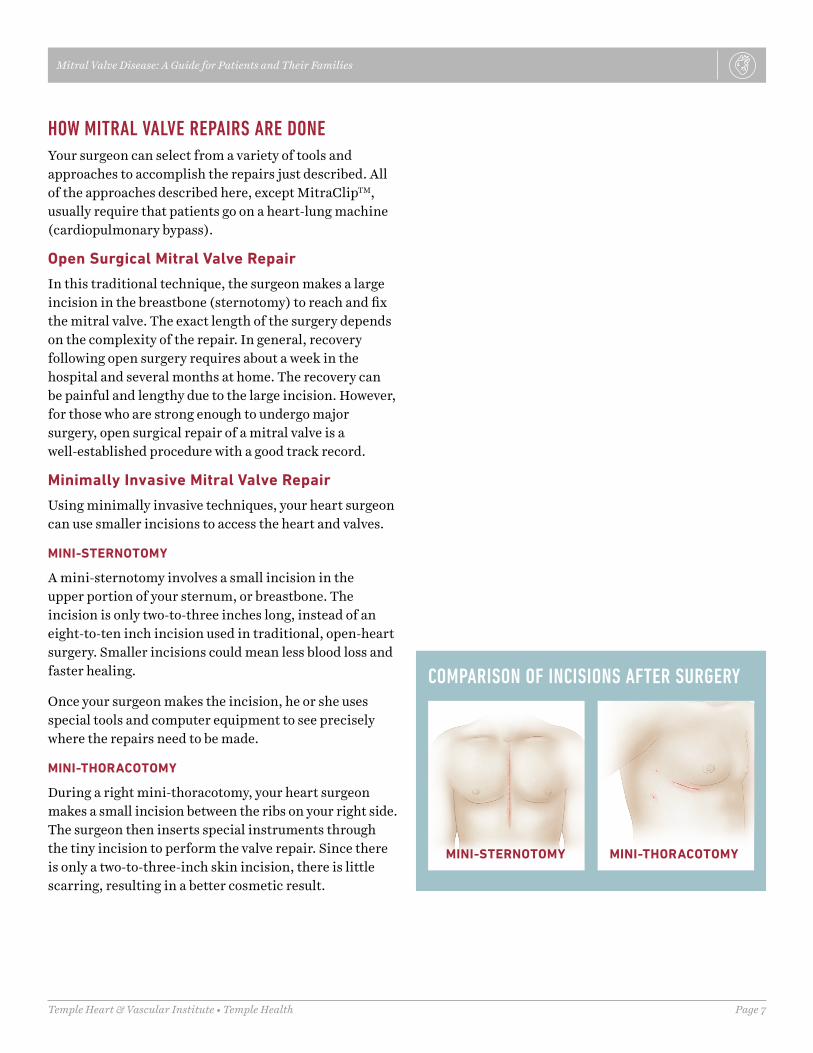
MINI-STERNOTOMY
A mini-sternotomy involves a small incision in the upper portion of your sternum, or breastbone. The incision is only two-to-three inches long, instead of an eight-to-ten inch incision used in traditional, open-heart surgery. Smaller incisions could mean less blood loss and faster healing.
Once your surgeon makes the incision, he or she uses special tools and computer equipment to see precisely where the repairs need to be made.
MINI-THORACOTOMY
During a right mini-thoracotomy, your heart surgeon makes a small incision between the ribs on your right side. The surgeon then inserts special instruments through the tiny incision to perform the valve repair. Since there is only a two-to-three-inch skin incision, there is little scarring, resulting in a better cosmetic result.
COMPARISON OF INCISIONS AFTER SURGERY
MINI-STERNOTOMY MINI-THORACOTOMY
Temple Heart & Vascular Institute • Temple Health Page 8
Mitral Valve Disease: A Guide for Patients and Their Families
Percutaneous Repair
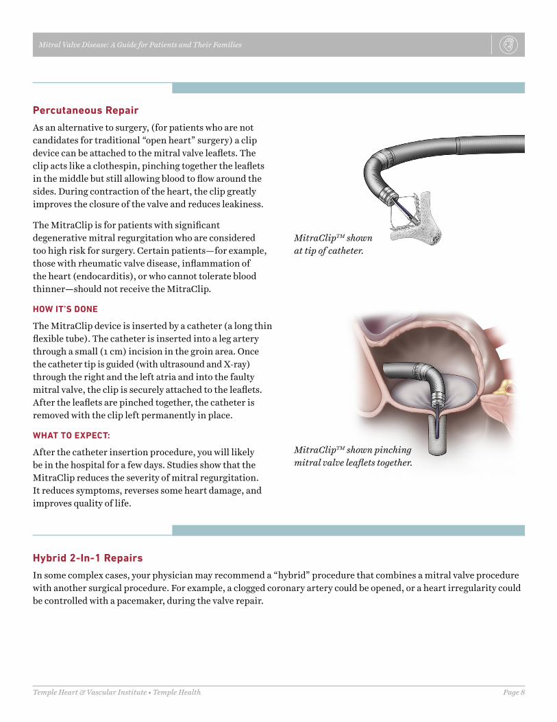
As an alternative to surgery, (for patients who are not candidates for traditional “open heart” surgery) a clip device can be attached to the mitral valve leaflets. The clip acts like a clothespin, pinching together the leaflets in the middle but still allowing blood to flow around the sides. During contraction of the heart, the clip greatly improves the closure of the valve and reduces leakiness.
The MitraClip is for patients with significant degenerative mitral regurgitation who are considered too high risk for surgery. Certain patients—for example, those with rheumatic valve disease, inflammation of the heart (endocarditis), or who cannot tolerate blood thinner—should not receive the MitraClip.
HOW IT’S DONE
The MitraClip device is inserted by a catheter (a long thin flexible tube). The catheter is inserted into a leg artery through a small (1 cm) incision in the groin area. Once the catheter tip is guided (with ultrasound and X-ray) through the right and the left atria and into the faulty mitral valve, the clip is securely attached to the leaflets. After the leaflets are pinched together, the catheter is removed with the clip left permanently in place.
WHAT TO EXPECT:
After the catheter insertion procedure, you will likely be in the hospital for a few days. Studies show that the MitraClip reduces the severity of mitral regurgitation. It reduces symptoms, reverses some heart damage, and improves quality of life.
MitraClipTM shown at tip of catheter.
MitraClipTM shown pinching mitral valve leaflets together.
Hybrid 2-In-1 Repairs
In some complex cases, your physician may recommend a “hybrid” procedure that combines a mitral valve procedure with another surgical procedure. For example, a clogged coronary artery could be opened, or a heart irregularity could be controlled with a pacemaker, during the valve repair.
Temple Heart & Vascular Institute • Temple Health Page 9
Mitral Valve Disease: A Guide for Patients and Their Families
WHY CHOOSE TEMPLE HEART & VASCULAR INSTITUTE?Temple cardiovascular specialists are highly experienced in all types of mitral valve repair surgeries and procedures. They are leaders in minimally invasive methods. They also have the special skills and facilities needed for hybrid heart procedures involving mitral valve repair.
Our faculty continue to pioneer new surgical repair methods. They excel at finding new solutions for patients with complex or serious valve conditions and often can offer novel therapeutic options.
Although our surgeons and interventional cardiologists are leaders in minimally invasive valve procedures, they realize every patient is different, and they always evaluate patients carefully before recommending the most appropriate treatment.
The Temple Heart & Vascular Institute emphasizes comprehensive and supportive care for our patients throughout the surgical process. From your first visit to the end of your recovery process, we are committed to providing compassionate care.
Our cardiovascular specialists are available for immediate evaluations and second opinions. You can meet face-to-face with our physicians to discuss your options. If surgery is needed, they can tell you about the risks and benefits of your specific procedure.
OUR TEAM
CARDIOVASCULAR SURGERY
Yoshiya Toyoda, MD, PhDChief, Cardiovascular Surgery, Temple University Hospital
Director, Mechanical Circulatory Support, Temple University Hospital
Vice-Chair, Thoracic Medicine and Surgery
Co-Surgical Director, Temple Heart & Vascular Institute
Surgical Director, Heart and Lung Transplantation, Temple University Hospital
William Maul Measey Chair in Cardiothoracic Surgery and Professor, Surgery, Lewis Katz School of Medicine at Temple University
Robert S. Boova, MDChief, Cardiovascular Surgery, Jeanes Hospital
Vice Chief, Cardiovascular Surgery, Temple University Hospital
Clinical Professor of Surgery, Lewis Katz School of Medicine at Temple University
Ravishankar Raman, MBBSAssistant Professor, Surgery, Lewis Katz School of Medicine at Temple University

Temple Heart & Vascular Institute • Temple Health Page 10
Mitral Valve Disease: A Guide for Patients and Their Families
CARDIOVASCULAR IMAGING
Daniel Edmundowicz, MS, MDChief, Cardiology, Temple University Hospital
Medical Director, Temple Heart & Vascular Institute
Professor & Vice Chair of Program Development, Department of Medicine, Lewis Katz School of Medicine at Temple University
Andrew Chen, MDAssistant Professor, Medicine, Lewis Katz School of Medicine at Temple University
Martin G. Keane, MDMedical Director, Echocardiography Laboratory
Director, Echocardiography Research/Quantitative Echo, Temple University Hospital
Professor, Medicine, Lewis Katz School of Medicine at Temple University
John Panidis, MDProfessor, Medicine, Lewis Katz School of Medicine at Temple University
Pravin V. Patil, MDAssistant Professor, Medicine
Associate Director, Cardiovascular Disease Training Program, Lewis Katz School of Medicine at Temple University
William Van Decker, MDDirector, Nuclear Cardiology, Temple University Hospital
Professor, Medicine, Lewis Katz School of Medicine at Temple University
Susan E. Wiegers, MDSenior Associate Dean, Faculty Affairs
Senior Associate Dean, Graduate Medical Education
Professor, Medicine, Lewis Katz School of Medicine at Temple University
To schedule an appointment with one of our cardiovascular specialists, or for more information, call 800-Temple-MED.
INTERVENTIONAL CARDIOLOGY
Vikas Aggarwal, MD, MPHAssistant Professor, Medicine, Lewis Katz School of Medicine at Temple University
Riyaz Bashir, MDProfessor, Medicine, Lewis Katz School of Medicine at Temple University
Brian O’Murchu, MDAssociate Professor, Clinical Medicine, Lewis Katz School of Medicine at Temple University
Brian O’Neill, MDAssistant Professor, Medicine, Lewis Katz School of Medicine at Temple University
Temple Heart & Vascular Institute • Temple Health Page 11
Mitral Valve Disease: A Guide for Patients and Their Families
CARDIOVASCULAR IMAGING
Daniel Edmundowicz, MS, MDChief, Cardiology, Temple University Hospital
Medical Director, Temple Heart & Vascular Institute
Professor & Vice Chair of Program Development, Department of Medicine, Lewis Katz School of Medicine at Temple University
Andrew Chen, MDAssistant Professor, Medicine, Lewis Katz School of Medicine at Temple University
Martin G. Keane, MDMedical Director, Echocardiography Laboratory
Director, Echocardiography Research/Quantitative Echo, Temple University Hospital
Professor, Medicine, Lewis Katz School of Medicine at Temple University
John Panidis, MDProfessor, Medicine, Lewis Katz School of Medicine at Temple University
Pravin V. Patil, MDAssistant Professor, Medicine
Associate Director, Cardiovascular Disease Training Program, Lewis Katz School of Medicine at Temple University
William Van Decker, MDDirector, Nuclear Cardiology, Temple University Hospital
Professor, Medicine, Lewis Katz School of Medicine at Temple University
Susan E. Wiegers, MDSenior Associate Dean, Faculty Affairs
Senior Associate Dean, Graduate Medical Education
Professor, Medicine, Lewis Katz School of Medicine at Temple University
To schedule an appointment with one of our cardiovascular specialists, or for more information, call 800-Temple-MED.
INTERVENTIONAL CARDIOLOGY
Vikas Aggarwal, MD, MPHAssistant Professor, Medicine, Lewis Katz School of Medicine at Temple University
Riyaz Bashir, MDProfessor, Medicine, Lewis Katz School of Medicine at Temple University
Brian O’Murchu, MDAssociate Professor, Clinical Medicine, Lewis Katz School of Medicine at Temple University
Brian O’Neill, MDAssistant Professor, Medicine, Lewis Katz School of Medicine at Temple University