mixed complexes of cobalt(iii) with phenanthroline and galactose or arabinose
TRANSCRIPT

Polyhedron 20 (2001) 2223–2230
Mixed complexes of cobalt(III) with phenanthroline and galactoseor arabinose
Jose Parada a, Sergio Bunel a,*, Exequiel Moraga a, Carmen Ibarra a,Nicholas D. Gillitt b, Clifford A. Bunton b
a Departamento de Quımica Inorganica y Analıtica, Facultad de Ciencias Quımicas y Farmaceuticas, Uni�ersidad de Chile, Casilla 233,Santiago, Chile
b Department of Chemistry and Biochemistry, Uni�ersity of California, Santa Barbara, CA 93106, USA
Received 9 October 2000; accepted 26 April 2001
Abstract
Mixed complexes of bis-phenanthroline cobalt(III) and �-D-galactose or �-D- or L-arabinose are identified in aqueous solutionfrom their 1H NMR and circular dichroism (CD) spectra. Galactose forms only the �-complex, but D-arabinose givespreferentially �-, and L-arabinose preferentially �-complexes, consistent with structural optimization with PM3 (tm) parameters.The �-complex is formed initially from �-D-arabinose, but the �-complex is preferred thermodynamically. Examination of theabsorption and CD spectra gives information on configuration at cobalt(III) and allows assignments of electronic transitions inCo(III) and the phenanthroline residues. © 2001 Elsevier Science Ltd. All rights reserved.
Keywords: Cobalt(III) complexes; Carbohydrate cobalt complexes; Circular dichroism; Galactose and arabinose complexes; Stereoselectivereactions; Sugar complexes; 1H NMR
www.elsevier.com/locate/poly
1. Introduction
Sugars and related polyols exhibit a rich chemistry informing a variety of complexes with metal ions, which,in favorable cases, can be isolated as stable solids,although evidence of complexation is, often due tonecessity, based on reaction mixtures in solution [1–3].Mixed complexes involving a sugar and another ligand,e.g. a diamine, have also been characterized. We haveidentified mononuclear complexes of sugars and 1,10-phenanthroline (phen) with cobalt(III), with the com-position Co(III)(phen)2sugar [4]. The configuration atCo(III) is given by the circular dichroism (CD) oroptical rotatory dispersion (ORD) spectra [5]; NMRspectroscopy provides additional information on theconformation and carbohydrate–metal ion bondingsites [4,6]. The 1H NMR signals of phen are wellseparated from those of the sugar, and Co(III) is dia-magnetic. The relative rigidity of phenanthroline sim-plifies structural modeling.
Complexes are prepared in water from the sugar and[Co(III)(phen)2Cl2]Cl:
[Co(phen)2Cl2]+ ���H2O
pH 8[Co(phen)2(H2O)(OH)]2+
���sugar
pH 8[Co(phen)2(sugar)]2+
To date we have used this method with keto andamino-sugars with, in some cases, isolation of com-plexes, typically as sparingly soluble salts with I3
− [4,7].Where these mixed complexes can be isolated theirspectral properties are the same as those of complexesprepared ‘in situ’ without isolation.
In the present work we examine complexes of alde-hydo-sugars in solution by observing their 1H NMRspectra and separating the complexes chromatographi-cally, and comparing their CD and spectra with thoseof products ‘in situ’. We relate the spectral evidence tostructures based on semi-empirical structural optimiza-tion. The free sugars have no CD signals in the area ofinterest, Co(III) bis-phenanthroline complexes areracemic and decomposition does not introduce extrane-ous CD signals.
* Corresponding author. Fax: +56-2-737-0567.E-mail address: [email protected] (S. Bunel).
0277-5387/01/$ - see front matter © 2001 Elsevier Science Ltd. All rights reserved.PII: S 0 2 7 7 -5387 (01 )00826 -9
J. Parada et al. / Polyhedron 20 (2001) 2223–22302224
For ‘in situ’ preparation we allowed complexes toform under conditions which reduce formation ofCo(II) derivatives, which give extensive NMR linebroadening, a major problem with these aldehydo-sug-ars. The H-1 and H-2 1H NMR signals of the com-plexes are separated from those of the sugars, andprovide information on the anomeric composition ofthe diastereomeric complexes [6].
Examination of the CD spectra identifies configura-tion at Co(III) and allows assignment of electronic
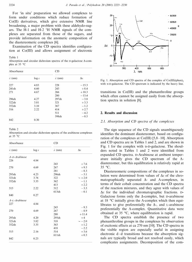
Fig. 1. Absorption and CD spectra of the complex of Co(III)(phen)2
with �-D-galactose. The CD spectrum is indicated by the heavy line.
Table 1Absorption and circular dichroism spectra of the D-galactose �-com-plex at 35 °C
CDAbsorbance
� (nm)log � ��� (nm)
−15.3227 4.83 228241sh +8.42434.60
266 +10.34.67271−13.4280
4.27296sh 297sh −3.8322sh 3.81 321 +3.3
−1.2367352sh 3.102.22516 414 +2.7
−4.2511598sh −0.5
0.30842
transitions in Co(III) and the phenanthroline groupswhich often cannot be assigned easily from the absorp-tion spectra in solution [8].
2. Results and discussion
2.1. Absorption and CD spectra of the complexes
The sign sequence of the CD signals unambiguouslyidentifies the dominant diastereomer, based on configu-ration of the complexes at Co(III) [5,8–10]. Absorptionand CD spectra are in Tables 1 and 2, and are shown inFig. 1 for the complex with �-D-galactose. The shoul-ders noted in Tables 1 and 2 were identified fromexpanded CD spectra. D-Arabinose at ambient temper-ature initially gives the CD spectrum of the �-diastereomer, but this equilibration is relatively rapid at35 °C.
Diastereomeric compositions of the complexes in so-lution were determined from values of �� of the chro-matographically separated �- and �-complexes, interms of their cobalt concentration and the CD spectraof the reaction mixtures, and they agree with values of�� for the individual chromatographic fractions. D-Galactose forms only the �-complex, but D-arabinoseat 18 °C initially gives the �-complex which then equi-librates to give preferentially the �-, and L-arabinosepreferentially the �-complex. Quantitative data wereobtained at 35 °C, where equilibration is rapid.
The CD spectra establish the presence of twophenanthroline groups in the complexes by observationof excitonic effects at ca 270 nm [10]. The CD spectra inthe visible region are especially useful in assigningelectronic d–d transitions because the absorption sig-nals are typically broad and not resolved easily, whichcomplicates assignments. Decomposition of the com-
Table 2Absorption and circular dichroism spectra of the arabinose complexesat 35 °C
CDAbsorbance
log � � (nm) ��� (nm)
�-D-Arabinose226 4.84
243 +7.6270 +9.52674.69
281 −8.34.23293sh 296sh −3.13.76321sh 322 �+2.03.35 362 −1.1352sh
413 +2.22.22515 512 −3.5
613sh −0.1840 0.27
�-L-Arabinose4.84227
243 −9.3−8.92674.69270
280 +11.4293sh 4.20 295sh +4
3.82321sh 318 �−2.0352sh 3.35 363 +1.2
−2.24182.16 514515 +3.6
+0.3611sh0.23842
J. Parada et al. / Polyhedron 20 (2001) 2223–2230 2225
plexes and the presence of excess sugar do not affectlocations of the CD signals.
2.2. Analysis of electronic transitions
Absolute configurations at Co(III) are given by thesign sequence of the CD ���* transitions at 266 and280 nm due to the excitonic effect [8–10] under the pband of phenanthroline at ca 271 nm in chiral com-plexes with two or more of these ligands (Tables 1 and2). In the following discussion we consider CD signalsof complexes with the D-sugars unless specified.
There are CD signals at 241–243 nm that correspondto a long axis polarized transition of phenanthroline, ��,[11] and are not observed in the absorption spectra, dueto overlap with other signals. This electronic transitionbecomes optically active whenever there are at least twocis-phenanthroline ligands [12,13], as in our complexes,and the transition gives a shoulder at the foot of the �band at 223 nm and is without optical activity. Theabsorption band at 227 nm could be due to the shortaxis polarized � band that gives rise to a negative CDsignal at 228 nm with the D-galactose complex, but wasnot observed in the complexes with D- or L-arabinose.The �-band of phenanthroline, seen in the expandedspectra, appears as a shoulder at 296 nm on one side ofthe p band in both the absorption and CD spectra. ThisCD signal appears in complexes containing one ormore phenanthroline ligands and, depending on theresolution of the CD spectrometer, the vibronic natureof the � band can be seen [9].
The p and � bands of phenanthroline partially over-lap the absorption spectrum of Co(III) at ca 350 nm.However, there are two CD signals at 318–322 nm(positive) and 362–367 nm (negative) assigned to the1T2g state of the CoN4O2 system [14] (N and O denotenitrogen and oxygen ligands, respectively). The low ��
(1–3) is understandable because, in the classical de-scription, the magnetic moment is zero for strict octahe-dral symmetry. All the Co(III) complexes with twophenanthroline ligands and one carbohydrate, aldose,ketose or an amino sugar, exhibit only one visibleabsorption band [4,7]. In the present complexes it iscentered at 515–516 nm. Two CD signals of oppositesigns appear at 414 and 511 nm in the D-galactosecomplex. The band with negative sign is located almostdirectly under the absorption maximum. These two CDsignals probably correspond to the 1T1g splitting due torhombic distortion of the metal ion.
In these CD spectra a weak negative signal appearsnear the edge of the band at 511–514 nm, and wasresolved by gaussian analysis. Its location at ca 600 nmand �� value allows us to ascribe it to the 3T2g state ofCo(III). A very weak, low-energy absorption at 840 nmcould correspond to the 3T1g state of Co(III). Locations
of these signals coincide with those of spin forbiddenbands of complexes with similar ligands around Co(III)[14].
We can derive values of the repulsion parameters andcontributions of galactose and arabinose to perturba-tions of the d orbitals of Co(III) from these spectraldata [14]. For example for �-[Co(phen)2D-galactose](Table 1): 1A1g�1T1g=19 379 cm−1; 1A1g�3T1g=11 876 cm−1 and 1T1g�3T1g=2C, C=3751 cm−1.Similarly for �-[Co(phen)2D-arabinose] we obtain C=3756 cm−1.
A decrease in the value of C is a measure of theradial expansion of the d orbitals, and comparison ofits values in the sugar complexes with 3720 cm−1 for[Co(NH3)4CO3]+ [14] shows that in our complexes ex-pansion through delocalization over the ligands is rela-tively unimportant.
Assuming that the complexes obey D4h holohedrizedsymmetry corresponding to CoN4O2, the Dq values ofthe carbohydrates can be obtained by the ‘averageenvironment’ empirical rule. Taking 2410 cm−1 as theDq [14] value for phenanthroline, we estimate Dq val-ues of 2119 cm−1 for D-galactose and 2131 cm−1 forD-arabinose. These calculated values reflect the averageDq values produced by the oxygen atoms bonded toCo(III) which, in these complexes, correspond to O−
and OH, because the predominant species in solutionare dicationic and their Dq values are similar to thoseobtained with H2O, CO3
2− and oxalate ligands [14].Calculation of the energy levels corresponding to the
1T1g state by the ligand field method [15,16] yields: 1A2
— 5Dq0+5DqN−C and 1E — 3(Ds0−DsN)+25/7Dq0+45/7DqN−C where Ds, Dq, and C relate tocrystal field parameters. As DqN�Dq0, and 3(Ds0−DsN)=15/84(Dq0−DqN), the 1A2 state has the lowerenergy [15,17]. The negative CD band at 511–514 nmcorresponds to the 1A2 state, whereas that at 413–421nm, positive, should correspond to the 1E state.
In this and the other compounds it is the 1A2 statethat correlates with the sign sequence of the excitoniceffect at ca 270 nm and with the absolute configurationof the complex [5,8–10].
There are differences in literature assignments ofsome UV absorption bands of phenanthroline ligands[18]. Our assignments are in essential agreement withthe literature [9,11], and we have identified all thetransitions which generate CD signals of our, and re-lated complexes.
2.3. NMR spectroscopy
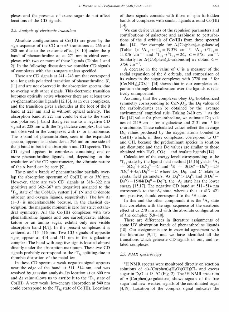
1H NMR spectra were monitored directly on reactionsolutions of cis-[Co(phen)2(H2O)(OH)]Cl2 and excesssugar in D2O at 18 °C (Fig. 2). The 1H NMR spectrumof �-[Co(phen)2D-galactose] shows signals of the freesugar and new, weaker, signals of the coordinated sugar[4,19]. Location of the complex signal indicates the

J. Parada et al. / Polyhedron 20 (2001) 2223–22302226
presence of the �-anomer of galactose in its 4C1 pyra-nose form [6,20,21]. There is a doublet with the appro-priate coupling constant in the region of the anomeric�-hydrogen of the sugar that is absent in the spectrumof the �-anomeric sugar (Table 3). The COSY spectrum
allows assignment of the signal of H-2 of the coordi-nated sugar with a 0.3 ppm high field shift relative tothe free sugar (Table 3). The �-anomer coordinates as abidentate ligand through the 1-O− and 2-OH groups.The J1,2 coupling constant is similar to that of the free
Fig. 2. 1H NMR spectra of the complexes and parent sugars: (A) with �-D-galactose; (B) with �-D-arabinose after 6 days at 18 °C; and (C) asB after 15 days.
Table 3Chemical shifts � (ppm) of the complexes and parent sugars a
�-D-GalactoseCo(III) �-D-ArabinoseCo(III) �-L-ArabinoseCo(III)
�� �� �
5.40 (2.9)5.39 (2.7) 5.375.37 (4.2)5.33 (2.9)H-1H-2 3.54 3.603.60
�-D-Arabinose �-D-Arabinose�-D-Galactose�-D-Galactose
5.31 (3.2)H-1 5.30 (3.3) 4.62 (7.8) 4.59 (7.7)3.90H-2 3.87 3.53 3.58
a Referred to TSP, �=0 ppm; values in parentheses are J (Hz).
J. Parada et al. / Polyhedron 20 (2001) 2223–2230 2227
sugar, which, from the Karplus or related equations[6d,20,21], indicates only a small change in the dihedralangles of the O−, OH groups (Table 3).
The 1H NMR spectra of the equilibrated complexeswith D- and L-arabinose are essentially the same. Weinitially saw, at 18 °C, signals of only one complex,which from the CD signals was the �-diastereomer, butwith time a second set of signals of the �-diastereomerappeared. These signals were broadened slightly due todecomposition generating Co(II). The new complexsignals were assigned as for the galactose complex.From relative intensities we relate them to the �-D (or�-L) and �-L (or �-D) diastereomers, respectively, ofthe � sugars in the pyranose 1C4 form [6,20,21] (Fig. 2and Table 3). From the COSY spectra we identify theH-2 signals of the coordinated sugar in the predomi-nant equilibrated diastereomer, i.e. the �-anomer withD-arabinose and the �-anomer with L-arabinose. Wecould not identify H-2 signals in the COSY spectra ofthe minor �-[Co(phen)2D-arabinose] and �-[Co(phen)2-L-arabinose] complexes, because they are overlappedwith signals of the predominant isomer, or of the sugar(Table 3).
The intensities of the complex signals were muchlower than those of the parent sugars, even after 2weeks (Fig. 2). The complexes equilibrate with thesugar and Co(phen)2 and dissociation is favored in ourconditions.
The 1H NMR signals of H-1 and H-2 in complexeswith D-galactose and D- or L-arabinose are shiftedsimilarly, relative to those of the free sugars (Table 3),consistent with bonding with cis–ax–eq groups whichfavors coordination without disruption of the sugarconformation [1,2]. The ease of deprotonation of 1-OHrelative to the other OH groups [22] favors preferentialcoordination by axial 1-O− and equatorial 2-OHgroups to the metal ion. Examination of the 1H NMRspectra of the complexes was limited to H-1 and H-2 ofthe sugar residue because the remaining signals over-lapped those of the free sugar.
2.4. Structural optimization
Structures of complexes with cis-1-O− and 2-OHgroups were optimized by using SPARTAN (wavefunc-tion) software with semi-empirical PM3 (tm) parame-ters which extend the PM3 basis set [23] to transitionmetal ions [24]. The PM3 (tm) parameters had beenused to treat structures of complexes of amino sugarswhich were similar to, but not identical to those ob-tained earlier with MM2 parameters [4,7]. Applicationsof semi-empirical structural treatments have been criti-cally reviewed [25].
Aquo complexes of Co(III) typically have pKa�6,and anomeric hydroxyl groups are considerably moreacidic than water [22]. Potentiometric determination of
the pKa of our chromatographically isolated complexes(Section 2) gives values of ca 3. We therefore simulatedthe structures of the dicationic complexes, althoughpredicted structures are similar for tricationic com-plexes. Hydration is neglected on the assumption that itwill affect �- and �- complexes similarly, to a firstapproximation.
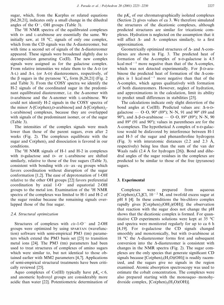
Geometrically optimized structures of �- and �-com-plexes are shown in Fig. 3. The predicted heat offormation of the �-complex of �-D-galactose is 4.5kcal mol−1 more negative than that of the �-complex,which was not detected experimentally. For �-D-ara-binose the predicted heat of formation of the �-com-plex is 1 kcal mol−1 more negative than that of the�-complex, which agrees qualitatively with formationof both diastereomers. However, neglect of hydration,and approximations in the calculation, limit its abilityto predict small differences in stabilities [25].
The calculations indicate only slight distortion of thebond angles at Co(III). Predicted values are: �-�-D-galactose — O�O, 90° (89°); N�N, 91 and 90° (89 and90°), and �-�-D-arabinose — O�O, 89° (89°); N�N, 90and 89° (91 and 90°); values in parentheses are for the�-complexes. The hypothetical � complex of �-D-galac-tose would be disfavored by interference between H-1and H-3 of the sugar and phenanthroline hydrogens(Fig. 3) with interatomic distances (2.2 and 2.3 A� ,respectively) being less than the sum of the van derWaals radii (2.4 A� for hydrogen [26]). Bond and dihe-dral angles of the sugar residues in the complexes arepredicted to be similar to those of the free (pyranose)sugars.
3. Experimental
Complexes were prepared from aqueous[Co(phen)2Cl2]Cl, 10−2 M, and twofold excess sugar atpH 8 [4]. In these conditions the bis-chloro complexrapidly gives [Co(phen)2(OH2)(OH)]; the observationthat reaction with the sugar does not change the pHshows that the dicationic complex is formed. For quan-titative CD experiments solutions were kept at 35 °Cand spectra were monitored regularly for up to ca 6 h[4,19]. For D-galactose the CD signals changedsmoothly and monotonically, but with D-arabinose at18 °C the �-diastereomer forms first and subsequentconversion into the �-diastereomer is consistent withchanges in the NMR spectra (Fig. 2). The sugar com-plexes are the only species that generate significant CDsignals because [Co(phen)2(H2O)(OH)] is readily racem-ized, and the sugars give no signals in the regionexamined. Atomic absorption spectroscopy was used toestimate the cobalt concentration. The complexes werealso prepared starting with the monoaquo–monohy-droxide complex, [Co(phen)2(H2O)(OH)].

J. Parada et al. / Polyhedron 20 (2001) 2223–22302228
Fig. 3. Optimized structures of the complexes of the D-sugars, with bonding to Co(III) at positions 1 and 2. Oxygen 1 is behind the plane of thepaper in all the structures except that of the �-D-arabinose complex.
In earlier work with amino-sugars we isolated andanalyzed complexes with the structure [Co(phen)2-aminosugar] (I3)3, which are sparingly soluble in water[4,7], but with D-galactose and the arabinoses, isolationof the respective triiodides gave mixtures of di- andtri-cations, as shown by elemental analyses. Because wecould not isolate solid complexes we used freshly chro-matographed solutions for examination of absorptionand CD spectra.
3.1. Chromatography
The �- and �-complexes were separated in solutionby following the general methods used for other com-plexes [4,19] with a Sephadex C25 column and elutionwith 0.1 M NaCl.
3.2. Determination of pKa
Potentiometric titration [22] of freshly separated frac-tions of the complexes and complex concentration gavepKa�3.0 for the D-galactose complex and 2.9 for theD- or L-arabinose complexes.
3.3. Spectrophotometry
Aqueous solutions of sugar and [Co(phen)2C12]Clwere mixed and absorption and CD spectra were mon-itored over time. Absorption and CD spectra wereobtained as rapidly as possible with chromatographi-cally separated material after elution of the complexes.The CD spectra were monitored in a Jobin-Yvon CD 6spectrometer and absorption spectra were monitored inthis instrument or in a Unicam UV3 spectrometer.

J. Parada et al. / Polyhedron 20 (2001) 2223–2230 2229
3.4. NMR spectroscopy
1H NMR spectra were obtained on the reactionsolutions (2×10⁻² M) in D2O (with solvent suppres-sion) in a Bruker DRX 00 instrument (300 MHz for1H), and are referred to TSP, �=0 ppm. In order tolimit reduction of Co(III) we used only twofold excesssugar at 18 °C and left solutions for at least 1 week. Inthese conditions there is limited formation of the com-plexes and we could not identify signals which areobscured by those of the excess sugars. Over severalweeks at 18 °C these mixed complexes disproportionateforming (Co(III)phen3)3+ and Co(II) due to reductionby the aldehydo-sugar which broadens the NMR sig-nals [4]. The signals of H-2 of the galactose and ara-binose complexes were identified from the 1H COSYspectra in D2O, but there is extensive overlap betweenother signals and those of the sugars and we did notattempt to identify them. We saw a small signal, proba-bly of a galactofuranose at 5.26 ppm, which is veryevident in DMSO [20,21]. The 1H NMR spectrum ofthe complex of �-D-arabinose (Fig. 2) is consistent withevidence from the CD signals which shows that the�-complex is initially formed preferentially and, at18 °C it equilibrates with the �-complex although line-broadening due to formation of Co(II) limits the quan-titative utility of NMR spectroscopy. Thereproducibility of the NMR data is shown by thefollowing H-1 chemical shifts, �, and coupling con-stants, J, for D- and L-xylose anomeric mixtures. Valuesfor the �-anomer are in parentheses: D-xylose — �,5.20 (4.60) ppm; J, 3.6 (7.8) Hz; L-xylose — �, 5.22(4.60) ppm; J, 3.6 (7.8) Hz. Chemical shifts and cou-pling constants are identical for the D- and L-arabinoseanomers and, as for galactose, agree with the literaturevalues [20,21]. Data for the sugars are given for com-parison with those for the complexes.
3.5. Structure optimization
Calculations were with PM3 (tm) parameters (wave-function, SPARTAN Pro), following an MMFF confor-mational search [23–25]. Initial structures wereperturbed, followed by resimulation, to test for falseminima [1b].
4. Conclusions
Examination of the CD spectra of complexes ofCo(phen)2 with D-galactose or D- and L-arabinose pro-vides evidence on the stoichiometry and configurationof these complexes in solution. It allows assignment ofthe electronic transitions and comparison with those forother Co(III)(phen)2 complexes. The absorption spectraare less readily assigned because of overlap of some
bands and possible interference by racemic productsgenerated by decomposition. There is considerableoverlap of 1H NMR signals of the parent sugars andthe complexes, but those at the anomeric center provideinformation on the ligating groups of the sugar whichcan be compared with that from structural optimizationbased on semi-empirical PM3 (tm) parameters. How-ever, reduction of Co(III) by aldehydo-sugars compli-cates quantitative use of NMR spectroscopy.
The combined use of CD and NMR spectra providesevidence on structures of complexes which exist insolution but cannot be isolated as crystalline solids, andshows that the �-complex of D-arabinose is formedinitially under kinetic control. We saw only the �-com-plex of D-galactose in all conditions. The �-complex ofD-arabinose is the kinetically preferred diastereomer atambient temperature, but it gradually equilibrates withthe �-complex, and equilibration is relatively fast at35 °C which was therefore used for the quantitativeCD measurements.
Acknowledgements
We acknowledge instrumental support from CE-PEDEQ, Facultad de Ciencias Quımicas y Farmaceuti-cas, Universidad de Chile and Professor Ines Ahumada.J.P. acknowledges support from FONDECYT Project2970036.
References
[1] (a) D.W. Whitfield, S. Stojkovski, B. Sarkar, Coord. Chem. Rev.122 (1993) 171;(b) M. Zimmer, Chem. Rev. 95 (1995) 2629;(c) S. Yano, Coord. Chem. Rev. 92 (1988) 113;(d) B. Gyurcsik, L. Nagy, Coord. Chem. Rev. 203 (2000) 81.
[2] (a) H. Kozlowski, P. Decock, I. Olivier, G. Micera, A. Pucino,L.D. Pettit, Carbohydr. Res. 197 (1990) 109;(b) J.M. Harrowfield, M. Mocerino, B.W. Skelton, W. Wei,A.H. White, J. Chem. Soc., Dalton Trans. (1995) 783;(c) R.P. Bandwar, M.D. Sastry, R.M. Kadam, C.P. Rao, Carbo-hydr. Res. 297 (1997) 333;(d) S.J. Angyal, Adv. Carbohydr. Chem. Biochem. 47 (1989) 1.
[3] K. Hegetschweiler, Chem. Soc. Rev. 28 (1999) 239.[4] S. Bunel, C. Ibarra, E. Moraga, J. Parada, A. Blasko, C.
Whiddon, C.A. Bunton, Carbohydr. Res. 312 (1998) 191 (andreferences therein).
[5] (a) L.J. Katzin, J. Eliezer, Coord. Chem. Rev. 7 (1972) 331;(b) in: K. Nakanishi, N. Berova, R.W. Woody (Eds.), CircularDichroism, Principles and Applications, VCH, Cambridge, 1994.
[6] (a) J. Keeler, Chem. Soc. Rev. 19 (1990) 381;(b) M. Jaseja, A.S. Perlin, P. Dais, Magn. Reson. Chem. 28(1990) 283;(c) F. Franks, Pure Appl. Chem. 59 (1987) 1189;(d) C. Altona, G.A. Haasnoot, Org. Magn. Reson. 13 (1980)417.
[7] (a) A. Blasko, C.A. Bunton, E. Moraga, S. Bunel, C. Ibarra,Carbohydr. Res. 278 (1995) 315;

J. Parada et al. / Polyhedron 20 (2001) 2223–22302230
(b) J. Parada, S. Bunel, C. Ibarra, G. Larrazabal, E. Moraga,N.D. Gillitt, C.A. Bunton, Carbohydr. Res. 329 (2000) 195.
[8] J. Ferguson, C.J. Hawkins, N.A.P. Kane-Maguire, H. Lip, In-org. Chem. 8 (1969) 771.
[9] S.F. Mason, B.J. Peart, J. Chem. Soc., Dalton Trans. (1973) 949.[10] B. Bosnich, Acc. Chem. Res. 2 (1969) 266 (and references
therein).[11] N. Sanders, P. Day, J. Chem. Soc. A (1970) 1190.[12] A. Tatehata, Inorg. Chem. 21 (1982) 2496.[13] T. Yasui, B.E. Douglas, Inorg. Chem. 10 (1971) 97.[14] V.S. Sastri, C.H. Langford, Can. J. Chem. 47 (1969) 4237.[15] R. Krishnamurthy, W.B. Schaap, J. Chem. Educ. 47 (1970)
433.[16] (a) C.E. Schaffer, C.K. Jorgensen, Mat. Fys. Medd. Dan. Vid.
Selsk. 34 (1965) 1;(b) J. Josephsen, C.E. Schaffer, Acta Chem. Scand., A 31 (1977)813.
[17] M. Ban, J. Csaszar, Acta Chim. Acad. Sci. Hung. 57 (1968)153.
[18] C.J. Hawkins, Absolute Configuration of Metal Complexes,Wiley, New York, 1971.
[19] A. Blasko, C.A. Bunton, S. Bunel, E. Moraga, C. Ibarra, J.Parada, Bol. Soc. Chil. Quim. 40 (1995) 449.
[20] P. Hobley, O. Howarth, R.N. Ibbett, Magn. Reson. Chem. 34(1996) 755.
[21] R.U. Lemieux, J.D. Stevens, Can. J. Chem. 44 (1966) 249.[22] (a) L.E. Bennett, R.H. Lane, M. Gilroy, F.A. Sedor, J.P. Ben-
nett, Inorg. Chem. 12 (1973) 1200;(b) T. Nishide, K. Ogino, J. Fujita, K. Saito, Bull. Chem. Soc.Jpn. 47 (1974) 3057.
[23] (a) J.J.P. Stewart, J. Comput. Chem. 10 (1989) 209;(b) J.J.P. Stewart, J. Comput. Chem. 11 (1990) 543.
[24] W.J. Hehre, J. Yu, Abstracts of the 221st ACS National Meet-ing, New Orleans, LA, March, 1996.
[25] I.N. Levine, Quantum Chemistry, 5th ed., Prentice Hall, UpperSaddle River, NJ, 2000 (chap. 17).
[26] L. Pauling, The Nature of the Chemical Bond, 3rd ed., CornellUniversity Press, Ithaca, NY, 1960 (p. 260).
.