mkp-3 has essential roles as a negative regulator of the ras
TRANSCRIPT

MOLECULAR AND CELLULAR BIOLOGY, Jan. 2004, p. 573–583 Vol. 24, No. 20270-7306/04/$08.00�0 DOI: 10.1128/MCB.24.2.573–583.2004Copyright © 2004, American Society for Microbiology. All Rights Reserved.
MKP-3 Has Essential Roles as a Negative Regulator of theRas/Mitogen-Activated Protein Kinase Pathway during
Drosophila DevelopmentMyungjin Kim,1 Guang-Ho Cha,1 Sunhong Kim,1 Jun Hee Lee,1 Jeehye Park,1
Hyongjong Koh,1 Kang-Yell Choi,2 and Jongkyeong Chung1*National Creative Research Initiatives Center for Cell Growth Regulation and Department of Biological Sciences,
Korea Advanced Institute of Science and Technology, Taejon 305-701,1 and Departmentof Biotechnology, Yonsei University College of Engineering,
Seoul 120-752,2 Korea
Received 20 June 2003/Returned for modification 4 August 2003/Accepted 20 October 2003
Mitogen-activated protein kinase (MAPK) phosphatase 3 (MKP-3) is a well-known negative regulator in theRas/extracellular signal-regulated kinase (ERK)-MAPK signaling pathway responsible for cell fate determi-nation and proliferation during development. However, the physiological roles of MKP-3 and the mechanismby which MKP-3 regulates Ras/Drosophila ERK (DERK) signaling in vivo have not been determined. Here, wedemonstrated that Drosophila MKP-3 (DMKP-3) is critically involved in cell differentiation, proliferation, andgene expression by suppressing the Ras/DERK pathway, specifically binding to DERK via the N-terminalERK-binding domain of DMKP-3. Overexpression of DMKP-3 reduced the number of photoreceptor cells andinhibited wing vein differentiation. Conversely, DMKP-3 hypomorphic mutants exhibited extra photoreceptorcells and wing veins, and its null mutants showed striking phenotypes, such as embryonic lethality and severedefects in oogenesis. All of these phenotypes were highly similar to those of the gain-of-function mutants ofDERK/rl. The functional interaction between DMKP-3 and the Ras/DERK pathway was further confirmed bygenetic interactions between DMKP-3 loss-of-function mutants or overexpressing transgenic flies and variousmutants of the Ras/DERK pathway. Collectively, these data provide the direct evidences that DMKP-3 isindispensable to the regulation of DERK signaling activity during Drosophila development.
The mitogen-activated protein kinase (MAPK) signalingpathway is critically involved in diverse biological processes,including mitogenesis, neuronal differentiation, apoptosis, anddevelopment (3, 37, 41). Core signaling modules of the MAPKpathway consist of MAPK kinase kinase, MAPK kinase, andMAPK. MAPK kinase kinase activates MAPK kinase by phos-phorylation, which, in turn, phosphorylates and activatesMAPK (3, 37, 41). Subsequently, activated MAPK translocatesinto the nucleus, where it phosphorylates various nuclear tar-gets leading to specific cellular processes (5, 30). In eukaryotes,three major MAPKs—extracellular signal-regulated kinase(ERK), p38, and c-Jun N-terminal kinase (JNK)—have beencharacterized and found to be highly conserved among variousspecies. These three MAPK signaling cascades convey abun-dant information to numerous target effectors in the cell, al-lowing various responses to the environment.
The specific roles of Ras/Drosophila ERK (DERK) signalingin cell differentiation and proliferation have been intensivelystudied in the developing eyes and wings of Drosophila (21, 22,25, 29, 51). A well-known step in the photoreceptor cell fatedetermination is the recruitment of the final R7 cell (2, 51, 54).In this step, Ras/DERK signaling acts as a binary switch totrigger one of the nonneuronal cone cells to differentiate intoa R7 neuronal cell. This was proved by a series of experiments
in which gain-of-function mutations of the components of theRas/DERK signaling pathway induce ectopic R7 cell differen-tiation in the absence of upstream inducing signals (4, 6, 38,39). For example, the rlsem mutant, containing an Asp334-to-Asn point mutation in DERK, showed additional photorecep-tor cells during Drosophila eye development. It was also dem-onstrated that the gain-of-function alleles of the componentsof the Ras/DERK pathway enhance wing vein formation (38,42). Consistently, downregulation of the Ras/DERK signalingpathway inhibits wing vein formation (12, 15, 23). Meanwhile,it was reported that the activated form of Ras was able to drivethe eye and wing imaginal discs to hyperplastic growth (27),implicating that Ras/DERK signaling is required for cell pro-liferation as well.
The duration and strength of the activities of Ras/MAPKsignaling are tightly regulated at many different levels withinthe pathway (3, 37, 41). Especially, the activity of MAPK isreversibly regulated by MAPK kinase-dependent phosphoryla-tion and MAPK phosphatase (MKP)-dependent dephosphor-ylation. The MKPs belong to a subclass of the dual-specificityphosphatase superfamily and dephosphorylate the critical thre-onine and tyrosine residues of MAPK (46, 56). Among MKPs,MKP 3 (MKP-3) tightly binds to ERK via the N-terminalERK-binding domain (NBD) and negatively modulates ERKactivities but not p38 and JNK (8, 19, 44, 45, 60). Recently, wehave demonstrated that Drosophila MKP-3 (DMKP-3) ishighly homologous to mammalian MKP-3 and plays specificand dominant roles in negatively regulating DERK activities inSchneider cells (32, 34). Therefore, understanding the function
* Corresponding author. Mailing address: Department of BiologicalSciences, Korea Advanced Institute of Science and Technology, 373-1Kusong-Dong, Yusong, Taejon 305-701, Republic of Korea. Phone:82-42-869-2620. Fax: 82-42-869-8260. E-mail: [email protected].
573
on February 16, 2018 by guest
http://mcb.asm
.org/D
ownloaded from

of DMKP-3 in Ras/DERK signaling at the organism levelwould provide a significant clue for deciphering the in vivo roleof the Ras/ERK signaling pathway in mammals.
To better understand how MKP-3 regulate Ras/DERK sig-naling in a cellular and developmental context, we investigatedthe physiological role of MKP-3 in Drosophila by using highlyconvenient genetic and histochemical methods. In the presentstudy, we characterized both the transgenic flies overexpress-ing DMKP-3 and the loss-of-function mutant flies of DMKP-3.The genetic analyses of these flies clearly demonstrated thatDMKP-3 functions as a negative regulator of the Ras/DERKpathway by directly inhibiting DERK-MAPK. Furthermore,we demonstrated that MKP-3 plays a critical role in the ERK-MAPK-mediated developmental processes, such as cell fatedetermination and cell proliferation in Drosophila.
MATERIALS AND METHODS
Fly strains. The fly strains, EP(3)3142, sev-PTP-ER101, RafHM7, RafF179,Rase1b, rl1, y/w; CyO, P[�2-3]/Bc Egfr, rhoAA69, rlsem, and UAS-lacZ and variousGAL4 lines such as sevenless (sev)- and apterous (ap)-GAL4 (7) were obtainedfrom the Bloomington Drosophila Stock Center (Bloomington, Ind.). The e16E-GAL4 line (24, 59) was kindly provided by J. Kim (Korea Advanced Institute ofScience and Technology). The MS1096-GAL4 driver line (9, 40) was a gift fromM. Freeman (Medical Research Council [United Kingdom]). All Drosophilastocks were maintained and cultured with standard cornmeal-yeast-agar mediumat 25°C, while RafHM7 hemizygous males were maintained at 18°C.
Plasmid construction and generation of transgenic flies. To induce ectopicexpression of DMKP-3, we used UAS/GAL4 system (49). A full-length ofDMKP-3 cDNA was generated by reverse transcription-PCR (RT-PCR) by usingthe 5� primer 5�-GCGAGATCTATGCCAGAAACGGAGCACGAG-3� and the3� primer 5�-CGCCTCGAGTCAGGCCGCATCCTCATCGTA-3� and thensubcloned into the BglII-XhoI site of the pUAST vector. The PCR-clonedDMKP-3 was confirmed by DNA sequencing. We also generated various mu-tants of DMKP-3—C302A (Cys302 to Ala302), R56/57A (Arg56 and Arg57 toAla56 and Ala57), and R56/57A/C302A—by site-directed mutagenesis (Strat-agene) and cloned them into pUAST vector (32, 34). �N, a truncated mutantlacking the NH2-terminal 170 amino acids, was also generated and cloned intopUAST. To obtain transgenic flies, various pUAST plasmids and a helper plas-mid, p�25.1, were microinjected into embryos prior to pole cell formation byusing a microinjector model IM30 (Narishige) and an Axiovert 25 micromanip-ulator (Carl Zeiss).
P-element mobilization-mediated DMKP-3 mutagenesis. To generateDMKP-3 loss-of-function mutants, we used the P-element local hopping mu-tagenesis by crossing EP(3)3142 with y/w; CyO, �2-3/Bc Egfr flies (52), as previ-ously described (31). We then isolated 2,000 independent lines with mobilized Pelements from 2.6 � 105 screened flies generated from this mutagenesis andfurther narrowed the total down to three DMKP-3 loss-of-function mutants:DMKP-3P1, DMKP-3P2, and DMKP-3P3. Their new P-element insertion sites weredetermined by PCR-based mapping and sequencing analyses (14). To isolate thehomozygous DMKP-3 mutant individuals, we used a green fluorescent protein(GFP) balancer chromosome (TM3, P{ActGFP}JMR2, Ser1) as a tracking mark-er; the GFP-negative embryos were selected as homozygous DMKP-3 mutants(10).
Analysis of eye and wing phenotypes. Scanning electron micrograph imageswere obtained by using a LEO 1455VP in a variable pressure secondary electronmode. Eye section experiments were performed as previously described (35). Toquantify the eye phenotypes presented in Fig. 2, we calculated the percentage ofommatidia with extra R cells, the ratio of the number of ommatidia with extra Rcells to the total number of ommatidia, in a single eye. In addition, the meanvalue of the number of photoreceptor cells per ommatidium was also calculatedfor each genotype. All of these experiments were independently performed byexamining �100 ommatidia per eye from 4 different flies with the same genotype.
Adult wing blades were mounted in 50% Canadian Balsam (Sigma) in methylsalicylate (Fischer). To quantify wing phenotypes, the mean number of extraveins per wing for each genotype was calculated from 110 different wings.
Feeding drugs. To test the effects of the MEK inhibitors U0126 (20) andPD98059 (1), larvae were fed a 100 �M or a 1 mM concentration of U0126 anda 250 or a 500 �M concentration of PD98059.
Immunostaing and histological analysis. Third-instar larvae were dissected inDrosophila Ringer’s solution, and eye and wing imaginal discs were fixed in 4%paraformaldehyde phosphate-buffered saline (PBS) solution for 30 min at roomtemperature. After being washed with PBS–0.1% Triton X-100 (PBST), the discswere blocked for 30 min at room temperature in PBS containing 3% bovineserum albumin. The discs were further incubated with primary antibodies(mouse anti-�-galactosidase [40-1a, The Developmental Study Hybridoma Bank,University of Iowa], goat anti-Sevenless [Santa Cruz Biotechnology], rat anti-Elav [rat-Elav-7E8A10; The Developmental Study Hybridoma Bank, Universityof Iowa], and mouse anti-phospho-specific DERK [Sigma]) and secondary anti-bodies conjugated to fluorescein isothiocyanate or TRITC (tetramethyl rhoda-mine isothiocyanate; Jackson Laboratories). The antibody-bound samples werewashed with PBS and mounted in 50% glycerol-PBS solution for confocal mi-croscopic observations.
X-Gal (5-bromo-4-chloro-3-indolyl-�-D-galactopyranoside) staining was per-formed as previously described (35).
Propidium iodide staining experiment. Eggs collected from the female flieswith DMKP-3P1 germ line clones were immersed in 50% bleach for 3 min fordechorionation and then rinsed repeatedly with PBST. The dechorionated eggswere agitated vigorously for 20 min in equal volume of 4% formaldehyde andheptane. The bottom formaldehyde phase of the solution was removed, and thenmethanol was added. The samples were shaken vigorously for 15 s, and theremaining heptane phase and the unsunken eggs were removed. After the sam-ples were washed with PBST, the devitellinized eggs were incubated with 10 mgof RNase/ml at 37°C for 2 h. After several washes in PBST, 50% glycerol-PBSmounting medium with 1 �g of propidium iodide/ml was added to the eggs. Theeggs in the mounting medium were transferred onto a glass microscope slide forconfocal microscopic observations.
BrdU labeling experiment. Third-instar larvae cultured at 25°C were dissectedin Ringer’s solution and then incubated in the presence of 100 �g of 5-bromo-2�-deoxyuridine (BrdU/ml; Roche) in M3 medium for 1 h at room temperature.The samples were fixed in Carnoy’s fixative (ethanol-acetic acid-chloroform[6:3:1]) for 30 min at 25°C and sequentially treated with 70% ethanol in PBST(0.3% Triton X-100 in 1� PBS), 50% ethanol in PBST, and 30% ethanol inPBST at 25°C for 3 min. Next, the samples were incubated in 2 N HCl for 1 h,and the incorporated BrdU was visualized by using an mouse anti-BrdU antibody(Roche) and Alexa 568 (Molecular Probe). The samples were observed under aconfocal laser microscope (Carl Zeiss).
In situ hybridization. For in situ RNA hybridization analysis, antisense andsense digoxigenin-labeled riboprobes were prepared by using T7 and SP6 RNApolymerases and linearized T vector–DMKP-3 as a template. Digoxigenin label-ing was performed according to the manufacturer’s instructions (Roche Bio-chemicals). In situ hybridization of Drosophila embryos and larval tissues wasconducted as previously described (16, 17).
Mitotic clonal analysis. DMKP-3 alleles were recombined into FRT79D chro-mosomes, and clonal analysis was performed by using the FLP/FRT system asdescribed previously (57). For the generation of clones in the adult eye and wing,larvae of the genotype yw hs Flp; FRT79D/DMKP-3P1 FRT79D were subjected at24 to 48 h after egg deposition to heat shock for 1 h at 37°C to induce mitoticrecombination.
The germ line clones of DMKP-3P1 were generated by using the autosomalFLP-DFS technique (11). In brief, females with a genotype of FRT79D DMKP-3P1/TM6B were crossed with males with a genotype of y w hs-FLP/Y;;P[ovoD1]FRT79D/Sb. Their progenies, the larvae at first-instar stage, were heat shockedat 37°C for 2 h. The y w hs-FLP/X;;P[ovoD1] FRT79D/DMKP-3P1 FRT79D fe-males (3 or 5 days old) were selected and crossed with w1118 males to obtainDMKP-3P1 germ line clones. The lengths of eggs were determined by using thearbitrary unit scale according to the previous report (26).
RESULTS
Ectopic expression of DMKP-3 interferes with eye and wingvein development. To examine the physiological roles ofDMKP-3 in vivo, we generated transgenic flies carrying UAS-DMKP-3. Ectopic expression of wild-type DMKP-3 in the de-veloping Drosophila eye by using the sev-GAL4 driver resultedin roughened eye phenotypes (Fig. 1B and C), which variedfrom mild (Fig. 1B, sev-GAL4DMKP-315) to severe (Fig. 1C,sev-GAL4DMKP-310) compared to the control (Fig. 1A). Wefound that some ommatidia in the sev-GAL4DMKP-315 flies
574 KIM ET AL. MOL. CELL. BIOL.
on February 16, 2018 by guest
http://mcb.asm
.org/D
ownloaded from

contain six photoreceptor cells (Fig. 1E), whereas control fliesnormally have seven photoreceptor cells in each ommatidium(Fig. 1D). We also observed that the number of photoreceptorcells in the ommatidium of sev-GAL4DMKP-310 flies wasfurther reduced to fewer than six (Fig. 1F), a finding whichcorrelated with the more severe eye phenotype (Fig. 1C).
To investigate the role of DMKP-3 in wing development,wild-type DMKP-3 was ectopically expressed by using wing-specific GAL4 drivers. The wings of MS1096-GAL4DMKP-310 flies displayed a mild vein-loss phenotype (Fig. 1H) com-pared to the control (Fig. 1G). In addition, the ectopicexpression of DMKP-3 by using a more severe transgenic al-lele, DMKP-36, caused a large reduction in the overall wing size
and disruptions in the anterior cross veins and the longitudinalveins L3, L4, and L5 (Fig. 1I). When DMKP-3 was overex-pressed by using the same alleles as in Fig. 1H and 1I under thee16E-GAL4 driver, similar phenotypes were observed: dis-rupted veins and reduced wing size in the wing posterior region(Fig. 1K and L, respectively).
In agreement with the observation that DMKP-3 involves inthe eye and wing development, we detected ubiquitous expres-sion of the endogenous DMKP-3 transcripts in wild-type eye(Fig. 1N) and wing (Fig. 1Q) imaginal discs. In addition, ec-topic expression of DMKP-3 using sev- or MS1096-GAL4 wasalso confirmed by in situ hybridization (Fig. 2O and R).
Generation and characterization of DMKP-3 loss-of-func-tion mutants. To further investigate the physiological roles ofDMKP-3, we generated loss-of-function mutants of DMKP-3by using the local P-element mutagenesis method as describedin Materials and Methods. By mobilizing the P element ofEP(3)3142 strain, we obtained three DMKP-3 mutant lineswith a P-element insertion within the DMKP-3 coding region.We referred to these strains as DMKP-3P1, DMKP-3P2, andDMKP-3P3 (Fig. 2A). To determine the exact insertion sites ofthe P element in these DMKP-3 alleles, we sequenced theflanking regions of each P element in the mutants. DMKP-3P1
and DMKP-3P3 had a P element inserted into intron 1, specif-ically located 1,024 and 489 nucleotides downstream from thetranslation start site of DMKP-3, respectively (Fig. 2A). On theother hand, DMKP-3P2 contained a P element at the 5� un-translated region located 721 nucleotides upstream from thetranslation start site of DMKP-3 (Fig. 2A).
To examine whether the DMKP-3 transcripts are expressedin these mutant flies, we conducted RT-PCR analysis. Wedetected a weak DMKP-3 RT-PCR signal from the homozy-gous DMKP-3P3 embryos and could not detect any DMKP-3signals from the homozygous DMKP-3P1 and DMKP-3P2 em-bryos (Fig. 2B, upper panel). Furthermore, these data wereconfirmed by in situ hybridization (Fig. 2C-F). Collectively,these results implicated that DMKP-3P1 and DMKP-3P2 areDMKP-3-null alleles, whereas DMKP-3P3 is a hypomorphicallele.
While analyzing DMKP-3P1 and DMKP-3P2 homozygotes, weobserved that the majority (�70% [data not shown]) of themwere embryonic lethal, suggesting that DMKP-3 is involved inother critical physiological functions as well as in eye and wingdevelopment. On the other hand, homozygous DMKP-3P3 fliessurvived up to adulthood. To confirm whether the lethalityobserved in both homozygous DMKP-3P1 and DMKP-3P2 mu-tant embryos was due to the P-element insertion, we preciselyexcised out the P element from the insertion loci. Resultingrevertants displayed no defects on viability and fertility (datanot shown). In addition, these revertants were able to fullycomplement DMKP-3P1 and DMKP-3P2 alleles (data notshown).
Meanwhile, homozygous DMKP-3P3 flies displayed extraphotoreceptor cells in some ommatidia (Fig. 2J and S). Fur-thermore, in the transallelic mutants of DMKP-3, DMKP-3P2/DMKP-3P3 flies, the number of ommatidia with extra pho-toreceptor cells increased significantly (Fig. 2M and S),demonstrating that a reduction in DMKP-3 gene dosage cor-relates with extra photoreceptor cell phenotypes. Because thenewly appeared extra photoreceptor cells are notably small and
FIG. 1. Phenotypes induced by ectopic expression of DMKP-3.Scanning electron micrographs of the compound eyes (A to C) andtheir tangential cross sections (D to F) are shown. (G to L) Adult wingphenotypes were examined by light microscopy: L1 to L5, longitudinalveins; AC, anterior cross vein; PC, posterior cross vein. In situ hybrid-ization analysis in eye (M to O) and wing imaginal discs (P to R): Mand P, discs with sense control probe; N, O, Q, and R, discs withantisense DMKP-3 probe. For panels N and O, DMKP-3 expressionwas induced by sev-GAL4 in the region below the dotted line. (A, D,M, and N) sev-GAL4/�; (B, E, and O) sev-GAL4/�; UAS-DMKP-315/�; (C and F) sev-GAL4/�; UAS-DMKP-310/�; (G, P, and Q)MS1096-GAL4/�; (H and R) MS1096-GAL4/�; �/�; UAS-DMKP-310/�; (I) MS1096-GAL4/�; UAS-DMKP-36/�; (J) e16E-GAL4/�;(K) e16E-GAL4/�; UAS-DMKP-310/�; (L) e16E-GAL4/UAS-DMKP-36.
VOL. 24, 2004 MKP-3 AND DROSOPHILA DEVELOPMENT 575
on February 16, 2018 by guest
http://mcb.asm
.org/D
ownloaded from

are located in the centermost ommatidium, surrounded byother photoreceptor cells, we suspected that these are extra R7cells. To further demonstrate this, we performed immunohis-tochemistry with anti-Sevenless antibody, an R7-specific
marker (55). As expected, multiple R7 signals were detected inDMKP-3P3 mutant ommatidia (Fig. 2W and X) but not inwild-type ommatidia (Fig. 2U and V), demonstrating that extraphotoreceptor cells have R7 cell fates. However, in DMKP-3
FIG. 2. Loss-of-function mutants of DMKP-3. (A) The insertion sites of the P element in the loss-of-function mutants of DMKP-3. The openboxes indicate the exons of DMKP-3 gene, and the breaks between the boxes indicate the introns. The triangles represent the P elements, and thearrows indicate their directions. (B) DMKP-3 expression in the loss-of-function mutants of DMKP-3. The amount of DMKP-3 transcripts wasvisualized by RT-PCR experiments from the following samples: a DMKP-3P1/TM3, GFP, Ser (�/) embryo; a DMKP-3P1/DMKP-3P1 (/)embryo; a DMKP-3P2/TM3, GFP, Ser (�/) embryo; a DMKP-3P2/DMKP-3P2 (/) embryo; a DMKP-3P3/TM3, GFP, Ser (�/) embryo; and aDMKP-3P3/DMKP-3P3 (/) embryo. Rp49 transcripts were used as a control. (C to F) In situ hybridization analysis of stage 10 embryos: awild-type embryo with sense control probe (C), a wild-type embryo with antisense DMKP-3 probe (D), a DMKP-3P3/DMKP-3P3 embryo withantisense DMKP-3 probe (E), and a DMKP-3P1/DMKP-3P1 embryo with antisense DMKP-3 probe (F). Scanning electron micrographs of thecompound eyes (G, H, K, and L) and their tangential cross sections (I, J, M, and N) are shown. (O to R) Microscopic views of the wings are shown,and the arrows point to ectopic veins. (S) Quantified data of I, J, and M. (T) Quantified data of O to Q. (U and W) R7 cells in eye imaginal discswere stained with anti-Sevenless antibody (green). (V and X) Photoreceptor cells in eye imaginal discs were stained with anti-Elav antibody (red).(G, I, O, U, and V) w1118; (H, J, P, W, and X) DMKP-3P3/DMKP-3P3; (K, M, and Q) DMKP-3P2/DMKP-3P3; (L, N, and R) yw hs Flp; �/�;DMKP-3P1 FRT79D/FRT79D.
576 KIM ET AL. MOL. CELL. BIOL.
on February 16, 2018 by guest
http://mcb.asm
.org/D
ownloaded from

eye null clones, we observed extra photoreceptor cells otherthan R7-type cells (Fig. 2N). Hence, we conclude thatDMKP-3 is a general regulator of photoreceptor cell differen-tiation, including the R7 cell.
We also observed the homozygous DMKP-3P3 flies with ec-topic veins in their wings (Fig. 2P and T) and more severephenotypes in the transallelic mutants (DMKP-3P2/DMKP-3P3)(Fig. 2Q and T). Consistently, mitotic clonal analyses forDMKP-3 mutants also showed an extra vein phenotype in thewings with DMKP-3-null clones (Fig. 2R).
Collectively, by analyzing the loss-of-function mutant flies ofDMKP-3, we conclude that DMKP-3 suppresses the photore-ceptor cell differentiation and wing vein formation. Since theRas/DERK signaling pathway positively regulates these twodevelopmental processes, it is likely that DMKP-3 negativelymodulates the Ras/DERK pathway in vivo.
Genetic interactions between DMKP-3 and the Ras/DERKpathway. To further understand the roles of DMKP-3 in de-veloping eyes and wings in vivo, we investigated whetherDMKP-3 genetically interacts with the Ras/DERK pathway.Ectopic expression of DMKP-3 in a reduced gene dosage back-ground of Ras (Fig. 2B and F), Raf (Fig. 2D and H), or DERK(Fig. 2J and N) resulted in a more severe rough eye phenotypewith a further decreased number of photoreceptor cells thanthe eyes overexpressing DMKP-3 in a wild-type genetic back-ground (Fig. 1B and E) or the control eyes in mutant geneticbackgrounds (Fig. 3A, C, E, G, I, and M).
Conversely, we coexpressed DMKP-3 with constitutively ac-tive mutants of Ras (Fig. 2K and O) or DERK (Fig. 2Q and U)in the eye. The phenotype of a constitutively active Ras(RasV12) was almost completely suppressed by coexpression ofDMKP-3 (Fig. 3L and P). However, coexpression of DMKP-3and a constitutively active DERK (rlsem) did not cause anychanges in the phenotypes with rlsem expression only (Fig. 3Rand V). We thought that these data result from the pointmutation in rlsem gene, which abolishes the binding ability ofMKP-3 to ERK (6, 8, 19).
Next, we examined the relationship between DMKP-3 andPTP-ER, which is another DERK phosphatase. Interestingly,coexpression of DMKP-3 with PTP-ER resulted in severelyreduced eye size (Fig. 3T) and number of photoreceptor cells(Fig. 3X) compared to the eyes expressing DMKP-3 (Fig. 1Band E) or PTP-ER only (Fig. 3S and W) in a wild-type geneticbackground, suggesting the cooperative interaction betweenDMKP-3 and PTP-ER.
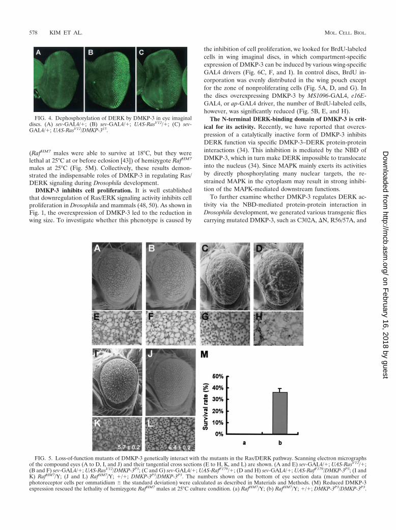
To further support these genetic interactions betweenDMKP-3 and DERK, we monitored the phosphorylationstatus of DERK in tissues by using an anti-phospho-specificantibody for ERK-MAPK (33). As shown in Fig. 4, RasV12-induced phosphorylation of DERK (Fig. 4B) was down-regulated by coexpression of DMKP-3 (Fig. 4C). The resultprovides the direct evidence that DMKP-3 dephosphorylatesDERK in vivo.
Finally, to verify that endogenous DMKP-3 affects the Ras/DERK pathway, we examined the activities of Ras/DERKsignalings in a DMKP-3 loss-of-function genetic background.Ectopic expression of either constitutively active Ras (RaslV12)or Raf (RafF179) caused roughened eye phenotypes (Fig. 5Aand C, respectively) with extra photoreceptor cells in mostommatidia (Fig. 5E and G, respectively). All of these pheno-
types were enhanced in DMKP-3 hypomorphic backgrounds,and the ommatidia structures of these flies were severely dis-organized and fused (Fig. 5B, D, F, and H).
If DMKP-3 does indeed negatively regulate the Ras/DERKpathway in vivo, a loss-of-function mutation of DMKP-3 couldrescue the deleterious phenotypes of loss-of-function muta-tions of the components in the Ras/DERK pathway. As ex-pected, homozygous DMKP-3P3 alleles strongly suppressed therough eye phenotype and the decreased number of photore-ceptor cell phenotype of RafHM7 hemizygote (compare Fig. 5Iand K to 5J and L, respectively). In addition, hypomorphicDMKP-3 partially rescued temperature-dependent lethality
FIG. 3. Genetic interactions between DMKP-3 and the compo-nents of the Ras/DERK pathway. Scanning electron micrographs ofthe compound eyes (A to D, I to L, and Q to T) and their tangentialcross sections (E to H, M to P, and U to X) are shown: sev-GAL4/�;Rase1B/� (A and E); sev-GAL4/�; Rase1B/UAS-DMKP-315 (B and F);RafHM7/Y; sev-GAL4/� (C and G); RafHM7/Y; sev-GAL4/�; UAS-DMKP-315/� (D and H); sev-GAL4/rl1 (I and M); sev-GAL4/rl1; UAS-DMKP-315/� (J and N); sev-GAL4/�; UAS-RasV12/� (K and O);sev-GAL4/�; UAS-RasV12/DMKP-315 (L and P); sev-GAL4/�; UAS-rlsem/� (Q and U); sev-GAL4/�; UAS-rlsem/UAS-DMKP-315 (R and V);sev-PTP-ER101/X; sev-GAL4/� (S and W); and sev-PTP-ER101/X; sev-GAL4/�; DMKP-315/� (T and X). The numbers shown on the bottomof eye section data (mean number of photoreceptor cells per omma-tidium � the standard deviation) were calculated as described in Ma-terials and Methods.
VOL. 24, 2004 MKP-3 AND DROSOPHILA DEVELOPMENT 577
on February 16, 2018 by guest
http://mcb.asm
.org/D
ownloaded from
(RafHM7 males were able to survive at 18°C, but they werelethal at 25°C at or before eclosion [43]) of hemizygote RafHM7
males at 25°C (Fig. 5M). Collectively, these results demon-strated the indispensable roles of DMKP-3 in regulating Ras/DERK signaling during Drosophila development.
DMKP-3 inhibits cell proliferation. It is well establishedthat downregulation of Ras/ERK signaling activity inhibits cellproliferation in Drosophila and mammals (48, 50). As shown inFig. 1, the overexpression of DMKP-3 led to the reduction inwing size. To investigate whether this phenotype is caused by
the inhibition of cell proliferation, we looked for BrdU-labeledcells in wing imaginal discs, in which compartment-specificexpression of DMKP-3 can be induced by various wing-specificGAL4 drivers (Fig. 6C, F, and I). In control discs, BrdU in-corporation was evenly distributed in the wing pouch exceptfor the zone of nonproliferating cells (Fig. 5A, D, and G). Inthe discs overexpressing DMKP-3 by MS1096-GAL4, e16E-GAL4, or ap-GAL4 driver, the number of BrdU-labeled cells,however, was significantly reduced (Fig. 5B, E, and H).
The N-terminal DERK-binding domain of DMKP-3 is crit-ical for its activity. Recently, we have reported that overex-pression of a catalytically inactive form of DMKP-3 inhibitsDERK function via specific DMKP-3–DERK protein-proteininteractions (34). This inhibition is mediated by the NBD ofDMKP-3, which in turn make DERK impossible to translocateinto the nucleus (34). Since MAPK mainly exerts its activitiesby directly phosphorylating many nuclear targets, the re-strained MAPK in the cytoplasm may result in strong inhibi-tion of the MAPK-mediated downstream functions.
To further examine whether DMKP-3 regulates DERK ac-tivity via the NBD-mediated protein-protein interaction inDrosophila development, we generated various transgenic fliescarrying mutated DMKP-3, such as C302A, �N, R56/57A, and
FIG. 4. Dephosphorylation of DERK by DMKP-3 in eye imaginaldiscs. (A) sev-GAL4/�; (B) sev-GAL4/�; UAS-RasV12/�; (C) sev-GAL4/�; UAS-RasV12/DMKP-315.
FIG. 5. Loss-of-function mutants of DMKP-3 genetically interact with the mutants in the Ras/DERK pathway. Scanning electron micrographsof the compound eyes (A to D, I, and J) and their tangential cross sections (E to H, K, and L) are shown. (A and E) sev-GAL4/�; UAS-RasV12/�;(B and F) sev-GAL4/�; UAS-RasV12/DMKP-3P3; (C and G) sev-GAL4/�; UAS-RafF179/�; (D and H) sev-GAL4/�; UAS-RafF179/DMKP-3P3; (I andK) RafHM7/Y; (J and L) RafHM7/Y; �/�; DMKP-3P3/DMKP-3P3. The numbers shown on the bottom of eye section data (mean number ofphotoreceptor cells per ommatidium � the standard deviation) were calculated as described in Materials and Methods. (M) Reduced DMKP-3expression rescued the lethality of hemizygote RafHM7 males at 25°C culture condition. (a) RafHM7/Y; (b) RafHM7/Y; �/�; DMKP-3P3/DMKP-3P3.
578 KIM ET AL. MOL. CELL. BIOL.
on February 16, 2018 by guest
http://mcb.asm
.org/D
ownloaded from

R56/57A/C302A (Fig. 7A), as described in Materials andMethods. R56/57 is important for the specific binding betweenDMKP-3 and its substrate DERK, and C302 mutation cannullify the phosphatase activity of DMKP-3 (32).
Ectopic expression of �N (Fig. 7E and K) or R56/57A (Fig.7F and L) in the eye by sev- (Fig. 7B to G) or gmr-GAL4 (Fig.7H to M) driver did not show any defects compared to thedriver alone (Fig. 7B and H) in spite of the presence of theirintact phosphatase domain (PD), suggesting that the NBD iscritical for the DMKP-3-mediated dephosphorylation and in-hibition of DERK in photoreceptor cell differentiation. In ad-dition, ectopic expression of DMKP-3 C302A (Fig. 7D) orR56/57A/C302A (Fig. 7G) under sev-GAL4 driver also failedto affect photoreceptor cell differentiation. However, intrigu-ingly, when we expressed these DMKP-3 mutants using astronger driver, the gmr-GAL4 driver, transgenic flies express-ing DMKP-3 C302A showed a mild gain-of-function pheno-type of DMKP-3 with missing photoreceptor cells (Fig. 7J).
Likewise, overexpression of DMKP-3 �N (Fig. 7Q), R56/57A (Fig. 7R), or R56/57A/C302A (Fig. 7S) in the wing did notdisplay any significant phenotypic changes compared to theMS1096-GAL4 driver alone (Fig. 7N). However, overexpres-sion of DMKP-3 C302A (Fig. 7P) caused several defects, in-cluding disruption of the posterior cross vein and the longitu-dinal vein L4, which is similar to the phenotype ofoverexpression of wild-type DMKP-3 (Fig. 7O).
Expression of rhomboid is inhibited by DMKP-3. To furtherconfirm whether the NBD of DMKP-3 is required for the
repression of DERK function, we examined the transcriptionallevels of rhomboid (rho) in imaginal discs. The transcription ofrho is directly regulated by DERK (35) and can be monitoredby anti-�-galactosidase staining using the rhoAA69 enhancertrap line, as previously described (13). The rho-lacZ expressionin the eye imaginal discs was dramatically suppressed by over-expression of DMKP-3 wild type (Fig. 8B) compared to thesev-GAL4 driver alone (Fig. 8A). Expression of rho-lacZ in theeye disc overexpressing DMKP-3 C302A (Fig. 8C) or DMKP-3R56/57A (Fig. 8D) under the control of the sev-GAL4 driverwas, however, both quantitatively and qualitatively similar tothe control eye disc (Fig. 8A). Likewise, overexpression ofDMKP-3 wild type by MS1096-GAL4 driver strongly inhibitedrho-lacZ expression in the primordial vein of the wing imaginaldisc (Fig. 8F) compared to the MS1096-GAL4 alone (Fig. 8E).On the other hand, ectopic expression of DMKP-3 C302Asignificantly reduced rho expression (Fig. 8G), whereasDMKP-3 R56/57A was unable to suppress rho expression inthe primordial vein (Fig. 8H).
From the results of domain analysis in eyes and wings, wecould draw a conclusion that both NBD and PD of DMKP-3are necessary for Drosophila developmental processes, which isconsistent with our previous data (32, 34).
DMKP-3 is required for proper oogenesis and early embry-ogenesis. Because hypomorphic DMKP-3 flies displayed a re-duced fertility and DMKP-3 transcripts were enriched in theovary (32; data not shown), we speculated that DMKP-3 mightbe involved in the female reproductive system. We generated
FIG. 6. Inhibited cell proliferation in the DMKP-3-overexpressing wing imaginal discs. Wing imaginal discs were stained with an anti-BrdUantibody as described in Materials and Methods. (A) MS1096-GAL4/�; (B) MS1096-GAL4/�; UAS-DMKP-36/�; (D) e16E-GAL4/�; (E) e16E-GAL4/UAS-DMKP-36; (G) ap-GAL4/�; (H) ap-GAL/UAS-DMKP-36. The expression pattern of GAL4 lines was tested with X-Gal staining in wingimaginal discs. (C) MS1096-GAL4/�; UAS-lacZ; (F) e16E-GAL4/UAS-lacZ; (I) ap-GAL4/UAS-lacZ.
VOL. 24, 2004 MKP-3 AND DROSOPHILA DEVELOPMENT 579
on February 16, 2018 by guest
http://mcb.asm
.org/D
ownloaded from

germ line clones of DMKP-3 by using the FLP-FRT-DFS tech-nique (11). Interestingly, females with DMKP-3P1 germ lineclone laid only a small number of eggs compared to the controlflies (less than �30%), implying severe defects in oogenesis.Even the laid eggs were abnormal: approximately 77% of thelaid eggs were shorter than the normal ones (Fig. 9D; compareFig. 9B to A), and some displayed severe defects in chorion(egg shell) formation (Fig. 9C). Furthermore, none of theDMKP-3P1 germ line clone embryos could escape the embry-onic stage and progress beyond the two-nuclei stage (Fig. 9Fand H) compared to the wild type (Fig. 9E and G). Theseresults supported that DMKP-3 is required for the properoogenesis and early embryogenesis.
DISCUSSION
We investigated in vivo the roles of DMKP-3 in Drosophiladevelopment. Our studies revealed novel functions ofDMKP-3 as a negative regulator in a variety of developmentalprocesses including cell differentiation and proliferation con-trolled by the Ras/DERK pathway. Ectopic expression ofDMKP-3 in the eye strongly suppressed photoreceptor celldifferentiation (Fig. 1D to F), and this phenotype was furtherenhanced by genetic reduction of the Ras/DERK-dependentsignaling activity (Fig. 3). In addition, overexpressed DMKP-3also caused suppression in wing vein differentiation, which iscorrelated with the inhibition of the Ras/DERK signaling path-
FIG. 7. The NBD and PD of DMKP-3 is required for photoreceptor cell differentiation and wing vein formation. (A) Various DMKP-3 mutantswere generated in the present study as described in Materials and Methods. Functionally important amino acid residues in the NBD and PD areindicated. (B to M) Tangential cross sections of the compound eyes expressing mutant DMKP-3 are shown. (B) sev-GAL4/�; (C) sev-GAL4/�;UAS-DMKP-310/�; (D) sev-GAL4/UAS-DMKP-3 C302A; (E) sev-GAL4/UAS-DMKP-3 �N; (F) sev-GAL4/�; UAS-DMKP-3 R56/57A/�; (G) sev-GAL4/UAS-DMKP-3 R56/57A/C302A; (H) gmr-GAL4/�; (I) gmr-GAL4/�; UAS-DMKP-310/�; (J) gmr-GAL4/UAS-DMKP-3 C302A; (K) gmr-GAL/UAS-DMKP-3 �N; (L) gmr-GAL4/�; UAS-DMKP-3 R56/57A/�; (M) gmr-GAL4/UAS-DMKP-3 R56/57A/C302A. (N to S) Microscopic viewsof the wings expressing DMKP-3. (N) MS1096-GAL4/�; (O) MS1096-GAL4/�; �/�; UAS-DMKP-310/�; (P) MS1096-GAL4/�; UAS-DMKP-3C302A/�; (Q) MS1096-GAL4/�; UAS-DMKP-3 �N/�; (R) MS1096-GAL4/�; �/�; UAS-DMKP-3 R56/57A/�; (S) MS1096-GAL4/�; UAS-DMKP-3 R56/57A/C302A/�. The numbers shown on the bottom of eye section data (mean number of photoreceptor cells per ommatidium � thestandard deviation) were calculated as described in Materials and Methods.
580 KIM ET AL. MOL. CELL. BIOL.
on February 16, 2018 by guest
http://mcb.asm
.org/D
ownloaded from

way during wing development (Fig. 1G to L). Conversely, hy-pomorphs of DMKP-3 (Fig. 2J and P) or DMKP-3-null clones(Fig. 2N and R) displayed ectopic differentiation of photore-ceptor cells and wing vein cells. These results strongly suggestthat DMKP-3 plays an essential role in the regulation of celldifferentiation during Drosophila development by restrainingthe activities of the Ras/DERK signaling pathway.
In addition, we also observed that DMKP-3 overexpressioncauses a dramatic reduction in the overall size of adult wingblades (Fig. 1I and L) and larval wing imaginal discs (data notshown), as well as the defects in wing vein cell differentiation.In support of this phenotype, the active cell proliferation inwing imaginal discs, monitored by measuring BrdU incorpora-tion, was strongly inhibited by DMKP-3 overexpression (Fig.6). Therefore, we conclude that DMKP-3 is able to negativelymodulate cell proliferation in vivo. Consistently, numerousprevious results also have shown that downregulation of Ras/ERK signaling activity inhibits cell proliferation (18, 36, 50,53). Collectively, these results provide strong in vivo evidencethat DMKP-3 suppresses cellular proliferation, as well as dif-ferentiation, both of which are promoted by the Ras/DERKpathway during development.
Because the females of hypomorphic DMKP-3 mutants dis-played a phenotype of decreased number of laid eggs, wesuspected that there is another function of DMKP-3 in thefemale reproductive system. When the maternal DMKP-3 wasdepleted in the female germ line, the number of laid eggs wasfurther reduced to that of hypomorphic DMKP-3 flies, andeven the laid eggs showed severe morphological defects, in-cluding chorion malformation, which is known to be caused bydefects in the vitellogenesis process during oogenesis (58).Although some eggs appeared to have normal shape, theyfailed to show any further embryonic development, just arrest-ing at the single-nucleus or two-nucleus stage (Fig. 9E to H).
FIG. 8. Expression of rhomboid is inhibited by DMKP-3. Antibody staining of �-galactosidase in rhoAA69 reveals rho expression. (A) sev-GAL4/�; rhoAA69/�; (B) sev-GAL4/�;UAS-DMKP-310/rhoAA69; (C) sev-GAL4/UAS-DMKP-3 C302A; rhoAA69/�; (D) sev-GAL4/�;UAS-DMKP-3R56/57A/rhoAA69; (E) MS1096-GAL4/�; �/�; rhoAA69/�; (F) MS1096-GAL4/�; �/�; UAS-DMKP-310/rhoAA69; (G) MS1096-GAL4/�; UAS-DMKP-3 C302A/�; rhoAA69/�; (H) MS1096-GAL4/�; �/�; UAS-DMKP-3 R56/57A/rhoAA69.
FIG. 9. Defective embryos in DMKP-3P1 germ line clones. Scan-ning electron micrographs of w1118 (A) and DMKP-3P1 germ line cloneeggs (B and C). (D) Measurements of egg lengths in wild type (w1118)and in DMKP-3P1 germ line clone are shown (1 arbitrary unit � 30.6�m). (E to H) Propidium iodide-stained embryos. (E and F) Eggs 0.5 hafter egg laying; (G and H) eggs 15 h after egg laying; (E and G) w1118;(F and H) DMKP-3P1 germ line clones.
VOL. 24, 2004 MKP-3 AND DROSOPHILA DEVELOPMENT 581
on February 16, 2018 by guest
http://mcb.asm
.org/D
ownloaded from

These results suggest that DMKP-3 is also required for properoogenesis and early embryogenesis besides the other develop-mental processes, as we have described, such as eye and wingdevelopment. Interestingly, various gain-of-function mutantsof DERK also have shown female sterility with severe defectsin vitellogenesis in previous studies (38), demonstrating thatthe DMKP-3-deficient phenotypes in female germ lines may bethe consequences of DERK hyperactivation.
It should be noted that another DERK phosphatase, PTP-ER, has been identified in the Drosophila system (28). Eye-specific overexpression of PTP-ER suppresses DERK activity,causing a reduction in the photoreceptor cell number (Fig.3W). However, the null mutants of PTP-ER develop well andonly show mild defects in photoreceptor cell differentiation(28), whereas the loss-of-function DMKP-3 mutants becomelethal from the embryonic or early larval stages. Since themammalian homologues of PTP-ER are expressed in a neu-ron-specific manner (28, 47), it seems that Drosophila PTP-ERacts specifically in neuronal photoreceptor cells. In contrast toPTP-ER, DMKP-3 was shown to be involved not only in pho-toreceptor cell specification but also in other developmentalprocesses such as vein cell differentiation, oogenesis, and earlyembryogenesis. Moreover, DMKP-3 is ubiquitously expressedthroughout all developmental stages (Fig. 1N and Q and 2D[32]). Therefore, we conclude that DMKP-3 is a generalDERK phosphatase controlling various processes of Drosophiladevelopment.
In conclusion, we provide here firm genetic evidence tosupport that DMKP-3 acts as an essential antagonist againstthe Ras/DERK-dependent signaling pathway during Drosophiladevelopment. Further analyses of DMKP-3 functions in Dro-sophila may provide abundant insights for understanding the invivo roles of the Ras/DERK-dependent signaling pathway inthe context of both cell differentiation and proliferation.
ACKNOWLEDGMENTS
We are indebted to M. Freeman and J. Kim for kindly providing flystocks. The 40-1a and rat-Elav-7E8A10 antibodies developed by J. R.Sanes and G. M. Rubin were obtained from the Developmental Stud-ies Hybridoma Bank (University of Iowa at Iowa City).
REFERENCES
1. Alessi, D. R., A. Cuenda, P. Cohen, D. T. Dudley, and A. R. Saltiel. 1995. PD098059 is a specific inhibitor of the activation of mitogen-activated proteinkinase kinase in vitro and in vivo. J. Biol. Chem. 270:27489–27494.
2. Biggs, W. H., III, K. H. Zavitz, B. Dickson, A. van der Straten, D. Brunner,E. Hafen, and S. L. Zipursky. 1994. The Drosophila rolled locus encodes aMAP kinase required in the sevenless signal transduction pathway. EMBO J.13:1628–1635.
3. Blenis, J. 1993. Signal transduction via the MAP kinases: proceed at yourown RSK. Proc. Natl. Acad. Sci. USA 90:5889–5892.
4. Bott, C. M., S. G. Thorneycroft, and C. J. Marshall. 1994. The sevenmakergain-of-function mutation in p42 MAP kinase leads to enhanced signallingand reduced sensitivity to dual specificity phosphatase action. FEBS Lett.352:201–205.
5. Brunet, A., D. Roux, P. Lenormand, S. Dowd, S. Keyse, and J. Pouyssegur.1999. Nuclear translocation of p42/p44 mitogen-activated protein kinase isrequired for growth factor-induced gene expression and cell cycle entry.EMBO J. 18:664–674.
6. Brunner, D., N. Oellers, J. Szabad, W. H. Biggs III, S. L. Zipursky, and E.Hafen. 1994. A gain-of-function mutation in Drosophila MAP kinase acti-vates multiple receptor tyrosine kinase signaling pathways. Cell 76:875–888.
7. Calleja, M., E. Moreno, S. Pelaz, and G. Morata. 1996. Visualization of geneexpression in living adult Drosophila. Science 274:252–255.
8. Camps, M., A. Nichols, C. Gillieron, B. Antonsson, M. Muda, C. Chabert, U.Boschert, and S. Arkinstall. 1998. Catalytic activation of the phosphataseMKP-3 by ERK2 mitogen-activated protein kinase. Science 280:1262–1265.
9. Capdevila, J., and I. Guerrero. 1994. Targeted expression of the signalingmolecule decapentaplegic induces pattern duplications and growth alter-ations in Drosophila wings. EMBO J. 13:4459–4468.
10. Cho, K. S., J. H. Lee, S. Kim, D. Kim, H. Koh, J. Lee, C. Kim, J. Kim, andJ. Chung. 2001. Drosophila phosphoinositide-dependent kinase-1 regulatesapoptosis and growth via the phosphoinositide 3-kinase-dependent signalingpathway. Proc. Natl. Acad. Sci. USA 98:6144–6149.
11. Chou, T. B., and N. Perrimon. 1996. The autosomal FLP-DFS technique forgenerating germline mosaics in Drosophila melanogaster. Genetics 144:1673–1679.
12. Clifford, R. J., and T. Schupbach. 1989. Coordinately and differentiallymutable activities of torpedo, the Drosophila melanogaster homolog of thevertebrate EGF receptor gene. Genetics 123:771–787.
13. Conley, C. A., R. Silburn, M. A. Singer, A. Ralston, D. Rohwer-Nutter, D. J.Olson, W. Gelbart, and S. S. Blair. 2000. Crossveinless 2 contains cysteine-rich domains and is required for high levels of BMP-like activity during theformation of the cross veins in Drosophila. Development 127:3947–3959.
14. Dalby, B., A. J. Pereira, and L. S. Goldstein. 1995. An inverse PCR screen forthe detection of P element insertions in cloned genomic intervals in Dro-sophila melanogaster. Genetics 139:757–766.
15. Diaz-Benjumea, F. J., and E. Hafen. 1994. The sevenless signalling cassettemediates Drosophila EGF receptor function during epidermal development.Development 120:569–578.
16. Dorstyn, L., P. A. Colussi, L. M. Quinn, H. Richardson, and S. Kumar. 1999.DRONC, an ecdysone-inducible Drosophila caspase. Proc. Natl. Acad. Sci.USA 96:4307–4312.
17. Dorstyn, L., S. H. Read, L. M. Quinn, H. Richardson, and S. Kumar. 1999.DECAY, a novel Drosophila caspase related to mammalian caspase-3 andcaspase-7. J. Biol. Chem. 274:30778–30783.
18. Downward, J. 1997. Cell cycle: routine role for Ras. Curr. Biol. 7:R258–R260.
19. Farooq, A., G. Chaturvedi, S. Mujtaba, O. Plotnikova, L. Zeng, C. Dhalluin,R. Ashton, and M. M. Zhou. 2001. Solution structure of ERK2 bindingdomain of MAPK phosphatase MKP-3: structural insights into MKP-3 ac-tivation by ERK2. Mol. Cell 7:387–399.
20. Favata, M. F., K. Y. Horiuchi, E. J. Manos, A. J. Daulerio, D. A. Stradley,W. S. Feeser, D. E. Van Dyk, W. J. Pitts, R. A. Earl, F. Hobbs, R. A.Copeland, R. L. Magolda, P. A. Scherle, and J. M. Trzaskos. 1998. Identi-fication of a novel inhibitor of mitogen-activated protein kinase kinase.J. Biol. Chem. 273:18623–18632.
21. Freeman, M. 1996. Reiterative use of the EGF receptor triggers differenti-ation of all cell types in the Drosophila eye. Cell 87:651–660.
22. Garcia-Bellido, A., F. Cortes, and M. Milan. 1994. Cell interactions in thecontrol of size in Drosophila wings. Proc. Natl. Acad. Sci. USA 91:10222–10226.
23. Guichard, A., B. Biehs, M. A. Sturtevant, L. Wickline, J. Chacko, K. Howard,and E. Bier. 1999. rhomboid and Star interact synergistically to promoteEGFR/MAPK signaling during Drosophila wing vein development. Devel-opment 126:2663–2676.
24. Harrison, D. A., R. Binari, T. S. Nahreini, M. Gilman, and N. Perrimon.1995. Activation of a Drosophila Janus kinase (JAK) causes hematopoieticneoplasia and developmental defects. EMBO J. 14:2857–2865.
25. Hou, X. S., T. B. Chou, M. B. Melnick, and N. Perrimon. 1995. The torsoreceptor tyrosine kinase can activate Raf in a Ras-independent pathway. Cell81:63–71.
26. Jordan, P., and R. Karess. 1997. Myosin light chain-activating phosphoryla-tion sites are required for oogenesis in Drosophila. J. Cell Biol. 139:1805–1819.
27. Karim, F. D., and G. M. Rubin. 1998. Ectopic expression of activated Ras1induces hyperplastic growth and increased cell death in Drosophila imaginaltissues. Development 125:1–9.
28. Karim, F. D., and G. M. Rubin. 1999. PTP-ER, a novel tyrosine phosphatase,functions downstream of Ras1 to downregulate MAP kinase during Dro-sophila eye development. Mol. Cell 3:741–750.
29. Karim, F. D., H. C. Chang, M. Therrien, D. A. Wassarman, T. Laverty, andG. M. Rubin. 1996. A screen for genes that function downstream of Ras1during Drosophila eye development. Genetics 143:315–329.
30. Khokhlatchev, A. V., B. Canagarajah, J. Wilsbacher, M. Robinson, M. At-kinson, E. Goldsmith, and M. H. Cobb. 1998. Phosphorylation of the MAPkinase ERK2 promotes its homodimerization and nuclear translocation. Cell93:605–615.
31. Kim-Ha, J., J. Kim, and Y. J. Kim. 1999. Requirement of RBP9, a DrosophilaHu homolog, for regulation of cystocyte differentiation and oocyte determi-nation during oogenesis. Mol. Cell. Biol. 19:2505–2514.
32. Kim, S. H., H. B. Kwon, Y. S. Kim, J. H. Ryu, K. S. Kim, Y. Ahn, W. J. Lee,and K. Y. Choi. 2002. Isolation and characterization of a Drosophila homo-logue of mitogen-activated protein kinase phosphatase-3 which has a highsubstrate specificity toward extracellular-signal-regulated kinase. Biochem. J.361:143–151.
33. Kumar, J. P., F. Hsiung, M. A. Powers, and K. Moses. 2003. Nuclear trans-location of activated MAP kinase is developmentally regulated in the devel-oping Drosophila eye. Development 130:3703–3714.
582 KIM ET AL. MOL. CELL. BIOL.
on February 16, 2018 by guest
http://mcb.asm
.org/D
ownloaded from

34. Kwon, H. B., S. H. Kim, S. E. Kim, I. H. Jang, Y. Ahn, W. J. Lee, and K. Y.Choi. 2002. Drosophila extracellular signal-regulated kinase involves the in-sulin-mediated proliferation of Schneider cells. J. Biol. Chem. 277:14853–14858.
35. Lee, J. H., K. S. Cho, J. Lee, D. Kim, S. B. Lee, J. Yoo, G. H. Cha, andJ. Chung. 2002. Drosophila PDZ-GEF, a guanine nucleotide exchange factorfor Rap1 GTPase, reveals a novel upstream regulatory mechanism in themitogen-activated protein kinase signaling pathway. Mol. Cell. Biol. 22:7658–7666.
36. Leone, G., J. DeGregori, R. Sears, L. Jakoi, and J. R. Nevins. 1997. Myc andRas collaborate in inducing accumulation of active cyclin E/Cdk2 and E2F.Nature 387:422–426.
37. Lewis, T. S., P. S. Shapiro, and N. G. Ahn. 1998. Signal transduction throughMAP kinase cascades. Adv. Cancer Res. 74:49–139.
38. Lim, Y. M., K. Nishizawa, Y. Nishi, L. Tsuda, Y. H. Inoue, and Y. Nishida.1999. Genetic analysis of rolled, which encodes a Drosophila mitogen-acti-vated protein kinase. Genetics 153:763–771.
39. Lorenzen, J. A., S. E. Baker, F. Denhez, M. B. Melnick, D. L. Brower, andL. A. Perkins. 2001. Nuclear import of activated D-ERK by DIM-7, animportin family member encoded by the gene moleskin. Development 128:1403–1414.
40. Lunde, K., B. Biehs, U. Nauber, and E. Bier. 1998. The knirps and knirps-related genes organize development of the second wing vein in Drosophila.Development 125:4145–4154.
41. Marshall, C. J. 1995. Specificity of receptor tyrosine kinase signaling: tran-sient versus sustained extracellular signal-regulated kinase activation. Cell80:179–185.
42. Martin-Blanco, E., F. Roch, E. Noll, A. Baonza, J. B. Duffy, and N. Perrimon.1999. A temporal switch in DER signaling controls the specification anddifferentiation of veins and interveins in the Drosophila wing. Development126:5739–5747.
43. Melnick, M. B., L. A. Perkins, M. Lee, L. Ambrosio, and N. Perrimon. 1998.Developmental and molecular characterization of mutations in the Drosophila-raf serine/threonine protein kinase. Development 118:127–138.
44. Muda, M., A. Theodosiou, C. Gillieron, A. Smith, C. Chabert, M. Camps, U.Boschert, N. Rodrigues, K. Davies, A. Ashworth, and S. Arkinstall. 1998. Themitogen-activated protein kinase phosphatase-3 N-terminal noncatalytic re-gion is responsible for tight substrate binding and enzymatic specificity.J. Biol. Chem. 273:9323–9329.
45. Muda, M., U. Boschert, R. Dickinson, J. C. Martinou, I. Martinou, M.Camps, W. Schlegel, and S. Arkinstall. 1996. MKP-3, a novel cytosolic
protein-tyrosine phosphatase that exemplifies a new class of mitogen-acti-vated protein kinase phosphatase. J. Biol. Chem. 271:4319–4326.
46. Neel, B. G., and N. K. Tonks. 1997. Protein tyrosine phosphatases in signaltransduction. Curr. Opin. Cell Biol. 9:193–204.
47. Ogata, M., M. Sawada, Y. Fujino, and T. Hamaoka. 1995. cDNA cloning andcharacterization of a novel receptor-type protein tyrosine phosphatase ex-pressed predominantly in the brain. J. Biol. Chem. 270:2337–2343.
48. Pages, G., P. Lenormand, G. L’Allemain, J. C. Chambard, S. Meloche, andJ. Pouyssegur. 1993. Mitogen-activated protein kinases p42mapk andp44mapk are required for fibroblast proliferation. Proc. Natl. Acad. Sci.USA 90:8319–8323.
49. Phelps, C. B., and A. H. Brand. 1998. Ectopic gene expression in Drosophilausing GAL4 system. Methods 14:367–379.
50. Prober, D. A., and B. A. Edgar. 2000. Ras1 promotes cellular growth in theDrosophila wing. Cell 100:435–446.
51. Raabe, T. 2000. The sevenless signaling pathway: variations of a commontheme. Biochim. Biophys. Acta 1496:151–163.
52. Robertson, H. M., C. R. Preston, R. W. Phillis, D. M. Johnson-Schlitz, W. K.Benz, and W. R. Engels. 1988. A stable genomic source of P element trans-posase in Drosophila melanogaster. Genetics 118:461–470.
53. Tapon, N., K. H. Moberg, and I. K. Hariharan. 2001. The coupling of cellgrowth to the cell cycle. Curr. Opin. Cell Biol. 13:731–737.
54. Therrien, M., D. K. Morrison, A. M. Wong, and G. M. Rubin. 2000. Agenetic screen for modifiers of a kinase suppressor of Ras-dependent rougheye phenotype in Drosophila. Genetics 156:1231–1242.
55. Tomlinson, A., D. D. Bowtell, E. Hafen, and G. M. Rubin. 1987. Localizationof the sevenless protein, a putative receptor for positional information, in theeye imaginal disc of Drosophila. Cell 51:143–150.
56. Tonks, N. K., and B. G. Neel. 2001. Combinatorial control of the specificityof protein tyrosine phosphatases. Curr. Opin. Cell Biol. 13:182–195.
57. Xu, T., and G. M. Rubin. 1993. Analysis of genetic mosaics in developing andadult Drosophila tissues. Development 117:1223–1237.
58. Yan, Y. L., and J. H. Postlethwait. 1990. Vitellogenesis in Drosophila: se-questration of a yolk polypeptide/invertase fusion protein into developingoocytes. Dev. Biol. 140:281–290.
59. Ye, Y., and M. E. Fortini. 1999. Apoptotic activities of wild-type and Alz-heimer’s disease-related mutant presenilins in Drosophila melanogaster.J. Cell Biol. 146:1351–1364.
60. Zhou, B., L. Wu, K. Shen, J. Zhang, D. S. Lawrence, and Z. Y. Zhang. 2001.Multiple regions of MAP kinase phosphatase 3 are involved in its recognitionand activation by ERK2. J. Biol. Chem. 276:6506–6515.
VOL. 24, 2004 MKP-3 AND DROSOPHILA DEVELOPMENT 583
on February 16, 2018 by guest
http://mcb.asm
.org/D
ownloaded from