mode of action study for a novel class of antimicrobial
TRANSCRIPT

This document is downloaded from DR‑NTU (https://dr.ntu.edu.sg)Nanyang Technological University, Singapore.
Mode of action study for a novel class ofantimicrobial polymers againstmethicillin‑resistant Staphylococcus aureus
Shi, Zhenyu
2019
Shi, Z. (2019).Mode of action study for a novel class of antimicrobial polymers againstmethicillin‑resistant Staphylococcus aureus. Master's thesis, Nanyang TechnologicalUniversity, Singapore.
https://hdl.handle.net/10356/136786
https://doi.org/10.32657/10356/136786
This work is licensed under a Creative Commons Attribution‑NonCommercial 4.0International License (CC BY‑NC 4.0).
Downloaded on 19 Feb 2022 21:00:12 SGT

MODE OF ACTION STUDY FOR A NOVEL CLASS OF ANTIMICROBIAL POLYMERS
AGAINST METHICILLIN-RESISTANT STAPHYLOCOCCUS AUREUS
SHI ZHENYU
(B.Eng. (Hons), NTU)
School of Chemical and Biomedical Engineering
A thesis submitted to Nanyang Technological University in partial fulfilment of the requirement for the degree of Master
of Engineering
2019
Supervisor:
Professor Chan Bee Eng, Mary


Acknowledgement
I would like to express my deepest appreciation to my thesis supervisor
and mentor, Professor Chan Bee Eng, Mary. I am grateful for her generous
guidance and advice along the way, especially when I ran into a trouble spot.
She consistently steered me in the right direction. Without her help this thesis
would not have been possible.
I would also like to express my gratitude to Professor Angelika
Gründling at Imperial College London, who invited me to her laboratory for a
6-month visit. I learned tremendously in the field of microbiology and
bacteriology during my time at Imperial College London. I am gratefully
indebted to her for her expertise and valuable comments.
In addition, a thank you to my family and friends for their unfailing
support and continuous encouragement. I would also like to thank all my lab
members, who are always there to provide help.

1
Table of Contents
Abstract ............................................................................................................... 3
Chapter 1: Introduction .................................................................................... 4
1.1 Literature review ................................................................................................ 4
1.2.1 Antibiotic targets and resistance in Staphylococcus aureus .................... 4
1.2.2 Cationic antimicrobial polymers ............................................................... 5
1.2 Objectives and scope of project ......................................................................... 8
Chapter 2: Materials and Methods .................................................................. 9
2.1 Strains and culture conditions ........................................................................... 9
2.2 Determination of minimum inhibitory concentrations (MICs) .................... 10
2.3 Membrane permeability assay ........................................................................ 11
2.4 Membrane potential assay ............................................................................... 12
2.5 Macromolecular synthesis ............................................................................... 13
2.6 Bacterial respiration ......................................................................................... 14
2.7 Resistance study ................................................................................................ 15
2.8 Whole genome sequencing ............................................................................... 16
2.9 Menaquinone extraction and quantification .................................................. 18
2.10 Rhodamine-conjugated polymer binding assay ........................................... 20
Chapter 3: Results and Discussions ............................................................... 21
3.1 Impact of PIM1 on the physiology of the MRSA ........................................... 21
3.1.2 Impact of PIM1 on macromolecular synthesis ....................................... 24
3.1.3 PIM1 blocks bacterial respiration ........................................................... 26
3.2 Development of resistant mutants and whole genome sequencing analysis 28
3.2.1 S. aureus develops resistance to PIM1 upon serial passage .................. 28
3.2.2 Genomic variations in PIM1-resistant strains ........................................ 30

2
3.3 Binding affinity to bacterial surface influences PIM’s activity .................... 37
3.3.1 Inactivation of CAMP resistance pathway confers sensitivity to PIM137
3.3.2 Electrostatic adsorption is the first step of PIM1-bacteria interaction 37
3.4 PIM’s activity is dependent on respiration level ........................................... 39
3.4.1 Respiratory-deficient mutants are less susceptible to PIM ................... 39
3.4.2 Intracellular menaquinone content decreases under exposure to PIM 42
3.5 Relationship between the 2 categories of genomic variations ...................... 44
Chapter 4: Conclusion ..................................................................................... 46
Supplementary information ............................................................................ 48
Reference .......................................................................................................... 53

3
Abstract
The development of antibiotics has been hampered by the rapid evolution
of antimicrobial resistance and lack of new mechanisms discovered. Membrane-
polarizing cationic polymers have attracted attention due to their low propensity
to induce resistance. A cationic polymer (labelled as PIM1) has been found to
exert excellent antimicrobial efficacy with low cell toxicity. To understand the
mechanism of action of PIM1, investigations were carried out in methicillin-
resistant Staphylococcus aureus (MRSA). It was found that PIM1 had a
distinctively different mechanism from common cationic polymers, since it did
not physically disrupt and perforate the cytoplasmic membrane. Instead, the
polymer blocked aerobic respiration, leading to pleiotropic effects on multiple
biosynthetic pathways in the bacteria. Whole genome sequencing of PIM1-
resistant MRSA strains revealed that genes associated with bacteria surface
charge and the electron transport chain (ETC) were crucial for the activity of
PIM1. Studies with respiratory mutants implied a strong relationship between the
level of aerobic respiration and the efficacy of PIM1. Moreover, PIM1 was found
to decrease the intracellular menaquinone content at sub-inhibitory
concentrations, suggesting a direct interaction between PIM1 and the ETC
component menaquinone. The results suggest that PIM1 binds to the bacteria
surface through electrostatic attraction, and interact with the membrane-bound
ETC which leads to cell death.
Declaration: Due to patent filing, the exact structure of the cationic polymer PIM
is not disclosed here. I apologise for the inconvenience caused.

4
Chapter 1: Introduction
1.1 Literature review
1.2.1 Antibiotic targets and resistance in Staphylococcus aureus
Staphylococcus aureus is a Gram-positive round-shaped bacterium that
can be both commensal and pathogenic. It is a leading cause of clinical infections
including bacteraemia, infective endocarditis, skin and soft tissue, osteoarticular,
and hospital-acquired infections [1]. Moreover, S. aureus is a major foodborne
pathogen, which is capable of producing enterotoxins responsible for food
poisoning [2]. Major intracellular targets for antibiotics developed against S.
aureus are the cell envelope, protein synthesis, and nucleic acid synthesis which
are absent or significantly different from human cells [3]. Nonetheless, antibiotic
resistance can develop through either horizontal transfer of mobile genetic
elements or acquisition of mutations. Horizontally acquired resistance can result
in enzymatic modification of drug or target site, drug efflux, or bypassing the
drug target [4]. Acquisition of mutation usually lead to modification of drug
target, derepression of drug efflux pumps, or accumulation of mutations that alter
the bacteria surface to reduce drug access [5]. One examples of cell envelope
targeting drug is ß-lactams, which were the earliest generation of S. aureus
antibiotics. Penicillin, methicillin and oxacillin all belong to this category. The
inhibitory target of ß-lactams is the penicillin binding protein 2a (PBP2a)
responsible for crosslinking peptidoglycan in the cell wall [6]. Methicillin-
resistant S. aureus (MRSA) was observed shortly after the first clinical use of ß-
lactams. The resistance is acquired by horizontal transfer of staphylococcal
cassette chromosome to the PBP2a gene mecA, which led to lower binding

5
affinity of PBP2a to ß-lactams [7, 8]. Since then MRSA has become a formidable
clinical threat, causing high morbidity and mortality. Vancomycin, a
glycopeptide that blocks peptidoglycan synthesis, has been used to treat serious
infections by MRSA [9]. However, genetic adaptation has given MRSA the most
feared vancomycin resistance. The vancomycin-intermediate S. aureus emerged
after prolonged vancomycin treatment. Another category of high level
vancomycin-resistant S. aureus accepted plasmid transfer of vanA operon from
vancomycin-resistant Enterococcus faecalis [10].
S. aureus, like most bacterial pathogens, has extraordinary ability to
develop resistance to any antibiotic it is exposed to. There has been a lack of new
chemical classes of antibiotics since the 1980s [11]. The organism has at least
one resistance mechanism towards all clinically available antibiotics. The current
clinical solution to treat multidrug resistant MRSA is to switch drugs or employ
different drug combinations. Recently, there has been some discovery of new
antimicrobial targets. One promising molecule named triclosan targets the fatty
acid biosynthesis protein FabI [12]. The latest development was teixobactin,
which inhibits peptidoclycan and wall teichoic acid biosynthesis by binding to
lipid II [13]. Due to the structure and composition of lipid II, the binding site
cannot be easily modified by point mutations. Nonetheless, there remains an
obvious need for development of newer antibiotic drugs and discovery of
antibiotic targets that are difficult to acquire resistance.
1.2.2 Cationic antimicrobial polymers
With the intensive development of polymer science and characterisation
methods since 1980s, various macromolecular structures have been devised for

6
antimicrobial applications. These polymer structures often show enhanced
bactericidal activities and low cytotoxicity compared to small amphiphilic
molecules. Moreover, antimicrobial polymers have the advantage of being
tethered to surfaces without losing their biological activities, delivering a variety
of applications like wound dressing, antifouling and food packaging [14]. Many
different types of antimicrobial polymers have been designed, and the main
categories include cationic antimicrobial polymer [15], and biocide-releasing
polymers [16]. The cationic antimicrobial polymers have been described to
contain two major functional components: the cationic group and the
hydrophobic group [17]. The fundamental mode of action of synthetic cationic
antimicrobial polymers is based on membrane activity. One important feature of
the bacterial cell envelope is the net negative charge provided by teichoic acid in
Gram-positive bacteria cell wall, lipopolysaccharide and phospholipids in Gram-
negative outer membrane, and the phospholipid bilayer in the cytoplasmic
membrane [18]. Cationic antimicrobial polymers take advantage of the negative
charge and target the cytoplasmic membrane. The widely accepted antimicrobial
mechanism of cationic polymers consists of multiple steps: (i) the polymers are
first adsorbed onto the bacterial surface with the aid of cationic groups; (ii) the
high binding affinity enhanced by hydrophobic groups cause cytoplasmic
membrane disorganisation; (iii) cytoplasmic contents leak out, eventually leading
to cell lysis [19, 20]. It is believed that this membrane-active mechanism has a
low potential of inducing microbial drug resistance [21].
Ever since the first cationic antimicrobial polymer polyhexamethylene
biguanide chloride (PHMB) was introduced in 1983 [22, 23], there has been a
constant exploration of structural design to optimize functionality of the polymer.

7
Examples of the cationic centre are ammonium groups (including primary,
secondary, tertiary and quaternary ammonium) [24], and iminium structures
(pyridinium, imidazolium, and guanidinium) [25-27]. The unique feature of
iminium groups is that the positive charge is delocalized evenly through the
conjugated system, which give rise to better microbial efficacy compared with
quaternary ammonium groups [28]. The hydrophobic groups have also been
studied extensively to facilitate the penetration into the cytoplasmic membrane.
Common strategies involve alteration of alkyl chain length, and adopting linear
versus cyclic structures [17]. Although many polymer systems have been
developed to date, the understanding towards the antimicrobial mechanism is still
limited. There has been a lack of new potential mechanisms explored for the past
decade. In-depth mode of action studies will establish fundamentals for
improving structure-activity relationships.

8
1.2 Objectives and scope of project
A series of PIM salts has been synthesized, out of which PIM1 exhibited
low cell toxicity and good antimicrobial activity against a wide range of
microorganisms, including both Gram-positive and Gram-negative bacteria. The
objective of this project is to investigate the mechanism of action of PIM1
through microbiological and biochemical approaches. The model organism is
MRSA, which has been well characterized and has high clinical relevance and
significance. The investigation began with studying the impact of PIM1 on the
physiology of MRSA. In the second stage, a resistance evolution study was
carried out and the genomic variations associated with resistant strains were
critically analysed. Based on findings from the resistance study, further
characterizations were carried out to determine the specificity of the activity of
PIM1. All results were discussed and interpreted after data presentation.
Therefore, no separate discussion section was included. Conclusions from this
study would provide a deeper understanding into the field of antimicrobial
polymers, and lay foundation for future work.

9
Chapter 2: Materials and Methods
2.1 Strains and culture conditions
Bacteria strains used in this study are listed in Table 1. MRSA strains
were cultured in TSB (tryptic soy broth) or on TSA (tryptic soy agar). Freezer
stocks of strains were made with TSB and 30% glycerol. Transposon mutants
were constructed from the Nebraska library [29]. For transposon mutants, 10
µg/mL Erythromycin (Erm) was added to the culture media.
Table 1. List of MRSA strains used in this study
Strains Relevant characteristics Source
LAC CA-MRSA USA300 strain [30]
LAC ∆menD Deletion of menD from LAC [31]
LAC ∆hemB Deletion of hemB from LAC [31]
LAC mprf::tn mprf transposon mutant, Erm-resistant [29]
LAC* Erm-sensitive derivative of LAC [32]
LAC*qoxB::tn qoxB transposon mutant, Erm-resistant [33]
LAC*cydA::tn cydA transposon mutant, Erm-resistant [33]
LAC*∆dltD Deletion of dltD from LAC [29]
LAC*∆dlt operon Deletion of dlt operon from LAC [29]

10
2.2 Determination of minimum inhibitory concentrations (MICs)
MICs were determined in TSB by the broth microdilution method. A
single bacteria colony was picked and grown overnight. Next day the cultures
were diluted to an OD of 0.01 and grown to exponential phase (OD of
approximately 1). Next, the cultures were adjusted to approximately 5x10⁵ cells
per mL (1000-fold dilution of OD=1 culture) and 50 µl of these cultures were
added to 50 µl of serial dilutions of the test compound in TSB medium. After 18
h of incubation at 37°C with constant shaking at 500 rpm, the absorbance at
600nm (OD600) was measured using a SPECTROstar Nano microplate reader
(BMG Labtech). The MIC90 is defined as the concentration at which ≥ 90% of
the isolates in a test population are inhibited [34]. Experiments were performed
with 3 independent biological samples.

11
2.3 Membrane permeability assay
Permeability of cytoplasmic membrane was monitored with nucleic acid
dye SYTOX green (ThermoFisher). The method was adapted from previous
studies on S. aureus membrane permeability [35, 36]. The bacterial strains were
grown overnight in TSB medium. Next day the cultures were diluted to an OD
of 0.01 and grown to exponential phase (OD of approximately 1). Next, the cells
were pelleted by centrifugation at 7,000 rpm for 10 min, and resuspended to OD
of 2 in 0.85% NaCl solution. The dye SYTOX green (1 µM) was added together
with compounds of interest (PIM1, nisin at 8 x MIC). The cell suspension was
incubated in dark for 1 h. Next, the mixture was washed by pelleting and
resuspending to OD of 2 in 0.85% NaCl solution. 2 µl of the washed cell
suspension was spotted on a slide with a thin 1.5% agarose gel patch and covered
with a coverslip. The observation was performed at x1000 magnification using
an Axio Imager.A2 Zeiss microscope equipped with a GFP filter set. The images
were taken with the ZEN 2012 (blue edition) software. Cells were counted using
the ImageJ software. Experiments were performed with 3 independent biological
samples.

12
2.4 Membrane potential assay
The measurement of membrane potential was performed with the
voltage-sensitive fluorescent dye 3,3’-Dipropylpylthiadicarbocyanine iodide
DiSC3(5) (Sigma-Aldrich). Bacterial strains were grown in the same condition as
for membrane permeability assay. The exponential phase bacteria cells from a
culture aliquot were pelleted by centrifugation at 7,000 rpm for 10 min, and
resuspended to OD of 0.2 in PBS supplemented with 0.5 mg/mL BSA. BSA was
added to reduce the absorption of the DiSC3(5) dye to the polystyrene surfaces
of the plates [37]. 200 µl of these cell suspensions were aliquoted into black-wall
transparent bottom 96-well microplate (Greiner) and the fluorescence signal was
measured at 622 nm excitation and 670 nm emission using a Tecan Infinite
M200Pro plate reader. After measuring baseline fluorescence for 2 minutes,
DiSC3(5) was added to a final concentration of 1 µM. The fluorescence
quenching was monitored until signal the intensity stabilized. The compounds of
interest (PIM1, nisin, DI water) were then added. The final concentration in the
medium was 8 µg/mL for PIM1 and 64 µg/mL for nisin. The fluorescence signal
was subsequently followed for 60 min. Experiments were performed with 3
independent biological samples.

13
2.5 Macromolecular synthesis
Protocol for measuring incorporation rate of radioactive-labelled
precursors was adapted from Ling et al. [38] and Bowman et al. [39]. MRSA
strain LAC* cells were grown in minimal medium (0.02 M HEPES, 0.002M
MgSO4, 0.0001M CaCl2, 0.4% succinic acid, 0.043M NaCl, 0.5% (NH4)2SO4, 5%
TSB) to exponential phase. Cells were pelleted by centrifugation for 10 min at
8,000 rpm and 4°C. The supernatant was removed, and the pellet was
resuspended in fresh minimal medium to an OD of 0.2 (108 cells per mL). 350 µl
of this cell suspension was aliquoted to 50 mL conical tubes, and 100 µl was
removed for background measurement. Antibiotics were added to the cell
suspension, immediately followed by addition of radioactive precursors. The
precursors used were N-acetyl-glucosamine, D-[6-3H] (1 mCi mL-1), thymidine,
[methyl-3H] (0.25 mCi mL-1), uridine, [5-3H] (1 mCi mL-1), L-glutamine, 14C(U)
(0.1 mCi mL-1) (Hartmann Analytics). 100 µl cell suspension was removed and
added to 500 µl ice cold 25% trichloroacetic acid (TCA) and 1% casamino acid
(CA) solution. The mixture was filtered through nitrocellulose filter
(PerkinElmer Life Sciences), followed by washing with 2 mL ice cold 25% TCA
+ 1% CA solution, and 16 mL ice cold water. Measurements were taken
immediately upon addition of precursor, and after 20 min incubation at 37°C and
500 rpm. Afterwards, the filters were dissolved in 9 mL of scintillation liquid
Filter Count (PerkinElmer Life Sciences). Incorporated radioactivity was
measured with Wallac 1409 DSA liquid scintillation counter. The count/min
(cpm) values were normalized to the OD of cell suspension, and experiments
were performed with 3 independent biological samples.

14
2.6 Bacterial respiration
The oxygen consumption rate (OCR) of LAC* and resistant mutants were
measured using a Seahorse XFp Analyzer (Agilent). Overnight culture was
diluted to OD of 0.01 in fresh TSB, and grown to early exponential phase (OD
of 0.2 to 0.5). Next, the cultures were diluted to a final OD of 0.01, and 100 µl of
the diluted cells were added to a XFp Tissue Culture Microplate precoated with
poly-D-lysine (PDL). Cells were centrifuged at 1400 x g for 10 min to attach
them to the PDL-coated surfaces. After centrifugation, 100 µl of fresh TSB was
added in each well. For LAC*, baseline OCR were measured for 3 cycles (9 min)
before the antibiotics were injected. Readings were followed for 10
measurements after injection. For the PIM1 resistant mutants, baseline
respiration was measured for 10 cycles. Each data point plotted was the average
and standard deviation from 3 biological replicates.

15
2.7 Resistance study
In order to obtain PIM1-resistant mutants, the MRSA strain LAC* was
serially passed with increasing PIM1 concentrations. On day one, exponentially
growing bacteria cells were seeded in 100 µl of TSB containing different
concentrations (0.25xMIC, 0.5xMIC, 1xMIC, 2xMIC, 4xMIC) of PIM1 in a 96-
well microplate. The plate was incubated at 37°C and 500 rpm. At 24 hr intervals,
growth was checked by measuring OD600. Cultures from the highest PIM1
concentration that allowed 50% growth was used to inoculate the next passage.
The concentration of PIM1 was increased accordingly as resistance developed
(0.25x, 0.5x, 1x, 2x, 4x of the new MIC). The sequential passaging was repeated
until cultures reached 128x the original MIC of wildtype strain. 5 µl of the liquid
culture was streaked on a TSA plate. Single colonies were picked from the plates
and their resistance was confirmed by MIC measurements. As a control,
ciprofloxacin resistant mutants were selected using the same procedure [40]. 3
independent experiment of sequential passaging were done, all yielding cultures
with high resistance.

16
2.8 Whole genome sequencing
Genomic DNA of wildtype LAC* and a select number of PIM1-resistant
mutant strains was isolated with the Wizard Genomic DNA purification kit
(Promega). Concentration of isolated DNA was measured with a NanoDrop
spectrophotometer (ThermoFisher), and adjusted to 50 ng/µl. The DNA was
cleaned up further with a DNA Clean up and Concentration Kit (Zymogen). The
gDNA concentration was measured using a Qubit dsDNA HS Assay Kit
(ThermoFisher), and diluted to 0.5 ng/µl in buffer (5 mM Tris pH8.5, 0.1%
Tween 20). Next the samples were prepared for Illumina sequencing. To this end,
tagmentation reaction were performed using the Nextera DNA Library
Preparation Kit (Illumina). The tagmented DNA samples were amplified and
barcoded by PCR using indexing primers. Tagmented and barcoded DNA
samples were run on a 1.5% agarose gel at 100V for 20 min. Band intensity was
analysed by ImageJ to normalize DNA concentrations, and samples were pooled.
Large DNA fragments were removed by running the pooled DNA sample on a
1.5% agarose and excising the DNA bands ranging from 250 to 800 bp. The DNA
was subsequently extracted from the gel with MinElute Gel Extraction Kit
(QIAGEN) using a standard protocol, with one modification. The step requiring
incubation at 50°C for 10 min to dissolve the gel was performed at room
temperature using 2x the amount of buffer until the gel slice dissolved. As further
DNA quality control, the purified sample was loaded on a High Sensitivity DNA
Chip (Agilent) and the size distribution of DNA fragments was analysed using a
Agilent 2100 Bioanalyzer instrument. The sample was sequenced on an Illumina
Miseq instrument using a paired end 150 kit MRC LMS Genomics lab (Imperial
College London Hammersmith Campus).

17
The CLC Genomics Workbench Software was used to identify genomic
alterations in the PIM1-resistant mutants. Briefly, the Illumina reads of the WT
LAC* strains were mapped to the USA300 FPR3757 genome sequence and
annotations were transferred. The consensus sequence was generated for the
LAC* strain and used as reference genome. Illumina reads for the PIM1-resistant
mutants were mapped onto the consensus, and genomic variations (single
nucleotide variations, small deletions and insertions) with a frequency at or above
65% were detected. Large deletions were found by manually searching the whole
genome for zero coverage regions.

18
2.9 Menaquinone extraction and quantification
Intracellular menaquinone was extracted following a modified Bligh and
Dyer protocol [41, 42]. Briefly, bacteria day cultures were grown to exponential
phase (OD of 1). The cultures were diluted to 108 cfu/mL and treated with
corresponding MIC concentrations of PIM1 (32 µg/mL), PIM5 (128 µg/mL), or
daptomycin (32 µg/mL) for 15 or 30 min. Treatment by the same volume of DI
water was used as control. At the end of treatment period, bacteria cells were
pelleted by centrifugation at 7000 rpm for 10 min. The cells were washed twice
in PBS. The washed bacteria cells were pelleted again and the supernatant was
removed. The bacteria pellets were frozen at -80°C and lyophilized in the
FreeZone 2.5 Plus Benchtop Freeze Dryer (Labconco). To extract menaquinone
from the dry biomass, 1 mL methanol-water (10:1) mixture and 1 mL hexane
were added to 10 mg dry mass. The tubes were mixed on tube rotator for 15 min,
and the upper layer was transferred to a small vial. Another 1 mL of hexane was
added and the above-mentioned procedure was repeated. The extracted
menaquinone was obtained by evaporating the combined upper layer in a stream
of nitrogen gas (<37°C). Menaquinone samples were analysed with high
performance liquid chromatography (HPLC) at the Antimicrobial Resistance
Interdisciplinary Research Group (AMR IRG) in Singapore-MIT Alliance for
Research and Technology (SMART). The analysis was performed with Agilent
1290 ultrahigh pressure liquid chromatography system (Agilent), equipped with
ZORBAX 300SB-C18 2.1x50mm 1.8µm RRHD column (Agilent). The major
forms of menaquinone in MRSA contains seven (MK-7) or eight (MK-8)
isoprene units [43]. Therefore, MK-7 and MK-8 were used as standards in the
HPLC analysis. The relative abundance of menaquinone were calculated by

19
normalizing the peak intensity in the UV-vis spectra (254 nm) to bacterial dry
mass.

20
2.10 Rhodamine-conjugated polymer binding assay
PIM1 was conjugated with the fluorescent dye rhodamine B (Sigma-
Aldrich), to quantify the binding of polymers to the bacterial surface. The idea of
using rhodamine B as a fluorescent label to track the interaction between PIM1
and S. aureus came from previous works where rhodamine B was conjugated to
either peptides or synthetic polymers to investigate biochemical interactions [44,
45]. A single colony of bacteria was picked and used to inoculate the overnight
culture in in 5 mL TSB. On the next day, overnight culture was backdiluted to
OD of 0.01 in fresh TSB, and grown to exponential phase (OD of 0.2 to 0.5).
Bacteria cells were harvested by centrifuging 20 min day culture at 7,000 rpm
for 10 min. The harvested cells were washed twice in PBS, and the final OD was
adjusted to 0.4. Next, 4 mL aliquots of the bacteria suspension were transferred
to Falcon tubes. Cells were pelleted and the supernatant was removed. The pellets
were resuspended in 4 mL PBS containing 20 µg/mL PIM1-rhodamine B
conjugate, and incubated at room temperature. At 0, 30, 60 min time points, 1
mL bacteria suspension was removed and pelleted by centrifugation at the top
speed for 2 min. The supernatant was removed and cells were resuspended in
fresh PBS. The washing procedure was repeated to reduce background signal.
The fluorescent output of bacteria suspension was measured at 553 nm excitation
and 627 nm emission. Experiments were performed with 3 independent
biological samples.

21
Chapter 3: Results and Discussions
3.1 Impact of PIM1 on the physiology of the MRSA
3.1.1 PIM1 has minimal membrane damaging effect
Due to the fact that bacteria surfaces are negatively charged [46], most
cationic antibiotic polymers exert their antimicrobial action by electrostatic
interactions with the cell envelope and physically disrupting the cytoplasmic
membrane [47, 48]. To evaluate if PIM1 falls within the category of membrane-
disrupting cationic polymers, membrane depolarization assay was performed
with the fluorescent dye SYTOX green. Wildtype LAC* cells were incubated
with SYTOX green after treatment by PIM1 (Fig. 1). Nisin, a known pore-
forming antibiotic [49, 50], was used as a control. SYTOX green is a nucleic acid
dye which cannot penetrate the cytoplasmic membrane under normal
physiological conditions. The dye can only diffuse inside the bacteria when the
physical integrity of the membrane is compromised. Therefore, a positive signal
from the dye indicate a permeablized cytoplasmic membrane. After 1 h of
treatment, only 11% of bacteria cells were permeablized by PIM1, while the
membrane depolarizing antibiotic caused more than 30% depolarized cells with
much stronger fluorescent signal (Table 1). This strongly suggests that, unlike
conventional cationic antimicrobial polymers, physical disruption to the bacterial
cell envelope is not the major mechanism for cell death caused by PIM1.

22
Figure 1. Cytoplasmic membrane permeability was assessed by nucleic acid dye
SYTOX green. Intact cytoplasmic membrane is impermeable to SYTOX green.
Microscopic pictures of LAC* WT treated 1 h with (A) control (DI water), (B) 8x MIC
PIM1, (C) 8x MIC nisin are shown. The number of cells were counted with ImageJ
software. The percentage of bacteria cells permeablized by PIM1 is significantly lower
than the percentage caused by nisin. Images are representative of 3 independent
biological samples.
Table 2. Percentage of permeablized cells after treatment
Treatment % of permeable cells No treatment 2.67% PIM1 8x MIC 10.56% Nisin 8x MIC 33.47%
The other criteria to look at membrane damage is proton motive force (PMF) or
(trans)membrane potential. Many antibiotics target the cell membrane and
cause dissipation of membrane potential, leading to dysregulation of a
multitude of cellular functions [51, 52]. The impact of PIM1 on transmembrane
potential was investigated with the fluorescent dye DiSC3(5) (Fig. 2). DiSC3(5)
is a cationic membrane-permeable dye that can act as a potentiometric probe to
reflect the PMF. Upon addition to energized cells, the dye penetrates the lipid

23
bilayer and accumulates inside the cell, as reflected by the initial decrease in
fluorescent signal. The dye is rapidly released upon membrane depolarization,
and the increase in fluorescence intensity is proportional to the extent of
membrane potential dissipation. All agents were added at sub-lethal
concentrations, and the data shown is representative of three biological
replicates. As predicted from the SYTOX green assay, PIM1 induced minimal
membrane potential dissipation compared to nisin during the 2 h measurement
period. This observation, together with the SYTOX green assay, lead to the
conclusion that the bacterial cytoplasmic membrane remained intact under the
exposure to PIM1. Nisin is a representative membrane-perforating agent, and
biosynthetic cationic antimicrobial polymers (e.g. ε-polylysine, α-poly-L-
lysine, α-poly-D-lysine) have been reported to elicit the same concentration-
dependent depolarization profile as nisin [53]. Since the response profile
induced by PIM1 is distinctively different from that of nisin, it follows that
PIM1 is not a membrane-active compound, despite its positive charges. It is
likely that PIM1 has a different mode of action compared to common types of
cationic polymers. The distinctive mechanism could be the reason for PIM1’s
superior broad spectrum antimicrobial activity against both Gram-positive and
Gram-negative organisms.

24
Figure 2. Membrane potential assay using the fluorescent dye DiSC3(5). Fluorescence
intensity changes of DiSC3(5) in LAC* cell suspensions treated with PIM1 (blue), nisin
(red) and DI water (green) are plotted. Time points of dye and test compound addition
are indicated with arrows. DiSC3(5) is quenched upon accumulation inside the cell,
leading to initial decrease in signal. Subsequent membrane potential dissipation results
in increase in fluorescence. The data shown are representative of 3 biological samples.
3.1.2 Impact of PIM1 on macromolecular synthesis
Since PIM1 was found to be distinctively different from common types
of membrane active cationic polymers, the question arises whether PIM1 behaves
like small molecule antibiotics that target specific intracellular enzyme or protein.
Therefore, the impact of PIM1 on major macromolecular synthesis pathways was
investigated. In order to gain insight into the cellular pathways targeted by PIM1,
DNA, RNA, peptidoglycan and protein synthesis was assessed in MRSA strain
LAC* in the absence or presence of PIM1 (1x MIC or 4x MIC) and control
antibiotics ciprofloxacin, rifampicin, vancomycin, erythromycin (4x MIC)
known to inhibit one of the four specific macromolecular synthesis pathways [54-
57]. This was done by following the incorporation of the radioactive-labelled
precursors (3H-thymidine, 3H-uridine, 3H-N-acetyl-glucosamine, 14C-L-
0 50 1000
1000
2000
3000
Time (min)
DiS
C3(
5) fl
uore
scen
ce (a
.u.)
PIM1 Nisin DI water
DiSC3(5)
PIM1/nisin/water

25
glutamine) to assess DNA, RNA, peptidoglycan and protein synthesis,
respectively. Ciprofloxacin, rifampicin, vancomycin and erythromycin inhibited
as expected DNA, RNA, peptidoglycan and protein synthesis in LAC*. During
the 20 min labelling period, PIM1 had virtually no effect on any of the
biosynthesis pathways at 1x MIC (Fig. 3). DNA, RNA and protein synthesis (but
not peptidoglycan synthesis) were inhibited when cells were treated with PIM1
at 4x MIC. However, the inhibition was weaker as compared to the control
antibiotics used at 4x the MIC. Taken together, these data highlight that PIM1
affects multiple macromolecular biosynthesis pathways, since inhibition of DNA,
RNA and protein synthesis was all observed. Therefore, PIM1 has pleiotropic
effects on the cell and the inhibition of multiple cellular pathways is likely a
downstream effect of the main action of PIM1.
Figure 3. Impact of PIM1 on major macromolecular biosynthesis pathways. 3H-
thymidine, 3H-uridine, 3H-N-glucosamine, 14C-glutamine were used as precursors for
DNA, RNA, peptidoglycan, and protein synthesis. PIM1 was tested at 1x and 4x MIC.
Samples treated with ciprofloxacin, rifampicin, vancomycin, and erythromycin (4x
MIC) were used as controls. The average and standard deviation from 3 biological
replicates are plotted.
DNARNA
Peptid
oglycan
Prote
in0
20
40
60
80
100
120
Per
cent
age
inco
rpor
atio
n PIM1 1x MIC
PIM1 4x MIC
Antibiotic control (4x MIC)
Cip
roflo
xaci
n
Vanc
omyc
in
Rifa
mpi
cin
Ery
thro
myc
in

26
3.1.3 PIM1 blocks bacterial respiration
The impact of PIM1 on bacterial respiration was assessed by measuring
the real-time oxygen consumption rate (OCR) with a Seahorse XFp Analyzer,
where changes in oxygen concentration just above a bacteria monolayer is
monitored using a fluorophore on a sensor chip [58, 59]. The platform measures
OCR at picomole resolution, which can be used as a proxy to bacterial respiration
rate [60]. Wildtype LAC* cells were tested in a standard rich medium (TSB), and
the logarithmic increase in OCR for untreated sample is consistent with the
doubling time of exponential growth phase. Treatment of LAC* with PIM1
induced rapid decrease in OCR (Fig. 4). A sub-MIC dose partially suppressed
respiration, and doses above MIC concentration caused the OCR to decrease to
near base-line level for the 60-min measurement period. The inhibition was dose-
dependent and instantaneous, which suggests that the respiration suppression is
an event prior to bacteria death. It was evidenced that PIM1 causes a block in the
respiration chain. However, the mechanism leading to respiration inhibition by
PIM1 and what role this plays in the overall mode of action remains unclear.
Figure 4. Antibiotics suppress bacterial respiration. Real-time changes in oxygen
consumption rate (OCR) were measured on a Seahorse XFp Extracellular Flux Analyser.
The first three data points are basal respiration rates, and the time point of compound
injection is indicated an by arrow. Changes in OCR of wildtype LAC* treated with PIM1
0 20 40 60 80 1000
100
200
300
400
500
Time (minutes)
OC
R (p
mol
es/m
in)
Untreated control
PIM1 0.25X MIC
PIM1 1X MIC
PIM1 4X MIC
Rifampicin 4X MIC
Erythromycin 4X MIC

27
at 0.25x, 1x, 4x MIC was compared against cells treated with rifampicin and
erythromycin at 4x MIC. The data shown are representative of 3 biological replicates.

28
3.2 Development of resistant mutants and whole genome sequencing
analysis
3.2.1 S. aureus develops resistance to PIM1 upon serial passage
To test if S. aureus can develop resistance to PIM1, 20 independent
cultures of MRSA strain LAC* were passed in TSB containing increasing
concentrations of PIM1 (for experimental details refer to the methodology
section). Acquisition of resistance to ciprofloxacin was carried out as a
comparison. To screen for potential strains of LAC* that became resistant to
PIM1 by acquiring mutations in their genome, wild type bacteria cells were
challenged with sub-inhibitory concentrations of PIM1 in liquid medium (Fig. 5).
By the 15th day of the serial passage, all 20 cultures acquired resistance to PIM1.
The speed of resistance acquisition was comparable to that of ciprofloxacin. The
resistant cultures were able to withstand PIM1 treatment as high as 128 times the
MIC observed for the wildtype LAC* strain. The liquid cultures were plated and
single colonies were picked. After confirmation of resistance by MIC assays, the
genomic DNA of 21 selected PIM1-resistant strains was isolated and variations
in the genome were analysed by whole genome sequencing (Table 3).
0 5 10 15
0.5
1
2
4
8
16
32
64
128
256
Time (days)
Fold
cha
nge
in M
IC
PIM1Ciprofloxacin

29
Figure 5. Resistance acquisition during sequential passage in the presence of sub-
inhibitory concentrations of antibiotics. The y-axis represents the highest concentration
in which the cells grew to more than half of the untreated control population. The speed
of resistance acquisition for PIM1 was comparable to ciprofloxacin. The highest
concentration tested was 128x MIC for PIM1 and 64x MIC for ciprofloxacin. The figure
is representative of 20 independent cultures treated by PIM1 and 4 independent cultures
treated by ciprofloxacin.
Table 3. PIM1-resistant strains analysed by whole genome sequencing
Strain Day of evolution Resistance (# x MIC) LAC* - -
ANG4944 13 64 ANG4945 12 >128 ANG4946 15 >128 ANG4947 12 >128 ANG4948 15 >128 ANG4949 13 >128 ANG4950 16 >128 ANG4951 15 >128 ANG4952 8 128 ANG5103 15 >128 ANG5104 15 >128 ANG5105 15 >128 ANG5106 15 >128 ANG5107 15 128 ANG5108 15 128 ANG5109 15 128 ANG5110 15 128 ANG5111 15 >128 ANG5112 15 >128 ANG5113 15 >128 ANG5114 15 >128

30
3.2.2 Genomic variations in PIM1-resistant strains
The whole genome sequencing analysis revealed that most PIM1-
resistant strains had gene mutations belonging to 2 major pathways (Table 4).
The full list of genomic variations can be found in the supplementary information
(Table S1). Largest number of mutations were found related to the aerobic
respiration. Every mutant strain sequenced contained at least one mutation
related to the electron transport chain (ETC). Reoccurring mutations were also
found on the cationic antimicrobial peptide (CAMP) resistance pathway, which
is an innate mechanism bacteria developed to evade the activity of cationic
antimicrobial peptides. These mutations provided valuable insights into the mode
of action of PIM1.
Table 4. Common genetic variations found on multiple PIM1-resistant strains
Pathway Gene ID Annotation Strain AA change*
Cationic-antimicrobial peptide (CAMP) resistance
SAUSA300_0645 (graR)
DNA-binding response regulator
ANG4947, 4948 Phe13Val
ANG4951 Thr11Ala
ANG5107 Asp182Gly
ANG5108 Glu184Lys
ANG5111 Glu184Val
SAUSA300_0646 (graS)
Signal transduction histidine kinase ANG5108 Leu169Val
SAUSA300_0648 (vraG) ABC transporter permease ANG4952 Gln454Pro
SAUSA300_0647 (vraF)
ABC transporter ATP-binding protein ANG5112 Ser195Pro
SAUSA300_1255 (fmtC)
Oxacillin/methicillin resistance-related FmtC protein, controls lysylation of phosphatidylglycerol
ANG5104 Ala96Val
ANG5105 Arg50Cys
ANG5106 Gly61Glu
ANG5109 Ser295Leu
ANG5111 Arg50Cys
ANG5112 Ser295Leu

31
Aerobic respiration
SAUSA300_0945 (menF) Isochorismate synthases ANG5103 Gly222Val
SAUSA300_0946 (menD)
2-succinyl-6-hydroxy-2, 4-cyclohexadiene-1-carboxylate synthase
ANG5104 Ser287Tyr
ANG5105 Thr525fs
ANG5109 Lys289fs
SAUSA300_1737 (menE)
O-succinylbenzoate-CoA ligase
ANG4947, 4948 Ala34fs
ANG4944, 4951 Thr314Lys
ANG5106 Ser59Leu
ANG5111 Trp205*
SAUSA300_0944 (menA)
1,4-dihydroxy-2 naphthoate octaprenyltransferase
ANG5107 Ala288del
ANG5113 Arg19Ser
SAUSA300_0948 (menB) Naphthoate synthase ANG4949,
4950 Gly122Asp
SAUSA300_1359 (hepT) Polyprenyl synthase ANG5110 Arg39Ser
SAUSA300_0249 (ispD)
2-C-methyl-D-erythritol 4-phosphate cytidylyltransferase
ANG4945, 4946 Ile49fs
ANG4949, 4950
Gly16Asp
Ile49fs
ANG5112 Arg207*
SAUSA300_0844 (ndh2) NADH dehydrogenase ANG5108 Trp49*
SAUSA300_0962 (qoxB) Cytochromeaa3 ANG5108 Trp277*
* Amino acid change: fs refers to frameshift. * indicates stop codon.
3.2.2.1 Mutations related to bacterial surface charge
The CAMP resistance pathway is an innate defence mechanism bacteria
developed to evade the activity of antimicrobial peptides. The main function of
CAMP resistance pathway is repulsion of cationic polymers and peptides via
modification to cell wall and membrane surface charges [61]. There are multiple
components in the CAMP resistance mechanism (Fig. 6). The two-component
GraRS regulatory system senses the presence of CAMP and upregulates the
expression of the dltABCD, mprF, and vraFG operons [62]. In Gram-positive
bacteria, the DltABCD enzymes mediate the D-alanylation of wall teichoic acid

32
(WTA) and lipoteichoic acid (LTA), and MprF converts the membrane lipid
phosphatidylglycerol to lysyl-phosphatidylglycerol. Both processes contribute to
repulsion, or reduced binding of CAMP, by neutralizing the bacterial surface
charge. Moreover, GraRS also activates VraFG, which is in charge of exporting
CAMP from the cell. The repulsion and exporting mechanisms together confer
resistance to positively-charged membrane-active agents.
Several PIM1-resistant strains contained base substitution mutations in
graR,S, vraG,F, and fmtC genes (fmtC directly controls the lysylation of
phosphatidylglecerol). Notably, no frameshift or nonsense mutations were
detected in these genes, and the base substitutions only led to single amino acid
variations. Previously, it was shown that a mutant deficient in graR became more
susceptible to CAMP [63]. Taking into consideration the functions of CAMP
resistance genes, the mutations found in the PIM1-resistant strains most likely
resulted in gain-of-function changes. These mutations likely increased the
activity of CAMP resistance pathway, leading to a less negatively charged cell
envelope, which reduced the electrostatic binding of PIM1. Notably, the gain-of-
function changes were also observed for daptomycin-resistant strains [64],
indicating that CAMP resistance related mutations are a common response to
cationic antimicrobial agents.
33
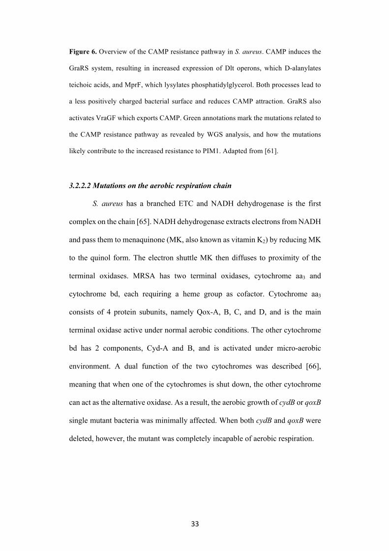
Figure 6. Overview of the CAMP resistance pathway in S. aureus. CAMP induces the
GraRS system, resulting in increased expression of Dlt operons, which D-alanylates
teichoic acids, and MprF, which lysylates phosphatidylglycerol. Both processes lead to
a less positively charged bacterial surface and reduces CAMP attraction. GraRS also
activates VraGF which exports CAMP. Green annotations mark the mutations related to
the CAMP resistance pathway as revealed by WGS analysis, and how the mutations
likely contribute to the increased resistance to PIM1. Adapted from [61].
3.2.2.2 Mutations on the aerobic respiration chain
S. aureus has a branched ETC and NADH dehydrogenase is the first
complex on the chain [65]. NADH dehydrogenase extracts electrons from NADH
and pass them to menaquinone (MK, also known as vitamin K2) by reducing MK
to the quinol form. The electron shuttle MK then diffuses to proximity of the
terminal oxidases. MRSA has two terminal oxidases, cytochrome aa3 and
cytochrome bd, each requiring a heme group as cofactor. Cytochrome aa3
consists of 4 protein subunits, namely Qox-A, B, C, and D, and is the main
terminal oxidase active under normal aerobic conditions. The other cytochrome
bd has 2 components, Cyd-A and B, and is activated under micro-aerobic
environment. A dual function of the two cytochromes was described [66],
meaning that when one of the cytochromes is shut down, the other cytochrome
can act as the alternative oxidase. As a result, the aerobic growth of cydB or qoxB
single mutant bacteria was minimally affected. When both cydB and qoxB were
deleted, however, the mutant was completely incapable of aerobic respiration.

34
Figure 7. Schematic showing aerobic respiration in S. aureus. The ETC is branched,
with two terminal oxidases, namely cytochrome aa3 and bd complexes. The dominant
terminal oxidase, cytochrome aa3, is composed of QoxA-D proteins, and requires heme
A and heme B as cofactors. The other terminal oxidase, cytochrome bd, consists of CydA
and CydB, and has heme B and and heme O as cofactors [66]. Red annotations indicate
how ETC mutations found in the PIM1-resistant strains affect aerobic respiration.
Mutations include NADH dehydrogenase gene ndh2, menaquinone synthesis genes ispD,
hepT, menA, menB, menD, menE, menF, and cytochrome aa3 gene qoxB. Adapted from
[33, 67].
Mutations in genes coding for ETC components were found in all mutant
strains (Fig. 7). The mutations included NADH dehydrogenase gene ndh2 and
cytochrome aa3 gene qoxB. Nonetheless, majority of mutations were related to
the MK biosynthesis. Genomic variations were detected in ispD, hepT, menA,
menB, menD, menE, menF, which all code for enzymes on the MK biosynthesis
pathway. Synthesis of MK in bacteria is a well-established process [68], which
includes formation of 1,4-dihydroxy-2-naphthoate (DHNA) from chorismate,
and attachment of a polyprenyl chain to DHNA (Fig. 8). Frameshift and nonsense
mutations were found in ispD, which codes for the 4-diphosphocytidyl-2-C-
methyl-D-erythritol (CDP-ME) synthase in the non-mevalonate pathway [69].
As a result, the amount of MK precursors, isopentenyl diphosphate (IPP) and its

35
isomer dimethylallyl diphosphate (DMAPP), were likely to be greatly reduced.
Moreover, a great number of men genes were inactivated, leading to less
conversion of chorismate to DHNA. The overall consequence of these gene
mutations was reduced production of menaquinone. Therefore, the PIM1-
resistant strains likely have a less active aerobic respiration.
Figure 8. Isoprenoid production from the non-mevalonate pathway and production of
MK. Red annotations show how inactivating mutations in the MK synthesis pathway
reduce overall MK production. Adapted from [68, 69].
To confirm that the ETC mutations in PIM1-resistant strains were
inactivating mutations, the basal respiration rates of the resistant strains in TSB
were measured and compared against the wildtype and ∆hemB mutant (Fig. 9).
Since the heme group is a necessary cofactor for both cytochromes, the ∆hemB
mutant was completely unable to respire, as reflected by the flat OCR curve.
Although the basal respiration rates of PIM1-resistant strains (ANG4944-4952)

36
varied depending on the exact mutations, they were all much slower than the
exponentially increasing rate of the wildtype bacteria. This observation strongly
suggests that aerobic respiration was reduced/blocked in the PIM1-resistant
strains as part of their effort to escape the toxicity of PIM1.
Figure 9. Basal respiration rate of PIM1-resistance mutants. OCR assays were
conducted on a Seahorse XFp Extracellular Flux Analyser. The bacteria strains were
tested in TSB. The basal respiration rate was monitored for wildtype LAC*, LAC ∆hemB,
PIM1-resistant strains ANG4944-4952 for 1 h. The data shown are representative of 3
biological replicates.
0 10 20 30 40 50 60 700
100
200
300
400
500
Time (minutes)
OC
R (p
mol
es/m
in)
LAC* WT
LAC ΔhemB
ANG4944
ANG4945
ANG4946
ANG4947
ANG4948
ANG4949
ANG4950
ANG4951
ANG4952

37
3.3 Binding affinity to bacterial surface influences PIM’s activity
3.3.1 Inactivation of CAMP resistance pathway confers sensitivity to PIM1
Since mutations in several CAMP resistance genes were observed in
PIM1-resistant strains, the effect of bacterial surface charge on antimicrobial
activity of PIM1 was further investigated. Mutants with increased sensitivity to
CAMP was obtained, namely ∆dltD, ∆dlt operon and mprF::tn (Table 5). The
dlt mutants lack D-alanine in their teichoic acid [70], and the mprF mutant is
unable to transfer L-lysine residue to membrane lipid phosphatidylglycerol [71].
As a result, both dlt and mprF mutants are in particular vulnerable to cationic
polymers. Despite that PIM1 does not depolarize cytoplasmic membrane like
common CAMPs as discussed in section 4.1.2, the mutants ∆dltD, ∆dlt operon
and mprF::tn are still more sensitive to PIM1 (Table 5). It seems that although
PIM1 does not kill bacteria by membrane disruption, the electrostatic interaction
with bacteria surface is nonetheless a critical condition for PIM1 to bind.
Table 5. PIM1 activity against surface charge mutants
Strain PIM1 MIC90 (µg/mL)
LAC* 2
LAC*∆dltD 0.25
LAC*∆dlt operon 0.25
LAC 2
LAC mprF::tn 0.5 3.3.2 Electrostatic adsorption is the first step of PIM1-bacteria interaction
To test the effect of surface charge mutations on PIM1’s antimicrobial
efficacy, the binding affinity of PIM1 to PIM1-resistant strain was evaluated. In
order to visualize the accumulation of polymer on bacterial surface, PIM1 was
labelled with the fluorescent dye rhodamine B. Wildtype LAC* and a PIM1-

38
resistant strain ANG4951 were exposed to a sub-inhibitory concentration of
PIM1-rhodamine. The treated bacteria were washed thoroughly to make sure the
fluorescence signal was proportional to the amount of polymer attached to the
bacteria surface. The fluorescent intensity was normalized to OD600 of bacterial
suspension. From the binding assay (Fig. 10A), PIM1 binds instantaneously to
wildtype bacteria, as reflected by the high fluorescent signal at 1 min. The
polymer gradually accumulates on the surface, thus the increase in signal
intensity over the 60 min period. In contrast, PIM1 had a hard time binding to the
PIM1-resistant strain, the amount of PIM1 accumulated on the surface of
ANG4951 was only half of the amount on wildtype cells. The reduced binding,
likely caused by the less negatively charged bacteria surface, partly contributes
to the resistance of ANG4951 (Fig. 10B). This strongly suggests that electrostatic
attraction is the first step dominating PIM1-bacteria interaction.
Figure 10. (A) PIM1-rhodamine binding assay. Wildtype MRSA LAC* and PIM1-
resistant strain AND4951 was treated with rhodamine-B labelled PIM1 during a 60 min
period. The fluorescent signal was normalized to OD600 of the bacterial suspension, and
was directly proportional to the amount of PIM1-rhodamine bound to the surface of
bacteria. (B) Dose-response curves of PIM1 against LAC* and ANG4951. The data
shown is average and standard deviation from 3 biological replicates.

39
3.4 PIM’s activity is dependent on respiration level
3.4.1 Respiratory-deficient mutants are less susceptible to PIM
The WGS analysis revealed that MRSA are less susceptible to PIM1
when aerobic respiration is inhibited. To investigate the specific relationship
between respiration level and the activity of PIM1, the antimicrobial efficacy of
PIM1 against several respiratory-deficient mutants was measured. A structural
analog of PIM1 named PIM5 was also tested for comparison. PIM5 contains two
extra oxygen atoms on the alkyl chain and thus exhibits higher hydrophilicity
than PIM1. The list of respiratory-deficient mutants tested is shown in Table 6.
Silencing qoxB inactivates the main terminal oxidase cytochrome aa3 on the ETC,
while silencing cydA effectively shuts down the alternative terminal oxidase
cytochrome bd. It should be noted that the respiration and growth rates of
qoxB::tn and cydA::tn were minimally affected due to the dual function of the
cytochromes. When any one of the cytochromes was inhibited, bacteria could
respire through the alternative cytochrome, hence the MIC is similar to wild type.
In the case of ∆hemB and ∆menD where aerobic respiration was completely
disabled, the bacteria was markedly more resistant to PIM1. Interestingly, the
lack of heme and MK increased PIM1 resistance to different extents. Although
both mutants have a completely inactive respiration chain, the resistance of
∆menD is higher than the resistance of ∆hemB. The difference is especially
pronounced for PIM5. This suggests that MK could potentially have direct
interactions with the PIM series polymers.

40
Table 6. PIM1 MIC against respiratory-deficient MRSA mutants
Strain MIC90 (µg/mL)
PIM1 PIM5
LAC/LAC* 2 8
LAC ∆menD 16 >=2048
LAC ∆hemB 4 128
LAC*qoxB::tn 2 32
LAC*cydA::tn 1 8
When exogenous MK and heme was supplemented in the growth medium
for ∆menD and ∆hemB, the resistance was partially reversed (Fig. 11A and B).
Addition of MK and heme restored the ETC of MK- and heme-deficient mutants,
and promoted bacterial growth. The untreated ∆menD grew to OD600 0.3
overnight without MK supplementation. In contrast, the overnight OD600 of
untreated sample was 0.5 in the presence of 10 µg/mL MK or 0.6 in 100 µg/mL
MK. Similarly, addition of heme increased the overnight OD600 of untreated
sample from 0.3 to 0.6. This strengthened the observation that PIM’s
antimicrobial activity is closely linked to the level of aerobic respiration.
Interestingly, heme sensitized facultative anaerobe Enterococcus faecalis
to PIM1 and PIM5. E. faecalis shares some phenotypic properties of respiratory-
deficient MRSA strains because it lacks a functional ETC. Despite that genes
encoding for type a and b cytochromes are present in E. faecalis, the strain is
naturally defective for heme production [72, 73]. However, E. faecalis contains
a heme uptake system, and therefore aerobic respiration can be activated in the
presence of exogenous heme. The OD600 of untreated E. faecalis increased from
0.3 to 0.55 with heme supplemented to growth medium. For both PIM1 and PIM5,
the activation of aerobic respiration led to higher susceptibility (Fig. 11C).

41
Therefore, the relationship between PIM1’s antimicrobial activity and aerobic
respiration is not limited to S. aureus, but also applicable to other organism.
Figure 11. Dose-response curves of PIM1 and PIM5 against (A) LAC ∆menD with and
without supplementation of 10 or 100 µg/mL exogenous MK, (B) LAC ∆hemB and (C)
E. faecalis VR583 with and without supplementation of 8 µg/mL heme. The data shown
are average and standard deviation from 3 biological replicates.

42
3.4.2 Intracellular menaquinone content decreases under exposure to PIM
The significantly higher resistance of ∆menD compared to ∆hemB raised
the possibility that MK has direct interactions with PIM1 which might contribute
to the antimicrobial activity. To investigate this further, the intracellular MK
content in PIM-treated MRSA was measured by high performance liquid
chromatography (HPLC). The major forms of MK in MRSA contains seven
(MK-7) or eight (MK-8) isoprene units [43]. Therefore, MK-7 and MK-8 were
used as standards in the HPLC analysis (Fig. 12A). The relative abundance of
MK was calculated by normalizing the peak intensity in the LC-MS spectra (254
nm) to bacterial dry mass from which MK was extracted.
Changes in MK content after exposure to PIM1 and PIM5 are shown in
Fig. 12B and C. Compared to control sample treated by DI water, both PIM1 and
PIM5 induced significant decreases in the intracellular MK level. The decrease
in MK-8 is more pronounced, mostly like because it is the more prominent form
of MK in MRSA. It should be noted that the polymers were dosed at MIC
concentrations and did not cause cell-death over the period of measurement (15
and 30 min). Therefore, the drop in MK content is most likely the cause for
impaired bacterial fitness, instead of the result of cell death. Furthermore,
samples treated with daptomycin did not lead to reduction in MK content (Fig.
12D), corroborating that the changes in MK content was not caused by membrane
disruption. This observation suggests that MK not only affects the level aerobic
respiration, but also has direct interactions with PIM1 and PIM5. There was a
report of MK-targeting antibiotic lysocin E with cyclic peptide structure [28].
However, the interaction of lysocin E with MK was through non-covalent
binding and subsequent membrane depolarization, which differs from the

43
mechanism observed for PIM1. Nonetheless, further biochemical investigations
are needed to discover the detailed reaction mechanism between PIM and MK.
Figure 12. (A) UV-vis spectrum (254 nm) of MK extract from MRSA dry mass. The
elution peak for MK-7 was observed at 10.4 min, and the peak for MK-8 was eluted at
12 min. Changes in relative MK content after 15 and 30 min exposure to MIC
concentration of (B) PIM1, (C) PIM5 and (D) daptomycin were observed. The LC-MS
peak intensity were normalized to dry biomass. The data shown are average and standard
deviation from 3 independent biological samples.

44
3.5 Relationship between the 2 categories of genomic variations
From the whole genome sequencing analysis discussed in section 3.2, 2
major pathways have been identified to be responsible for the resistance
mechanism, namely the CAMP resistance pathway and aerobic respiration.
Section 3.3 and 3.4 have been dedicated to understanding the role these 2
pathways play in the resistance mechanism.
The main function of CAMP resistance pathway is to add positive
charges to the bacteria envelope and decrease binding of cationic agents. In
section 3.3, strains lacking CAMP resistant genes (∆dltD, ∆dlt operon and
mprF::tn mutants) were found to be more susceptible to PIM1. Therefore,
electrostatic adsorption was one of the determinants of PIM1’s antimicrobial
activity. This was confirmed by the assay with rhodamine B-conjugated
polymer, where PIM1-resistant strains attracted less polymer to the surface
comparing to the wildtype strain.
In section 3.4, a series of experiments were conducted to show that
PIM1’s activity was dependent on the level of aerobic respiration. When
components of the aerobic respiration chain were knocked out, the bacteria
show decreased susceptibility to PIM1. Moreover, menaquinone was found to
be a potential target of PIM1, since deletion of menaquinone synthesis gene had
the most impact on resistance, and PIM1 treatment induced a reduction in
intracellular menaquinone content.
It is hard to judge which pathway plays a more important role in the
resistance mechanism. The 2 pathways affect different stages of PIM1’s
activity. Firstly, as a cationic agent, PIM1 adsorbs onto the negatively-charged
bacteria envelope by electrostatic attraction, a process influenced by the activity

45
of CAMP resistance pathway. When CAMP resistance genes acquire gain-of-
function mutations, bacteria have more positive charges on the surface, leading
to decreased adsorption of PIM1 and increased resistance. Secondly,
perturbation of aerobic respiration occurs after the electrostatic adsorption of
PIM1. Strains with a less active aerobic respiration chain show less
susceptibility to PIM1. It was no surprise to observe genomic variations in both
pathways for the PIM1-resistant strains. In fact, it is plausible to conclude that
the activity of both pathways need to be altered in order to achieve a high
resistance.

46
Chapter 4: Conclusion
With rapid development of antimicrobial resistance, there is an ever-
growing need for new strategies to combat bacterial infections. However, the lack
of new antimicrobial mechanisms and targets has hindered the discovery of
antibiotics. The PIM-series antimicrobial polymers also carry positive charges.
Nonetheless, they have been found to exert a distinctively different mechanism
from cationic polymers previously characterized. PIM1 did not physically disrupt
the membrane or cause membrane potential dissipation, which sets it apart from
common membrane active compounds. Moreover, PIM1 had pleiotropic effects
on multiple biosynthesis pathways in MRSA, including DNA, RNA, protein
synthesis. This indicated that PIM1 did not have a specific enzyme target. Real-
time monitoring of OCR revealed that PIM1 instantaneously blocked aerobic
respiration, preceding cell death.
WGS analysis of PIM1-resistant MRSA strains developed by resistance
evolution revealed 2 major pathways crucial for PIM1’s activity. Firstly, many
genomic variations were related to the CAMP-resistance pathway. These
mutations likely resulted in a less negatively charged bacteria surface, reducing
the electrostatic adsorption of PIM1. By labelling PIM1 with fluorescent dye
rhodamine B, it was found that PIM1 binds significantly less to PIM1-resistant
strains with mutations in CAMP-resistance pathway. This evidenced that despite
PIM1 does not cause membrane perturbations like other cationic polymers, the
electrostatic interaction still dominates the initial binding stage. Secondly,
several genes related to synthesis of ETC components were found, which
validates the postulation that PIM1 blocks aerobic respiration. Above all, the
synthesis of electron shuttle MK was found to be most disabled in response to

47
PIM1-treatment. Indeed, studies with respiratory-deficient MRSA mutants
revealed that the activity of PIM1 is strongly linked to the level of aerobic
respiration, and more importantly, that PIM1 potentially has direct interactions
with MK. By isolating the isoprenoid fraction from PIM1-treated bacteria and
detecting with HPLC, PIM1 and its derivative PIM5 were found to greatly reduce
the MK content.
This study discovered a new antimicrobial mechanism, which has not
been reported before. It adds a new dimension to the development of cationic
antimicrobial polymers. The action of PIM1 likely takes place in a 2-step process.
The cationic polymer first binds to the negatively charged bacteria surface via
electrostatic adsorption. The polymer then perturbs aerobic respiration, possibly
by direct interactions with ETC component MK. More in-depth characterization
and investigations are needed to find out the detailed interaction mechanism
between PIM1 and MK. Moreover, it is important to understand how the
perturbation of aerobic respiration leads to cell death.
The fact that S. aureus resistant cultures evolved within a period of 2
weeks and were able to withstand PIM1 treatment of 128x the original MIC did
not align with the low propensity to elicit resistance for common cationic
antimicrobial polymers. Nonetheless, this is very likely caused by PIM1 having
a distinctively different antimicrobial mechanism than other cationic polymers.
The PIM1-resistant strains were useful in understanding the mechanism of PIM1,
and the knowledge obtained for S. aureus can provide inspiration for studies in
other organisms.

48
Supplementary information
Table S 1. Full list of genomic alterations in PIM1-resistant mutants
Strain no. Type1 Ref2 Allele Freq. Annotation AA change3
ANG4945, ANG4946
INS - A 100 SAUSA300_0249 (ispD), 2-C-methyl-D-erythritol 4-phosphate cytidylyltransferase Ile49fs
DEL G - 100 SAUSA300_0993 (pdhA), pyruvate dehydrogenase E1 component Ala159fs
SNV C T 100 SAUSA300_1499 (aroK), Shikimate kinase Gly17Asp
ANG4947, ANG4948
SNV G A 100 SAUSA300_0250 (tarJ), Ribitol-5-phosphate dehydrogenase Gly333Asp
SNV T G 100 SAUSA300_0645 (graR), DNA-binding response regulator, involved in CAMP resistance
Phe13Val
INS - A 100 SAUSA300_1252 (alsT), Na+/alanine symporter Glu455fs
DEL G - 100 SAUSA300_1737 (menE), O-succinylbenzoate-CoA ligase Ala34fs
ANG4949, ANG4950
SNV G A 100 SAUSA300_0249 (ispD), 2-C-methyl-D-erythritol 4-phosphate cytidylyltransferase Gly16Asp
INS - A 98.3 Ile49fs
SNV T C 100 SAUSA300_0844 (ndh2), NADH dehydrogenase Leu270Ser
DEL‡ - - - fs
SNV G A 100 SAUSA300_0948 (menB), naphthoate synthase Gly122Asp
SNV T G 100 SAUSA300_0955 (atl), Autolysin. Asp343Ala
SNV G A 97.7 SAUSA300_1733, Dipeptidyl aminopeptidases Pro228Ser
SNV G A 100 SAUSA300_2025 (rsbU), Serine phosphatase Thr66Ile
INS - T 100 SAUSA300_2323 (cobI), Mg2+ and Co2+ transporters Lys173fs

49
SNV G A 100 SAUSA300_2386 (flp), beta-lactamase Trp260*
ANG4944, ANG4951
SNV C G 100 SAUSA300_0415 (lpl3), tandem lipoprotein. Asp152Glu
SNV A G 100 SAUSA300_0645 (graR), DNA-binding response regulator, involved in CAMP resistance
Thr11Ala
SNV A G 100 SAUSA300_1252 (alsT), Na+/alanine symporter Ile251Val
SNV A G 100 SAUSA300_0971 (purl), Phosphoribosylformylglycinamidine synthase II Thr314Lys
SNV G T 100 SAUSA300_1737 (menE), O-succinylbenzoate-CoA ligase Thr314Lys
ANG4952 SNV A C 100 SAUSA300_0648 (vraG), ABC transporter permease. Involved in CAMP resistance. Gln454Pro
SNV C A 100 SAUSA300_1442 (srrA), Respiratory response protein SrrA Glu31*
ANG5103 SNV G T 100 SAUSA300_0945 (menF), isochorismate synthases Gly222Val
SNV T A 100 SAUSA300_1077 (murD), peptidoglycan biosynthesis Leu2His
SNV G A 100 SAUSA300_2025 (rsbU), serine phosphatase Ala127Val
SNV G A 100 SAUSA300_2230 (modA) ABC-type molybdate transporter Thr150Ile
SNV C T 100 SAUSA300_2323 (cobI), Mg2+ and Co2+ transporters Gln232*
ANG5104 SNV C A 100 SAUSA300_0946 (menD), 2-succinyl-6-hydroxy-2, 4-cyclohexadiene-1-carboxylate synthase Ser287Tyr
SNV T A 100 SAUSA300_1167 (pnpA), polynucleotide phosphorylase/polyadenylase Leu338Ile
SNV C T 100 SAUSA300_1255 (fmtC), oxacillin/methicillin resistance-related FmtC protein Ala96Val
SNV C A 100 SAUSA300_1370 (ebpS), cell surface elastin binding protein Gly471Val
SNV A T 100 SAUSA300_2025 (rsbU), serine phosphatase Ile304Asn
SNV G C 100 SAUSA300_2323 (cobI), Mg2+ and Co2+ transporters Trp26Ser
ANG5105 SNV G A 100 SAUSA300_0022 (walH), uncharacterized protein Ala173Thr

50
SNV C T 100 SAUSA300_0279 (esaA), membrane protein, involved in protein secretion Gln372*
SNV G T 100 SAUSA_0910 (mgtE), magnesium transporter MgtE Asp237Tyr
DEL A - 91.7 SAUSA300_0946 (menD), 2-succinyl-6-hydroxy-2, 4-cyclohexadiene-1-carboxylate synthase Thr525fs
SNV C T 100 SAUSA300_1255 (fmtC), oxacillin/methicillin resistance-related FmtC protein Arg50Cys
SNV A T 100 SAUSA300_1267 (trpB), tryptophan synthase beta subunit Leu195Phe
INS - T 95.2 SAUSA300_2025 (rsbU), serine phosphatase Cys28fs
ANG5106 SNV C G 100 SAUSA300_0869 (rexB), exonuclease RexB, DNA metabolism Ala489Gly
SNV G T 100 SAUSA_0910 (mgtE), magnesium transporter MgtE Met102Ile
SNV C G 100 SAUSA300_1154 (cdsA), cytidylyltransferase Ala113Gly
INS - A 95.2 SAUSA300_1252 (alsT) Na+/alanine symporter Glu455fs
SNV G A 100 SAUSA300_1255 (fmtC), oxacillin/methicillin resistance-related FmtC protein Gly61Glu
SNV G A 100 SAUSA300_1737 (menE), o-succinylbenzoate-CoA ligase Ser59Leu
SNV G A 100 SAUSA300_2025 (rsbU), serine phosphatase Gln6*
ANG5107 SNV A G 100 SAUSA300_0645 (graR), DNA-binding response regulator, CAMP resistance Asp182Gly
DEL CAG - 87.5 SAUSA300_0944 (menA), 1,4-dihydroxy-2-naphthoate octaprenyltransferase Ala288del
DEK CTT - 83.9 SAUSA300_2022 (rpoF), RNA polymerase sigma factor SigB Glu149del
SNV C G 100 SAUSA300_2323 (cobI), Mg2+ and Co2+ transporters His129Asp
ANG5108 SNV G A 95.8 SAUSA300_0623 (tagA), teichoic acid biosynthesis proteins Trp227*
SNV G A 100 SAUSA300_0645 (graR), DNA-binding response regulator, CAMP resistance Glu184Lys
SNV C G 100 SAUSA300_0646 (graS), signal transduction histidine kinase, CAMP resistance Leu169Val

51
INS - T 100 SAUSA300_0668, recombination and DNA strand exchange inhibitor protein Ser139fs
SNV G A 100 SAUSA300_0844 (ndh2), NADH dehydrogenase Trp49*
SNV C T 100 SAUSA300_0962 (qoxB), heme/copper-type cytochrome/quinol oxidases Trp277*
DEL A - 100 SAUSA300_1193 (glpD), glycerol-3-phosphate dehydrogenase, energy metabolism Met139fs
ANG5109 SNV A C 100 SAUSA300_0279 (esaA), membrane protein, involved in protein secration Lys379Gln
SNV C T 100 SAUSA300_0800 (sek), staphylococcal enterotoxin K Val136Ile
DEL A - 93.75 SAUSA300_0946 (menD), 2-succinyl-6-hydroxy-2, 4-cyclohexadiene-1-carboxylate synthase Lys289fs
SNV T A 100 SAUSA300_1193 (glpD), glycerol-3-phosphate dehydrogenase, energy metabolism Leu146Ile
SNV C T 100 SAUSA300_1255 (fmtC), oxacillin/methicillin resistance-related FmtC protein Ser295Leu
SNV C T 100 SAUSA300_1483, hypothetical protein Arg47His
SNV G T 100 SAUSA300_1604 (mreD), rod shape-determining protein, cell envelope biogenesis Pro156Thr
SNV C A 100 SAUSA300_2359 (tcyA), amino acid ABC transporter Gly91Trp
DEL T - 100 SAUSA300_2454, membrane spanning protein Cys184fs
DEL T - 100 SAUSA300_2498 (crtN), squalene synthase Asn32fs
ANG5110 SNV C A 100 SAUSA300_0027, hypothetical protein Ala389Glu
SNV T C 100 SAUSA300_1255 (fmtC), oxacillin/methicillin resistance-related FmtC protein Leu341Ser
SNV G T 100 SAUSA300_1359 (hepT), polyprenyl synthase, involved in isoprenoid synthesis Arg39Ser
INS - A 96.2 SAUSA300_2127 (sepA), multidrug resistance efflux pump Leu31fs
ANG5111 SNV A G 100 SAUSA300_0369, hypothetical protein Tyr281His
SNV A T 100 SAUSA300_0645 (graR), DNA-binding response regulator, CAMP resistance Glu184Val

52
SNV C T 100 SAUSA300_0989 (mjA), mRNA degradation ribonucleases Arg209His
SNV C T 100 SAUSA300_1255 (fmtC), oxacillin/methicillin resistance-related FmtC protein Arg50Cys
SNV C T 100 SAUSA300_1737 (menE), o-succinylbenzoate-CoA ligase Trp205*
ANG5112 SNV C T 100 SAUSA300_0249 (ispD), 2-C-methyl-D-erythritol 4-phosphate cytildylyltransferase Arg207*
SNV T C 100 SAUSA300_0647 (vraF), ABC transporter ATP-binding protein Ser195Pro
SNV C T 100 SAUSA300_1255 (fmtC), oxacillin/methicillin resistance-related FmtC protein Ser295Leu
SNV G T 100 SAUSA300_1481, Gene: SAUSA300_1481 Thr224Lys
SNV G T 100 SAUSA300_1541 (grpE), heat shock protein GrpE Pro112Thr
ANG5113 SNV G T 100 SAUSA300_0251 (tarL), putative teichoic acid biosynthesis protein, cell envelope biogenesis Gly314Val
SNV C T 100 SAUSA300_0821 (sufU), NifU family SUF system FeS assembly protein Ala142Val
SNV G T 100 SAUSA300_0944 (menA), 1,4-dihydroxy-2-naphthoate octaprenyltransferase Arg19Ser
DEL TATC - 94.6 SAUSA300_2025 (rsbU), serine phosphatase Asp303fs
SNV G A 100 SAUSA300_2323 (cobI), Mg2+ and Co2+ transporters, inorganic ion transport and metabolism Val262Ile
1 Type of mutation: INS stands for insertion, DEL stands for deletion. DEL‡ denotes a multiple deletion of 54 bp. SNV is short for single nucleotide variant. 2 Base in reference genome 3 Amino acid change: fs refers to frameshift. * indicates stop codon.

53
Reference 1. Liu, C., A. Bayer, S.E. Cosgrove, R.S. Daum, S.K. Fridkin, R.J.
Gorwitz, . . . H.F. Chambers, Clinical practice guidelines by the infectious diseases society of america for the treatment of methicillin-resistant Staphylococcus aureus infections in adults and children: executive summary. Clin Infect Dis, 2011. 52(3): p. 285-92.
2. Le Loir, Y., F. Baron, and M. Gautier, Staphylococcus aureus and food poisoning. Genet Mol Res, 2003. 2(1): p. 63-76.
3. Ohlsen, K. and U. Lorenz, Novel targets for antibiotics in Staphylococcus aureus. Future Microbiol, 2007. 2(6): p. 655-66.
4. Jensen, S.O. and B.R. Lyon, Genetics of antimicrobial resistance in Staphylococcus aureus. Future Microbiol, 2009. 4(5): p. 565-82.
5. Foster, T.J., Antibiotic resistance in Staphylococcus aureus. Current status and future prospects. FEMS Microbiol Rev, 2017. 41(3): p. 430-449.
6. Giesbrecht, P., T. Kersten, H. Maidhof, and J. Wecke, Staphylococcal cell wall: morphogenesis and fatal variations in the presence of penicillin. Microbiol Mol Biol Rev, 1998. 62(4): p. 1371-414.
7. Hartman, B.J. and A. Tomasz, Low-affinity penicillin-binding protein associated with beta-lactam resistance in Staphylococcus aureus. J Bacteriol, 1984. 158(2): p. 513-6.
8. Katayama, Y., T. Ito, and K. Hiramatsu, A new class of genetic element, staphylococcus cassette chromosome mec, encodes methicillin resistance in Staphylococcus aureus. Antimicrob Agents Chemother, 2000. 44(6): p. 1549-55.
9. Zeng, D., D. Debabov, T.L. Hartsell, R.J. Cano, S. Adams, J.A. Schuyler, . . . J.L. Pace, Approved Glycopeptide Antibacterial Drugs: Mechanism of Action and Resistance. Cold Spring Harb Perspect Med, 2016. 6(12).
10. Weigel, L.M., D.B. Clewell, S.R. Gill, N.C. Clark, L.K. McDougal, S.E. Flannagan, . . . F.C. Tenover, Genetic analysis of a high-level vancomycin-resistant isolate of Staphylococcus aureus. Science, 2003. 302(5650): p. 1569-71.
11. Silver, L.L., Challenges of antibacterial discovery. Clin Microbiol Rev, 2011. 24(1): p. 71-109.
12. Schweizer, H.P., Triclosan: a widely used biocide and its link to antibiotics. FEMS Microbiol Lett, 2001. 202(1): p. 1-7.
13. Ling, L.L., T. Schneider, A.J. Peoples, A.L. Spoering, I. Engels, B.P. Conlon, . . . K. Lewis, A new antibiotic kills pathogens without detectable resistance. Nature, 2015. 517: p. 455.
14. Carmona-Ribeiro, A.M. and L.D. de Melo Carrasco, Cationic antimicrobial polymers and their assemblies. Int J Mol Sci, 2013. 14(5): p. 9906-46.
15. Ganewatta, M.S. and C. Tang, Controlling macromolecular structures towards effective antimicrobial polymers. Polymer, 2015. 63: p. A1-A29.
16. Stebbins, N.D., M.A. Ouimet, and K.E. Uhrich, Antibiotic-containing polymers for localized, sustained drug delivery. Adv Drug Deliv Rev, 2014. 78: p. 77-87.

54
17. Yang, Y., Z. Cai, Z. Huang, X. Tang, and X. Zhang, Antimicrobial cationic polymers: From structural design to functional control. Vol. 50. 2017.
18. Gilbert, P. and L.E. Moore, Cationic antiseptics: diversity of action under a common epithet. J Appl Microbiol, 2005. 99(4): p. 703-15.
19. Denyer, S.P., Mechanisms of action of antibacterial biocides. International Biodeterioration & Biodegradation, 1995. 36(3): p. 227-245.
20. Salton, M.R., Lytic agents, cell permeability, and monolayer penetrability. The Journal of general physiology, 1968. 52(1): p. 227-252.
21. Milovic, N.M., J. Wang, K. Lewis, and A.M. Klibanov, Immobilized N-alkylated polyethylenimine avidly kills bacteria by rupturing cell membranes with no resistance developed. Biotechnol Bioeng, 2005. 90(6): p. 715-22.
22. Broxton, P., P.M. Woodcock, and P. Gilbert, A study of the antibacterial activity of some polyhexamethylene biguanides towards Escherichia coli ATCC 8739. J Appl Bacteriol, 1983. 54(3): p. 345-53.
23. Broxton, P., P.M. Woodcock, F. Heatley, and P. Gilbert, Interaction of some polyhexamethylene biguanides and membrane phospholipids in Escherichia coli. J Appl Bacteriol, 1984. 57(1): p. 115-24.
24. Palermo, E.F. and K. Kuroda, Chemical structure of cationic groups in amphiphilic polymethacrylates modulates the antimicrobial and hemolytic activities. Biomacromolecules, 2009. 10(6): p. 1416-28.
25. Sambhy, V., B.R. Peterson, and A. Sen, Antibacterial and hemolytic activities of pyridinium polymers as a function of the spatial relationship between the positive charge and the pendant alkyl tail. Angew Chem Int Ed Engl, 2008. 47(7): p. 1250-4.
26. Demberelnyamba, D., K.S. Kim, S. Choi, S.Y. Park, H. Lee, C.J. Kim, and I.D. Yoo, Synthesis and antimicrobial properties of imidazolium and pyrrolidinonium salts. Bioorg Med Chem, 2004. 12(5): p. 853-7.
27. Gabriel, G.J., A.E. Madkour, J.M. Dabkowski, C.F. Nelson, K. Nüsslein, and G.N. Tew, Synthetic Mimic of Antimicrobial Peptide with Nonmembrane-Disrupting Antibacterial Properties. Biomacromolecules, 2008. 9(11): p. 2980-2983.
28. Ng, V.W.L., J.P.K. Tan, J. Leong, Z.X. Voo, J.L. Hedrick, and Y.Y. Yang, Antimicrobial Polycarbonates: Investigating the Impact of Nitrogen-Containing Heterocycles as Quaternizing Agents. Macromolecules, 2014. 47(4): p. 1285-1291.
29. Fey, P.D., J.L. Endres, V.K. Yajjala, T.J. Widhelm, R.J. Boissy, J.L. Bose, and K.W. Bayles, A genetic resource for rapid and comprehensive phenotype screening of nonessential Staphylococcus aureus genes. MBio, 2013. 4(1): p. e00537-12.
30. Diep, B.A., S.R. Gill, R.F. Chang, T.H. Phan, J.H. Chen, M.G. Davidson, . . . F. Perdreau-Remington, Complete genome sequence of USA300, an epidemic clone of community-acquired meticillin-resistant Staphylococcus aureus. Lancet, 2006. 367(9512): p. 731-9.
31. Pader, V., E.H. James, K.L. Painter, S. Wigneshweraraj, and A.M. Edwards, The Agr quorum-sensing system regulates fibronectin binding

55
but not hemolysis in the absence of a functional electron transport chain. Infect Immun, 2014. 82(10): p. 4337-47.
32. Boles, B.R., M. Thoendel, A.J. Roth, and A.R. Horswill, Identification of genes involved in polysaccharide-independent Staphylococcus aureus biofilm formation. PLoS One, 2010. 5(4): p. e10146.
33. Zeden, M.S., C.F. Schuster, L. Bowman, Q. Zhong, H.D. Williams, and A. Grundling, Cyclic di-adenosine monophosphate (c-di-AMP) is required for osmotic regulation in Staphylococcus aureus but dispensable for viability in anaerobic conditions. J Biol Chem, 2018. 293(9): p. 3180-3200.
34. Schwarz, S., P. Silley, S. Simjee, N. Woodford, E. van Duijkeren, A.P. Johnson, and W. Gaastra, Editorial: assessing the antimicrobial susceptibility of bacteria obtained from animals. J Antimicrob Chemother, 2010. 65(4): p. 601-4.
35. DuMont, A.L., T.K. Nygaard, R.L. Watkins, A. Smith, L. Kozhaya, B.N. Kreiswirth, . . . V.J. Torres, Characterization of a new cytotoxin that contributes to Staphylococcus aureus pathogenesis. Molecular Microbiology, 2011. 79(3): p. 814-825.
36. Roth, B.L., M. Poot, S.T. Yue, and P.J. Millard, Bacterial viability and antibiotic susceptibility testing with SYTOX green nucleic acid stain. Appl Environ Microbiol, 1997. 63(6): p. 2421-31.
37. Te Winkel, J.D., D.A. Gray, K.H. Seistrup, L.W. Hamoen, and H. Strahl, Analysis of Antimicrobial-Triggered Membrane Depolarization Using Voltage Sensitive Dyes. Front Cell Dev Biol, 2016. 4: p. 29.
38. Ling, L.L., T. Schneider, A.J. Peoples, A.L. Spoering, I. Engels, B.P. Conlon, . . . K. Lewis, A new antibiotic kills pathogens without detectable resistance. Nature, 2015. 517(7535): p. 455-9.
39. Bowman, L., M.S. Zeden, C.F. Schuster, V. Kaever, and A. Grundling, New Insights into the Cyclic Di-adenosine Monophosphate (c-di-AMP) Degradation Pathway and the Requirement of the Cyclic Dinucleotide for Acid Stress Resistance in Staphylococcus aureus. J Biol Chem, 2016. 291(53): p. 26970-26986.
40. Didier, J.P., R. Villet, E. Huggler, D.P. Lew, D.C. Hooper, W.L. Kelley, and P. Vaudaux, Impact of ciprofloxacin exposure on Staphylococcus aureus genomic alterations linked with emergence of rifampin resistance. Antimicrob Agents Chemother, 2011. 55(5): p. 1946-52.
41. Bligh, E.G. and W.J. Dyer, A rapid method of total lipid extraction and purification. Can J Biochem Physiol, 1959. 37(8): p. 911-7.
42. Minnikin, D.E., A.G. O'Donnell, M. Goodfellow, G. Alderson, M. Athalye, A. Schaal, and J.H. Parlett, An integrated procedure for the extraction of bacterial isoprenoid quinones and polar lipids. Journal of Microbiological Methods, 1984. 2(5): p. 233-241.
43. Nahaie, M.R., M. Goodfellow, D.E. Minnikin, and V. Hajek, Polar lipid and isoprenoid quinone composition in the classification of Staphylococcus. J Gen Microbiol, 1984. 130(9): p. 2427-37.
44. Rami, M., A. Innocenti, J.-L. Montero, A. Scozzafava, J.-Y. Winum, and C.T. Supuran, Synthesis of rhodamine B–benzenesulfonamide conjugates and their inhibitory activity against human α- and bacterial/fungal β-carbonic anhydrases. Bioorganic & Medicinal Chemistry Letters, 2011. 21(18): p. 5210-5213.

56
45. Bucki, R., J.J. Pastore, P. Randhawa, R. Vegners, D.J. Weiner, and P.A. Janmey, Antibacterial Activities of Rhodamine B-Conjugated Gelsolin-Derived Peptides Compared to Those of the Antimicrobial Peptides Cathelicidin LL37, Magainin II, and Melittin. Antimicrobial Agents and Chemotherapy, 2004. 48(5): p. 1526.
46. Jucker, B.A., H. Harms, and A.J. Zehnder, Adhesion of the positively charged bacterium Stenotrophomonas (Xanthomonas) maltophilia 70401 to glass and Teflon. J Bacteriol, 1996. 178(18): p. 5472-9.
47. Deka, S.R., A.K. Sharma, and P. Kumar, Cationic polymers and their self-assembly for antibacterial applications. Curr Top Med Chem, 2015. 15(13): p. 1179-95.
48. Phillips, D.J., J. Harrison, S.J. Richards, D.E. Mitchell, E. Tichauer, A.T.M. Hubbard, . . . M.I. Gibson, Evaluation of the Antimicrobial Activity of Cationic Polymers against Mycobacteria: Toward Antitubercular Macromolecules. Biomacromolecules, 2017. 18(5): p. 1592-1599.
49. Ruhr, E. and H.G. Sahl, Mode of action of the peptide antibiotic nisin and influence on the membrane potential of whole cells and on cytoplasmic and artificial membrane vesicles. Antimicrob Agents Chemother, 1985. 27(5): p. 841-5.
50. Hobbs, J.K., K. Miller, A.J. O'Neill, and I. Chopra, Consequences of daptomycin-mediated membrane damage in Staphylococcus aureus. J Antimicrob Chemother, 2008. 62(5): p. 1003-8.
51. Strahl, H. and L.W. Hamoen, Membrane potential is important for bacterial cell division. Proc Natl Acad Sci U S A, 2010. 107(27): p. 12281-6.
52. Lohner, K., New strategies for novel antibiotics: peptides targeting bacterial cell membranes. Gen Physiol Biophys, 2009. 28(2): p. 105-16.
53. Venkatesh, M., V.A. Barathi, E.T.L. Goh, R. Anggara, M.H.U.T. Fazil, A.J.Y. Ng, . . . R. Lakshminarayanan, Antimicrobial Activity and Cell Selectivity of Synthetic and Biosynthetic Cationic Polymers. Antimicrobial agents and chemotherapy, 2017. 61(10): p. e00469-17.
54. Ferrero, L., B. Cameron, B. Manse, D. Lagneaux, J. Crouzet, A. Famechon, and F. Blanche, Cloning and primary structure of Staphylococcus aureus DNA topoisomerase IV: a primary target of fluoroquinolones. Mol Microbiol, 1994. 13(4): p. 641-53.
55. Jansen van Rensburg, M.J., A.C. Whitelaw, and B.G. Elisha, Genetic basis of rifampicin resistance in methicillin-resistant Staphylococcus aureus suggests clonal expansion in hospitals in Cape Town, South Africa. BMC Microbiol, 2012. 12: p. 46.
56. Watanakunakorn, C., Mode of action and in-vitro activity of vancomycin. J Antimicrob Chemother, 1984. 14 Suppl D: p. 7-18.
57. Leclercq, R. and P. Courvalin, Resistance to macrolides and related antibiotics in Streptococcus pneumoniae. Antimicrob Agents Chemother, 2002. 46(9): p. 2727-34.
58. Lobritz, M.A., P. Belenky, C.B. Porter, A. Gutierrez, J.H. Yang, E.G. Schwarz, . . . J.J. Collins, Antibiotic efficacy is linked to bacterial cellular respiration. Proc Natl Acad Sci U S A, 2015. 112(27): p. 8173-80.

57
59. Kashyap, D.R., M. Kuzma, D.A. Kowalczyk, D. Gupta, and R. Dziarski, Bactericidal peptidoglycan recognition protein induces oxidative stress in Escherichia coli through a block in respiratory chain and increase in central carbon catabolism. Mol Microbiol, 2017. 105(5): p. 755-776.
60. Bettenbrock, K., H. Bai, M. Ederer, J. Green, K.J. Hellingwerf, M. Holcombe, . . . R.K. Poole, Towards a systems level understanding of the oxygen response of Escherichia coli. Adv Microb Physiol, 2014. 64: p. 65-114.
61. Joo, H.S. and M. Otto, Mechanisms of resistance to antimicrobial peptides in staphylococci. Biochim Biophys Acta, 2015. 1848(11 Pt B): p. 3055-61.
62. Rajagopal, M., M.J. Martin, M. Santiago, W. Lee, V.N. Kos, T. Meredith, . . . S. Walker, Multidrug Intrinsic Resistance Factors in Staphylococcus aureus Identified by Profiling Fitness within High-Diversity Transposon Libraries. MBio, 2016. 7(4).
63. Falord, M., U. Mader, A. Hiron, M. Debarbouille, and T. Msadek, Investigation of the Staphylococcus aureus GraSR regulon reveals novel links to virulence, stress response and cell wall signal transduction pathways. PLoS One, 2011. 6(7): p. e21323.
64. Bayer, A.S., T. Schneider, and H.G. Sahl, Mechanisms of daptomycin resistance in Staphylococcus aureus: role of the cell membrane and cell wall. Ann N Y Acad Sci, 2013. 1277: p. 139-58.
65. Artzatbanov, V. and V.V. Petrov, Branched respiratory chain in aerobically grown Staphylococcus aureus--oxidation of ethanol by cells and protoplasts. Arch Microbiol, 1990. 153(6): p. 580-4.
66. Hammer, N.D., M.L. Reniere, J.E. Cassat, Y. Zhang, A.O. Hirsch, M. Indriati Hood, and E.P. Skaar, Two heme-dependent terminal oxidases power Staphylococcus aureus organ-specific colonization of the vertebrate host. MBio, 2013. 4(4).
67. Gotz, F. and S. Mayer, Both terminal oxidases contribute to fitness and virulence during organ-specific Staphylococcus aureus colonization. MBio, 2013. 4(6): p. e00976-13.
68. Meganathan, R. and O. Kwon, Biosynthesis of Menaquinone (Vitamin K2) and Ubiquinone (Coenzyme Q). EcoSal Plus, 2009. 3(2).
69. Heuston, S., M. Begley, C.G. Gahan, and C. Hill, Isoprenoid biosynthesis in bacterial pathogens. Microbiology, 2012. 158(Pt 6): p. 1389-401.
70. Peschel, A., M. Otto, R.W. Jack, H. Kalbacher, G. Jung, and F. Gotz, Inactivation of the dlt operon in Staphylococcus aureus confers sensitivity to defensins, protegrins, and other antimicrobial peptides. J Biol Chem, 1999. 274(13): p. 8405-10.
71. Andra, J., T. Goldmann, C.M. Ernst, A. Peschel, and T. Gutsmann, Multiple peptide resistance factor (MprF)-mediated Resistance of Staphylococcus aureus against antimicrobial peptides coincides with a modulated peptide interaction with artificial membranes comprising lysyl-phosphatidylglycerol. J Biol Chem, 2011. 286(21): p. 18692-700.
72. Winstedt, L., L. Frankenberg, L. Hederstedt, and C. von Wachenfeldt, Enterococcus faecalis V583 contains a cytochrome bd-type respiratory oxidase. J Bacteriol, 2000. 182(13): p. 3863-6.

58
73. Painter, K.L., A. Hall, K.P. Ha, and A.M. Edwards, The Electron Transport Chain Sensitizes Staphylococcus aureus and Enterococcus faecalis to the Oxidative Burst. Infect Immun, 2017. 85(12).