modeling a dehalogenase fold into the 8-Å density map for
TRANSCRIPT

Modeling a Dehalogenase Fold into the 8-Å Density Map forCa21-ATPase Defines a New Domain Structure
David L. Stokes* and N. Michael Green†
*Skirball Institute of Biomolecular Research, Department of Cell Biology, New York University School of Medicine, New York,New York 10016 USA, and †National Institute for Medical Research, London NW7 1AA, England
ABSTRACT Members of the large family of P-type pumps use active transport to maintain gradients of a wide variety ofcations across cellular membranes. Recent structures of two P-type pumps at 8-Å resolution have revealed the arrangementof transmembrane helices but were insufficient to reveal the architecture of the cytoplasmic domains. However, recentproposals of a structural homology with a superfamily of hydrolases offer a new basis for modeling these domains. In thecurrent work, we have extended the sequence comparison for the superfamily and delineated domains in the 8-Å density mapof Ca21-ATPase. The homology suggests a new domain structure for Ca21-ATPase and, specifically, that the phosphorylationdomain adopts a Rossman fold. Accordingly, the atomic structure of L-2 haloacid dehalogenase has been fitted into therelevant domain of Ca21-ATPase. The resulting model suggests the existence of two ATP sites at the interface between twodomains. Based on this new model, we are able to reconcile numerous results of mutagenesis and chemical cross-linkingwithin the catalytic domains. Furthermore, we have used the model to predict the configuration of MgzATP at its binding site.Based on this prediction, we propose a mechanism, involving a change in Mg21 liganding, for initiating the domainmovements that couple sites of ion transport to ATP hydrolysis.
INTRODUCTION
In all cells, ionic homeostasis is maintained by members ofthe large family of P-type ion pumps (Axelsen andPalmgren, 1998). Ca21-ATPase and Na1/K1-ATPase arethe two best-studied members, and they have been charac-terized in great detail with regard to reaction kinetics, chem-ical modification, and site-directed mutagenesis (Andersen,1995; Moller et al., 1996; MacLennan et al., 1997). Struc-tural studies by electron microscopy have recently culmi-nated in structures at 8-Å resolution for both Ca21-ATPase(Zhang et al., 1998) and H1-ATPase (Auer et al., 1998).These structures revealed similar arrangements of 10 trans-membrane helices, and, in the case of Ca21-ATPase, con-nections were identified between four of these transmem-brane helices and the corresponding stalk helices. Inaddition, a particular pathway for calcium through the trans-membrane domain was proposed and the sequences of stalkand transmembrane helices were assigned, using a variety ofconstraints. Similar assignments were not possible for thecytoplasmic domains because they contain mainlyb-strandsand short helices, which are not easily identified at 8-Åresolution.
Nevertheless, based on secondary structure predictionsand on functional considerations, a sophisticated model hasevolved for the cytoplasmic portion of Ca21-ATPase, whichincludes the delineation of several domains (MacLennan et
al., 1985; Taylor and Green, 1989; Green and Stokes, 1992).According to this model, the main cytoplasmic loop be-tween transmembrane helices M4 and M5 begins with aphosphorylation domain that is separated from a nucleotide-binding domain by the dominant tryptic cleavage site (atR505) and ends with a 60-residue “hinge” domain, whichwas postulated to fold back onto the phosphorylation do-main to produce the tertiary fold. An alternative to thesethree sequential domains is a model in which the nucleotide-binding domain forms an insert relative to a combinedphosphorylation/hinge domain. Although the sequential do-main model was never entirely satisfactory, the alternativewith an inserted domain was unusual and, at that time, wasnot supported by evidence for an insertion site.
However, Aravind et al. (1998) recently proposed theexistence of a superfamily of HAD hydrolases that includesthe P-type pumps and that specifically supports a modelwith an inserted nucleotide-binding domain. This superfam-ily is based on homology between the phosphorylationdomains of the P-type ATPases and the catalytic domains ofserine/threonine phosphatases, phosphoglycomutases, andhaloacid dehalogenases (HADs). Besides the sequence ho-mology, these families also share the use of an aspartyl esterintermediate (Collet et al., 1998), the dynamics of which, inthe pumps, are closely linked to the occlusion and transportof cations across the membrane. Here we show how theproposed homology leads to a new domain structure for theP-type ATPases, which we correlate with domains identi-fied in the cytoplasmic portion of the 8-Å density map forCa21-ATPase. By fitting the crystal structure for a haloaciddehalogenase to this map, we propose a model not only ofthe phosphorylation domain, but also of the structural com-ponents coupling this domain with the ion transport siteswithin the membrane. This model provides insight into the
Received for publication 7 October 1999 and in final form 12 December1999.
Address reprint requests to Dr. David L. Stokes, Skirball Institute ofBiomolecular Research, Department of Cell Biology, New York UniversitySchool of Medicine, New York, NY 10016. Tel.: 212-263-1580; Fax:212-263-1580; E-mail: [email protected].
© 2000 by the Biophysical Society
0006-3495/00/04/1765/12 $2.00
1765Biophysical Journal Volume 78 April 2000 1765–1776

effects of site-directed mutation and chemical modification;it supports the existence of two overlapping nucleotide-binding sites, and it suggests a role for Mg21 in initiatingthe conformational changes required for Ca21 translocation.
RESULTS
Sequence alignment and domain structure ofdehalogenases and P-type pumps
A homology between the L-2 haloacid dehalogenase fromPseudomonassp. YL (1JUD) and P-type pumps was pro-posed by Aravind et al. (1998), based on four sequencemotifs that contribute to the active sites of these divergentenzymes. We begin our comparison by reviewing evidencefrom a wide variety of sources that support this homology.The crystal structure of 1JUD reveals the existence of twodistinct domains (Fig. 1) (Hisano et al., 1996), the coredomain being ana/b sandwich with the topology of aRossmann fold (b-strand order 3-2-1-4-5-6). The catalyticaspartate is at the C-terminus ofb1 and is followed first bya five-residue coil (cyan in Fig. 1) and then by the insertedsubdomain, which consists of a distorted four-helix bundle(gray in Fig. 1). The chain then returns to the first helix (a5)of the core domain and completes the Rossman fold. Thesequence motifs involved in the proposed homology are infour of the loops at the top of theb-sheet (red residues inFig. 1b, including the catalytic aspartate and the cyan coil).According to mutagenesis studies (Kurihara et al., 1995)and the crystal structures of dehalogenases in the presenceof a substrate analog (Li et al., 1998; Ridder et al., 1997),most of these conserved residues contribute directly to theactive site. R41 is the only residue from thea-helical sub-domain that is directly involved in dehalogenation andserves to stabilize and to abstract the halide (Ridder et al.,1997). This role suggests that the subdomain is primarilyinvolved in substrate specificity and binding, which wouldexplain its variability across the proposed superfamily.
We have expanded the original alignment of these foursequence motifs to encompass the entire sequence betweenM4 and M6. Fig. 2 compares the sequence from 1JUD withfour representative P-type ATPases from three main sub-families (Axelsen and Palmgren, 1998): the type I heavymetal ATPases (CadA), the type III plant and fungal H1-ATPases (H-ATP), and the type II animal Na1/K1- andCa21-ATPases (NaATP, CaATP). The sequences homolo-gous to the dehalogenase core domain are referred to as thephosphorylation or P-domain and the inserted subdomain(140–240 residues in the ATPases) as the adenosine-bind-ing or A-domain. The presence of this inserted subdomainin Ca21-ATPase is strongly supported by the recent isola-tion and characterization of a proteolytic fragment withtermini (T357-T608) close to those of the putative A-domain(Champeil et al., 1998). Furthermore, this proteolytic frag-ment was shown to retain tertiary structure, to bind nucle-
FIGURE 1 Structure of chloroacetate dehalogenase (1JUD). (a) Thisribbon model was generated by ICM (Abagyan et al., 1994), using a pdbfile of 1JUD, in which important functional residues were mutated byQuanta (MSI, San Diego, CA) to the corresponding residues of the se-quence of the Ca21-ATPase (shown inred in b). The backbone of theRossmann fold is color-coded from blue at the N-terminus to red at theC-terminus. The inserted four-helix subdomain is gray. Catalytic residuesare colored as follows: Asp (351, 601, 703, 707) are yellow, Lys (352, 684)are green, Arg (678) is purple, theb1-loop (KTGTL356) is cyan, and VO3
2
is white. (b) The topology diagram shows the positions of three majorinserts by Ca21-ATPase into the sequence of 1JUD (dotted lines,with thenumber of inserted residues in brackets). The largest insert replacesa4 inthe A-domain and includes sites of TNP-ATP and FITC labeling (K492 andK515). The two short strands that link the domains form a hinge and havebeen redesignated ash1 andh2, and the strands of theb-sheet have beenrenumbered fromb1 to b6. Residue numbers correspond to CaATPase.
1766 Stokes and Green
Biophysical Journal 78(4) 1765–1776

FIGURE 2 Alignment of the phosphorylation (P) and adenosine binding (A) domains of the ATPases with 1JUD. Four sequences of ATPases(SWISSPROT codes cadaostaau, pma1oneucr, atn1osheep, atcborabit) are aligned together with secondary structures predicted independently for threefamilies by PHD (Rost and Sander, 1993). The predicted structure was used to supplement the sequence motifs and to guide the alignment to 1JUD. TheA-domain has been divided at a conserved intron site (dashed line) into an N-terminal A1 region, with some similarity to 1JUD, and a C-terminal A2 regioncontaining adenosine-binding sites, but with no resemblance to 1JUD. The intron sites for CadA and H-ATP were deduced from homologous positions ofthe Menkes Cu21-ATPase (Tumer et al., 1995) and theNicotianaH1-ATPase (Perez et al., 1992) genes. Sites of chemical labeling (1–9), mutation (a–j),proteolysis (..) and Ca21 binding (11) are shown for the Ca21-ATPase. Many of these sites are also relevant to Na1/K1-ATPase, but three unique sites
Modeling Ca21-ATPase from Dehalogenase 1767
Biophysical Journal 78(4) 1765–1776

otides, and to react specifically with fluorescein isothiocya-nate (FITC), supporting its proposed functional role inbinding nucleotide. The independently predicted secondarystructures of the P-domains are consistent both with eachother and with the crystal structure of 1JUD. The onlysignificant disparities are a variable insert followinga6 andsome differences in the loop betweenb5 andb6 (which isthe site of a two-helix insert in the dehalogenase fromXanthobacter autrophicus; Ridder et al., 1997). In view ofthe low overall sequence identity (8–14%), further supportwas obtained from the structure-based GenTHREADERprogram, which picked 1JUD from the structure database asthe only certain match (p 5 1) to any of the ATPases in Fig.2 (N. M. Green and J. Saldanha, unpublished observations).All together, these results provide firm ground for fitting the
structure of the 1JUD core domain into the 8-Å density mapof Ca21-ATPase, which we describe below.
Given the dissimilarity of their substrates, it is not sur-prising that the subdomain of 1JUD differs from the A-do-mains of the pumps, which are all much larger. Neverthe-less, near the N-terminus of the A-domain in Ca21-ATPasethere are three predicted helices that could parallel those of1JUD, one of which (a3) includes a notable sequence sim-ilarity (EATETAL/QATEDAL). In 1JUD these three heli-ces are reconnected to the core domain by a fourth helix,whereas in the pumps, this connection expands to form aregion of mixed secondary structure, including sites labeledby FITC and by azido-adenosine nucleotides (Figs. 1b and2). These differences in homology and secondary structureas well as the existence of a conserved intron site (dashed
TABLE 1 Functional Sites of the Na1/K1-ATPase and Ca21-ATPase
Site Label Domain
Activity
Ref.ATPase E1P AcPase E2P
Ca21-ATPase1 C344 ANSmal P:b1 1 1 1 1 Bigelow and Inesi (1992)
NBDCl “2 C364 ANSmal A1 1 1 1 1 “3 K394 Phospholamban* A1 James et al. (1989)
K4004 K464 ErITC A1 0.2* 1 1 1 Huang et al. (1998)5 K492 PAL A2 2 2 1 1 McIntosh (1998)
ADPpal 2 2 “TNPAzATP 2 2 1 1 “AMCA 1 1 1 1 “Glutarald.* 2 1 E1P 2 “
6 K515 FITC A2 2 2 1 1 Murphy (1988)7 651–665 A52 antibody P:a6-b5 McIntosh (1998)8 C674 IAF P:b3 1 1 1 1 Bigelow and Inesi (1992)
IAEDANS9 R678 Glutarald.* P:b3 2 1 E1P 2 McIntosh (1998)i K684 ADPpal/Ca P:b3-a7 2 2 “
Na1/K1-ATPase49 C457 IAF A1 1 1 1 1 “59 G502 AzATP A2 0.5* 2 Tran et al. (1994)5 K480 TNPAzADP A2 2 2 1 1 Ward and Cavieres (1998b)j D703 ClRATP P:b4-a8 2 2 McIntosh (1998)k D707 ClRATP P:a8 2 2 “69 K719 FSBA P:a8 2 1 “
Site designations refer to Fig. 2, where primes relate exclusively to the Na1/K1-ATPase and lowercase letters refer to sites that have also been mutatedand therefore also appear in Table 2. The A-domain has been subdivided into A1 and A2 by the intron site in Fig. 2, and elements of secondary structurewithin the P-domain refer to the Rossman fold of 1JUD in Fig. 1. The catalytic and phosphorylation characteristics are given approximately as1 (normal)and2 (,10% active); when more complex effects are observed (*) the original papers should be consulted. ATPase and AcPase indicate hydrolysis ofATP and acetylphosphate, respectively; E1P and E2P indicate formation of phosphoenzyme from ATP and Pi, respectively. Most references are to two majorreviews. ADPpal, pyridoxal-59-diphosphoadenosine; AMCA, 7-amino-4-methylcoumarin-3-acetic acid; ANSmal, 2-(4-maleimidoanilino)naphthalene-6-sulfonate; AzATP, 8-azido-ATP; ClRATP,g-(4-N-2-chloroethyl-N-methylamino)-benzylamide-ATP; ErITC, erythrosin-59-isothiocyanate; FITC, fluores-cein-59-isothiocyanate; FSBA, 59-(4-fluorosulfonylbenzoyl) adenosine; IAEDANS,N-iodoacetyl-N9-(5-sulfo-1-naphthyl)ethylenediamine; IAF, 59-iodoac-etamidofluorescein; NBDCl, 7-chloro-4-nitrobenzo-2-oxa-1,3-diazole; PAL, pyridoxal-59-phosphate; TNPAzATP, 29,39-O-trinitrophenyl-8-azido-adenosine triphosphate.
for Na1/K1-ATPase are designated with primes (49, 59, and 69). These sites are characterized in more detail in Tables 1 and 2. Finally, residues directlyinvolved in the catalysis of 1JUD are indicated by x’s below their sequence.
1768 Stokes and Green
Biophysical Journal 78(4) 1765–1776
line in Fig 2; McIntosh, 1998) suggest that the A-domainmay be subdivided into two parts (A1 and A2).
The proposed functional distinction between the P- andA-domains is supported by the effects of site-directed mu-tagenesis and chemical modification of Ca21-ATPase andNa1/K1-ATPase (Tables 1 and 2). ATP analogs with reac-tive groups on the ring invariably label residues in theA-domain (sites 4, 5, 59, 6); as a result, these analogs blockATP-activated phosphorylation and transport, but not hy-drolysis of acetyl phosphate or phosphorylation by Pi. Incontrast, reactive groups near theg-phosphate label residuesin the P-domain (sites 69, j, k); the modified ATPases arecompletely inactive and phosphorylation either by ATP orby Pi is blocked. Similarly, mutations of residues in theA-domain (sites 5, 59, and 6) have much smaller effects onactivity than do those in the P-domain (sites a–k), and themutations at site 5 are the only ones with specific effects onATP binding (McIntosh et al., 1996).
Domain boundaries in the density map
These considerations suggest that the cytoplasmic part ofCa21-ATPase is made up of three domains: the A- andP-domains for the loop between M4 and M5 and the “b-domain” for the loop between M2 and M3, which is pre-dicted to be predominantlyb-strand (Green and Stokes,1992). We have therefore looked for these three domains inthe cytoplasmic portion of our Ca21-ATPase density map(Fig. 3). The transmembrane and stalk regions had previ-ously been fitted witha-helices (Zhang et al., 1998), and wetherefore considered only the densities between the top ofthe stalk and the top of the molecule. In particular, weexamined the map for low-density regions that might rep-resent boundaries between domains, both in contour plots ofmap sections (Fig. 3,d–f) and in surface representations of
the molecule (Fig. 3,a–c). The molecular surface dependson the density level that is chosen, and the figure shows thecytoplasmic region at a relatively high density threshold,with three domains (colored yellow, orange, and blue). Theboundaries of the yellow domain were the best defined anddelineate a block-like domain that sits on top of the stalk.On the other hand, the boundary between orange and bluedomains was unclear, and we have left the correspondingpart of the surface uncolored in this region. Contour plotscorrespond to cross sections through a dimer, with domainsoutlined on one of the two protomers.
According to the homology with dehalogenases, the P-domain composing the Rossman fold begins and ends onlya few residues from the ends of stalk helices S4 and S5,respectively, so it was logical to assign it to the yellowdomain in Fig. 3. Indeed, the volume of this yellow domaincorresponds closely to the expected mass of the P-domain(Table 3), leading us to fit the atomic coordinates for 1JUD(see next section). The nucleotide is expected to bind at theinterface between the P-domain and the A-domain, which isconsistent with the previously defined site of CrATP bind-ing (Yonekura et al., 1997). Although the 14-Å resolution ofthe CrATP difference map was insufficient to define bound-aries, the CrATP difference density lies on the boundarybetween the orange and yellow domains (# in Fig. 3a andarrow in Fig. 3 d). This suggests that the orange domaincorresponds to the A-domain and that the blue domaincorresponds to theb-domain
Two very high densities are apparent in Fig. 3f, which webelieve are good candidates for the decavanadate used toinduce crystallization. One of these is present on the dyadaxis (* in Fig. 3 f), and, given its position on a symmetryaxis, it is unlikely to represent protein. The peak is;10 Åin diameter and is therefore the right size and shape for adecavanadate ion (Day et al., 1987). Accordingly, this peak
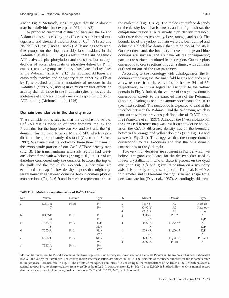
TABLE 2 Mutation-sensitive sites of Ca21-ATPase
Site Mutant Domain Type Site Mutant Domain Type
a D351-N P:b1 P2 5 F487-S A2 Katp oc2-T P2 5 K492-Y A2 Katp oc2
6 K515-E A2 slowb K352-R P: L P2 g D601-E P: h2 P2
-Q P2 -N E1Pc T353-A P: L E1P h D627-A P:b2-a6 P2
-S Slow -N E1Pd T355-A P: L Slow i K684-R P:b3-a7 E1P
-S WT -H P2e L356-T P: L Slow j D703-A P:b4-a8 P2 oc1
-I WT k D707-A P: a8 P2f T357-A P: h1 P2
-S WT
Most of the mutants in the P- and A-domains that have large effects on activity are shown and most are in the P-domain; the A-domain has been subdividedinto A1 and A2 by the intron site. The corresponding lowercase letters are shown in Fig. 2. The elements of secondary structure for the P-domain referto the proposed Rossman fold in Fig. 1. The effects of mutagenesis are classified according to the nomenclature of Andersen (1995), which provides ageneral review: P2, no phosphorylation from MgATP or from Pi; E1P, transition from E1-P z Mg z Ca2 to E2MgPi is blocked; Slow, cycle is normal exceptthat the transport rate is slow; oc2, unable to occlude Ca21 with CrATP; WT, cycle is normal.
Modeling Ca21-ATPase from Dehalogenase 1769
Biophysical Journal 78(4) 1765–1776

was omitted from the molecule in previous publications(Zhang et al., 1998; Toyoshima et al., 1993). Further exam-ination of Fig. 3 f reveals a second peak of high densitywithin the A-domain, which is much higher than that fromana-helix (magenta in Fig. 3,a andc; triple arrowheadinFig. 3 f). Again, this peak is;10 Å in diameter and mayrepresent a second, internal site for decavanadate. If so, it
would imply a total of 1.5 moles of decavanadate per moleof ATPase. Previous measurement of decavanadate binding(Csermely et al., 1985) determined a stoichiometry of 1.5–2moles of decavanadate per mole of Ca21-ATPase, which isconsistent with our model.
The assignment of this high-density peak to decavanadateleaves only a narrow neck of density (green line, Fig. 3,a,
FIGURE 3 Domains in the cytoplasmic re-gion of Ca21-ATPase. (a–c) Surface represen-tation of the density map in which the threedomains composing the cytoplasmic head havebeen contoured at a higher density and colored.Yellow corresponds to the P-domain, orange tothe A-domain, blue to theb-domain, and purpleto the putative decavanadate site; the green lineindicates a possible delineation between the twoparts of the A-domain. Domain boundarieswere difficult to discern at the top of the mol-ecule, which has therefore been left uncolored.The molecule is shown in three orientationsrelated by;90° rotations about the vertical (z)axis. The three zones indicated at the rightmargins correspond to the sections shown ind–f. The white arrows on the left correspond tothe levels shown in Fig. 4,a andc. (d–f) Sec-tions through two molecules related by a two-fold axis, showing the low density boundariesbetween the three cytoplasmic domains. Thecolored contours represent a single moleculemasked from the surrounding crystal lattice andcorrespond to four superimposed 2-Å-thick sec-tions. Green, dashed contours are lowest, blackare intermediate, and magenta are the highestdensity; thus, domain boundaries are likely toexist in regions filled with green contour lines.Twofold related molecules are represented by asingle 2-Å section with only black contours,and the domains have been outlined in colorscorresponding to those ina–c. The single arrowin d and # in a show the cavity occupied byCr-ATP (Yonekura et al., 1997). The doublearrowhead ine indicates a deep, curved cavitythat is postulated as a potential second nucleo-tide binding site. Densities corresponding totwo proposed decavanadate binding sites areindicated inf by the asterisks and by the triplearrowhead; the latter is colored magenta inaandc.
1770 Stokes and Green
Biophysical Journal 78(4) 1765–1776

c, f) between the upper and lower parts of the A-domain.These could correspond to the division of the correspondingsequence into A1 and A2 subdomains (see above), the upperregion (A1) adjoining the hinge region of the P-domain andthe lower region (A2) lining the cavities that bind thenucleotide. Indeed, A2 contains all of the functional sites inthe A-domain, and its calculated molecular weight is con-sistent with the size of this part of the map (Table 3). It isalso noteworthy that the internal decavanadate site contactsboth parts of the A-domain as well as the P- andb-domainsand so is in an ideal position to stabilize their interactions.This stabilization could account for the compact structure ofthis E2 form of the pump compared to the E1Ca2 form(Stokes et al., 1999).
Docking of dehalogenase core domain intoCa21-ATPase
Our next step was to dock the atomic structure of thedehalogenase core domain into the yellow density assignedto the Ca21-ATPase P-domain (Figs. 4 and 5). We tried twodifferent automated methods for docking, but neither pro-duced convincing or consistent results. Docking was there-fore done manually, and the general orientation was chosento be consistent with short connections between the stalkhelices S4 and S5 and the ends of the dehalogenase fold, sothat theb-strands run away from the membrane. The patternof density within the P-domain was then used for a moreprecise positioning of the dehalogenase fold. Generallyspeaking,a-helices generate relatively strong density peaksat 8 Å resolution, whereasb-strands remain undefined.Thus we positioneda5 anda9 in a plausible region of highdensity along the back of the P-domain, which also matcheda6 to a high-density rod that protrudes from the side of theP-domain. In addition, a low-density region in the P-domainwas matched with a low-density region in maps generatedfrom the dehalogenase coordinates betweena5, a9, and theb-sheet (Figs. 1a and 6). There were, however, severaldiscrepancies remaining between the two structures, whichare not surprising given the large differences in sequenceand the probability that extensions of stalk helices S2 andS3 run through this region and thus contribute extra density.In particular, two regions of 1JUD project beyond the yel-
low envelope of the P-domain. One is the L56 loop (Fig. 4a), which is variable among dehalogenases and is predictedto be slightly shorter anda-helical for Ca21-ATPase (wehave remodeled this loop in Fig. 6 to minimize this protru-sion). The other is the loop betweenb3 anda7 (Fig. 4 c),which is actually a short 310 helix in the dehalogenase andis two residues shorter in Ca21-ATPase. On the other hand,there are unassigned densities in the P-domain, the largestbeing at the bottom of Fig. 4c. This particular density isnext to the loop betweena6 andb3, which consists of ashort 310 helix in the dehalogenase but has an extra 28residues in Ca21-ATPase; these extra residues could plau-sibly fill this unassigned space. We can find no well-resolved connecting density between theb-domain andstalk helices, although chains could be accommodated byadjustments ofa8 and L56, which occupy a relatively large,high-density region at the interface with the A-domain (Fig.3 d). Alternatively, theb-domain terminates as a smallcolumn of density that resembles a helix (Fig. 3d), and it ispossible that it is connected to the stalk by a disordered, andtherefore invisible, loop.
DISCUSSION
Modeling of the catalytic site
To evaluate the consistency of this model with the results ofmutagenesis and chemical modification, a particular bind-ing configuration for ATP must be specified. Although theactive site for nucleophilic attack on theg-phosphate isdefined by the dehalogenase structure, the binding of therest of the ATP molecule is not, both because it is substan-tially different from dehalogenase ligands and because thecontribution of the subdomain to this binding pocket iscurrently undefined. Nevertheless, according to our model,the binding pocket for ATP should occur at the interfacebetween the A-domain and the P-domain, and two low-density cavities are seen at this interface (labeleda andb inFig. 6, single and double arrowheadsin Fig. 3, d and e).These two cavities ultimately merge and provide plausiblebinding pockets for either one or possibly two adenosinering systems. They are both close enough to the catalyticaspartate (D351) to permit interaction with theg-phosphate;
TABLE 3 Size of domains in cytoplasmic head of Ca21-ATPase
SequenceMolecular mass
(kDa)
Fraction of cyt. head
Molecular mass(%)
Volume(%)
P-domain (234–247, 338–358, 601–750) 19.82 33.5 31.6A2-domain* (376–600) 14.21 24.1 25.5A11b-domain* (359–375, 120–233) 24.97 42.3 42.9
*Because the domain boundaries between the A-domain andb-domain were ambiguous, it was not possible to determine their relative masses from themap. However, the uncertainty was confined to the A1 region; A2 was well defined and corresponded in size to the sequence segment following the intronsite shown in Fig. 2.
Modeling Ca21-ATPase from Dehalogenase 1771
Biophysical Journal 78(4) 1765–1776
however, the location of the difference density produced byCrATP is consistent with the position of cavitya, eventhough the two cavities could not be resolved in the corre-
sponding maps because of their lower resolution (Yonekuraet al., 1997). Cavityb might contribute to a second ATPbinding site, for which there is considerable evidence in theliterature (see below).
The g-phosphate itself, which is modeled as a transitionstate VO3, occupies an active site at the topological switchpoint betweenb-strands 1 and 4; such a switch point isgenerally favorable for an active site ina/b-type structures(Branden, 1980) and specifically matches the active site ofthe dehalogenases (Hisano et al., 1996; Ridder et al., 1997).The side chains shown in Fig. 6 correspond to mutation-sensitive residues in Table 2, which have been modeled intothe dehalogenase fold. The location of all of these residuesat the ligand binding surface, mostly in close juxtapositionwith the g-phosphate, is strongly supportive of our model.In particular, theg-phosphate is surrounded by four muta-tionally sensitive residues (D351, D703, D707, K684), whosehomologs in the dehalogenase are directly involved in nu-cleophilic attack on the substrate. The signature sequencefor P-type pumps (DKTGTL356) is represented by the cyanloop, followingb1, which links D351 to the hinge and whichis stabilized by a continuous network of H-bonds in thedehalogenase structure. The loop followingb2 containsD627, which is more distant, but could indirectly affect thedisposition of the preceding several residues (T625 andG626), which also contribute ligands to the hydrogen-bond-ing system at the active site of dehalogenases; indeed, thesepreceding residues constitute the fourth sequence motif thatwas used to define the superfamily (Aravind et al., 1998).The final residue shown is R678 at the C-terminus ofb3,whose orientation in Fig. 6 was determined by energyminimization. This residue is located on the surface of theP-domain, pointing toward the A-domain, consistent withits observed cross-linking by glutaraldehyde to K492 in theA-domain. The ability of decavanadate to block this cross-link is evidence for the effect of this reagent on domaininteractions.
Chemical modification and evidence for twonucleotide binding sites
The binding of nucleotides and nucleotide analogs has beenthe subject of many types of experiment, and the complexityof the results has given rise to a variety of different expla-nations. The basic observation is that the high-affinity cat-alytic site binds ATP with aKd of ;5 mM to give aphosphoenzyme whose subsequent transformations are fur-ther activated by ATP binding at sites of medium (50mM)and low (1 mM) affinity (e.g., Moller et al., 1980). In otherexperiments the binding constants for inhibitors or for pro-tection by ATP against irreversible inhibitors have beenmeasured. The simplest explanation (Champeil et al., 1988;Moczydlowski and Fortes, 1981) postulates a single nucle-otide site that changes in affinity after phosphorylation ofD351, giving rise to low-affinity modulations. Others assume
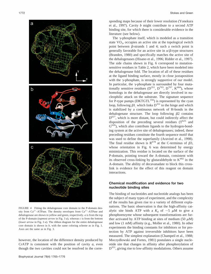
FIGURE 4 Fitting the dehalogenase core domain to the P-domain den-sity from Ca21-ATPase. The density envelopes from Ca21-ATPase anddehalogenase are shown in yellow and green, respectively.a is from the topof the P-domain (topmost arrowin Fig. 3a), whereasc is from the bottom(lower arrow in Fig. 3 a). The ribbon diagram for the entire dehalogenasecore domain is shown inb, with the same coloring scheme as in Fig. 1.Axes are the same as in Fig. 3.
1772 Stokes and Green
Biophysical Journal 78(4) 1765–1776

separate catalytic and regulatory sites, which may be on thesame peptide chain (Ward and Cavieres, 1998a) or onopposing chains of a dimeric unit (Linnertz et al., 1998).
Our observation of two cavities at the domain interface isconsistent with the existence of two nucleotide sites withina single molecule (a andb in Fig. 6). Becauseb-g-CrATPoccupies sitea and has the same stereochemistry as MgATP(as found in the active sites of myosin and F1 ATPase), weassume that this is the catalytic ATP site. Siteb is near K678
and extends upward out of the section in Fig. 6. As men-tioned, K678 can be cross-linked to K492 by glutaraldehyde,which in turn can be cross-linked to K515 by 4,49-diisothio-cyanostilbene-2,29-disulfonic acid, suggesting that thesethree residues are close together. Association with a nucle-otide binding site is suggested by the fact that K492 ismodified by a number of nucleotide analogs and that FITClabeling of K515 blocks ATP binding (Table 1). Thus wepropose that the well-studied residues K492 and K515 areassociated with siteb and form a secondary nucleotidebinding site.
In fact, there is ample evidence from various ATP ana-logs that suggest the existence of two nucleotide bindingsites that can be simultaneously occupied. For example,although labeling of K492 or K515 generally blocks phos-phorylation by ATP, D351 can still react with smaller sub-
strates such as Pi, acetyl phosphate, orp-nitrophenyl phos-phate (and even, in the Na1/K1-ATPase, withO-methylfluorescein phosphate). In addition, nucleotides and theiranalogs are still able to bind to the labeled enzyme withmoderate affinity and inhibit its reactivity with these smallsubstrates, supporting the existence of a site distinct fromK492 and K515 (Champeil et al., 1988; Mignaco et al., 1996;Scheiner-Bobis et al., 1993; Ward and Cavieres, 1998b).One explanation for the complexity of these effects is thatreagents bound at the catalytic sitea produce a conforma-tional change that closes down the secondary siteb, whereasreagents bound at siteb have mainly local effects. Thisexplanation is supported by the general observation that thereactivity of K492and K515is blocked when the catalytic sitea is occupied. Furthermore, the reactivity of 11 of 16cytoplasmic thiol groups is reduced after ATP binding to thecatalytic site (Thorley-Lawson and Green, 1977), but reac-tion of FITC at the secondary site does not protect thiolgroups to any significant extent (N. M. Green, unpublishedobservations) and does not prevent phosphorylation of D351
with Pi or with p-nitrophenyl phosphate. Assuming thatATP binds preferentially to the catalytic site, this couldexplain why direct binding measurements have failed tosupport the existence of two sites. An inconsistency exists inthe large effects of mutations at F487 and K492 on binding of
FIGURE 5 New model for the ar-chitecture of Ca21-ATPase, showingtransmembrane and stalk helices aswell as the P-domain. Transmem-brane and stalk domains are as fittedby Zhang et al. (1998) with hypothet-ical connecting loops. The hinge be-tween the P-domain and A-domain isindicated by h, and the putative deca-vanadate peak is indicated by V. TheN- and C-termini of the P-domain areindicated inb, as is the location of thecatalytic D351 (star). Axes are thesame as in Figs. 3 and 4.
Modeling Ca21-ATPase from Dehalogenase 1773
Biophysical Journal 78(4) 1765–1776

ATP to the catalytic site (McIntosh et al., 1996), but suchinconsistencies could be reconciled by postulating ligand-induced domain movements, an idea that is supported bypreliminary observations of the Ca21-bound, E1 state, dis-cussed below. Moreover, it should be remembered that wedo not yet have a 3D structure for the E1 form and that ourstructure for the E2 form includes both decavanadate andthapsigargin, which may influence the relations between thedomains.
The multiplicity of binding sites is further extended byour observation of two sites for decavanadate distinct fromthe CrATP site, both of which remain occupied in thehelical arrays containing CrATP (Yonekura et al., 1997). Atthe same time there is evidence for competition betweendecavanadate and ATP (Coan et al., 1986), although thedata did not allow quantitative conclusions about the disso-ciation constants. The competition was not caused by or-thovanadate, because the latter does not prevent firm bind-ing of ATP (Andersen and Moller, 1985). This impliesallosteric interaction between the decavanadate sites and anATP site, which is also suggested by a decreased decavana-
date binding by the FITC derivative of the ATPase(Csermely et al., 1985).
Mechanistic implications of theP-domain structure
This model also provides potential insights into the couplingbetween sites of phosphorylation and Ca21 transport. Someearlier proposals have emphasized effects transmitted by thedirect link between the Ca21 binding site in M4 and thephosphorylation site, while others have considered transportto be the result of more global changes (reviewed by McIn-tosh, 1998). In our proposed structure, the phosphorylatedD351 is closely linked to the hinge segmenth1 (TTN359) bythe short, fully conserved sequence KTGTL356. In the de-halogenase, this connection forms a five-residue loop,which is stabilized by two hydrogen bonds betweeni andi 1 5 positions. Also according to this analogy, anothermutation-sensitive sequence, DPP603, composes the returnhalf of the hinge (segmenth2) and is linked to TTN359 by
FIGURE 6 Catalytic region of Ca21-ATPase superimposed on a section from the density map. The section is at the same orientation and position as thoseof Fig. 3 d. For this figure, the pdb file of 1JUD was mutated in Quanta, replacing functional residues in the loops at the ends ofb1, b2, b3, andb4 bythe corresponding residues of the Ca21-ATPase (as for Fig. 1). In addition, the L56 loop was shortened and remodeled as two turns of helix, based on thepredicted secondary structure of the corresponding Ca21-ATPase sequence. This remodeling would reduce the projection of this loop outside the densityenvelope of the ATPase as seen in Fig. 4a. ADP-Mg-VO3 was imported from the structure of myosin (1VOM; Smith and Rayment, 1996) and orientedbetween D351 and sitea of the density map. Energy minimization of the whole structure was performed using the CHARMm option of Quanta. Only theremodeling of L56 required changes in backbone configuration; other changes were restricted to the side chains. The functionally important residues areshown in full, as is R678, which can be cross-linked by glutaraldehyde to the A-domain. The figure was prepared using the program MOLSCRIPT (Kraulis,1991).
1774 Stokes and Green
Biophysical Journal 78(4) 1765–1776
two H-bonds. We suggest that this close linking between theKTGTL356 loop and the hinge allows changes at the cata-lytic site to initiate domain movements.
In particular, there is evidence that the bonding of Mg21
changes markedly during the reaction cycle. Previous stud-ies have measured a fall in the rate constant for Mg21
release from 80 s21 in the EzMgzATP complex (Reinsteinand Jencks, 1993) to less than 0.5 s21 in E1;PzMg(Ca21)2
(Wakabayashi and Shigekawa, 1984). This dramatic in-crease in Mg21 affinity after phosphate transfer and disso-ciation of ADP suggests that loss of phosphate coordinationhas been compensated for by new protein ligands withMg21. Similar large changes in the Mg21 off-rate accom-pany phosphorylation from Pi (Ogurusu et al., 1991), andanalogous behavior is observed in Na1/K1-ATPase, usingCo21 as a substitute for Mg21 (Richards, 1988). So far theseresults have not resulted in any detailed mechanistic pro-posals, because of lack of a good structural model. How-ever, the H-bond network between the MgATP site and thehinge in our model provides a basis for Mg21-bonding toinitiate movements of the P- and A-domains. Such move-ments could ultimately be transmitted via the stalk to theCa21 sites within the membrane.
There are several plausible candidates for Mg21 ligandsamong the mutation-sensitive sites of Ca21-ATPase, includ-ing T353and T355of the catalytic loop, D627, D703, and D707.A recent structural comparison (Ridder and Dijkstra, 1999)between the catalytic sites of CheY and 1JUD, has sug-gested D351, D703, and D707 as Mg21 ligands. In the relatedFixJ, the conformational change that accompanies forma-tion of the aspartyl phosphate involved reorientation of T625
to bind phosphate together with a 6 Å movement of thehistidine corresponding to D627 towards the KTGTL356 loop(Birck et al., 1999). In Ca21-ATPase, analogous changesare bound to influence Mg21 bindings as well as the con-figuration of the hinge region.
The structure of Ca21-ATPase used for our fitting islikely to represent the E2 conformation of the enzyme,because crystallization is prevented by Ca21 and promotedby thapsigargin (Stokes and Lacapere, 1994). The bindingof Ca21 to the transport site initiates a major structuraltransition from the E2 to the E1 conformation, which acti-vates the catalytic ATP site and leads to nucleophilic attackof D351 on ATP. Comparison of the fitted 3D structure withprojection images of Ca21-ATPase in the presence of sat-urating calcium concentrations (i.e., E1zCa2; Cheong et al.,1996; Ogawa et al., 1998) and with the structure of H1-ATPase in the E1 conformation (Ku¨hlbrandt et al., 1998;Stokes et al., 1999) reveals a substantial rearrangement ofthe cytoplasmic nose. According to our model, this rear-rangement can be explained by a movement of the A-do-main relative to the P-domain. In particular, the interfacethat contains the putative ATP-binding site appears to openup and produce a gap between the domains, perhaps pro-moting the binding of nucleotide. The two hinge segments
between P- and A-domains could provide the necessaryflexibility for such a rearrangement. Indeed, analogous do-main movements have been shown to modulate the acces-sibility of the ligand binding site of various structurallyrelated enzymes, such as the family of phosphoribosyl trans-ferases (Smith, 1999). Clearly, revealing the nature of thisconformational change is an important step in understand-ing the structural basis for active transport.
We thank Steve Smerdon for his help with Fig. 6.
This work was supported by National Institutes of Health grant AR40997to DLS and by the MRC.
REFERENCES
Abagyan, R. A., M. M. Totrov, and D. N. Kuznetsov. 1994. ICM: a newmethod for protein modeling and design.J. Comp. Chem.15:488–506.
Andersen, J. P. 1995. Dissection of the functional domains of the sarco-plasmic reticulum Ca21-ATPase by site-directed mutagenesis.Biosci.Rep.15:243–261.
Andersen, J. P., and J. V. Moller. 1985. The role of Mg21 and Ca21 in thesimultaneous binding of vanadate and ATP at the phosphorylation site ofsarcoplasmic reticulum Ca21-ATPase.Biochim. Biophys. Acta.815:9–15.
Aravind, L., M. Y. Galperin, and E. V. Koonin. 1998. The catalytic domainof the P-type ATPase has the haloacid dehalogenase fold.Trends Biol.Sci.23:127–129.
Auer, M., G. A. Scarborough, and W. Ku¨hlbrandt. 1998. Three-dimensional map of the plasma membrane H1-ATPase in the openconformation.Nature.392:840–843.
Axelsen, K. B., and M. G. Palmgren. 1998. Evolution of substrate speci-ficities in the P-type ATPase superfamily.J. Mol. Evol.46:84–101.
Bigelow, D. J., and G. Inesi. 1992. Contributions of chemical derivatizationand spectroscopic studies to the characterization of the Ca21 transportATPase of sarcoplasmic reticulum.Biochim. Biophys. Acta.1113:323–338.
Birck, C., L. Mourey, P. Gouet, B. Fabry, J. Schumacher, P. Rousseau, D.Kahn, and J.-P. Samama. 1999. Conformational changes induced byphosphorylation of the FixJ receiver domain.Structure. 7:1505–1515.
Branden, C. I. 1980. Relation between structure and function of alpha/beta-proteins.Q. Rev. Biophys.13:317–338.
Champeil, P., T. Menguy, S. Soulie, B. Juul, A. G. de Gracia, F. Rusconi,P. Falson, L. Denoroy, F. Henao, M. le Maire, and J. V. Moller. 1998.Characterization of a protease-resistant domain of the cytosolic portionof sarcoplasmic reticulum Ca21-ATPase: nucleotide- and metal-bindingsites.J. Biol. Chem.273:6619–6631.
Champeil, P., S. Riollet, S. Orlowski, F. Guillain, C. J. Seebregts, and D. B.McIntosh. 1988. ATP regulation of sarcoplasmic reticulum Ca21-ATPase. Metal-free ATP and 8-bromo-ATP bind with high affinity tothe catalytic site of phosphorylated ATPase and accelerate dephosphor-ylation. J. Biol. Chem.263:12288–12294.
Cheong, G.-W., H. S. Young, H. Ogawa, C. Toyoshima, and D. L. Stokes.1996. Lamellar stacking in three-dimensional crystals of Ca21-ATPasefrom sarcoplasmic reticulum.Biophys. J.70:1689–1699.
Coan, C., D. J. Scales, and A. J. Murphy. 1986. Oligovanadate binding tosarcoplasmic reticulum ATPase. Evidence for substrate analogue behav-ior. J. Biol. Chem.261:10394–10403.
Collet, J. F., V. Stroobant, M. Pirard, G. Delpierre, and E. Van Schaftingen.1998. A new class of phosphotransferases phosphorylated on an aspar-tate residue in an amino-terminal DXDX(T/V) motif.J. Biol. Chem.273:14107–14112.
Csermely, P., S. Varga, and A. Martonosi. 1985. Competition betweendecavanadate and fluorescein isothiocyanate on the Ca21-ATPase ofsarcoplasmic reticulum.Eur. J. Biochem.150:455–460.
Day, V. W., W. G. Klemperer, and D. J. Maltbie. 1987. Where are theprotons in H3V10O28? J. Am. Chem. Soc.109:2991–3002.
Modeling Ca21-ATPase from Dehalogenase 1775
Biophysical Journal 78(4) 1765–1776

Green, N. M., and D. L. Stokes. 1992. Structural modelling of P-type ionpumps.Acta Physiol. Scand.146:59–68.
Hisano, T., Y. Hata, T. Fujii, J.-Q. Liu, T. Kurihara, N. Esaki, and K. Soda.1996. Crystal structure of L-2-haloacid dehalogenase fromPseudomo-nassp.YL. J. Biol. Chem.271:20322–20330.
Huang, S., S. Negash, and T. C. Squier. 1998. Erythrosin isothiocyanateselectively labels lysine464 within an ATP-protectable binding site onthe Ca-ATPase in skeletal sarcoplasmic reticulum membranes.Biochem-istry. 37:6949–6957.
James, P., M. Inui, M. Tada, M. Chiesi, and E. Carafoli. 1989. Nature andsite of phospholamban regulation of the Ca21 pump of sarcoplasmicreticulum.Nature.342:90–92.
Kraulis. 1991. MOLSCRIPT—a program to produce detailed and sche-matic plots of protein structures.J. Appl. Crystallogr.24:946–950.
Kuhlbrandt, W., M. Auer, and G. A. Scarborough. 1998. Structure ofP-type ATPases.Curr. Opin. Struct. Biol.8:510–516.
Kurihara, T., J. Q. Liu, V. Nardi-Dei, H. Koshikawa, N. Esaki, and K.Soda. 1995. Comprehensive site-directed mutagenesis of L-2-halo aciddehalogenase to probe catalytic amino acid residues.J. Biochem.117:1317–1322.
Li, Y.-F., Y. Hata, T. Fujii, T. Hisano, M. Nishihara, T. Kurihara, and N.Esaki. 1998. Crystal structures of reaction intermediates of L-2 haloaciddehalogenase and implications for the reaction mechanism.J. Biol.Chem.273:15035–15044.
Linnertz, H., P. Urbanova, T. Obsil, P. Herman, E. Amler, and W. Schoner.1998. Molecular distance measurements reveal an (ab)2 dimeric struc-ture of Na1/K1-ATPase. High affinity ATP binding site and K1-activated phosphatase reside on different alpha-subunits.J. Biol. Chem.273:28813–28821.
MacLennan, D. H., C. J. Brandl, B. Korczak, and N. M. Green. 1985.Amino-acid sequence of a Ca21 1 Mg21- dependent ATPase fromrabbit muscle sarcoplasmic reticulum, deduced from its complementaryDNA sequence.Nature.316:696–700.
MacLennan, D. H., W. J. Rice, and N. M. Green. 1997. The mechanism ofCa21 transport by sarco(endo)plasmic reticulum Ca21 ATPases.J. Biol.Chem.272:28815–28818.
McIntosh, D. 1998. The ATP binding sites of P-type ion transportATPases.Adv. Mol. Cell Biol.23A:33–99.
McIntosh, D. B., D. G. Woolley, B. Vilsen, and J. P. Andersen. 1996.Mutagenesis of segment 487Phe-Ser-Arg-Asp-Arg-Lys492 of sarcoplas-mic reticulum Ca21-ATPase produces pumps defective in ATP binding.J. Biol. Chem.271:25778–25789.
Mignaco, J. A., O. H. Lupi, F. T. Santos, H. Barrabin, and H. M. Scofano.1996. Two simultaneous binding sites for nucleotide analogs are kinet-ically distinguishable on the sarcoplasmic reticulum Ca21-ATPase.Bio-chemistry.35:3886–3891.
Moczydlowski, E. G., and P. A. Fortes. 1981. Inhibition of sodium andpotassium adenosine triphosphatase by 29,39-O-(2, 4, 6-trinitrocyclo-hexadienylidene) adenine nucleotides. Implications for the structure andmechanism of the Na:K pump.J. Biol. Chem.256:2357–2366.
Moller, J. V., K. E. Lind, and J. P. Andersen. 1980. Enzyme kinetics andsubstrate stabilization of detergent-solubilized and membranous (Ca21-Mg21)- activated ATPase from sarcoplasmic reticulum.J. Biol. Chem.255:1912–1920.
Moller, J. V., B. Juul, and M. le Maire. 1996. Structural organization, iontransport, and energy transduction of ATPases.Biochim. Biophys. Acta.1286:1–51.
Murphy, A. J. 1988. Affinity labeling of the active site of the Ca21-ATPaseof sarcoplasmic reticulum.Biochim. Biophys. Acta.946:57–65.
Ogawa, H., D. L. Stokes, H. Sasabe, and C. Toyoshima. 1998. Structure ofthe Ca21 pump of sarcoplasmic reticulum: a view along the lipid bilayerat 9-Å resolution.Biophys. J.75:41–52.
Ogurusu, T., S. Wakabayashi, and M. Shigekawa. 1991. Activation ofsarcoplasmic reticulum Ca21-ATPase by Mn21: a Mn21 binding study.J. Biochem.109:472–476.
Perez, C., B. Michelet, V. Ferrant, P. Bogaert, and M. Boutry. 1992.Differential expression within a three-gene subfamily encoding a plasma
membrane H1-ATPase inNicotiana plumbaginifolia. J. Biol. Chem.267:1204–1211.
Reinstein, J., and W. P. Jencks. 1993. The binding of ATP and Mg21 to thecalcium adenosinetriphosphatase of sarcoplasmic reticulum follows arandom mechanism.Biochemistry.32:6632–6642.
Richards, D. E. 1988. Occlusion of cobalt ions within the phosphorylatedforms of the Na1-K1 pump isolated from dog kidney.J. Physiol.(Lond.).404:497–514.
Ridder, I. S., and B. W. Dijkstra. 1999. Identification of the Mg21-bindingsite in the P-type ATPase and phosphatase members of the HAD(haloacid dehalogenase) superfamily by structural similarity to the re-sponse regulator protein CheY.Biochem. J.339:223–226.
Ridder, I. S., H. J. Rozeboom, K. H. Kalk, D. B. Janssen, and B. W.Dijkstra. 1997. Three-dimensional structure of L-2-haloacid dehaloge-nase fromXanthobacter autotrophicusGJ10 complexed with the sub-strate-analogue formate.J. Biol. Chem.272:33015–33022.
Rost, B., and C. Sander. 1993. Prediction of protein secondary structure atbetter than 70% accuracy.J. Mol. Biol. 232:584–599.
Scheiner-Bobis, G., J. Antonipillai, and R. A. Farley. 1993. Simultaneousbinding of phosphate and TNP-ADP to FITC-modified Na1,K1-ATPase.Biochemistry.32:9592–9599.
Smith, C. A., and I. Rayment. 1996. X-ray structure of the magnesium(II).ADP.vanadate complex of theDictyostelium discoideummyosin motordomain to 1.9 Å resolution.Biochemistry.35:5404–5417.
Smith, J. L. 1999. Forming and inhibiting PRT active sites.Nature Struct.Biol. 6:502–504.
Stokes, D. L., M. Auer, P. Zhang, and W. Kuehlbrandt. 1999. Comparisonof H1-ATPase and Ca21-ATPase suggests that a large conformationalchange initiates P-type ion pump reaction cycles.Curr. Biol. 9:672–679.
Stokes, D. L., and J.-J. Lacapere. 1994. Conformation of Ca21-ATPase intwo crystal forms: effects of Ca21, thapsigargin, AMP-PCP, and Cr-ATPon crystallization.J. Biol. Chem.269:11606–11613.
Taylor, W. R., and N. M. Green. 1989. The predicted secondary structuresof the nucleotide-binding sites of six cation-transporting ATPases lead toa probable tertiary fold.Eur. J. Biochem.179:241–248.
Thorley-Lawson, D. A., and N. M. Green. 1977. The reactivity of the thiolgroups of the adenosine triphosphatase of sarcoplasmic reticulum andtheir location on tryptic fragments of the molecule.Biochem J.167:739–748.
Toyoshima, C., H. Sasabe, and D. L. Stokes. 1993. Three-dimensionalcryo-electron microscopy of the calcium ion pump in the sarcoplasmicreticulum membrane.Nature.362:469–471.
Tran, C. M., E. E. Huston, and R. A. Farley. 1994. Photochemical labelingand inhibition of Na, K-ATPase by 2-azido-ATP.J. Biol. Chem.269:6558–6565.
Tumer, Z., B. Vural, T. Tonnesen, J. Chelly, A. P. Monaco, and N. Horn.1995. Characterization of the exon structure of the Menkes disease geneusing vectorette PCR.Genomics.26:437–442.
Wakabayashi, S., and M. Shigekawa. 1984. Role of divalent cation boundto phosphoenzyme intermediate of sarcoplasmic reticulum ATPase.J. Biol. Chem.259:4427–4436.
Ward, D. G., and J. D. Cavieres. 1998a. Affinity labeling of two nucleotidesites on Na, K-ATPase using 29(39)-O-(2,4,6-trinitrophenyl)8-azidoadenosine 59-[a-32P]diphosphate (TNP-8N3-[a-32P]ADP) as aphotoactivatable probe. Label incorporation before and after blockingthe high affinity ATP site with fluorescein isothiocyanate.J. Biol. Chem.273:33759–33765.
Ward, D. G., and J. D. Cavieres. 1998b. Photoinactivation of fluoresceinisothiocyanate-modified Na, K-ATPase by 29(39)-O-(2,4,6-trinitrophe-nyl)8-azidoadenosine 59-diphosphate. Abolition of E1 and E2 partialreactions by sequential block of high and low affinity nucleotide sites.J. Biol. Chem.273:14277–14284.
Yonekura, K., D. L. Stokes, H. Sasabe, and C. Toyoshima. 1997. TheATP-binding site of Ca21-ATPase revealed by electron image analysis.Biophys. J.72:997–1005.
Zhang, P., C. Toyoshima, K. Yonekura, N. M. Green, and D. L. Stokes.1998. Structure of the calcium pump from sarcoplasmic reticulum at 8 Åresolution.Nature.392:835–839.
1776 Stokes and Green
Biophysical Journal 78(4) 1765–1776