modeling of cardiac muscle thin films pre-stretch, passive and
TRANSCRIPT
Journal of Biomechanics ] (]]]]) ]]]–]]]
Contents lists available at SciVerse ScienceDirect
journal homepage: www.elsevier.com/locate/jbiomech
Journal of Biomechanics
0021-92
doi:10.1
n Corr
E-m
bertoldi
PleasBiom
www.JBiomech.com
Modeling of cardiac muscle thin films: Pre-stretch, passive andactive behavior
Jongmin Shim a,n, Anna Grosberg b, Janna C. Nawroth c, Kevin Kit Parker b, Katia Bertoldi a,n
a School of Engineering and Applied Science, Harvard University, Cambridge, MA, United Statesb Disease Biophysics Group, Wyss Institute for Biologically Inspired Engineering, School of Engineering and Applied Science, Harvard University, Cambridge, MA, United Statesc Division of Biology, California Institute of Technology, Pasadena, CA, United States
a r t i c l e i n f o
Article history:
Accepted 4 October 2011Recent progress in tissue engineering has made it possible to build contractile bio-hybrid materials that
undergo conformational changes by growing a layer of cardiac muscle on elastic polymeric membranes.
Keywords:
Cardiomyocytes
Cell alignment
Bio-hybrid thin film
Constitutive model
Finite element simulation
Pre-stretch
Isometric twitch stress
90/$ - see front matter & 2011 Elsevier Ltd. A
016/j.jbiomech.2011.11.024
esponding authors.
ail addresses: [email protected] (J. Shim
@seas.harvard.edu (K. Bertoldi).
e cite this article as: Shim, J., et al.,echanics (2012), doi:10.1016/j.jbiom
a b s t r a c t
Further development of such muscular thin films for building actuators and powering devices requires
exploring several design parameters, which include the alignment of the cardiac myocytes and the
thickness/Young’s modulus of elastomeric film. To more efficiently explore these design parameters, we
propose a 3-D phenomenological constitutive model, which accounts for both the passive deformation
including pre-stretch and the active behavior of the cardiomyocytes. The proposed 3-D constitutive
model is implemented within a finite element framework, and can be used to improve the current
design of bio-hybrid thin films and help developing bio-hybrid constructs capable of complex
conformational changes.
& 2011 Elsevier Ltd. All rights reserved.
1. Introduction
The field of tissue engineering is rapidly moving towardrebuilding living tissues and organs through the development ofstem-cell-derived cells and in vitro manufacturing of extracellularmatrix proteins (Place et al., 2009). Organs and tissues adoptcomplex 3-D configurations and dynamics, so the capability tomodel the conformation of engineered tissues is essential for theirdesign. Contractile bio-hybrid materials have been built bygrowing a monolayer of spatially aligned cardiac myocytes onsynthetic elastomeric thin films (Feinberg et al., 2007). When thebio-hybrid film is released into solution and electrically stimu-lated, the myocytes contract, forcing the construct into adopting a3-D conformation (Feinberg et al., 2007; Alford et al., 2010). Thedevelopment of such muscular thin films (MTFs, i.e., a rectangularconstruct actuated by aligned cardiomyocytes) for building actua-tors and powering devices requires exploring several design para-meters which include the arrangement of the cardiac myocytes andthe thickness and Young’s modulus of the elastomeric film.
Finite element (FE) analysis provides an efficient way toexplore those design parameters, and it will allow researchersto create contractile constructs with non-trivial 3-D geometries(e.g., flexible pumps). Such modeling capability could play an
ll rights reserved.
),
Modeling of cardiac musclech.2011.11.024
important role in designing constructs that would finish theirbuilding process, or self-assembly in vivo. Additionally, it couldgreatly aid in the development of in vitro assays to test pharma-ceutical agents for efficacy and toxicity as well as evaluation ofstem-cell-derived tissues. For example, a completed FE modelwhich includes fluid–MTF coupling would allow quantification ofthe resistance experienced by the films at different field frequencyand more accurate evaluation of the effect of chronotropic drugs.
Along with the advances in tissue engineering, constitutivemodels have been proposed to investigate the behavior ofanisotropic bio-materials (Spencer, 1984; Weiss et al., 1996) andskeletal muscle (Blemker et al., 2005; Bol and Reese, 2008; Calvoet al., 2010). Cardiac muscle is, like skeletal muscle, characterizedby highly organized striated myofibrils; however, it also exhibitsspontaneous activity as well as mechanical, chemical, and elec-trical cell-to-cell coupling (functional syncytium) leading tothrough-conduction of impulses and, subsequently, a highlysynchronous response. Recently, a few researchers (Sainte-Marieet al., 2006; Sermesant et al., 2006; Chapelle et al., 2010) proposedphysiology-based models for cardiomyocytes, which considersthe mechanism of the involuntary contractile behavior of cardiacmuscle due to the action potential under calcium flux andcaptures the characteristic energy dissipative process of thecardiomyocyte active contribution. However, those physiology-based models are mathematically complex and the identificationof the model parameters is very challenging. To overcome thesedifficulties, Bol et al. (2009) proposed a phenomenological model,which neglects the energy dissipation of the active contribution,
e thin films: Pre-stretch, passive and active behavior. Journal of
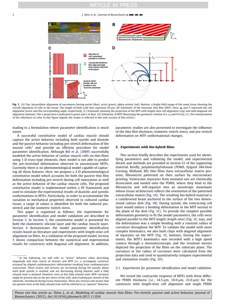
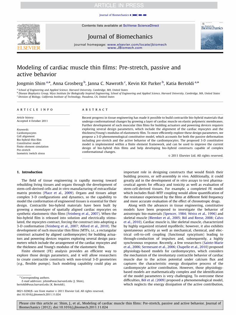
Fig. 1. (A) Top: intracellular alignment of sarcomeres having nuclei (blue), actin (green), alpha-actinin (red). Bottom: a bright-field image of the same tissue showing the
overall alignment of cells in the tissue. The length of both scale bars represent 20 mm. (B) Schematic of the muscular thin film (MTF). Here, a0 and y represent the cell
alignment vector and the corresponding angle, respectively. (C) Schematic showing the projection of the MTF with length-wise cell alignment (top) and with diagonal cell
alignment (bottom). The x-projection is indicated in green and a in blue. (D) Schematic of MTF illustrating the geometric relation of x, L0 and R in Eq. (1). (For interpretation
of the references to color in this figure legend, the reader is referred to the web version of this article.)
J. Shim et al. / Journal of Biomechanics ] (]]]]) ]]]–]]]2
leading to a formulation where parameter identification is mucheasier.
A successful constitutive model of cardiac muscle shouldcapture the active behavior including both systole and diastoleand the passive behavior including pre-stretch deformation of themuscle cells1 and provide an efficient procedure for modelparameter identification. Although Bol et al. (2009) successfullymodeled the active behavior of cardiac muscle cells on thin filmsusing 1-D truss-type elements, their model is not able to predictthe pre-stretched deformation observed in unconstraint MTFs.Currently there is no phenomenological model capable of captur-ing all those features. Here, we propose a 3-D phenomenologicalconstitutive model which accounts for both the passive thin filmdeformation including pre-stretch during cell maturation as wellas the active behavior of the cardiac muscle cells. The proposedconstitutive model is implemented within a FE framework andused to simulate the experimental results of diastolic and systolicconformations in MTFs. Particularly, in order to accommodate thevariation in mechanical properties observed in cultured cardiactissue, a range of values is identified for both the induced pre-stretch and the isometric twitch stress.
This paper is organized as follows: the experiments forparameter identification and model validation are described inSection 2. In Section 3, the constitutive model is presented forboth the elastomeric silicone layer and the cardiac muscle layer.Section 4 demonstrates the model parameter identificationresults based on literature and experiments with length-wise cellalignment on films. As a validation of the proposed model, Section5 shows comparison between the numerical and experimentalresults for constructs with diagonal cell alignment. In addition,
1 In the following, we will refer to ‘‘active’’ behavior when describing
magnitude and time course of stresses and MTF (i.e., a rectangular construct
actuated by aligned cardiomyocytes) deformation resulting from cardiomyocytes
contraction. These strains and stresses are increasing during the systolic phase
until peak systole is reached, and are decreasing during diastole until a fully
relaxed state is attained. However, even at this fully relaxed state, MTF curvature
might be present due to the pre-stress of the cells, and resulting pre-stretch of the
substrate, introduced during tissue maturation. These pre-stresses and strains that
are present even at the fully relaxed state will be referred to as ‘‘passive’’ behavior.
Please cite this article as: Shim, J., et al., Modeling of cardiac musclBiomechanics (2012), doi:10.1016/j.jbiomech.2011.11.024
parametric studies are also presented to investigate the influenceof the thin film thickness, isometric twitch stress, and pre-stretchdeformation on MTF conformational changes.
2. Experiments with bio-hybrid films
This section briefly describes the experiments used for identi-fying parameters and validating the model, and experimentaldetails and methods are provided in Section S1 of the supportingmaterial. Briefly, polydimethylsiloxane (PDMS, Sylgard 184-DowCorning, Midland, MI) thin films have extracellular matrix pro-teins, fibronectin patterned on their surface by microcontactprinting. Ventricular myocytes from neonatal rats are chemicallydissociated and seeded onto the PDMS, where they bind to thefibronectin and self-organize into an anisotropic monolayerwhose tissue architecture reflects the orientation of the patternedextracellular matrix (Fig. 1A). The resulting bio-hybrid material isa cantilevered beam anchored to the surface of the two dimen-sional culture dish (Fig. 1B). During systole, the contracting celllayer would induce a bending deformation in the MTF normal tothe plane of the dish (Fig. 1C). To provide the simplest possibledeformation geometry to fit the model parameters, the cells werealigned parallel to the MTF length (length-wise) (Fig. 1C, top), andthe deformation was a simple bending with a constant radius ofcurvature throughout the MTF. To validate the model with morecomplex kinematics, we also built chips with diagonal alignmentof myocytes on the MTF (Fig. 1C, bottom). During the experi-ments, the MTF’s kinematics was recorded with a high speedcamera through a stereomicroscope, and the resultant moviesdepicted the projection of the films on the substrate plane. Thecurvature or the radius of curvature were calculated from theprojection data and used to quantitatively compare experimentaland simulation results (Fig. 1D).
2.1. Experiments for parameter identification and model validation
We tested the contractile response of MTFs with three differ-ent PDMS thickness (i.e., 14:5 mm, 18:0 mm, 23:0 mm) for theconstructs with length-wise cell alignment and single PDMS
e thin films: Pre-stretch, passive and active behavior. Journal of
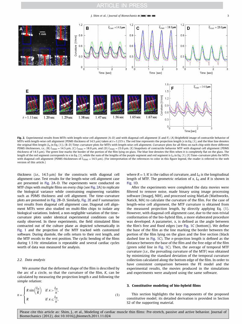
Fig. 2. Experimental results from MTFs with length-wise cell alignment (A–D) and with diagonal cell alignment (E and F). (A) Brightfield image of contractile behavior of
MTFs with length-wise cell alignment (PDMS thickness of 14:5 mm) taken at t¼1.215 s. The red line represents the projection length (x in Eq. (1), and the blue line denotes
the original film length (L0 in Eq. (1)). (B–D) Time–curvature plots for MTFs with length-wise cell alignment. Curvature plots for all films on each chip with three different
PDMS thicknesses, i.e., (B) ~tPDMS ¼ 14:5 mm, (C) ~t PDMS ¼ 18:0 mm, and (D) ~t PDMS ¼ 23:0 mm. (E) Snapshots of contractile behavior MTF with diagonal cell alignment (PDMS
thickness of 14:5 mm). The green line marks the border of the portion of the film lying on glass. The blue line denotes the film when it is completely flat on the glass. The
length of the red segment corresponds to x in Eq. (1), while the sum of the lengths of the purple segment and red segment is L0 in Eq. (1). (F) Time–curvature plots for MTFs
with diagonal cell alignment (PDMS thicknesses of ~t PDMS ¼ 14:5 mm). (For interpretation of the references to color in this figure legend, the reader is referred to the web
version of this article.)
J. Shim et al. / Journal of Biomechanics ] (]]]]) ]]]–]]] 3
thickness (i.e., 14:5 mm) for the constructs with diagonal cellalignment case. Test results for the length-wise cell alignment caseare presented in Fig. 2A–D. The experiments were conducted onMTF chips with multiple films on every chip (see Fig. 2A) to replicatethe biological variance while constraining engineering variablessuch as PDMS thickness and cell alignment. The time–curvatureplots are presented in Fig. 2B–D. Similarly, Fig. 2E and F summarizestest results from diagonal cell alignment case. Diagonal cell align-ment MTFs were also studied on multi-film chips to isolate anybiological variations. Indeed, a non-negligible variation of the time–curvature plots under identical experimental conditions can beeasily observed. In these experiments, the field stimulated filmscontracted out of the culture plane as depicted schematically inFig. 1 and the projection of the MTF tracked with customizedsoftware. During diastole, the cells return to their rest length, andthe MTF recoils to the rest position. The cyclic bending of the filmsduring 1:5 Hz stimulation is repeatable and several cardiac cyclesworth of data was measured for analysis.
2.2. Data analysis
We assume that the deformed shape of the film is described bythe arc of a circle, so that the curvature of the film, K, can becalculated by measuring the projection length x and following thesimple relation:
x¼
R sinL0
R
� �if x4
L0
p,
R if xrL0
p,
8>>><>>>:
ð1Þ
Please cite this article as: Shim, J., et al., Modeling of cardiac musclBiomechanics (2012), doi:10.1016/j.jbiomech.2011.11.024
where R¼ 1=K is the radius of curvature, and L0 is the longitudinallength of MTF. The geometric relation of x, L0 and R is shown inFig. 1D.
After the experiments were completed the data movies werefiltered to remove noise, made binary using image processingsoftware (ImageJ, NIH), and processed using MatLab (Mathworks,Natick, MA) to calculate the curvature of the film. For the case oflength-wise cell alignment, the MTF curvature is obtained fromthe measured projection length, by directly applying Eq. (1).However, with diagonal cell alignment case, due to the non-trivialconformation of the bio-hybrid film, a more elaborated procedurewas developed. A parameter, a, is defined as the angle betweenthe film’s free and fixed edges (see Fig. 1C (bottom)). We definethe base of the film as the line marking the border between theportion of the film lying on the glass and the free section (blackdashed line in Fig. 1C). The x-projection length is defined as thedistance between the base of the film and the free edge of the film(green solid line in Fig. 1C). Then, the average of temporal MTFcurvature (i.e., the prevailing curvature of the MTF) was obtainedby minimizing the standard deviation of the temporal curvaturecollection calculated along the bottom edge of the film. In order tohave consistent comparison between the FE model and theexperimental results, the movies produced in the simulationsand experiments were analyzed using the same software.
3. Constitutive modeling of bio-hybrid films
This section highlights the key components of the proposedconstitutive model; its detailed derivation is provided in SectionS2 of the supporting material.
e thin films: Pre-stretch, passive and active behavior. Journal of
J. Shim et al. / Journal of Biomechanics ] (]]]]) ]]]–]]]4
Let F¼ @x=@X be the deformation gradient mapping a materialpoint from the reference position X to its current position x, andJ¼ detF be its determinant. The behavior of nearly incompressiblematerials is effectively described by splitting the deformationlocally into volumetric (denoted by superscript v) and isochoric(denoted by superscript i) components as
F¼ Fv� Fi, where Fv
¼ J1=31, Fi¼ J�1=3F: ð2Þ
3.1. Elastomeric substrate
The elastomeric substrate is fabricated using PDMS, and itsbehavior is well captured using a neo-Hookean model. Based on
the kinematic assumption shown in Eq. (2) with Ci¼ FiT
� Fi and
I1 ¼ trCi, a decoupled form of the strain energy density (Gurtinet al., 2010) is given by
c¼cvðJÞþci
ðI1Þ ¼~k2ðJ�1Þ2þ
~E
6ðI1�3Þ, ð3Þ
where ~k and ~E denote the bulk modulus and the initial elasticmodulus of the elastomer, respectively. The Cauchy stress T is
found by differentiating c with respect to C, yielding
T¼ ~kðJ�1Þ1þ~E
3JdevðBi
Þ: ð4Þ
where Bi¼ Fi� FiT and ‘‘dev’’ stands for deviatoric part of 2nd
order tensors.
3.2. Cardiac muscle cells
The characteristic energy dissipative process of the cardio-myocyte active contribution can be captured by recently devel-oped physiology-based models, which consider the mechanism ofthe involuntary contractile behavior of cardiac muscle due to theaction potential under calcium flux (Sainte-Marie et al., 2006;Sermesant et al., 2006; Chapelle et al., 2010). However, thisapproach is mathematically complex, and model parameteridentification is challenging. In this article, instead, we take asimple, but effective phenomenological approach, neglecting theenergy dissipative process. While recently Bol et al. (2009)developed a phenomenological model for cardiac muscle cells,their formulation neglects the important effect of the pre-stretchthat the muscle cells develop as they mature. This paper presentsa 3-D phenomenological model that captures the two majorfeatures of cardiac muscles: the passive behavior including pre-stretched deformation and their active behavior including systoleand diastole.
Kinematics including pre-stretch. Even when resting, musclecells in vivo experience a state of stress due to their pre-stretchedconditions. This can be clearly observed from the experimentaldata reported in Fig. 2B–D and F, showing a non-negligible MTFcurvature during diastole. In order to account for such pre-stretched conditions, previous constitutive models introducedan ad hoc strain-shift in the stress–strain curve (Blemker et al.,2005; Bol et al., 2009; Calvo et al., 2010) leading to a non-uniquematerial response that depends on the level of pre-stretch ofmuscle cells. However, such formulation does not capture thenon-negligible MTF curvature observed during diastole. Thus, inthis paper, a different approach is adopted to model pre-stretch;inspired by the multiplicative decomposition introduced byKroner (1960) and Lee (1969), the isochoric deformation gradientFi in Eq. (2) is decomposed into load-induced, FiL, and pre-
Please cite this article as: Shim, J., et al., Modeling of cardiac musclBiomechanics (2012), doi:10.1016/j.jbiomech.2011.11.024
stretched, FiS, contributions
Fi¼ FiL
� FiS: ð5Þ
Here, for the sake of simplicity, the pre-stretch is assumed to be fullydeveloped during cell differentiation and maturation prior to theexperiment, and not to be affected by the cell active response. If wealso assume that the pre-stretch deformation is incompressible andthe cells deform affinely, the pre-stretched contribution to thedeformation gradient FiS is given by
FiS¼QKQ T , ð6Þ
with
K¼ lSe1 � e1þðlSÞ�1=2ðe2 � e2þe3 � e3Þ, ð7Þ
Q ¼ cos yðe1 � e1þe2 � e2Þ
þsin yð�e1 � e2þe2 � e1Þþe3 � e3, ð8Þ
where lS is the pre-stretch induced into the cardiac myocytes lying inthe x1–x2 plane during maturation (i.e., myogenesis and tissuedevelopment) and y is the angle identifying the cell alignment inthe undeformed configuration (see Fig. 1B). The diastole curvatures ofthe experimental data reported in Fig. 2B–D and F clearly show adeviation in the pre-stretched response of the cells as a result of thediversity in cell conditions. Therefore, we expect the parameter lS notto be uniquely determined, but to be influenced by experimentalfactors such as, but not limited to, small variations in temperature/humidity, local density variations, local intracellular architecture, andcardiac origin of the cells (left or right ventricle).
Constitutive equations for passive and active behavior. When anisotropic material is reinforced by a family of fibers with directiona0 ¼ cos ye1þsin ye2 in the reference configuration (Fig. 1B), theisochoric part of the strain energy can be expressed as a functionof not only the invariants of C, but also additional invariantsdepending on a0. To describe both passive (representing restingstatus) and active (including systole and diastole) behavior ofcardiomyocytes, we specify a decoupled form of free energy as
c¼cvðJÞþci
isoðI1ÞþcipaniðI4Þþc
iaaniðI4,qÞ, ð9Þ
where I4 ¼ a0 � Ci� a0 and q is the activation level of cardiac
muscle cells. In the proposed model, while cv and ciiso reflect
the volumetric and the isotropic contributions of the intercellularpart, respectively, cip
ani and ciaani represent the passive and the
active contributions of anisotropic effect of the myofibril, respec-tively. Their detailed forms can be found in Section S2 of thesupporting material. By differentiating c with respect to C, theresulting Cauchy stress for the cardiac muscle cells is obtained as
T¼ TvþTi
isoþTipaniþTia
ani, ð10Þ
where
Tv¼ kðJ�1Þ1, ð11Þ
Tiiso ¼
Ec
3JdevðBi
Þ, ð12Þ
Tipani ¼
EplaJ½eaðl�1Þ�1�devða � aÞ, ð13Þ
Tiaani ¼
Pq
J1�
l�lo
1�lo
!224
35devða � aÞ if 1olo ð2lo�1Þ,
0 otherwise:
8>><>>: ð14Þ
e thin films: Pre-stretch, passive and active behavior. Journal of
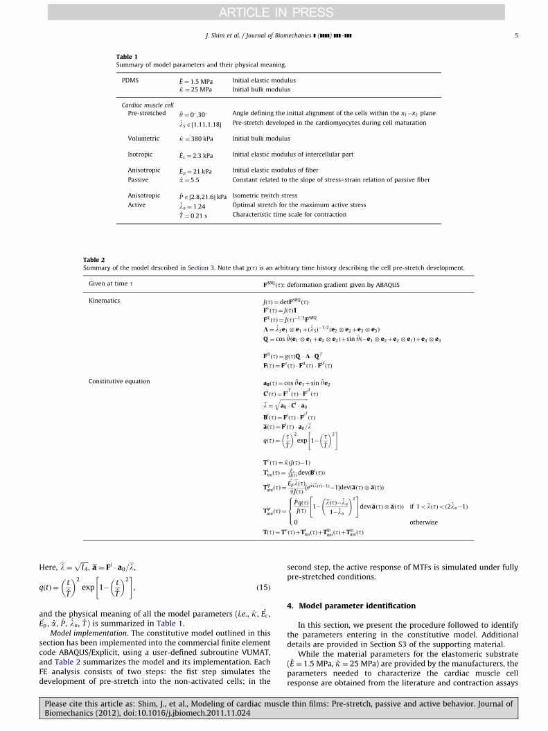
Table 1Summary of model parameters and their physical meaning.
PDMS ~E ¼ 1:5 MPa Initial elastic modulus
~k ¼ 25 MPa Initial bulk modulus
Cardiac muscle cell
Pre-stretched y ¼ 01,301 Angle defining the initial alignment of the cells within the x1�x2 plane
lS A ½1:11,1:18� Pre-stretch developed in the cardiomyocytes during cell maturation
Volumetric k ¼ 380 kPa Initial bulk modulus
Isotropic Ec ¼ 2:3 kPa Initial elastic modulus of intercellular part
Anisotropic Ep ¼ 21 kPa Initial elastic modulus of fiber
Passive a ¼ 5:5 Constant related to the slope of stress–strain relation of passive fiber
Anisotropic P A ½2:8,21:6� kPa Isometric twitch stress
Active lo ¼ 1:24 Optimal stretch for the maximum active stress
T ¼ 0:21 s Characteristic time scale for contraction
Table 2Summary of the model described in Section 3. Note that gðtÞ is an arbitrary time history describing the cell pre-stretch development.
Given at time t FABQðtÞ: deformation gradient given by ABAQUS
Kinematics JðtÞ ¼ detFABQðtÞ
FvðtÞ ¼ JðtÞ1
FiLðtÞ ¼ JðtÞ�1=3FABQ
K¼ lSe1 � e1þðlSÞ�1=2ðe2 � e2þe3 � e3Þ
Q ¼ cos yðe1 � e1þe2 � e2Þþsin yð�e1 � e2þe2 � e1Þþe3 � e3
FiSðtÞ ¼ gðtÞQ �K � Q T
FðtÞ ¼ FvðtÞ � FiL
ðtÞ � FiSðtÞ
Constitutive equation a0ðtÞ ¼ cos ye1þsin ye2
CiðtÞ ¼ FiT
ðtÞ � FiTðtÞ
l ¼ffiffiffiffiffiffiffiffiffiffiffiffiffiffiffiffiffiffiffiffiffiffia0 � C
i� a0
qBiðtÞ ¼ Fi
ðtÞ � FiTðtÞ
aðtÞ ¼ FiðtÞ � a0=l
qðtÞ ¼ tT
� �2
exp 1�tT
� �2" #
TvðtÞ ¼ kðJðtÞ�1Þ
TiisoðtÞ ¼
E c3JðtÞdevðBi
ðtÞÞ
TipaniðtÞ ¼
EplðtÞaJðtÞ
½ea ðlðtÞ�1Þ�1�devðaðtÞ � aðtÞÞ
TipaniðtÞ ¼
PqðtÞJðtÞ 1�
lðtÞ�lo
1�lo
!224
35devðaðtÞ � aðtÞÞ if 1olðtÞoð2lo�1Þ
0 otherwise
8>><>>:
TðtÞ ¼ TvðtÞþTi
isoðtÞþTipaniðtÞþTia
aniðtÞ
J. Shim et al. / Journal of Biomechanics ] (]]]]) ]]]–]]] 5
Here, l ¼ffiffiffiffiffiI4
p, a ¼ Fi
� a0=l,
qðtÞ ¼t
T
� �2
exp 1�t
T
� �2" #
, ð15Þ
and the physical meaning of all the model parameters (i.e., k, Ec ,Ep , a, P , lo, T ) is summarized in Table 1.
Model implementation. The constitutive model outlined in thissection has been implemented into the commercial finite elementcode ABAQUS/Explicit, using a user-defined subroutine VUMAT,and Table 2 summarizes the model and its implementation. EachFE analysis consists of two steps: the fist step simulates thedevelopment of pre-stretch into the non-activated cells; in the
Please cite this article as: Shim, J., et al., Modeling of cardiac musclBiomechanics (2012), doi:10.1016/j.jbiomech.2011.11.024
second step, the active response of MTFs is simulated under fullypre-stretched conditions.
4. Model parameter identification
In this section, we present the procedure followed to identifythe parameters entering in the constitutive model. Additionaldetails are provided in Section S3 of the supporting material.
While the material parameters for the elastomeric substrate( ~E ¼ 1:5 MPa, ~k ¼ 25 MPa) are provided by the manufacturers, theparameters needed to characterize the cardiac muscle cellresponse are obtained from the literature and contraction assays
e thin films: Pre-stretch, passive and active behavior. Journal of
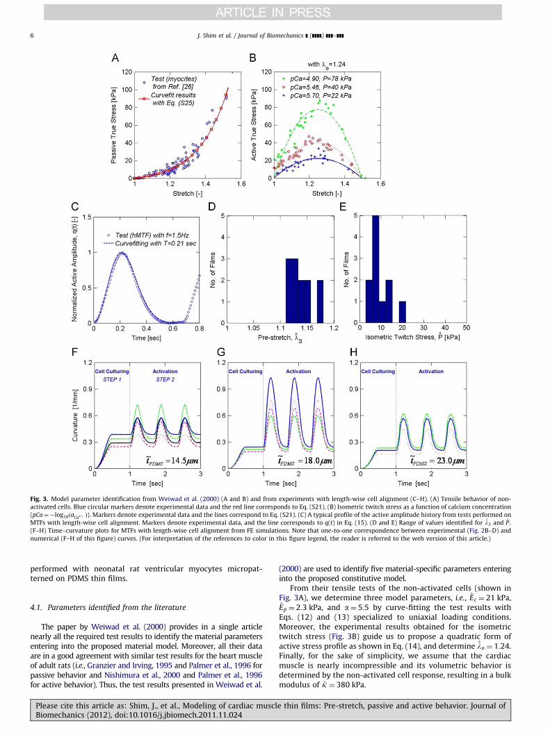
Fig. 3. Model parameter identification from Weiwad et al. (2000) (A and B) and from experiments with length-wise cell alignment (C–H). (A) Tensile behavior of non-
activated cells. Blue circular markers denote experimental data and the red line corresponds to Eq. (S21). (B) Isometric twitch stress as a function of calcium concentration
(pCa¼�log10ðaCa2þ Þ). Markers denote experimental data and the lines correspond to Eq. (S21). (C) A typical profile of the active amplitude history from tests performed on
MTFs with length-wise cell alignment. Markers denote experimental data, and the line corresponds to q(t) in Eq. (15). (D and E) Range of values identified for lS and P .
(F–H) Time–curvature plots for MTFs with length-wise cell alignment from FE simulations. Note that one-to-one correspondence between experimental (Fig. 2B–D) and
numerical (F–H of this figure) curves. (For interpretation of the references to color in this figure legend, the reader is referred to the web version of this article.)
J. Shim et al. / Journal of Biomechanics ] (]]]]) ]]]–]]]6
performed with neonatal rat ventricular myocytes micropat-terned on PDMS thin films.
4.1. Parameters identified from the literature
The paper by Weiwad et al. (2000) provides in a single articlenearly all the required test results to identify the material parametersentering into the proposed material model. Moreover, all their dataare in a good agreement with similar test results for the heart muscleof adult rats (i.e., Granzier and Irving, 1995 and Palmer et al., 1996 forpassive behavior and Nishimura et al., 2000 and Palmer et al., 1996for active behavior). Thus, the test results presented in Weiwad et al.
Please cite this article as: Shim, J., et al., Modeling of cardiac musclBiomechanics (2012), doi:10.1016/j.jbiomech.2011.11.024
(2000) are used to identify five material-specific parameters enteringinto the proposed constitutive model.
From their tensile tests of the non-activated cells (shown inFig. 3A), we determine three model parameters, i.e., Ec ¼ 21 kPa,Ep ¼ 2:3 kPa, and a¼ 5:5 by curve-fitting the test results withEqs. (12) and (13) specialized to uniaxial loading conditions.Moreover, the experimental results obtained for the isometrictwitch stress (Fig. 3B) guide us to propose a quadratic form ofactive stress profile as shown in Eq. (14), and determine lo ¼ 1:24.Finally, for the sake of simplicity, we assume that the cardiacmuscle is nearly incompressible and its volumetric behavior isdetermined by the non-activated cell response, resulting in a bulkmodulus of k ¼ 380 kPa.
e thin films: Pre-stretch, passive and active behavior. Journal of
J. Shim et al. / Journal of Biomechanics ] (]]]]) ]]]–]]] 7
4.2. Parameters identified from MTF tests with length-wise cell
alignment
Experiments with length-wise cell alignment are used toidentify the remaining three model parameters (P , lS, T ). WhileP and lS are experimental-conditions-specific, T denotes thecharacteristic time scale of contractile behavior under the stimu-lating electric pulse. First, T ¼ 0:21 s is identified by curve-fitting atypical profile of the active amplitude history shown in Fig. 3Cwith q(t) in Eq. (15).
Secondly, values for both lS and P are determined using theexperimental data presented in Fig. 2B–D. As in the experiments,MTFs with PDMS substrate of thickness ~t ¼ 14:5, 18.0, or 23:0 mmand cardiac myocytes thickness t ¼ 4 mm are modeled and simu-lated. Since the experiments show a significant variability in thematerial response for each curvature history shown in Fig. 2B–D,both lS and P are determined to achieve the best fit while leavingall the other model parameters unchanged. Thus, a range ofvalues is identified for lS and P whose distributions are shownin Fig. 3D and E, respectively. More specifically, we obtain(a) lSA ½1:11, 1:16� and P A ½2:8, 7:3� kPa for ~t ¼ 14:5 mm,(b) lSA ½1:12, 1:14� and P A ½9:2, 21:6� kPa for ~t ¼ 18:0 mm, and(c) lSA ½1:16, 1:18� and P A ½6:9, 8:5� kPa for ~t ¼ 23:0 mm. While arather small variation in the pre-stretch ranging from lS ¼ 1:11 to1.18 is observed, we find a quite large variation in the isometrictwitch stress ranging from P ¼ 2:7 to 21.6 kPa. Finally, Fig. 3F–Hreports the curvature histories of MTFs obtained from FE simula-tions, clearly showing that the model correctly capture thecurvature induced by pre-stretch curvature during cell matura-tion and the active response.
5. Numerical simulation results
In this section, we first validate the proposed model bysimulating MTFs with diagonal cell alignments, and then presenta series of parametric studies to investigate the effect of PDMSthickness, isometric twitch stress, and pre-stretch of cells.
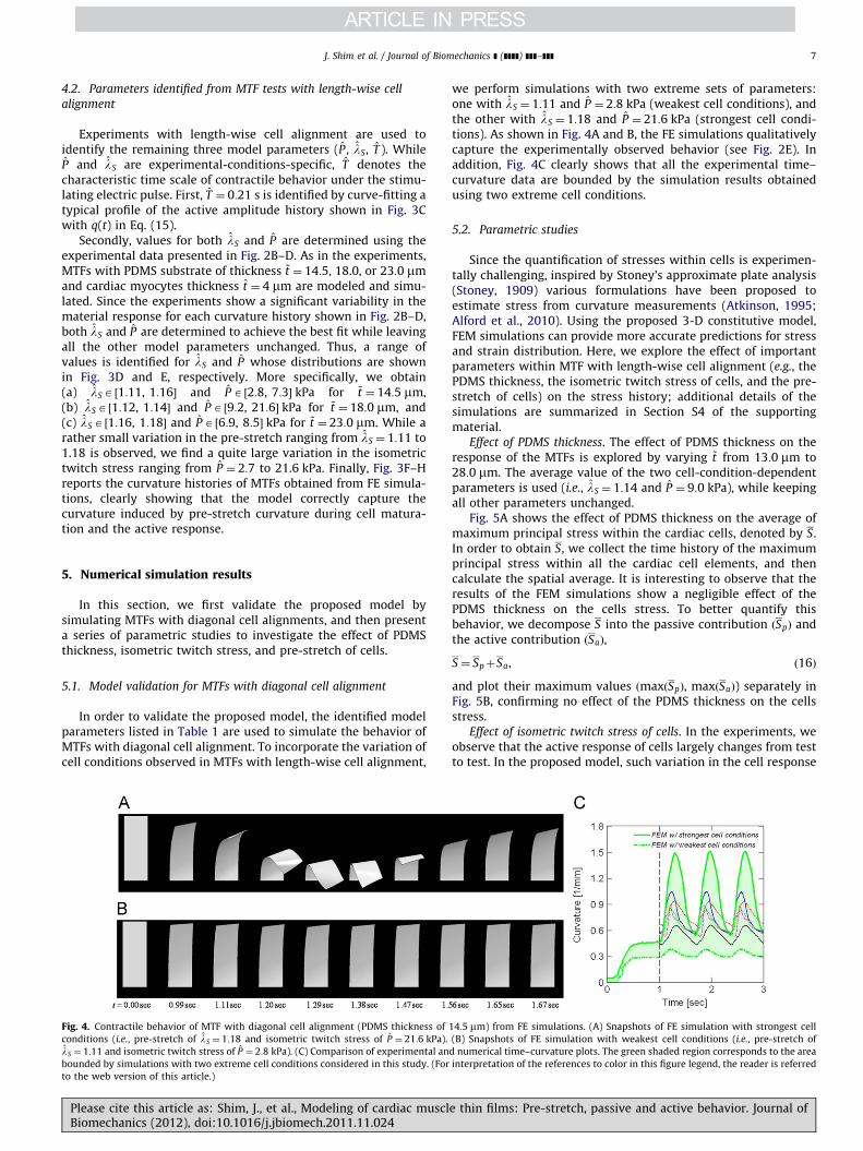
5.1. Model validation for MTFs with diagonal cell alignment
In order to validate the proposed model, the identified modelparameters listed in Table 1 are used to simulate the behavior ofMTFs with diagonal cell alignment. To incorporate the variation ofcell conditions observed in MTFs with length-wise cell alignment,
Fig. 4. Contractile behavior of MTF with diagonal cell alignment (PDMS thickness of
conditions (i.e., pre-stretch of lS ¼ 1:18 and isometric twitch stress of P ¼ 21:6 kPa).
lS ¼ 1:11 and isometric twitch stress of P ¼ 2:8 kPa). (C) Comparison of experimental an
bounded by simulations with two extreme cell conditions considered in this study. (For
to the web version of this article.)
Please cite this article as: Shim, J., et al., Modeling of cardiac musclBiomechanics (2012), doi:10.1016/j.jbiomech.2011.11.024
we perform simulations with two extreme sets of parameters:one with lS ¼ 1:11 and P ¼ 2:8 kPa (weakest cell conditions), andthe other with lS ¼ 1:18 and P ¼ 21:6 kPa (strongest cell condi-tions). As shown in Fig. 4A and B, the FE simulations qualitativelycapture the experimentally observed behavior (see Fig. 2E). Inaddition, Fig. 4C clearly shows that all the experimental time–curvature data are bounded by the simulation results obtainedusing two extreme cell conditions.
5.2. Parametric studies
Since the quantification of stresses within cells is experimen-tally challenging, inspired by Stoney’s approximate plate analysis(Stoney, 1909) various formulations have been proposed toestimate stress from curvature measurements (Atkinson, 1995;Alford et al., 2010). Using the proposed 3-D constitutive model,FEM simulations can provide more accurate predictions for stressand strain distribution. Here, we explore the effect of importantparameters within MTF with length-wise cell alignment (e.g., thePDMS thickness, the isometric twitch stress of cells, and the pre-stretch of cells) on the stress history; additional details of thesimulations are summarized in Section S4 of the supportingmaterial.
Effect of PDMS thickness. The effect of PDMS thickness on theresponse of the MTFs is explored by varying ~t from 13:0 mm to28:0 mm. The average value of the two cell-condition-dependentparameters is used (i.e., lS ¼ 1:14 and P ¼ 9:0 kPa), while keepingall other parameters unchanged.
Fig. 5A shows the effect of PDMS thickness on the average ofmaximum principal stress within the cardiac cells, denoted by S.In order to obtain S, we collect the time history of the maximumprincipal stress within all the cardiac cell elements, and thencalculate the spatial average. It is interesting to observe that theresults of the FEM simulations show a negligible effect of thePDMS thickness on the cells stress. To better quantify thisbehavior, we decompose S into the passive contribution ðSpÞ andthe active contribution ðSaÞ,
S ¼ SpþSa, ð16Þ
and plot their maximum values ðmaxðSpÞ, maxðSaÞ) separately inFig. 5B, confirming no effect of the PDMS thickness on the cellsstress.
Effect of isometric twitch stress of cells. In the experiments, weobserve that the active response of cells largely changes from testto test. In the proposed model, such variation in the cell response
14:5 mm) from FE simulations. (A) Snapshots of FE simulation with strongest cell
(B) Snapshots of FE simulation with weakest cell conditions (i.e., pre-stretch of
d numerical time–curvature plots. The green shaded region corresponds to the area
interpretation of the references to color in this figure legend, the reader is referred
e thin films: Pre-stretch, passive and active behavior. Journal of
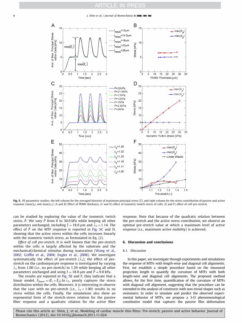
Fig. 5. FE parametric studies: the left column for the averaged histories of maximum principal stress (S), and right column for the stress contribution of passive and active
response (maxðSpÞ and maxðSaÞ). (A and B) Effect of PDMS thickness, (C and D) effect of isometric twitch stress of cells, (E and F) effect of cell pre-stretch.
J. Shim et al. / Journal of Biomechanics ] (]]]]) ]]]–]]]8
can be studied by exploring the value of the isometric twitchstress, P . We vary P from 0 to 30.0 kPa while keeping all otherparameters unchanged, including ~t ¼ 18:0 mm and lS ¼ 1:14. Theeffect of P on the MTF response is reported in Fig. 5C and D,showing that the active stress within the cells increases linearlywith the isometric twitch stress, as formulated in Eq. (2).
Effect of cell pre-stretch. It is well known that the pre-stretchwithin the cells is largely affected by the substrate and themechanical/chemical stimulus during maturation (Wang et al.,2002; Griffin et al., 2004; Engler et al., 2008). We investigatesystematically the effect of pre-stretch (lS); the effect of pre-stretch on the cardiomyocyte response is investigated by varyinglS from 1.00 (i.e., no pre-stretch) to 1.39 while keeping all otherparameters unchanged and using ~t ¼ 18:0 mm and P ¼ 9:0 kPa.
The results are reported in Fig. 5E and F, they indicate that alinear model, Slinear ¼ ðEcþ EpÞ ln lS, poorly captures the stressdistribution within the cells. Moreover, it is interesting to observethat the case with no pre-stretch (i.e., lS ¼ 1:00) results in nostress within the cells. Finally, the simulations also show anexponential form of the stretch-stress relation for the passivefiber response and a quadratic relation for the active fiber
Please cite this article as: Shim, J., et al., Modeling of cardiac musclBiomechanics (2012), doi:10.1016/j.jbiomech.2011.11.024
response. Note that because of the quadratic relation betweenthe pre-stretch and the active stress contribution, we observe anoptimal pre-stretch value at which a maximum level of activeresponse (i.e., maximum active mobility) is achieved.
6. Discussion and conclusions
6.1. Discussion
In this paper, we investigate through experiments and simulationsthe response of MTFs with length-wise and diagonal cell alignments.First, we establish a simple procedure based on the measuredprojection length to quantify the curvature of MTFs with bothlength-wise and diagonal cell alignments. The proposed methodallows, for the first time, quantification of the curvature of MTFswith diagonal cell alignment, suggesting that the procedure can beextended to the analysis of constructs with non-trivial shapes such asswimmers. In order to simulate and predict the observed experi-mental behavior of MTFs, we propose a 3-D phenomenologicalconstitutive model that captures the passive film deformation
e thin films: Pre-stretch, passive and active behavior. Journal of
J. Shim et al. / Journal of Biomechanics ] (]]]]) ]]]–]]] 9
including pre-stretch as well as the active behavior of cardiomyo-cytes. Inspired by the multiplicative decomposition introduced byKroner (1960) and Lee (1969), the isochoric deformation gradient isdecomposed into load-induced and pre-stretched contributions sothat the effect of the pre-stretch can be independently investigated.To simplify the identification of parameters entering in the model, wepropose a phenomenological formulation where the cardiomyocyteactive contribution is described by an elastic model and all thedifferent aspects of constitutive behaviors are decoupled. Data fromliterature and experiments conducted on MTFs with length-wise cellalignment are used to identify all the model parameters. Moreover,the variation in mechanical properties observed during experimentsis accounted for by identifying a range of values for the induced pre-stretch (lS) and the isometric twitch stress (P). The resultingformulation captures qualitatively and quantitatively the experimen-tally observed response of MTFs with diagonal cell alignment. MTFsare characterized by a very complicated chemo-mechanical behavior.To capture the salient features of their response with a sufficientlysimple formulation, several assumptions need to be made: here, theenergy dissipating mechanism is neglected and a decoupled form offree energy is adopted.
Recently proposed physiology-based constitutive models con-sider the mechanism of the involuntary contractile behavior ofcardiac muscle due to the action potential under calcium flux(Sainte-Marie et al., 2006; Sermesant et al., 2006; Chapelle et al.,2010). These formulations capture the characteristic energy dissi-pative process of the cardiomyocyte active contribution and satisfythe thermodynamics laws. However, these models are mathemati-cally complex and lead to sophisticated procedures for the identi-fication of the model parameters (Sermesant et al., 2006), makingunrealistic the use of such models as design tools for MTF actuators.Instead, we take a simple, but effective phenomenological approachin the spirit of Blemker et al. (2005) and Bol et al. (2009), where theenergy dissipative behavior of the active contribution is neglected.To describe the active response of cardiomyocytes, an elastic modelis adopted and a scalar internal variable (q in Eq. (15)) is introduced,leading to a formulation that does not satisfy the free energyimbalance conditions.
Several theories for metal plasticity (Brown et al., 1989),approximately incompressible elastic materials (Anand, 1996),and fiber-reinforced composite (Spencer, 1984) are based on aseparable form of free energy (the so-called separability hypothesis
Gurtin et al., 2010) and consider separately the contributions ofplastic, volumetric and anisotropic deformation. This formulationprovides an effective way to incorporate various aspect of thecomplex material behavior into a relatively simple model.Inspired by this approach, we decouple all the aspects of themuscle response (see Eq. (9)) although there is no experimentalevidence for such assumption. However, this formulation leads toa substantially simplified procedure for the identification of themodel parameters; most of them can be identified from literature,and only a few need to be identified from reference experiments.
6.2. Conclusions
We present a 3-D phenomenological constitutive model thatcaptures the passive film deformation including pre-stretch dur-ing cell maturation as well as the active behavior of the cardiacmuscle cells. While a range of values is identified for two modelparameters (induced pre-stretch l and isometric twitch stress P)to account for the variation in mechanical properties observed inexperiments, all other model parameters are obtained from theliterature. The proposed model is implemented within a FEframework and used to simulate the experimental results ofdiastolic and systolic conformations in a muscular thin film. Withthe identified model parameters, the proposed model
Please cite this article as: Shim, J., et al., Modeling of cardiac musclBiomechanics (2012), doi:10.1016/j.jbiomech.2011.11.024
qualitatively captured the behavior of MTFs with diagonal cellalignment, and all the experimental time–curvature data arebounded by the numerical results obtained using two extremecell conditions.
The proposed constitutive model can be immediately extendedto the analysis of constructs with non-trivial 3-D initial geometries.This will greatly aid in the engineering of soft muscle-poweredrobots, but it will require a computationally efficient implementa-tion of the model and the use of shell elements within the FEframework. Furthermore, the model has the potential to take intoaccount fluid–structure interactions, opening avenues for the designof swimming constructs, actuators, and micro-fluidic devices.
Conflict of interest statement
The authors report that no conflicts of interest, financial orotherwise, influenced this work.
Acknowledgements
This work has been supported by the Harvard MaterialsResearch Science and Engineering Center under NSF awardnumber DMR-0820484 (KB), DMR-0213805 (KKP), and NIH grant1 R01 HL079126 (KKP). We are grateful to the Center of NanoscaleSystems at Harvard University for the use of their cleanroomfacilities, to Alexander P. Nesmith for developing the brickpatterned stamps, and to Harvard SEAS Academic Computing fortheir support.
Appendix A. Supplementary data
Supplementary data associated with this article can be foundin the online version at doi:10.1016/j.jbiomech.2011.11.024.
References
Alford, P.W., Feinberg, A.W., Sheehy, S.P., Parker, K.K., 2010. Biohybrid thin filmsfor measuring contractility in engineered cardiovascular muscle. Biomaterials31 (13), 3613–3621.
Anand, L., 1996. A constitutive model for compressible elastomeric solids.Computational Mechanics 18 (5), 339–355.
Atkinson, A., 1995. British Ceramic Proceedings 54 (1).Blemker, S.S., Pinsky, P.M., Delp, S.L., 2005. A 3D model of muscle reveals the
causes of nonuniform strains in the biceps brachii. Journal of Biomechanics 38,657–665.
Bol, M., Reese, S., 2008. Micromechanical modelling of skeletal muscles based onthe finite element method. Computer Methods in Biomechanics and Biome-dical Engineering 11, 489–504.
Bol, M., Reese, S., Parker, K.K., Kuhl, E., 2009. Computational modeling of muscularthin film for cardiac repair. Computational Mechanics 43, 535–544.
Brown, S.B., Kim, K.H., Anand, L., 1989. An internal variable constitutive model forhot-working of metals. International Journal of Plasticity 5 (2), 95–130.
Calvo, B., Ramirez, A., Alonso, A., Grasa, J., Soteras, F., Osta, R., Munoz, M.J., 2010.Passive nonlinear elastic behaviour of skeletal muscle: experimental resultsand model formulation. Journal of Biomechanics 43, 318–325.
Chapelle, D., Gerbeau, J.F., Sainte-Marie, J., Vignon-Clementel, I.E., 2010.A poroelastic model valid in large strains with applications to perfusion incardiac modeling. Computational Mechanics 46, 91–101.
Engler, A.J., Carag-Krieger, C., Johnson, C.P., Raab, M., Tang, H.-Y., Speicher, D.W.,Sanger, J.W., Sanger, J.M., Discher, D.E., 2008. Embryonic cardiomyocytes beatbest on a matrix with heart-like elasticity: scar-like rigidity inhibits beating.Journal of Cell Science 121, 3794–3802.
Feinberg, A.W., Feigel, A., Shevkoplyas, S.S., Sheehy, S., Whitesides, G.M., Parker,K.K., 2007. Muscular thin films for building actuators and powering devices.Science 317 (5843), 1366–1370.
Granzier, H.L., Irving, T.C., 1995. Passive tension in cardiac muscle: contribution ofcollagen, titin, microtubules, and intermediate filaments. Biophysical Journal68, 1027–1044.
Griffin, M.A., Engler, A.J., Barber, T.A., Healy, K.E., Sweeney, H.L., 2004. Patterning,prestress, and peeling dynamics of myocytes. Biophysical Journal 86,1209–1222.
e thin films: Pre-stretch, passive and active behavior. Journal of
J. Shim et al. / Journal of Biomechanics ] (]]]]) ]]]–]]]10
Gurtin, M.E., Fried, E., Anand, L., 2010. The Mechanics and Thermodynamics ofContinua. Cambridge University Press, New York.
Kroner, E., 1960. Allgemeine kontinuums theorie der versetzungen und eigen-spannungen. Archive for Rational Mechanics and Analysis 4, 273–334.
Lee, E.H., 1969. Elastic–plastic deformation at finite strains. Journal of AppliedMechanics 36, 1.
Nishimura, S., Yasuda, S., Katoh, M., Yamada, K.P., Yamashita, H., Saeki, Y.,Sunagawa, K., Nagai, R., Hisada, T., Sugiura, S., 2000. Single cell mechanics ofrat cardiomyocytes under isometrica unloaded and physiologically loadedconditions. AJP—Heart and Circulatory Physiology 287, H196–H202.
Palmer, R.E., Brady, A.J., Roos, K.P., 1996. Mechanical measurements from isolatedcardiac myocytes using a pipette attachment system. AJP—Cell Physiology270, C697–C704.
Place, E.S., Evans, N.D., Stevens, M.M., 2009. Complexity in biomaterials for tissueengineering. Nature Materials 8 (6), 457–470.
Sainte-Marie, J., Chapelle, D., Cimrman, R., Sorine, M., 2006. Modeling andestimation of the cardiac electromechanical activity. Computers and Struc-tures 84, 1743–1759.
Please cite this article as: Shim, J., et al., Modeling of cardiac musclBiomechanics (2012), doi:10.1016/j.jbiomech.2011.11.024
Sermesant, M., Moireau, P., Camara, O., Sainte-Marie, J., Andriantsimiavona, R.,Cimrman, R., Hill, D.L.G., Chapelle, D., Razavi, R., 2006. Cardiac functionestimation from MRI using a heart model and data assimilation: advancesand difficulties. Medical Image Analysis 10, 642–656.
Spencer, A.J.M., 1984. Continuum Theory of the Mechanics of Fibre-reinforcedComposites. Springer-Verlag, Wien-New York.
Stoney, G.G., 1909. The tension of metallic films deposited by electrolysis.Proceedings of the Royal Society of London A 82, 172–175.
Wang, N., Tolic-Norrelykke, I.M., Chen, J., Mijailovich, S.M., Butler, J.P., Fredberg, J.J.,Stamenovic, D., 2002. Cell prestress. I. Stiffness and prestress are closelyassociated in adherent contractile cells. AJP—Cell Physiology 282, C606–C616.
Weiss, J.A., Marker, B.N., Govindjee, S., 1996. Finite element implementation ofincompressible, transversely isotropic hyperelasticity. Computer Methods inApplied Mechanics and Engineering 135, 107–128.
Weiwad, W.K.K., Linke, W.A., Wussling, M.H.P., 2000. Sarcomere length–tensionrelationship of rat cardiac myocytes at lengths greater than optimum. Journalof Molecular and Cellular Cardiology 32, 247–259.
e thin films: Pre-stretch, passive and active behavior. Journal of