modelling meristem development in plants heisler,...
TRANSCRIPT

LUND UNIVERSITY
PO Box 117221 00 Lund+46 46-222 00 00
Modelling meristem development in plants
Heisler, Marcus G.; Jönsson, Henrik
Published in:Current Opinion in Plant Biology
DOI:10.1016/j.pbi.2006.11.005
Published: 2007-01-01
Link to publication
Citation for published version (APA):Heisler, M. G., & Jönsson, H. (2007). Modelling meristem development in plants. Current Opinion in PlantBiology, 10(1), 92-97. DOI: 10.1016/j.pbi.2006.11.005
General rightsCopyright and moral rights for the publications made accessible in the public portal are retained by the authorsand/or other copyright owners and it is a condition of accessing publications that users recognise and abide by thelegal requirements associated with these rights.
• Users may download and print one copy of any publication from the public portal for the purpose of privatestudy or research. • You may not further distribute the material or use it for any profit-making activity or commercial gain • You may freely distribute the URL identifying the publication in the public portalTake down policyIf you believe that this document breaches copyright please contact us providing details, and we will removeaccess to the work immediately and investigate your claim.

Modelling Meristem Development in Plants
Marcus. G. Heisler1 and Henrik Jönsson
2
1Corresponding author, Division of Biology, California Institute of Technology,
Pasadena, CA, USA, [email protected]
2Computational Biology and Biological Physics, Department of Theoretical Physics,
Lund University, Lund, Sweden, [email protected]
Running title: Modelling meristem development

Summary
Meristems continually supply new cells for post embryonic plant development
and coordinate the initiation of new organs such as leaves and flowers. Meristem function
is regulated by a large and interconnected dynamical system that includes transcriptional
networks, intercellular protein signalling, polarized transport of hormones and a
constantly changing cellular topology. Mathematical modelling, in which the dynamics of
a system are simulated using explicitly defined interactions, can serve as a powerful tool
for examining the expected behaviour of such a system given our present knowledge and
assumptions. Modelling can also help to investigate new hypotheses in silico both to
validate ideas and to obtain inspiration for new experiments. Several recent studies use
new molecular data together with modelling and computational techniques to investigate
meristem function.
Introduction
Due to the often regular and symmetrical patterns generated by plants, plant
architecture has not only fascinated biologists but also mathematicians and artists alike
for centuries [1]. However over the past couple of years mathematical and modelling
approaches to understanding plant development have gained fresh momentum [2,3],
partially due to inexpensive computing power but also due to the rapid increase in
detailed molecular data related to plant development. The origin of many plant
developmental patterns can be traced back to meristems located at the growing tips of
roots and shoots, from which most postembryonic structures are derived. In this review
we will focus on how modelling and computational techniques have recently been
combined with detailed molecular data to help understand meristem development and
function.
Meristem maintenance
One of the astounding features of meristems is their ability to maintain a specific
cellular structure and growth pattern throughout the lifetime of the plant, which can in
some cases last thousands of years. Experimental studies have shown that shoot apical
meristem (SAM) size, in particular the region of undifferentiated cells at the very apex, is
regulated via a negative feedback loop between the transcription factor WUSCHEL
(WUS) and a small secreted peptide CLAVATA3 (CLV3). WUS is expressed in the
organizing centre, a small subapical region in the interior of the shoot. It promotes stem
cell identity and positively regulates CLV3 transcription. CLV3 in turn acts together with
the receptor kinase CLAVATA1 to repress WUS expression, thus creating a negative
feedback loop [4,5,6,7,8]. Although this feedback loop provides an intuitively simple
system for regulating the size of the WUS expression domain, and hence proliferation of
stem cells in the SAM, it leaves several questions open, including the question of how the
pattern of the WUS expression domain is specified.
The WUS expression pattern is intriguing since it is capable of reorganising itself
after disruption by laser ablation [9], mirroring the ability of meristems in general to self-

organize after wounding . Self-organization is also a property exhibited by reaction-
diffusion systems as proposed by Turing and Meinhardt [10,11]. As a test to see how well
such a scheme might work to control WUS expression, Jönsson et al (2005) examined a
model in which WUS expression was placed under the control of a reaction-diffusion
mechanism [••12]. The model was simulated on a two-dimensional cellular template
extracted from a transverse confocal microscope section of the SAM centre (Figure 1a-c).
The model created one maximum of WUS expression within the meristem region, which
was centred within the tissue using a hypothetical repressive signal emanating from the
epidermal, or L1, cell layer. Model parameters were tuned by comparing the output of the
model directly with real WUS expression, also extracted from the template data (Figure
1d). When the central cells expressing WUS were removed to simulate the effect of laser
ablation, WUS expression reappeared on either side of the ablated region in a similar
fashion to the observed experimental response. An alternative model using constitutive
expression modulated by the repressive signal from the L1 failed to re-organize WUS
expression in this way. Although this model is abstracted from the biology to a
considerable degree, it suggests that the basic mechanism of long-range inhibition
together with local reinforcement may account for the self-organizational properties of
not only WUS expression but also meristems in general. While CLV3 is a prime
candidate for a WUS-induced long-range inhibitor, recent experiments also show that
members of the HD-ZIP class of transcription factors play a repressive role in regulating
WUS expression [13,14]. It will be interesting to test whether the expression of these
genes is, like CLV3, also dependant on WUS activity and whether models can be used for
discriminating between different scenarios of long-range inhibition.
How is auxin distributed in meristems?
Auxin flow within the root meristem is coordinated via multiple members of the
PIN family of auxin efflux mediators as well as the auxin influx mediator AUX1
[•15,16]. In Swarup et al. (2005) experiments and modelling were combined to
investigate the role of auxin in mediating gravitropism [••17]. A three dimensional model
corresponding to the elongation zone proximal to the meristem was constructed using the
stereotypical arrangement of root cells as well as detailed distribution patterns of PIN1,
PIN2, and AUX1 (Figure 2a). The model used these data together with the chemiosmotic

transport theory [18,19] to show that AUX1 expression in the epidermal cells should be
sufficient to mediate apical transport of auxin from the root tip with only moderate
diffusion. Given an asymmetric pulse of auxin from the root apex in response to an
altered gravity vector, epidermal cells are predicted to maintain this asymmetry
throughout the elongation zone in order to promote differential growth. In fact the authors
go on to predict that PIN2 should not be required in the epidermis for this transport
function since weak PIN1 in these cells should suffice. Instead PIN2 is suggested to be
only necessary for auxin efflux into the epidermis from the lateral root cap. This remains
an untested prediction from the paper. This study represents an important step towards
modelling the flux of auxin throughout the root meristem and it would be interesting to
see whether such a complete model might account for the specific auxin-induced
expression and protein degradation patterns of the various PIN proteins.
In the SAM, PIN1 expression and localization in the epidermal layer appears to be
important for determining the auxin distribution in relation to where new primordia are
formed [20, •21] (see below). However PIN1 localization patterns in these cells are
harder to interpret compared to the root. Some cells are not clearly polarized and PIN
localization patterns change constantly as primordia development proceeds around the
meristem periphery [•21]. To try to deduce auxin distribution patterns in these cells
Barbier de Reuille et al. (2006) used confocal imaging to visualize and document PIN1
immunolocalization patterns in the L1 [••22] (Figure 2b). After hand-marking these
patterns such that each side of each cell was designated to either contribute or not
contribute transport to the adjoining cell they introduced a simplified model for PIN1
dependant auxin flow (not based on the chemiosmotic transport mechanism) that
included diffusion. Encouragingly their simulations predicted auxin peaks at positions
where new primordia were about to form, as has been shown to occur experimentally.
Unexpectedly, their model also predicted high auxin levels at the shoot apex (Figure 2b).
In support of this prediction the authors were able to successfully detect IAA specifically
in the meristem apex using an IAA specific monoclonal antibody and Gas
chromatography-Mass spectrometry (GCMS). By also showing that the synthetic auxin
reporter DR5 was not sensitive to exogenous auxin in this region the authors concluded
that perception of auxin is suppressed at the level of primary auxin response genes at the

meristem centre (however contradictory results are presented in [••23]). Lastly, the
simulations of their flow model lead the authors to hypothesize that auxin levels increase
at new primordial positions because of over accumulation of auxin in the meristem centre
and depletion of auxin by neighbouring primordia. However the question of how PIN1
polarity is coordinated to produce this flow pattern is left open (see below).
The heart of phyllotaxis – an auxin mediated spacing mechanism
The positioning of lateral organs (phyllotaxis) has long been the subject of
modelling studies. One of the key findings from these studies is that many, if not most, of
the complex patterns of organs observed in nature can be generated by any kind of
regular spacing mechanism combined with a gradually expanding generative region such
as a meristem growing over time [24,25,26,27,28]. Recent experimental studies have
shown that an essential part of the mechanism involves the transport of auxin, via the
asymmetrically localized PIN1 auxin efflux carrier, to positions where primordia are
destined to form [29,30,31]. Thus a central question is what coordinates PIN1
localization in such a way? Two recent studies have proposed a mechanism that is not
only capable of generating close to observed PIN1 localization patterns but is also able to
spontaneously generate regularly spaced peaks and troughs of auxin concentration [••23,
••32]. At the core of these models is a feedback system in which PIN1 protein gets
localized to the membranes of a cell closest to neighbouring cells that contain the most
auxin. Thus cells with high auxin content polarize their neighbours towards them, further
increasing their auxin content until the flux due to polar transport is balanced by
diffusion.
Although both models are based on the same hypothesis for PIN polarization,
they differ in their approaches. Smith et al do not always use equations that are easily
interpretable in terms of biochemical mechanisms and to stabilize their patterns they
include additional rules for localizing PIN1 that abstractly relate to primordial
differentiation. However their model parameters are tuned by comparing the resulting
phyllotactic patterns with experimental data from Arabidopsis, and a good
correspondence is achieved. Lastly, their model is run on a global growth template
defined by the authors to resemble real meristem tissue growth (Figure 3a). In contrast,
Jönsson et al create a dynamic cellular template using spring mechanics to model growth.
Although such a growth model does not appear very “plant-like” it represents an early
attempt to couple real cell wall mechanics to a signaling and gene regulatory model
(Figure 3b). The auxin transport model is also defined differently using mechanistic
equations, and an experimental template is used together with the chemiosmotic auxin
transport theory to estimate parameter values for modeling PIN1 polarization. This model
is also capable of generating phyllotactic patterns, although not as stably as those
generated by the Smith et al model. Overall, the picture that emerges is that even if true,
PIN1 polarization by cytoplasmic auxin concentrations in neighboring cells can only be
part of the story. Perhaps this is not surprising given the existence of many other players
such as the PID kinase [33], SHOOTMERISTEMLESS (STM) and CUP-SHAPED
COTYLEDONS1/2 (CUC1/2) which also mediate organ positioning and growth [34].
Nevertheless these initial models provide us with a novel potential positioning
mechanism based on up-to-date molecular data and hypotheses that are experimentally
testable.
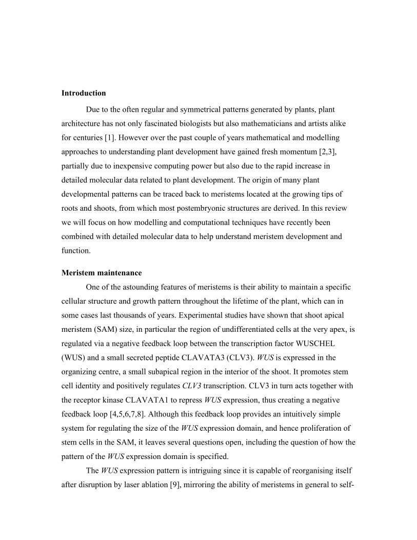
Integration of gene regulatory models with morphogenesis and mechanics
So far we have been discussing discrete models that deal with gene expression
patterns and the distribution of signalling molecules within “virtual” cellular templates.
Each template has been different, from the static 3D root architecture of Swarup et al. to
the mechanically driven 2D template from Jönsson et al. Ideally the model template
should resemble the real plant tissue as closely as possible and such a template may not
only involve creating a cellular architecture of the tissue under study but may also
involve the use of gene expression patterns, protein distributions and the time-evolution
of these data. This is where confocal live-imaging techniques potentially offer significant
new advantages for assessing and developing plant developmental simulations. In
deReuille et al. a novel semi-automatic protocol for obtaining the cellular architecture of
the meristem epidermal layer is presented [••35]. The procedure is based on segmenting
epidermal cell outlines using a membrane localized fluorescent dye or GFP imaged using
confocal microscopy (Figure 4a and b). From these data the neighbour relationships can
be determined and by conducting time-lapse imaging, the shapes of these cells can be
extracted over time (Figure 4c). Although this type of data has also been obtained using a
simpler non-invasive replica method [36,37,38], this study potentially represents a first
step towards using confocal imaging to extract a multivariable modelling template that
includes gene expression patterns and protein localization data along with cell shape
dynamics.
Conclusion
It is an exciting time for understanding the role of auxin in meristems and this is
reflected in the choice of papers we have focused on in this review. The use of new
imaging and perturbation techniques is also providing us with data for use in models in
unprecedented detail [•21,39,•40,•41,•42]. In fact, we feel that these new experimental
techniques are of great help in providing the detailed dynamic data required for inspiring
mechanistic models as well as for testing the models adequately.
A challenge not yet addressed in the literature is to link gene regulatory models
with realistic models for cell wall mechanics since only then can models bridge the gap
between signalling and morphogenesis. This may not be a trivial task, given the
anisotropic and visco-elastic nature of plant cell walls [43]. Another future challenge, as
more and more data becomes available, is to somehow integrate and combine models and
hopefully this is where efforts at creating standardized languages such as Systems
Biology Markup Language (SBML) may bear fruit [44].
Acknowledgements
We thank Eric Mjolsness, Elliot M. Meyerowitz and Adrienne Roeder for
discussion and critical comments. HJ acknowledge support from the Swedish Research
Council and Human Frontier Science Program. MGH was supported by the National
Science Foundation’s Frontiers in Biological Research (FIBR) program, award number
EF-0330786; and Department of Energy Grant DOE FG02-88ER13873.
REFERENCES
1. Adler I, Barabe D, Jean RV: A history of the study of phyllotaxis. Annals of Botany
1997, 80:231-244.
2. Prusinkiewicz P, Rolland-Lagan AG: Modeling plant morphogenesis. Current
Opinion in Plant Biology 2006, 9:83-88.

3. Kwiatkowska D: Structural integration at the shoot apical meristem: Models,
measurements, and experiments. American Journal of Botany 2004, 91:1277-1293.
4. Fletcher JC, Brand U, Running MP, Simon R, Meyerowitz EM: Signaling of cell fate
decisions by CLAVATA3 in Arabidopsis shoot meristems. Science 1999, 283:1911-
1914.
5. Schoof H, Lenhard M, Haecker A, Mayer KF, Jurgens G, Laux T: The stem cell
population of Arabidopsis shoot meristems in maintained by a regulatory loop
between the CLAVATA and WUSCHEL genes. Cell 2000, 100:635-644.
6. Mayer KF, Schoof H, Haecker A, Lenhard M, Jurgens G, Laux T: Role of
WUSCHEL in regulating stem cell fate in the Arabidopsis shoot meristem. Cell
1998, 95:805-815.
7. Trotochaud AE, Jeong S, Clark SE: CLAVATA3, a multimeric ligand for the
CLAVATA1 receptor-kinase. Science 2000, 289:613-617.
8. Clark SE, Williams RW, Meyerowitz EM: The CLAVATA1 gene encodes a putative
receptor kinase that controls shoot and floral meristem size in Arabidopsis. Cell
1997, 89:575-585.
9. Reinhardt D, Frenz M, Mandel T, Kuhlemeier C: Microsurgical and laser ablation
analysis of interactions between the zones and layers of the tomato shoot apical
meristem. Development 2003, 130:4073-4083.
10. Turing aM: The Chemical Basis of Morphogenesis. Philosophical Transactions of
the Royal Society of London Series B-Biological Sciences 1952, 237:37-72.
11. Gierer a, Meinhardt.H: Theory of Biological Pattern Formation. Kybernetik 1972,
12:30-39.
••12. Jonsson H, Heisler M, Reddy GV, Agrawal V, Gor V, Shapiro BE, Mjolsness E,
Meyerowitz EM: Modeling the organization of the WUSCHEL expression domain in
the shoot apical meristem. Bioinformatics 2005, 21:I232-I240.
The authors discriminate among different models for how a sharp domain of
WUS expression can be maintained by comparing directly with WUS extracted from an
experimental template as well as with perturbation experiments. This paper provides an
early example of the potential of direct integration of modelling techniques and confocal
data.
13. Prigge MJ, Otsuga D, Alonso JM, Ecker JR, Drews GN, Clark SE: Class III
homeodomain-leucine zipper gene family members have overlapping, antagonistic,
and distinct roles in Arabidopsis development. Plant Cell 2005, 17:61-76.
14. Williams L, Grigg SP, Xie MT, Christensen S, Fletcher JC: Regulation of
Arabidopsis shoot apical meristem and lateral organ formation by microRNA
miR166g and its AtHD-ZIP target genes. Development 2005, 132:3657-3668.
•15. Blilou I, Xu J, Wildwater M, Willemsen V, Paponov I, Friml J, Heidstra R, Aida M,
Palme K, Scheres B: The PIN auxin efflux facilitator network controls growth and
patterning in Arabidopsis roots. Nature 2005, 433:39-44.
Detailed expression and polarization data for five PIN proteins is presented,
which illuminates the auxin flow within the root meristem. In addition several multiple
mutants are investigated and interactions with the PLETHORA root stem cell
specification genes are shown. This paper provides detailed data for a module for root
meristem maintenance, integrating auxin, polarized transport and transcription factors.

16. Swarup R, Friml J, Marchant A, Ljung K, Sandberg G, Palme K, Bennett M:
Localization of the auxin permease AUX1 suggests two functionally distinct
hormone transport pathways operate in the Arabidopsis root apex. Genes Dev 2001,
15:2648-2653.
••17. Swarup R, Kramer EM, Perry P, Knox K, Leyser HM, Haseloff J, Beemster GT,
Bhalerao R, Bennett MJ: Root gravitropism requires lateral root cap and epidermal
cells for transport and response to a mobile auxin signal. Nat Cell Biol 2005, 7:1057-
1065.
The authors combine modelling and tissue specific induction of the AUX1 auxin
influx mediator to show which cell types are important for mediating the root gravitropic
response. The presented model use detailed information of the polarization of the auxin
transport mediators as input and use the chemiosmotic transport theory with quantitative
estimates for parameter values, which results in a model capable of providing quantitative
predictions for the auxin flow and distribution in the root elongation zone, both in wild
type and during gravitropic response.
18. Rubery PH, Sheldrake AR: Carrier-Mediated Auxin Transport. Planta 1974,
118:101-121.
19. Raven JA: Transport of indoleacetic-acid in plant-cells in relation to pH and
electrical potential gradients, and its significance for polar IAA transport. Edited by;
1975:163.
20. Reinhardt D, Pesce ER, Stieger P, Mandel T, Baltensperger K, Bennett M, Traas J,
Friml J, Kuhlemeier C: Regulation of phyllotaxis by polar auxin transport. Nature
2003, 426:255-260.
•21. Heisler MG, Ohno C, Das P, Sieber P, Reddy GV, Long JA, Meyerowitz EM:
Patterns of auxin transport and gene expression during primordium development
revealed by live imaging of the Arabidopsis inflorescence meristem. Curr Biol 2005,
15:1899-1911.
This paper gives a broad overview temporal and spatial changes to gene
expression patterns that occur during primordium development on the Arabidopsis
inflorescence meristem. Correlations between gene expression changes and changes to
PIN1 polarity patterns suggest various causal hypotheses that can be investigated both by
modelling and in future experiments.
••22. de Reuille PB, Bohn-Courseau I, Ljung K, Morin H, Carraro N, Godin C, Traas J:
Computer simulations reveal properties of the cell-cell signaling network at the
shoot apex in Arabidopsis. Proc Natl Acad Sci U S A 2006, 103:1627-1632.
The complex PIN1 polarization pattern is manually extracted for the epidermal
cells at the SAM from confocal data in multiple plants. This polarization data is used as
an input to an auxin transport model to give an estimate of the auxin distribution in the
SAM. The model is verifying high auxin concentrations at sites of new primordium
initiation and, equally important, predicts high auxin levels at the apex.
••23. Smith RS, Guyomarc'h S, Mandel T, Reinhardt D, Kuhlemeier C, Prusinkiewicz P:
A plausible model of phyllotaxis. Proc Natl Acad Sci U S A 2006, 103:1301-1306.

This paper (and ref. [••32]) investigates a novel hypothesis for a global spacing
mechanism based on local cell-cell interactions and in accordance with current data on
auxin transport and PIN polarization. Simulations on a 2D growing meristem template
show that the model is potent to drive phyllotactic patterning. Together with [••32] this
work represents a first attempt to bridge the gap between the long history of models for
phyllotaxis and molecular biology.
24. Mitchison GJ: Phyllotaxis and Fibonacci Series. Science 1977, 196:270-275.
25. Douady S, Couder Y: Phyllotaxis as a dynamical self organizing process.1. The
spiral modes resulting from time-periodic iterations. Journal of Theoretical Biology
1996, 178:255-274.
26. Douady S, Couder Y: Phyllotaxis as a dynamical self organizing process.2. The
spontaneous formation of a periodicity and the coexistence of spiral and whorled
patterns. Journal of Theoretical Biology 1996, 178:275-294.
27. Douady S, Couder Y: Phyllotaxis as a dynamical self organizing process.3. The
simulation of the transient regimes of ontogeny. Journal of Theoretical Biology 1996,
178:295-&.
28. Douady S, Couder Y: Phyllotaxis as a Physical Self-Organized Growth-Process.
Physical Review Letters 1992, 68:2098-2101.
29. Okada K, Ueda J, Komaki MK, Bell CJ, Shimura Y: Requirement of the Auxin
Polar Transport System in Early Stages of Arabidopsis Floral Bud Formation. Plant
Cell 1991, 3:677-684.
30. Reinhardt D, Wittwer F, Mandel T, Kuhlemeier C: Localized upregulation of a new
expansin gene predicts the site of leaf formation in the tomato meristem. Plant Cell
1998, 10:1427-1437.
31. Reinhardt D, Mandel T, Kuhlemeier C: Auxin regulates the initiation and radial
position of plant lateral organs. Plant Cell 2000, 12:507-518.
••32. Jönsson H, Heisler MG, Shapiro BE, Mjolsness E, Meyerowitz EM: An auxin-
driven polarized transport model for phyllotaxis. Proc Natl Acad Sci U S A 2006,
103:1633-1638.
See comment on ref [••23].
33. Friml J, Yang X, Michniewicz M, Weijers D, Quint A, Tietz O, Benjamins R,
Ouwerkerk PBF, Ljung K, Sandberg G, et al.: A PINOID-dependent binary switch in
apical-basal PIN polar targeting directs auxin efflux. Science 2004, 306:862-865.
34. Furutani M, Vernoux T, Traas J, Kato T, Tasaka M, Aida M: PIN-FORMED1 and
PINOID regulate boundary formation and cotyledon development in Arabidopsis
embryogenesis. Development 2004, 131:5021-5030.
••35. de Reuille PB, Bohn-Courseau I, Godin C, Traas J: A protocol to analyse cellular
dynamics during plant development. Plant Journal 2005, 44:1045-1053.
This paper describes a computer-assisted protocol for segmenting the cells that
make up the SAM epidermis. By repeating this procedure over time the authors are also
able to measure growth rates. Future improvements on this method may enable the
extraction of a dynamic three dimensional template that could contain information for
gene expression and protein localization in addition to cell architecture.

36. Kwiatkowska D: Flower primordium formation at the Arabidopsis shoot apex:
quantitative analysis of surface geometry and growth. Journal of Experimental
Botany 2006, 57:571-580.
37. Kwiatkowska D: Surface growth at the reproductive shoot apex of Arabidopsis
thaliana pin-formed 1 and wild type. Journal of Experimental Botany 2004, 55:1021-
1032.
38. Kwiatkowska D, Dumais J: Growth and morphogenesis at the vegetative shoot
apex of Anagallis arvensis L. J Exp Bot 2003, 54:1585-1595.
39. Reddy GV, Heisler MG, Ehrhardt DW, Meyerowitz EM: Real-time lineage analysis
reveals oriented cell divisions associated with morphogenesis at the shoot apex of
Arabidopsis thaliana. Development 2004, 131:4225-4237.
•40. Reddy GV, Meyerowitz EM: Stem-cell homeostasis and growth dynamics can be
uncoupled in the Arabidopsis shoot apex. Science 2005, 310:663-667.
Using dexamethasone (Dex) -induced double-stranded RNA interference, the
authors assess the immediate consequences of the loss of CLV3 function to Arabidopsis
inflorescence meristems using confocal microscopy. The expression of GFP under the
control of the CLV3 promoter is used as a marker for central zone identity while cell
divisions were tracked using a membrane localized yellow fluorescent protein. The
authors found that upon treatment with Dex the number of cells expressing GFP (under
the CLV3 promoter) increased and this was later followed by an increase of the SAM
size, followed again by an enlargement of the CLV3 expression domain. These
observations show that peripheral zone cells get re-specified as central zone cells when
CLV3 function is compromised and this response can be separated temporally from the
increased number of cell divisions that give rise to the enlargement of the meristem. This
study provides an example of the type of detailed data required to formulate realistic
models of meristem function.
•41. Muller R, Borghi L, Kwiatkowska D, Laufs P, Simon R: Dynamic and
compensatory responses of Arabidopsis shoot and floral meristems to CLV3
signaling. Plant Cell 2006, 18:1188-1198.
The authors used an ethanol-inducible gene expression system to over-activate
CLV3 in its normal expression domain in the inflorescence meristem. While a sustained
CLV3 induction caused termination of shoot meristem development, a pulsed induction
led to a rapid but transient repression of WUS expression. Both WUS and endogenous
CLV3 expression were measured following the pulsed CLV3 induction using quantitative
RT-PCR. In addition they helped to characterize the non-linearity and robustness of the
CLV-WUS feedback loop by showing that the CLV3 signal strength can vary tenfold
without disrupting shoot meristem development. This paper gives quantitative insight
into the dynamics of the CLV3-WUS feedback loop.
•42. Xu J, Hofhuis H, Heidstra R, Sauer M, Friml J, Scheres B: A molecular framework
for plant regeneration. Science 2006, 311:385-388.
This study documents dynamic changes in auxin transport patterns and cell type
patterning that occur during regeneration of the Arabidopsis root tip after laser ablation.
Using time-lapse confocal imaging the authors find that changes to the auxin distribution
occur rapidly, followed by changes to cell identity, which in turn is followed by changes
to PIN polarity patterns. The PLETHORA genes as well as SHORTROOT and
SCARECROW are required for proper re-patterning to occur. By dissecting this process in
detail, the authors are able place auxin, cell fate patterning and polar auxin transport into
a linear pathway.
43. Niklas KJ: Plant biomechanics: an engineering approach to plant form and function.
Chicago: University of Chicago Press; 1992.
44. Hucka M, Finney A, Sauro HM, Bolouri H, Doyle JC, Kitano H, Arkin AP, Bornstein
BJ, Bray D, Cornish-Bowden A, et al.: The systems biology markup language
(SBML): a medium for representation and exchange of biochemical network
models. Bioinformatics 2003, 19:524-531.
Figure legends
Figure 1. Segmentation of 2D cell geometry and WUS expression from confocal data
(a) Original transverse confocal optical section through the Arabidopsis
inflorescence meristem. The cell membranes (red) are stained with FM4-64. Endoplasmic
reticulum localized GFP under the control of the WUS promoter is also shown (green).
(b-c) Cells extracted from the membrane data by removing the background (b), and using
a watershed-like algorithm for segmentation (c). (d) Quantitative estimation of WUS
expression from GFP intensities in segmented cells. Modified from [••12] with
permission from the National Academy of Sciences, Copyright 2004.
Figure 2. Output from auxin transport models of phyllotaxis.
(a) Whorled pattern of auxin maxima (light green) and PIN1 distribution (red)
generated by the model proposed by Smith et al. (2006) on expanding two-dimensional
template mimicking the meristem epidermis. Modified from [••23] with permission from
National Academy of Sciences, Copyright 2006.
(b) Spiral pattern of auxin maxima (red and orange) generated by the model
proposed by Jönsson et al. (2006) on mechanically growing layer of cells mimicking the
meristem epidermis. Modified from [••32] with permission from National Academy of
Sciences, Copyright 2006.
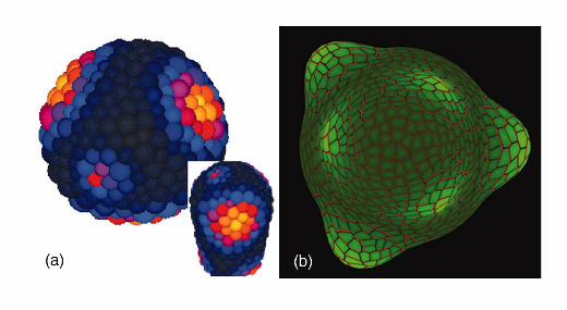
Figure 3. Modelled auxin distribution pattern in the root and shoot
(a) Illustration of the cylindrical template representing the root elongation zone
used in Swurap et al. (2006). The 3D model incorporates the epidermis, cortex, and
endodermis cells and auxin transport mediator locations illustrated in the inset (pink
PIN1/2, orange AUX1). The blue shades illustrate auxin concentrations in the epidermis
when auxin is deposited at the lower side from the lateral root cap (arrow) (Adapted with
permission from Macmillan Publishers Ltd: Nature Cell Biology [••17], Copyright 2006.
(b) Transverse section of the SAM showing anti-PIN1 immunolabeling (top) and
simulation output showing auxin concentrations (bottom) from Barbier et al. (2006). The
cells at the location of a new primordia (circle) as well as the apex have high auxin

concentrations. Modified from [••22] with permission from National Academy of
Sciences, Copyright 2006.
Figure 4. 4D segmentation of the shoot meristem epidermis
(a) Top view of the meristem epidermis extracted from a stack of transverse
confocal images where the background has been removed and the cells manually
extracted. (b) Three dimensional view of the epidermis of the meristem. (c) Vertex
movements as calculated from two consecutive time-points of the surface reconstruction.
Note also new cell walls marked in blue. Modified from [••35] with permission from
Blackwell Publishing Ltd, Copyright 2005.

A. B.
C. D.
1