modification of drosophila p53 by sumo modulates … · modification of drosophila p53 by sumo...
TRANSCRIPT

Mauri et al.
1
MODIFICATION OF DROSOPHILA p53 BY SUMO MODULATES ITS
TRANSACTIVATION AND PRO-APOPTOTIC FUNCTIONS Federico Mauri*
1,4, Laura M. McNamee*
3, Andrea Lunardi*
1,2, Fulvio Chiacchiera
1,2,
Giannino Del Sal1,2
, Michael H. Brodsky3 and Licio Collavin
1,2
From the 1Laboratorio Nazionale Consorzio Interuniversitario Biotecnologie, AREA Science Park,
Padriciano 99, 34012 Trieste, ITALY and 2Dipartimento di Biochimica, Biofisica e Chimica delle
Macromolecole, Università degli Studi di Trieste, Via L. Giorgeri 1, 34129 Trieste, ITALY and 3Program in Gene Function and Expression and Program in Molecular Medicine, University of
Massachusetts Medical School, 55 Lake Avenue North Worcester, MA 01605, USA. 4Current address: Institute of Molecular Biotechnology of the Austrian Academy of Sciences, Dr. Bohr
Gasse 3-5, 1030 Vienna, Austria.
Running title: Sumoylation of Drosophila p53
Address correspondence to: Licio Collavin, LNCIB – AREA Science Park, Padriciano 99, 34012 Trieste
ITALY; Fax: (+39) 040-398990; E-mail: [email protected]
* FM, LMM and AL contributed equally to this work
Conjugation to SUMO is a reversible post-
translational modification that regulates several
transcription factors involved in cell
proliferation, differentiation, and disease. The
p53 tumor suppressor can be modified by
SUMO-1 in mammalian cells, but the
functional consequences of this modification
are unclear. Here, we demonstrate that the
Drosophila homolog of human p53 can be
efficiently sumoylated in insect cells. We
identify two lysine residues involved in SUMO
attachment, one at the C-terminus, between the
DNA binding and oligomerization domains, and
one at the N-terminus of the protein. We find
that sumoylation helps recruit Drosophila p53
to nuclear dot-like structures that can be
marked by human PML and the Drosophila
homologue of Daxx. We demonstrate that
mutation of both sumoylation sites dramatically
reduces the transcriptional activity of p53 and
its ability to induce apoptosis in transgenic flies,
providing in vivo evidence that sumoylation is
critical for Drosophila p53 function.
The p53 tumor suppressor is a highly
regulated transcription factor that coordinates
cellular responses to DNA damage, activation of
oncogenes, and a variety of other stress signals
(1); accordingly, p53 inactivation is the most
common mutation found in human cancers (2). A
complex array of post-translational modifications
regulate stability, localization, conformation, and
transcriptional activity of p53, with crucial
implications for its tumor suppressive function (3-
6).
SUMO-1 belongs to a family of small
ubiquitin-related proteins that are covalently
linked to lysine residues of protein substrates
(7,8). In contrast to ubiquitination, sumoylation
does not target modified proteins for degradation,
but can affect their localization, stability, and
functions (7-10). Human p53 can be modified by
SUMO-1 on a single C-terminal lysine (K386) but
the effects of this modification are controversial
(3,11). Initial studies indicated that SUMO
stimulates the activity of p53 (12-14). In contrast,
other work suggested that sumoylation does not
affect p53 transcriptional activity (15,16). In
addition, conflicting reports indicate that the
SUMO E3 ligase PIAS1 can either stimulate or
inhibit p53 activity (15,17). Overexpression of
SUMO-1 stimulates recruitment of p53 to PML
Nuclear Bodies (NBs), with implications for p53
pro-apoptotic activity, but mutation of the SUMO
http://www.jbc.org/cgi/doi/10.1074/jbc.M710186200The latest version is at JBC Papers in Press. Published on May 20, 2008 as Manuscript M710186200
Copyright 2008 by The American Society for Biochemistry and Molecular Biology, Inc.
at ST
OW
ER
S IN
ST
ITU
TE
FO
R M
ED
on July 11, 2008 w
ww
.jbc.orgD
ownloaded from

Mauri et al.
2
acceptor site does not prevent p53 localization to
NBs (16,18). Two knock-in mouse models have
been generated in which all C-terminal lysine
residues in p53 have been mutated, including the
sumoylation site; despite extensive cell culture
data indicating critical roles of these residues for
p53 function, these mice are similar to wild type,
and MEFs and thymocytes derived from these
animals display normal apoptotic responses after
DNA damage (19,20). These results suggest that
several post-translational modifications of the C-
terminus, including sumoylation, may not be
crucial for p53 function in mammalian cells (5).
Other members of the p53 family are also
sumoylated at their C-terminus (3). In cell culture,
sumoylation of p63 destabilizes the protein and
decreases its transactivation function (21,22) while
sumoylation of p73 modulates its nuclear
localization and turnover (23). Therefore, although
the biological effects of sumoylation may vary
among p53-related proteins, modification with
SUMO is a common feature of the p53 family,
suggesting an ancient regulatory mechanism
inherited from a common ancestor gene.
In Drosophila melanogaster there is a
single p53 family member, with the same domain
structure of mammalian p53 proteins. The core
DNA binding domain has the greatest sequence
similarity, while the N- and C-terminal domains
show little sequence conservation but retain
similar structural and functional features (24-27).
Drosophila p53 binds the same consensus
sequence as human p53, and transactivates
reporter constructs driven by p53 responsive
elements (24-26). Drosophila mutants lacking p53
function are viable and fertile, but are defective for
induction of apoptosis by DNA damage or
unprotected telomeres (24,26,28-31). Drosophila
p53 induces cell death when overexpressed in eye
imaginal discs (24,26), upregulates pro-apoptotic
genes including reaper, sickle, hid and Eiger, and
binds a specific DNA damage responsive element
within the reaper promoter (28-30,32). Activation
of Drosophila p53-dependent apoptosis following
DNA damage depends on the protein kinase
Mnk/Chk2, which phosphorylates p53 (28,33).
Other post-translational modifications of
Drosophila p53 have not been demonstrated.
Here we show that Drosophila p53 can be
modified by SUMO on two independent residues.
We present evidence that a sumoylation-defective
p53 mutant is markedly less active than the wild-
type counterpart, in cell culture and in vivo,
implicating sumoylation in the biochemical
circuitry that positively regulates Drosophila p53
function.
Experimental procedures
Plasmids - The cDNA for Drosophila p53
was picked from the Drosophila Gene Collection
(DGC1.0). Mutants K26R, K302R and KRKR
were generated by PCR-based mutagenesis. The cDNAs for Drosophila SUMO and DLP(ct) were retrieved from DGC1.0, while full length DLP was obtained from the Drosophila Genomics Resource Center (DGRC). Coding regions were amplified by PCR and inserted in pAc5.1 vectors (Invitrogen) modified for expression of N-terminally RGS-His-, HA- or GFP- tagged proteins. In the SUMO-KRKR chimera, Drosophila SUMO (aa 1 to 85) is fused to residue 18 of the p53 KRKR mutant. Expression of the fusion protein at the expected molecular weight was verified by immunoblotting (not shown). For
luciferase assays, untagged wild-type p53 and K to
R mutants were cloned in pAc5.1 vectors. All
constructs involving PCR were fully sequenced.
pRpr150-LUC was constructed inserting the 150
bp EcoRI-XhoI fragment from the pH150-LacZ
reporter (24) into the EcoRI-XhoI sites of the
pGL2-promoter vector (Promega).
Cell culture, transfection and luciferase
assays - S2 cells were cultured at 26˚C in
Schneider’s Drosophila medium (Invitrogen) with
10% scomplemented fetal calf serum (FCS),
penicillin (50 U/ml) and streptomycin (50 mg/ml).
Transfections were performed by calcium
phosphate co-precipitation. For luciferase assays,
S2 cells in 3 cm petri dishes were transfected with
500 ng of the reporter, and 250 ng or 500 ng of
p53 expression plasmids. In all samples, 100 ng of
pPacLacZ were included for normalization of
transfection efficiency. After 36 hours, cells were
lysed and assayed for Luciferase and beta-
galactosidase activity. Fold induction is the ratio
at ST
OW
ER
S IN
ST
ITU
TE
FO
R M
ED
on July 11, 2008 w
ww
.jbc.orgD
ownloaded from
Mauri et al.
3
of luciferase over beta-galactosidase, normalized
to the activity of the reporter co-transfected with
empty vector. Expression levels of transfected
proteins were verified by immunoblotting of the
same lysates; gel loading was normalized for
transfection efficiency using beta-galactosidase
levels.
Western blotting, immuno-precipitation
and immunofluorescence - Immunoblotting was
performed in standard conditions. For
immunoprecipitations, S2 cells seeded in 6 cm
Petri dishes were collected 24 hours after
transfection and lysed in RIPA buffer (300mM
NaCl) containing 10 mM N-ethylmaleimide, 1
mM PMSF, and protease inhibitors. Clarified
lysates were incubated at 4˚C with anti RGS-His
primary antibody cross-linked to Protein G-
Sepharose (GE Healthcare). For immuno-
fluorescence, 36 hours after transfection S2 cells
were plated on glass coverslips coated with 0.5
mg/ml Concanavalin A (Sigma) or 0.5 mg/ml
poly-lysine (Sigma). After 2 hours, cells were
washed with PBS and fixed in 4%
paraformaldehyde at RT for 20 min. Cells were
permeabilized in PBS plus 0.1% Triton X-100.
Images were captured using a laser-scanning
microscope (Zeiss Axiocam 100M). The following
primary antibodies were used: mouse anti-RGS-
His (Qiagen), rabbit anti-GFP (self produced),
rabbit anti-SUMO (34), mouse anti-HA (mAb
12CA5), mouse anti-Drosophila p53 (24).
Electrophoretic mobility shift assay - For
EMSA, approximately 5x10⁶ S2 cells seeded in 6
cm petri dishes were transfected with p53
expression plasmids and harvested 48 hr after
transfection in lysis buffer (10mM Tris-HCl [pH
7.5], 1 mM EDTA, 0.5% NP40, 150 mM NaCl, 1
mM DTT, 10% glycerol, 0.5 mM PMSF, and
protease inhibitors). After 20 minutes on ice,
extracts were centrifuged at 16000xg for 20 min at
4°C to remove cell debris. Protein concentration in
supernatants was determined using Bio-Rad
protein assay. Expression levels of transfected
proteins were verified by immunoblotting of the
same lysates. A 26-mer DNA oligonucleotide
containing the cis-acting p53 responsive sequence
from the Reaper enhancer (5’-
ACCTGACATGTTTGAACAAGTCGAGC -3’)
was end-labeled with 32
P and annealed to the
complementary strand. For binding reactions, 30
μg of whole cell extract were added to gel shift
buffer (20 mM HEPES [pH 8], 25 mM KCl, 0.1
mM EDTA, 2 mM MgCl2, 0.5 mM DTT, 0.025%
NP-40, 2 mM spermidine, 10% glycerol, 0.1
mg/ml acetylated BSA, 120 ng double-stranded
poly(d[I-C])) containing the labeled
oligonucleotide in a final volume of 30 μl.
Competition was done adding 500 ng of unlabeled
double stranded oligonucleotide. Reactions were
incubated for 30 min at RT, and electrophoresed
on a non-denaturing 4% polyacrylamide gel before
autoradiography.
Transgenes and genetics - Flies were
raised at 250C. Wild type p53 and p53
KRKR were
expressed using the GUS vector which contains
both the UAS promoter for inducible expression
by Gal4, and Glass binding sites from GMR for
low to moderate expression in the developing eye
(24,28). Cloning of wild type p53 was previously
described (28), while GUSp53KRKR
was
constructed using Gateway cloning. For high
levels of p53 expression, GMRGal4 animals were
crossed to four independent lines of GUSp53+ and
GUSp53KRKR
. For lower levels of expression and
rescue of damage-induced apoptosis, p53- animals
were crossed to GUSp53+; p53
- and GUSp53
KRKR;
p53- flies.
Irradiation and immuno-histochemistry -
Climbing third instar larvae were irradiated with
4000 rads using a faxitron X-ray cabinet or mock
treated. Four hours following irradiation, eye discs
were dissected and stained with antibodies as
previously described (24). Discs were incubated
with primary antibodies in PBTN (PBS, 0.3%
triton, 5% normal goat serum) overnight at 4°C,
and with secondary antibodies in PBTN for two
hours at room temperature. The primary antibodies
used were rabbit anti-cleaved caspase-3 (1:100,
Cell Signaling), rabbit anti-SUMO (1:1000, gift from L.C. Griffith) (34), and mouse anti-
Drosophila p53 (1:10) (24). The secondary
antibodies used were donkey anti-mouse Alexa
488 and donkey anti-rabbit Alexa 555 (1:2000,
Molecular Probes). TUNEL staining was
performed using ApopTag Fluorescein In Situ
Apoptosis Detection Kit (Chemicon). Eye
at ST
OW
ER
S IN
ST
ITU
TE
FO
R M
ED
on July 11, 2008 w
ww
.jbc.orgD
ownloaded from
Mauri et al.
4
imaginal discs were fixed in 4% formaldehyde,
washed 5 times with PBTw (PBS+ 0.3% Tween-
20), and post fixed with cold ethanol/PBS (2:1).
Following rehydration and washing, discs were
treated with TdT mix for 1hr at 370C. After the
reaction was stopped, discs were incubated with
Fluorescein-conjugated anti-dig antibody for 30
minutes and mounted with Vectashield.
Confocal microscopy and quantification of
TUNEL and active caspase staining - Localization
of p53 and SUMO was visualized using a Leica
SP2 AOBS confocal microscope with a 63X
objective. For an overall view of the GMR region,
a Z-series was taken through the eye disc at
intervals of 284 nm, at a zoom of 1.75. For a
higher magnification view, a Z-series was taken
through the eye disc at intervals of 122 nm, at
zoom of 4. To quantify the amount of cleaved
caspase-3 in the eye disc, a Z-series was taken
through the entire eye disc at intervals of 1.42 m
with a 20x objective. A 3-D reconstruction of each
eye disc was generated using Imaris 5.0 image
analysis software (Bitplane AG). Only the
posterior region of the eye disc in which the p53
transgene is expressed was analyzed. The volume
positive for cleaved caspase-3 was determined
using a high intensity threshold, while the total
disc volume was determined using a low intensity
threshold (Supplementary Fig. S4). The caspase-
positive index for the posterior of the eye disc was
calculated by dividing the cleaved caspase-3
volume by the total volume. To quantify the
number and location of TUNEL positive cells in
the eye disc, a Z-series was taken through the
entire eye using a Zeiss Axioplan2 microscope and
an Hamamatsu ORCA-ER camera with a 20x
objective. Images were deconvolved using an
inverse filter algorithm in the Zeiss Axiovision 4.5
image analysis software. Imaris 5.0 image analysis
software was used to create a 3-D reconstruction
of the TUNEL staining in the eye disc. Individual
positive cells were marked using the “spot”
function, which identifies local maxima of signal
intensity (Supplementary Fig. S4). The distance of
TUNEL positive cells from the morphogenetic
furrow was determined by subtracting the position
of the furrow from the position of each cell.
RESULTS
Identification of two functional
sumoylation sites in Drosophila p53 – In yeast
two-hybrid screens, we and others have found
interactions between Drosophila p53 and
lesswright/dUbc9 (an E2 SUMO ligase), Su(var)2-
10/dPIAS (an E3 SUMO ligase) and Ulp1 (a
SUMO specific peptidase), suggesting that p53
may be sumoylated (35,36)(M.H.B. and Garson
Tsang, unpublished results). Within the p53
sequence there are two consensus sites for
sumoylation, one on lysine 302 in the C-terminal
region of the protein, the other on lysine 26 within
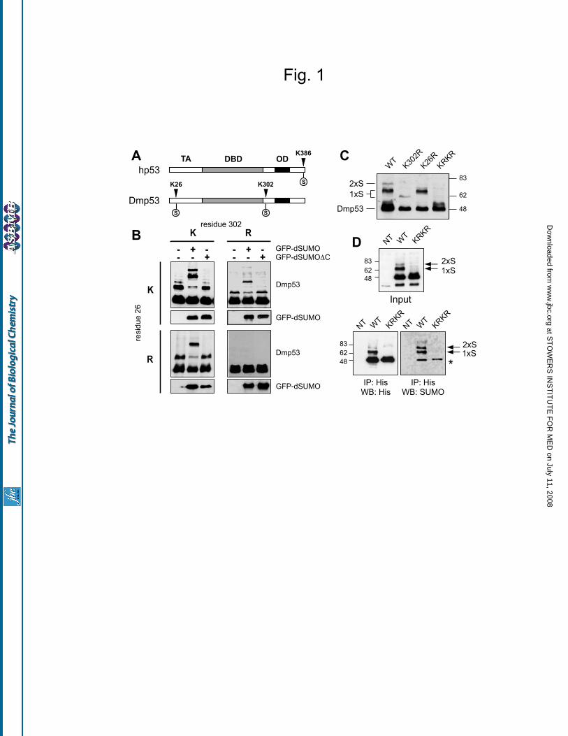
the N-terminal transactivation domain (Fig. 1A).
These sites do not directly correspond to the single
site identified at the extreme C-terminus of
mammalian p53, p63 or p73 (3).
To test if Drosophila p53 can be
sumoylated, Drosophila S2 cells were transfected
with His-tagged p53 (HT-Dmp53) alone, with
Drosophila SUMO fused with GFP (GFP-
dSUMO), or with a non-conjugatable version of
SUMO lacking the C-terminal glycines necessary
for attachment to substrates (GFP-dSUMO C).
Transfected p53 was visualized by immuno-
blotting with an antibody to the RGS-His tag. As
shown in Figure 1B-C, transfected p53 migrates as
one primary band and two slower migrating bands;
the apparent molecular weights are compatible
with attachment of one or two SUMO molecules.
In cells transfected with GFP-dSUMO, the upper
bands shift to higher molecular weights,
compatible with covalent attachment of one and
two GFP-dSUMO molecules. This shift is not
observed in cells transfected with the non-
conjugatable GFP-dSUMO C (Fig. 1B).
To test the requirement of lysine 302 and
lysine 26 for conjugation, they were replaced with
arginine by site-directed mutagenesis. When either
lysine 26 or lysine 302 are altered, the resulting
proteins (p53 K26R and p53 K302R) display a
single slower migrating band (Fig. 1B). When
both residues are mutated, the resulting protein
(p53 KRKR) is no longer modified. Comparison
of the various mutants suggests that lysine 302
may be sumoylated more efficiently than lysine
26. In addition, modification at K302 apparently
at ST
OW
ER
S IN
ST
ITU
TE
FO
R M
ED
on July 11, 2008 w
ww
.jbc.orgD
ownloaded from

Mauri et al.
5
induces a greater shift in migration than
modification at K26 (Fig. 1C).
To verify that endogenous SUMO is
covalently attached to p53, HT-Dmp53 was
immuno-precipitated from transfected S2 cells and
probed with an antibody to Drosophila SUMO
(Fig. 1D). Based on these results, we conclude that
a significant fraction of Dmp53 is sumoylated
when expressed in S2 cells, with lysine 302 being
the primary modification site.
Drosophila p53 localizes to nuclear dots -
The distribution of wild-type and non
sumoylatable p53 was analyzed in transfected S2
cells. As shown in Figure 2A, transfected p53 is
found throughout the nucleus with marked
accumulation in dot-like structures. p53 forms
nuclear dots in 70-80 percent of transfected cells,
with most nuclei having 2 or 3 dots. This
localization was not dependent on the adhesion
substrate (Concanavalin A or poly-lysine) and did
not change using N-terminally tagged or untagged
p53 (not shown). The p53 K to R mutants form
nuclear dots with similar frequency, shape, and
size as the wild-type protein. When co-transfected with GFP-dSUMO, wild type p53 and single lysine mutants co-localize with SUMO in nuclear dots. However, only a subset of nuclear dots formed by the non-sumoylatable p53 KRKR mutant overlap with dots formed by GFP-dSUMO (Fig. 2 and Supplementary Fig. S1).
Localization of wild type and non-
sumoylatable p53 was also analyzed in transgenic
flies, using the GAL4/UAS system. With a GMR-
GAL4 driver, high levels of p53 are produced in
the posterior of the developing eye imaginal disc,
sufficient to induce ectopic apoptosis in the
absence of DNA damage (24,26,28). In the
absence of GAL4 driver, p53 is expressed at low
levels, insufficient to induce apoptosis (see Fig. 5).
In both conditions, wild type p53 and p53 KRKR
are found throughout the nucleus with a sub-
nuclear domain of elevated staining, similar to that
seen in cell culture (Fig. 2C-D). We used a
polyclonal antibody to visualize endogenous
SUMO in these cells (34). Endogenous SUMO is
not detected in cells expressing less p53, probably
due to low levels of SUMO throughout the entire
nucleoplasm. At the higher p53 expression levels,
endogenous SUMO accumulates in nuclear dots
with wild type, but not with non-sumoylatable p53
(Fig 2C).
In contrast with cell culture results, p53
KRKR transgenic cells displayed lower overall
levels of immuno-staining, suggesting reduced
expression levels. This result was observed in four
independent transformants per line (data not
shown), and therefore is not a consequence of
genomic insertion sites. No p53 immunostaining
was detected in cells solely expressing endogenous
levels of p53 (data not shown). Following
exposure to ionizing radiation (IR), wild type p53
and p53 KRKR are still detected throughout the
nucleus and in nuclear dots (Fig. 2D). Together,
these results confirm that p53 accumulates in sub-
nuclear structures in cultured cells and in normal
developing tissues. The non-sumoylatable p53
KRKR mutant can also form nuclear dot-like
structures, but has reduced capacity to recruit
SUMO.
Sumoylation affects p53 localization to nuclear dots defined by human PML and Drosophila Daxx-like protein – We next asked if nuclear dots formed by wild type or non-sumoylatable p53 are in fact the same structures. In mammalian cells, p53 accumulates within PML nuclear bodies (NBs) under specific conditions (16,18,37,38). Markers for such structures are PML and Sp100 (39,40), but there are no Drosophila homologs of these proteins. However, transfected human PML IV forms nuclear dots that co-localize with SUMO in Drosophila cells (41). When co-transfected with hPML IV, wild type p53 and single lysine mutants co-localize with PML in nuclear dots. On the contrary, only a subset of nuclear dots formed by the non-sumoylatable p53 KRKR mutant overlap with those formed by PML (Fig. 2 and Supplementary Fig. S2).
Daxx, a transcriptional repressor and scaffolding protein, is also found in mammalian PML-NBs (42,43). The Drosophila homolog of Daxx, referred to as Daxx-like protein (DLP), has been described very recently (44): it encodes a large peptide with similarity to Daxx in the C-terminus. We observed that DLP accumulates in
at ST
OW
ER
S IN
ST
ITU
TE
FO
R M
ED
on July 11, 2008 w
ww
.jbc.orgD
ownloaded from

Mauri et al.
6
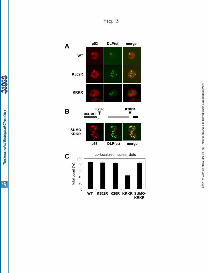
nuclear dots when overexpressed in Drosophila S2 cells, and these dots co-localize with GFP-dSUMO and human PML IV (Supplementary Fig. S3). A DLP deletion lacking the first 710 aminoacids, named DLP(ct), shows similar behavior, indicating that the region of Daxx similarity is sufficient for the observed localization (Fig. S3). We used GFP-DLP(ct) as a marker to analyze localization of p53 mutants; we counted the fraction of p53 nuclear dots co-localized with DLP(ct) in confocal images from independent co-transfection experiments (Fig. 3A). As summarized in Figure 3C, 85 to 90% of the dots formed by wild type p53 or single lysine mutants co-localize with DLP(ct). In contrast, only 45% of nuclear dots formed by the non-sumoylatable p53 KRKR co-localize with DLP(ct). Notably, the co-localization of the p53 KRKR mutant with DLP is restored to wild type levels when SUMO is fused to p53 KRKR to mimic constitutive K26 sumoylation (Fig. 3B). Together, these results indicate that sumoylation affects the recruitment of Drosophila p53 to specific nuclear domains.
Non-sumoylatable p53 is less active than
the wild-type protein in cultured cells - Given the
conflicting data on the functional relevance of
sumoylation in mammalian p53, we asked whether
sumoylation might affect the transcriptional
activity of Drosophila p53. Initially, we used the
pG13-LUC plasmid, a reporter responsive to
mammalian p53 (25). We transfected this
construct with increasing amounts of expression
vectors encoding untagged versions of p53
mutants, and assayed for luciferase. With this
reporter, equal expression levels of non-
sumoylatable p53 KRKR mutant have
significantly less transcriptional activity than wild-
type p53 (Fig. 4). In contrast, mutants with
substitution of either single lysine display a
transactivation activity similar to wild-type p53.
To confirm this behavior with a Drosophila
promoter element, a luciferase reporter driven by a
DNA damage responsive cis-regulatory sequence
from the reaper locus, containing a p53 binding
site (Rpr150 enhancer) (24), was constructed and
tested as above. The non-sumoylatable p53 KRKR
mutant was also less active than wild-type p53 or
single lysine mutants using this reporter (Fig. 4B).
To test if the reduced transcriptional
activity of p53 KRKR is due to impaired
sequence-specific DNA binding, we prepared
lysates from transfected S2 cells and performed
electrophoretic mobility shift assays (EMSA)
using a double stranded DNA oligonucleotide
containing the p53-binding element from the
Rpr150 enhancer. As shown in Figure 4C and D,
the p53 KRKR mutant binds efficiently to the
oligonucleotide probe, indicating that mutation of
both lysines does not prevent sequence-specific
DNA binding.
p53 sumoylation sites are essential for in
vivo function - To determine if sumoylation affects
p53 function in vivo, we compared the activity of
wild type and non-sumoylatable p53 in the
developing eye. We examined the ability of p53 to
induce apoptosis under two conditions, when
highly overexpressed and when activated
following exposure to ionizing radiation (IR).
Overexpression of p53 using GMR-Gal4 and
GUS-p53 results in a rough, reduced eye
phenotype, accompanied by a loss of pigmentation
in the center of the eye (26,28)(Fig. 5A, B).
Overexpression of p53 KRKR induces a similar
rough eye, but with less loss of pigmentation (Fig.
5C), suggesting a difference in wild type and
mutant p53 activity during eye development.
GMR-Gal4 induces target gene expression
beginning in the morphogenetic furrow, which
marks cells in the eye imaginal disc as they initiate
synchronous cell cycle progression and
differentiation. The furrow first forms in cells at
the posterior of the disc and moves to increasingly
anterior cells. As a result, cells near the furrow
have just begun to express the transgene, while
more posterior cells have expressed it for longer
times. Overexpressed wild type p53 induces a high
level of apoptosis in a band of cells immediately
posterior to the furrow, as assayed by activated
caspase and TUNEL staining (Fig. 5D, E, G and
data not shown). In eye discs overexpressing p53
KRKR, the band of apoptosis is initially weaker
and extends further posterior from the furrow (Fig.
5F and G). In these experiments, the mean
at ST
OW
ER
S IN
ST
ITU
TE
FO
R M
ED
on July 11, 2008 w
ww
.jbc.orgD
ownloaded from

Mauri et al.
7
distance of apoptotic cells is increased from 38.0
(s.e.m.=3.0) for wild type p53 to 59.3 (s.e.m.=2.5)
for p53KRKR
(p=0.036, two-tailed student’s t-test).
However, the total number of apoptotic cells is
similar; the average number of TUNEL positive
cells is 568 (s.e.m.=55.5) for wild type p53 and is
474 (s.e.m.=37.4) for p53KRKR
(p=0.20). Since the
distance from the morphogenetic furrow
corresponds to the length of time cells have been
overexpressing p53, these experiments indicate a
delay in the induction of apoptosis by non-
sumoylatable p53. This delay could reflect either
lower levels of p53 KRKR expression, decreased
transcriptional activity of p53 KRKR, or both.
The role of sumoylation in p53 function
was also examined during DNA damage induced
apoptosis. Drosophila p53 is required for the rapid
induction of apoptosis by ionizing radiation (IR)
(24,26) (Fig. 5H, L, I, M, P). In the absence of a
GAL4 driver, the GUS vector expresses sufficient
p53 in the posterior of the eye disc to fully restore
IR-induced apoptosis in a p53-null background
(28) (Fig. 5J, N, P). In contrast, expression of p53
KRKR only weakly rescues IR-induced apoptosis
(Fig. 5K, O, P). Under these conditions, apoptosis
is induced at the same time in all cells, and
quantification of cleaved caspase-3 reveals a six-
fold decrease in the level of IR-induced apoptosis
in p53KRKR
transgenics (Fig. 5P, p=0.008, student’s
t-test). The decrease is greatest near the furrow,
resulting in a change in the pattern of apoptotic
cells. It is important to emphasize that the failure
to fully rescue apoptosis is not due to insufficient
expression levels, since both the wild type and
mutant p53 transgenes are expressed at higher
levels than endogenous p53 (endogenous p53 is
not detected by immunofluorescence). These
results confirm that the p53 KRKR mutant is less
active than the wild-type protein and demonstrate
that sumoylation sites are critical for induction of
apoptosis by p53 following DNA damage in vivo.
DISCUSSION
In this work, we find that Drosophila p53
has two sites of sumoylation: one at the N-
terminus and the other in the C-terminal region,
before the oligomerization domain. Human p53, as
well as p63 and p73, are sumoylated on a single
residue at the extreme C-terminus (3,19).
Therefore, the modification is conserved, but its
position has changed during evolution. In
mammalian p53 the last C-terminal amino acids
are not required for oligomerization, and serve a
regulatory function. In contrast, in Drosophila p53
the C-terminal 24 amino acids form an alpha-helix
that interacts with the oligomerization domain and
is required for tetramerization (27). Thus, both in
mammals and Drosophila, C-terminal sumoylation
of p53 occurs on a site where it should not
interfere with oligomerization.
Because of very low expression levels, it
is extremely difficult to detect endogenous p53 in
Drosophila tissues or cell lines, even after DNA
damage stimulation. Although we have not been
able to examine endogenous p53, we have
demonstrated that p53 is efficiently modified in
cells in which SUMO and the SUMO ligating
enzymes are present at physiological levels.
Exploration of the signaling pathways that regulate
p53 sumoylation in vivo will require the
development of reagents and/or techniques to
efficiently detect endogenous p53 protein in
Drosophila cells.
p53 forms nuclear dots when over-
expressed in Drosophila cells in culture and in
developing eye imaginal discs. In tissue culture,
dots formed by p53 co-localize with dots formed
by human PML IV and Drosophila Daxx. It is
tempting to speculate that such structures may be
related to mammalian PML nuclear bodies.
However, an important caveat is that these
observations rely on ectopic expression of
transfected proteins, since no reagents are
available to detect endogenous counterparts. The
absence of an obvious PML homolog in
Drosophila clearly indicates that these structures
are not identical in insects and mammals;
however, the recruitment of human PML IV to
dots containing Drosophila SUMO, p53, and Daxx
homologs does suggest that some aspects are
conserved. We find that non-sumoylatable p53
also forms nuclear dots. However, it seems likely
that nuclear dots formed by non-sumoylatable p53
are qualitatively different from those formed by
the wild-type protein, as suggested by the reduced
at ST
OW
ER
S IN
ST
ITU
TE
FO
R M
ED
on July 11, 2008 w
ww
.jbc.orgD
ownloaded from

Mauri et al.
8
co-localization with dots marked by GFP-SUMO,
hPML IV and DLP. Thus, the change in
localization to specific sub-nuclear domains
correlates with the reduced activity of the p53
KRKR mutant.
Our experiments demonstrate that
sumoylation sites are important for the activity of
Drosophila p53 both in tissue culture and in vivo.
However, the sumoylation-deficient p53 KRKR
mutant is not completely inactive; it retains
sequence-specific DNA binding, and moderately
transactivates both reporter constructs tested (Fig.
4). p53 KRKR also retains some residual ability to
induce apoptosis in irradiated imaginal discs,
indicating that sumoylation sites are not absolutely
required for p53 activation by DNA damage (Fig.
5), but are essential for optimal activity.
It is important to note that single mutation
of either sumoylation site had no significant effect
on the activity of p53. This implies that
modification of a specific lysine is not critical;
rather, it is important that SUMO can be attached
to the protein. This observation indicates that
sumoylation does not simply function to compete
with another modification of the same residue (i.e.
ubiquitination or acetylation). Our observation that
sumoylation is not required for DNA binding
suggests that this modification may mediate
recruitment of additional factors needed for p53
dependent transcription of target genes.
Alternatively, sumoylation may indirectly control
p53 modification on other residues via interaction
with specific modifying enzymes.
Sumoylation affects turnover of human
p63 and p73 (3), but we see no difference in the
expression levels of transfected wild-type or
mutant p53 in S2 cells, where p53 KRKR has
clearly reduced transcriptional activity (Fig.1, Fig.
4 and data not shown). In contrast, in developing
eye discs, p53 KRKR seems to be expressed at
lower levels than wild type. This difference may
contribute to the difference in apoptosis induced
by strong p53 overexpression. However, p53
KRKR cannot rescue DNA damage-induced
apoptosis in p53 mutant animals, despite being
expressed at much higher levels than endogenous
p53 in wild-type animals (see Fig. 2 and Fig. 5.
Endogenous p53 was not detectable in wild-type
cells). This observation demonstrates that
sumoylation sites are critical for induction of
apoptosis by p53 in vivo, regardless of the
difference in expression levels detected between
exogenous wild-type and KRKR p53 proteins in
eye discs.
Our results in Drosophila are consistent
with studies in mammalian cells reporting that
sumoylation promotes p53 function (12,13,45), but
are in contrast with knock-in mouse models
demonstrating that C-terminal lysines are not
crucial for p53 function in vivo (5,19,20). There
are several possible explanations for this
discrepancy. First, since knock-in p53 models had
mutations in all C-terminal lysines, it is possible
that loss of other modification sites masks a
specific requirement for sumoylation. Second,
human p53 might be sumoylated at additional non-
canonical residues (16,46); a weak secondary site
may compensate for loss of the primary
sumoylation site. Third, interaction with
sumoylated proteins may be sufficient to substitute
for direct sumoylation of mammalian p53; these
interacting partners may not be present in
Drosophila (e.g. PML)(18). Finally, specific
features of the molecular regulation of Drosophila
p53 may account for a more stringent requirement
for sumoylation.
In conclusion, our data demonstrate that
SUMO attachment is a modification of p53 that is
evolutionarily conserved from insects to
mammals. Specific requirements for this
modification may have changed with the
emergence of three p53 paralogs in vertebrates,
but sumoylation sites are clearly important for
function of the single p53 protein in Drosophila.
Our results support the general hypothesis that
sumoylation has an important role in regulation of
metazoan p53 and p53-related proteins.
at ST
OW
ER
S IN
ST
ITU
TE
FO
R M
ED
on July 11, 2008 w
ww
.jbc.orgD
ownloaded from

Mauri et al.
9
REFERENCES
1. Vousden, K. H., and Lane, D. P. (2007) Nat Rev Mol Cell Biol 8, 275-283
2. Harris, S. L., and Levine, A. J. (2005) Oncogene 24, 2899-2908
3. Watson, I. R., and Irwin, M. S. (2006) Neoplasia 8, 655-666
4. Bode, A. M., and Dong, Z. (2004) Nat Rev Cancer 4, 793-805
5. Toledo, F., and Wahl, G. M. (2006) Nat Rev Cancer 6, 909-923
6. Horn, H. F., and Vousden, K. H. (2007) Oncogene 26, 1306-1316
7. Muller, S., Hoege, C., Pyrowolakis, G., and Jentsch, S. (2001) Nat Rev Mol Cell Biol 2, 202-210
8. Hay, R. T. (2005) Mol Cell 18, 1-12
9. Muller, S., Ledl, A., and Schmidt, D. (2004) Oncogene 23, 1998-2008
10. Gill, G. (2005) Curr Opin Genet Dev 15, 536-541
11. Hoeller, D., Hecker, C. M., and Dikic, I. (2006) Nat Rev Cancer 6, 776-788
12. Rodriguez, M. S., Desterro, J. M., Lain, S., Midgley, C. A., Lane, D. P., and Hay, R. T. (1999)
Embo J 18, 6455-6461
13. Gostissa, M., Hengstermann, A., Fogal, V., Sandy, P., Schwarz, S. E., Scheffner, M., and Del Sal,
G. (1999) Embo J 18, 6462-6471
14. Muller, S., Berger, M., Lehembre, F., Seeler, J. S., Haupt, Y., and Dejean, A. (2000) J Biol Chem
275, 13321-13329
15. Schmidt, D., and Muller, S. (2002) Proc Natl Acad Sci U S A 99, 2872-2877
16. Kwek, S. S., Derry, J., Tyner, A. L., Shen, Z., and Gudkov, A. V. (2001) Oncogene 20, 2587-2599
17. Megidish, T., Xu, J. H., and Xu, C. W. (2002) J Biol Chem 277, 8255-8259
18. Fogal, V., Gostissa, M., Sandy, P., Zacchi, P., Sternsdorf, T., Jensen, K., Pandolfi, P. P., Will, H.,
Schneider, C., and Del Sal, G. (2000) Embo J 19, 6185-6195
19. Feng, L., Lin, T., Uranishi, H., Gu, W., and Xu, Y. (2005) Mol Cell Biol 25, 5389-5395
20. Krummel, K. A., Lee, C. J., Toledo, F., and Wahl, G. M. (2005) Proc Natl Acad Sci U S A 102,
10188-10193
21. Huang, Y. P., Wu, G., Guo, Z., Osada, M., Fomenkov, T., Park, H. L., Trink, B., Sidransky, D.,
Fomenkov, A., and Ratovitski, E. A. (2004) Cell Cycle 3, 1587-1596
22. Ghioni, P., D'Alessandra, Y., Mansueto, G., Jaffray, E., Hay, R. T., La Mantia, G., and Guerrini, L.
(2005) Cell Cycle 4, 183-190
23. Minty, A., Dumont, X., Kaghad, M., and Caput, D. (2000) J Biol Chem 275, 36316-36323
24. Brodsky, M. H., Nordstrom, W., Tsang, G., Kwan, E., Rubin, G. M., and Abrams, J. M. (2000) Cell
101, 103-113
25. Jin, S., Martinek, S., Joo, W. S., Wortman, J. R., Mirkovic, N., Sali, A., Yandell, M. D., Pavletich,
N. P., Young, M. W., and Levine, A. J. (2000) Proc Natl Acad Sci U S A 97, 7301-7306
26. Ollmann, M., Young, L. M., Di Como, C. J., Karim, F., Belvin, M., Robertson, S., Whittaker, K.,
Demsky, M., Fisher, W. W., Buchman, A., Duyk, G., Friedman, L., Prives, C., and Kopczynski, C.
(2000) Cell 101, 91-101
27. Ou, H. D., Lohr, F., Vogel, V., Mantele, W., and Dotsch, V. (2007) Embo J 26, 3463-3473
28. Brodsky, M. H., Weinert, B. T., Tsang, G., Rong, Y. S., McGinnis, N. M., Golic, K. G., Rio, D. C.,
and Rubin, G. M. (2004) Mol Cell Biol 24, 1219-1231
29. Lee, J. H., Lee, E., Park, J., Kim, E., Kim, J., and Chung, J. (2003) FEBS Lett 550, 5-10
30. Sogame, N., Kim, M., and Abrams, J. M. (2003) Proc Natl Acad Sci U S A 100, 4696-4701
31. Oikemus, S. R., McGinnis, N., Queiroz-Machado, J., Tukachinsky, H., Takada, S., Sunkel, C. E.,
and Brodsky, M. H. (2004) Genes Dev 18, 1850-1861
32. Akdemir, F., Christich, A., Sogame, N., Chapo, J., and Abrams, J. M. (2007) Oncogene 26, 5184-
5193
at ST
OW
ER
S IN
ST
ITU
TE
FO
R M
ED
on July 11, 2008 w
ww
.jbc.orgD
ownloaded from

Mauri et al.
10
33. Peters, M., DeLuca, C., Hirao, A., Stambolic, V., Potter, J., Zhou, L., Liepa, J., Snow, B., Arya, S.,
Wong, J., Bouchard, D., Binari, R., Manoukian, A. S., and Mak, T. W. (2002) Proc Natl Acad Sci
U S A 99, 11305-11310
34. Long, X., and Griffith, L. C. (2000) J Biol Chem 275, 40765-40776
35. Stanyon, C. A., Liu, G., Mangiola, B. A., Patel, N., Giot, L., Kuang, B., Zhang, H., Zhong, J., and
Finley, R. L., Jr. (2004) Genome Biol 5, R96
36. Formstecher, E., Aresta, S., Collura, V., Hamburger, A., Meil, A., Trehin, A., Reverdy, C., Betin,
V., Maire, S., Brun, C., Jacq, B., Arpin, M., Bellaiche, Y., Bellusci, S., Benaroch, P., Bornens, M.,
Chanet, R., Chavrier, P., Delattre, O., Doye, V., Fehon, R., Faye, G., Galli, T., Girault, J. A., Goud,
B., de Gunzburg, J., Johannes, L., Junier, M. P., Mirouse, V., Mukherjee, A., Papadopoulo, D.,
Perez, F., Plessis, A., Rosse, C., Saule, S., Stoppa-Lyonnet, D., Vincent, A., White, M., Legrain, P.,
Wojcik, J., Camonis, J., and Daviet, L. (2005) Genome Res 15, 376-384
37. Melchior, F., and Hengst, L. (2002) Cell Cycle 1, 245-249
38. Gostissa, M., Hofmann, T. G., Will, H., and Del Sal, G. (2003) Curr Opin Cell Biol 15, 351-357
39. Lallemand-Breitenbach, V., Zhu, J., Puvion, F., Koken, M., Honore, N., Doubeikovsky, A., Duprez,
E., Pandolfi, P. P., Puvion, E., Freemont, P., and de The, H. (2001) J Exp Med 193, 1361-1371
40. Salomoni, P., and Pandolfi, P. P. (2002) Cell 108, 165-170
41. Lehembre, F., Badenhorst, P., Muller, S., Travers, A., Schweisguth, F., and Dejean, A. (2000) Mol
Cell Biol 20, 1072-1082
42. Michaelson, J. S. (2000) Apoptosis 5, 217-220
43. Salomoni, P., and Khelifi, A. F. (2006) Trends Cell Biol 16, 97-104
44. Bodai, L., Pardi, N., Ujfaludi, Z., Bereczki, O., Komonyi, O., Balint, E., and Boros, I. M. (2007) J
Biol Chem 282, 36386-36393
45. Li, T., Santockyte, R., Shen, R. F., Tekle, E., Wang, G., Yang, D. C., and Chock, P. B. (2006) J
Biol Chem 281, 36221-36227
46. Jakobs, A., Koehnke, J., Himstedt, F., Funk, M., Korn, B., Gaestel, M., and Niedenthal, R. (2007)
Nat Methods 4, 245-250
FOOTNOTES
We thank L.C. Griffith for providing the SUMO antibody, and O. Stemann for sharing S2 cells and
plasmids. We wish to acknowledge the contribution of S. Luppi, who performed the very first experiment
on Drosophila p53 sumoylation.
This work was supported by grants from AIRC (Italian Association for Cancer Research), MIUR (Italian
Ministry for University and Research) and EC FP6 contract 503576 (Active p53) to LC and GDS, and by
a Research Scholar Grant from the American Cancer Society and a New Scholar in Aging Award from
the Ellison Medical Foundation to MHB.
Supplementary material is available on the publisher’s website.
at ST
OW
ER
S IN
ST
ITU
TE
FO
R M
ED
on July 11, 2008 w
ww
.jbc.orgD
ownloaded from
Mauri et al.
11
FIGURE LEGENDS
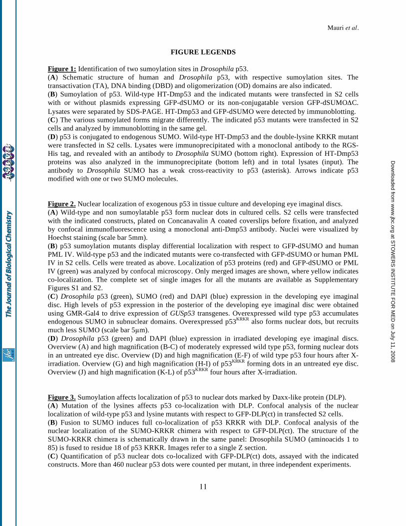
Figure 1: Identification of two sumoylation sites in Drosophila p53.
(A) Schematic structure of human and Drosophila p53, with respective sumoylation sites. The
transactivation (TA), DNA binding (DBD) and oligomerization (OD) domains are also indicated.
(B) Sumoylation of p53. Wild-type HT-Dmp53 and the indicated mutants were transfected in S2 cells
with or without plasmids expressing GFP-dSUMO or its non-conjugatable version GFP-dSUMO C.
Lysates were separated by SDS-PAGE. HT-Dmp53 and GFP-dSUMO were detected by immunoblotting.
(C) The various sumoylated forms migrate differently. The indicated p53 mutants were transfected in S2
cells and analyzed by immunoblotting in the same gel.
(D) p53 is conjugated to endogenous SUMO. Wild-type HT-Dmp53 and the double-lysine KRKR mutant
were transfected in S2 cells. Lysates were immunoprecipitated with a monoclonal antibody to the RGS-
His tag, and revealed with an antibody to Drosophila SUMO (bottom right). Expression of HT-Dmp53
proteins was also analyzed in the immunoprecipitate (bottom left) and in total lysates (input). The
antibody to Drosophila SUMO has a weak cross-reactivity to p53 (asterisk). Arrows indicate p53
modified with one or two SUMO molecules.
Figure 2. Nuclear localization of exogenous p53 in tissue culture and developing eye imaginal discs.
(A) Wild-type and non sumoylatable p53 form nuclear dots in cultured cells. S2 cells were transfected
with the indicated constructs, plated on Concanavalin A coated coverslips before fixation, and analyzed
by confocal immunofluorescence using a monoclonal anti-Dmp53 antibody. Nuclei were visualized by
Hoechst staining (scale bar 5mm).
(B) p53 sumoylation mutants display differential localization with respect to GFP-dSUMO and human
PML IV. Wild-type p53 and the indicated mutants were co-transfected with GFP-dSUMO or human PML
IV in S2 cells. Cells were treated as above. Localization of p53 proteins (red) and GFP-dSUMO or PML
IV (green) was analyzed by confocal microscopy. Only merged images are shown, where yellow indicates
co-localization. The complete set of single images for all the mutants are available as Supplementary
Figures S1 and S2.
(C) Drosophila p53 (green), SUMO (red) and DAPI (blue) expression in the developing eye imaginal
disc. High levels of p53 expression in the posterior of the developing eye imaginal disc were obtained
using GMR-Gal4 to drive expression of GUSp53 transgenes. Overexpressed wild type p53 accumulates
endogenous SUMO in subnuclear domains. Overexpressed p53KRKR
also forms nuclear dots, but recruits
much less SUMO (scale bar 5 m).
(D) Drosophila p53 (green) and DAPI (blue) expression in irradiated developing eye imaginal discs.
Overview (A) and high magnification (B-C) of moderately expressed wild type p53, forming nuclear dots
in an untreated eye disc. Overview (D) and high magnification (E-F) of wild type p53 four hours after X-
irradiation. Overview (G) and high magnification (H-I) of p53KRKR
forming dots in an untreated eye disc.
Overview (J) and high magnification (K-L) of p53KRKR
four hours after X-irradiation.
Figure 3. Sumoylation affects localization of p53 to nuclear dots marked by Daxx-like protein (DLP).
(A) Mutation of the lysines affects p53 co-localization with DLP. Confocal analysis of the nuclear
localization of wild-type p53 and lysine mutants with respect to GFP-DLP(ct) in transfected S2 cells.
(B) Fusion to SUMO induces full co-localization of p53 KRKR with DLP. Confocal analysis of the
nuclear localization of the SUMO-KRKR chimera with respect to GFP-DLP(ct). The structure of the
SUMO-KRKR chimera is schematically drawn in the same panel: Drosophila SUMO (aminoacids 1 to
85) is fused to residue 18 of p53 KRKR. Images refer to a single Z section.
(C) Quantification of p53 nuclear dots co-localized with GFP-DLP(ct) dots, assayed with the indicated
constructs. More than 460 nuclear p53 dots were counted per mutant, in three independent experiments.
at ST
OW
ER
S IN
ST
ITU
TE
FO
R M
ED
on July 11, 2008 w
ww
.jbc.orgD
ownloaded from
Mauri et al.
12
Figure 4. Mutation of both sumoylation sites affects transcriptional activity of p53 but not its DNA
binding.
(A) Transactivation of a human p53-responsive promoter. The pG13-LUC reporter plasmid was
transfected in S2 cells together with increasing amounts of vector expressing wild type p53 or
sumoylation mutants. A plasmid constitutively expressing beta-galactosidase was included as a control
for transfection efficiency. p53 transcriptional activity was measured by luciferase assay, while the levels
of expressed proteins were analyzed by immunoblotting of the same lysates (lower panel). Fold induction
values of the p53 KRKR mutant are indicated. Error bars indicate s.e.m. (n=4).
(B) Transactivation of a Drosophila p53-responsive promoter. The pRpr150-LUC reporter carrying the
p53 binding site from the Reaper DNA-damage responsive enhancer was transfected and assayed as
described above. Error bars indicate s.e.m. (n=3).
(C) Electrophoretic mobility shift assay (EMSA). Wild-type p53 and lysine mutants were tested for
sequence specific DNA binding by gel shift, using a double stranded oligonucleotide containing the p53-
responsive element form the Reaper enhancer (Rpr150). Specificity of the binding was confirmed by
competition with cold Rpr150 oligonucleotide. Lane 1, free probe. Lanes 2 to 9, whole cell lysates from
S2 cells untransfected (NT) or transfected with the indicated p53 constructs.
(D) Protein levels of transfected p53 mutants were assayed by immunoblotting of the lysates used for
EMSA.
Figure 5. p53KRKR
does not induce apoptosis as efficiently as wild type p53, and is unable to fully rescue
DNA damage-induced apoptosis
(A-F) High levels of p53 expression in the posterior of the developing eye imaginal disc were obtained
using GMR-Gal4 to drive expression of GUSp53 transgenes. (A) Wild type adult eye. (B) Adult eye
overexpressing wild type p53. (C) Adult eye overexpressing p53KRKR
. (D-F) TUNEL staining for
apoptotic cells in eye imaginal discs. (D) Wild type eye imaginal disc. (E) Eye disc overexpressing wild
type p53. (F) Eye disc overexpressing p53KRKR
. Scale bar: 20μm. (G) Distribution profiles of the distance
of TUNEL positive cells from the furrow in p53+ expressing cells versus p53KRKR
expressing cells. All
samples were normalized to calculate the mean percentage of apoptotic cells at a given distance from the
furrow out of the total number of apoptotic cells in the disc. Distribution profiles were generated to plot
percent of apoptotic cells at each distance from the furrow (n=5).
(H-O) Cleaved caspase-3 staining of eye imaginal discs mock-treated, or four hours after X-irradiation. In
the absence of a Gal4 driver, the Glass/multimer promoter of GUSp53 transgenes expresses levels of p53
that can rescue DNA damage induced apoptosis in a p53 mutant tissue, but are too low to induce
apoptosis without an external stress. The transgene expression domain in the posterior of each eye disc is
indicated with brackets. (H-K) Untreated eye discs. (L-O) Eye discs stained for cleaved capase-3 four
hours after X-irradiation. (P) Quantification of relative volumes of cleaved caspase-3 staining in the
regions marked by brackets. See methods for details of caspase quantification. Bars indicate standard
error of the mean (n=5). A two tailed t-test was used to determine the significance of the observed
changes.
at ST
OW
ER
S IN
ST
ITU
TE
FO
R M
ED
on July 11, 2008 w
ww
.jbc.orgD
ownloaded from
�� �
�����
�� �
�����
�
�
�
�� �����
����������������
���������������
�
�
��
�� �
�����
�����
�����
��
����
���
����
�
�
�� !�"
��
���
�
�
�
� �
#$��%�$���
&'�(%�����&'�(%����Δ)
���
���
���
!�"
&'�(%����
!�"
&'�(%����
���
���
���
���
���
#$��%�$��
���� ����
�� ��� ��
*�"
!�"
�
��
�����
��
'�+,�
at ST
OW
ER
S IN
ST
ITU
TE
FO
R M
ED
on July 11, 2008 w
ww
.jbc.orgD
ownloaded from

������� ���
��
����
����
���������
�������
�� !� ������
�
�
�
�
������
at ST
OW
ER
S IN
ST
ITU
TE
FO
R M
ED
on July 11, 2008 w
ww
.jbc.orgD
ownloaded from
�����
�� ����
���������������
���������������
������
��
��
����
��
�
���
���
����
��
���
���
�
��
��
���
�
�� ��������� �������
�� ���� �� �� ������
��
������
at ST
OW
ER
S IN
ST
ITU
TE
FO
R M
ED
on July 11, 2008 w
ww
.jbc.orgD
ownloaded from

��
���
���
���
���
���
���
��������
��
���
���
���
���
�������
��������������
���������
������������
���
���
���
����
����
����
����
�
�������
��
��� ���
��� ���
��� ���
��� ���
�������
��������������
��� ��
�����
��
�
�������
� � � � � � � �
� � �
�
����
��
� ! �
��"����#����$%
�
�
�
�
��$���
at ST
OW
ER
S IN
ST
ITU
TE
FO
R M
ED
on July 11, 2008 w
ww
.jbc.orgD
ownloaded from

at ST
OW
ER
S IN
ST
ITU
TE
FO
R M
ED
on July 11, 2008 w
ww
.jbc.orgD
ownloaded from