modification of the photosystem ii acceptor side function ... · indicative of the functioning of...
TRANSCRIPT
Ž .Biochimica et Biophysica Acta 1322 1997 60–76
Modification of the photosystem II acceptor side function in a D1 mutantž /arginine-269-glycine of Chlamydomonas reinhardti
Jin Xiong a,1, Ronald S. Hutchison b, Richard T. Sayre b, Govindjee a,2,c,)
a Department of Plant Biology, 265 Morrill Hall, 505 S. Goodwin AÕe., UniÕersity of Illinois at Urbana-Champaign, Urbana, IL61801-3707, USA
b Departments of Plant Biology and Biochemistry, Ohio State UniÕersity, Columbus, OH 43210, USAc Center for Biophysics and Computational Biology, UniÕersity of Illinois at Urbana-Champaign, Urbana, IL 61801, USA
Received 14 May 1997; revised 25 June 1997; accepted 4 July 1997
Abstract
Ž .Bicarbonate anions have a strong positive influence on the electron and proton transfers in photosystem II PS II . It hasbeen suggested that bicarbonate binds to the non-heme iron and the Q binding niche of the PS II reaction center. ToB
Ž .investigate the potential amino acid binding environment of bicarbonate, an arginine residue R269 of the D1 protein of PSŽ .II of Chlamydomonas reinhardtii was mutated into a glycine; our characterization of the resultant mutant D1-R269G
shows that both the Tyrq and Qy Fe2q EPR signals are substantially reduced and assembly of the tetranuclear Mn is lostD AŽ Ž . .R.S. Hutchison, J. Xiong, R.T. Sayre, Govindjee, Biochim. Biophys. Acta 1277 1996 83–92 . In order to understand the
Ž .molecular implications of this mutation on the electron acceptor side of PS II, we used chlorophyll Chl a fluorescence as aprobe of PS II structure and function, and herbicide binding as a probe for changes in the Q binding niche of PS II. ChlB
Ž .fluorescence measurements with the heterotrophically grown D1-R269G mutant cells or thylakoids , as compared to that ofthe wild type, show that: rate of electron transfer from Qy to the plastoquinone pool, measured by flash-induced Chl aA
fluorescence decay kinetics, is reduced by ;17 fold; the minimum Chl a fluorescence yield when all Qy is oxidized, isA
elevated by 2 fold; the level of stable charge separation as inferred from variable Chl fluorescence is reduced by 44%;binary oscillation pattern of variable Chl a fluorescence obtained after a series of light flashes is absent, indicative of the
Žloss of functioning of the two-electron gate on the PS II acceptor side; 77 K PS II Chl a fluorescence emission bands F685. Ž .and F695 are reduced by 20–30% assuming no change in the PS I emission band . Thermoluminescence data with
thylakoids show the absence of the S Qy and S Qy bands in the mutant. Herbicide 14C-terbutryn binding measurements,2 A 2 B
Abbreviations: Chl, chlorophyll; D1-R269G or R269G, mutant of Chlamydomonas reinhardtii with a glycine substitution at residueŽ .269 in the D1 protein of photosystem II; DCMU, 3- 3,4-dichlorophenyl -1,1-dimethylurea; EDTA, ethylenediaminetetraaccetic acid;
EPR, electron paramagnetic resonance spectroscopy; F , maximal level of chlorophyll fluorescence; F , initial measured level ofm 0Žchlorophyll fluorescence in dark-adapted cells and thylakoids; F , variable level of chlorophyll fluorescence; HEPES, N- 2-hydroxy-v
. X Ž . qethyl piperazine-N - 2-ethane sulfonic acid ; LHCIIb, light harvesting complex IIb; P680 and P680 , the reduced and oxidized forms ofthe primary electron donor of photosystem II; PCC, Pasteur culture collection; PS II, photosystem II; Q , primary plastoquinone electronA
acceptor of photosystem II; Q , secondary plastoquinone electron acceptor of photosystem II; TAP, tris-acetate-phosphate cultureB
medium; Tyr , a slow donor to P680q, tyrosine 160 of the D2 protein of photosystem IID) Ž .Corresponding author. Fax: q1 217 244-7246; E-mail: [email protected] Present address: Department of Biology, Indiana University, Bloomington, IN 47405, USA.2 Ž .On sabbatical 1996–1997 from the University of Illinois, Urbana, IL, USA.
0005-2728r97r$17.00 q 1997 Elsevier Science B.V. All rights reserved.Ž .PII S0005-2728 97 00063-7
( )J. Xiong et al.rBiochimica et Biophysica Acta 1322 1997 60–76 61
also with thylakoids, show that the Q niche of the mutant is significantly modified, at least 7–8 fold increased terbutrynBŽ .dissociation constant is shown 220 nM in the mutant versus 29 nM in the wild type ; the PS II sensitivity to
bicarbonate-reversible formate inhibition is reduced by 5 fold in the mutant, although the formaterbicarbonate binding sitestill exists in the mutant. This suggests that D1-R269 must play some role in the binding niche of bicarbonate. On the basisof the above observations, we conclude that the D1-R269G mutation has not only altered the structure and function of PS IIŽ .Q niche being abnormal , but may also have a decreased net excitation energy transfer from the PS II core to the reactionB
center andror an increased number of inactivated reaction center II. We also discuss a possible scenario for these effectsusing a recently constructed three dimensional model of the PS II reaction center. q 1997 Elsevier Science B.V.
ŽKeywords: Bicarbonate effect; D1 protein; Formate inhibition; Photosystem II reaction center; Site-directed mutagenesis; Chlamy-.domonas reinhardtii
1. Introduction
Ž .Electron transfer in photosystem II PS II hasbeen shown by numerous studies to be regulated bybicarbonate anions in higher plants, algae and
Ž w x.cyanobacteria see reviews 2–5 . There are severalstudies which show a donor side effect of bicarbonateŽ w x.see e.g., Refs. 6–10 on PS II. Furthermore, deple-tion of bicarbonate causes a significant inhibition ofthe electron transfer on the acceptor side of PS II,
y y Žparticularly on the Q to Q step references inA Bw x.2–5,10 .
w xMichel and Deisenhofer 11 suggested that bicar-bonate may be a functional homologue to the aminoacid residue E232 of the M subunit of theRhodopseudomonas Õiridis reaction center, and mayplay an important role in liganding to the non-hemeiron in PS II; bicarbonate may provide the fifthandror the sixth ligand to the non-heme iron. A closeassociation of bicarbonate with the non-heme iron inPS II was already known from EPR spectroscopic
w xstudies 12,13 . Fourier transform infrared differencespectroscopy study using 13C-labeled bicarbonate hasconfirmed that bicarbonate is a ligand of the non-heme
w xiron in PS II like M-E232 14 . However, this sug-gested equivalence of E232 on the M subunit andbicarbonate has its limitations since site-directed mu-tagenesis of M-E232 to several amino acid residuesŽ .R, V, A, Q, etc. in bacterial reaction centers did not
y Ž y. w xmodify the Q to Q or to Q electron flow 15 .A B B
Since anionic bicarbonate may be the active speciesw xfunctioning in the PS II reaction center 16 , it is
expected that the binding would be electrostatic innature and therefore positively charged amino acidresidues are likely to participate in bicarbonate bind-
ing. Further, bicarbonate has been suggested to aid in2y w xprotonation of Q 2,4,17,18 . Only a few positivelyB
charged D1 and D2 residues are found near theputative non-heme iron based on homology studiesŽ w x.see 4 . Some of these positively charged residuesincluding D1-R139, D2-R233, D2-R251, D2-K264and D2-R265 have been studied through site-directedmutagenesis in relation to the bicarbonate effectw x3,5,19 . The resultant mutants show significantlyvaried degrees of bicarbonaterformate binding affin-ity compared with the wild type. However, D1-R139Hdisplays wild type characteristics.
Sequence analyses of the D1 and D2 proteinsindicate that D1-R269, D2-K264 and D2-R265 arethe basic residues near the putative non-heme ironsite. However, D2-K264 and D2-R265 are locatedroughly in between Q and the non-heme iron,A
whereas D1-R269 is the only basic residue betweenthe non-heme iron and the Q according to our recentB
w xPS II model 20 . This residue is thought to belocated on the stromal side of the putative transmem-brane helix E and may be separated from D1-H272,one of the four putative non-heme iron ligands, by
Žapproximately 3r4 of a helical turn according to athree dimensional model of the PS II reaction centerw x.20 . Thus, a hypothesis that a close interactionbetween the arginine and the iron-liganding bicarbon-ate may exist has emerged. This hypothesis is par-tially supported by the analogy found in the X-raycrystal structure of human lactoferrin which has aŽ . w xbi carbonate binding to an iron at the active site 21 .
Ž .In this protein, the bi carbonate is stabilized byhydrogen bonding interactions with an arginine andseveral other adjacent amino acid residues. X-raycrystal structure of hemoglobin and myoglobin with a
( )J. Xiong et al.rBiochimica et Biophysica Acta 1322 1997 60–7662
Ž .formate a bicarbonate analog bound to the hemeiron also indicates the involvement of an arginine
w xresidue interacting with the formate 22 . Thus, it ispossible that a similar binding motif may exist in theHCOyrFe site of the PS II reaction center.3
To investigate whether D1-R269 is a bicarbonateliganding residue, we have constructed and partiallycharacterized a site-directed mutant on this residue
Ž .from a unicellular green alga Chlamydomonas C.reinhardtii, in which the arginine has been converted
Ž . w xto a non-conservative glycine D1-R269G 1 . Themutant was found to be defective on the donor sideof PS II even when the mutation was on the acceptorside. To understand the full implication of this muta-tion, we have focused here on the acceptor side of PSII. In this study, we have characterized the PS IIelectron transfer between Q and Q of the mutantA B
and their relation to bicarbonate-reversible formateinhibition; and the binding niche of a PS II herbicide
Ž .terbutryn. We show that 1 the heterotrophicallygrown mutant has a significantly reduced rate ofelectron transfer from Qy to the plastoquinone pool;AŽ .2 the binary oscillation pattern of variable Chl afluorescence, after a series of single-turnover flashes,indicative of the functioning of the two-electron gate
Ž .of the PS II acceptor side, is lost in the mutant; 3the mutant has a significantly elevated true F level0
suggesting either a decrease in the excitation energytransfer from the antenna to the PS II reaction centeror an increase in the back energy transfer from thereaction center to antenna, the latter is possible if the
Žreaction centers are photochemically inactive see. Ž .Section 3 ; 4 thermoluminescence bands due to
recombination of S with Qy and of S with Qy are2 A 2 BŽ .absent in the mutant; 5 77 K Chl a fluorescence
Žemission spectra has a slightly decreased ratio ;. Ž . Ž20–30% of F685 from CP43 and F695 from
. Ž . Ž .CP47 to F715 from PS I ; 6 the Q binding nicheB
of the mutant was drastically altered as there is a 7–8fold decreased affinity of the herbicide 14C-terbutryn
Ž .in the mutant; and 7 the sensitivity to the formateinhibition is reduced by ;5 fold compared to that ofthe wild type. However, the bicarbonate binding stillexists since bicarbonate can readily recover the for-mate inhibition. Results presented here and in refer-
w xence 1 show that a mutation on the acceptor sidesignificantly alters the structure and function of thePS II complex on both the donor and acceptor sides,
and may indirectly perturb the bicarbonaterformatebinding and functionality in vivo.
2. Materials and methods
2.1. Mutagenesis of psbA gene
Ž .The plasmid pWT which contains exons 4 and 5of the psbA gene cloned onto a phagemid vector
Ž .pBS q was used for site-directed mutagenesis ofthe D1 protein. The mutagenesis on the R269 locatedon the exon 5 of psbA DNA was according to
w x w xKunkel et al. 23 and Eggenberger et al. 24 . Argi-nine 269 was changed into a non-conservative residueglycine in an attempt to create a deletion-like muta-tion. A silent mutation at valine 307 was made,introducing a new Sal I restriction site, which wasdesigned to facilitate the subsequent screening of themutant DNA from C. reinhardtii transformants. Theresulting mutagenized psbA DNA was used to trans-form the C. reinhardtii chloroplasts. The algalcolonies containing the homoplasmic mutations wereselected and the introduced mutations were furtherverified with DNA sequencing and Southern blot
w xanalyses. For details, see Hutchison et al. 1 .
2.2. Growth of C. reinhardtii cells
Ž .The C. reinhardtii wild type CC-125 and theD1-R269G mutant cells were grown at 228C in total
Ž .darkness in a liquid tris-acetate-phosphate TAPw xmedium 25 . The wild type strain was maintained in
TAP agar plates with 100 mgrml ampicillin and themutant strain was maintained in TAP plates with 200mgrml spectinomycin and 100 mgrml ampicillin.The addition of ampicillin was to inhibit the potentialbacterial contamination. The growth of the greenalgae which are eukaryotic is not affected by theantibiotic. The cell culture reaching the late logarith-
Ž 6mic phase 750 nm O.D., ; 0.65; ; 6 = 10.cellsrml was harvested and used for the subsequent
measurements and preparations for thylakoids. At thisstage, Chl concentration of the culture was ;5mgrml. Chl concentration was determined by sus-pending the cells in 80% acetone at 408C for 20 min.The samples were centrifuged at 14 000=g for 1min, and the resulting pellet was discarded. The

( )J. Xiong et al.rBiochimica et Biophysica Acta 1322 1997 60–76 63
absorbance of the supernatant was measured at 663.6and 646.6 nm using a dual-beam spectrophotometerŽ .Shimadzu UV160U, Shimadzu, Kyoto, Japan . Chlconcentrations were calculated according to the equa-
w xtions of Porra et al. 26 . The growth rate of wild typeand the mutant was determined by measuring theoptical density of the cells in the TAP culture mediumw x25 at 750 nm.
In the heterotrophic growth condition, both thewild type and the mutant had near identical growth
Ž .rate 22 h .
2.3. Thylakoid preparation
The thylakoid preparation was as described earlierw x27 with slight modifications. The late log-phasecells were centrifuged at 2000=g for 4 min at 48C.The pellet was washed twice with a buffer containing
Ž .350 mM sucrose, 20 mM HEPES pH 7.5 , 2.0 mMMgCl . The cells were resuspended with the above2
buffer to ;0.5 mg Chlrml and passed through aFrench press once at 14 000 lbsrin.2. The broken
Ž .cells thylakoids were centrifuged at 100 000=g for20 min at 48C. The pellet was resuspended in a buffer
Žcontaining 400 mM sucrose, 20 mM HEPES pH.7.5 , 5.0 mM MgCl , 5.0 mM EDTA, 1.0 mgrml2
Ž .bovine serum albumin, and 20% vrv glycerol. Itwas further homogenized with a tissue grinder and
Ž .briefly centrifuged 1000=g, 10 s to remove theunbroken cells. The supernatant containing the th-ylakoids was re-centrifuged at 14 000=g for 1 min.The pellet was resuspended with the above describedbuffer to a concentration of ;1 mg Chlrml in 1.5ml microcentrifuge tubes. The aliquots of resuspen-sion was quickly frozen in liquid nitrogen and storedat 77 K.
2.4. Chlorophyll a fluorescence induction kineticsand measurements of F0
The Chl a fluorescence induction of cell or thyl-akoid samples was measured with a commercial
Žpulse-amplitude-modulated fluorimeter Walz PAM-.2000, Effeltrich, Germany . Actinic and measuring
Ž .beams were provided by the built-in red 650 nm -light-emitting diodes. The intensity of the measuringlight was 0.7 mErm2 s and the intensity of the actiniclight was 470 mErm2 s. Before the measurements,
the cells were resuspended in TAP medium, and theŽthylakoids in a buffer containing 20 mM HEPES pH
.7.0 , 100 mM sorbitol, 10 mM KCl, 10 mM MgCl ,2
0.1 mM NH Cl. Chl concentration of the samples4
was 5 mgrml. All samples were prepared in theŽ 2 .presence of weak -0.3 mErm s background green
light; and the measurements were done with samplesdark-adapted for 5 min. When a light treatment ofcells was needed, the dark-grown cells were illumi-nated with 70 mErm2 s white light for 1 h in theTAP medium while being stirred and bubbled withair.
In view of the fact that conclusions regarding thephotochemical activity are obtained from a knowl-
Ž .edge of the variable fluorescence F whose value isvŽdependent upon the precise value of true F see,0
w x.e.g., Ref. 28 , it is necessary to understand thecomplexity of the measured F , when the true F of0 0
w xPS II is defined as F of PS II with all Q in the0 A
oxidized state. The cell or thylakoid samples are alldark-adapted for at least 5 min. before F measure-0
ments to assure that all the Q are in the oxidizedA
state. We, however, understand that this measured F0Ž . Žin cells is due to true F qF PS I qF due to the0
prescence of some Qy in dark due either to PS IIA
inactive in transferring electrons from Qy to Q orA B
to the presence of Qy in the dark which leads toBy y.some Q Q from Q Q . Oxidants such as quinonesA B A B
do not work in Chlamydomonas cells to easily oxi-y Ž w x.dize Q see Vernotte et al. 29 without drasticallyB
decreasing the F . In the fluorescence transient mea-v
surements of intact cells obtained with actinic light,involving F to F rise, the measured F may have0 p 0
all of the above mentioned components, and, thus itsŽ .yield FrI will not be constant with increasing light
Ž .intensity I . In view of the above, the measured F0Ž .may be labeled as F or F . However, this doesi Žinitial.
not allow any better understanding and it has theŽserious potential of causing confusion since F orI
.F is used already for the fluorescence intensity at ‘I’iŽ w x.in the ‘O-I-P’ fluorescence transient see, e.g. 28 .
True PS II F is the yield of fluorescence when0w xQ is maximal, i.e., when the yield of photochem-A
istry is maximal; it reflects the PS II antenna fluores-cence that is obtained in competition with excitationenergy transfer to the reaction center. Since we wereinterested in knowing if the mutant had a differenttrue F than the WT, special efforts were made to0
( )J. Xiong et al.rBiochimica et Biophysica Acta 1322 1997 60–7664
measure F at the lowest possible intensities. At the0
very low light intensities, the quantum yield of trueŽPS II F i.e., F rI, where I is the intensity of0 0
.excitation must be independent of I. The PS I F ,0
that does not vary with light intensity at all, has aŽ . Žconstant, but small 10% of total F value see, e.g.,0
w x.Refs. 30,31 . Further, the addition of herbicideDCMU in darkness is not expected to increase thevalue of true F since DCMU is not expected to0
affect the excitation energy transfer process. Thus, F0
was measured as a function of light intensities in theŽ 2 .low range 0.01–0.45 mErm s with and without 10
mM DCMU to evaluate how close we were in mea-suring the true F . This was done using a different0
Žpulse-modulated fluorimeter Walz PAM-103, Effel-.trich, Germany than used for complete fluorescence
transient measurements. Light intensity was variedwith Oriel neutral density filters.
2.5. Flash induced chlorophyll a fluorescence changes
The kinetics of Chl a fluorescence changes indarkness after single-turnover actinic flashes weremeasured with a laboratory-made multiflash fluo-
w xrimeter 32 . All sample manipulations were done inŽthe dark with weak background green light -0.3
2 .mErm s . Measurements were made with cells sus-pended in the TAP medium, or with thylakoids sus-pended in the buffer and conditions described inSection 2.4. Chl concentration of the measured sam-ples was 5 mgrml. When necessary, the cells andthylakoids were treated with 5 mM NH OH, or 52
mM hydroquinone, andror 10 mM DCMU in totaldarkness. In spite of known difficulties in fully reoxi-dizing all Qy, treatment of 5 min dark-adapted cellsB
with 100 mM p-benzoquinone was made, and sam-ples with partially reoxidized Qy showed binary,B
albeit shallow, oscillation pattern.Chl fluorescence changes with time after the flashes
were deconvoluted into three exponential componentswith the KaleidaGraphe program. The fitting equa-tion used was:
FvsA exp trt qA exp trt qA exp trtŽ . Ž . Ž .1 1 2 2 3 3F0
where ‘ A’ represents the amplitude and ‘t ’ the life-Žtime of the components. We have assumed here see,
w x. Ž .e.g., Ref. 33 that the fast component A , t in1 1
sub-ms range reflects combination of the kinetics ofdirect reoxidation of Qy by Q , and, by Qy ; theA B B
Ž .intermediate component A , t in ms range reflects2 2w yxthe equilibrium Q , partially controlled by theA
movement of plastoquinone to PS II without boundŽ .Q ; and the slow component A , t in secondsB 3 3
range reflects both the back-reaction between Qy andA
the S-states and between Qy and the S-states. SinceB
there are no S-states in the mutant sample due to thew xlack of Mn cluster 1 , the slow component may
y Ž .reflect the back reaction of Q to donor Z D1-Y161 .A
2.6. Thermoluminescence
Thermoluminescence that probes both the donorŽ w xand acceptor sides of PS II see reviews by Inoue 34
w x.and Vass and Govindjee 35 was measured as de-w xscribed by Kramer et al. 36 . Thylakoid samples
Ž .;1 mg Chlrml, pH 7.6 were isolated from theheterotrophically grown cells that were briefly adapted
Ž 2 .by light 20 min, 70 mErm s white light . Themeasurement was done by illuminating with onesaturating flash in the presence and absence of 10mM DCMU. The sample cooling rate was )58Crs.The thermoluminescence curves were recorded fromy55 to q558C at a heating rate of 18Crs. Theflashes were given at y108C. For details see Ref.w x36 .
2.7. Bicarbonate depletion and recoÕery treatments
Bicarbonate depletion of cells by formate wascarried out with a formate treatment procedure de-
w xscribed in El-Shintinawy et al. 6 with modifications.The harvested dark-grown cells were resuspended to100 mg Chlrml in a buffer containing 100 mM
Ž .HEPES pH 5.83 , 40 mM NaCl, and 5 mM MgCl .2
The resuspended samples were treated with variousconcentrations of sodium formate for 5 min undergentle vacuum, after which they were diluted to 5 mgChlrml in the same buffer at pH 6.5. The sampleswere immediately used for the Chl fluorescence yield
Ž .measurements. Sodium bicarbonate 10 mM wasadded to the formate-treated samples to check thereversibility of the formate inhibition. The control
( )J. Xiong et al.rBiochimica et Biophysica Acta 1322 1997 60–76 65
samples were subject to similar treatments exceptwithout formate and bicarbonate in the buffers.
2.8. Low temperature fluorescence spectra
Measurements of the low temperature fluorescenceemission spectra of the heterotrophically grown cellsand the thylakoids isolated from these cells wereperformed at 77 K in TAP medium containing 20%
Ž . Žglycerol for cells or in the thylakoid buffer for.thylakoids as described in Sections 2.3 and 2.4 with
the addition of 20% glycerol. Chl concentration was30 mgrml. Measurements were made using a Perkin
ŽElmer LS-5 fluorescence spectrophotometer Perkin.Elmer, Oak Brook, IL which was equipped with a
Žred-sensitive photomultiplier R928, Hamamatsu,.Shizuoka-ken, Japan . The samples were placed in a
Dewar flask with an optical clear region throughwhich the fluorescence was excited and measured.The samples were frozen in liquid nitrogen prior tomeasurement. The excitation wavelength was set at435 nm and the monochromator bandwidth 10 nm forexcitation and 3 nm for emission. The fluorescenceemission was collected from the front surface of thesample. The obtained emission spectra were correctedfor the wavelength dependence of the photomultipliersensitivity, but not the monochromator. The emissionspectra for different samples were normalized at the715-nm band.
2.9. Herbicide binding assay
The herbicide 14C-terbutryn binding assay was donew xaccording to Vermaas et al. 37 . Thylakoid samples
Ž .25 mg Chlrml were incubated with various concen-14 Žtrations of C-terbutryn 24 mCirmg, kindly pro-
.vided by Dr. Donald Ort , known to bind at the QBŽsite, in the thylakoid buffer described above Sections
.2.3 and 2.4 at 258C in darkness for 15 min withoccasional shaking. The thylakoids were then cen-trifuged for 10 min at 14 000=g, and the supernatant
Žwas mixed with a scintillation cocktail Scintiverse II,.Fisher Scientific, Fair Lawn, NJ . The radioactivity
of the liquid mixture was counted in a scintillationŽcounter LS1701, Beckman Instruments, Fullerton,
.CA to 1% error. To eliminate the contribution of theunspecific binding, the samples were measured in thepresence and the absence of another herbicide, atra-
Ž .zine 20 mM , that also binds at the Q site. TheB
specific binding was obtained by subtracting the atra-zine-replaceable terbutryn binding from the total ter-butryn binding.
3. Results and discussion
3.1. Chlorophyll a fluorescence induction
To obtain information on the electron transferreactions on the PS II acceptor side in the D1-R269G
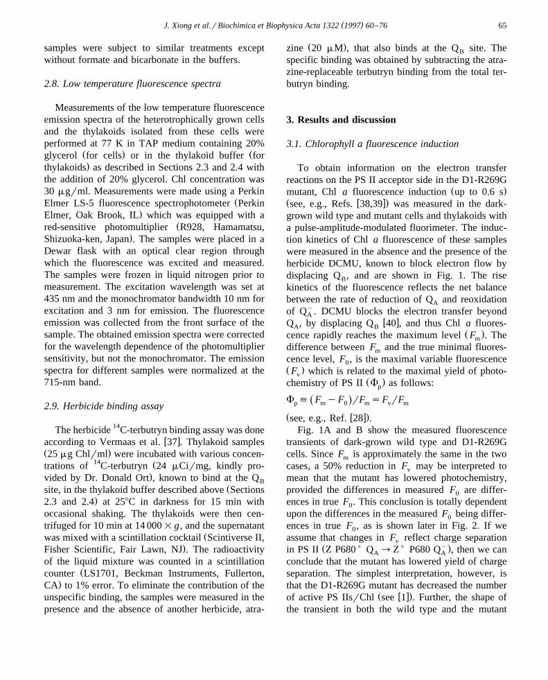
Ž .mutant, Chl a fluorescence induction up to 0.6 sŽ w x.see, e.g., Refs. 38,39 was measured in the dark-grown wild type and mutant cells and thylakoids witha pulse-amplitude-modulated fluorimeter. The induc-tion kinetics of Chl a fluorescence of these sampleswere measured in the absence and the presence of theherbicide DCMU, known to block electron flow bydisplacing Q , and are shown in Fig. 1. The riseB
kinetics of the fluorescence reflects the net balancebetween the rate of reduction of Q and reoxidationA
of Qy. DCMU blocks the electron transfer beyondAw xQ , by displacing Q 40 , and thus Chl a fluores-A B
Ž .cence rapidly reaches the maximum level F . Them
difference between F and the true minimal fluores-m
cence level, F , is the maximal variable fluorescence0Ž .F which is related to the maximal yield of photo-v
Ž .chemistry of PS II F as follows:p
F ' F yF rF sF rFŽ .p m 0 m v m
Ž w x.see, e.g., Ref. 28 .Fig. 1A and B show the measured fluorescence
transients of dark-grown wild type and D1-R269Gcells. Since F is approximately the same in the twom
cases, a 50% reduction in F may be interpreted tov
mean that the mutant has lowered photochemistry,provided the differences in measured F are differ-0
ences in true F . This conclusion is totally dependent0
upon the differences in the measured F being differ-0
ences in true F , as is shown later in Fig. 2. If we0
assume that changes in F reflect charge separationvŽ ) q y.in PS II Z P680 Q ™Z P680 Q , then we canA A
conclude that the mutant has lowered yield of chargeseparation. The simplest interpretation, however, isthat the D1-R269G mutant has decreased the number
Ž w x.of active PS IIsrChl see 1 . Further, the shape ofthe transient in both the wild type and the mutant
( )J. Xiong et al.rBiochimica et Biophysica Acta 1322 1997 60–7666
shows that dark-grown cells are not normal, as is wellŽ w x.known see, e.g., 41 : the water oxidation machin-
ery is not functional and one observes, perhaps, onlyone turnover of PS II following slow oxidation ofQy.A
Fig. 1C and D show the Chl a fluorescence tran-Žsient of light adapted cells dark grown cells exposed
2 .to 1 h of 70 mErm s white light . The light treat-ment causes the wild type kinetics to display a nor-
mal fluorescence transient, i.e., a slower rise phase,w xlabeled as O-I-D-P phase 42 . F is shifted to ap
Žlonger time 400 ms versus 25 ms after actinic light.illumination since both the donor and acceptor sides
of PS II are now functional: the F to F rise is0 pŽbiphasic with an intermediate F , or F not to bei I
confused with F , used by some authors to indicatei.‘initial’ fluorescence ; the rise reflects the filling up
of the plastoquinone pool with electrons from thedonor side of PS II. The phenomenon of photoadapta-tion which is a slower rise of transient kinetic curveto F due to the recovery of the PS II donor side wasp
observed earlier in C. reinhardtii by Guenther et al.w x43 . However, this transition is not observed in theR269G mutant, indicating that the mutation has inhib-ited the necessary photoadaptation process. In our
w xearlier paper 1 , we have shown that Mn centerswhich constitute the S-state complex for the donorside function are missing in the mutant. Further, aftera light treatment, the measured F of the mutant cellsv
Žbecomes further reduced only 35% of that of the.wild type , and F rF reflecting photochemistry isv m
reduced by 60%. This significant lowering of the Fv
after light treatment indicates an increased suscepti-
ŽFig. 1. Chl a fluorescence transients as a function of time of.illumination of the dark-grown C. reinhardtii wild type and
D1-R269G in the absence and the presence of 10 mM DCMUmeasured with a PAM-2000 fluorimeter. The full scale is 200mV. F is the measured F of this instrument, and F is the0 0 m
Ž . Ž .F . A The transient of the wild type cells. B ThemaximumŽ .transient of the mutant cells. C The transient of the light-adapted
Ž 2 .wild type cells treated with 70 mErm s white light for 1 h .Note the transformation of the kinetics from the dark phase to thelight phase indicated by the shift of the time of F . The Fp iŽdenoting fluorescence intensity at the ‘I’ step of O-I-D-P tran-
. Ž .sient should not be confused with the F F of otheri initialŽ . Ž 2 .authors. D The transient of the light-treated 70 mErm s
mutant cells. Note the absence of the kinetic transformation and amore elevated measured F and a decreased F , suggesting a0 v
Ž .possible photo-damage to the mutant PS II. E The transient ofŽ .the wild type thylakoid isolated from the dark-grown cells. F
The transient of the mutant thylakoid isolated from the dark-grownŽ .cells. G The transient of the wild type thylakoids isolated from
the light-adapted cells. Note the F is basically unchangedvŽ .compared with the cells. H The transient of the mutant th-
ylakoids isolated from the light-adapted cells. Note the evenw xlowered F in this sample. In all measurements, the Chl of thev
samples was 5 mgrml, and the actinic illumination was 470mErm2 s. Thy s thylakoids.
( )J. Xiong et al.rBiochimica et Biophysica Acta 1322 1997 60–76 67
bility of the mutant PS II to photoinhibition, confirm-w xing conclusions in our earlier paper 1 . Fig. 1E and F
are the Chl a fluorescence transient of the thylakoidsisolated from the dark-grown cells. The measured Fv
of the mutant thylakoids is 60% of the wild typelevel. After the light treatment of the cells, the mea-sured F of the isolated mutant thylakoids is reducedv
Žto only ;20% of the wild type level Fig. 1G and.H , indicating that the combination of the steps in-
volved in biochemical preparations and the lighttreatment may cause further lesions to the mutant PSII. It is noted that the maximal fluorescence level
Ž .after DCMU treatment F is slightly lower than themŽ .F in the control Fig. 1G . This lowering is thoughtp
to be due to the quenching of fluorescence by theŽ w xplastoquinone see review 28 , and Vernotte et al.
w x.44 .
3.2. The measured F and the true F0 0
D1-R269G mutant samples have a consistentlyelevated measured F compared to that of the wild0
type. If this information can be confirmed by measur-ing the true F , that is the minimal level of Chl0
fluorescence originating from antenna, in competitionwith energy transfer to the PS II reaction center, anincrease in F would indicate a decreased excitation0
energy transfer to the active PS II reaction center,possibly due to the disconnection between antennaeand the reaction center II, provided we can assumethat the PS I component of F is small and remains0
constant. A second possibility is the existence ofw xinactive PS II 45 incompetent in trapping excitons
arriving from antennae, which then can return toantennae and fluoresce; an increased number of inac-tive PS II reaction centers would lead to an apparentincrease in antennaeractive PS II reaction center.Our data do not allow us to distinguish between thepossibilities. However, as shown in Fig. 1, the wildtype cells or thylakoids display a small increase in F0
when measured in the presence of DCMU whichblocks the electron transfer beyond Qy; this may beA
due to back reaction of electrons from reduced QB
and Q . The slight increase in measured F in theA 0
presence of DCMU could also include a contributiondue to the small actinic effect of the measuring beamof the fluorimeter. Thus, the measured F required0
further investigation according to the rationale givenunder Section 2.
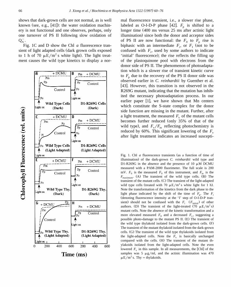
Independent measurement of F for the wild type0
and D1-R269G mutant cells at very low light intensi-ties of the measuring beam indicate that F intensity0
Ž . Ž .is a linear function of light intensities I Fig. 2A .Ž .The quantum yield F rI remains constant as it0
should for true F which should be independent of0Ž . Ž w x.photochemistry Fig. 2B see, e.g., Ref. 46 . Fur-
ther, addition of DCMU did not cause significantincreases in the measured F levels, especially at the0
w xlowest intensities used, as it should since all QA
remains essentially unchanged. Mutant F and F rI0 0Žwere consistently double that in the wild type ;
.200% . Thus, true F of the D1-R269G mutant is0
higher than the wild type, and F is, therefore,v
reduced in the mutant, as interpreted in Fig. 1.
Ž . Ž .Fig. 2. A Baseline Chl a fluorescence F measured as a0
function of light intensities in the low intensity range, in thepresence and the absence of 10 mM DCMU. Measurements ofthe heterotrophically grown wild type C. reinhardtii and D1-
Ž .R269G mutant cells were made as in Section 2.4. B TheŽ .quantum yield of measured F , or the ratio of fluorescence F0
Ž .to the light intensity I of the measuring beam, basically doesnot change with the light intensities indicating that the measuredmeasured F in this experiment is very close to the true F . It is0 0
further confirmed by the absence of any significant effect ofDCMU. Data suggest that D1-R269G mutant has a two times trueF than the wild type cells.0

( )J. Xiong et al.rBiochimica et Biophysica Acta 1322 1997 60–7668
3.3. Chlorophyll a fluorescence changes after a flash
To locate the effect of the mutation on the specificreaction of the acceptor side of PS II, we measuredChl a fluorescence changes on the microsecond timescale after a series of single turnover flashes. Thekinetics of these changes correspond mainly to the
y y Želectron transfer from Q to Q or Q see, e.g.,A B Bw x.Refs. 17,47 ; however, the fluorescence yield at Fm
and in the sub-ms range also include effects ofchanges in electron flow on the donor side of PS II,
w qxi.e. changes in P680 , a quencher of Chl fluores-w xcence 48 . A slowed equilibrium between S mZmn
P680 results in a lowering of F and a rise phase ofmŽ w x.fluorescence see, e.g., Refs. 32,49,50 . Fig. 3 shows
Chl a fluorescence yield changes for the dark-grown,
non-oxygen evolving cells and thylakoids in the pres-Ž .ence and absence of DCMU 10 mM and of hydrox-
Ž . Ž .ylamine NH OH, 5 mM and hydroquinone 5 mM ,2q Ždonors to P680 . For a rationale of using these
w x .chemicals, see Metz et al. 51 .We will first discuss the donor side effects. When
compared to the wild type, cells and thylakoids ofŽ .D1-R269G mutant show cf. Fig. 3A with B signifi-
cant decreases in the F rF ratio. Further, the mutantv 0
thylakoids have even lowered F rF level comparedv 0
to not only the wild type thylakoids, but also mutantcells. These results may suggest, in addition to theexistence of inactive PS IIs in the mutant, inhibitions
Žon the donor side of PS II in the mutant also see.Section 3.5 .
The addition of both 5 mM NH OH and 10 mM2
Fig. 3. Flash-induced Chl a fluorescence yield kinetics of the dark-grown wild type and the dark-grown D1-R269G mutant of C.Ž . Ž . Ž . Ž .reinhardtii, treated with or without DCMU 10 mM , or with DCMU 10 mM and NH OH 5 mM , or with DCMU 10 mM and2
Ž .hydroquinone HQ, 5 mM . The kinetic measurements were done with 5 mg Chlrml samples. Only the second-flash kinetic traces areŽ . Ž .shown. A Kinetics of fluorescence change from the wild type cells. B Kinetics of fluorescence change from the D1-R269G mutant
Ž . ycells. Data show a lowered yield of photochemistry calculated from F rF and a slowed rate of electron flow from Q to thev m AŽ . Ž .plastoquinone pool in the mutant. C Kinetics of fluorescence change from the wild type thylakoids. D Kinetics of fluorescence change
from the D1-R269G thylakoids. Note the slowed decay kinetics and the lowered F rF in the mutant samples suggesting an inhibition onv m
the PS II acceptor side and the presence of high proportion of inactive PS II reaction centers. The addition of the good donorŽ .hydroquinone is unable to restore the Chl a fluorescence level in the mutant cells or thylakoids to the wild type level.

( )J. Xiong et al.rBiochimica et Biophysica Acta 1322 1997 60–76 69
DCMU is unable to restore the maximum level ofŽ .F rF in the mutant sample cells or thylakoids tov 0
that of the wild type level. However, the addition ofŽ .hydroquinone 5 mM results in a variable fluores-
cence level equivalent to the DCMU treatment with-out added donors. This is simply because hydro-quinone is a much better electron donor to P680q
w xthan hydroxylamine 51,52 . The consistent lowerlevel of variable fluorescence in the D1-R269G mu-tant, even when DCMU and hydroquinone are pre-sent, indicates that the functional PS IIs may be muchlower than those in the wild type similar to ourobservations of the mutant using EPR spectroscopyw x1 . Fig. 3 also lists the increased measured F in the0
mutant cells and thylakoids relative to the wild type:this confirms the observation in the above fluores-
Ž .cence induction measurements Fig. 1 .We now discuss the acceptor side effects. The
decay of fluorescence is slower in the mutant than inthe wild type cells. The lifetime of the first compo-
Ž .nent t after the first flash is 90 ms in the wild type1
vs. 1.6 ms in the mutant; t after the second flash is1
120 ms in the wild type vs. 2.0 ms in the mutant.There is ;17 fold decrease in the rate of fluores-cence decay. The first component of the wild typeconstitutes ;66% of the total fluorescence decayprocess and that of the mutant only constitutes ;30%of the decay. In spite of the slight difference betweenflash 1 and 2 results, they do not represent the true
y Ž y . yQ to Q or Q steps, since Q to Q ratio inA B B B B
dark-adapted untreated intact cells is not too far fromŽ .1 see Section 3.6 . The decay rate for the mutant
Ž .thylakoid samples was even slower ;50 fold com-Ž .pared to that of the wild type Fig. 3C and D ,
consistent with other lines of evidence that the mu-tant PS II has much less structural stability in thethylakoid preparations. The addition of high concen-
Ž .tration 10 mM of DCMU inhibits the electron trans-fer in both the wild type and the mutant cells andthylakoids.
3.4. Functioning of the two-electron gate
PS II variable Chl a fluorescence is controlled bythe redox state of the primary plastoquinone, Q , aA
Žone electron carrier, which is oxidized by Q orBy. ŽQ , a two electron carrier ‘the two electron gate’,B
w x. y53,54 . Since the electron flow from Q to Q isA B
Fig. 4. Flash oscillation pattern for the variable Chl a fluores-cence of the dark-grown wild type and D1-R269G cells of C.reinhardtii measured at 200 ms after an actinic flash. The sam-ples were treated, by vacuum infiltration, with p-benzoquinoneŽ . y Ž100 mM to convert some of the Q to Q and NH OH 5B B 2
.mM to block the S-state transitions, if any, prior to measure-w xment. The Chl of the samples was 5 mgrml, the flash frequency
during the measurement was 0.67 Hz. The data indicate a loss ofthe period two oscillation pattern in the mutant.
faster than from Qy to Qy, it is reflected in a ‘deep’A B
period two oscillation pattern in the Chl a fluores-Ž w x.cence decays see 55 in samples that start with
Q : Qy ratio of 1 : 0. It is generally known that theB B
Q : Qy ratio is close to 1 : 1 in intact photosyntheticB Bw xcells 56,57 . Although, as noted earlier, it is not easy
y w xto fully oxidize Q in cells 29 , partial effects areB
obtained if we pretreat the dark-grown wild type cellsŽ . Žwith p-benzoquinone 100 mM and NH OH 52
.mM , and illuminate the samples with a series ofsingle turnover flashes. In this assay, the flash fre-quency was 0.67 Hz, and the variable fluorescencevalues at 200 ms are shown. NH OH was added to2
eliminate most of the period four oscillations due tothe donor side activities and to serve as an electrondonor, albeit poorer than hydroquinone, since thecells were unable to oxidize water. An obvious,although not very deep, binary oscillation pattern ofthe Chl variable fluorescence is observed for the wild
Ž . w xtype Fig. 4 . Crofts et al. 58 have used a benzo-quinone treatment method that yields deep period twooscillations, but, then the F is drastically reduced. Inv
the mutant sample, however, this period two oscilla-tion pattern was eliminated suggesting a defectivetwo-electron gate mechanism caused by the mutation.The data indicate that the PS II acceptor side reac-tions are significantly modified due to the R269Gmutation.
( )J. Xiong et al.rBiochimica et Biophysica Acta 1322 1997 60–7670
3.5. Thermoluminescence
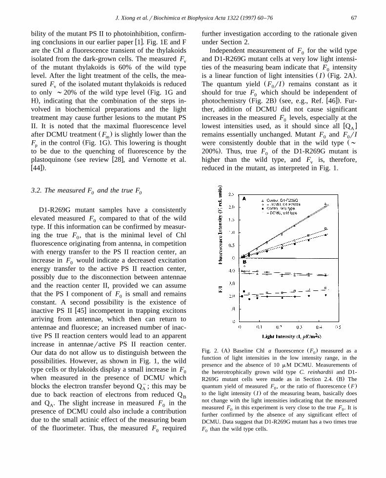
Ž w x.Since thermoluminescence see 34,35 measuresthe recombination of charges between S and Qy
2 AŽ . Ž . ythe D or the Q band and between S or S and Q2 3 BŽ . Ž w x.the B band see, e.g., 36 , it can be used to checkif the mutant is blocked in the S-state transition. Fig.5 shows that the wild type thylakoids have the nor-
Ž .mal B band a broad band in the 30–358C region .Ž .Upon treatment with DCMU 10 mM , the B band isŽ .abolished, and the Q band a broad band at 158C
appears due to inhibition of electron transfer fromQy™Q enabling stabilization of Qy. The mutant,A B A
however, lacks both the B and Q bands confirmingthat it is unable to store charges on the S-states.
ŽHowever, a difference thermoluminescence curve for.DCMU-treated minus untreated mutant thylakoids
showed a slight negative band in the B band region,and a slight positive band in the Q band region, but itwas within the noise level of our measurements. Ahigh temperature band that includes a ‘C’ band,suggested to arise from recombination of Yq and Qy
D Aw x59 , is however, present. A good part of the hightemperature band, observed here, was unrelated to
Fig. 5. Thermoluminescence of wild type C. reinhardtii andŽw x .D1-R269G mutant thylakoid Chl is ;1 mgrml in the pres-Ž .ence and absence of DCMU 10 mM . The samples were mea-
sured after one saturating, single-turnover, flash. The heating ratewas 18Crs. Data obtained by David Kramer.
Ž .photosynthesis data not shown ; but this does notaffect the clear conclusions noted above. Thermolu-minescence results confirm the defective nature of
w xthe mutant on the donor side of its PS II 1 .
3.6. Bicarbonate depletion and recoÕery
Since our hypothesis was that D1-R269 is involvedin HCOy binding, we determined the effect of for-3
Ž . ymate analog of bicarbonate inhibition on the Q toAŽ y.Q or Q electron transfer. Flash-induced Chl fluo-B B
rescence decay of the dark-grown wild type and theD1-R269G cells with or without formate or formateplus bicarbonate after the second flash is shown in
Ž .Fig. 6 data after the first flash are not shown . Sincein intact untreated cells, the ratio of Q to Qy inB B
w x y ydarkness is close to 1 56,57 , Q to Q and Q toA B A
Qy reactions are not easily separable. The additionBŽ .of 25 mM pH 6.5 formate slows down the electron
y Ž y. Žflow from Q to Q or Q in the wild type Fig.A B B.6A . Formate inhibition is almost fully reversed by
Ž . ythe addition of bicarbonate 10 mM . The Q to QA BŽ y.or Q reaction in the mutant samples are alsoB
inhibited by formate; bicarbonate readily reverses thisŽ . w xeffect Fig. 6B . It is established 6–10 that formate
has additional effects on the donor side; the insets inFig. 6 show that formate causes decrease in F rF atv 0
Ž-250 ms before an increase can be observed alsow x.see Refs. 6,10 . This confirms the dual effect of
bicarbonate-reversible formate effect in thylakoids ofboth wild type and D1-R269G Chlamydomonas cells.
A quantitative assay of the formate inhibition andbicarbonate recovery for both the wild type and themutant samples is shown in Fig. 7. The dark-growncells were treated with various concentrations of for-mate in the absence or presence of bicarbonate andassayed for Chl a fluorescence decay as above. Theresulting decay curves were deconvoluted with three
Ž w x.exponential components see, e.g., Ref. 33 . Thefast decay component represents the kinetics of Qy
AŽ y.to Q or Q electron transfer. Thus, we plotted theB B
Ž .normalized lifetime of the first component t as a1
function of formate concentration with the t of the1Ž .control without formate treatment as 100 in arbi-
trary units. For the second flash, the t of the wild1
type in the control is 120 ms and that of the mutant is2.0 ms, an ;17 fold effect. As shown in Fig. 7, atincreasing formate concentration, the t of the wild1
( )J. Xiong et al.rBiochimica et Biophysica Acta 1322 1997 60–76 71
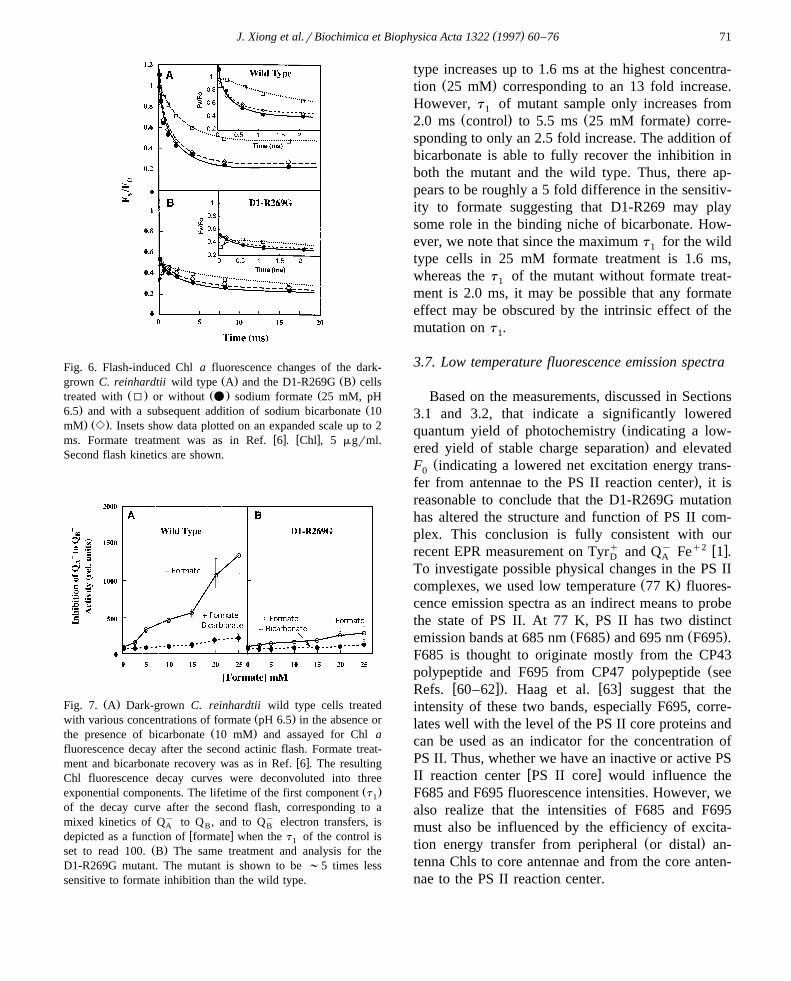
Fig. 6. Flash-induced Chl a fluorescence changes of the dark-Ž . Ž .grown C. reinhardtii wild type A and the D1-R269G B cells
Ž . Ž . Žtreated with I or without v sodium formate 25 mM, pH. Ž6.5 and with a subsequent addition of sodium bicarbonate 10. Ž .mM e . Insets show data plotted on an expanded scale up to 2
w x w xms. Formate treatment was as in Ref. 6 . Chl , 5 mgrml.Second flash kinetics are shown.
Ž .Fig. 7. A Dark-grown C. reinhardtii wild type cells treatedŽ .with various concentrations of formate pH 6.5 in the absence or
Ž .the presence of bicarbonate 10 mM and assayed for Chl afluorescence decay after the second actinic flash. Formate treat-
w xment and bicarbonate recovery was as in Ref. 6 . The resultingChl fluorescence decay curves were deconvoluted into three
Ž .exponential components. The lifetime of the first component t1
of the decay curve after the second flash, corresponding to amixed kinetics of Qy to Q , and to Qy electron transfers, isA B B
w xdepicted as a function of formate when the t of the control is1Ž .set to read 100. B The same treatment and analysis for the
D1-R269G mutant. The mutant is shown to be ;5 times lesssensitive to formate inhibition than the wild type.
type increases up to 1.6 ms at the highest concentra-Ž .tion 25 mM corresponding to an 13 fold increase.
However, t of mutant sample only increases from1Ž . Ž .2.0 ms control to 5.5 ms 25 mM formate corre-
sponding to only an 2.5 fold increase. The addition ofbicarbonate is able to fully recover the inhibition inboth the mutant and the wild type. Thus, there ap-pears to be roughly a 5 fold difference in the sensitiv-ity to formate suggesting that D1-R269 may playsome role in the binding niche of bicarbonate. How-ever, we note that since the maximum t for the wild1
type cells in 25 mM formate treatment is 1.6 ms,whereas the t of the mutant without formate treat-1
ment is 2.0 ms, it may be possible that any formateeffect may be obscured by the intrinsic effect of themutation on t .1
3.7. Low temperature fluorescence emission spectra
Based on the measurements, discussed in Sections3.1 and 3.2, that indicate a significantly lowered
Žquantum yield of photochemistry indicating a low-.ered yield of stable charge separation and elevated
ŽF indicating a lowered net excitation energy trans-0.fer from antennae to the PS II reaction center , it is
reasonable to conclude that the D1-R269G mutationhas altered the structure and function of PS II com-plex. This conclusion is fully consistent with our
q y q2 w xrecent EPR measurement on Tyr and Q Fe 1 .D A
To investigate possible physical changes in the PS IIŽ .complexes, we used low temperature 77 K fluores-
cence emission spectra as an indirect means to probethe state of PS II. At 77 K, PS II has two distinct
Ž . Ž .emission bands at 685 nm F685 and 695 nm F695 .F685 is thought to originate mostly from the CP43
Žpolypeptide and F695 from CP47 polypeptide seew x. w xRefs. 60–62 . Haag et al. 63 suggest that the
intensity of these two bands, especially F695, corre-lates well with the level of the PS II core proteins andcan be used as an indicator for the concentration ofPS II. Thus, whether we have an inactive or active PS
w xII reaction center PS II core would influence theF685 and F695 fluorescence intensities. However, wealso realize that the intensities of F685 and F695must also be influenced by the efficiency of excita-
Ž .tion energy transfer from peripheral or distal an-tenna Chls to core antennae and from the core anten-nae to the PS II reaction center.
( )J. Xiong et al.rBiochimica et Biophysica Acta 1322 1997 60–7672
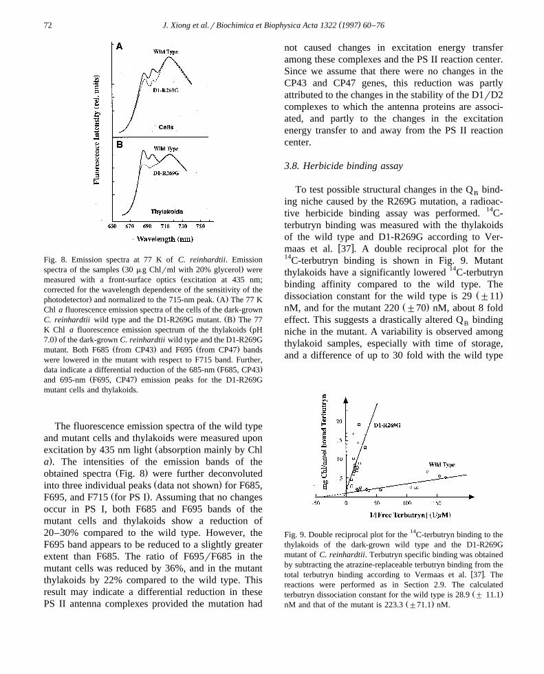
Fig. 8. Emission spectra at 77 K of C. reinhardtii. EmissionŽ .spectra of the samples 30 mg Chlrml with 20% glycerol were
Žmeasured with a front-surface optics excitation at 435 nm;corrected for the wavelength dependence of the sensitivity of the
. Ž .photodetector and normalized to the 715-nm peak. A The 77 KChl a fluorescence emission spectra of the cells of the dark-grown
Ž .C. reinhardtii wild type and the D1-R269G mutant. B The 77ŽK Chl a fluorescence emission spectrum of the thylakoids pH
.7.0 of the dark-grown C. reinhardtii wild type and the D1-R269GŽ . Ž .mutant. Both F685 from CP43 and F695 from CP47 bands
were lowered in the mutant with respect to F715 band. Further,Ž .data indicate a differential reduction of the 685-nm F685, CP43
Ž .and 695-nm F695, CP47 emission peaks for the D1-R269Gmutant cells and thylakoids.
The fluorescence emission spectra of the wild typeand mutant cells and thylakoids were measured upon
Žexcitation by 435 nm light absorption mainly by Chl.a . The intensities of the emission bands of the
Ž .obtained spectra Fig. 8 were further deconvolutedŽ .into three individual peaks data not shown for F685,
Ž .F695, and F715 for PS I . Assuming that no changesoccur in PS I, both F685 and F695 bands of themutant cells and thylakoids show a reduction of20–30% compared to the wild type. However, theF695 band appears to be reduced to a slightly greaterextent than F685. The ratio of F695rF685 in themutant cells was reduced by 36%, and in the mutantthylakoids by 22% compared to the wild type. Thisresult may indicate a differential reduction in thesePS II antenna complexes provided the mutation had
not caused changes in excitation energy transferamong these complexes and the PS II reaction center.Since we assume that there were no changes in theCP43 and CP47 genes, this reduction was partlyattributed to the changes in the stability of the D1rD2complexes to which the antenna proteins are associ-ated, and partly to the changes in the excitationenergy transfer to and away from the PS II reactioncenter.
3.8. Herbicide binding assay
To test possible structural changes in the Q bind-B
ing niche caused by the R269G mutation, a radioac-tive herbicide binding assay was performed. 14C-terbutryn binding was measured with the thylakoidsof the wild type and D1-R269G according to Ver-
w xmaas et al. 37 . A double reciprocal plot for the14C-terbutryn binding is shown in Fig. 9. Mutantthylakoids have a significantly lowered 14C-terbutrynbinding affinity compared to the wild type. The
Ž .dissociation constant for the wild type is 29 "11Ž .nM, and for the mutant 220 "70 nM, about 8 fold
effect. This suggests a drastically altered Q bindingB
niche in the mutant. A variability is observed amongthylakoid samples, especially with time of storage,and a difference of up to 30 fold with the wild type
Fig. 9. Double reciprocal plot for the 14C-terbutryn binding to thethylakoids of the dark-grown wild type and the D1-R269Gmutant of C. reinhardtii. Terbutryn specific binding was obtainedby subtracting the atrazine-replaceable terbutryn binding from the
w xtotal terbutryn binding according to Vermaas et al. 37 . Thereactions were performed as in Section 2.9. The calculated
Ž .terbutryn dissociation constant for the wild type is 28.9 " 11.1Ž .nM and that of the mutant is 223.3 "71.1 nM.
( )J. Xiong et al.rBiochimica et Biophysica Acta 1322 1997 60–76 73
was observed in certain trials. This reflects the insta-bility of the mutant thylakoids after they are preparedfrom the cells. The notion that the PS II stability isaffected in the mutant is supported from assays such
Ž .as the 77 K fluorescence emission spectra Fig. 8 ,Ž .fluorescence kinetics measurements Fig. 3 , as well
as the EPR, and western blot analyses publishedw xelsewhere 1 . Since mutant thylakoids are unstable,
we speculate that the differences between herbicidebinding in wild type and mutant cells would be muchless than those observed in thylakoids.
3.9. Modeling: bicarbonate binding sites
A recently constructed three dimensional PS IIw xreaction center model 20 suggests that D1-R269 is
not a direct binding residue for bicarbonate at thenon-heme iron site. The geometric position of D1-
˚R269 is modeled 8–11 A from bicarbonaterironcenter, which does not support a direct interaction.D1-R269 is located near the N-terminal region of the
transmembrane a-helix E of D1. According to themodel, D1-R269 is separated from D1-H272, one ofthe non-heme iron ligands, by nearly 3r4 of a helical
˚Ž .turn ;5 A . This close vicinity to D1-H272 whichis located approximately equally in between Q andA
Q , may help explain a structural perturbation ofB
D1-R269G mutation on the functionality of the ironw x1 and the liganding of bicarbonate and formateŽ .Figs. 6 and 7 .
In addition to D1-R269, several other positivelycharged residues on the D2 protein of cyanobacterianear the non-heme iron have also been investigatedfor involvement in bicarbonaterformate effectw x3,5,19 . D2-R233 and D2-R251 have been shown toincrease the PS II susceptibility to formate inhibitionof full chain electron transfer by 10 fold relative tothe wild type and are suggested to function in stabi-
w xlizing bicarbonate binding in vivo 19 . However, aŽ .mutation on D2-R139 D1-R139H showed no effect
w xon bicarbonate-reversible formate inhibition 5 sug-gesting specificity of other arginines. However, the
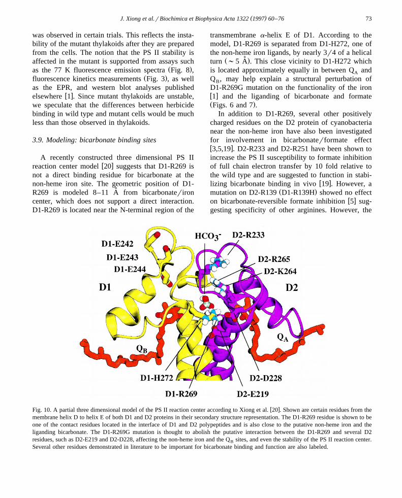
w xFig. 10. A partial three dimensional model of the PS II reaction center according to Xiong et al. 20 . Shown are certain residues from themembrane helix D to helix E of both D1 and D2 proteins in their secondary structure representation. The D1-R269 residue is shown to beone of the contact residues located in the interface of D1 and D2 polypeptides and is also close to the putative non-heme iron and theliganding bicarbonate. The D1-R269G mutation is thought to abolish the putative interaction between the D1-R269 and several D2residues, such as D2-E219 and D2-D228, affecting the non-heme iron and the Q sites, and even the stability of the PS II reaction center.B
Several other residues demonstrated in literature to be important for bicarbonate binding and function are also labeled.
( )J. Xiong et al.rBiochimica et Biophysica Acta 1322 1997 60–7674
lack of difference in phenotype compared to the wildtype may possibly be a result of the potentiallyconservative mutation. D2-K264 was suggested to bea strong candidate for bicarbonate binding at thenon-heme iron site, as its site-directed mutants wereconsiderably slower in electron transfer from Qy toA
Qy compared to the wild type, and were very resis-BŽtant to formate and NO treatment unpublished data,
w x.see Ref. 3 . This mutant required much higherbicarbonate concentrations than the wild type to ac-
Žcelerate Q rQ electron transfer unpublished data,A Bw x.see Ref. 3 , suggesting that the residue may be
intimately involved in binding of bicarbonate. Muta-tions at a nearby residue D2-R265 also showed simi-lar effects, though to a lesser extent.
In addition to liganding to the iron, bicarbonatemay also function in promoting the protonation of
2y Ž w x.Q see, e.g., Refs. 2,4,17,47 . Kinetic studies byBw xBlubaugh and Govindjee 64 suggested the possibil-
ity of two tight bicarbonate binding sites in PS II.One of these binding sites may well be at the non-ironsite, while the other may be related to the protonationof plastoquinone and thus likely to be near the QB
w xsite in the D1 protein. Maenpaa et al. 65 demon-¨ ¨¨strated that a strain of Synechocystis PCC 6803 withdeletion of residues D1-E242 to D1-E244 near theQ site exhibits a 7 fold higher resistance to formateB
inhibition than the wild type. Our recent site-directedmutagenesis experiments on D1-R257 of C. rein-hardtii have indicated that this residue is intimately
Žinvolved in affecting the formate binding J. Xiong,J. Minagawa, A.R. Crofts and Govindjee, unpub-
.lished data .w xIn the three dimensional PS II model 20 , based
on the analogy with a water transport channel in thew x w xbacterial reaction center 66 , Xiong et al. 20 pro-
posed a channel for transporting bicarbonate anionsand water molecules for protonating Q and provid-B
ing ligands to the non-heme iron in PS II reactioncenter. This channel or a large binding niche involvesa series of charged residues of the D1 and D2proteins, including D1-R269. Substitution on D1-R269 is thus expected to cause perturbations onformate effects, which is supported by the data inFigs. 6 and 7.
w xOur model 20 indicates that D1-R269 may alsobe involved in D1rD2 interaction. The correct as-sembly and stability of the D1rD2 complex of the
PS II reaction center rely partly on the interactions ofthe contact residues located on the transmembranespans. As shown in Fig. 10, D1-R269 may be acontact residue located in the interface of D1 and D2polypeptides and may provide interactions importantfor maintaining the conformation of D1 and D2polypeptides. Our model shows that D1-R269 mayhave electrostatic or hydrogen bonding interactionswith certain D2 residues, such as D2-E219 and D2-
Ž .D228 Fig. 10 . The glycine mutation may thus abol-ish such interactions affecting the stability of the PSII reaction center, resulting in a series of primary andsecondary effects such as lowered charge separation,slowed PS II electron transfer, decreased binding ofQ rherbicide, perturbed bicarbonaterformate func-B
tioning at the non-heme iron andror Q site, etc.B
Therefore, it may be interesting to test this hypothesisby mutagenizing the D2-E220 or D2-D228 residueand see whether a similar structural instability effecton PS II may exist in these mutants.
In the model, D1-R269 is located on the acceptorside, the transduction of the mutational effect fromthe acceptor to the donor side can be due to D1-R269being a contact residue affecting the assembly of thePS II altering the structure and function of both the
w xdonor and acceptor sides, as observed here and in 1 .Similar transduction of mutational effects betweenthe donor side and acceptor side has previously been
w xobserved. Etienne and Kirilovsky 67 and Constantw xet al. 68 showed that several herbicide-resistant
mutations at the Q site affect the S-state function.B
Similar effects have previously been observed formutants lacking the PS II 43 kDa Chl binding protein
w xand in site-directed mutants of the D1 protein 69,70 .w xCarpenter et al. 71 have observed that the S and S2 3
Ž .states on the D1rD2 are affected by changes inw xCP-43 protein, and Kless et al. 72 have shown that
alterations in the Q region of the D2 protein affectsA
DCMU affinity in the D1 protein.
4. Conclusions
D1 residue R269 is critically important for thestructure and function of PS II complex. D1-R269Gmutation drastically alters the PS II photosyntheticapparatus and has profound impacts on both thedonor and acceptor sides of PS II chemistry. The
( )J. Xiong et al.rBiochimica et Biophysica Acta 1322 1997 60–76 75
Ž .functional impacts include: 1 significant modifica-tions on the Q binding niche of PS II, affecting theB
electron transfer process from Qy to the plasto-AŽ .quinone pool; 2 possible destabilization or inhibi-
tion of assembly of the PS II complex andror ablockage of excitation energy transfer to the PS IIreaction center, affecting the level of stable chargeseparation, the PS II susceptibility to photodamage,and the donor side functions. Though the in vivobicarbonaterformate activity is clearly perturbed bythe mutation, the bicarbonate binding site is stillpresent. We propose that the residue may not be adirect liganding residue to the bicarbonate anion butmay affect the bicarbonaterformate binding indi-rectly through a general conformational changeandror through its involvement in bicarbonaterwater
Ž w x.transport for details, see Ref. 20 . This residue isalso considered to play a structural role for maintain-ing the proper D1rD2 conformation.
Acknowledgements
We thank Dr. David Kramer for thermolumines-cence measurements, Dr. Antony Crofts for the useof the multiflash fluorimeter in his laboratory, Dr.Donald Ort for providing us with the 14C-terbutryn,and Dr. Vladimir Shinkarev for help in deconvolutionof the low temperature fluorescence spectra. We alsothank Dr. Shankar Subramaniam for participation inthe PS II computer modeling work. This work was
Žsupported by an NSF grant 91-16838, supplement.1994 and a grant from the Research Board of the
University of Illinois to G.; and by a U.S. Dept. ofŽ .Energy grant DOE ER20076 to R.T.S. J.X. was
Ž .supported by a training grant DOE 92ER20095Ž .from the Triagency DOErNSFrUSDA and its ex-
Ž .tension NSF DBI96-02240 for Collaborative Re-search in Plant Biology. During the preparation ofthis paper, Govindjee was supported by a Fulbright
Ž .Grant to India November, 1996 to February, 1997 ;and by a Visiting Fellowship at the Australian Na-
Ž .tional University March–May, 1997 .
References
w x1 R.S. Hutchison, J. Xiong, R.T. Sayre, Govindjee, Biochim.Ž .Biophys. Acta 1277 1996 83–92.
w x Ž .2 D. Blubaugh, Govindjee, Photosyn. Res. 19 1988 85–128.w x3 B. Diner, V. Petrouleas, J.J. Wendoloski, Physiol. Plant. 81
Ž .1991 423–436.w x4 Govindjee, J.J.S. van Rensen, in: J. Deisenhofer, J. Norris
Ž .Eds. , The Photosynthetic Reaction Center, Vol. I, Aca-demic Press, San Diego, 1993, pp. 357–389.
w x Ž .5 Govindjee, Z. Naturforsch. 48c 1993 251–258.w x6 F. El-Shintinawy, C. Xu, Govindjee, J. Plant. Physiol. 136
Ž .1990 421–428.w x Ž .7 F. El-Shintinawy, Govindjee, Photosyn. Res. 24 1990
189–200.w x8 V.V. Klimov, S.I. Allakhverdiev, Y.M. Feyziev, S.V. Bara-
Ž .nov, FEBS Lett. 363 1995 251–255.w x9 H. Wincencjusz, S.I. Allakverdiev, V.V. Klimov, H.J. van
Ž .Gorkom, Biochim. Biophys. Acta 1273 1996 1–3.w x10 Govindjee, C. Xu, J.J.S. van Rensen, Z. Naturforsch. 52c
Ž .1997 24–32.w x Ž .11 H. Michel, J. Deisenhofer, Biochemistry 27 1988 1–7.w x Ž .12 W.F.J. Vermaas, A.W. Rutherford, FEBS Lett. 175 1984
243–248.w x13 V. Petrouleas, B.A. Diner, Biochim. Biophys. Acta 1015
Ž .1990 131–140.w x Ž .14 R. Hienerwadel, C. Berthomieu, Biochemistry 34 1995
16288–16297.w x15 X. Wang, J. Cao, P. Maroti, W.V. Stilz, U. Finkele, C.
Lauterwasse, W. Zinth, D. Oesterhelt, Govindjee, C.A.Ž .Wraight, Biochim. Biophys. Acta 1100 1992 1–8.
w x Ž .16 D. Blubaugh, Govindjee, Biochim. Biophys. Acta 848 1986147–151.
w x17 J.J. Eaton-Rye, Govindjee, Biochim. Biophys. Acta 935Ž .1988 237–247.
w x18 C. Xu, S. Taoka, A.R. Crofts, Govindjee, Biochim. Biophys.Ž .Acta 1098 1991 32–40.
w x19 J. Cao, W.F.J. Vermaas, Govindjee, Biochim. Biophys. ActaŽ .1059 1991 171–180.
w x Ž .20 J. Xiong, S. Subramaniam, Govindjee, Protein Sci. 5 19962054–2073.
w x21 B.F. Anderson, H.M. Baker, G.E. Norris, D.W. Rice, E.N.Ž .Baker, J. Mol. Biol. 209 1989 711–734.
w x22 S. Aime, M. Fasano, S. Paoletti, F. Cutruzzola, A. Desideri,`Ž .M. Bolognesi, M. Rizzi, P. Ascenzi, Biophys. J. 70 1996
482–488.w x23 T.A. Kunkel, J.D. Roberts, R.A. Zakour, Method. Enzymol.
Ž .154 1987 367–382.w x24 A.L. Eggenberger, P. De Ciechi, H. Pakrasi, in: M.
Ž .Baltscheffsky Ed. , Current Research in Photosynthesis,Vol. I, Kluwer Academic Publishers, Dordrecht, 1990, pp.363–366.
w x25 E.H. Harris, in: The Chlamydomonas Sourcebook, A Com-prehensive Guide to Biology and Laboratory Use, AcademicPress, San Diego, 1989, pp. 25–31.
w x26 R.J. Porra, W.A. Thompson, P.E. Kriedemann, Biochim.Ž .Biophys. Acta 975 1989 384–394.
w x Ž .27 H. Shim, J. Cao, Govindjee, Photosyn. Res. 26 1990223–228.
w x Ž .28 Govindjee, Aust. J. Plant Physiol. 22 1995 131–160.

( )J. Xiong et al.rBiochimica et Biophysica Acta 1322 1997 60–7676
w x29 C. Vernotte, J.-M. Briantais, C. Astier, Govindjee, Biochim.Ž .Biophys. Acta 1229 1995 269–301.
w x Ž .30 J. Lavorel, Colloques Int. du C.N.R.S. 119 1963 161–176.w x Ž .31 D. Wong, Govindjee, Photochem. Photobiol. 33 1981
103–108.w x32 D.M. Kramer, H.R. Robinson, A.R. Crofts, Photosyn. Res.
Ž .26 1990 181–193.w x33 Govindjee, C. Xu, G. Schansker, J.J.S. van Rensen, J.
Ž .Photochem. Photobiol. 37 1997 107–117.w x Ž .34 Y. Inoue, in: J. Amesz, A. Hoff Eds. , Biophysical Tech-
niques in Photosynthesis, Kluwer Academic Publishers,Dordrecht, 1996, pp. 93–107.
w x Ž .35 I. Vass, Govindjee, Photosyn. Res. 48 1996 117–126.w x36 D. Kramer, R. Roffey, Govindjee, R.T. Sayre, Biochim.
Ž .Biophys. Acta 1185 1994 228–237.w x Ž .37 W. Vermaas, J. Charite, G. Shen, Biochemistry 29 1990´
5325–5332.w x Ž .38 G. Papageorgiou, in: Govindjee Ed. , Bioenergetics of Pho-
tosynthesis, Academic Press, New York, 1975, pp. 319–371.w x39 J.-M. Briantais, C. Vernotte, G.H. Krause, E. Weis, in:
Ž .Govindjee, J. Amesz, D.C. Fork Eds. , Light Emission byPlants and Bacteria, Academic Press, Orlando, 1986, pp.539–583.
w x Ž .40 B.R. Velthuys, FEBS Letters 126 1981 277–281.w x Ž .41 G.M. Cheniae, I.F. Martin, Photochem. Photobiol. 17 1973
441–459.w x42 R.J. Strasser, A. Srivastava, Govindjee, Photochem. Photo-
Ž .biol. 61 1995 32–42.w x43 J.E. Guenther, J.A. Nemson, A. Melis, Photosyn. Res. 24
Ž .1990 35–46.w x44 C. Vernotte, A.-L. Etienne, J.-M. Briantais, Biochim. Bio-
Ž .phys. Acta 545 1979 519–527.w x Ž .45 J. Lavergne, J.-M. Briantais, in: D.R. Ort, C. Yocum Eds. ,
Oxygenic Photosynthesis: The Light Reactions, KluwerŽ .Academic Publishers, Dordrecht, 1996 in press .
w x Ž .46 J.C.M. Munday Jr., Govindjee, Biophys. J. 9 1969 22–35.w x47 J.J. Eaton-Rye, Govindjee, Biochim. Biophys. Acta 935
Ž .1988 248–257.w x Ž .48 W.L. Butler, Proc. Natl. Acad. Sci. USA 69 1972 3420–
3422.w x49 V.P. Shinkarev, Govindjee, Proc. Natl. Acad. Sci. USA 90
Ž .1993 7466–7469.w x50 V.P. Shinkarev, C. Xu, Govindjee, C. Wraight, Photosynth.
Ž .Res. 51 1997 43–49.w x51 J.G. Metz, P.J. Nixon, M. Rogner, G.W. Brudvig, B.A.
Ž .Diner, Biochemistry 28 1989 6960–6969.w x52 R.A. Roffey, D.M. Kramer, Govindjee, R.T. Sayre, Biochim.
Ž .Biophys. Acta 1185 1994 257–270.
w x53 B.R. Velthuys, J. Amesz, Biochim. Biophys. Acta 325Ž .1974 138–148.
w x Ž .54 B. Bouges-Bocquet, Biochim. Biophys. Acta 314 1973250–256.
w x Ž .55 J. Bowes, A.R. Crofts, Biochim. Biophys. Acta 590 1980373–384.
w x56 A.W. Rutherford, Govindjee, Y. Inoue, Proc. Natl. Acad.Ž .Sci. USA 81 1984 1107–1111.
w x57 C. Xu, S.M.D. Rogers, C. Goldstein, Govindjee, Photo-Ž .synth. Res. 21 1988 93–106.
w x58 A.R. Crofts, I. Baroli, D. Kramer, Taoka, Z. Naturforsch.Ž .48c 1993 259–266.
w x59 G.N. Johnson, A. Boussac, A.W. Rutherford, Biochim. Bio-Ž .phys. Acta 1184 1994 85–92.
w x60 H.Y. Nakatani, B. Ke, E. Dolan, C.J. Arntzen, Biochim.Ž .Biophys. Acta 765 1984 347–352.
w x61 Govindjee, K. Satoh, in: Govindjee, J. Amesz, D.C. ForkŽ .Eds. , Light Emission by Plants and Bacteria, AcademicPress, Orlando, 1986, pp. 497–537.
w x62 J.P. Dekker, A. Hassoldt, A. Petterson, H. van Roon, M.L.Ž .Groot, R. van Grondelle, in: P. Mathis Ed. , Photosynthe-
sis: from Light to Biosphere, Vol. I, Kluwer, Dordrecht,1995, pp. 53–56.
w x63 E. Haag, J.J. Eaton-Rye, G. Renger, W.F.J. Vermaas, Bio-Ž .chemistry 32 1993 4444–4454.
w x Ž .64 D. Blubaugh, Govindjee, Biochim. Biophys. Acta 936 1988208–214.
w x65 P. Maenpaa, T. Miranda, E. Tyystjarvi, T. Tyystjarvi,¨ ¨¨ ¨ ¨Govindjee, J.-M. Ducruet, A.-L. Etienne, D. Kirilovsky,
Ž .Plant Physiol. 107 1995 187–197.w x66 U. Ermler, G. Fritzsch, S.K. Buchanan, H. Michel, Structure
Ž .2 1994 925–936.w x Ž .67 A.-L. Etienne, D. Kirilovsky, Photosyn. Res. 38 1993
387–394.w x68 S. Constant, I. Perewoska, L. Nedbal, T. Miranda, A.-L.
Ž .Etienne, D. Kirilovsky, Plant Sci. 115 1996 165–174.w x69 C. de Vitry, J. Olive, D. Drapier, M. Recouvreur, F.-A.
Ž .Wollman, J. Cell Biol. 109 1989 991–1006.w x Ž .70 R.H. Hutchison, R.T. Sayre, in: P. Mathis Ed. , Photosyn-
thesis: from Light to Biosphere, Vol. I, Kluwer AcademicPublishers, Dordrecht, 1995, pp. 471–474.
w x71 S.D. Carpenter, I. Ohad, W.F.J. Vermaas, Biochim. Bio-Ž .phys. Acta 1144 1993 204–212.
w x72 H. Kless, M. Oren-Shamir, I. Ohad, M. Edelman, W.F.J.Ž .Vermaas, Z. Naturforsch. 48c 1993 185–190.