modulation of mechanosensitive gastro-oesophageal …
TRANSCRIPT
___________________________________________________________________
________________________________________________________________ 1
MODULATION OF MECHANOSENSITIVE GASTRO-OESOPHAGEAL VAGAL AFFERENTS BY NOVEL TARGETS
JAMES ARTHUR SLATTERY
Discipline of Physiology
School of Medical Sciences
The University of Adelaide
DECEMBER 2010

___________________________________________________________________
________________________________________________________________ 2
This work contains no material which has been accepted for the award of any other
degree or diploma in any university or tertiary institution, and to the best of my
knowledge, contains no material previously published or written by another person
except where due reference has been made in the text,
I give consent to this copy of my thesis, when deposited in the university library, to
be made available for loan and photocopying, subject to provisions of the Copyright
Act 1968.
James Arthur Slattery B. Sci (Biomed) (Hons, First Class), MBBS
December 2010

___________________________________________________________________
________________________________________________________________ 3
TABLE OF CONTENTS
ACKNOWLEDGEMENTS 7
Publications arising from this thesis 9
Conference Proceedings 9
ABBREVIATIONS 11
SUMMARY 13
INTRODUCTION 16
1. Anatomy of Innervation of the Gastro-intestinal tract 17
1.1 Vagus Nerve 18 Figure 1. Vagal afferent and efferent pathway. 20
1.1 a) Intra Ganglionic Laminar Endings (IGLE) 22
1.1 b) Intramuscular Arrays 24
1.1 c) Mucosal Afferents 25
1.2 Spinal Afferents 26
2. Functional properties of visceral afferent endings 30
2.1 Vagal Afferents 31 2.1 a) Mucosal Receptors 31 2.1 b) Tension Receptors 33 2.1 c) Tension Mucosal (TM) Receptors 34 2.2 Spinal Afferents 34
Gastro-Oesophageal: 34 Distal Gastrointestinal tract: 36
2.2 a) Mucosal Receptors 37 2.2 b) Muscular Receptors 38
Lumbar Splanchnic Nerve: 38 Pelvic Nerve: 38
2.2 c) Serosal and Mesenteric 40
3. Pharmacology of Visceral Afferents 41
3.1 Excitatory Receptors 42 3.1 a) Adenosine triphosphate (ATP) 42 3.1 b) Bradykinin 44 3.1 c) Cholecystokinin (CCK) 46 3.1 d) Ionotropic Glutamate Receptors (iGluRs) 48 3.1 d i) NMDA receptors: 49 3.1 d ii) AMPA receptors: 49 3.1 d iii) Kainate Receptors: 50 3.1 e) Metabotropic glutamate receptors (mGluR) 51 3.1 f) Prostaglandin Receptors: 53 3.1 g) 5-Hydroxytryptamine (5-HT): 54 3.1 h) Vanilloid Receptors: transient receptor potential channels: 55
3.2 Inhibitory Receptors 57 3.2 a) γ-Amino butyric acid (GABA): 57 3.2 b) Group II and III mGluR: 58 3.2 c) Galanin: 59 3.2 d) Ghrelin: 60
___________________________________________________________________
________________________________________________________________ 4
3.2 e) Opioids: 61
4. Molecular Mechanisms of Mechanotransduction 62
4.1 Mechanosensory Ion Channels 62 Acid Sensing Ion Channels (ASIC): 65 Transient Receptor Potential (TRP) Channels: 67
TRPV1: 68 TRPV4: 69 TRPA1: 70
5. Clinical Aspects of Gastro-oesophageal Reflux Disease (GORD) 70
5.1 Epidemiology of GORD 72
5.2 Pathophysiology: The lower oesophageal sphincter (LOS) and reflux 73
5.3 Neural pathway of transient LOS relaxations (TLOSR) 75
5.4 Current treatments for GORD 76
5.5 Pharmacology of TLOSR pathways 78
AIMS 84
CHAPTER 1 : Identification of Receptors Responsible for Neuromodulation of Mouse Gastro-oesophageal Vagal Afferents by Galanin 86
SUMMARY 87
INTRODUCTION 88
MATERIALS AND METHODS 90 Generation of GalR1-/- Mutant Mice 91 Nodose Ganglia Dissection and RNA extraction for RT-PCR and Quantitative RT-PCR 91 Determination of galanin receptor transcript expression and relative galanin receptor transcript expression using Quantitative RT-PCR 92 In vitro mouse gastro-oesophageal afferent preparation 94 Characterisation of gastro-oesophageal vagal afferent properties 95 Effects of Galanin on mechanosensitivity of vagal afferents 97 Effect of a GalR3 antagonist on the inhibitory effects of Galanin 98 Effects of AR-M961 on mechanical sensitivity of GalR1 -/- vagal afferents 98 Data Recording and Analysis 99 Drugs 100
RESULTS 100 Expression of Galanin receptors in mouse nodose ganglion 100 Quantitative RT-PCR 100 Electrophysiology 101 Effect of galanin on mechanosensitivity of gastro-oesophageal vagal afferents 101 Effect of a GalR3 antagonist on the inhibitory effect of galanin 102 Effect of a GalR1/2 agonist on the mechanosensitivity of gastro-oesophageal vagal afferents 102
DISCUSSION 103 Sources of endogenous galanin 105 Galanin receptors on vagal afferents 106 Role of GalR1 107 Role of GalR2 108 Role for GalR3 109
CONCLUSION 109
___________________________________________________________________
________________________________________________________________ 5
Figure 1 112 Figure 2. 114 Figure 3. 116 Figure 4. 118 Figure 5. 120 Figure 6. 122
CHAPTER 2: Potentiation of Vagal Afferent Mechanosensitivity by Ionotropic and Metabotropic Glutamate Receptors 123
SUMMARY 124
INTRODUCTION 125
MATERIALS AND METHODS 127 Nodose Ganglia Dissection and RNA extraction for RT-PCR and Quantitative RT-PCR 128 RT-PCR analysis of iGluR and mGluR subunit expression in mouse nodose ganglia 129 In Vitro mouse gastro-oesophageal afferent preparation 130 Characterisation of gastro-oesophageal vagal afferent properties 131 Effect of GluR agonists and antagonists on mechanosensitivity of vagal afferents 132 Data Recording and Analysis 134 Drugs 134
RESULTS 135 Expression of glutamate receptor subunits in vagal (nodose) cell bodies 135 Electrophysiological Studies 135 Effects of non-selective iGluR blockade on vagal afferents 136 Effects of NMDA receptor ligands on vagal afferents 136 Effects of AMPA/Kainate receptor ligands on vagal afferents 137 Effects of mGluR5 antagonist on vagal afferents 138
DISCUSSION 139 Peripheral role for excitatory glutamate receptors 140 Positive and negative modulation of mechanosensitivity by glutamate 141 Endogenous glutamate plays a role in modulation of vagal afferents 142 Glutamate receptor subtypes have varying effects on subtypes of primary afferents 143 All Subtypes of GluR are expressed in the nodose ganglion 143 Roles for glutamate receptors in the vagal and spinal sensory system 146
Table 1 149 Figure 1. 151 Figure 2. 153 Figure 3. 155 Figure 4. 157 Figure 5. 159 Figure 6. 161
CHAPTER 3: Ghrelin Selectively Reduces Mechanosensitivity of Upper Gastrointestinal Vagal Afferents 162
SUMMARY 163
INTRODUCTION 164
MATERIALS AND METHODS 167 In Vitro ferret and mouse gastro-oesophageal afferent preparations 167 Characterisation of gastro-oesophageal vagal afferent properties 168 Effect of ghrelin on mechanosensitivity of vagal afferents 170 Effect of [D-Lys-3]-GHRP-6 on mechanosensitivity of vagal afferents 171 Data Recording and Analysis 172 Drugs 172

___________________________________________________________________
________________________________________________________________ 6
Nodose Ganglia Dissection and RNA extraction for RT-PCR and Quantitative RT-PCR 173 Determination of ghrelin and ghrelin receptor transcript expression in Nodose Ganglia using RT-PCR 174 Determination of Relative ghrelin and ghrelin receptor transcript expression using Quantitative RT-PCR 175
RESULTS 177 RT PCR localisation of ghrelin and ghrelin receptor transcripts 177 Quantitative RT-PCR comparing relative transcript expression 177 Electrophysiology 178 Effect of ghrelin on the mechanosensitivity of gastro-oesophageal vagal afferents 178
Mouse 178 Effect of specific ghrelin receptor antagonist D-Lys-3]-GHRP-6 179
Ferret 179
DISCUSSION 180 Figure 1. 187 Figure 2. 189 Figure 3. 191 Figure 4. 193 Table 1. 195
CONCLUSIONS 196
BIBLIOGRAPHY 203

___________________________________________________________________
________________________________________________________________ 7
ACKNOWLEDGEMENTS I would firstly like to acknowledge my two supervisors Dr. Amanda Page and
Professor L. Ashley Blackshaw for the supervision they have given me during the
time of this thesis. Whilst encouraging creative thought and resourcefulness, they
have been an unending source of support and ideas. I am truly thankful for their
tireless work and every opportunity they have provided. They have both always led
by example and I cannot thank them enough for the wonderful opportunities to learn
new skills, develop knowledge and to travel and present work at international
conferences.
I would also like to thank members of the nerve gut lab for their support and comic
relief. It is a hub of innervation and I thoroughly enjoy seeing the hard work
culminate into the success it so deserves. Special thanks to Dr. Stuart Brierley who
was heavily involved in introducing the RT-PCR technique to the lab and was just
as involved in ironing out many methodical problems I had with the procedure.
Several people need to be recognised for their contribution to work included in
chapter 3. Tracey O’Donnell, Caitlin Wilte, Rheanna Laker and my supervisor, Dr.
Amanda Page all contributed to electrophysiological mouse and ferret studies using
ghrelin and ghrelin receptor analogues included in this work. Although I completed
the bulk of the work, the contribution to study numbers was invaluable and much
appreciated. Dr Amanda Page needs to be recognised for her contribution of MTEP
data presented in Chapter 2 which is also greatly appreciated.

___________________________________________________________________
________________________________________________________________ 8
I would like to thank the University of Adelaide for the opportunity to undertake
this process and for the scholarship I received during my time spent in the
laboratory. I would also like to acknowledge the work of Associate Professor Mike
Nordstrom, who always made himself available at short notice and was always
helpful.
Finally I would like to thank my Mum, Dad and three brothers Chris, Charles and
Henry, I am extremely grateful for everything they have done for me. Most of all I
would like to thank my own family, my wonderful wife, France, and my two main
men, Lachlan and Alexander, who have enabled me to persevere with all my
endeavours and are a continual source of love and support.

___________________________________________________________________
________________________________________________________________ 9
Publications arising from this thesis Chapter1:
Page, A.J, Slattery, J.A, Brierley S.M, Jacoby, A.S, Blackshaw, L.A (2007).
Involvement of galanin receptors 1 and 2 in the modulation of mouse vagal afferent
mechanosensitivity. The Journal of Physiology. 583 (Issue 2); 675-684
Chapter 2: Slattery, J.A, Page, A. J, Dorian, C.L, Brierley, S.M, Blackshaw, L.A (2006).
Potentiation of mouse vagal afferent mechanosensitivity by ionotropic and
metabotropic glutamate receptors. The Journal of Physiology. 577; 295-306
Chapter 3: Page, A.J, Slattery, J.A, Milte, C, Laker, R, O’Donnell, T, Dorian, C.L, Brierley,
S.M, Blackshaw, L, A (2007). Ghrelin selectively reduces mechanosensitivity of
upper gastrointestinal vagal afferents. American Journal of Physiology.
Gastrointestinal and Liver Physiology. 292: G1376-G1384.
Conference Proceedings
Slattery JA, Page AJ, Brierley SM, Cooper NJ, Young RL & Blackshaw LA.
(2005). The Galanin 3 Receptor- a potential vagal-specific modulator of gastric
mechanosensory function. European Society for Neurogastroenterology and
Motility Meeting. Toulouse, France
___________________________________________________________________
________________________________________________________________ 10
Slattery JA, Page AJ, Blackshaw LA & Brierley SM. (2006). Ionotropic Glutamate
Receptor Modulation of Vagal Afferent Mechanosensitivity in Mouse. Australian
Neuroscience Society. Sydney Australia
Slattery JA, Page AJ, Brierley SM, Dorian CL & Blackshaw LA. (2005) Ionotropic
Glutamate Receptor Modulation of Vagal Afferent Mechanosensitivity in Mouse.
Digestive Disease Week (AASLD, AGA, ASGE, SSAT). Chicago, USA
Slattery JA, Page AJ, Cheng E & Blackshaw LA. (2003). Potent Inhibition and
Reversal of Vagal Mechanosensitivity by Galanin. Autonomic Neuroscience: Basic
and Clinical. International Society of Autonomic Neurosciences (ISAN) Meeting.
Calgary, Canada
Slattery JA, Page AJ, O’Donnell TA, Cooper NJ, Young RL & Blackshaw LA.
(2005). Modulation of gastro-oesophageal vagal afferents by galanin in mouse and
ferret. Visceral Pain Satellite of the World Congress on Pain. Adelaide, Australia
2005
___________________________________________________________________
________________________________________________________________ 11
ABBREVIATIONS α,β-meATP; α,β-methylene adenosine 5’-triphosphate
AMPA; α-amino-3-hydroxy-5-methyl-4-isoxazolepropionic acid
AP-5, D-(-)-2-amino-5-phosphonopentanoic acid
ASIC; Acid Sensing Ion Channels
BK; Bradykinin
C; carboxyl terminus
CCK; Cholecystokinin
CNS; central nervous system
CRD; colorectal distension
CT; Cycle threshold
DEG/ENaC; Degenerin/Epithelial Na+ Channel
∆CT; (Cycle threshold (CT) of GalR/iGluR/ghrelin receptor transcript - Cycle
threshold (CT) of β-actin)
DRG; dorsal root ganglia
GalR; galanin receptor
GABA; γ-Amino butyric acid
Glu-IR; Glutamate immunoreactivity
IMG; inferior mesenteric ganglion
IGLEs; intraganglionic laminar endings
IMAs; intramuscular arrays
iGluR; ionotropic glutamate receptors
LSN; lumbar splanchnic nerve
mGlur; metabotropic glutamate receptor

___________________________________________________________________
________________________________________________________________ 12
MTEP, 3-[(2-Methyl-1, 3-thiazol-4-yl)ethynyl]-pyridine
N; amino terminus
NBQX, 2,3-dioxo-6-nitro-1, 2, 3,4-tetrahydrobenzo[f]quinoxaline-7-sulfonamide
NO; Nitric oxide
-/- ; null mutant
NMDA; N-methyl-D-aspartate
PN; sacral pelvic nerve
PCR; polymerase chain reaction
QRT-PCR; Quantitative reverse transcription polymerase chain reaction
RA; rapidly adapting mechanoreceptor
rIGLEs; rectal intraganglionic laminar endings
RNA; ribonucleic acid
RT; reverse transcription
5-HT; serotonin
spikes / sec; spikes per second
SD; standard deviation
TM; transmembrane domain
TLOSRs; transient lower oesophageal relaxations
TRP; transient receptor potential
TRPV1; transient receptor potential vanilloid receptor 1
VIP; Vasoactive intestinal peptide
ANOVA; analysis of variance
+/+; wild-type

___________________________________________________________________
________________________________________________________________ 13
SUMMARY
Modulation of signals from peripheral vagal afferent mechanoreceptors to the
central nervous system has been identified as the most accessible target for control
of neuronal pathways and reflexes central to gastrointestinal disorders such as
GORD, disordered food intake and functional dyspepsia.
There are numerous candidates for modulation of vagal afferent signals from the
gastrointestinal tract to the CNS, all of which may represent novel targets for
therapeutic treatment of gastrointestinal disorders. These candidates include
excitatory ionotropic receptors as well as inhibitory and excitatory (metabotropic)
G-protein coupled receptors. Four were chosen for study in this thesis. These are:
1) Galanin receptors, which may be excitatory or inhibitory GPCRs
depending on their subtype
2) Excitatory ionotropic glutamate receptors, and their relative contribution
compared with excitatory metabotropic glutamate receptors.
3) Ghrelin receptors, which may have excitatory or inhibitory actions on
nerves elsewhere.
Aims
Determine the roles of four groups of identified receptors in modulation of
mechanosensitivity of peripheral gastro-oesophageal mechanoreceptors and to
identify endogenous ligands and receptors in vagal cell bodies to complement their
known location in stomach.

___________________________________________________________________
________________________________________________________________ 14
Methods:
Novel in vitro mouse and ferret vagal gastro-oesophageal preparations have been
previously reported. Accurate quantification of mechanical responses was
performed according to the primary stimulus for the type of afferent. Mechanical
sensitivity of primary afferents was established by mechanical stimulation of the
preparation via circumferential tension (0.5-7g) or mucosal stroking with von Frey
hairs (10-1000mg). Afferent responses to mechanical stimulus were tested in the
presence of selective agonists and antagonists of galanin, ionotropic and
metabotropic glutamate as well as ghrelin receptors. In additional studies, the effects
of galanin and selective receptor agonists and antagonist on GalR1 wild type (+/+)
and null mutant (-/-) mice were determined.
Results:
Two types of vagal afferent mechanoreceptors were identified in the mouse model,
decribed as tension and mucosal sensitive afferents. An additional sub-type, tension-
mucosal was identified in the ferret oesophagus.
1) Galanin induced potent inhibition of mechanosensitivity of both types of
mouse afferent, an effect mimicked by a GalR1/2 agonist but was absent in
null mutant GalR1 (-/-) mice. A GalR1/2 agonist demonstrated minor
potentiation of mechanosensitivity in null mutant GalR1 (-/-) mice. There
was no significant effect of GalR3 selective ligands observed however.
2) Selective iGluR receptor agonists AMPA and NMDA dose dependently
potentiated responses of vagal afferents to mechanical sensitivity, an effect
reversed by both selective and non-selective antagonists, whilst the mGluR5
antagonist MTEP concentration dependently inhibited mechanosensitivity.
___________________________________________________________________
________________________________________________________________ 15
Efficacy of agonists and antagonists for the various receptor sub-types
differed between mucosal and tension receptors. No role for Kainate
receptors was observed in this study.
3) In a mouse model ghrelin significantly reduced the response of tension
sensitive afferents to circumferential tension, an effect reversed by a
selective receptor antagonist. This effect was not observed in mouse
mucosal receptors. In the ferret model, ghrelin significantly reduced the
response of mucosal and tension mucosal receptors to mucosal stroking
however did not affect responses to circumferential tension.
Conclusions:
The current study highlights the complex interaction between excitatory and
inhibitory receptors, located on peripheral vagal afferent terminals, that serve to
modulate afferent signalling to the CNS and thus allows precise control over gut
reflex and secretory function. This study further adds to an expanding list of
modulators of peripheral vagal afferent mechanoreceptors, providing additional
possible novel therapeutic candidates for treatment of upper gastro-intestinal
dysfunction.

___________________________________________________________________
________________________________________________________________ 16
INTRODUCTION
___________________________________________________________________
________________________________________________________________ 17
1. Anatomy of Innervation of the Gastro-intestinal tract
The gastrointestinal tract (GIT) is innervated by several neuronal systems
that ensure precise control over digestive function. The rich afferent innervation of
the GIT convey sensory information regarding intra-, and extra-luminal
environment as well as initiating gut reflex functions, and includes both extrinsic
and intrinsic primary afferents [1]. Most basic motor and secretory functions of the
GIT are controlled by the intrinsic nervous system, which is composed of three
neuronal structures contained entirely within the gut wall namely, a) submucosal
plexus, which is predominantly involved in nutrient signalling through the gut
epithelium, b) myenteric plexus, located between longitudinal and circular smooth
muscle of the GIT and primarily involved in co-ordination of motor pattern, and c)
intestinofugal fibres, that convey sensory signals as far as sympathetic pre-vertebral
ganglia, have their cell bodies contained within myenteric ganglia and form
synapses in sympathetic ganglia, as well as forming part of the afferent limb of
intestine-enteric reflexes [2]. Enteric (intrinsic) primary afferent neurones form
connections with interneurones and motor neurones of enteric nerve pathways as
well as with other intrinsic afferent neurones [1-3]. Given their synaptic
connections, none of these afferent pathways can make a direct contribution to
mechanisms of intestinal sensation. These intrinsic neuronal plexuses allow the
intestine to have a considerable degree of independent neural control, however the
stomach and oesophagus are almost completely dependent on extrinsic nervous
inputs arising from the central nervous system (CNS) [4].
CNS control of gut function is mediated via parasympathetic or sympathetic
pathways that either originate in, or are controlled by neural circuits in the caudal
___________________________________________________________________
________________________________________________________________ 18
brainstem [4]. Extrinsic innervation of the GIT provides the connection between the
viscera and the CNS and includes afferent and efferent neurones of two major
systems, namely vagal and spinal nerves, the latter comprised of thoracolumbar
splanchnic nerves (splanchnic pathway), and via paired pelvic nerves (sacral spinal
afferent pathway) [5].
1.1 Vagal Afferents
Vagal afferents carry a considerable volume of information regarding the
physiological status of the gut directly to brainstem circuits regulating GI function.
Previous studies in a number of animal models have shown the vagus to contain
between 18500 and 45000 afferent and efferent fibres, with approximately 90%
being afferent. The afferent limb of the vagus nerve is particularly important in the
sensory innervation of the upper GIT with afferent endings largely concentrated in
the upper GIT, whilst innervation caudal of the splenic flexure becomes sparse [4,
6]. Visceral sensory vagal axons are almost exclusively thin myelinated Aδ or
unmyelinated C fibres and vary in diameter from 1-28 µM [6].
Two separate vagus nerves contribute to the innervation of the gastro-
intestinal tract. The anterior and posterior vagi exit the thoracic cavity via the
diaphragmatic hiatus and contribute to the oesophageal plexus, thereafter sending
multiple branches to the rest of the gastrointestinal tract. The two vagus nerves
ascend with the internal jugular vein, then the common and internal carotid arteries.
The nerve then tracks between the olive and inferior cerebral peduncle before
reaching the anterolateral surface of the upper part of the medulla oblongata [7].
The nerve contains its cell bodies within the nodose and jugular ganglia close to its
entry into the cranial fossa. Neurones in the nodose ganglion are bipolar and

___________________________________________________________________
________________________________________________________________ 19
connect the gut directly with nucleus tractus solitarius (NTS) neurones in the
brainstem with no intervening synapse [4].
Stimulation of sensory vagal afferent pathways activates second order
neurones particularly in subnuclei of the lower part of the NTS as well as the area
sub-postrema [8]. Projections to the area sub-postrema have not been found in rat
studies possibly explaining the lack of emetic reflex in the rat [8]. Visceral sensory
afferents are organised in an organised topographic manner within the NTS
subnuclei. Terminal fields from the intestine are represented in the subnuclei
commisuralis and medialis, the stomach terminating in the subnuclei medialis and
gelatinosus, and the subnucleus centralis receives afferents almost exclusively from
the oesophagus [4, 9-11]. Efferent fibres then ascend to the thalamus and a number
of hypothalamic nuclei, with remaining fibres eventually reaching the postcentral
gyrus [7]. Studies have also shown extensive information exchange from the
abdominal vagus to the hypothalamus, which is responsible for coordinating
autonomic reflexes. Studies showed the hypothalamus receives information from
the gut via vagal afferents concerning the chemical nature of its contents, the degree
of distension and absorbed glucose levels [12-14]. Importantly, neurones ascending
from the NTS control output of dorsal motor nucleus of the vagus (DMV) cells,
which, in turn, control gastric function and completes the vago-vagal loop (Figure
1.) [4].
The DMV contains the cell bodies for the majority of parasympathetic
efferent motor fibres that project to the upper GIT. The DMV is a paired structure in
the dorsal caudal medulla adjacent to the central canal, the majority of whose cells
project to neurones in the myenteric plexus or onto interstitial cells of cajal (ICC) of
___________________________________________________________________
________________________________________________________________ 20
the proximal GIT, with the highest density of efferent fibres terminating in the
stomach [4, 15-19].
Using an in vitro vagus-gastric myenteric plexus preparation, Schemann and
Grundy, (1992) [9], demonstrated nicotinic acetylcholine receptors located on
myenteric ganglia are the likely termination point for vagal efferent neurones.
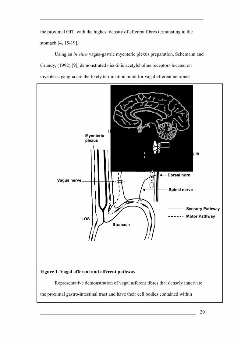
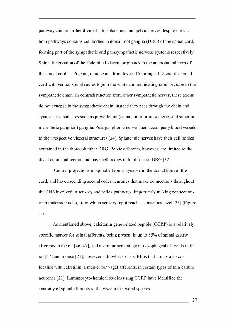
Figure 1. Vagal afferent and efferent pathway.
Representative demonstration of vagal afferent fibres that densely innervate
the proximal gastro-intestinal tract and have their cell bodies contained within
NTS
Stomach LOS
Dorsal horn
Myentericplexus
DRG
Vagus nerve
Nodose ganglia
Spinal nerve
Sensory Pathway
Motor Pathway
DMV
Hypothalamus
Thalamus
___________________________________________________________________
________________________________________________________________ 21
nodose ganglia. Ascending afferents terminate in the medulla oblongata of the
midbrain. Projections from the medulla oblongatta make connections with the
hypothalamus, thalamus and the NTS in the brainstem. Output of the nucleus tractus
solitarius (NTS) to synapse with dorsal motor nucleus of the vagus (DMV) cells is
critical in this investigation as a program generator within the DMV controls
efferent neurones that project to the gut, in particular to the lower oesophageal
sphincter (LOS) and synapse with myenteric neurones or interstitial cells of cajal.
Vagal efferents may activate nitrergic motor neurones and can cause smooth muscle
relaxation. This figure also demonstrates spinal afferents that have their cell bodies
contained within dorsal root ganglia. These neurones then synapse in the dorsal
horn of the spinal cord. Central projections of spinal afferents synapse in the dorsal
horn and ascending second order neurones make connections throughout the CNS.
Activation of vagal efferents can produce both excitatory and inhibitory
effects on GI smooth muscle [4], therefore it is believed that both excitatory and
inhibitory postganglionic neuroeffectors are released from enteric neurones in
response to excitatory vagal input. Acetylcholine is the principal excitatory
postganglionic neurotransmitter and acts on muscarinic receptors in gastric smooth
muscle, augmenting smooth muscle activity driving peristalsis and tone [10], as
well as parietal cells [4]. Inhibitory postganglionic neurones comprise the non-
adrenergic non-cholinergic (NANC) link between the nicotinic preganglionic vagal
efferent fibres and gastric smooth muscles and ICC’s. The two most likely
candidates for mediating this connection are nitric oxide (NO) or vasoactive
intestinal polypeptide (VIP), other mediators have been implicated however [4].

___________________________________________________________________
________________________________________________________________ 22
Of key importance to the current investigation is the afferent limb of the
vago-vagal pathway. The vagus has afferent endings located in all layers of the gut
wall. There are several types of visceral receptors and afferent fibres. Vagal afferent
terminals have been classified according to their morphology and anatomical
location into mucosal endings [11, 12], intraganglionic laminar endings (IGLE), and
intramuscular arrays (IMA) [13, 14]. These afferents have in common low
thresholds of activation and reach maximal responses within physiological levels of
mechanical stimulation [15, 16].
1.1 a) Intra Ganglionic Laminar Endings (IGLE)
Neuronal labelling techniques have been used to isolate origins of both vagal
and spinal afferents in the distinct layers of the gut wall. A number of studies have
utilised chemical coding with calretinin, a calcium binding protein, anterograde
tracing with DiI, a red fluorescent carbocyanine dye, and wheat germ agglutinin-
conjugated horseradish peroxidases to identify vagal afferents [11, 17], whilst
calcitonin gene-related peptide (CGRP) largely labels spinal afferents, although a
pitfall is that some vagal afferents also contain this peptide [11, 18].
IGLEs were first identified in the striated oesophageal muscle of the dog
[19], and were further described in oesophageal and gastric cardia smooth muscle in
cat and rat [20]. Rodrigo, et al (1982)[21], described IGLEs as nerve endings that
surround myenteric ganglia and lie between the longitudinal and circular smooth
muscle layers of the oesophagus. Anterograde tracing from nodose ganglia and
nodose ganglionectomy techniques comprehensively described IGLEs to be of
vagal origin [14, 21, 22].

___________________________________________________________________
________________________________________________________________ 23
IGLEs have dense concentration in the proximal GIT including in the
stomach, where about one half to one third of myenteric ganglia receive at least one
IGLE. However, they have been described throughout the gastrointestinal tract
including small and large intestine of the rat [13, 14, 23-25]. It was found that whilst
the jejunum contained the highest overall number of IGLE, the relative proportion
of myenteric ganglia supplied by IGLE decreased throughout the small intestine,
and the fewest identified in the caecum and colon [13, 14, 25, 26].
Later studies have found a close correlation in the morphology and regional
distribution patterns previously mapped in the rat model in the mouse and guinea
pig [26, 27].
They are localised to longitudinal, and to a lesser extent, circular smooth
muscle layers, with fibres penetrating the muscle and running parallel to the
respective fibres for several millimetres [23]. Individual axons appear to form
multiple terminal branches forming varicosities which interdigitate with myenteric
ganglia as well as having intimate association with surrounding connective tissue
components, NADPH positive enteric neurones and interstitial cells of cajal [11, 26,
28].
Individual parent axons supply multiple IGLE fields to adjacent myenteric
ganglia with proposed overlapping of receptive fields providing a network of
afferent innervation to myenteric plexuses [26, 27, 29].
It was proposed that IGLE act as mechanoreceptors detecting intra-mural
tension/shearing forces between orthogonal smooth muscle layers. Given their
topographic distribution and morphology, IGLE were implicated in mediation of a
number of reflex actions involving myenteric neurones, including rhythmical motor
___________________________________________________________________
________________________________________________________________ 24
programs such as coordination of peristalsis, swallowing peristalsis and emptying
[11, 26].
In vitro preparations using guinea pig stomach have since shown, using a
combination of electrophysiology and anterograde tracing techniques from vagus
nerve branches to oesophagus and stomach, that IGLE correlate closely with
mechanosensitive “hot spots”. IGLEs are now considered the transduction sites of
tension sensitive mechanoreceptors [27, 29, 30]. It is assumed that IGLEs found in
other rodent species correspond also to tension receptive sites responsible for
signalling active contraction and passive distension. Zagorodnyuk, et al (2003) [40],
further demonstrated modulation of sensitivity of these afferents, with the
transduction process itself involving mechanosensitive ion channels on the afferent
ending and to be relatively independent of chemical transmission.
IGLEs are not unique to vagal afferents, rather similar structures have now
been described on afferents of the pelvic nerve in the rectum [31]. Recordings from
pelvic afferent bundles innervating rectum revealed low threshold, slowly adapting
mechanoreceptors. Functionally similar to their vagal counterparts in the
oesophagus and stomach, morphologically they typically appear as flattened leaf-
like endings located in myenteric ganglia, however are smaller and simpler in
function, having fewer leaflets and less extensive branching patterns than in the
upper GIT [31].
1.1 b) Intramuscular Arrays
The other type of vagal afferent ending to the muscularis propria is the
intramuscular array (IMA). IMA anterogradely labelled in vagus nerve stomach
preparations were observed to be specialised endings consisting of several long and
___________________________________________________________________
________________________________________________________________ 25
straight varicose nerve fibres, ranging from hundreds of microns up to several
millimetres in length. They too, are located between longitudinal and circular
smooth muscle layers, branching and running parallel to smooth muscle fibres.
Individual fibres bear close proximity, are connected by short oblique or
perpendicular bridging collaterals and are commonly associated with interstitial
cells of cajal [23, 26, 29, 32].
In contrast to IGLEs, IMAs appear to have a distinct distribution, and are
densely concentrated innervating sphincter regions including the lower oesophageal
and pyloric sphincters, as well as a dense concentration in the proximal stomach
becoming more sparse toward the corpus and antrum [11, 13, 14, 23, 25, 26]. It was
postulated that IMA act as ‘in series’ length or stretch receptors, responding to
passive stretch and active contraction, due to their location where intraluminal
matter collects (forestomach), or passes (sphincters) [26], however in vitro studies
of the guinea pig stomach failed to correlate anterogradely labelled IMA and
mechanosensory transduction sites, and thus direct morphological evidence for
IMAs as length sensitive afferents is lacking [27, 29, 32].
1.1 c) Mucosal Afferents
The third morphologically distinct vagal afferent has its endings in the
mucosal layer of the gut. These fibres penetrate the muscle layer and the
submucosa, forming networks of branching axons in the lamina propria of crypts
and villi [32, 33]. Vagal afferent mucosal innervation has biphasic distribution,
being densely distributed to uppermost cervical oesophagus immediately distal to
pharyngeo-oesophageal junction, and a second dense concentration of afferents in
abdominal regions, with innervation sparse in the lower cervical and thoracic

___________________________________________________________________
________________________________________________________________ 26
oesophagus [11, 12, 22, 28]. This pattern is reflected in animal and human studies
where variable sensory properties of afferents occur in different regions of the gut,
with the upper oesophagus more sensitive to mucosal mechanical and chemical
stimuli [11, 12].
Anterograde labelling techniques from the nodose ganglia have identified up
to four types of fibre supplying the mucosa. Wank, et al (2001) [12], described
small varicose intra-epithelial processes invading the superficial epithelial layers.
Dutsch, et al (1998) [21], commented on similar structures densely innervating the
upper oesophagus. These thick calibre fibres, running parallel to the oesophagus
were highly branched laminar structures, with their terminal ramifications directed
toward the basal epithelial layer and appearing to penetrate it, and are thus ideally
positioned to sense luminal contact and chemical mediators released from local
structures [32, 33]. Thinner axon fibres were also observed in the upper cervical
oesophagus having dense networks in the submucosal layer, with finger like
projections penetrating toward the basal epithelium. Other types of mucosal fibres
supply submucosal and perivascular tissue and appear to have functional importance
[12].
No attempt has been made to identify histologically vagal and spinal afferent
endings in the mucosa of the large intestine, however functional evidence shows
vagal, pelvic, and splanchnic afferents between them, to supply all regions of the
GIT mucosa [32].
1.2 Spinal Afferents
Like vagal sensory afferents, spinal afferents innervating the GI tract are
almost exclusively thin myelinated Aδ or unmyelinated C fibres. The spinal
___________________________________________________________________
________________________________________________________________ 27
pathway can be further divided into splanchnic and pelvic nerves despite the fact
both pathways contains cell bodies in dorsal root ganglia (DRG) of the spinal cord,
forming part of the sympathetic and parasympathetic nervous systems respectively.
Spinal innervation of the abdominal viscera originates in the anterolateral horn of
the spinal cord. Preganglionic axons from levels T5 through T12 exit the spinal
cord with ventral spinal routes to join the white communicating rami en route to the
sympathetic chain. In contradistinction from other sympathetic nerves, these axons
do not synapse in the sympathetic chain, instead they pass through the chain and
synapse at distal sites such as prevertebral (celiac, inferior mesenteric, and superior
mesenteric ganglion) ganglia. Post-ganglionic nerves then accompany blood vessels
to their respective visceral structures [34]. Splanchnic nerves have their cell bodies
contained in the thoracolumbar DRG. Pelvic afferents, however, are limited to the
distal colon and rectum and have cell bodies in lumbosacral DRG [32].
Central projections of spinal afferents synapse in the dorsal horn of the
cord, and have ascending second order neurones that make connections throughout
the CNS involved in sensory and reflex pathways, importantly making connections
with thalamic nuclei, from which sensory input reaches conscious level [35] (Figure
1.).
As mentioned above, calcitonin gene-related peptide (CGRP) is a relatively
specific marker for spinal afferents, being present in up to 85% of spinal gastric
afferents in the rat [46, 47], and a similar percentage of oesophageal afferents in the
rat [47] and mouse [21], however a drawback of CGRP is that it may also co-
localise with calretinin, a marker for vagal afferents, in certain types of thin calibre
neurones [21]. Immunocytochemical studies using CGRP have identified the
anatomy of spinal afferents to the viscera in several species.

___________________________________________________________________
________________________________________________________________ 28
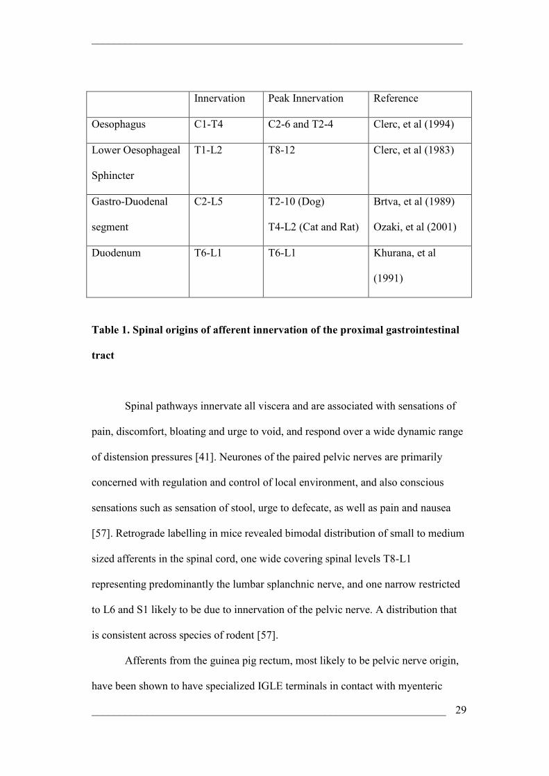
Spinal afferent innervation of the gastro-oesophageal region arises from a
segment of the cord spanning upper cervical (C1) to upper lumbar (L2) (Table 1.).
These afferents are conveyed in the greater splanchnic and the thoracic spinal
nerves [36, 37], 51-53], and are generally considered to give rise to non-specialised
free endings in the mucosa, muscle and serosa, arising from thin varicose fibres in
apposition with myenteric ganglia, possibly connected to muscle tension receptors
as well as distributed between or passing to other structures and also releasing
neurotransmitter [49, 50]. These fibres are fairly evenly distributed along the
gastrointestinal tract in contrast to vagal innervation as described previously [21, 37,
48].
Retrograde labelling studies have enabled mapping of distribution of these
afferents in the oesophagus revealing bimodal distribution of afferents to the
oesophagus with dense innervation at cervical (C2-C6) and thoracic (T2-T4) parts
of the oesophagus [37, 38]. Fibres innervating the LOS are distributed in the cord
from T1-L2, having a peak distribution in the T8-T12 region [36].
The sensory innervation of the gastro-duodenal segment extends from C2-
L5 with a peak innervation field of the stomach spanning the cranial, middle, and
the immediately adjoining caudal thoracic ganglia (T2-T10) in the dog, and T4-L2
in cat and rat [39, 40]. The duodenum has peak innervation originating in the middle
and caudal thoracic ganglia and cranial lumbar (T6-L1) ganglia. There is a
recognizable viscerotopic organization in the sensory innervation of the post-
pharyngeal foregut; successively more caudal sectors of this region of the
alimentary canal are supplied with sensory fibres from successively more caudal
spinal dorsal root ganglia [37].
___________________________________________________________________
________________________________________________________________ 29
Innervation Peak Innervation Reference
Oesophagus C1-T4 C2-6 and T2-4 Clerc, et al (1994)
Lower Oesophageal
Sphincter
T1-L2 T8-12 Clerc, et al (1983)
Gastro-Duodenal
segment
C2-L5 T2-10 (Dog)
T4-L2 (Cat and Rat)
Brtva, et al (1989)
Ozaki, et al (2001)
Duodenum T6-L1 T6-L1 Khurana, et al
(1991)
Table 1. Spinal origins of afferent innervation of the proximal gastrointestinal
tract
Spinal pathways innervate all viscera and are associated with sensations of
pain, discomfort, bloating and urge to void, and respond over a wide dynamic range
of distension pressures [41]. Neurones of the paired pelvic nerves are primarily
concerned with regulation and control of local environment, and also conscious
sensations such as sensation of stool, urge to defecate, as well as pain and nausea
[57]. Retrograde labelling in mice revealed bimodal distribution of small to medium
sized afferents in the spinal cord, one wide covering spinal levels T8-L1
representing predominantly the lumbar splanchnic nerve, and one narrow restricted
to L6 and S1 likely to be due to innervation of the pelvic nerve. A distribution that
is consistent across species of rodent [57].
Afferents from the guinea pig rectum, most likely to be pelvic nerve origin,
have been shown to have specialized IGLE terminals in contact with myenteric
___________________________________________________________________
________________________________________________________________ 30
ganglia in the guinea pig rectum. These endings bear similar function to their vagal
counterparts in the upper gut however are morphologically slightly different as
mentioned previously. Rectal IGLEs are concentrated in rectal nerves and are absent
in colonic nerves indicating their function in rectal reflexes such as defecation [31].
2. Functional properties of visceral afferent endings
Much of our knowledge about the electrophysiology and function of GI
sensory afferents has arisen from in vitro and in vivo whole animal experiments
using ‘single fibre’ recording techniques first established by Paintal, (1953)[42] and
Iggo, (1955)[43]. Most visceral afferents are unmyelinated C fibres with few being
Aδ fibres [5]. Visceral afferents were traditionally thought to be best described
based on the layer of gut containing their afferent terminals, and their general
response properties including response to mechanical stimulation, however Brierley,
et al (2005)[60], demonstrated lumbar splanchnic, and pelvic nerve pathways differ
in their chemosensitivity to known noxious stimuli. Also pertinent to this project
however, is the general response profiles of visceral afferents to mechanical stimuli.
Visceral afferent responses to mechanical stimuli, acting within the GI wall,
is consistent with the location in mucosal layer, muscle, and in serosal mesenteric
attachments. With this in mind, functional studies using in vitro and in vivo
electrophysiological techniques have enabled vagal and spinal afferents to be
classified into different classes according to their response to mechanical
stimulation [35].
Vagal and spinal extrinsic afferent nerves exhibit contrasting stimulus
response properties, highlighting their differing roles in sensory signalling. Clinical
___________________________________________________________________
________________________________________________________________ 31
evidence demonstrates spinal nerves to be involved in the signalling of visceral
pain, as any pain originating from visceral structures can be alleviated by splanchnic
nerve blockade [34]. In vitro and in vivo animal studies have demonstrated spinal
afferents in the upper gut to as far as the transverse colon, give rise to sensations of
pain, discomfort, bloating and fullness. Afferents to the distal colon and rectum,
give rise to urge to defecate, discomfort as well as pain evoked by more intense
stimuli [42, 56].
In contrast vagal mechanoreceptors are thought to be involved in regulatory
physiological processes such as reflexes involving intestinal secretion and motility
including involvement in the emetic reflex [6], as well as contributing to nausea and
malaise [44].
Afferent fibre labelling studies have enabled the localisation of the precise
terminations of afferent endings and have enabled greater understanding of
functional correlates of their endings. Tracing techniques have demonstrated the
localisation of vagal and spinal afferents to numerous layers of the gastrointestinal
tract. Functional roles correlate with layer of gut and exposure to mechanical and
chemical stimuli. The focused distribution of vagal and pelvic afferents may
correspond to regions of the gut where graded, innocuous sensations can be evoked
by distension whereas regions that predominantly receive splanchnic innervation
appear to generate less graded sensation, rather discomfort and frank pain are the
first responses to increasing levels of distension [32].
2.1 Vagal Afferents
2.1 a) Mucosal Receptors

___________________________________________________________________
________________________________________________________________ 32
The general consensus is that mucosal receptors are implicated in the
regulation of gastric motility and secretion. Mucosal afferents have endings in the
mucosal lamina propria, where they are ideally positioned to detect material
absorbed across the mucosal epithelium or released from epithelial and sub-
epithelial cells [32].
The existence of these terminal endings has been verified by in vivo and in
vitro electrophysiological studies in a number of animal models demonstrating them
to be exquisitely sensitive to mechanical deformation of the mucosa by mucosal
stroking with calibrated von Frey hairs (10-1000mg) or probing, as might occur
with particulate material within the lumen. In general mucosal receptors do not
show resting activity, and show rapidly adapting responses in a force dependent
manner to mucosal stroking with calibrated von Frey hairs, whilst being insensitive
to muscular stimuli such as contraction or distension [32, 45-51]. Mucosal receptors
are polymodal with numerous studies demonstrating, in addition to light stroking,
they are also responsive to luminally applied drugs and chemical stimuli including
inorganic and fatty acids, bile, hypo- and hyperosmolality, as well as 5-
hydroxytryptamine (5-HT), cholecystokinin (CCK), α,β-meATP, prostaglandins
(PG) and bradykinin (BK) [44, 46-53].
These afferents have been extensively characterised in the vagus nerve
where in addition to those functions mentioned above, are also thought to play a
role in generation of sensations of satiety, nausea and vomiting [45, 47-49, 51, 53,
54].
___________________________________________________________________
________________________________________________________________ 33
2.1 b) Tension Receptors
Nerve terminals in the muscle and serosa convey mechanosensitive
information corresponding to distension or contraction of the gut wall [35], however
electrophysiological studies have demonstrated information originating from vagal
and spinal mechanoreceptive afferents differs.
Vagal mechanoreceptors located in the muscle layer often have resting
discharge and are specifically sensitive to muscular contraction and distension, but
are unresponsive to mucosal stroking with calibrated von Frey hairs (10-50mg),
have low thresholds of activation, and reach maximal responses within
physiological levels of distension [6, 35, 43, 46, 47, 51, 55]. These basic
characteristics of vagal distension sensitive afferents are conserved in dog [56],
opossum [57], mouse [46] and ferret [45]. In contrast, sub-populations of spinal
afferents have thresholds of activation within the physiological range, however are
generally are considered to be critical in detecting stimuli in the supra-physiological
range [58]. Vagal muscular afferents, like pelvic muscular afferents, show slowly
adapting maintained responses to distension, in contrast to splanchnic muscular
afferents which are more rapidly adapting [32].
Of particular importance in the current investigation are tension sensitive
afferents, which have been comprehensively described in the muscle layer of the
gastro-oesophageal region.
Vagal afferent terminals form specialized endings called IGLEs found
throughout the length of the GI tract. Studies in the guinea pig have demonstrated
IGLEs to be the transduction sites of vagal tension sensitive mechanoreceptors in
the oesophagus and stomach.

___________________________________________________________________
________________________________________________________________ 34
Tension sensitive afferents are thought to serve as pure mechanoreceptors
involved in physiological regulation such as mediating the non painful sensations of
fullness, bloating and nausea, as well as gut reflex function such as the triggering of
transient lower oesophageal sphincter relaxations (TLOSRs) which are of
importance to the current investigation [56-59].
2.1 c) Tension Mucosal (TM) Receptors
The ferret gastro-oesophageal in vitro preparation has also identified a
further vagal afferent receptor subtype with endings in the oesophagus, that
responds not only to mucosal stimulation with calibrated von Frey hairs 10-1000mg,
but also to circumferential tension, thus has both mucosal and tension sensitive
afferent properties. There are two possible anatomical explanations for the site of
the TM receptive field. There are either two receptive fields, topographically
superimposed with one being in the mucosal and the other in the muscular layer, or
like a vagal afferent, Davison et al, (1972)[53], had previous described in the
duodenum, there is one receptive field interposed in the muscularis mucosae. These
afferents are thought to be important in the detection of rapidly moving boli of food
and/or liquid along the oesophagus [60].
2.2 Spinal Afferents
Gastro-Oesophageal:
Afferent innervation of the gastro-oesophageal region conveying sensory
information to the central nervous system occurs via the splanchnic nerve, or
thoracic sympathetic chain [58]. Characterisation of mechanosensitive splanchnic
afferents have been studied in the rat stomach [40], and the opossum oesophagus
___________________________________________________________________
________________________________________________________________ 35
[58]. There exist separate populations of low- and high-threshold afferent fibres that
innervate many viscera. The low threshold afferent fibres are assumed to mediate
sympatho-sympathetic or sympatho-vagal regulatory reflexes which, under normal
physiological conditions, are generally not sensed. Low threshold fibres also likely
play a role in non-painful sensations such as bloating, fullness, nausea. High
threshold afferent fibres are believed to mediate nociception [77]. Mechanosensitive
afferents have been classified into low threshold (wide dynamic range)
mechanoreceptors or high threshold (phasic) nociceptors depending on their ability
to encode either non-noxious mechanical stimuli, noxious mechanical stimuli or
both [58].
Low threshold (phasic) mechano-nociceptors were observed in both preparations
and respond to a wide range of stimuli from innocuous to noxious intensity. They
have a low threshold of stimulation, responding to peristaltic contractions and a
linear stimulus response to graded distension of the viscera within a narrow
physiological pressure range [58]. Discharge is saturated in opossum oesophagus at
innocuous levels and therefore does not distinguish noxious from innocuous stimuli
[58], in contrast to the low threshold mechanoreceptors population in the rat
stomach that encoded distending stimuli throughout range of distending pressures
and are thus considered to contribute to sensations such as discomfort and pain [40].
This subtype of mechanoreceptive afferent differs from gastro-oesophageal vagal
mechanoreceptive afferents in magnitude of response and saturation of receptor
activity at physiological pressures [40]. Given they are activated at low thresholds
of mechanical stimulus they are implicated in non painful mechanical signalling

___________________________________________________________________
________________________________________________________________ 36
giving rise to sensations such as bloating, fullness and nausea as well as
physiological regulatory functions such as storage, propulsion and emptying [40].
High Threshold (phasic) mechanoreceptors are insensitive to peristaltic contraction
and are activated only by mechanical stimuli considered within noxious range [40,
58]. The majority of these afferents have little spontaneous resting activity; however
resting activity could be altered by chemical or mechanical stimuli. These afferents
also show ability to be sensitised by insult or even non injurious stimuli, and are
thus considered to contribute to altered sensation arising from the gastro-
oesophageal region such as central hyperexcitability and visceral hyperalgesia [40].
Given the response profiles of these afferents, their presence is taken as evidence for
the presence of nociceptors that give rise to sensations of discomfort and pain [40].
Responses of either subtype of afferent to gastric distension was conserved across
species, and generally exhibited a monotonic increase in firing with increasing
distension pressure of the viscus, typically exhibiting slowly adapting responses
during maintained distension [40, 58].
Distal Gastrointestinal tract:
Mechanosensitive afferents have been well described in the distal
gastrointestinal tract where they have been identified using in vivo and in vitro
electrophysiological techniques in lumbar splanchnic and pelvic nerves of the cat
[61, 62], rat [63, 64] and mouse [65], including afferents that respond to mechanical
stimulation of the colonic mucosa, muscle layer, serosa and/or mesentery. Major
differences between anatomical location of receptive fields and response profiles of
lumbar splanchnic and pelvic nerves to mechanical stimulus have been identified

___________________________________________________________________
________________________________________________________________ 37
[65]. Brierley, et al (2004)[65], identified five different classes of afferent fibre
based on mechanical response profiles in the mouse colon. Three types were
conserved across pelvic and splanchnic pathways (muscular, serosal and mucosal),
and each nerve supplied a unique type of afferent. A general discussion of afferent
types is presented here.
2.2 a) Mucosal Receptors
Mucosal receptive afferents have been previously described in the distal
colon and perianal mucosa [52, 63, 65]. Duthie and Gairns, (1960)[66], provided the
only morphological evidence of free nerve endings in the anal mucosa epithelium,
exhibiting multiple branching and presence of varicose structures. An in vitro
electrophysiological study of the rat identified pelvic nerve afferents in the distal
colon mucosa which, like vagal afferents innervating the upper gastro-oesophageal
tract, were not spontaneously active but all responded with rapidly adapting burst of
discharge in an incremental manner to full range of von Frey hair stroking, and the
majority were unresponsive to circular stretch [52]. These results are also consistent
with those of mucosal afferents in the cat [67].
In vitro electrophysiological studies have also identified mucosal afferents in
LSN innervating the colon. These colonic mucosal afferent fibres have a
comparable response to von Frey hair stroking to that of the upper gastrointestinal
mucosal fibres recorded in vitro, and similarly show no response to circumferential
tension [45]. These colonic fibres exhibited limited or no resting discharge,
consistent with previous findings in the ferret [45], mouse [65] as well as vagal
mucosal afferents in a number of species [45, 47, 68]. Mucosal colonic afferents
were also found to be polymodal with most responding to one of hypertonic saline,

___________________________________________________________________
________________________________________________________________ 38
HCL, bile and capsaicin, which is in contrast to what was found in ferret
oesophageal vagal afferents where only a small proportion were responsive to
chemical stimulus [52].
2.2 b) Muscular Receptors
The response profiles of LSN and PN afferents to colonic distension has
been characterised extensively using a variety of in vitro and in vivo
electrophysiological techniques [52, 61, 62, 69]. Spinal afferents with terminals in
the muscle layers are likely to encode both physiological and noxious levels of
stimulation.
Whilst both pathways have similar response properties to mechanical
stimuli, and have been shown to have similar response profiles to noxious colorectal
distension, discrepancies do exist [61, 69-72].
Lumbar Splanchnic Nerve:
Muscular afferents of the LSN do not respond to low intensity mucosal
stroking but do respond to probing of the receptive field and most consistently to
distension of the colon [63]. Blumberg, (1983)[61], categorised distension sensitive
afferents in the cat LSN to exhibit different characteristic responses to phasic
distension, classifying fibres into 4 types based on their adaptation to phasic colonic
distension, ranging between tonic and phasic discharge in response to colonic
distension. These afferents display responses to normal physiological stimuli (non-
noxious) however they also are thought to encode stimuli into the noxious range, as
well as being chemosensitive [62].
Pelvic Nerve:

___________________________________________________________________
________________________________________________________________ 39
Two populations of slowly adapting afferent fibre have been described
arising from the pelvic nerve based on their responses to graded colorectal
distension (CRD) in the rat [63]. Low threshold responding to ≤10mmHg, and high
threshold responding to ≥28mmHg, indicating functionally different afferent fibres
in the pelvic nerve [63]. Pelvic fibres innervating the colon of the rat responsive to
noxious CRD were mainly C fibres, were active at rest and exhibited either a
dynamic response followed by slow adaptation or only a tonic response to phasic
CRD.
Pelvic high threshold afferents are thought to be important for visceral
nociception, whilst low threshold afferents may mediate sensation such as fullness,
as well as mediating sympatho-sympathetic or sympatho-vagal regulatory reflexes,
but do not appear to elicit painful sensation.
Pelvic nerve afferents in the cat were subdivided into phasic and tonic
afferents. Phasic being rapidly adapting and tonic being slowly adapting throughout
distension of the colon. These fibres responded to low thresholds of intraluminal
pressure [70].
Brierley, et al (2004)[65], identified a further receptor subtype unique to the
pelvic nerve that possessed properties of both muscular and mucosal receptors,
responding to both mucosal stroking with von Frey hairs (10-1000mg) and also to
circumferential stretch and was termed muscular/mucosal receptors.
In contrast to the lower colon, the guinea pig rectum is richly innervated by
distension sensitive afferents arising from the pelvic nerve which display low
thresholds and are slowly adapting. The receptive fields of these stretch sensitive
mechanoreceptive afferents have been morphologically identified as IGLEs. These

___________________________________________________________________
________________________________________________________________ 40
IGLEs termed rectal IGLEs (rIGLE) bear many similarities with IGLEs in the upper
GIT however are less complex and have less branching [52].
2.2 c) Serosal and Mesenteric
Afferents to the serosa and mesenteric attachments of the colon are the most
common afferent identified from the lumbar splanchnic nerve in animal in vitro
preparations accounting for over 50% of recorded afferents [65]. Afferents have
their endings located on or close to blood vessels or branching points of capillaries
supplying the serosa and mesentery [65], and have the ability to respond beyond
physiological levels and encode both physiological and noxious levels of intestinal
distension [73].
Serosal afferents often have multiple punctuate receptive fields (2-4mm2)
that extend from the point of division of the left colic artery to the branch points
beneath the serosa of the colon [68, 82, 90]. Previous studies have found afferents to
respond to micromanipulator rod stimulation over receptive fields as well as
responding to tension, exhibiting a phasic and tonic component of discharge [74].
Responses to circumferential tension are either absent or only seen at onset of
stretch, bearing no relationship with tension or length of tissue. In addition they do
not respond to mucosal stroking with calibrated von Frey hairs less than 50mmHg
[50, 51, 63].
These afferents have sporadic resting discharge, and are characterised by
their ability to respond to firm blunt probing on the mucosal surface, responding at
lower mechanical threshold to stimulation of the reflected serosal surface than of the
mucosal surface [68, 78, 79, 82, 91]. They demonstrate a burst of firing rather than a
prolonged response [52], and have a high threshold for response to mechanical

___________________________________________________________________
________________________________________________________________ 41
stimulation [52, 75]. Mesenteric afferents are specifically sensitive to distortion of
mesenteric attachments, therefore a force strong enough to distort the mesentery or
serosa is required for activation of both types. Contraction and distension of the
bowel wall are sufficient stimuli to activate these afferents post a sensitising event
such as colonic inflammation [76]. It has been proposed these afferents may detect
twisting and torsion of the colon and pulsatile changes in blood pressure in
mesenteric blood vessels, possibly critical during plasma extravasation resulting
from colonic inflammation [65].
In addition to their mechanoreceptive properties, these afferents are
generally chemosensitive. Responsiveness to bradykinin, capsaicin, hypertonic
saline, normal saline and hydrochloric acid (HCl) applied extra-luminally have been
previously observed [62, 77], indicating possible roles for transmission of signals
related to noxious and inflammatory events and sensing composition of luminal
content [52].
3. Pharmacology of Visceral Afferents
In addition to mechanical stimuli, vagal and spinal afferents are also
responsive to chemical stimuli, a characteristic vital for their role in participating in
central and local reflex control of gastrointestinal motor and secretory function. The
location of visceral afferent terminals supplying the gut near myenteric ganglia,
blood vessels and luminal material, combined with the fact that these afferents
express a variety of membrane receptors to a multitude of chemical mediators
generated from both within and outside the gut wall [73], mean they are well suited
to sense and be modulated by chemical stimuli [61].

___________________________________________________________________
________________________________________________________________ 42
Sensory signal transduction is highly dependent on the excitability of the
sensory neurone, which in turn, is governed by the relative contribution of a variety
of voltage-dependent ion channels controlling ion movement, thus charge, across
the cell membrane. These and other channels present in nerve terminals transmit and
amplify this code into generation of action potential, which conveys the message
onwards toward the central nervous system [73].
Electrophysiological, immunocytochemical and molecular biological
techniques have identified a variety of chemical mediators acting on functionally
active receptors expressed on sensory nerve terminals. Several mediators have been
shown to be active at nerve terminals of extrinsic afferents supplying the gut wall,
altering the excitability of the sensory neurone; a general discussion is presented
below.
These substances are thought to produce their effects via three distinct
mechanisms. 1) Direct activation, which generally involves the opening of ion
channels present on nerve terminals, 2) Sensitization, which may only occur in the
absence of direct stimulation but which usually results in hyperexcitability to both
chemical and mechanical modalities, and 3) Alteration of the phenotype of the
afferent nerve, for example through alterations in the expressions of mediators,
channels and receptors or modulating activity of these by changing the ligand-
binding characteristics or coupling efficiency of other receptors [78].
3.1 Excitatory Receptors
3.1 a) Adenosine triphosphate (ATP)
Within the gut, there is an abundance of evidence to suggest that adenosine
triphosphate (ATP) acts as a neurotransmitter, released from either extrinsic

___________________________________________________________________
________________________________________________________________ 43
sympathetic nerves (as a co-transmitter with nor-adrenaline and neuropeptide Y)
[79, 80], intrinsic neurones [81], or from tissue injury, where by cell damage causes
the release of cytoplasmic contents rich in ATP. ATP may further induce the
synthesis of prostaglandins which are, in turn, mediators of inflammation [80, 82,
83].
ATP exerts its extracellular actions as a neurotransmitter via cell surface
receptors, P2 purinoceptors. Purinoceptors are further divided into two major
classes based on structural and pharmacological profiles. P2X are ligand-gated ion
channels causing a voltage dependent calcium influx upon activation, thus
mediating fast synaptic transmission. The second subclass, P2Y receptors are
metabotropic G-Protein coupled receptors that mediate signalling via inositol
trisphosphate, leading to intracellular calcium release. P2Y receptors are therefore
considered to play a modulatory role rather than directly mediating purinergic
transmission [82, 84].
P2X and P2Y receptors are both present in dorsal root and nodose ganglia
[80, 84, 85]. Both P2X and P2Y receptors have been shown to be located on
afferent sensory neurones including vagal [86], and splanchnic afferent fibres [80,
87]. A subclass of P2X receptors, the P2X3 receptor, is selectively expressed in
sensory neurones and is implicated in the mediation of nociception [85].
Immunohistochemistry demonstrated binding of ATP throughout the human and rat
nodose ganglion and brainstem, including the ventrolateral medulla and NTS, with
ATP binding specifically P2Y receptors. Bilateral ligature of the vagus nerves and
accumulation of both P2Y immunoreactivity and ATP binding sites present adjacent
to ligatures of the nerve suggest bidirectional transport of the P2Y1 receptor along
the vagus nerve [84, 88].

___________________________________________________________________
________________________________________________________________ 44
Page, et al (2000, 2002) [46, 83], showed two distinct responses of gastro-
oesophageal vagal afferents to the purinergic agonist α, β-meATP. A proportion of
mouse afferents were directly excited by the purinergic agonist, in contrast to the
ferret, where no gastro-oesophageal mucosal receptors were excited by direct
application. However, mechanosensitivity was reduced in inflamed tissue in the
ferret and was enhanced in the presence of a purinergic agonist back to uninflamed
levels [83]. This phenomenon is also demonstrated by Dang, et al (2005)[87], who
showed that inflammation increases excitability in both rat DRG and nodose
ganglion neurones that innervate the stomach, increasing the fraction of neurones
that are responsive, as well as their peak response to α,β-meATP.
This combination of morphological and functional electrophysiological
evidence demonstrating the expression of purinergic receptors on peripheral sensory
neurones implicate a role for purinergic receptors in not only mediation of
nociceptive input into the spinal cord, but also in mechanosensation of gastro-
oesophageal afferents, particularly post inflammation.
3.1 b) Bradykinin
Bradykinin (BK), a metabolite of the Kallikrein-Kinin system, is released
from ischaemic and inflamed tissues and has been shown to be an important algesic
stimulus on sensory nerves and mediates pain responses to tissue injury [89]. It is a
well recognized nociceptive stimulus on skeletal muscle receptors and cutaneous
nociceptors. Importantly, bradykinin stimulates visceral nociceptors including
cardiac as well as splanchnic afferents innervating much of the gastrointestinal tract
[59, 62, 77, 90-95]. Bradykinin exerts its effect via two different subtypes of G-
protein coupled receptors termed B1 and B2. The B2 receptor is extensively
___________________________________________________________________
________________________________________________________________ 45
expressed in a constitutive fashion whereas the B1 receptor is expressed at low
levels in healthy tissue, but becomes rapidly up-regulated following tissue injury by
various pro-inflammatory kinin metabolites [89, 96]. Sengupta, et al (1992)[90],
describes an additional three receptors termed B3-5, B3 and B4 being excitatory
receptors, involved in smooth muscle contraction and B5, an inhibitory receptor
involved in relaxation of the lower oesophageal sphincter.
Both vagal and spinal afferents innervating the viscera are responsive to
bradykinin. Bradykinin directly increases firing of serosal afferents in the rat
jejunum, with results consistent with an action directly on the B2 receptor located on
serosal afferent terminals [113]. Colonic distension sensitive splanchnic afferents of
the cat were also sensitive to BK [62]. This result was reinforced in the opossum
where all distension sensitive wide dynamic range and high threshold afferents were
responsive to bradykinin in a dose dependent and pharmacologically reversible
manner, an action believed to be directly mediated by the B2 receptor [90]. In the
abdominal viscera of the mouse, BK potently stimulates splanchnic afferents
innervating much of the gastrointestinal tract [90], but few pelvic afferents [60]. In
addition, mechanically insensitive afferents recruited by BK are exclusive to LSN
[60].
Bradykinin also activates vagal afferent fibres supplying the guinea pig
airway where all afferents originating in the jugular ganglion responded to BK,
however in the nodose ganglion, whilst 100% of C fibres responded to BK, there
were no Aδ responsive fibres [97]. This stimulation of vagal afferent fibres
innervating the guinea pig airway was not dependent on smooth muscle tone nor on
BK induced production of prostaglandins, and was mediated via B2 type receptor.
More importantly to the current investigation is that bradykinin has been shown to
___________________________________________________________________
________________________________________________________________ 46
increase signalling of vagal afferents innervating the gastrointestinal tract.
Bradykinin was shown to cause excitation of vagal distension sensitive afferents
innervating the duodenum of the sheep, and the oesophagus of the opossum [90,
98]. However in each case, there was close coupling of vagal afferent activation and
longitudinal smooth muscle contraction, suggesting distortion of the tissue caused
direct activation of mechanosensitive receptors. Sengupta et al, (1992)[90],
demonstrated this effect in opossum to be reproducible by activation of a post
junctional B4 receptor, suggesting a role for B4 receptor in this effect of contraction
of longitudinal smooth muscle.
These results led to the proposition of two possible explanations for the
actions of BK on sensory afferents innervating the GIT. Either BK acts directly on
chemosensitive nerve endings or can stimulate pure mechanoreceptors secondary to
muscle contraction. However, Page and Blackshaw (1998) [45], have demonstrated,
using an in vitro ferret preparation, a tension sensitive afferent to be directly
responsive to bradykinin, unrelated to smooth muscle contraction, as was found for
vagal afferents innervating the guinea pig oesophagus [99].
3.1 c) Cholecystokinin (CCK)
Cholecystokinin (CCK) is a peptide hormone originally isolated from
mammalian GIT, where the predominant form, CCK-octapeptide (CCK-8), exists in
sulfated-, and non-sulfated states [100, 101]. CCK is a hormonal regulator of
digestive processes produced by two separate cell types, enteroendocrine I-cells in
the upper GIT and various neurones in the GIT and CNS [102, 103]. Receptors for
CCK are classified into two distinct subtypes, CCK1, and CCK2. CCK1 receptors
occur in a few localised areas of central and peripheral nervous system, but
___________________________________________________________________
________________________________________________________________ 47
predominate in the peripheral tissue including the gastrointestinal tract where they
influence feeding behaviour. The CCK2 subtype predominates in the central nervous
system where they are distributed throughout the CNS and spinal cord, and
modulate anxiety, analgesia, arousal and neuroleptic activity [100, 101].
CCK is an important satiety factor, having a marked effect on reflex and
behavioural responses to food, and this effect is dependent on an intact vagal
afferent pathway [104].
CCK binding sites are present both within the vagus nerve proper, and also
within the cell bodies in the nodose ganglia [103, 105, 106]. This result confirmed
by the presence of CCK receptor mRNA isolated within nodose ganglia [103, 107-
109], and by autoradiography demonstrating CCK binding sites at ligation points in
rat vagal afferents, suggesting peripheral transport [110]. CCK has also been shown
to activate gastric mechanoreceptors in both muscle and mucosal layers. Distension
sensitive vagal afferents in both stomach and duodenum have been shown to be
activated upon application of CCK, demonstrating integration of signals elicited by
CCK and gastric loads at the level of the vagal afferent fibre [47, 104, 111, 112].
However, whilst Davison, et al (1988)[104] found this effect to be independent of
intraluminal pressure, and thus gastric wall tension, Blackshaw & Grundy,
(1990)[47], found mechanoreceptor discharge to closely follow intraluminal wall
pressure, indicating sensitivity of mechanoreceptors could be secondary to a direct
action on the muscle itself, or a reflex pathway involving muscle tone, rather than
directly attributed to CCK [47, 105].
In contrast, Blackshaw and Grundy, (1990)[47], found CCK to similarly
cause an increase in firing of mechanosensitive vagal afferents innervating the
mucosa of the ferret gastroduodenal region, and found this to be an effect mediated
___________________________________________________________________
________________________________________________________________ 48
directly by CCK. Also, Richards, et al (1996) [105], recording from mesenteric
nerve bundles found that 70% were responsive to CCK, demonstrating an increase
rate of firing. This effect was abolished by vagal degeneration indicating the effect
exclusively mediated by vagal, and not splanchnic and enteric nerves. These
responsive nerves were believed also to have endings in the mucosal layer as these
afferents were unresponsive to distension. Whilst there is no evidence for effects of
CCK on spinal gastro-oesophageal afferents, evidence does exist for CCK receptors
in dorsal root ganglia [113, 114].
3.1 d) Ionotropic Glutamate Receptors (iGluRs)
Glutamate is the major excitatory neurotransmitter in the central nervous
system. In addition to mediating synaptic transmission, it is also involved as a
modulator at both pre- and post-synaptic sites. Glutamate exerts its effects via
metabotropic (mGluR) or ionotropic receptors (iGluR). Ionotropic glutamate
receptors are ligand gated ion channels. There are three major types of iGluR,
named after the selective agonists that were originally identified to activate them,
and are thus called N-Methyl-D-Aspartate (NMDA), α-amino-3-hydroxy-5-methyl-
isoxazoleproprionate (AMPA) and Kainate receptors. Each of the major functional
subtypes of iGluR are made up of a family of genes forming an agonist binding
domain, interrupted by insertion of three transmembrane pore loop ion channels
[115]. Native receptors of all of these families are likely tetrameric assemblies
comprising more than one type of subunit [116]. Binding of agonist to an iGluR
triggers opening of an ion pore, allowing the flow of selected cations across the
membrane. In the continuing presence of agonist, the receptor typically desensitises,
closing the channel [117].
___________________________________________________________________
________________________________________________________________ 49
3.1 d i) NMDA receptors:
NMDA receptors are likely of tetrameric structure and are likely composed
of pairs, or dimers, of dimers [116]. The NMDA receptor family is composed of
seven subunits, NR1, NR2A-D and NR3A and B, which are all products of separate
genes. Eight functional splice variants and one non-functional truncated splice
variant of NR1 have been described [118].
All NMDA receptors contain an obligate NR1 subunit that serves two roles.
NR1 contain a glycine binding site, with NMDA receptor activation requiring
glycine occupation of this site. In addition, it is required for trafficking of
heteromeric receptor assemblies to the plasma membrane [119]. Functional NMDA
receptors appear to be comprised of at least one NR2 subunit, or NR1 and both NR2
and NR3 subunits [116].
Of the different iGluR subtypes, NMDA receptor channels are highly
permeable to Ca++ ions giving them additional physiological importance as triggers
in secondary messenger cascades [115]. An additional unique feature of NMDA
receptors that demonstrates their important in synaptic physiology is that Mg++ is an
extremely fast NMDA receptor channel blocker [115]. NMDA is the most
commonly used agonist of NMDA receptors, however drawbacks such as poor
selectivity between receptor subtypes, and being less potent than glutamate, mean
NMDA is much more effective in intact preparations [116].
3.1 d ii) AMPA receptors:
AMPA receptors are composed of a four-subunit family (GluR1-4) that are
products of separate genes and are believed to assemble as functional tetramers.
Like NMDA receptors, AMPA receptors are likely heteromeric in composition. The

___________________________________________________________________
________________________________________________________________ 50
GluR2 subunit plays a critical role in determination of the permeability of
heteromeric receptors to Ca++. Thus, AMPA receptors that do not contain GluR2 are
Ca++-impermeable and show marked inward rectification, which is the result of a
voltage dependent block of the ion channel by intracellular polyamines. Glutamate
and AMPA are full agonists and induce rapidly desensitising responses. Kainate
acts as a partial agonist and induces AMPA receptor responses that show little
desensitisation [116].
3.1 d iii) Kainate Receptors:
The composition of native kainate receptor remains unclear. The study of
kainate receptor physiology is restricted by the lack of selective ligands. In addition
to glutamate, both recombinant and native kainate receptors are activated by
agonists including kainate and domoate. However, both compounds also elicit non-
desensitising responses at AMPA receptors and exhibit only moderate sensitivity for
kainate receptors [116]. Kainate receptors are composed of two related subunit
families GluR 5-7 and KA-1 and 2. Native receptors are likely tetrameric
combinations, possibly of both homomeric and heteromeric combinations. KA-1
and KA-2 subunits don’t form functional homomeric receptors but do form high-
affinity kainate binding sites and combine in heteromeric assemblies with members
of the GluR5-7 subfamily to form functional receptors resembling native kainate
receptors. Unlike KA subunits, GluR5-7 subunits can form functional homomeric
receptors as well as combine with KA-1 and 2 to form heteromeric receptors with
distinct pharmacological properties. Similar to AMPA receptor subunits, GluR5, 6,
7 all undergo alternative splicing and RNA editing, introducing additional
heterogeneity [116].

___________________________________________________________________
________________________________________________________________ 51
Expression of iGluRs in nodose ganglia has been demonstrated in several
species with transport of receptors towards peripheral endings [135, 136], although
there is no evidence to support the existence of functional glutamate receptors on
guinea pig vagal afferents [40].
In vitro studies in the rat indicated that iGluR may be involved in excitatory
modulation of vagal afferents [120], consistent with findings of Page, et al
(2005)[121], who found an inhibitory effect of glutamate on metabotropic glutamate
receptors in the mouse, an effect that was only apparent upon blocking iGluR
effects, suggesting an excitatory (iGluR) and inhibitory (mGluR) balance of vagal
afferent mechanosensitivity.
NMDA receptors have been implicated in signalling painful distension of
the colon, as peripheral administration of an ionotropic receptor antagonist,
memantine, caused a reduction in afferent and behavioural responses to colorectal
distension [139]. However as yet, there are no studies that identify a role for iGluR
activity on gastro-oesophageal spinal afferents.
3.1 e) Metabotropic glutamate receptors (mGluR)
In addition to fast synaptic transmission at ionotropic glutamate receptors
(iGluR), glutamatergic neurotransmission is also mediated by metabotropic
glutamate receptors (mGluR). These G-protein coupled receptors can be divided
into 8 molecular subtypes, which fall into three pharmacological and functional
groups. In general Group I (mGlu 1 & 5) activate phospholipase C and increase
neuronal excitability, whereas group II (mGlu 2 & 3) and group III (mGlu 4, 6, 7, 8)
___________________________________________________________________
________________________________________________________________ 52
are negatively coupled to adenylyl cyclase and inhibit glutamatergic transmission,
decreasing neuronal excitability and will be discussed separately [56, 140, 141].
All eight mGluR transcripts are present in nodose ganglia of a number of species
including humans, with the exception of mGluR 3 and 6 in human nodose ganglion.
Ligation studies further demonstrated the distal transport of mGluR 1, 4, 5, 7 & 8
along vagus neurones [121]. In addition, using both in vivo and in vitro techniques,
a specific mGlu5 receptor antagonist has been shown to significantly inhibit
mechanosensitivity of gastro-oesophageal tension sensitive vagal afferents, acting at
peripheral terminals [142].
This effect has been shown to translate to potent inhibition of transient lower
oesophageal sphincter relaxations in the ferret and dog [122, 123], and has
implicated the mGlu5 receptor, like the GABAB receptor, as a potential novel target
for the treatment of GORD.
Group I, II, and III receptors are expressed on DRG cells or their peripheral
or central processes [124]. Particular focus has been placed on the mGlu5 receptors,
as in addition to its effects on vagal afferents, the mGluR 5 receptor has also been
implicated in nociceptive and mechanosensory signalling from the colon. An mGlu5
receptor antagonist was shown to potently inhibit responses to colorectal distension
in conscious rats [125]. This was reflected in an in vitro preparation whereby an
mGluR5 antagonist was shown to potently inhibit pelvic mechanoreceptive
afferents in a similar manner to the in vivo preparation, indicating the effect
observed was at least partly due to antagonism of mGluR5 at peripheral colorectal
endings [143].

___________________________________________________________________
________________________________________________________________ 53
3.1 f) Prostaglandin Receptors:
Prostaglandins are phospholipid by-products, arising from the arachadonic
acid pathway, being mediators in the response to immune challenges [126]. They
are inflammatory mediators that excite and sensitise cell bodies [127, 128], and
peripheral nerve terminals of primary vagal sensory neurones [129], as well as
acting on central projections of vagal afferents to modulate glutamatergic
transmission in the NTS [126].
Subtypes of prostaglandins, PGE1, E2, and F2α have all been shown to
sensitise vagal cardiac baroreceptors as well as enhance pulmonary vagal afferent
signalling in response to chemical and electrical stimuli [130]. More specifically
prostaglandin E2 (PGE2) has been shown to be involved in physiological reflexes
initiated by primary vagal afferents by acting directly on receptors located at vagal
afferent terminals [127, 130]. PGE2 which binds four subtypes of G-protein
coupled receptors EP1-4, specifically located in both nodose and dorsal root ganglia
[130, 131], has previously been shown to directly excite a proportion of gastro-
oesophageal vagal afferents in vitro [45].
A number of studies have shown prostaglandins to have a sensitisation effect
on sensory afferents innervating the viscera [127, 130, 131], including augmenting
both vagal and spinal afferent responses to inflammation by increasing the number
of action potentials, and decreasing the threshold of nociceptors. However, whether
this effect of PGE2 is directly on sensory endings or by augmenting actions of other
inflammatory mediators is still unclear [130].
___________________________________________________________________
________________________________________________________________ 54
3.1 g) 5-Hydroxytryptamine (5-HT):
The primary source of the neurotransmitter 5-hydroxytryptamine (5-HT) in
the human body is within the gastrointestinal tract. It is released from a population
of enterochromaffin cells in the intestinal mucosa, as well as enteric neurones and
mast cells in response to meals, toxins and chemotherapeutic agents [60, 132, 133].
5-HT receptors represent a superfamily of receptors. To date fifteen receptor
subtypes from seven families (5-HT1-7) have been identified, all being G-protein
coupled with the exception of the 5-HT3 family which are ion channels [134]. To
date, only 5-HT1-4 receptors have been shown to mediate actions within the gut
[133].
5-HT has been previously shown to modulate extrinsic afferents innervating
the gastrointestinal tract, an effect that was shown to be mediated by various 5-HT
receptors. Importantly, the 5-HT3 receptor subtype has been shown to be involved in
direct activation of vagal afferent endings in the upper gastrointestinal tract
mediating luminal stimuli [135], as well as mesenteric afferents containing both
vagal and spinal afferents innervating the rat jejunum [133]. Functional evidence
also exists to show 5-HT3 receptors to be expressed on colonic distension sensitive
afferents [132].
Blackshaw and Grundy, (1993)[49], showed 5-HT to activate vagal
mechanosensitive afferents in the ferret, however this direct effect on mucosal
afferents was thought to be mediated via changes in motor activity evoked by 5-HT
rather than a direct effect of 5-HT on afferent endings. Since then, studies using
mouse and ferret in vitro preparations have showed 5-HT to directly activate vagal
tension, mucosal and tension/mucosal receptors consistent with expression of 5-HT

___________________________________________________________________
________________________________________________________________ 55
receptors on vagal afferent endings as opposed to a secondary effect via muscle
contraction [45, 46].
3.1 h) Vanilloid Receptors: transient receptor potential channels:
Full transient receptor potential (TRP) channels are proposed to be primary
transducers of thermal, mechanical and chemical stimuli. The TRP family is made
up of 5 subfamilies (TRPV, TRPA, TRPC, TRPM and TRPP) of selective cation
channels, generally composed of 6 transmembrane domains and exist as tetramers
[136].
The TRPV family, or vanilloid family are best known for TRPV1 that is
present only on sensory neurones, and is activated by heat (>43○C), protons and the
exogenous ligand, capsaicin [137]. Capsaicin is a pungent vanilloid that is used as a
tool for chronic ablation or functional impairment of sensory C-fibres, or as an acute
stimulus for nociceptive afferents [138]. Thus the TRPV1 receptor can be viewed as
an integrator of chemical and physical stimuli that elicit pain [139].
It has been demonstrated vagal afferent pathways can be activated by
capsaicin infused intraluminally into the distal oesophagus of the ferret [138].
Functional studies demonstrated significant reduction in afferent responses to
intraluminally applied HCl in TRPV1 -/- mice compared to wild type [140]. It has
also been shown that TRPV1 -/- mice develop significantly less acid induced
oesophagitis compared with wild type mice [137]. These results are reflected in the
clinical setting whereby TRPV1 positive endings are upreguated in patients with
oesophagitis [141].
An action of capsaicin on afferents has two phases; initial excitation which
leads to transmitter release, followed by desensitisation and damage after prolonged

___________________________________________________________________
________________________________________________________________ 56
or repeated exposure [138]. In the ferret vagal afferent preparation, capsaicin caused
desensitisation of responses to further capsaicin application in a proportion of
afferents. In addition it caused cross desensitisation to mechanical stimuli, an effect
also seen in afferents that did not directly respond to capsaicin [138].
Studies demonstrated acute excitation by capsaicin of all subclasses of vagal
afferents innervating the gut in both in vivo and in vitro ferret preparations [138],
with responses restricted to less than one third of fibres recorded in each subclass.
These results are comparable with studies of vagal mucosal afferents supplying the
rat stomach which showed 32% of afferents were directly excited by capsaicin [64].
In contrast to the vagus, all splanchnic afferents arising from the serosa and
mesentery of the cat were activated by capsaicin [77].
TRPV1 receptor mRNA in the nodose ganglion was isolated in 80% of
neuronal cells, however, it was considered only slightly more than half of these
neurones functionally expressed TRPV1 receptors by eliciting a response to
capsaicin [142]
Neurones retrogradely labelled from the gastrointestinal tract to both nodose
and dorsal root ganglia contain TRPV1 receptors. Vagal afferents to the
gastrointestinal tract express TRPV1 receptors, but at low level. Immunoreactivity
was mainly limited to thoracic and cervical extensions of the vagus, while few
fibres of the subdiaphragmatic portion of the vagus showed TRPV1
immunoreactivity.TRPV1 was only co-localised with vagal afferent endings of the
mucosal layer, and there was no colocalisation with IGLEs, rather there was dense
innervation of the myenteric and submucosal plexi with neurones that showed dense
TRPV1 immunoreactivity, however these neurones showed colocalisation with
CGRP indicating these afferents were of spinal origin [159]. These results are

___________________________________________________________________
________________________________________________________________ 57
consistent with the finding of a much greater TRPV1immunoreactivity in dorsal
root compared with nodose ganglia [155].
3.2 Inhibitory Receptors
3.2 a) γ-Amino butyric acid (GABA):
γ-Amino butyric acid (GABA) is the most prevalent inhibitory
neurotransmitter in the CNS. GABA exerts its effects through 3 main classes of
receptor, GABA A,B,&C. Type A and C are ligand gated ion channels that allow entry
of chloride into the cell leading to rapid hyperpolarisation, whereas GABAB
receptors are coupled via G-proteins, giving rise to slower mechanisms of signalling
[143]. GABAB receptors are functionally expressed on vagal afferents innervating
the gastro-oeosophageal region as demonstrated in both in vivo and in vitro
electrophysiological studies, whereby inhibition of mechansensory signalling from
gastro-oesophageal vagal afferents in a dose dependent manner by a selective
GABAB agonist was observed [144, 145]. Studies have also isolated GABAB
receptors on 93% of vagal afferents retrogradely labelled from the ferret stomach
[161-163]. An in vitro study in the ferret demonstrated the inhibitory effect
mentioned in 100% of tension and mucosal but only 60% of oesophageal
tension/mucosal afferents in the ferret [146]. Subsequent in vivo studies
demonstrated a similar result for tension sensitive afferents but not for mucosal
afferents, however the in vivo study evaluated only sensitivity of mucosal receptors
to chemical stimulus, whereas the in vitro study evaluated only mechanical
sensitivity, therefore it is not known whether GABAB receptors have differing
effects in vivo compared to in vitro or chemo- versus mechanosensitivity [144].
___________________________________________________________________
________________________________________________________________ 58
The ability to modulate mechanosensitivity of vagal afferents innervating the
gastro-oesophageal region has implicated the GABAB receptor as a possible target
for the treatment of gastro-oesophageal reflux disease (GORD), via reduction in
transient lower oesophageal sphincter relaxations in response to gastric distension
[163-165], the major underlying pathophysiological mechanism of GORD.
3.2 b) Group II and III mGluR:
In addition to the observed excitatory effects on mechanosensitivity of
mGluR5 receptors, group II and III metabotropic glutamate receptors (mGluR 2, 3,
4, 6, 7, 8), which are linked to inhibition of neuronal excitability have also been
implicated in modulation of vagal afferent mechanosensation. After a conditioning
mechanical stimulation of an in vitro preparation, a known trigger of endogenous
glutamate release, a group III antagonist increased the response to the subsequent
stimuli, however this antagonist had no effect on stimulus response without
conditioning stimulation, indicating that endogenous glutamate release modulates
mechanosensitivity, but resting levels of glutamate are insufficient [138].
Glutamate was shown to inhibit mechanosensitivity of gastro-oesophageal vagal
afferents in vitro in both mouse and ferret via group II and III receptors, however
this effect was only observed after the addition of a non-selective ionotropic
glutamate receptor antagonist Kynurenate, indicating the inhibitory effect of mGluR
is opposed by an excitatory effect of ionotropic or group I metabotropic glutamate
receptors [138].

___________________________________________________________________
________________________________________________________________ 59
3.2 c) Galanin:
Galanin is a peptide found throughout both central and peripheral nervous
systems. It may have either excitatory or inhibitory effects on motor function and
neuronal excitability, depending on which of the three galanin receptors it binds to.
The galanin receptors GalR1-3 are G-protein coupled with GalR1 and GalR3 being
inhibitory, acting via adenylyl cyclase, whereas GalR2 is excitatory acting via
phospholipase C [147].
Galanin has both inhibitory and excitatory effects in gastric vagal pathways
[167, 168], and has been shown to potently influence responses to mechanical
stimuli in several types of gastro-oesophageal vagal afferent in the ferret. Galanin
was demonstrated to mostly inhibit gastro-oesophageal vagal afferent
mechanosensitivity, although potentiation was observed in a minority of
experiments [148]. Several endogenous sources of galanin have been identified,
including cell bodies in the nodose ganglion as well as on vagal neurones that
project to the stomach [148]. A role for endogenous galanin to modulate peripheral
vagal afferent mechanosensitivity was observed when a non specific galanin
receptor antagonist was shown to potentiate mechanosensitivity directly in specific
types of ferret afferent [148].
Galanin has also been shown to have comparable effects on somatic
afferents and skin nociceptors in the rat [170, 171]. Both the peptide and its
receptors are localised in DRG and in the dorsal horn. In addition, 50% of primary
sensory DRG afferents contain galanin [172, 173], however as yet there is no
information regarding the effect of galanin on modulation of mechanical sensitivity
of spinal afferents supplying the gastrointestinal tract.

___________________________________________________________________
________________________________________________________________ 60
3.2 d) Ghrelin:
Ghrelin is a 28-amino acid peptide, acylated by octanoic acid that was first
characterised as the natural ligand of the growth hormone secretagogue receptor
(GHS-R), and as an endogenous mediator of growth hormone (GH) release [174,
175]. Ghrelin is mainly produced by discrete types of endocrine cells in the oxyntic
mucosa of the stomach implicating the peptide as a regulator of food intake [149].
The ghrelin receptor belongs to a rhodopsin-like, seven trans-membrane domain
receptor family. Alternative splicing leads to the formation of two splice variants of
GHS-R. The full length receptor, GHS-R1a, contains 366 amino-acids and has 7
trans-membrane spanning alpha helices, connected by alternating intra- and extra-
cellular loops. The type 1b receptor is a carboxyl truncated form and contains 5
trans-membrane domains. Unlike GHS-R1a, the 1b receptor is not activated by the
GHSs or ghrelin, and it is unclear whether it is actually a functional receptor [149].
Despite the unknown physiological significance of the GHS-R1b variant of the
ghrelin receptor, mRNA of this subtype has more widespread expression than the
type 1a variant [149]. The ghrelin receptor is highly constitutively active. Exposure
of GHS-R to ghrelin results in rapid attenuation of the receptor responsiveness,
resulting from uncoupling of the receptor from heteromeric G-proteins and of the
internalisation of the cell surface receptors to intracellular compartments [174, 176].
Ghrelin is well known to be orexigenic, intricately involved in mechanisms
influencing satiety and body weight, including via modulation of vagal afferent
signalling and GH secretion [175]. Ghrelin and GHS-R immunoreactivity has been
previously demonstrated within sensory neuronal cell bodies and fibres innervating
the human stomach and colon. Date, et al (2002)[175], demonstrated both ghrelin
and GHS-R to be present in cell bodies of mouse nodose ganglion, as well as
___________________________________________________________________
________________________________________________________________ 61
peripheral vagal afferents, transported to peripheral afferent terminals in the rat.
Ghrelin has been previously shown to suppress vagal afferent discharge, which in
turn led to a reduction in ghrelin induced feeding and GH secretion [175]. Studies
have since demonstrated ghrelin to modulate vagal afferent mechanosensitivity of
jejunal afferents, whereby ghrelin augmented jejunal afferent responses to
distension, an effect reversed by vagotomy and a selective ghrelin antagonist, D-
Lys-3-GHRP-6, confirming an action at ghrelin receptors on vagal afferent endings
[150].
3.2 e) Opioids:
Opioid receptors located in central and peripheral neural pathways, are best
known for their analgesic properties as targets for morphine and its derivatives.
Three major subtypes of opioid receptor, µ, δ, and κ, have been identified, all are
GPCRs. Both vagal and spinal neurones express all three subtypes, but at differing
levels. Both µ- and δ- opioid receptors have been isolated in nodose ganglia cells
and on vagal neurones, with relative expression studies demonstrating greater
expression of µ- than of δ-opioid receptors. [177-179]. All three receptors have been
isolated in DRG and along sensory neurones transported to peripheral endings from
DRG [180-182].
Despite the inhibitory function of opioids in the CNS, opioids cause
excitation of afferents from the small intestine, unrelated to muscle contraction
[151]. Subsequent electrophysiological studies of rat vagal afferent response to
gastric distension confirmed a dose dependent attenuation of response by κ- but not
µ- or δ-opioid agonists [184]. Further studies elucidated the effect of opioid receptor
analogues on all subtypes of vagal afferent, including mucosal and TM receptors in

___________________________________________________________________
________________________________________________________________ 62
the oesophagus of the ferret. Page, et al (2008)[185] demonstrated, like Ozaki, et al
(2000)[184], inhibition of tension sensitive afferents by a κ-opioid agonist, whilst
TM afferents were unaffected. In contrast to Ozaki, et al (2000)[184], tension
sensitive afferents as well as mucosal afferents, were significantly inhibited also by
a µ-opioid receptor agonist, but had no significant effect on TM afferents [152].
Like Ozaki, et al (2000) [184], there was no effect of δ-opioid receptor agonist on
mechanical sensitivity of vagal afferents [185].
Modulation of spinal afferent sensitivity by opioid receptors has been
observed in the rat colon, where pelvic afferent response to colorectal distension
confirmed a dose dependent attenuation of response by κ- but not µ- or δ-opioid
agonists, a similar effect to that seen in the proximal gastrointestinal tract on vagal
afferents [186, 187].
4. Molecular Mechanisms of Mechanotransduction
4.1 Mechanosensory Ion Channels
Although a variety of mechanoreceptors have been described, little is known
about the molecules utilized by any of these cells to transduce mechanical signals.
Mechanically sensitive cells transmit valuable information about external and
internal environment to the brain and are essential for the senses of hearing, touch,
proprioception and interoception, therefore, to the life of an organism [153].
The identity of mechanotransduction mechanisms in the viscera remain unclear,
however we gain insight from knowledge of mechanoreception in other systems.
The major candidates as transducers of mechanical stimuli are two families of ion
___________________________________________________________________
________________________________________________________________ 63
channel, the degenerin/ epithelial sodium (DEG/ENaC) ion channels and the
transient receptor potential (TRP) ion channels families [189].
Observations in vertebrate hair cells has led to models in which
mechanosensory transduction is mediated by physical manipulation of a sensory
channel or channel complex [154]. The identification of components needed to
sense mechanical signals using biochemical or molecular biological approaches is
not currently feasible [154], however genetic studies suppressing and enhancing
mutations have hypothesised a general model of a mechanosensory apparatus, in
which a mechanosensory complex is formed by a rigid cytoskeleton, an intracellular
linking protein, a mechanosensitive channel, and an extracellular linking protein
[190-192].
Electrophysiological studies have demonstrated that any molecule, for it to
be the basis of mechanotransduction must satisfy several criteria. Firstly, rapid
response times of vertebrate hair cells and drosophila mechanosensitive bristles led
to the assumption that the mechanosensory transduction mechanism must be an ion
channel rather than intracellular messaging cascades, although this is yet to be
proven [188, 190]. Secondly, the molecule must direct the maximum amount of
stimulus energy to the transducing channel, and thus be highly sensitive. Thirdly for
mechanical forces to be directed toward specific ion channels, which can open
rapidly and amplify the signal by permitting large number of ions to pass [153]. The
channel should also be located in the correct cells and correct position within the
cells. The channel must be necessary for the electrical response of the cell rather
than for subsequent activity of the cell and mechanical forces must gate the channel
expressed in both heterologous cells and in its native environment [154].
___________________________________________________________________
________________________________________________________________ 64
Candidate transduction mechanisms have arisen from electrophysiological
and genetic screening studies of the two most commonly studied mechanoreceptor
models, the nematode (Caenorhabditis elegans) and the fruitfly (Drosophila
melanogaster). The identities and interaction of cloned mutant genes of C. elegans
defective in mechanosensation, revealed a mechanoreceptive complex, with a
mechanically sensitive ion channel connected to both intracellular cytoskeletal
components and extracellular matrix proteins to form a transduction apparatus,
consistent with the general model for mechanotransduction proposed [153].
Screening for a set of mutant genes responsible identified a set of genes encoding
family of ion channels, the degenerins (DEG). The DEG family encode ion channels
related to vertebrate epithelial sodium channels (ENaCs), responsible for sodium
ion absorption, and are sensitive to amiloride. These molecules form
heteromultimeric channels with combination of α, β, γ and δ subunits likely in either
a tetrameric or octomeric configuration [188, 192, 193]. Vertebrates also have a
large array of DEG/ENaC channels. Members of this family traverse the membrane
twice, with a large extracellular loop thought to function as the channel pore [153].
Novel members of this ion channel family have since been identified, however the
C. elegans sequence database still includes at least 14 uncharacterised degenerin
homologues [155].
Amiloride and its analogue benzamil inhibit DEG/ENaC channels [156].
Consistent with a role for DEG/ENaC ion channels in mechanosensory
transduction, both these drugs inhibit mechanosensory function of several types of
visceral afferent in the vasculature, pulmonary and gastrointestinal systems [40,
195, 196].
___________________________________________________________________
________________________________________________________________ 65
Acid Sensing Ion Channels (ASIC):
The DEG/ENaC family in mammals is comprised mainly of acid sensing ion
channels (ASICs) [188, 193]. ASICs have been localised to peripheral
mechanosensory structures and sensory ganglia [189, 197-200]. They were
implicated as candidates to serve as components of molecular transduction
mechanisms in visceral and somatic mechanoreceptors by their close relationship to
invertebrate channels, without which there is major deficits in body touch
perception [188, 193].
These channels were originally shown to be involved in perception of pain
that accompanies tissue acidosis as a result of inflammation, and ischemic tissue
conditions. A total of four H+-gated cation channel subunits have been cloned.
ASIC1 has two variants (ASIC1a and 1b), and is activated by relatively high pH and
inactivates rapidly. ASIC2, also known as BNC1 (brain Na+ channel-1) or MDEG
(mammalian degenerin) and ASIC3, originally named DRASIC and is isolated
specifically in sensory neurones [188, 194].
All four subunits form heteromultimeric channels and have been shown to
have a widespread effect on the sensitivity of mechanoreceptors and are therefore
considered to be required for normal mechanotransduction. Evidence for this stems
from targeted disruption of the acid sensing ion channel gene, which leads to major
alterations in responses of afferents innervating the skin and viscera [189, 194, 198,
199, 201, 202].
ASIC mutations have both positive and negative effects on
mechanosensitivity, suggesting a complexity in their contribution to
mechanotransduction. The picture that emerges from these positive and negative
___________________________________________________________________
________________________________________________________________ 66
effects of ASIC mutations is of a complex interplay between individual members of
the heteromultimer.
In the mouse, ASIC2 and 3 have been found in cutaneous sensory structures
[197-199]. A specific role for ASIC2 and 3 in cutaneous mechanosensation was
discovered when disruption of these channels impaired cutaneous sensation [198,
199]. In contrast however, ASIC1 had no effect on any of the 5 types of cutaneous
mechanoreceptors
Electrophysiological studies recording from visceral afferents has shown
contrasting effects of ASIC suggesting different mechanisms underly
mechanosensory function in the gut and skin [189].
ASIC1 has been shown to be expressed in sensory ganglia [157]. A role for
ASIC1 was shown by consistently increased mechanical sensitivity of gastric,
oesophageal and colonic afferents from ASIC1-/- mice by single fibre recordings
[189]. Therefore ASIC1 is required for normal visceral mechanosensory function of
vagal neurones innervating the stomach and oesophagus, and DRG neurones
innervating the colon [189], but not cutaneous mechanoreceptor function [198,
199]. The actual contribution to mechanotransduction of ASIC1 however, is
debatable given, without it, mechanosensitivity is universally increased in visceral
afferents, suggesting an inhibitory role of the ASIC1 channel, and that the
heteromultimeric mechanotransducer becomes more efficient in its absence [158].
The role of ASIC2 differs widely across subclasses of afferent. Disruption of
ASIC2 had variable effects on visceral mechanosensitivity. It increased
mechanosensitivity in low threshold gastro-oesophageal tension receptors, but
attenuated high threshold colonic serosal endings and had no effect on colonic
mesenteric endings [201, 203].

___________________________________________________________________
________________________________________________________________ 67
ASIC3 makes a critical positive contribution to mechanosensitivity in three
out of four classes of visceral afferent [203]. In ASIC3-/- mice, all colonic afferent
classes arising from DRG had marked reduction in afferent mechanosensitivity.
This effect was also seen in vagal tension sensitive afferents innervating the gastro-
oesophageal region but not in mucosal receptors [158].
Identification of the contribution of specific ACIC channel subtypes to
mechanosensory mechanisms in the colon was further elucidated by studies
demonstrating ASIC3-/- mice to be significantly less responsive to colorectal
distension than control [202]. As well, the ability of benzamil (a non selective
DEG/ENaC antagonist) to inhibit colonic afferent mechanosensitivity was markedly
reduced in ASIC2-/- and ASIC3-/- mice, but unchanged in ASIC1a-/- mice. Gastro-
oesophageal afferents were shown however, to require none of the ASIC channels
individually for inhibition by benzamil suggesting alternate mechanisms in upper
gastrointestinal afferents with different pharmacology than afferents innervating the
colon [203].
Transient Receptor Potential (TRP) Channels:
TRP channels were first described in Drosophila. As previously mentioned,
the TRP family comprises 5 main members (TRPA, C, M, P and V) in mammals,
and all are putative 6 transmembrane spanning polypeptide subunits that assemble
as tetramers to form cation permeable pores. They are ubiquitously expressed and
most have splice variants [204, 205]. TRP family members have been implicated in
osmosensation, a similar sense to mechanotransduction. A C. elegans TRP channel
(OSM-9) is expressed in dendrites of some ciliated sensory neurones and is required

___________________________________________________________________
________________________________________________________________ 68
for osmosensation and nose touch sensation [153]. Candidate TRP channels in
mechanotransduction are TRPV1, V4 and A1 [156, 192, 206, 207].
TRPV1:
TRPV1, which has capsaicin as a ligand, is a Ca++ permeable cation channel
that is potentiated by heat (>43°C) and decreased pH, and is inhibited by PIP2 as
described in 3.1h.
TRPV1-/- mice are deficient in nociception, inflammatory and hyperthermic
responses to vanilloid compounds, supporting the impression that TRPV1 contribute
to thermal nociception and hyper-algesia after tissue injury [159]. TRPV1 has also
been shown to be essential for normal mechanically evoked purinergic signalling by
the urothelium [206].
The TRPV1 receptor has been identified in both intrinsic and extrinsic
neurones of the GIT [208, 209]. In vitro studies have shown only one quarter of
gastric vagal mechanosensitive neurones showed TRPV1 immunoreactivity [210],
suggesting TRPV1 does not play a major role in sensing mechanical stimulus under
physiological condition. Supporting this, responses of jejunal afferents to tension
were significantly lower in TRPV1-/- than wild type (or TRPV+/+) mice, with a
marked downward shift in the pressure response curve for wide dynamic range
(WDR) fibres in TRPV1-/- mice compared to control. In addition, the TRPV1
antagonist capsazepine significantly attenuated afferent nerve responses to
distension, as well as spontaneous discharge in TRPV +/+ mice, and acute
desensitisation of afferents by capsaicin resulted in profound reduction in afferent
sensitivity in TRPV+/+ but not TRPV1-/- mice [140]. This effect, which is only
seen in wide dynamic range afferents, points to a role for TRPV1 receptors in

___________________________________________________________________
________________________________________________________________ 69
mechanical transduction from splanchnic nerves innervating the small intestine.
This effect is not observed in low threshold afferents, which are considered to be
vagal afferents [211], suggesting TRPV1 receptors to be less important in
mechanosensory transduction in vagal afferents [140]. However, TRPV1 has not yet
been shown to be mechanosensitive, but alternatively may sensitise small intestinal
afferent neurones [140].
TRPV4:
TRPV4, the mammalian homologue of the C. elegans gene OSM-9, was
originally found to be involved in osmosensation. TRPV4 is critical in mechanical
avoidance suggesting that it functions to detect noxious mechanical stimuli [205]. It
has since been shown that temperature may sensitise the channel to other stimuli
including mechanical stress, clearly pointing to a role of this channel in
mechanosensory phenomena. The recent demonstration that mammalian TRPV4 is
sensitive to activation by mechanical stress in a heterologous expressed system
provides strong evidence that TRPV4 channels and perhaps TRPV4 related proteins
such as OSM-9 are transducers of mechanical stimuli [212].
Recent studies have demonstrated mechanosensory responses of colonic
serosal and mesenteric afferents to be enhanced by a TRPV4 agonist and
dramatically reduced by a targeted deletion of TRPV4, or by a TRP antagonist.
Other types of vagal and pelvic afferents in contrast, were unaffected, indicating a
specific role for mechanotransduction in colonic serosal and mesenteric afferents
[205].
___________________________________________________________________
________________________________________________________________ 70
TRPA1:
In addition to the TRPV family, the TRPA subfamily has also been
implicated in mechanosensation. The TRPA family has one member, TRPA1,
which is the mammalian orthologue of the Drosophila gene, Painless, where it is
expressed in polymodal nociceptors endings and contributes to detection of
mechanical stimuli [136]. TPRA1 is known to be localised to extrinsic afferents
supplying the gut, being expressed in 55% of gastro-oesophageal, 54% of
splanchnic colonic and 58% of pelvic colonic afferents [205]. Deletion of the
TRPA1 gene has been correlated with reduced mechanosensitivity of mucosal
afferents in vagal and spinal pathways as well as decreasing the response-function
of high threshold colonic splanchnic, and pelvic serosal-mesenteric afferents.
However, TRPA1 had no effect on vagal tension sensitive, as well as pelvic
muscular, and muscular-mucosal afferents, suggesting TRPA1 may play a selective
role in both tactile and nociceptive afferents [205, 207].
As yet there is no evidence demonstrating a role for the further TRP
subfamilies in signalling from the gastrointestinal tract, however a role for the
calcium channel effector TRPC is currently under investigation [136].
5. Clinical Aspects of Gastro-oesophageal Reflux Disease (GORD)
Abdominal vagal afferents that sense distension are a well known trigger of
belching and transient relaxations of the lower oesophageal sphincter (TLOSR),
allowing refluxate of gastric contents, which is the most common mechanism
underlying gastro-oesophageal reflux disease (GORD) [213-215]. GORD as

___________________________________________________________________
________________________________________________________________ 71
generally defined, is a common condition that results from reflux of gastric material
through the lower oesophageal sphincter (LOS), into the oesophagus or oropharynx,
causing symptoms and/or injury to oesophageal tissue.
The term covers both symptoms and pathophysiological changes to the
oesophageal mucosa, which occur as a result of exposure of the distal oesophagus to
acidic gastric contents after episodes of gastro-oesophageal reflux.
Some degree of gastro-oesophageal reflux into the oesophagus is a normal
occurrence in most individuals, however protective mechanisms such as
oesophageal clearance and mucosal resistance are normally able to maintain a
physiologically balanced state and such episodes are not usually associated with
pathological signs or symptoms. When these mechanisms are overcome by the
injurious properties of the refluxate which contains gastric acid, bile, pepsin, and
duodenal contents, reflux becomes pathological.
Pathological reflux episodes are of longer duration and are more frequent.
Pathological acid reflux can result in a spectrum of symptoms and oesophageal
changes that characterise the disease, including most commonly heartburn and acid
regurgitation, but also dysphagia/odynophagia and non cardiac chest pain. In
addition to oesophageal symptoms, other symptoms include laryngitis/pharyngitis,
chronic sinusitis, dental erosions, asthma and chronic cough.
Long standing cases of GORD can possibly lead to complications which
may include oesophagitis, oesophageal erosions and ulcerations with resultant scar
tissue contributing to stricture formation. Importantly, replacement of oesophageal
squamous epithelium by a metaplastic columnar epithelium characterises
development of Barrett’s oesophagus, a known complication of long standing
GORD, which significantly increases the risk of oesophageal adenocarcinoma

___________________________________________________________________
________________________________________________________________ 72
[160]. In fact, symptomatic GORD is a strong risk factor for oesophageal
adenocarcinoma [161].
The most common presentation of reflux is heartburn, classically described
as a retrosternal burning sensation radiating to the pharynx. Patients may also
complain of unexplained substernal chest pain resembling myocardial infarction
without evidence of coronary artery disease, termed non-cardiac chest pain (NCCP).
GORD is the most common gastrointestinal cause of NCCP. This obviously
presents a diagnostic dilemma to the physician.
5.1 Epidemiology of GORD
Longitudinal studies reporting the incidence of GORD in a population of
patients aged 2-79 years, as diagnosed by physicians in the UK, reported an overall
incidence of GORD to be 4.5 per 1000 person years (95% CI 4.4-4.7).
Using a strict symptom frequency criteria to define GORD, whereby symptoms
must be present at least once per week, thus considered sufficient to impair quality
of life, its prevalence in the western world may be as high as 20% [162]. GORD,
therefore, is currently one of the most common clinical problems of our society
[162].
Recent work in twin studies has also identified a familial vulnerability to
GORD, demonstrating aggregation of GORD symptoms in monozygotic twins,
compared with dizygotic twins [163]. Other studies have demonstrated there to be a
correlation between patients with Barrett’s oesophagus or adenocarcinoma and
family members with GORD symptoms [164].

___________________________________________________________________
________________________________________________________________ 73
5.2 Pathophysiology: The lower oesophageal sphincter (LOS) and reflux
Constriction of the lower oesophageal sphincter (LOS) prevents
regurgitation of stomach contents into the oesophagus. The LOS is the main anti-
reflux barrier allowing the passage of boluses during food ingestion to the stomach,
whilst preventing reflux of gastric content back into the oesophagus. The normal
LOS is a 3-4cm segment of tonically contracted smooth muscle located at the
gastro-oesophageal junction. Anatomically, the gastro-oesophageal junction is made
up of two components: the true LOS in the distal oesophagus and the crural portion
of the diaphragm, which both contribute to the gastro-oesophageal sphincter
competence [165].
The primary aetiology of GORD has long been considered to involve
disordered control of the gastro-oesophageal barrier, arising from motor dysfunction
of the LOS, allowing gastric content passage to the oesophagus. Mechanisms
thought to contribute to the development of gastro-oesophageal reflux (GOR),
involve decreased LOS resting tone, which can be provoked by a variety of
hormonal and neuronal agents, commonly used medications, food, impaired
oesophageal acid clearance, delayed gastric emptying, decreased salivation, and
impaired tissue resistance.
A minority of patients with GORD (20%) have, as their primary underlying
motility disorder, LOS incompetence due to either decreased LOS pressure,
increased abdominal pressure (obesity or pregnancy), or a shorter than normal LOS
[221, 222]. Many patients with GORD however have normal LOS pressure. In this
group of patients, frequent transient LOS relaxations (TLOSRs), are often found as
the underlying cause of pathological reflux [166]. Although TLOSRs are not more
frequent in GORD, a higher proportion of them are accompanied by reflux [165].

___________________________________________________________________
________________________________________________________________ 74
McNally, et al (1964)[167], first described non-swallow related LOS relaxations as
a mechanism of belching, the freeing of gastric gas however, TLOSRs were not
described until 1980 [168]. TLOSRs are brief episodes of LOS relaxation that are
unrelated to swallowing or peristalsis [224, 225]. Lasting approximately 5-35
seconds, TLOSRs decrease LOS pressure to gastric levels and are associated with
inhibition of the crural diaphragm, gastric fundus and oesophagus [213, 222].
Among patients with GOR, TLOSRs are considered the primary underlying cause
of pathological reflux in the presence of a normal resting LOS tone.
They are strongly associated with both physiological and pathological GOR.
TLOSRs are common to both healthy and GORD patients in order to vent gas
trapped above gastric contents, during which a little gastric acid may reflux
simultaneously, but not usually enough to cause oesophageal mucosal damage [226,
227].
The dominant pattern of LOS dysfunction in patients with GORD is an
increased proportion of TLOSRs accompanied by reflux [169], as well as an
increased number of inappropriate TLOSRs [170], thus the time that acid is in
contact with oesophageal mucosa is increased in patients with GORD, increasing
their risk of symptoms and oesophageal injury [165].
In normal subjects, gastro-oesophageal reflux occurs only during TLOSRs
and swallow induced LOS relaxations, accounting for 94% of GOR episodes.
Excessive acid reflux due to TLOSRs is the most common causative mechanism in
patients with GORD, accounting for 48-73% of reflux episodes [225, 230-232].
Manifestations of GORD are thus dependent on rate of TLOSRs and
composition of refluxate (acid vs non acid).

___________________________________________________________________
________________________________________________________________ 75
5.3 Neural pathway of transient LOS relaxations (TLOSR)
Whilst the precise mechanism responsible for TLOSRs remains to be
elucidated, the most potent trigger among both normal individuals, and those with
GORD, is known to be gastric distension caused by anything that increases
intragastric volume, such as postprandial fullness or air [221, 233-236].
A number of lines of evidence exist to suggest that TLOSRs are neurally mediated.
TLOSRs have an abrupt onset and brief time course, lasting from 5 to 35 seconds
[170]. Moreover they are abolished by cooling of the cervical vago-sympathetic
trunks [171], general anaesthesia [172], and during sleep [173], indicating they are
mediated via a vagal pathway, and are under central control.
The fact that TLOSRs are absent in achalasia patients, a condition
characterised by partial oesophageal denervation, reinforces the notion that TLOSRs
are neurally mediated [174].
Neurophysiology studies indicate that TLOSRs are visceral motor
programmes with vagal afferent and efferent pathways that transmit information to
and from the dorsal motor nucleus of the vagus [240, 241]. TLOSRs are mediated
by a vago-vagal reflex initiated by mechanoreceptor response to distension in the
gastric wall [174]. The trigger zone was identified using partitioning studies of the
dog stomach to be localised within the proximal (cardia) portion of the stomach
[175]. Animal species possess mechanoreceptors located in this part of the stomach
musculature that are responsive to local tension in smooth muscle, as opposed to its
length [55]. These vagal afferents have central terminals in the nucleus tractus
solitarius of the brain stem [243], whereupon they synapse with other neurones
which constitute a central program generator [168]. There are several outputs from
the program: first is a brief and powerful activation of vagal preganglionic motor

___________________________________________________________________
________________________________________________________________ 76
neurones (in adjacent dorsal vagal motor nucleus) projecting to the LOS, which
activate nitrergic motor neurones of the enteric system, causing smooth muscle
relaxation (Figure 1.). Second is a suppression of oesophageal body peristalsis
[243], presumably due to interruption of excitatory vagal output [243]. Third is a
suppression of motor output to the crural diaphragm leading to opening of the
external striated muscle sphincter [213, 244-246].
5.4 Current treatments for GORD
GORD is typically a chronic condition, requiring continued management
using medications and lifestyle modifications. In early stages of the disease,
uncomplicated GORD can be treated by lifestyle modification and changing eating
habits. Despite the fact that gastric acid secretion is actually normal in most GORD
patients [176], recognition that symptoms and oesophagitis are mainly due to the
damaging effects of acid and pepsin means focussed attention on gastric acid
suppression is currently the mainstay of treatment, which has been effective in
relieving symptoms and oesophageal lesions [177].
Pharmacotherapy, particularly the use of anti-secretory agents to minimise
acid content of refluxate present in the oesophagus, has probably modified the
natural history of GORD. The various agents currently used for treating GORD
include mucoprotective substances, antacids, H2 blockers, prokinetics, and the
mainstay of treatment, proton pump inhibitors, which reduce gastric acid
production. Proton pump inhibitor (PPI) use, in particular, has had an enormous
impact on treatment, significantly improving erosive oesophagitis healing rates and
bettering symptom control [165]. Although these drugs are effective, they do not
necessarily influence the underlying causes of the disease, in particular reducing
___________________________________________________________________
________________________________________________________________ 77
frequency of TLOSRs [178]. In addition, GORD refractory to PPI therapy is very
common, and may affect up to 40% of patients prescribed daily PPI therapy [179,
180]. Refractory GORD can present as incomplete or lack of response to PPI
therapy, possibly due to weakly acidic reflux, duodenogastro-oesophageal/bile
reflux, visceral hypersensitivity, delayed gastric emptying, and concomitant
functional bowel disorders. Reduced PPI bioavailability, rapid PPI metabolism, PPI
resistance, nocturnal reflux and Helicobacter Pylori infection status are not however
thought to be responsible [248, 249].
The underlying aetiology of GORD is characterised by an increased number
of reflux episodes, rather than acid hypersecretion [250]. TLOSRs are the
mechanism underlying most episodes of gastro-oesophageal reflux in healthy
subjects and in most patients with reflux disease [229-232]. It is thus an attractive
target for drug therapy that could effectively decrease the occurrence of reflux
episodes [251-253].
Gastric distension, activating tension sensitive mechanoreceptors (sensing
gastric volume) appear to be most relevant factor in triggering transient LOS
relaxations [213, 250]. Animal studies have demonstrated that tension
mechanoreceptors are present in the stomach [71, 254-257]. Whilst it is still
unknown which of these receptors is more relevant to triggering TLOSRs, the
proximal stomach is an area of particular interest with regard to modulation of these
events [168]. A treatment that addresses the fundamental cause of reflux would be
expected to provide a better success rate, and of course be more appropriate to the
disease aetiology.

___________________________________________________________________
________________________________________________________________ 78
5.5 Pharmacology of TLOSR pathways
Current pharmacological targets for management of GORD are aimed at
acid suppression, prokinetic agents, or both. These approaches do not address the
major mechanism underlying reflux of gastric contents into the oesophagus,
transient LOS relaxation, which is clearly the most prevalent mechanism in most
patients with reflux disease. Therefore control of reflux through pharmacological
inhibition of TLOSRs would provide a novel pharmacological approach for reflux
disease based on the pathophysiology outlined above [181]. Intense interest in the
area has led to numerous studies that have demonstrated pharmacological reduction
of the rate of TLOSRs [241, 259-263].
The gamma amino butyric acid (GABA) receptor type B agonist, baclofen,
has been shown to be a potent inhibitor of TLOSRs in both normal subjects and
patients with GORD. Baclofen, was first implicated to be a potent inhibitor of
TLOSRs in the dog [264], these findings were subsequently confirmed in the ferret
[165].
Baclofen has since been shown to significantly reduce the rate of TLOSRs in
both healthy subjects and patients with reflux disease by up to 60%, with a similar
rate of reduction of GOR [258, 265].
GABA is a major inhibitory neurotransmitter within the central nervous
system. Evidence exists for modulation by GABAB receptors at several points along
the vagal pathway controlling TLOSRs.
GABAB receptors are present at many sites in both the central, peripheral
and enteric nervous systems, including expression at central and peripheral
terminals of primary vagal afferents. There is dense distribution of inhibitory
GABAB receptors along the central vagal pathways in the nucleus tractus solitarii

___________________________________________________________________
________________________________________________________________ 79
(NTS) and dorsal vagal nucleus [182], as well as in vagal LOS-projecting pre-
ganglionic motor neurones [183].
This distribution implicates GABAB receptors in control of the oesophageal
phase of swallowing at the level of the central program generator [268]. GABAB
receptors have been implicated in attenuation of mechanosensory input into brain
stem neurones [144, 184], inhibition of centrally evoked LOS relaxation via a site of
action associated with vagal motor outflow to the LOS [268], as well as a discreet
pre-synaptic inhibitory role for GABAB receptors on vagal preganglionic fibres
serving inhibitory motorneurones in the ferret LOS [183].
Retrograde labelling and immunohistochemistry were used to identify
GABAB receptors located on vagal afferent neurones in the nodose ganglion. Over
93% of retrogradely labelled neurones in the nodose ganglion expressed
immunoreactivity for GABAB receptors [145]. Page, et al (1999)[146], provided the
first direct evidence for the inhibitory modulation of peripheral mechanosensory
endings via GABAB receptors expressed on vagal afferent fibres. Inhibition was
dose dependent, pharmacologically reversible, and selective to certain aspects of
mechanosensitivity [146]. Baclofen, acting at GABAB receptors expressed on vagal
afferents also significantly inhibited the vagal afferent distension response curves in
the ferret proximal stomach [145]. This is likely a contributing mechanism to the
efficacy of GABAB receptor agonist in reducing TLOSRs and reflux episodes in
vivo [145].
These findings suggest that GABAB agonists may be useful as therapeutic
agents for the management of reflux disease and sparked intense clinical and basic
interest [60].

___________________________________________________________________
________________________________________________________________ 80
GABA however, is an important inhibitory neurotransmitter within the
central nervous system and is responsible for mediating at least 40% of all
inhibitory synaptic events within higher centres of the mammalian brain [185], and
is associated with numerous centrally mediated side effects. Recently, a peripherally
acting GABAB receptor agonist AZD3355 potently stimulated recombinant human
GABA receptors and inhibited TLOSRs in dogs [186]. Its peripheral effects were
further confirmed by in vitro studies demonstrating AR-HO61719 (the racemate of
AZD3355), to inhibit responses of ferret mechanoreceptors to gastric distension
[186]. Several additional pharmacological agents have been shown to inhibit
triggering of TLOSRs mediating gastro-oesophageal reflux.
Cholecystokinin (CCK) appears to play a role in triggering of TLOSRs in
dogs and healthy subjects [187-189] . A CCK1 receptor antagonist, Loxiglumide,
was shown to antagonise mechanical and chemical induction of TLOSRs. This
effect was possibly mediated by an effect on vagal afferents [188]. However, whilst
loxiglumide has been shown to reduce meal induced TLOSRs in healthy subjects
and GORD patients, its effects on GOR has been minimal [190]. This, as well as a
side effect profile that includes inhibition of gall bladder emptying, leading to stone
formation, means CCK antagonists have a limited role in the clinical setting.
Metabotropic Glutamate receptors (mGluR) are present in cell bodies of the
vagus nerve in the nodose ganglia, as well as in vagal nerves themselves, where
they are demonstrated to be transported peripherally [146]. A total of 8 mGLuRs
belong to a family of G-protein coupled receptors and can be divided into three
groups (I, II, III). Group I(mGluR 1 and 5) are excitatory, Group II (mGluR 2 and 3)
and Group III (mGluR 4, 6, 7 and 8) are inhibitory. Electrophysiological in vitro

___________________________________________________________________
________________________________________________________________ 81
studies demonstrated modulation of certain subtypes of vagal afferent
mechanosensitivity by these receptors [146, 191].
In vivo studies in ferret [122], and dog [123], have demonstrated that
antagonism of the group I receptor mGlu5, produces a dose dependent inhibition of
gastric distension induced TLOSRs, an effect which correlates with a reduction of
reflux episodes. Frisby, et al (2005) [122], further identified an mGluR8 agonist to
also reduce the number of TLOSRs, however its effect was not as potent as the
mGluR5 antagonist which reduced TLOSRs by up to 90%.
Recent evidence comparing in vitro peripheral responses of ferret gastro-
oesophageal vagal afferents to graded mechanical stimuli, and central responses of
NTS neurones with gastric input in vivo, in the presence or absence of mGluR5
antagonist, demonstrated the principal site of action of the mGluR 5 antagonist
previously seen on vagal afferent mechanosensitivity was on afferent terminals in
the periphery. In contrast, minor central effects of the mGluR5 antagonist, as well as
sparse receptor immuno-labelling in NTS subnuclei, suggest mGluR5 plays only a
minor role in the central vagal pathway linked to gastric distension [191].
The Muscarinic Receptor antagonist atropine has been shown to reduce
basal LOS tone in dogs and humans, presumably by a reduction in cholinergic
neural input [192, 193]. However, atropine has been shown to reduce the frequency
of reflux in normal subjects, and reflux disease patients, via inhibition of TLOSRs,
but also having the unwanted side effect of inhibition of swallow induced LOS
relaxations [193]. Atropine induced inhibition of TLOSRs is unlikely to be via an
effect on the cholinergic supply of the proximal stomach causing relaxation, rather
by a central action on the integrating mechanisms in the brainstem [194].
___________________________________________________________________
________________________________________________________________ 82
Nitric oxide (NO) is the major inhibitory neurotransmitter of post-ganglionic
neurones innervating the LOS that cause it to relax. Boulant, et al (1994)[187],
demonstrated a specific NO-synthase inhibitor to evoke a significant reduction of
gastric distension induced TLOSRs in dogs. This result was reflected in human
studies of normal subjects, whereby a NO-synthase inhibitor evoked a reduction in
TLOSRs by greater than 75%, suggesting that NO is involved in the triggering of
TLOSRs. In addition, there was no effect on the degree of swallow-induced LOS
relaxations [195].
Opioid receptor agonist morphine, has been shown to increase residual LOS
pressure during swallow induced TLOSRs [196]. In addition to this effect,
morphine was the first drug shown to decrease the rate of TLOSRs in GORD
patients, leading to a substantial decrease in the number of reflux episodes [197].
However, morphine also prolonged acid exposure to the oesophagus, presumably by
the unwanted effect of inhibition of swallow induced LOS relaxations [197].
There are many theoretical points of intervention to inhibit TLOSRs.
However the effects of morphine outlined above, as well as those of Mittal, et al
(1995) [193], with atropine, on the motor limb of the TLOSR pathway, highlight the
difficulty of devising a pharmacological approach that selectively decreases the rate
of transient LOS relaxations without affecting the central or efferent limb of the
vagal pathway responsible for TLOSRs, which are not desirable [197].
Signalling from gastric vagal tension receptors to the CNS is crucial in the
initiation of this motor pattern [144]. The most accessible target in modulation of
the neuronal pathway mediating TLOSRs is via the vagal afferent terminal
mechanoreceptors. Findings such as demonstrated by inhibitory G-protein coupled
GABAB and metabotropic glutamate receptors have important relevance to

___________________________________________________________________
________________________________________________________________ 83
strategies for selective reduction of sensory input to the CNS at a peripheral site.
Such peripheral targets offer advantages by minimising interference with motor
pathways that may produce unwanted side effects elsewhere in the GI tract as well
as in other target organs of the vagus [198].

___________________________________________________________________
________________________________________________________________ 84
AIMS
General aim:
Three major classes of receptors are candidates for modulation of vagal
afferent signals from the gastrointestinal tract: excitatory ionotropic receptors,
inhibitory and excitatory (metabotropic) G-Protein coupled receptors (GPCR). Four
were chosen for study in this thesis, since their endogenous ligands are present in
the stomach. These are galanin receptors (which may be excitatory or inhibitory
GPCR according to subtype), excitatory ionotropic glutamate receptors, and their
relative contribution compared with excitatory metabotropic glutamate receptors,
and finally G-protein coupled ghrelin receptors (which may have excitatory or
inhibitory actions on nerves elsewhere). These receptors have defined roles
elsewhere in the nervous system, but the critical focus of this investigation is their
roles at a peripheral site, where they may represent novel targets for therapeutic
modulation of gastro-oesophageal vagal signals to the central nervous system, and
therefore potential treatments for diseases such as GORD, disordered food intake,
and functional dyspepsia.
Specific aims of this study:
1) To identify and compare relative expression galanin receptors in vagal
sensory ganglia and to evaluate the contribution of individual receptor subtypes
involved in modulation of vagal afferent mechanosensitivity by galanin, using
quantitative RT-PCR as well as an in vitro electrophysiological approach utilising
transgenic knockout mice and receptor specific ligands.

___________________________________________________________________
________________________________________________________________ 85
2) To further understand and elucidate mechanisms of glutamatergic
modulation of vagal mechanosensitive afferents by systematically identifying
possible roles for individual ionotropic glutamate receptors in the excitatory
modulation of vagal afferents using quantitative RT-PCR and an in vitro
electrophysiological preparation, and to investigate a potential peripheral
mechanism of action of group I mGluR, mGluR5, to inhibit signalling of
mechanosensitivity.
3) To investigate the effect of ghrelin on the sensitivity of differing
populations of mechanosensitive gastro-oesophageal vagal afferents to mechanical
stimulation using RT-PCR and an in vitro electrophysiological preparation, and to
determine if any effects observed are conserved across species.

___________________________________________________________________
________________________________________________________________ 86
CHAPTER 1 : Identification of Receptors Responsible for
Neuromodulation of Mouse Gastro-oesophageal Vagal
Afferents by Galanin

___________________________________________________________________
________________________________________________________________ 87
SUMMARY
Galanin, a 29-amino acid brain-gut neuropeptide, is known to have
inhibitory and excitatory effects on neuronal excitability, acting via three galanin
receptors, GalR1, 2 and 3. We have previously demonstrated a predominantly
inhibitory effect of exogenously applied galanin in gastro-oesophageal vagal
afferents, whilst potentiating a minority. These actions may have implications for
therapeutic inhibition of gut afferent signalling. We performed quantitative RT-PCR
on RNA from vagal (nodose) sensory ganglia, which indicated that all three GalR
subtypes were expressed at similar levels. The responses of mouse gastro-
oesophageal vagal afferents to graded mechanical stimuli were investigated before
and during the application of galanin receptor ligands to their peripheral endings.
Two types of vagal afferents were tested: tension receptors, which responded to
circumferential tension, and mucosal receptors, which responded only to mucosal
stroking. Galanin induced potent inhibition of mechanosensitivity of both types of
afferent. This effect was absent in GalR1- null mutant mice. A GalR1/2 agonist was
shown to mimic the inhibitory effect of galanin in GalR1+/+ mice, but this effect
was reversed to cause modest, but significant potentiation in GalR1-/- mice,
indicating a minor role for GalR2 in potentiation of vagal afferents. We observed no
functional evidence of GalR3 involvement, despite its relative expression in nodose
ganglia. The current study highlights the complex interaction of galanin with
differing receptor subtypes, consistent with the function of galanin in other systems.
*I would like to acknowledge the work of Dr Stuart Brierley who assisted to
develop PCR protocols used in this and other chapters.

___________________________________________________________________
________________________________________________________________ 88
INTRODUCTION
Much basic and clinical interest has been recently directed toward novel
therapies for management of gastro-oesophageal reflux disease (GORD). The major
mechanism underlying pathological reflux is transient lower oesophageal sphincter
relaxations (TLOSRs), which are mediated by a vagal pathway and comprise a
motor pattern initiated by the activation of gastric mechanoreceptors by distension
of the proximal stomach [171, 172, 199, 200]. Therefore, modulation of peripheral
vagal afferent signalling from the upper gastro-intestinal tract has important
implications in the treatment of GORD.
Modulation of peripheral vagal afferent mechanosensation has been
previously demonstrated by numerous amino-acid neurotransmitters, ion channels
and peptides [146, 148, 157, 158, 201]. Importantly, potent inhibition of vagal
afferent mechanosensitivity by metabotropic receptors to the amino-acids γ-amino
butyric acid (GABA) and glutamate [121, 146], has been mirrored by in vivo animal
and human trials to correlate with inhibition of TLOSRs and subsequently gastro-
oesophageal reflux with therapeutic benefit [143, 181, 198, 202, 203].
We have recently identified an additional peptide target shown to modulate vagal
afferent mechanosensitivity from the upper GIT in both ferret and mouse models
[148]. Galanin is a 29-30 amino acid peptide found throughout the central and
enteric nervous system, known to have either inhibitory or excitatory effects on
motor function and neuronal excitability [147]. Galanin has previously
demonstrated these divergent effects on nociceptive transmission in the spinal cord
[204], gastric smooth muscle activity [205], and signalling in central gastric vagal
pathways [206, 207].

___________________________________________________________________
________________________________________________________________ 89
This property of galanin was replicated in our in vitro gastro-oesophageal
vagal afferent preparation whereby inhibition and potentiation of vagal afferent
mechanosensitivity was observed. We observed galanin to be predominantly
coupled to inhibition of vagal afferent mechanosensitivity [148]; prompting the
question whether variable expression of different galanin receptor subtypes on
gastro-oesophageal vagal afferents accounts for this phenomenon.
Vagal afferents that project to the stomach, including their cell bodies in the
nodose ganglion, in addition to enteric neurones, contain galanin, implicating both
extrinsic and intrinsic neurones as candidates for providing ongoing modulation of
mechanosensitivity by an endogenous source of galanin. In keeping, an endogenous
role of galanin on vagal afferent mechanoreceptors has been previously
demonstrated [148].
Galanin exerts its effects via three different receptor subtypes. GalR1 is
negatively coupled to adenylate cyclase via an inhibitory Gi G-protein, and can
influence neuronal Ca++ and inwardly rectifying K+ currents. GalR2 is coupled via
Go and Gq/11 G-proteins leading to activation of cellular inositol tri-phosphate
production and protein kinase C activity. The physiological role of GalR3 remains
unclear, however the receptor is known to utilize a signalling pathway linked
through G-proteins to inhibition of adenylate cyclase or modulation of other
effectors such as ATP dependent K+ or Ca++ channels [208].
The divergent effects of galanin are likely to be due to a result of activation
of multiple receptor subtypes with differing signal transduction pathways. It is
highly likely that the inhibitory effects we observed are mediated via GalR1 or
GalR3 and potentiating effects via GalR2. The resultant change in signalling from
peripheral afferents could be accounted for by widely different expression of

___________________________________________________________________
________________________________________________________________ 90
different GalR subtypes on different afferent fibres; however this remains to be
examined. The relatively higher frequency at which we encountered inhibitory
effects compared to the excitatory effects suggests agonists aimed at GalR1 or
GalR3 may have greater therapeutic potential by modulating mechanosensory
function than antagonists for GalR2.
There are several theoretical points of intervention to inhibit TLOSRs,
however, targeting specific receptors in order to reduce vagal afferent signalling is
important as to minimise side effects of therapeutic intervention, and to gain a better
understanding of mechanisms of modulation of vagal afferent endings. To date, the
specific role of each receptor on vagal afferents innervating the upper
gastrointestinal tract is yet to be determined. In order to better understand the
contribution of individual receptor subtypes involved in modulation of
mechanosensitivity we used specific galanin receptor ligands in conjunction with
GalR1 -/- transgenic knock-out mice to identify receptors mediating modulation of
mechanosensitivity caused by galanin.
MATERIALS AND METHODS
All studies were performed in accordance with the guidelines of the Animal
Ethics Committees of the Royal Adelaide Hospital and Institute for Medical and
Veterinary Science, Adelaide, Australia. These committees followed the Australian
code of practice for the care and use of animals for scientific purposes, 6th edition
1997. Every attempt was made to minimise the number of animals required and to
minimise their suffering.
___________________________________________________________________
________________________________________________________________ 91
Generation of GalR1-/- Mutant Mice
For exhaustive methods used to generate GalR1-/- null mutants, refer to
Jacoby et al, (2002)[209], and Blakeman et al, (2003)[210]. Briefly, W9.5
embryonic stem cells were transfected with a GalR1 gene targeting construct and
correctly targeted clones were isolated and injected into C57BL/6 blastocysts. The
resultant chimeric mice were mated with C57BL/6 mice to produce heterozygous
(Galr1+/-) mice that carried the GalR1-null allele in their germ line. These
heterozygotes were backcrossed to C57BL/6 mice to generate GalR1 +/-for mating
to produce GalR1-/- and GalR1 +/+ littermates for analysis. GalR1-/- mice on a
C57BL/6 background exhibit normal growth rates and lifespan and display no over
phenotype.
Nodose Ganglia Dissection and RNA extraction for RT-PCR and Quantitative
RT-PCR
Nodose ganglia were removed from five male or female C57 mice (20-30g).
Nodose ganglia were pooled in RNAlater® (QIAGEN, NSW, Australia) at -20°C
for subsequent RNA extraction. RNA isolation involved homogenisation with a
sterile glass pestle, of the nodose ganglia, after being snap frozen using liquid
nitrogen. A total of 1ml of TRIzol reagent (Invitrogen, Vic, Australia) was added
before the homogenized material was added to Qiashredders and centrifuged
(12,000 rpm) at room temperature. Tubes containing homogenate were capped after
Qiashredders were removed, vortexed and allowed to stand at room temperature for
15 minutes in order to promote dissociation of nucleoprotein complexes. After
which 0.2 ml of chloroform was added, tubes were vortexed and left to stand at
room temperature for a further 15 minutes. The samples were then cold centrifuged
___________________________________________________________________
________________________________________________________________ 92
(12,000 rpm, 4°C) for 15 minutes with the upper aqueous layer (containing RNA
substrate) being removed and transferred to a sterile eppendorf tube followed by
isopropanol precipitation of RNA, by which 0.5 ml isopropanol was added and cold
centrifuged for 10min (38, 47). The upper aqueous phase was discarded leaving an
RNA pellet to which 1 ml of 75% ethanol was added. A 1 min vortex and 4 min
cold centrifuge and then supernatant was removed with the pellet being re-
suspended in 100µl RNAase free sterile water and heated for 2 min at 60°C. RNA
was separated into 5µl aliquots and stored at -80°C.
RNA quantification was determined by measuring the absorbance at 260nm
(A260) using a spectrophotometer (Biorad, Sydney, Australia). RNA quality was
estimated by the A260: A280 nm ratio.
Determination of galanin receptor transcript expression and relative galanin
receptor transcript expression using Quantitative RT-PCR
Quantitative reverse transcription polymerase chain reactions (QRT-PCR)
were performed using a Qiagen QuantiTect® SYBR® green RT-PCR one step RT-
PCR kit, using QuantiTect® primer assays (Qiagen) optimized for the detection of
the known sequence of mouse GalR1, GalR2, GalR3 and β–actin transcripts
contained in the NCBI reference sequence database
(www.ncbi.nlm.nih.gov/RefSeq). All product lengths were restricted below 150bp
to maximize efficiency of the SYBR® Green reaction.
Real-time PCR mater mix was prepared according to product manufactures
specifications as follows: for each reaction 24.5µL of 2x QuantiTect SYBR Green
RT-PCR Master Mix, (each master mix containing HotStarTaq® DNA polymerase,
QuantiTect® SYBR green RT-PCR buffer, dNTP Mix including dUTP, SYBR®

___________________________________________________________________
________________________________________________________________ 93
Green I (Fluoresces in presence of double stranded DNA), ROX (passive reference
dye), 5mM MgCl2, and 0.5µL of QuantiTect® RT mix (Omniscript® Reverse
Transcriptase and Sensiscript® Reverse transcriptase).
For each reaction 25µL of the master mix, 10µL (3µM) forward primer,
10µL (3µM) backward primer and 5µL of 50ng of template RNA were added in MJ
White PCR tube (MJ Research). RNA template was replaced by RNAase free,
sterile water in control samples.
QRT-PCR reactions were performed using a Chromo4 (MJ research, Biorad)
real-time instrument (designed to detect SYBR green fluorescence, therefore
amplified cDNA products) attached to PTC-200 Peltier thermal cycler (MJ
Research) and analysed using Opticon Monitor Software (MJ Research).
The following amplification program followed: Reverse Transcription: 50°C
for 30mins; Initial PCR activation: 95°C for 15 mins; PCR cycles; denaturing:
94°C for 15 sec, annealing: 47°C for 30 sec and extension: 72°C for 30 sec repeated
for 44 cycles with final extension step of 72°C for 10 min. A melting curve program
was performed to verify the specificity and identity of the RT-PCR products, whilst
all other reactions were verified using gel electrophoresis. Assays were run in
triplicate in separate experiments. The size of the amplified products (5µL PCR
product + 2µL loading buffer) was confirmed by 1.5-3% agarose gel electrophoresis
and visualised by ethidium bromide staining.
Results for QRT-PCR were viewed via Opticon software and were
represented as cycle threshold (CT) required to achieve a certain level of
fluorescence (amount of double stranded PCR product). In all experiments CT was
obtained from the exponential/linear join of the PCR fluorescence curve. As CT is

___________________________________________________________________
________________________________________________________________ 94
proportional to the logarithm of total DNA in sample, comparing CT enabled
determination of the relative expression of transcripts.
Titration curves were obtained to confirm amplification efficiencies and
therefore the validity of the comparative cycle threshold. Template RNA of known
concentration from nodose ganglia was used to create a five log dilution series of
RNA. Equal amplification was indicated by the linearity of the plots assay over a
range of input RNA concentrations for β-actin, galanin and galanin receptor
subtypes. These curves demonstrated the efficiencies of targets and references were
equal.
The relative concentration of a sample can be determined by the number of
cycles required to reach a certain fluorescence level when compared with other
samples. The β-actin sample was the first sample to reach CT and thus galanin
receptor transcripts expression values were referenced relative to β-actin. The CT
for each was compared with that of β-actin using the equation of CT of target
transcript – CT of β-actin. To determine relative expression of these transcripts in
whole nodose ganglia, the ∆∆CT was calculated using the formula: ∆∆CT= ∆CT
[β-actin] - ∆CT [galanin receptor] and the relative fold differences calculated using
the formula 2-∆∆CT. CT values for β-actin were highly reproducible between samples
and between methods of RNA preparation. Quantitative data are expressed as mean
± SD, and significant differences in transcript expression determined by a Mann-
Whitney test, significance considered where p < 0.05.
In vitro mouse gastro-oesophageal afferent preparation
Male and female C57/BL6 and GalR1-/- mice (20-30g, 7-10 weeks) were
killed via CO2 inhalation. The thoracic and abdominal cavity was opened. At this

___________________________________________________________________
________________________________________________________________ 95
stage no overt phenotype was noted. The stomach, oesophagus and attached vagus
nerves were then removed and placed in a modified Krebs’ solution of the following
composition (mM): 118.1 NaCl, 4.7 KCl, 25.1 NaHCO3, 1.3 NaH2PO4, 1.2 MgSO4,
7 H2O, 1.5 CaCl2, 1.0 Citric acid and 11.1 glucose, bubbled with 95% O2-5% CO2.
The preparation temperature was maintained at 4°C during dissection to preserve
tissue integrity and prevent metabolic degradation. The peripheral organs, major
blood vessels and diaphragm were removed by further dissection, leaving the
stomach oesophagus and attached vagus nerves intact. The preparation was opened
longitudinally and pinned out flat, mucosa side up in a Perspex organ bath
(dimension 6 x 2.5 x 1.2 cm) lined with Sylgard® (Dow Corning) and perfused at a
rate of 12ml/min with carbogenated Krebs’ bicarbonate buffer solution maintained
at 34°C. The vagus nerves were pulled into a separate isolate recording chamber
which was filled with paraffin oil. The dimensions of the second chamber are 3.7
cm diameter and 1.2 cm deep. Under a dissecting microscope, the vagus sheath was
removed exposing the underlying nerve fibres, which were teased using fine forceps
into 16-24 bundles and one by one placed on platinum recording electrodes. A
reference electrode rested on the mirror surface in the bath in a small pool of
Krebs’. Nifedipine (1µM) was also added to the Krebs superfusate in order to
prevent smooth muscle contraction. Previous preliminary studies have demonstrated
nifedipine to have no effect on the mechanical sensitivity of gastro-oesophageal
vagal afferents [211].
Characterisation of gastro-oesophageal vagal afferent properties
Corresponding receptive fields of all types of afferent fibre were located by
mechanical stimulation throughout the preparation with a brush and then more

___________________________________________________________________
________________________________________________________________ 96
accurately with a von Frey hair. Accurate quantification of mechanical responses
was performed differently according to the primary adequate stimulus for the type
of fibre as has been described previously [46, 148, 157, 158]. The mechanical
sensitivity of afferents was characterised by mucosal stroking with calibrated von
Frey hairs (10mg-1000mg) and applied circular tension (0.5-5g).
Two types of mechanosensitive afferent were studied, those responding to
mucosal stroking but not to circular tension (mucosal receptors) and those
responding to mucosal stroking and circular tension (tension receptors) as reported
previously [46].
Mechanical thresholds of all types of fibre were determined using calibrated
von Frey hairs. The most reproducible, stimulus dependent response of these
afferents to mucosal stimuli was evoked when the probe was moved at a rate of
5mms-1 across the receptive field rather than being static. The mechanical sensitivity
of the receptor response to mucosal stroking was recorded as impulses evoked per
stroke of the von Frey hair for the middle 8 of 10 strokes given at 1s intervals as to
minimise experimental error. A bent von Frey hair (10-1000mg) stroking over the
preparation meant that an even force was distributed over the receptive field during
stroking stimulus.
Tension-response curves were also obtained for fibres responsive to circular
tension. Tension receptors also responded to von Frey hair stroking, however were
distinguished from mucosal receptors by their clear responsiveness to circular
tension which was slowly adapting.
Tension stimuli were applied via a thread attached to an unpinned point
adjacent to the receptive field. The thread was attached to a cantilever and pulley
system close to the preparation, with reference standard weights applied to the
___________________________________________________________________
________________________________________________________________ 97
opposite side of the cantilever. Each weight was applied in a step wise manner and
maintained for duration of 1 minute. Sensory receptors response to circular tension
was recorded as impulses evoked per second over a 1 min period. Because all
responses to tension were similarly slowly adapting, this method of assessment was
considered representative of physiological responsiveness to distension. The tension
response curves were produced by applying weights to the cantilever system in a
range of (1-5g) which was sufficient to evoke a maximum response without tissue
damage. A recovery period of at least 1 min was allowed between each tension
stimulus.
Effects of Galanin on mechanosensitivity of vagal afferents
After mechanical sensitivity of gastro-oesophageal vagal afferents has been
established, the effects of galanin on mechanical sensitivity were determined.
Galanin (1nM) was added to the superfusing Krebs’ solution, and for each dose was
allowed to equilibrate for 20 min, after which the tension-response curves and
stroke response curves were re-determined. This equilibration period was to ensure
complete penetration of the drug into all layers of tissue. This procedure was
repeated for galanin at increasingly higher doses (3-10nM). Concentrations used
were based on previous studies of ferret and mouse (C57BL/6) gastro-oesophageal
afferents in which galanin had a dose dependent effect between 1 and 10nM [148].
Time controlled experiments were performed in which there was no significant
change in the mechanical responses over a comparable duration (see Fig 6).
Concentration response curves were obtained in tissue from matched GalR1+/+ and
GalR1-/- mice for tension receptors. Because of the effects of galanin on tension
receptors were similar in the GalR1+/+ littermate mice and unmatched C57BL/6

___________________________________________________________________
________________________________________________________________ 98
mice, the latter were used as controls for studies of mucosal afferents. This was
necessary because of the limited availability of GalR1+/+ littermates.
Effect of a GalR3 antagonist on the inhibitory effects of Galanin
The effects of the selective GalR3 agonist SNAP 37889 (100nM) was also
determined. After mechanical sensitivity of the gastro-oesophageal vagal afferent
was established, the effect of galanin on mechanical sensitivity was observed. The
maximal concentrations of galanin (1nM for tension receptors and 10nM for
mucosal receptors) were added to the superfusing Krebs’ and allowed to equilibrate
for 20 mins. After which time, tension response and stroke response curves were
then re-determined. SNAP 37889 (10nM for tension receptors and 100nM for
mucosal receptors) was then added to the Krebs’ superfusate in addition to galanin
and allowed to equilibrate for 20 mins. Before tension and stroke response curves
were once again re-determined.
These doses of SNAP 37889 were calculated based on published potency of
SNAP 37889 [212].
Effects of AR-M961 on mechanical sensitivity of GalR1 -/- vagal afferents
In a separate experiment, the effects of selective GalR1 and GalR2 agonist
AR-M961 (10-100n M) on tension receptor responses to circular tension was
determined. AR-M961 was tested for afferents where galanin (100nM) had showed
significant inhibition of tension receptor sensitivity to mechanical stimuli. The
effects of galanin were then reversed with a 20 minute washout of the preparation
with Krebs’ superfusate before the addition of AR-M961 at increasingly higher
doses (10-100 nM), and for each dose an equilibration period of 20 mins before

___________________________________________________________________
________________________________________________________________ 99
stimulus response curves were re-determined. AR-M961 has been shown to have
potent agonist activity at both GalR1 and GalR2, with an IC50 of 0.403 nM and 1.74
nM, respectively [213].
In a separate series of experiments using GalR1-/- mice, the effect of AR-
M961 alone was determined. After mechanical sensitivity had been established,
AR-M961 (100nM) was added to the superfusing solution and the preparation was
allowed to equilibrate for 20min. The tension-response curve was then re-
determined.
Time control experiments (n=9) were preformed and showed there was no
significant change in mechanical responses over a comparable time period (Fig 6).
All experiments were performed in the presence of Nifedipine 1µM in order to limit
the effects of these drugs secondary to smooth muscle responses or extra-cellular
Ca++ - dependent mediator release.
Data Recording and Analysis
Afferent impulses were amplified with a biological amplifier (World
Precision Instruments, Sarasota, FL, DAM 50) and scaling amplifier (JRAK,
Melbourne, Australia), filtered (CWE Inc., Ardmore, PA band-pass filter-932) and
monitored using an oscilloscope (DL 1200A; Yakogawa, Tokyo, Japan). Single unit
action potentials were discriminated on the basis of size and shape using spike II
software (Cambridge Electronic Design, Cambridge, UK).
Statistical Analysis: All data were recorded and analysed off-line using a
personal computer. All data were expressed as mean ± SE with n = number of
individual afferents in all instances. Statistical difference between stimulus-response
curves were evaluated using a two-way ANOVA performed using graphpad Prism
___________________________________________________________________
________________________________________________________________ 100
(Version 3.00 for Windows, Graphpad Software, San Diego, California, USA).
Differences were considered significant where p < 0.05.
Drugs
Stock solutions of all drugs were kept frozen (-80°C) and diluted to their
final concentration in Krebs’ solution on the day of the experiment. Galanin was
obtained from SIGMA (Sydney, Australia) and SNAP 37889 was a kind donation
from AstraZeneca (Montreal, Canada).
RESULTS
Expression of Galanin receptors in mouse nodose ganglion
Using RT-PCR and gel electrophoresis we were able to confirm the presence
and size of amplified products generated by QuantiTect Primer Assays specific for
galanin receptors GalR1, GalR2 GalR3, and β-actin (Figure 1A.). Isolation of single
bands of correct predicted transcript size was achieved by UV analysis of the gel.
Bands of correct predicted product size for each galanin receptor was observed in
mouse nodose ganglia. Where template RNA was substituted with sterile water, no
evidence of amplified products was observed. Expression of the reference β-actin
gene was observed in all cases using template RNA.
Quantitative RT-PCR
Using gel electrophoresis we confirmed the size of amplified products
generated by the QuantiTect® Primer Assays specific for galanin receptors and β-
actin, showing intense single bands for all three galanin receptors and β-actin
transcripts (Figure 1A.).
___________________________________________________________________
________________________________________________________________ 101
Separate quantitative RT-PCR experiments were performed to compare
relative expression galanin receptors in whole nodose ganglion. Using β-actin as a
reference CT value, QRT-PCR analysis of whole nodose ganglia revealed there to
be no significant difference in relative transcript expression between galanin
receptors (Figure 1B.).
We also measured quantitative expression of GalR2 and GalR3 in whole
nodose ganglia of GalR1-/- mice, which showed no difference in their levels relative
to β-actin (data not shown), compared with wild-types; these data indicate no
adaptive regulation of other galanin receptors in GalR1-/- mice.
Electrophysiology
Effect of galanin on mechanosensitivity of gastro-oesophageal vagal afferents
The effect of galanin on mouse GalR1 +/+ and GalR1 -/- tension receptor
sensitivity to mechanical stimulation is illustrated in Figure 2. Galanin (1-10 nM)
significantly reduced the response of GalR1 +/+ tension receptors to circular tension
(n = 7; Figure 2A. and C). The inhibitory effect of galanin was reversible by
removing it from the Krebs’ superfusate in all experiments (Fig 2Cc). Galanin (1-10
nM) however had no significant effect on GalR1 -/- tension receptors response to
circular tension (n = 13; Figure 2B and D).
The effect of galanin on mouse C57BL/6 (GalR1 +/+ substitute) and GalR1 -
/- mucosal receptor sensitivity to mechanical stimulation is illustrated in figure 3.
Galanin (1-10 nM) significantly reduced the response of C57BL/6 mouse mucosal
receptors to mucosal stroking with calibrated von Frey hairs (10-1000mg) (n = 7;
Figure 3A and C). Galanin (1-10nM), however did not have any significant effect

___________________________________________________________________
________________________________________________________________ 102
on GalR1 -/- mice mucosal receptor response to mucosal stroking (n = 8; Figure 3B
and D.).
Mechanosensitivity of mucosal and tension receptors in the absence of
galanin showed no significant differences (P>0.05, two-way ANOVA) in control
stimulus-response curves between GalR1-/- and wild type mice (Data from Figs 2A
and B, and 3a and B)
Effect of a GalR3 antagonist on the inhibitory effect of galanin
The effect of a GalR3 selective antagonist SNAP 37889 on wild-type mouse
gastro-oesophageal vagal afferents is illustrated in fig 4. Galanin (1nM) alone
significantly reduced the response of tension receptors to circular tension (n=5; Fig
4A). When the GalR3 antagonist SNAP 37889 (10nM) was added to the superfusate
along with galanin (1nM), there was no significant change in the response to
circular tension compared with galanin (1nM) alone (Fig4A). Galanin (10nM) also
significantly reduced response of mucosal receptors (Fig 4B), which were
unchanged by SNAP 37889 (100nM; Fig 4B).
Effect of a GalR1/2 agonist on the mechanosensitivity of gastro-oesophageal
vagal afferents
The effects of the selective GalR1 and GalR2 agonist AR-M961 on tension
sensitive gastro-oesophageal afferents of GalR1 +/+ and GalR1 -/- mice is
illustrated in Figure 5.
In preparations of wild-type mice, galanin (100nM) significantly reduced
mechanical sensitivity of tension sensitive afferents to circular tension (n = 5; Fig
5A). After galanin had been washed out of the preparation by Krebs superfusate for

___________________________________________________________________
________________________________________________________________ 103
20 minutes and sensitivity had been shown to return to normal, the GalR1 and
GalR2 agonist AR-M961 (10-100nM) was then added, which dose dependently and
significantly inhibited mechanical sensitivity to circular tension mimicking the
inhibitory effects of galanin (n = 5; Figure 5A.). In contrast, in preparations
obtained from GalR1-/- mice, AR-M961 (100nM; n=6) caused a minor but
significant potentiation of mechanical sensitivity of tension sensitive afferents to
circular tension (Fig. 5B).
DISCUSSION
Modulation of vagal afferent signalling from the upper gastrointestinal tract
has been implicated as a novel therapeutic approach for the treatment of a number
of disorders of dysfunctional neuronal signalling from the gastro-intestinal tract to
the central nervous system (CNS), particularly gastro-oesophageal reflux disease
(GORD). The current data demonstrate potent, reversible modulation of vagal
afferent sensitivity of mouse gastro-oesophageal vagal afferents by exogenous
galanin. As we have observed previously [148], both inhibitory and facilitatory
effects of galanin were seen, however again, we observed a predominantly
inhibitory effect of galanin in our preparations. The inhibitory effect of galanin on
both types of vagal afferent mechanoreceptor observed in wild-type and C57BL/6
mice was not observed in GalR1-/- mice, indicating the inhibitory effect observed
was mediated through the GalR1 receptor. A dominant role for GalR1 to mediate
the observed inhibitory effect of galanin was further established when a GalR3
selective antagonist SNAP 37889 [212], failed to reverse the inhibitory effects of
galanin observed on both tension and mucosal sensitive afferents, suggestive of no

___________________________________________________________________
________________________________________________________________ 104
apparent functional role for GalR3 on gastro-oesophageal vagal afferent
mechanoreceptors.
Confirmation of the role for GalR1 mediating the inhibitory effects came
from studies using the selective GalR1/2 agonist AR-M961 [213], which mimicked
the inhibitory effect of galanin on tension sensitive afferents after galanin had been
washed out of the preparation.
Exogenously applied AR-M961 was also shown to significantly potentiate
tension sensitive afferents in mice lacking GalR1, implicating a functional role for
GalR2 to mediate the excitatory effects on vagal afferent mechanosensitivity
observed in several preparations.
The dihydropyridine calcium channel blocker Nifedipine (1µM), was added
to superfusate bathing the tissue in our electrophysiological preparations to prevent
smooth muscle contraction. In previous preliminary studies we have shown that
nifedipine has no effect on the mechanical sensitivity of gastro-oesophageal vagal
afferents [211]. Therefore the effects of galanin and its analogues we observed, was
due to an effect on mechanically sensitive afferents directly, rather than distortion of
gastrointestinal smooth muscle.
We identified the presence of all three receptor transcripts in whole mouse
nodose ganglia, which represented the cell bodies of vagal afferents. These
receptors have previously been shown to be present on vagal afferents and
transported to peripheral afferent terminals [148]. Quantitative RT-PCR was unable
to distinguish any difference in level of expression between the three receptors.
These findings suggest that whilst all three receptors are found to have equal
expression in nodose ganglia, their functional expression on vagal afferent
mechanoreceptors varies.

___________________________________________________________________
________________________________________________________________ 105
Sources of endogenous galanin
Several sources of endogenous galanin are proposed. Circulating galanin
levels found in previous studies are equal to or greater than doses used in this
investigation [214]. In addition, detailed distribution of galanin immuno-reactivity
in gut of the rat, mouse, guinea pig and pig as well as human oesophagus, reveal
galanin exists in neuronal structures throughout the gastrointestinal tract. Galanin
immunoreactive fibres were isolated in myenteric neurones of the submucosal and
myenteric plexi, coursing through the lamina propria, submucosa and circular
smooth muscle layers as well as innervating intramural ganglia [215-218].
Galanin immunoreactivity occurs in several functionally distinct classes of
enteric neurones, amongst which are neurones controlling motility, intestinal blood
flow as well as mucosal water and electrolyte transport [219].
Galanin has also been identified in the cell bodies of the nodose ganglion in
a number of species and importantly, vagal afferent fibres projecting to the stomach
have also been shown to contain galanin [148, 220].
These findings implicate a number of sources of endogenous galanin that
may be released locally in an autocrine or paracrine fashion from vagal afferents
themselves, or systemically in an endocrine manner by neurones or endocrine cells
of the gut to modulate activity of galanin receptors located on vagal
mechanoreceptive afferents.
Previous data identified a role for endogenous galanin acting on ferret
tension sensitive gastro-oesophageal vagal afferents [148], where by a number of
tension sensitive afferents were potentiated by a non-selective galanin receptor
antagonist, galantide. Results from the current study contradict this finding,

___________________________________________________________________
________________________________________________________________ 106
demonstrating baseline mechanosensitivity of tension and mucosal sensitive
afferents was unchanged in mice lacking GalR1 which argues against an ongoing
role for endogenous galanin in modulation of vagal afferent mechanosensitivity,
however a possible explanation for limited access of endogenous galanin to the
endings may include several factors including, an intact vascular supply, dimension
of tissue and species.
Galanin receptors on vagal afferents
Galanin exerts its effects on neuronal excitability by acting on specific G-
protein coupled receptors which in turn affect intra-cellular second messenger
signalling cascades. Three distinct receptor subtypes for galanin have been cloned;
Gal-R1, -R2 and -R3, and pharmacological studies strongly suggest the existence of
additional unidentified galanin receptors [221]. Activation of GalR1 and GalR3
receptors expressed in cell lines leads to modulation of inwardly rectifying calcium
and potassium channels resulting in inhibition of cyclic AMP levels. In contrast,
GalR2 has been shown to be excitatory, activating phospholipase C and increasing
phosphoinositol and intracellular calcium levels leading to cellular excitability [147,
222].
The expression of GalR 1 and 2 in sensory neurones is well established
[223-227], however, whilst it is widespread in central and peripheral sensory
neurones, little evidence exists regarding the existence of functional GalR3 in
sensory neurones [227, 228].
Previous reports have demonstrated galanin specific binding sites in the
nodose ganglion [229], however ours are the first data demonstrating expression of
galanin receptors in the nodose ganglion.
___________________________________________________________________
________________________________________________________________ 107
Role of GalR1
Both inhibitory and excitatory effects of galanin are well documented,
however several lines of anatomical and functional evidence exist to demonstrate a
predominant inhibitory role of galanin in neuronal processing including inhibition
of local gut reflexes affecting feeding behaviour, as well as neurotransmitter and
hormone release [230, 231], and modulation of nociceptive processing at the spinal
level where galanin has been shown to exert an analgesic effect and inhibit spinal
reflexes [205, 230-232]. In addition, studies of the effects of exogenous galanin on
sensory endings also demonstrate a predominant inhibitory effect, causing
modulation of peripheral nociceptive neurones [222], and importantly causing
predominant inhibition of mechanosensitivity of somatic and visceral primary
afferents [148, 233].
The predominant inhibitory effect of galanin in previous studies is reflected
in our current studies, whereby galanin exerted a predominantly inhibitory effect on
gastro-oesophageal vagal afferent mechanosensitivity. Based on the widespread
distribution of GalR1 mRNA in central and peripheral nervous systems, the
inhibitory actions of galanin in feeding [234], nociception [210, 235], gut motility
and secretion [205, 208], have largely been attributed to the Gal1 receptor.
In the present study we set out to determine the relative contribution of the three
galanin receptor subtypes in modulation of vagal afferent mechanosensitivity and
had the opportunity to utilise a combination of GalR1-/- mutant mice and receptor
specific ligands to test the hypothesis that GalR1 predominates in galanin’s effect
on mechanosensitivity. Previous studies in GalR1 -/- mice have focussed on roles
for galanin in modulation of neuropathic pain at the level of the spinal cord [210], as
well as its role for GalR1in the central nervous system where it plays an inhibitory

___________________________________________________________________
________________________________________________________________ 108
role in seizure activity, anxiety, learning and memory [209, 236-239]. The current
data demonstrate actions of galanin in the peripheral nervous system where it
mediates inhibition of vagal afferent mechanosensitivity via GalR1. As described in
other studies of GalR1-null mutant mice raised on the same background, no other
overt phenotypic differences could be observed, suggesting that the changes in
mechanical responsiveness and effects of galanin were directly as a result of loss of
the receptor
Role of GalR2
GalR2 was the only receptor of the three to be coupled to excitatory
mechanisms, activating phospholipase C and increasing intracellular calcium levels
[147, 222], and we therefore expected it to be responsible for the excitatory effects
observed. Our findings with a GalR1/2 agonist AR-M961 [213], reinforced our
initial findings with galanin, showing predominantly inhibition mediated through
GalR1. In the GalR1-/- mice however, AR-M961 was shown to modestly but
significantly potentiate mechanosensitivity, implicating a functional role for GalR2
in modulating mechanosensitivity of vagal afferents, with GalR1 having a greater
functional activity than GalR2. This observation supported prior observations made
of two wild type mice in which galanin augmented mechanosensitivity, where this
effect was mimicked by selective GalR2 agonist AR-M1896 [213] (Data not
shown). Both excitatory and inhibitory effects of galanin observed may perhaps
reflect differing receptor activation or proportion of receptors on afferents.

___________________________________________________________________
________________________________________________________________ 109
Role for GalR3
The other possible candidate for mediating the inhibitory effects of galanin
was via GalR3, however we were unable to identify a functional role for this
receptor in our preparation. A selective GalR3 antagonist SNAP 37889 [212], failed
to reverse any of the inhibitory effects of galanin on both tension and mucosal
afferents. This coupled with the fact that the inhibitory effect of galanin in GalR1-/-
mice was absent, argues against any role for GalR3 in modulation of gastro-
oesophageal vagal afferents. Surprisingly, the relative expression of all three
receptors was not significantly different in cell bodies of the nodose ganglia. This
contradicts our functional results, however our recent estimates of gastric-projecting
neurones suggest that GalR3 containing neurones represent only 10-15% of the total
number of neurones [148]. Consistent with this finding, measurement of GalR3 in
gut tissue has shown considerably lower expression compared with GalR1 and
GalR2 [240]. Therefore the possibility exists that GalR3 plays a predominant role in
other organs supplied by the vagus nerve, however histochemical detection of
GalR3 is equivocal and as yet no functional role for GalR3 has yet been identified
in the gastrointestinal tract.
CONCLUSION
In conclusion, the divergent effects of galanin have again been observed in
our preparation, facilitating both inhibitory and excitatory modulation of extrinsic
vagal afferents supplying the upper gastrointestinal tract, and importantly that this
effect is mediated by GalR1 and GalR2 respectively. However, we were unable to
identify any evidence of functional involvement of GalR3. The current study
highlights the complex interaction of not only galanin with multiple receptors, but

___________________________________________________________________
________________________________________________________________ 110
also the differential receptor activation which may reflect the proportion of
receptors expressed on vagal afferent terminals. The complex actions of galanin
observed are consistent throughout functional studies of galanin on sensory
innervation of somatic and visceral structures, as well as roles of galanin in gastric
motility and secretion. This study implicates a GalR1 agonist as the most likely
candidate of a novel therapeutic target for modulation of extrinsic afferent signalling
from the gastrointestinal tract, an effect which previous modulators of vagal afferent
sensitivity have shown to translate to therapeutic use in diseases of altered extrinsic
afferent signalling from the gastrointestinal tract.

___________________________________________________________________
________________________________________________________________ 111
(100bp)
(200bp) (300bp)
0.00000
0.00025
0.00050
0.00075
0.00100
0.00125
GalR1 GalR2 GalR3
β-ac
tin(1
08bp
)
β-ac
tinco
ntro
l
Gal
R1
(107
bp)
Gal
R1
cont
rol
Gal
R2
(61b
p)
Gal
R2
cont
rol
Gal
R3
cont
rol
Gal
R3
(113
bp)
A
B
mR
NA
leve
l rel
ativ
e to
β-ac
tin

___________________________________________________________________
________________________________________________________________ 112
Figure 1
Transcript expression and relative expression of galanin receptors in whole
mouse nodose ganglia
A. PCR products of quantitative RT-PCR experiments separated on a 3% agarose
gel to confirm the transcript expression of galanin receptors GalR1, GalR2 and
GalR3. The sizes of the amplified products were confirmed showing intense single
bands corresponding to the predicted sizes of each of the receptors transcript as well
as β-actin transcripts. B. Quantitative RT-PCR revealed there to be no significant
difference in transcript expression between galanin receptor subtypes (P>0.05,
Mann-Whitney U test). Experiments were performed in at least triplicate. Fold
differences of GalR1, 2 and 3 receptor expressions are calculated relative to β-actin
mRNA levels.

___________________________________________________________________
________________________________________________________________ 113
A B
Load (g)1 2 3 4 5
Impu
lses
/Sec
.
30
20
10
00
Impu
lses
/Sec
.
Load (g)1 2 3 4 5
30
20
10
00
*********
C D
5 Sec.
0.1mV
0.1mV
5 Sec.
3g
3g
3g
3g
3g
i)
ii)
iii)
i)
ii)
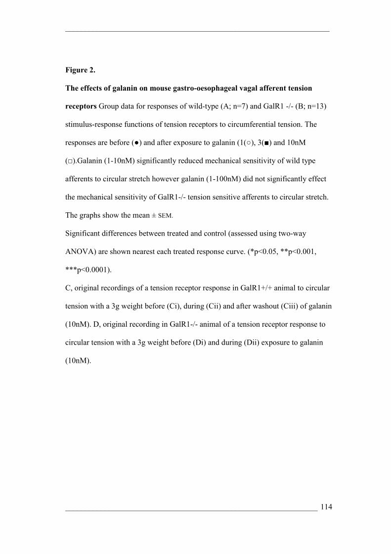
___________________________________________________________________
________________________________________________________________ 114
Figure 2.
The effects of galanin on mouse gastro-oesophageal vagal afferent tension
receptors Group data for responses of wild-type (A; n=7) and GalR1 -/- (B; n=13)
stimulus-response functions of tension receptors to circumferential tension. The
responses are before (●) and after exposure to galanin (1(○), 3(■) and 10nM
(□).Galanin (1-10nM) significantly reduced mechanical sensitivity of wild type
afferents to circular stretch however galanin (1-100nM) did not significantly effect
the mechanical sensitivity of GalR1-/- tension sensitive afferents to circular stretch.
The graphs show the mean ± SEM.
Significant differences between treated and control (assessed using two-way
ANOVA) are shown nearest each treated response curve. (*p<0.05, **p<0.001,
***p<0.0001).
C, original recordings of a tension receptor response in GalR1+/+ animal to circular
tension with a 3g weight before (Ci), during (Cii) and after washout (Ciii) of galanin
(10nM). D, original recording in GalR1-/- animal of a tension receptor response to
circular tension with a 3g weight before (Di) and during (Dii) exposure to galanin
(10nM).

___________________________________________________________________
________________________________________________________________ 115
A B
C D
Impu
lses
/str
oke
21
14
7
Impu
lses
/str
oke
12
8
4
10 50 200 1000von Frey hair (mg)
10 50 200 1000von Frey hair (mg)
******
0
120
Inst
ant.
Fre
q.(H
z)
0
120
Inst
ant.
Fre
q.(H
z)
0.2mV
0.5s
0.1mV
0.5s
0
120
0
120
Inst
ant.
Fre
q.(H
z)
Inst
ant.
Fre
q.(H
z)
i) i)
ii) ii)
00

___________________________________________________________________
________________________________________________________________ 116
Figure 3.
The effect of galanin on mouse gastro-oesophageal vagal mucosal receptor
Group data for stimulus-response functions from GalR1 +/+ (A; n=7) and GalR1 -/-
(B; n=8) mice of mucosal receptors to mucosal stroking with calibrated von Frey
hairs. The responses are before (●) and after exposure to galanin (1(○), 3 (■) and 10
nM (□)). Galanin (1-100nM). Galanin significantly inhibited wild-type mouse
mucosal receptor response to mucosal stroking however galanin had no statistically
significant effect on GalR1-/- mouse mucosal receptors. Significant differences
between treated and control (assessed using two-way ANOVA) are shown nearest
each treated response curve. (*p<0.05, **p<0.001, ***p<0.0001). C, original
recording of a mucosal receptor response from wild-type mouse to mucosal stroking
with a 200mg von Frey hair before (Ci) and during (Cii) exposure to galanin
(10nM). D, original recording from a GalR1-/- mouse of a mucosal receptor
response to mucosal stroking with a 200mg von Frey hair before (Di) and during
(Dii) exposure to galanin (10nM)

___________________________________________________________________
________________________________________________________________ 117
A
B
5
10
15
10 50 200 1000
Impu
lses
/str
oke
von Frey hair (mg)
*****
0
10
8
6
4
2
1 2 3 4 5
Impu
lses
/Sec
.
Load (g)
****
00
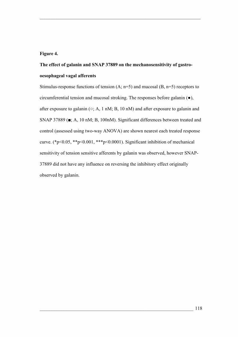
___________________________________________________________________
________________________________________________________________ 118
Figure 4.
The effect of galanin and SNAP 37889 on the mechanosensitivity of gastro-
oesophageal vagal afferents
Stimulus-response functions of tension (A; n=5) and mucosal (B, n=5) receptors to
circumferential tension and mucosal stroking. The responses before galanin (●),
after exposure to galanin (○; A, 1 nM; B, 10 nM) and after exposure to galanin and
SNAP 37889 (■; A, 10 nM; B, 100nM). Significant differences between treated and
control (assessed using two-way ANOVA) are shown nearest each treated response
curve. (*p<0.05, **p<0.001, ***p<0.0001). Significant inhibition of mechanical
sensitivity of tension sensitive afferents by galanin was observed, however SNAP-
37889 did not have any influence on reversing the inhibitory effect originally
observed by galanin.

___________________________________________________________________
________________________________________________________________ 119
A
B
Impu
lses
/Sec
.Im
puls
es/S
ec.
1 2 3 4 5Load (g)
0
8
16
24
0
32
*********
8
16
24
1 2 3 4 5Load (g)
00
32
**

___________________________________________________________________
________________________________________________________________ 120
Figure 5.
The effect of galanin and AR-M961 on the mechanosensitivity of gastro-
oesophageal vagal tension receptors
The effects of the selective GalR1 and GalR2 agonist AR-M961 are shown on the
responses to circular tension in tension sensitive afferents of GalR1 +/+ (A; n=5)
and GalR1-/- (B; n=6) mice. A, the responses of wild-type tension receptors before
(●), after exposure to galanin (○; 100nM) after washout of galanin (■), after
exposure to AR-M961 (▲; 100nM). Significant inhibition of mechanical sensitivity
of tension sensitive afferents by galanin was observed, galanin was then completely
washed out and the effect of AR-M961 was observed. AR-M961 (10-100nM) dose
dependently and significantly inhibited mechanical sensitivity to circular stretch
(average impulses s-1 over stimulus response of 1 min) of tension receptors when
compared to both control and washout.
B, the responses of GalR1-/- tension receptors before (●) and after exposure to AR-
M961 (○; 100nM). Significant differences between treated and control (assessed
using two-way ANOVA) are shown nearest each treated response curve. (*p<0.05,
**p<0.001, ***p<0.0001). AR-M961 (100nM) significantly potentiated the
mechanical sensitivity of GalR1-/- tension sensitive afferents to circular stretch.
Whilst the effect was not dramatic it was very reproducible.
___________________________________________________________________
________________________________________________________________ 121
0
1020304050
0
10
20
30
40
0 1 2 3 4 5
10 50 200 1000
Load (g)
von Frey hair (mg)
Impulses/second
Impulses/stroke
n=5
n=4
A) Tension receptor
B) Mucosal receptor
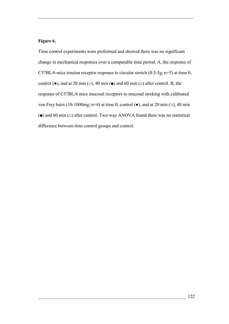
___________________________________________________________________
________________________________________________________________ 122
Figure 6.
Time control experiments were preformed and showed there was no significant
change in mechanical responses over a comparable time period. A, the response of
C57BL/6 mice tension receptor response to circular stretch (0.5-5g; n=5) at time 0;
control (●), and at 20 min (○), 40 min (■) and 60 min (□) after control. B, the
response of C57BL/6 mice mucosal receptors to mucosal stroking with calibrated
von Frey hairs (10-1000mg; n=4) at time 0; control (●), and at 20 min (○), 40 min
(■) and 60 min (□) after control. Two-way ANOVA found there was no statistical
difference between time control groups and control.

___________________________________________________________________
________________________________________________________________ 123
CHAPTER 2: Potentiation of Vagal Afferent
Mechanosensitivity by Ionotropic and Metabotropic
Glutamate Receptors

___________________________________________________________________
________________________________________________________________ 124
SUMMARY
Glutamate exerts its effects at central synapses via ionotropic (iGluR-
NMDA, AMPA & Kainate) and metabotropic (mGluR) receptors. Group I mGluR
are coupled to excitatory, whilst group II and III mGluR are coupled to inhibitory
mechanisms, but more importantly to the current investigation, inhibitory
modulation of gastro-oesophageal vagal afferent sensitivity. Here we identified an
opposing excitatory role for GluR in the modulation of gastro-oesophageal vagal
afferent mechanosensitivity, and described the specific subunit expression of
receptor subtypes in the nodose ganglion. The response of mouse gastro-
oesophageal vagal afferent endings to mechanical stimuli was examined and the
effect of agonists and antagonists to GluR was determined. Two types of vagal
afferents were tested: tension receptors responding to circumferential stretch and
mucosal receptors which responded only to mucosal stroking. The selective iGluR
agonists AMPA and NMDA concentration dependently potentiated afferent
responses. Their corresponding antagonists NBQX and AP-5 respectively, alone
attenuated mechanosensitivity responses as did the non- selective antagonist
Kynurenate. However the Kainate selective agonist had little effect on
mechanosensitivity, and its antagonist UBP 302 was ineffective also. The mGluR5
antagonist MTEP concentration dependently inhibited mechanosensitivity. Efficacy
of agonists and antagonist for the various receptor sub-types differed on mucosal
and tension receptors. We conclude that NMDA, AMPA, and mGluR5 are coupled
to excitatory modulation of afferent mechanosensitivity, and the role of each differs
according to receptor subtypes. This data was reinforced by the finding of complete
___________________________________________________________________
________________________________________________________________ 125
complement of subtypes of all receptors located in the nodose ganglion, therefore
implicating each receptor as a possible candidate for neuromodulation.
*I would like to acknowledge Dr Amanda Page who contributed the MTEP
electrophysiological data used in this chapter.
INTRODUCTION
Glutamate is the major excitatory neurotransmitter mediating synaptic
transmission and modulation at pre- and post-central nervous system (CNS)
synapses. Vagal afferent transmission from peripheral sites to the CNS is largely
glutamatergic, and is tightly regulated pre-synaptically via a number of glutamate
receptors [241-246]. The range of glutamate receptors in the CNS includes
ionotropic (NMDA, AMPA, Kainate) receptors (iGluR), which are ligand gated ion
channels [247], and metabotropic (group I, II, III) receptors (mGluR), which are G-
protein coupled receptors [248]. Metabotropic GluR exist as eight different
subtypes: group I mGluR (mGluR 1 and 5) exerting their effect through activation
of phospholipase C, whilst Group II (mGluR 2 & 3), and group III (mGluR 4,6 ,7,
8) are coupled to inhibition of adenylate cyclase causing altered calcium and
potassium current and slow hyperpolarisation [248]. The three mGluR groups were
initially designated based on similarities in their sequence and pharmacology. In
addition to effects in the CNS, a role for modulation of peripheral vagal gastro-
oesophageal afferents by group II and III mGluR has been demonstrated in ferret
and rodent studies [121]. Inhibition of peripheral gastro-oesophageal vagal afferents
was observed by glutamate and group II and III mGluR agonists and a selective
group III antagonist caused potentiation of mechanosensitivity. This data indicates
both exogenous and endogenous sources of glutamate are able to negatively
modulate vagal afferents.

___________________________________________________________________
________________________________________________________________ 126
Inhibition of gastro-oesophageal vagal afferents has been previously
demonstrated by GABAB [144, 146], importantly corresponding to associated
effects on gastro-oesophageal reflux in vivo [202]. Triggering of transient lower
oesophageal sphincter relaxations (TLOSR) by gastric distension was markedly
attenuated by GABAB receptor agonists, which correlated with a reduction in the
incidence of gastro-oesophageal acid reflux in ferrets, dogs, humans and gastro-
oesophageal reflux patients [181, 184, 198, 202, 249, 250]. This has focused
attention on peripheral vagal afferent endings as important potential targets for the
treatment of reflux disease. Importantly, the inhibitory effect of group III mGluR
agonists on vagal afferents in vitro was conserved across species [121], and is
translated into inhibition of TLOSR’s in vivo [122].
A number of important observations were made in the course of these
studies on mGluR. Firstly the effect of glutamate on vagal afferents was not seen
unless the effects of iGluR was blocked, suggesting an excitatory (iGluR and
mGluR) and inhibitory (mGluR) balance. Secondly, a group I (mGluR 5) antagonist
potently inhibited transient lower oesophageal sphincter relaxations, however a
central or peripheral mechanism for this effect remains unclear. Therefore, whilst
gaining an understanding of inhibitory mechanisms of regulation of vagal afferents
by mGluR, there remained major un-investigated possibilities regarding an
excitatory balance by mGluRs and/or iGluRs.
Several lines of evidence exist that implicate a role for iGluRs in the
mediation of the excitatory glutamatergic modulation of vagal afferent
mechanosensitivity. Morphological evidence demonstrates iGluR subtype
expression in nodose ganglia as well as peripheral sensory afferent neurones
innervating the viscera of several species [120, 137, 138]. In addition, functional in

___________________________________________________________________
________________________________________________________________ 127
vitro studies have demonstrated an NMDA receptor antagonist to cause a reduction
in afferent response to colorectal distension [141], and corresponding effects
observed in vivo in behavioural studies. Importantly, in vitro rat studies have
identified iGluR to play a modulatory role in vagal afferent mechanosensitivity,
whereby non-specific iGluR receptor antagonists dose dependently attenuated
mechanosensory properties of vagal afferents to mechanical stimulation [120],
however precise contributions of select iGluR and the potential for involvement of
excitatory group I mGluR remained undetermined.
Our aim in this study was to investigate systematically the iGluR that may
be involved in the excitatory modulation of vagal afferents, and to determine the
ability of mGluR 5 to have peripheral affects by influencing vagal afferent
mechanosensitivity in vitro. We also sought to identify expression of iGluR and
mGluR5 transcripts in the mouse nodose ganglion which contains the cell bodies of
vagal afferents, and thus gain insight as to the subtypes that may be implicated in
modulating afferent function.
MATERIALS AND METHODS
All studies were performed in accordance with the guidelines of the Animal
Ethics Committees of the Royal Adelaide Hospital and Institute for Medical and
Veterinary Science, Adelaide, Australia. These committees followed the Australian
code of practice for the care and use of animals for scientific purposes, 6th edition
1997. Every attempt was made to minimise the number of animals required and to
minimise their suffering.

___________________________________________________________________
________________________________________________________________ 128
Nodose Ganglia Dissection and RNA extraction for RT-PCR and Quantitative
RT-PCR
Nodose ganglia were removed from five male or female C57 mice (20-30g).
Nodose ganglia were pooled in RNAlater® (QIAGEN, NSW, Australia) at -20°C
for subsequent RNA extraction. RNA isolation involved homogenisation of the
nodose ganglia with a sterile glass pestle, after being snap frozen using liquid
nitrogen. A total of 1mL of TRIzol reagent (Invitrogen, Vic, Australia) was added
before the homogenized material was added to Qiashredders and centrifuged
(12,000 rpm) at room temperature. Qiashredders were removed and tubes
containing homogenate were capped, vortexed, and allowed to stand at room
temperature for 15 minutes in order to promote dissociation of nucleoprotein
complexes. After which 0.2 mL of chloroform was added, tubes were vortexed and
left to stand at room temperature for a further 15 minutes. The samples were then
cold centrifuged (12,00 rpm, 4°C) for 15 minutes and the upper aqueous layer
(containing RNA substrate) was removed and transferred to a sterile eppendorf tube
followed by isopropanol precipitation of RNA, by which 0.5 ml isopropanol was
added cold centrifuged for 10min (38, 47). The upper aqueous phase was discarded
leaving an RNA pellet to which 1 mL of 75% ethanol was added. A 1 min vortex
and 4 min cold centrifuge and then supernatant was removed with the pellet being
re-suspended in 100µL RNAase free sterile water and heated for 2 min. at 60°C.
RNA was separated into 5µL aliquots and stored at -80°C
RNA quantification was determined by measuring the absorbance at 260nm
(A260) using a spectrophotometer (Biorad, Sydney, Australia). RNA quality was
estimated by the A260: A280 nm ratio.
___________________________________________________________________
________________________________________________________________ 129
RT-PCR analysis of iGluR and mGluR subunit expression in mouse nodose
ganglia
Reverse transcription (RT) and polymerase chain reaction (PCR) was
performed using a Qiagen® one-step RT-PCR kit, with primers used as indicated in
Table 1. Previously published primers were used but were tested for specificity
using NCBI’s BLAST software. RT-PCR master-mix was prepared as follows: for
each reaction 25µL RT-PCR Master Mix, 10µL RNAse free water, 10µL of 5x
QIAGEN One-Step RT-PCR Buffer, 2µL dNTP Mix (containing 10 mM of each
dNTP), 2µL QIAGEN One-Step RT-PCR Enzyme Mix and ), 1µL of RNAse
inhibitor (Ambion).
RT–PCR reactions were performed using an alpha unit block for PTC DNA engine
systems (MJ Research) attached to a PTC-200 Peltier thermal cycler (MJ Research).
The following amplification program was applied: Reverse Transcription: 50°C for
30mins, Initial PCR activation: 95°C for 15 mins, PCR cycles: denaturing: 94°C for
1 min, annealing: 47°C for 1 min and extension: 72°C for 1 min repeated for 40
cycles followed by a final extension step of 72°C for 10 min.
For each reaction 25µL of Master Mix was added to 0.2mL clear thin walled
PCR tubes (Axygen) with 10µL (3µM) forward primer (for a final concentration of
0.6 µM), 10µL (3µM) backward primer (for a final concentration of 0.6µM) and
5µL of 50ng template RNA. For each reaction, RNA template was substituted with
5µL distilled RNA-free water as controls. Amplified products (5µL PCR
product/2µL loading buffer) were resolved by 1.5-3% agarose gel electrophoresis
and visualised by ethidium bromide staining.

___________________________________________________________________
________________________________________________________________ 130
In Vitro mouse gastro-oesophageal afferent preparation
Male and female C57/BL6 mice (20-30g, 7-10 weeks) were killed by CO2
inhalation. The stomach, oesophagus and attached vagus nerves were removed and
placed in a modified Krebs’ solution of the following composition (mM): 118.1
NaCl, 4.7 KCl, 25.1 NaHCO3, 1.3 NaH2PO4, 1.2 MgSO4.7H2O, 1.5 CaCl2, 1.0
Citric acid and 11.1 glucose, bubbled with 95% O2-5% CO2.
The preparation temperature was maintained at 4°C during dissection to
preserve tissue integrity and prevent metabolic degradation. The peripheral organs,
major blood vessels and diaphragm were removed by further dissection, leaving the
stomach oesophagus and attached vagus nerves intact. The preparation was opened
longitudinally and pinned out flat mucosa side up in a Perspex organ chamber
(dimension 6 x 2.5 x 1.2 cm) lined with Sylgard® (Dow Corning) and perfused at a
rate of 12mL/min with carbogenated Krebs’ bicarbonate buffer solution maintained
at 34°C. The vagus nerves were pulled into a separate isolate recording chamber
which was filled with paraffin oil. The dimensions of the second chamber are 3.7
cm diameter and 1.2 cm deep. Under a dissecting microscope, the vagus sheath was
removed exposing the underlying nerve fibres, which were teased using fine forceps
into 16-24 bundles and one by one placed on platinum recording electrodes. A
reference electrode rested on the mirror surface in the bath in a small pool of
Krebs’. Nifedipine (1µM) was also added to the Krebs superfusate in order to
prevent smooth muscle contraction. Previous preliminary studies have demonstrated
nifedipine to have no effect on the mechanical sensitivity of gastro-oesophageal
vagal afferents[148].
___________________________________________________________________
________________________________________________________________ 131
Characterisation of gastro-oesophageal vagal afferent properties
Corresponding receptive fields of all types of vagal afferent were located by
mechanical stimulation throughout the preparation with a brush and then more
accurately with a von Frey hair. Accurate quantification of mechanical responses
was performed differently according to the primary adequate stimulus for the type
of fibre as has been described previously [46, 148, 157, 158]. The mechanical
sensitivity of afferents was characterised by mucosal stroking with calibrated von
Frey hairs (10mg-1000mg) and applied circular tension (0.5-7g).
Two types of mechanosensitive afferent were studied, those responding to
mucosal stroking but not to circular tension (mucosal receptors) and those
responding to mucosal stroking and circular tension (tension receptors) as reported
previously [46].
Mechanical thresholds of all types of fibre were determined using calibrated
von Frey hairs. The most reproducible, stimulus dependent response of these
afferents to mucosal stimuli was evoked when the probe was moved at a rate of
5mms-1 across the receptive field rather than being stationary. A single test at each
intensity of calibrated von Frey hair is prone to error due to such small sizes of
receptive field, we therefore minimised experimental error by measuring mean
response to 10 standard strokes given at 1 second intervals, omitting the first and
last from analysis because of increased associated error. A bent von Frey hair (10-
1000mg) stroking over the preparation meant that an even force was distributed
over the receptive field during stroking stimulus. This protocol was found to give
reproducible data and was therefore used to assess the effects of agonists and
antagonists of iGluR and mGluR on vagal afferents.

___________________________________________________________________
________________________________________________________________ 132
Tension response curves were also obtained for afferent fibres responsive to
circular tension. Tension receptors also responded to von Frey hair stroking,
however were distinguished from mucosal receptors by their clear responsiveness to
circular tension which was slowly adapting. Tension stimuli was used in
combination with von Frey hair stroking to determine whether the receptive fields
of fibres were located in the mucosa, the muscle layer or both.
Tension stimuli were applied via a thread attached to an unpinned point
adjacent to the receptive field. The thread was attached to a cantilever and pulley
system close to the preparation, with reference standard weights applied to the
opposite side of the cantilever. Each weight was applied in a step wise manner and
maintained for duration of 1 minute, with a 1 minute rest between weight
applications. The response was measured as the mean discharge evoked, and
subsequent responses during drug exposure are expressed as the percentage of the
maximum control response (without drug addition), so that increases and decreases
in responsiveness may be directly compared. The tension response curves were
produced by applying weights to the cantilever system in a range of (1-5g) as
previously described was sufficient to evoke a maximum response without tissue
damage.
Effect of GluR agonists and antagonists on mechanosensitivity of vagal
afferents
After the mechanical sensitivity of gastro-oesophageal vagal afferents had
been established under control conditions, the effects of various agonists and
antagonists on mechanical sensitivity were determined. Kynurenate (0.1–1µM) was
added to the superfusing Krebs’ solution and was allowed to equilibrate for 20
___________________________________________________________________
________________________________________________________________ 133
minutes in order to ensure complete penetration of the drug into all layers of the
tissue. Tension response and stroke stimulus response curves were then re-
determined. The agonists NMDA (0.1– 1µM), AMPA (10–100µM), and SYM
2081 (1–10µM), were added to the superfusing solution. The antagonists AP-5 (1–
10µM), NBQX (1–10µM), and UBP 302 (0.1–10µM) were added to a ring placed
directly over the receptive field in the preparation. Concentrations used were based
on previous published potencies [247, 251-254].
Prior to this work, 6 pilot studies were performed in which the non-selective
iGluR antagonist Kynurenate was tested at concentrations varying between 10-7 –
10-4M. These experiments demonstrated concentration dependent effects and
ultimately complete abolition of vagal afferent discharge; ranges around the
subsequent EC50 were chosen for this study.
Drug addition to the ring was followed by a twenty minute equilibration
period before mechanical stimulus curves were once again established immediately
after removal of the ring. An equilibration period was essential for complete
penetration of the drug into all layers of tissue. Preliminary data has indicated that
drug effects are observed for approximately 10 min after removal of drug. Time
control experiments (n=9) were performed prior to the study and showed there to be
no significant change in mechanical sensitivity responses over a comparable time
period.
Five of nine experiments involving NMDA were performed in the absence
of Nifedipine and all other experiments were performed in the presence of
nifedipine (1µM), in order to limit the effects of drugs secondary to smooth muscle
responses.

___________________________________________________________________
________________________________________________________________ 134
Three additional experiments were performed with suspended, circular orientated
rings, to investigate length tension relationships. No change in these was seen with
any of the agonists used at any concentration. Also no effect was seen on smooth
muscle responses to electrical field stimulation in separate experiments
(O’Callaghan, unpublished observations). Statistical analysis of mechanical
stimulus response curves was by two-way ANOVA. Results were considered to be
significant where p < 0.05.
Data Recording and Analysis
Afferent impulses were amplified with a biological amplifier (world
Precision Instruments, Sarasota, FL, DAM 50) and scaling amplifier (JRAK,
Melbourne, Australia), filtered (CWE Inc., Ardmore, PA band-pass filter-932) and
monitored using an oscilloscope (DL 1200A; Yakogawa, Tokyo, Japan). Single unit
action potentials were discriminated on the basis of size and shape using spike II
software (Cambridge Electronic Design, Cambridge, UK).
Statistical Analysis: All data were recorded and analysed off-line using a
personal computer. All data were expressed as mean ± SE with n = number of
individual afferents in all instances. Statistical difference between stimulus response
curves were evaluated using a two-way ANOVA performed using graphpad Prism
(Version 3.00 for Windows, Graphpad Software, San Diego, California, USA).
Differences were considered significant where p < 0.05.
Drugs
Stock solutions of all drugs were kept frozen (-80°C) and diluted to their
final concentration in Krebs’ solution on the day of the experiment.

___________________________________________________________________
________________________________________________________________ 135
NMDA, AP-5, NBQX, UBP-302 and SYM-2081, were all obtained from
Astrazeneca (Astrazeneca, Molndal, Sweden). Kynurenate and AMPA were
obtained from Sigma (Sigma, Castle Hill, NSW, Australia).
RESULTS
Expression of glutamate receptor subunits in vagal (nodose) cell bodies
We were able to identify the expression of a number of transcripts
corresponding to iGluR subunits and mGluR in mouse nodose ganglia. RT-PCR and
gel electrophoresis confirmed the presence of amplified products of predicted sizes
generated by primers specific for all the NMDA receptor subunits (NMDAR1,
NR2A, NR2B, NR2C, NR2D and NR3A; Figure 1A). RT-PCR also revealed the
expression of all AMPA receptor subunits (GluR1, GluR2, GluR3, GluR4; Figure
1B), and all Kainate receptor subunits (KA1, GluR5, GluR6, GluR7; Figure 1C).
Expression of mGluR5 was also evident (Figure 1D) in mouse nodose ganglion.
Isolation of single bands of correct predicted transcript size was achieved by UV
analysis of the gel. Where template RNA was substituted for sterile water, no
evidence of amplified products was observed. Expression of the reference β-actin
gene was observed in all cases using template RNA.
Electrophysiological Studies
A role of peripheral iGluR and mGluR in modulation of mechanosensitivity
was determined by examining responses of single afferent fibres using an in vitro
gastro-oesophageal vagal afferent preparation. The effects of specific agonists and
antagonists directed at iGluR and mGluR are illustrated in Figures 2 – 6. Single
afferent fibres were classified as either mucosal or tension depending on their

___________________________________________________________________
________________________________________________________________ 136
response to mechanical stimulation. All afferents had their corresponding receptive
fields located either in the distal oesophagus or on the oesophageal sphincter. As
previously described mucosal receptors are distinct from tension receptors in their
response to circular tension where mucosal receptors are unresponsive, however
both types of afferents respond to mechanical stimulus by mucosal stroking with
calibrated von Frey hairs (10 – 1000mg) in a graded manner [46]. Tension receptors
elicited a graded increase in firing of action potentials in response to applied load
(1-5g).
Effects of non-selective iGluR blockade on vagal afferents
The effect of Kynurenate on both mouse mucosal and tension receptors is
illustrated in Figure 2. Kynurenate (0.1–1µM) was shown to significantly attenuate
both mucosal and tension receptors sensitivity to mechanical stimulus in a
concentration dependent manner with maximal effect at (1µM). These data
prompted further investigation of the role of endogenous glutamate at subtypes of
ionotropic glutamate receptors.
Effects of NMDA receptor ligands on vagal afferents
The effect of NMDA receptor agonist, NMDA, on mouse mucosal and
tension receptor responses to mechanical stimulation is illustrated in Figure 3 A &
B. Whilst a trend of potentiation of mechanical sensitivity was observed, NMDA
(0.1-1µM) did not significantly alter the mechanical sensitivity of tension receptors
to applied circular tension. Individual analysis of experiments revealed a clear
increase in sensitivity of tension receptors where responses returned to original
values after 10-20 minute wash-out of the drug from the preparation, possibly

___________________________________________________________________
________________________________________________________________ 137
suggesting sub-groups of responders and non-responders. These may be comparable
to two populations of tension-sensitive fibres found in ferrets which showed
differing pharmacology [121]. Effects of NMDA on responses to tension were
similar to their responses to local stimulation by von Frey hair stroking in two
additional experiments (Data not shown).
NMDA demonstrated significant potentiation of mechanical sensitivity of
mucosal receptors response to mucosal stroking with calibrated von Frey hairs
(Figure 3B; p < 0.001). These effects of NMDA were also reversible upon washout
with normal Krebs solution. A subset of experiments observing the effects of
NMDA on mechanical sensitivity of vagal afferents was performed in the absence
of nifedipine however all other experiments were performed in the presence of
nifedipine (1µM) in order to block smooth muscle contractions and therefore
remove any effects of drugs on smooth muscle. The effects of NMDA were
unaffected by this treatment indicating a direct effect on vagal nerve endings.
The effects of the competitive NMDA receptor antagonist AP-5 is illustrated
in Figure 3C & D. AP-5 (1-10µM) significantly and dose dependently reduced the
sensitivity of mucosal receptors to mucosal stroking with calibrated von Frey hairs
(p < 0.001) and of tension receptors to circular tension (p < 0.001 ).
Effects of AMPA/Kainate receptor ligands on vagal afferents
The effect of AMPA, the selective AMPA receptor agonist, on mouse
mucosal and tension receptors is illustrated in Figure 4 A and B. AMPA (10-
100µM) demonstrated significant and concentration dependent potentiation of
mechanical sensitivity of tension receptors response to circular tension (p < 0.001),
whilst also significantly increasing the effect of mucosal receptors response to

___________________________________________________________________
________________________________________________________________ 138
mucosal stroking by calibrated von Frey hairs (p < 0.001), in a similar dose
dependent manner. These effects of AMPA were shown to be washed out over an
equilibration period with discharge returning to near baseline levels.
Selective Kainate receptor agonist SYM-2081 at concentrations that had
been previously effective in other systems (1-10µM) [252], was shown to have no
significant effect on mechanical sensitivity of tension receptors response to circular
tension nor mucosal receptors response to mucosal stroking, however there was
some indication of slight potentiation of mucosal receptors to mucosal stroking. The
kainite receptor antagonist UBP 302 (0.1-10µM) also failed to have any significant
effect on mechanical sensitivity of either mucosal nor tension receptors sensitivity
to mechanical stimulation (Figure 5C and D). The AMPA/Kainate receptor
antagonist NBQX (1-10nM; Figure 4C and D) was shown to significantly attenuate
sensitivity of tension response to circular tension (p < 0.0001), but failed to have a
significant effect on mucosal receptors sensitivity to mucosal stroking.
Effects of mGluR5 antagonist on vagal afferents
The effects of the selective mGluR5 antagonist, MTEP on mouse mucosal
and tension receptors is illustrated in Figure 5 A and B. MTEP (1-30µM)
significantly inhibited sensitivity of mucosal receptors to mucosal stroking (10 -
1000mg; p<0.01 at 30µM). MTEP (1-30µM) also significantly inhibited mechanical
sensitivity of tension receptors to circular tension in a concentration-dependent
manner (p<0.0001 for all concentrations). Potency of MTEP on mucosal receptors
appeared less than on tension receptors as significance was only seen at (30µM,
p<0.01), the highest concentration applied.
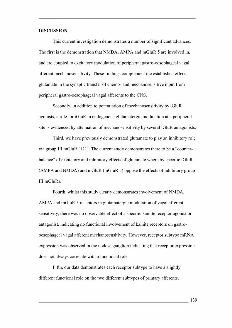
___________________________________________________________________
________________________________________________________________ 139
DISCUSSION
This current investigation demonstrates a number of significant advances.
The first is the demonstration that NMDA, AMPA and mGluR 5 are involved in,
and are coupled to excitatory modulation of peripheral gastro-oesophageal vagal
afferent mechanosensitivity. These findings complement the established effects
glutamate in the synaptic transfer of chemo- and mechanosensitive input from
peripheral gastro-oesophageal vagal afferents to the CNS.
Secondly, in addition to potentiation of mechanosensitivity by iGluR
agonists, a role for iGluR in endogenous glutamatergic modulation at a peripheral
site is evidenced by attenuation of mechanosensitivity by several iGluR antagonists.
Third, we have previously demonstrated glutamate to play an inhibitory role
via group III mGluR [121]. The current study demonstrates there to be a “counter-
balance” of excitatory and inhibitory effects of glutamate where by specific iGluR
(AMPA and NMDA) and mGluR (mGluR 5) oppose the effects of inhibitory group
III mGluRs.
Fourth, whilst this study clearly demonstrates involvement of NMDA,
AMPA and mGluR 5 receptors in glutamatergic modulation of vagal afferent
sensitivity, there was no observable effect of a specific kainite receptor agonist or
antagonist, indicating no functional involvement of kainite receptors on gastro-
oesophageal vagal afferent mechanosensitivity. However, receptor subtype mRNA
expression was observed in the nodose ganglion indicating that receptor expression
does not always correlate with a functional role.
Fifth, our data demonstrates each receptor subtype to have a slightly
different functional role on the two different subtypes of primary afferents.
___________________________________________________________________
________________________________________________________________ 140
Sixth, these data indicate that at least part of the effect of MTEP to cause
inhibition of transient lower oesophageal sphincter relaxations in response to gastric
distension may be mediated peripherally. This may correspond to a peripheral target
for the therapeutic treatment of gastro-oesophageal reflux disease.
Finally these results suggest a similar role for glutamate in vagal afferent
transmission similar to that in spinal transmission where it is evident that they may
underlie behavioural change in response to colonic distension. Each of the above
concepts is addressed below.
Peripheral role for excitatory glutamate receptors
Excitatory glutamate receptors are expressed in the caudal brainstem, and
have been shown to play a specific role in transmission from peripheral gastro-
oesophageal afferents to the NTS, particularly implicated in signalling of gastric
distension, swallowing, transient lower oesophageal sphincter relaxations, emetic
signalling and satiety [269-272]. Whilst establishing an important role for these
receptors in peripheral vagal afferent modulation, in addition to their involvement in
central transmission, these findings further add to and complement previous
findings in rat and ferret studies, that demonstrate an excitatory role for glutamate
receptors on vagal afferents [137, 273]. In contrast, in vitro investigations in the
guinea pig have been unable to identify functional glutamate and GABA receptors
on afferent endings. [30, 255].
Previous studies have demonstrated expression of iGluR and mGluR
subtypes in vagal afferent cell bodies of several species, with these studies in
addition demonstrating bi-directional axonal transport of iGluR and mGluR on
peripheral and central branches of the vagus nerve [121, 256-260], implicating a

___________________________________________________________________
________________________________________________________________ 141
peripheral role for these receptors. Glutamate receptors are also expressed and
function in peripheral terminals of sensory spinal afferents innervating the skin
[276-279], and may be involved in hyper-algesia associated with inflammation
[280, 281]. NMDA receptors have been implicated in signalling painful distension
of the colon, as peripheral administration of an ionotropic receptor antagonist,
memantine, caused a reduction in afferent and behavioural responses to colorectal
distension [139]. However memantine is a non-selective ligand and may have
effects other than at NMDA receptors.
Positive and negative modulation of mechanosensitivity by glutamate
Our previous in vitro studies in ferrets identified a possible counter-balance
of excitatory and inhibitory glutamatergic signalling from peripheral vagal afferents
whereby glutamate acts simultaneously at iGluR and mGluR [121]. The potent
inhibitory effect of glutamate, an effect mimicked by selective group II and III
agonists, on vagal afferent mechanosensitivity, was only revealed after the addition
of the non-specific iGluR antagonist kynurenate to the preparation. In vivo
observations in the rat indicated similarly, that iGluR may be involved in excitatory
modulation of vagal afferents [120], however which subtypes of iGluR were
involved, and in addition, the potential for involvement of excitatory group I
mGluR, remained undetermined. The current data clearly indicate a counter balance
of excitatory modulation of vagal afferent mechanosensitivity by iGluR and mGluR
5, and inhibitory modulation by group II and III mGluR. More specifically, agonist
and antagonist data of both AMPA and NMDA receptors indicate these two
receptors to play a major role in excitatory balance of modulation of
___________________________________________________________________
________________________________________________________________ 142
mechanosensitivity with kainite receptors having little if any effect under the current
conditions.
Endogenous glutamate plays a role in modulation of vagal afferents
The inhibitory effects of exogenous iGluR and mGluR antagonists,
particularly to NMDA, AMPA and mGluR5 receptors, indicate endogenous
glutamate to exert an ongoing tonic stimulation of mechanosensitivity. The question
arises as to the likely source of endogenous glutamate. Glutamate is detected in
approximately 60% of nodose ganglion cells and on vagal afferent fibres originating
from the nodose ganglion [261]. The vesicular glutamate transporter VGLUT2 is
widespread in the enteric nervous system as well as being contained within
peripheral endings of vagal afferents and is necessary for glutamate release [262,
263]. We therefore speculate vagal afferents themselves are a potential source,
likely to release glutamate both centrally and peripherally. There are also separate
sources of endogenous glutamate other than vagal afferents where glutamate is in
close proximity with vagal afferents. Studies have shown that whilst almost 40% of
ingested glutamate is pooled in skeletal muscle, a significant rise in circulating
plasma and intraluminal glutamate may both represent candidate sources of
glutamate causing the observed effects on vagal afferents [264]. An excess of
endogenous glutamate may account for the lack of effect on mechanosensitivity of
NMDA receptors of exogenous glutamate, in contrast to the potent inhibition of
mechanosensitivity by a NMDA receptor antagonist. Excess endogenous glutamate
may also provide an explanation for why a kainite receptor agonist had little effect,
and why a group I mGluR agonist had no effect in our previous study [121]. On the
___________________________________________________________________
________________________________________________________________ 143
other hand, endogenous activation of AMPA receptors may be overcome by
exogenous AMPA.
Glutamate receptor subtypes have varying effects on subtypes of primary
afferents
Selective agonists and antagonists of both iGluR and mGluR5 receptors
were effective on responses of both tension sensitive and mucosal receptors to
mechanical stimulation. In general, ligands had highly potent effects on vagal
afferents, however, discrepancies as to the effectiveness of ligands on specific
afferent types differed. NMDA exerted a more potent modulation of mucosal
receptors, whilst MTEPs effect on tension receptors was more powerful, suggesting
NMDA receptors may be more highly expressed by mucosal receptor cell bodies,
and mGluR5 receptors may be the predominant receptor expressed on tension
receptors. However localization of receptors on individual afferent populations
ending in different layers of the gut is yet to be investigated.
All Subtypes of GluR are expressed in the nodose ganglion
Expression of mRNA encoding all mGluR and some subtypes of iGluR in
nodose ganglia has been reported previously in several species [121, 257, 259, 260].
This study confirms the expression of all iGluR and mGluR5 receptor subunits in
mouse nodose ganglion, which from previous observations in the ferret we would
expect to be transported to peripheral endings.
All NMDA receptors function as tetrameric structures composed of multiple
subunits including an obligate NR1 subunit required for glycine occupation and
trafficking of the heteromeric assembly to the plasma membrane [117].

___________________________________________________________________
________________________________________________________________ 144
Functionality of NMDA receptors depends on their permeability to Ca++, affinity for
glutamate as well as Mg+ block, all which are dictated by composition of receptor
subunits and may be important in altering conductance of sensory neurones, and
thus influence nociception and mechanosensitivity. NMDA receptors must contain
at least one NR2, and may contain both NR2 and NR3 subunits to be functional
[265, 266]. Sub-unit composition of NMDA receptors in sensory neurones has been
previously described, those expressing NR1 subunits conjugated with NR2B
subunits which are present in both A and C fibres, and those with NR1 conjugated
with NR2D subunits, present only on A fibres. This study confirmed the expression
of all subunits in vagal afferent cell bodies in the nodose ganglion, however the
assembly of sub-units in individual vagal afferent neurones is beyond the scope of
the current investigation.
AMPA receptors also likely function as tetramers and are composed of a
four subunit family (GluR1-4). The GluR2 subunit plays a critical role in
determination of the permeability of heteromeric receptors to calcium ions and thus,
AMPA receptors lacking GluR2 are impermeable due to voltage dependent block of
the ion channel [103]. Whilst all AMPA receptors subunits are not required for full
function, previous studies of AMPA receptors in certain brain regions have found
that AMPA receptors contain the full complement of receptor subunits [247].
All AMPA receptor subunits were found to be expressed in the nodose
ganglion including GluR2, providing a consistent molecular correlate with
functional evidence of the AMPA receptor agonist and antagonist data gained in this
study. All subtypes of kainate receptors were found in the nodose ganglion,
however we identified little evidence to suggest a functional role of this receptor in
the modulation of mechanosensitivity of gastro-oesophageal vagal afferent endings.
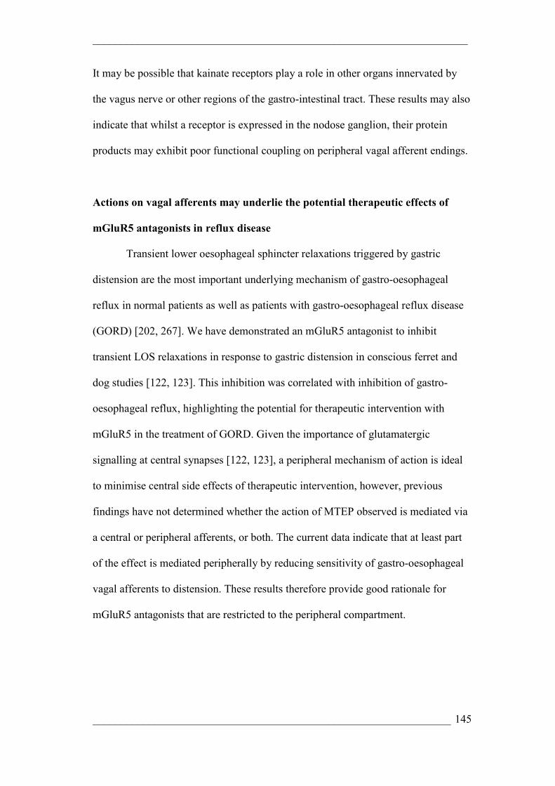
___________________________________________________________________
________________________________________________________________ 145
It may be possible that kainate receptors play a role in other organs innervated by
the vagus nerve or other regions of the gastro-intestinal tract. These results may also
indicate that whilst a receptor is expressed in the nodose ganglion, their protein
products may exhibit poor functional coupling on peripheral vagal afferent endings.
Actions on vagal afferents may underlie the potential therapeutic effects of
mGluR5 antagonists in reflux disease
Transient lower oesophageal sphincter relaxations triggered by gastric
distension are the most important underlying mechanism of gastro-oesophageal
reflux in normal patients as well as patients with gastro-oesophageal reflux disease
(GORD) [202, 267]. We have demonstrated an mGluR5 antagonist to inhibit
transient LOS relaxations in response to gastric distension in conscious ferret and
dog studies [122, 123]. This inhibition was correlated with inhibition of gastro-
oesophageal reflux, highlighting the potential for therapeutic intervention with
mGluR5 in the treatment of GORD. Given the importance of glutamatergic
signalling at central synapses [122, 123], a peripheral mechanism of action is ideal
to minimise central side effects of therapeutic intervention, however, previous
findings have not determined whether the action of MTEP observed is mediated via
a central or peripheral afferents, or both. The current data indicate that at least part
of the effect is mediated peripherally by reducing sensitivity of gastro-oesophageal
vagal afferents to distension. These results therefore provide good rationale for
mGluR5 antagonists that are restricted to the peripheral compartment.

___________________________________________________________________
________________________________________________________________ 146
Roles for glutamate receptors in the vagal and spinal sensory system
Functional GluR are present throughout the CNS where they modulate
neuronal excitability and synaptic transmission. Anatomical studies in skin have
demonstrated the expression of GluR on afferent endings, including mGluR5 and
NMDA receptors [265, 268-270]. Functional studies have demonstrated NMDA and
mGluR5 to be involved in nociceptive primary afferent signalling. Afferent and
behavioural studies demonstrate an effect of these receptors on sensory afferents
whereby they were responsible for increased signalling in response to an algesic
stimulus. This effect is reflected in our studies of primary vagal afferent which
demonstrate similarly, excitatory coupling of primary afferent signalling in sensory
neurones. A similar finding was made in the viscera, whereby the non-selective
ionotropic antagonist memantine reduced afferent and behavioural responses to
colorectal distension [271]. The functional role of iGluR receptor subtypes in spinal
afferents is unclear, and also if there are opposing influences of positively- and
negatively- coupled receptors. Therefore similarities between vagal and spinal
afferents in the actions of glutamate are possible. An interesting difference between
vagal and spinal afferents however, is the presence of VGLUT2 in vagal cell bodies
and endings [262, 272], which was not observed in their dorsal root ganglion
counterparts [273], which may in turn, be suggestive of a greater role for activation
of glutamate auto-receptors on vagal afferents than on spinal afferents.
CONCLUSION
In conclusion, this study in conjunction with previous findings by our group,
further clarifies the influence of all major GluR subtypes on vagal afferent
mechanosensitivity. There is clearly scope for both excitatory and inhibitory

___________________________________________________________________
________________________________________________________________ 147
modulation of afferent sensitivity by glutamate from both exogenous and
endogenous sources, providing a balance to achieve normal vagal afferent function.
This balance is clearly possible to manipulate pharmacologically. How it may be
altered in disease states we hope will be the subject of our continuing investigations.

___________________________________________________________________
________________________________________________________________ 148
Receptor Sequence Predicted Product size (bp)
GluR1 Fwd: GGACCACAGAGGAAGGCATGATC 358 Rev: CAGTCCCAGCCCTCCAATC GluR2 Fwd: TGTGTTTGTGAGGACTACGGCA 226 Rev: GGATTCTTTGCCACCTTCATTC GluR3 Fwd: GCAGAGCCATCTGTGTTTGTGAGTT 472 Rev: AGTTTTGGGTGTTCTTTGTGAGTT GluR4 Fwd: GCAGAGCCGTCTGTGTTCACTAG 220 Rev: CGGCAAGGTTTACAGGAGTTCTT GluR5 Fwd: GCCCCTCTCACCATCACGTAT 358 Rev: TGGTCGATAGAGCCTTGGGCA GluR6 Fwd: TTCCTGAATCCTCTCTCCCCT 259
Rev: CACCAAATGCCTCCCACTATC GluR7 Fwd: GCAGAGTCAGGCCTGCTGGA 300 Rev: ACTCCACACCCCGACCTTCT KA1 Fwd: CCCATCGAGTCTGTGGATGA 434 Rev: CTGTGGTCCTCCTCCTTGGG NMDAR1 Fwd: GCTGTACCTGCTGGACCGCT 210 Rev: GCAGTGTAGGAAGCCACTATGATC NR2A Fwd: GCTACGGGCAGACAGAGAAG 257 Rev: GTGGTTGTCATCTGGCTCAC NR2B Fwd: GCTACAACACCCACGAGAAGAG 314 Rev: GAGAGGGTCCACGCTTTCC NR2C Fwd: AACCACACCTTCAGCAGCG 464 Rev: GACTTCTTGCCCTTGGTGAG NR2D Fwd: CGATGGCGTCTGGAATGG 265 Rev: AGATGAAAACTGTGACGGCG NR3A Fwd: CCGCGGGATGCCCTACTGTTC 417 Rev: CCAGTTGTTCATGGTCAGGAT mGluR5 Fwd: GTCCTTCTGTTGATCCTGTC 118 Rev: ATGCAGCATGGCCTCCACTC β-ACTIN Fwd: ATCATGTTTGAGACCTTCAACAC 830 Rev: TCTGCGCAAGTTAGGTTTTGTC

___________________________________________________________________
________________________________________________________________ 149
Table 1
Nucleotide primer sequences for RT-PCR identification of ionotropic receptor
subunit and mGluR5 transcripts.

___________________________________________________________________
________________________________________________________________ 150
NM
DA
R1
(210
bp)
NR
2B (
314
bp)
NR
2A (
257
bp)
NR
2D (
265
bp)
bp
400300
200
400500
300
bp
NR
2C (
464
bp)
NR
3A (
417
bp)
A
bp
400
300
200
B
Glu
R1
(358
bp)
Glu
R2
(226
bp)
Glu
R3
(472
bp)
Glu
R4
(220
bp)
Cbp
400
300
200
KA
R1
(434
bp)
Glu
R5
(358
bp)
Glu
R6
(259
bp)
Glu
R7
(300
bp)
120
100
80
60
bp
mGluR5 (118 bp)
D
Figure 1
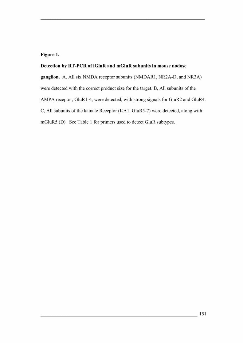
___________________________________________________________________
________________________________________________________________ 151
Figure 1.
Detection by RT-PCR of iGluR and mGluR subunits in mouse nodose
ganglion. A. All six NMDA receptor subunits (NMDAR1, NR2A-D, and NR3A)
were detected with the correct product size for the target. B, All subunits of the
AMPA receptor, GluR1-4, were detected, with strong signals for GluR2 and GluR4.
C, All subunits of the kainate Receptor (KA1, GluR5-7) were detected, along with
mGluR5 (D). See Table 1 for primers used to detect GluR subtypes.
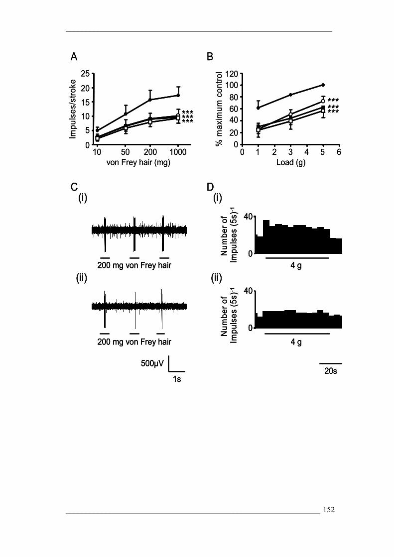
___________________________________________________________________
________________________________________________________________ 152
20s
40
120100
8060
200
0 1 2 3 4 5 610 50 200 10000
5
10
15
20
25Im
puls
es/s
trok
e
% m
axim
um c
ontr
ol
Load (g)von Frey hair (mg)
A B
*********
*********
C(i)
200 mg von Frey hair
1s
500µV
(ii)
200 mg von Frey hair
0
40N
umbe
r of
Impu
lses
(5s
)-1
D(i)
4 g
0
40
Num
ber
of
Impu
lses
(5s
)-1
(ii)
4 g
20s20s
40
120100
8060
200
0 1 2 3 4 5 610 50 200 10000
5
10
15
20
25Im
puls
es/s
trok
e
% m
axim
um c
ontr
ol
Load (g)von Frey hair (mg)
A B
*********
*********
C(i)
200 mg von Frey hair
C(i)
200 mg von Frey hair
1s
500µV
1s
500µV
(ii)
200 mg von Frey hair
(ii)
200 mg von Frey hair
0
40N
umbe
r of
Impu
lses
(5s
)-1
D(i)
4 g0
40
0
40N
umbe
r of
Impu
lses
(5s
)-1
D(i)
4 g
0
40
Num
ber
of
Impu
lses
(5s
)-1
(ii)
4 g
0
40
00
40
Num
ber
of
Impu
lses
(5s
)-1
(ii)
4 g

___________________________________________________________________
________________________________________________________________ 153
Figure 2.
The effect of Kynurenate on mouse gastro-oesophageal vagal afferents.
Stimulus-response functions of mucosal (A, n=5) and tension receptors (B, n=6) to
mucosal stroking and circumferential tension respectively. The responses are before
(●) and after exposure to Kynurenate (0.1µM (○), 0.3µM (■) & 1µM (□)). Asterisks
indicate significant difference from control using a two-way ANOVA
(***p<0.001). C, original recording of a mucosal receptor response to mucosal
stroking with a 200mg von Frey hair before (C i) and after exposure to Kynurenate
(1 µM, C ii). D, original recording of a tension receptor response to circumferential
tension using a 4g weight before (D (i)) and after (D (ii)) exposure to Kynurenate (1
µM).

___________________________________________________________________
________________________________________________________________ 154
0 1 2 3 4 5 610 50 200 10000
Impu
lses
/str
oke
% m
axim
um c
ontr
ol
Load (g)von Frey hair (mg)
A B
C D
8
16
24
32
0
50
100
150
0
5
10
15
20
25
0
40
80
120
Impu
lses
/str
oke
% m
axim
um c
ontr
ol
1 2 3 4 5 610 50 200 1000Load (g)von Frey hair (mg)
0
******
***
***********
0 1 2 3 4 5 610 50 200 10000
Impu
lses
/str
oke
% m
axim
um c
ontr
ol
Load (g)von Frey hair (mg)
A B
C D
8
16
24
32
0
50
100
150
0
5
10
15
20
25
0
40
80
120
Impu
lses
/str
oke
% m
axim
um c
ontr
ol
1 2 3 4 5 610 50 200 1000Load (g)von Frey hair (mg)
0
******
***
***********
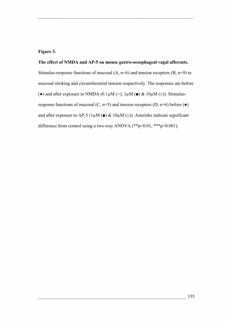
___________________________________________________________________
________________________________________________________________ 155
Figure 3.
The effect of NMDA and AP-5 on mouse gastro-oesophageal vagal afferents.
Stimulus-response functions of mucosal (A, n=6) and tension receptors (B, n=9) to
mucosal stroking and circumferential tension respectively. The responses are before
(●) and after exposure to NMDA (0.1µM (○), 1µM (■) & 10µM (□)). Stimulus-
response functions of mucosal (C, n=5) and tension receptors (D, n=6) before (●)
and after exposure to AP-5 (1µM (■) & 10µM (□)). Asterisks indicate significant
difference from control using a two-way ANOVA (**p<0.01, ***p<0.001).

___________________________________________________________________
________________________________________________________________ 156
A B
DC
10 50 200 1000von Frey hair (mg)
0
Impu
lses
/str
oke
10
20
30
40
*********
1 2 3 4 5 6
% m
axim
um c
ontr
ol
Load (g)
50
75
100
125
150
0
******
10 50 200 1000von Frey hair (mg)
0
2
4
6
8
10
Impu
lses
/str
oke
1 2 3 4 5 6
% m
axim
um c
ontr
ol
Load (g)
120
80
40
00
******
***
A B
DC
10 50 200 1000von Frey hair (mg)
0
Impu
lses
/str
oke
10
20
30
40
*********
1 2 3 4 5 6
% m
axim
um c
ontr
ol
Load (g)
50
75
100
125
150
0
******
10 50 200 1000von Frey hair (mg)
0
2
4
6
8
10
Impu
lses
/str
oke
10 50 200 1000von Frey hair (mg)
0
2
4
6
8
10
Impu
lses
/str
oke
1 2 3 4 5 6
% m
axim
um c
ontr
ol
Load (g)
120
80
40
00
******
***
1 2 3 4 5 6
% m
axim
um c
ontr
ol
Load (g)
120
80
40
00
******
***

___________________________________________________________________
________________________________________________________________ 157
Figure 4.
The effect of AMPA and NBQX on mouse gastro-oesophageal vagal afferents.
Stimulus-response functions of mucosal (A, n=5) and tension receptors (A, n=7) to
mucosal stroking and circumferential tension respectively. The responses are before
(●) and after exposure to AMPA (10µM (○), 30µM (■) & 100µM (□)). Stimulus-
response functions of mucosal (C, n=4) and tension receptors (D, n=5) before (●)
and after exposure to NBQX (1nM (○), 3nM (■) & 10nM (□)). Asterisks indicate
significant difference from control using a two-way ANOVA (***p<0.001).

___________________________________________________________________
________________________________________________________________ 158
Impu
lses
/str
oke
A B
C
10 50 200 1000von Frey hair (mg)
0
Impu
lses
/str
oke
5
10
15
**
1 2 3 4 5 6
% m
axim
um c
ontr
ol
Load (g)
0
25
50
75
100
0
10 50 200 1000von Frey hair (mg)
0
3
6
9
D
1 2 3 4 5 6Load (g)
50
75
100
125
0
% m
axim
um c
ontr
ol150
Impu
lses
/str
oke
A B
C
10 50 200 1000von Frey hair (mg)
0
Impu
lses
/str
oke
5
10
15
**
10 50 200 1000von Frey hair (mg)
0
Impu
lses
/str
oke
5
10
15
**
1 2 3 4 5 6
% m
axim
um c
ontr
ol
Load (g)
0
25
50
75
100
0 1 2 3 4 5 6
% m
axim
um c
ontr
ol
Load (g)
0
25
50
75
100
0
10 50 200 1000von Frey hair (mg)
0
3
6
9
10 50 200 1000von Frey hair (mg)
0
3
6
9
D
1 2 3 4 5 6Load (g)
50
75
100
125
0
% m
axim
um c
ontr
ol150
D
1 2 3 4 5 6Load (g)
50
75
100
125
0
% m
axim
um c
ontr
ol150
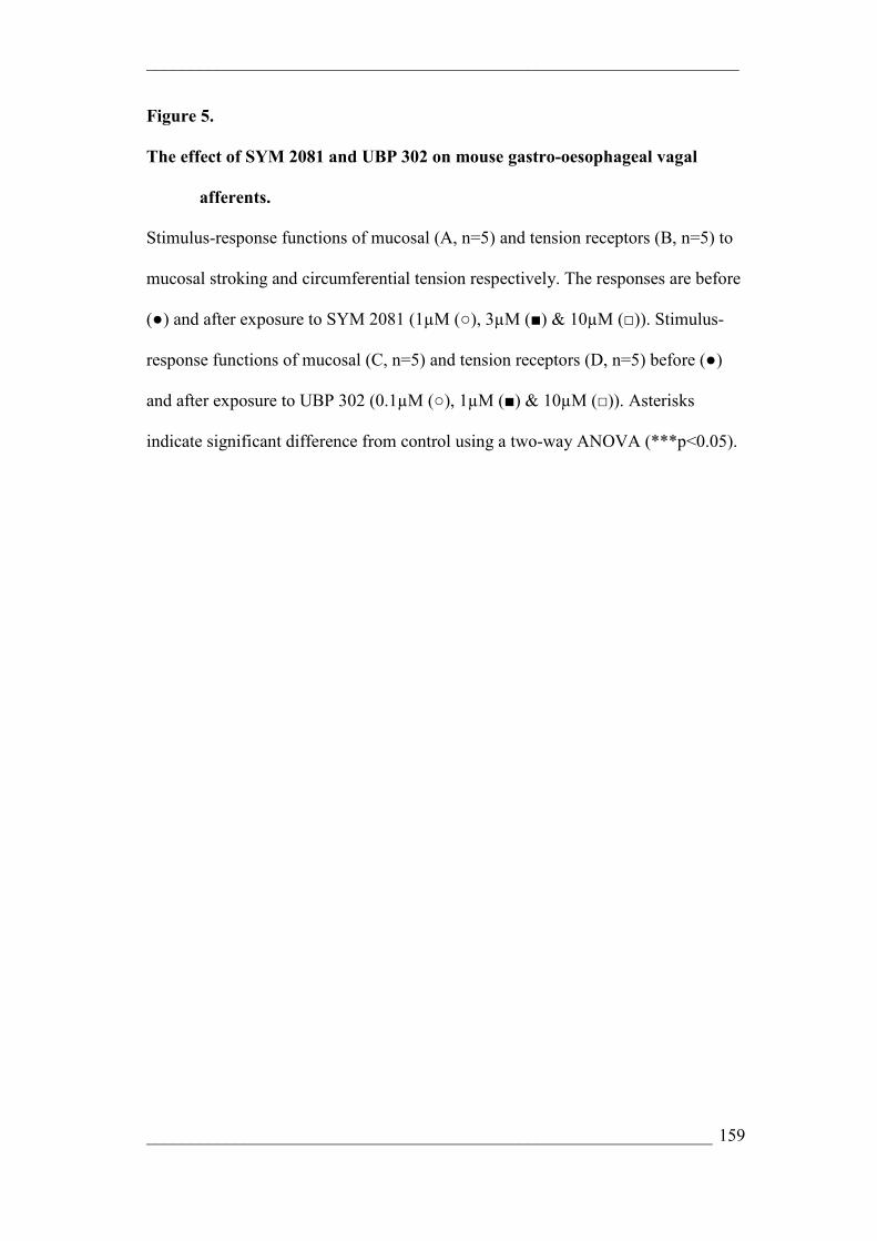
___________________________________________________________________
________________________________________________________________ 159
Figure 5.
The effect of SYM 2081 and UBP 302 on mouse gastro-oesophageal vagal
afferents.
Stimulus-response functions of mucosal (A, n=5) and tension receptors (B, n=5) to
mucosal stroking and circumferential tension respectively. The responses are before
(●) and after exposure to SYM 2081 (1µM (○), 3µM (■) & 10µM (□)). Stimulus-
response functions of mucosal (C, n=5) and tension receptors (D, n=5) before (●)
and after exposure to UBP 302 (0.1µM (○), 1µM (■) & 10µM (□)). Asterisks
indicate significant difference from control using a two-way ANOVA (***p<0.05).

___________________________________________________________________
________________________________________________________________ 160
0 1 2 3 4 5 6
% m
axim
um c
ontr
ol
Load (g)
30
60
90
120
B
************
10 50 200 1000von Frey hair (mg)
0
Impu
lses
/str
oke
3
6
9
A
**
0 1 2 3 4 5 6
% m
axim
um c
ontr
ol
Load (g)
30
60
90
120
B
************
0 1 2 3 4 5 60 1 2 3 4 5 6
% m
axim
um c
ontr
ol
Load (g)
30
60
90
120
30
60
90
120
B
************
10 50 200 1000von Frey hair (mg)
0
Impu
lses
/str
oke
3
6
9
A
**
10 50 200 100010 50 200 1000von Frey hair (mg)
0
Impu
lses
/str
oke
3
6
9
A
**

___________________________________________________________________
________________________________________________________________ 161
Figure 6.
The effect of MTEP on mouse gastro-oesophageal vagal afferents.
Stimulus-response functions of mucosal (A; n=9) and tension receptors (B; n=5) to
mucosal stroking and circumferential tension respectively. The responses are before
(●) and after exposure to MTEP (1µM (○), 3µM (■), 10µM (□) & 30 µM (▲)).
Asterisks indicate significant difference from control using a two-way ANOVA
(**p<0.01; ***p<0.001).

___________________________________________________________________
________________________________________________________________ 162
CHAPTER 3: Ghrelin Selectively Reduces
Mechanosensitivity of Upper Gastrointestinal Vagal
Afferents

___________________________________________________________________
________________________________________________________________ 163
SUMMARY
Ghrelin, a circulating peptide, predominantly released from endocrine cells
of the gastric fundus exerts its orexigenic effects via a vagal pathway. An in vitro
preparation enabled the responses of gastro-oesophageal afferents to graded
mechanical stimulation and the effect of ghrelin on these peripheral endings to be
determined. Three types of vagal afferent were recorded from two animal models,
namely the ferret and mouse, they include tension receptors responding to
circumferential tension, mucosal receptors responding only to von Frey hair
mucosal stroking, and a third afferent subtype specific to the ferret, tension/mucosal
(TM) receptors located in the ferret oesophagus which responded to both stimuli. In
the mouse, ghrelin significantly reduced the responses of tension receptors to
circumferential tension (p<0.005; two-way ANOVA) by up to 40%, an effect
reversed by a specific ghrelin receptor antagonist [D-Lys-3]-GHRP-6. Responses of
mucosal receptors to mucosal stroking with calibrated von Frey hairs were however
not significantly affected. In the ferret, ghrelin significantly reduced the responses
of mucosal and TM receptors to mucosal stroking with calibrated von Frey hairs,
however ghrelin did not have a significant effect to reduce the response to
circumferential tension in both tension and TM receptors. RT-PCR analysis
demonstrated the expression of both ghrelin and its receptor in vagal afferent cell
bodies in mouse nodose ganglia. In conclusion, ghrelin was shown to selectively
inhibit subpopulations of mechanically sensitive gastro-oesophageal vagal afferents,
however its effects were not conserved across species. Expression of both peptide
and its receptor indicates the potential for release from vagal afferents. These
findings implicate a possible role for ghrelin in mediation of satiety signalling via
vagal afferent mechanoreceptors.

___________________________________________________________________
________________________________________________________________ 164
*I would like to acknowledge Dr Amanda Page, Tracey O’Donnell, Camilla
Dorian, Rhianna Laker and Catherine Milte who contributed
electrophysiological ferret studies that were included in this chapter.
INTRODUCTION
Originally identified for its role in growth hormone (GH) secretion, ghrelin
is a 28 amino-acid acylated peptide found mainly in the stomach, and is an
endogenous ligand for the growth hormone secretagogue receptor 1a (GHS-R1a)
[274-276].
It is localised in distinct cells of the gastric mucosa, predominantly distributed in the
mid portion of the oxyntic gland characterised by P/D1 granules in man and X/A
like granules in rodents [275-278]. The extensive endocrine and extra-endocrine
biological activities of ghrelin are evidenced by the widespread distribution of the
peptide and expression of its receptor [275].
The ghrelin cells which synthesize round electron dense granules containing
ghrelin proteins, represents the second most frequent endocrine cell type after the
enterochromaffin-like cells in gastric oxyntic mucosa, indicating a relevant role in
physiological processing at the level of the stomach [275, 279-282].
Ghrelin is predominantly concentrated in the upper gastrointestinal tract, but is still
present distally where ghrelin concentration gradually diminishes form the
duodenum to the colon [283].
Ghrelin is one of several brain-gut neuroenteric peptides that regulate food
intake. To date, the most important role of ghrelin appears to be stimulation of
appetite and regulation of energy homeostasis where it favours adiposity [274].
Secretion of ghrelin is up-regulated and causes increased food intake and body
weight during conditions of negative energy balance such as starvation, insulin-

___________________________________________________________________
________________________________________________________________ 165
induced hypoglycaemia, cachexia and anorexia nervosa, and is down regulated
during conditions of positive energy balance such as feeding, hyperglycaemia, and
obesity [277].
Ghrelin, which is negatively regulated by leptin and IL-beta, is secreted by the
stomach and increases arcuate neuropeptide NPY expression, which in turn acts
through Y1 receptors to increase food intake and decrease energy expenditure [284].
Additional roles for ghrelin have been identified including alteration of gastric
motility as well as affecting gastric acid secretion implicating a role in
gastroprotection [274]. Ghrelin is structurally related to motilin and therefore may
play the role of motilin in rodents as rodents are natural motilin null mutants [275].
Neuronal and hormonal signals produced in the gastrointestinal tract transmit
messages of satiety and starvation to the brain via the afferent limb of the vagus
nerve or via circulation in an endocrine manner.
Ghrelin appears to be an endocrine signal however studies have
demonstrated ghrelin to exert its effect in the periphery via neural pathways, with
the vagus nerve being implicated in conveying ghrelinergic signal from the
periphery to the CNS [275, 277, 279].
An effect of ghrelin acting via vagal pathways has been demonstrated
previously whereby ghrelin stimulated gastric acid production via vagal activation
of gastric parietal cells [274]. In addition, recent work has demonstrated ghrelin is
the first neuro-enteric peptide that can stimulate appetite via an effect on the vagus
nerve in the periphery [277]. In addition, these findings demonstrated both central
and peripheral neuronal pathways to be responsible for the effect of ghrelin on
appetite [277].
___________________________________________________________________
________________________________________________________________ 166
Vagal afferent fibres have a continuous low-frequency spontaneous
discharge that is modulated by sensory inputs [285, 286]. Ghrelin, at a dose required
to stimulate feeding and GH secretion, suppressed the resting discharge of whole
vagus nerve resting discharge in both mouse and rat models [277, 279]. This is in
contrast to other anorexigenic peptides that transmit satiety signals via vagal
afferents such as CCK, which increase signalling to the nucleus of the solitary tract
via the vagus [277, 287, 288].
Involvement of vagal pathways were confirmed when lesion of gastric vagal
afferent fibres was also shown to inhibit appetite-stimulating actions of ghrelin
[277, 279]. Morphological evidence has shown ghrelin receptors are synthesised in
vagal afferent cell bodies and are transported to the periphery, and thus there exists
a close proximity between ghrelin producing cells and vagal afferent terminals in
the stomach [277, 289].
Recent studies have demonstrated ghrelin to have an effect on
mechanosensitivity of vagal afferents innervating the GIT whereby ghrelin was
shown to modulate mechanosensitivity of rat jejunal afferents. An effect on vagal
afferents confirmed by vagotomy and selective ghrelin receptor antagonist studies
[150].
Together, these results provide functional and molecular evidence for a role
of ghrelin acting on ghrelin receptors at peripheral vagal afferent endings.
In humans and animals, a major source of satiety signal from the stomach is gastric
filling and distension [290, 291]. These gastric distension-induced satiety signals are
transferred to the CNS via vagal afferent mechanisms [292]. To date, there is a gap
in our knowledge of this system as there is no direct information on the effect of
ghrelin on signalling of distension by gastro-oesophageal vagal afferent fibres.

___________________________________________________________________
________________________________________________________________ 167
We used an in vitro gastro-oesophageal vagal afferent preparation to study
accurately different populations of mechanosensory afferent fibres [45, 46]. Using
this preparation, we investigated the effect of ghrelin on afferent response to
mechanical stimulation. We studied all of the mechanosensitive gastro-oesophageal
vagal afferent subtypes to determine if ghrelin actions were selective for a particular
type of mechanosensitive afferent fibre. We also investigated the effect of ghrelin in
two species, namely mice and ferrets to determine if the effects are conserved across
species, which may be important since the vast majority of evidence for a role of
ghrelin so far originates from rodent studies.
MATERIALS AND METHODS
All studies were performed in accordance with the guidelines of the Animal
Ethics Committees of the Royal Adelaide Hospital and Institute for Medical and
Veterinary Science, Adelaide, Australia. These committees followed the Australian
code of practice for the care and use of animals for scientific purposes, 6th edition
1997. Every attempt was made to minimise the number of animals required and to
minimise their suffering.
In Vitro ferret and mouse gastro-oesophageal afferent preparations
Female ferrets (0.5-1kg; N=21) were deeply anaesthetized by sodium
pentobarbitone (50mg.Kg, i.p), with the thorax and abdomen opened by midline
incision. Ferrets were then exsanguinated by cardiac puncture. Female C57 mice
(20-30g; N=37) were killed via CO2 inhalation. Both ferret and mouse in vitro
gastro-oesophageal afferent preparations were carried out in a similar manner with
the exception of the size of the organ bath.

___________________________________________________________________
________________________________________________________________ 168
The stomach, oesophagus and attached vagus nerves were then removed and
placed in a modified Krebs’ solution of the following composition (mM): 118.1
NaCl, 4.7 KCl, 25.1 NaHCO3, 1.3 NaH2PO4, 1.2 MgSO4.7H2O, 1.5 CaCl2, 1.0
Citric acid and 11.1 glucose, bubbled with 95% O2-5% CO2.
The preparation temperature was maintained at 4°C during dissection to
preserve tissue integrity and prevent metabolic degradation. The peripheral organs,
major blood vessels and diaphragm were removed by further dissection, leaving the
stomach oesophagus and attached vagus nerves intact. The preparation was opened
longitudinally and pinned out flat mucosa side up in a perspex organ chamber
perfused at a rate of 12mL/min with Krebs’ bicarbonate buffer solution maintained
at 34°C. The vagus nerves were pulled into a separate isolate recording chamber
which was filled with paraffin oil. Under a dissecting microscope, the vagus sheath
was removed exposing the underlying nerve fibres, which were teased into small
bundles and placed on platinum recording electrodes. Nifedipine (1µM) was also
added to the Krebs superfusate in order to prevent smooth muscle contraction.
Previous preliminary studies have demonstrated nifedipine to have no effect on the
mechanical sensitivity of gastro-oesophageal vagal afferents [148].
Similar to the ferret preparation, female C57 mice (20-30g; N=37) were
killed via CO2 inhalation, and the thorax was opened by midline incision. The
stomach and oesophagus were then placed in an organ bath. This preparation has
been described in detail previously in previous chapters.
Characterisation of gastro-oesophageal vagal afferent properties
Corresponding receptive fields of all types of afferent fibre were located by
mechanical stimulation throughout the preparation with a brush. Accurate

___________________________________________________________________
________________________________________________________________ 169
quantification of mechanical responses was performed differently according to the
primary adequate stimulus for the type of fibre. The mechanical sensitivity of
afferents was characterised by mucosal stroking with calibrated von Frey hairs
(10mg-1000mg) and applied circular tension (0.5-7g).
In the ferret, three distinct types of afferent were recorded: those responding
to circular tension but not to low intensity mucosal stimuli (tension receptors), those
responding only to mucosal stroking (mucosal receptors) and those responding to
mucosal stroking and circular tension (tension/mucosal (TM); only found in the
ferret. These three types of afferent endings have been previously described in detail
[45, 146]. In the mouse preparation two types of mechanosensitive afferent were
studied, those responding to mucosal stroking but not to circular tension (mucosal
receptors) and those responding to mucosal stroking and circular tension (tension
receptors) as reported previously [146].
Mechanical thresholds of all types of fibre were determined using calibrated
von Frey hairs. The most reproducible, stimulus dependent response of these
afferents to mucosal stimuli was evoked when the probe was moved at a rate of
5mms-1 across the receptive field rather than being static. Because receptive fields
are small (1-3mm2), a single test at each intensity is prone to missing the centre of
the receptive field on occasions. In order to minimise experimenter error, we
measure the mean response to the middle 8 of 10 standard strokes given at 1s
intervals. A bent von Frey hair (10-1000mg) stroking over the preparation meant
that an even force was distributed over the receptive field during stroking stimulus.
This protocol was found to give reproducible data and was therefore used to assess
the effects of ghrelin on vagal afferents.

___________________________________________________________________
________________________________________________________________ 170
Tension response curves were also obtained for all afferent fibres, which
were used in combination with von Frey hair stroking to determine whether the
receptive fields of fibres were located in the mucosa, the muscle layer or both.
Tension stimuli were applied via a thread attached to an unpinned point
adjacent to the receptive field. The thread was attached to a cantilever and pulley
system close to the preparation, with reference standard weights applied to the
opposite side of the cantilever. Each weight was applied in a step wise manner and
maintained for duration of 1 minute. Sensory receptors response to circular tension
was recorded as mean discharge evoked over this period. Because all responses to
tension were similarly slowly adapting, this method of assessment was considered
representative of physiological responsiveness to distension. The tension response
curves were produced by applying weights to the cantilever system in a range of (1-
5g) and (0.5-7g) for mice and ferrets respectively as previously described [46, 211],
to evoke a maximum response without tissue damage. A recovery period of at least
1 minute was allowed between each tension stimulus.
Effect of ghrelin on mechanosensitivity of vagal afferents
After the mechanical sensitivity of gastro-oesophageal vagal afferents had
been established under control conditions, the effect of ghrelin on mechanical
sensitivity was determined. Ghrelin (1nM) was added to the superfusing Krebs’
solution and was allowed to equilibrate for 20 minutes in order to ensure complete
penetration of the drug into all layers of the tissue. Tension response and stroke
response curves were then re-determined. The procedure was repeated for ghrelin at
increasingly greater doses (3-10nM). Time control experiments had previously been

___________________________________________________________________
________________________________________________________________ 171
performed demonstrating no significant change in mechanical responses over a
comparable duration (see Chapter 1, Fig 6).
In a separate series of experiments in mice, an excess of the ghrelin
antagonist [D-Lys-3]-GHRP-6 (30-100µM) was used to reverse the effects of
ghrelin (3nM). After mechanical sensitivity of the gastro-oesophageal afferent had
been established, ghrelin (3nM) was added to the superfusing Krebs’ solution and
allowed to equilibrate for 20 min. Mechanical response curves were then re-
established. [D-Lys-3]-GHRP-6 (30µM) was then added to the superfusing Krebs’
solution along with ghrelin (3nM), and again this was allowed to equilibrate for 20
min before mechanical response relationships were re-determined. The time frame
as outlined for ghrelin experiments was carried out for higher doses of [D-Lys-3]-
GHRP-6 (100µM). Therefore any reversal of effect of ghrelin by [D-Lys-3]-GHRP-
6 is unlikely to be due to desensitisation to ghrelin. There was no obvious difference
in the effect of ghrelin on oesophageal and gastric afferents, and therefore data from
these afferents were pooled.
Effect of [D-Lys-3]-GHRP-6 on mechanosensitivity of vagal afferents
The ghrelin receptor antagonist [D-Lys-3]-GHRP-6 was used to determine
the involvement of endogenous ghrelin on mechanosensitivity of gastro-
oesophageal vagal afferents. After mechanical sensitivity of the mouse tension
sensitive gastro-oesophageal vagal afferent had been determined, the effect of [D-
Lys-3]-GHRP-6 on mechanical sensitivity was determined. [D-Lys-3]-GHRP-6
(0.1µM) was added to the superfusing Krebs’ solution and allowed to equilibrate for
20 min. after which time the tension-response and stroke-response curves were re-
determined. The concentration of [D-Lys-3]-GHRP-6 used was markedly lower

___________________________________________________________________
________________________________________________________________ 172
than that used in attempts to reverse the effects of exogenous ghrelin. [D-Lys-3]-
GHRP-6 had a different effect on gastric and oesophageal tension receptors and so
the afferents were compared according to location.
Data Recording and Analysis
Afferent impulses were amplified with a biological amplifier (World
Precision Instruments, Sarasota, FL, DAM 50) and scaling amplifier (JRAK,
Melbourne, Australia), filtered (CWE Inc., Ardmore, PA band-pass filter-932) and
monitored using an oscilloscope (DL 1200A; Yakogawa, Tokyo, Japan). Single unit
action potentials were discriminated on the basis of size and shape using spike II
software (Cambridge Electronic Design, Cambridge, UK).
Statistical Analysis: All data were recorded and analysed off-line using a
personal computer. All data were expressed as mean ± SE with n = number of
individual afferents in all instances. Statistical difference between stimulus-response
curves were evaluated using a two-way ANOVA performed using graphpad Prism
(Version 3.00 for Windows, Graphpad Software, San Diego, California, USA).
Differences were considered significant where p < 0.05.
Drugs
Stock solutions of all drugs were kept frozen (-80°C) and diluted to their
final concentration in Krebs’ solution on the day of the experiment. Ghrelin was
obtained from AUSPEP Pty Ltd (Victoria, Australia) and [D-Lys-3]-GHRP-6 was
obtained from Sigma (Sydney, Australia).
___________________________________________________________________
________________________________________________________________ 173
Nodose Ganglia Dissection and RNA extraction for RT-PCR and Quantitative
RT-PCR
Nodose ganglia were removed bilaterally from five male or female C57 mice
(20-30g). Nodose ganglia were pooled in RNAlater® (QIAGEN, NSW, Australia)
at -20°C for subsequent RNA extraction. RNA isolation involved homogenisation
with a sterile glass pestle, of the nodose ganglia, after being snap frozen using liquid
nitrogen. A total of 1ml of TRIzol reagent (Invitrogen, Vic, Australia) was added
before the homogenised material was added to Qiashredders and centrifuged
(12,000 rpm) at room temperature. Tubes containing homogenate were capped after
Qiashredders were removed, vortexed and allowed to stand at room temperature for
15 minutes in order to promote dissociation of nucleoprotein complexes. After
which 0.2 ml of chloroform was added, tubes were vortexed and left to stand at
room temperature for a further 15 minutes. The samples were then cold centrifuged
(12,00 rpm, 4°C) for 15 minutes with the upper aqueous layer (containing RNA
substrate) being removed and transferred to a sterile eppendorf tube followed by
isopropanol precipitation of RNA, by which 0.5 ml isopropanol was added cold
centrifuged for 10min (38, 47). The upper aqueous phase was discarded leaving an
RNA pellet to which 1 ml of 75% ethanol was added. A 1 min vortex and 4 min
cold centrifuge and then supernatant was removed with the pellet being re-
suspended in 100µl RNAase free sterile water and heated for 2 min at 60°C. RNA
was separated into 5µL aliquots and stored at -80°C
RNA quantification was determined by measuring the absorbance at 260nm
(A260) using a spectrophotometer (Biorad, Sydney, Australia). RNA quality was
estimated by the A260: A280 nm ratio.
___________________________________________________________________
________________________________________________________________ 174
Determination of ghrelin and ghrelin receptor transcript expression in Nodose
Ganglia using RT-PCR
Reverse transcription (RT) and polymerase chain reaction (PCR) was
performed using a Qiagen® one-step RT-PCR kit, with QuantiTect Primer Assays
(Qiagen) optimised for the detection of the known sequence of mouse ghrelin,
ghrelin receptor and β-actin. RT-PCR master-mix was prepared as follows: for each
reaction 25µL RT-PCR Master Mix, 10µL RNAse free water, 10µL of 5x QIAGEN
One-Step RT-PCR Buffer, 2µL dNTP Mix (containing 10 mM of each dNTP), 2µL
QIAGEN One-Step RT-PCR Enzyme Mix and ), 1µL of RNAse inhibitor
(Ambion).
RT–PCR reactions were performed using an alpha unit block for PTC DNA
engine systems (MJ Research) attached to a PTC-200 Peltier thermal cycler (MJ
Research). The following amplification program was applied: Reverse
Transcription: 50°C for 30min, Initial PCR activation: 95°C for 15 min, PCR
cycles: denaturing: 94°C for 1 min, annealing: 47°C for 1 min and extension: 72°C
for 1 min repeated for 40 cycles followed by a final extension step of 72°C for 10
min.
For each reaction 25µL of Master Mix was added to 0.2mL clear thin walled
PCR tubes (Axygen) with 10µL (3µM) forward primer (for a final concentration of
0.6 µM), 10µL (3µM) backward primer (for a final concentration of 0.6µM) and
5µL of 50ng template RNA. For each reaction, RNA template was substituted with
5µL distilled RNA-free water as controls. Amplified products (5µL PCR
___________________________________________________________________
________________________________________________________________ 175
product/2µL loading buffer) were resolved by 1.5-3% agarose gel electrophoresis
and visualised by ethidium bromide staining.
Determination of Relative ghrelin and ghrelin receptor transcript expression
using Quantitative RT-PCR
Quantitative reverse transcription polymerase chain reactions (QRT-PCR)
were performed using a Qiagen QuantiTect® SYBR® green RT-PCR one step RT-
PCR kit, using QuantiTect® primer assays (Qiagen) optimized for the detection of
the known sequence of mouse ghrelin, ghrelin receptor and β-actin transcripts
contained in the NCBI reference sequence database. Real-time PCR master mix was
prepared according to product manufactures specifications as follows: for each
reaction 24.5µL of 2x QuantiTect SYBR Green RT-PCR Master Mix, each master
mix containing HotStarTaq® DNA polymerase, QuantiTect® SYBR green RT-PCR
buffer, dNTP Mix including dUTP, SYBR® Green I (Fluoresces in presence of
double stranded DNA), ROX (passive reference dye), 5mM MgCl2, and 0.5µL of
QuantiTect® RT mix (Omniscript® Reverse Transcriptase and Sensiscript®
Reverse transcriptase).
For each reaction 25µL of the master mix, 10µL (3µM) forward primer,
10µL (3µM) backward primer and 5µL of 50ng of template RNA were added in MJ
White PCR tube (MJ Research). RNA template was replaced by RNAase free,
sterile water in control samples.
QRT-PCR reactions were performed using a Chromo4 (MJ research, Biorad)
real-time instrument (designed to detect SYBR green fluorescence, therefore
amplified cDNA products) attached to PTC-200 Peltier thermal cycler (MJ
Research) and analysed using Opticon Monitor Software (MJ Research).

___________________________________________________________________
________________________________________________________________ 176
The following amplification program followed: Reverse Transcription: 50°C for
30mins; Initial PCR activation: 95°C for 15 min; PCR cycles; denaturing: 94°C for
15 sec, annealing: 47°C for 30 sec and extension: 72°C for 30 sec repeated for 44
cycles with final extension step of 72°C for 10 min. A melting curve program was
performed to verify the specificity and identity of the RT-PCR products, whilst all
other reactions were verified using gel electrophoresis. Assays were run in triplicate
in separate experiments. The size of the amplified products (5µL PCR product +
2µL loading buffer) was confirmed by 1.5-3% agarose gel electrophoresis and
visualised by ethidium bromide staining.
Results for QRT-PCR were viewed via Opticon software and were
represented as cycle threshold (CT) required to achieve a certain level of
fluorescence (amount of double stranded PCR product). In all experiments CT was
obtained from the exponential/linear join of the PCR fluorescence curve. As CT is
proportional to the logarithm of total DNA in sample, comparing CT enabled
determination of the relative expression of transcripts.
Titration curves were obtained to confirm amplification efficiencies and
therefore the validity of the comparative cycle threshold. Template RNA of known
concentration from nodose ganglia was used to create a five log dilution series of
RNA. Equal amplification was indicated by the linearity of the plots assay over a
range of input RNA concentrations for β-actin, ghrelin and ghrelin receptor. These
curves demonstrated the efficiencies of targets and references were equal.
The relative concentration of a sample can be determined by the number of
cycles required to reach a certain fluorescence level when compared with other
samples. The β-actin sample was the first sample to reach CT and thus ghrelin and

___________________________________________________________________
________________________________________________________________ 177
ghrelin receptor transcripts expression values were referenced relative to β-actin.
The CT for each was compared with that of β-actin using the equation of CT of
target transcript – CT of β-actin. To determine relative expression of these
transcripts in whole nodose ganglia, the ∆∆CT was calculated using the formula:
∆∆CT= ∆CT [ghrelin receptor] - ∆CT [ghrelin] and the relative fold differences
calculated using the formula 2-∆∆CT. CT values for β-actin were highly reproducible
between samples and between methods of RNA preparation. Quantitative data are
expressed as mean ± SD, and significant differences in transcript expression
determined by a Mann-Whitney test. Significance considered where p < 0.05.
RESULTS
RT PCR localisation of ghrelin and ghrelin receptor transcripts
Using RT-PCR and gel electrophoresis we were able to confirm the presence
of amplified products generated by primers specific for ghrelin, ghrelin receptor,
and β-actin. Isolation of single bands of correct predicted transcript size was
achieved by UV analysis of the gel. Bands of correct predicted product size for both
ghrelin and ghrelin receptors was observed in mouse nodose ganglia. Where
template RNA was substituted for sterile water, no evidence of amplified products
was observed. Expression of the reference β-actin gene was observed in all cases
using template RNA.
Quantitative RT-PCR comparing relative transcript expression
Using gel electrophoresis we confirmed the size of amplified products
generated by the QuantiTect® Primer Assays specific for ghrelin, ghrelin receptor

___________________________________________________________________
________________________________________________________________ 178
and β-actin, showing intense single bands corresponding to predicted sizes of
ghrelin, ghrelin receptor and β-actin transcripts (Figure 4A.).
Separate quantitative RT-PCR experiments were performed to compare
expression between ghrelin and its receptor. Using β-actin as a reference CT value,
QRT-PCR analysis of whole nodose ganglia revealed the ghrelin receptor to have
significantly greater transcript expression (2.33 fold) than ghrelin (p < 0.05, Figure
4B).
Electrophysiology
Effect of ghrelin on the mechanosensitivity of gastro-oesophageal vagal
afferents
Mouse
The effect of ghrelin (1-10nM) on the mechanosensitivity of mouse gastro-
oesophageal vagal afferents is illustrated in figure 1. Ghrelin (1 – 10nM) did not
significantly affect the responses of 9 mucosal receptors to mucosal stroking
throughout range of stimuli (Calibrated von Frey hairs; 10-1000mg; Fig 1A).
However, ghrelin (3-10 nM) did significantly reduce the response of mouse tension
receptors (n = 11) to circular tension (1-5g; Fig 1B & D).
The spontaneous activity of mouse tension receptors was 11.54 ± 2.54
impulses/ sec. In the presence of ghrelin (3 & 10nM) the spontaneous activity was
significantly reduced (table 1). Mouse mucosal receptors tend to have no
spontaneous activity, thus it is not surprising that ghrelin (1 -10nM) had no
significant effect on the spontaneous activity of mouse mucosal receptors (Data not
illustrated).
___________________________________________________________________
________________________________________________________________ 179
The effect of ghrelin (3nM) and the ghrelin antagonist [D-Lys-3]-GHRP
(30-100µM) on mouse tension receptors (n=7) is illustrated in figure 1C. Ghrelin
alone significantly reduced the response curves of tension receptors to circular
stretch (1-5g; Figure 1C.). [D-Lys-3]-GHRP was added in addition with ghrelin
(3nM) to the Krebs’ superfusate and was shown to reverse the inhibitory effect of
ghrelin (Figure 1C.). [D-Lys-3]-GHRP (100µM) demonstrating complete reversal of
the inhibitory effects of ghrelin back to control levels. [D-Lys-3]-GHRP, however,
had no significant effect on the baseline discharge of tension sensitive afferents
(data not shown).
Effect of specific ghrelin receptor antagonist D-Lys-3]-GHRP-6
The ghrelin receptor antagonist [D-Lys-3]-GHRP-6 (0.1µM) alone on mouse
tension receptors as illustrated in figure 3. It significantly potentiated the response
of gastric tension receptors to circular tension (Fig 3A), but did not have a
significant effect on the response of oesophageal tension receptors to circular
tension (Figure 3B.). The antagonist had no significant effect on the spontaneous
activity of tension sensitive afferents (data not shown).
Ferret
The effect of ghrelin on the mechanical sensitivity of ferret gastro-
oesophageal vagal afferents is illustrated in figure 2. Ghrelin (3 - 10 nM)
significantly reduced the response of a subpopulation of mucosal receptors to
mucosal stroking with calibrated von Frey hairs (10 – 1000mg). The
mechanosensitivity of three, from a total of eight mucosal receptors studied was

___________________________________________________________________
________________________________________________________________ 180
reduced in presence of ghrelin (3-10 nM). This reduction of mechanosensitivity was
reversed upon washout of ghrelin from the Krebs’ superfusate (Figure 2D).
Ghrelin (1–10 nM), did not significantly alter the response of tension receptors (n =
7) to circular tension (1-5g; Figure 2B.). The effect of ghrelin (1–10 nM) on the
mechanosensitivity of TM receptors (n = 11) is illustrated in Figure 1C. Ghrelin (1–
10 nM) significantly reduced the response of TM afferents to mucosal stroking with
calibrated von Frey hairs (10 – 1000mg) (Figure 2C (i)), whilst it had no effect on
the response of the same TM receptors to circular stretch (1 -5g; Fig 2C (ii)).
Ghrelin (1-10 nM) did not significantly affect on the spontaneous activity of ferret
mucosal (data not illustrated due to the fact that these receptors are generally silent),
TM, or tension receptors (Table 1).
DISCUSSION
The current study, investigating the effects of ghrelin on the sensitivity of
vagal afferent mechanoreceptors across separate species, provides the first evidence
that ghrelin receptors are involved in transduction of mechanical stimuli by the
gastro-oesophageal vagal afferents. In particular ghrelin is coupled to inhibition of
mechanotransduction in a subset of primary sensory endings. This study identified
ghrelin, the endogenous ligand for the growth hormone secretagogue receptor, to
inhibit ascending vagal afferents signalling in both mice and ferret models. This was
reversed by a ghrelin receptor antagonist [D-Lys-3]-GHRP-6, confirming its
selectivity for this receptor.
Our data revealed important differences between species in the afferent
subtypes influenced by ghrelin. In mice, the inhibitory effect of ghrelin was specific
to tension receptors. This is in contrast to ferret studies where ghrelin had no effect
___________________________________________________________________
________________________________________________________________ 181
on the responses of tension and TM receptors to circumferential tension, however it
inhibited afferent responses to mucosal stroking of TM and mucosal receptors.
These results indicate the possibility of specific roles for ghrelin on specific afferent
pathways, which are discussed in detail below.
Vagal afferents provide the main neuro-anatomical link between the brain
and the upper GIT and are involved in the triggering of behavioural responses [41,
44], reflexes controlling digestive function [293], and motor patterns leading to
gastro-oesophageal reflux [143, 168]. Several physiological and pathophysiological
processes can be triggered by upper gastrointestinal vagal afferents. These include
retching, vomiting, transient lower oesophageal sphincter relaxations (proposed
mechanism of gastro-oesophageal reflux disease), secondary peristalsis, relaxation
of the proximal stomach, gastric emptying and most importantly to the current
investigations, sensations associated with satiety, nausea or discomfort. Modulation
or alteration of this pathway may have important implications in a number of
pathological states. Our data implicate ghrelin acting at ghrelin receptors located on
vagal afferent endings to exert an endogenous influence on this pathway.
Exogenous infusion of ghrelin has been shown previously to enable larger meal
sizes [294]. This may correspond with our finding that ghrelin reduces the response
of tension receptors to circular stretch. Therefore, it would take more distension (i.e.
larger meal size) to give the same degree of inhibition of food intake.
Vagal afferents provide a means of signalling of satiety and influence food
intake via multiple mechanisms including meal related metabolites, monoamines,
peptides as well as mechanical and chemical stimuli [295-297]. Distension of the
stomach has been shown to inhibit feeding via stimulation of vagal afferent
___________________________________________________________________
________________________________________________________________ 182
mechanoreceptors [290]. In addition, the gastrointestinal tract releases various
hormones that regulate short-term food intake via vagal afferent neurones [298]. For
example leptin and cholecystokinin (CCK) receptors are expressed in nodose
ganglia and vagal afferents transmit leptin or CCK signals from the gastrointestinal
tract to the CNS [103, 299, 300]. Therefore vagal afferents play a key role in the
inhibition of food intake [301]. Distension sensitive vagal afferents in both stomach
and duodenum have been shown to be activated upon application of CCK,
demonstrating integration of signals elicited by CCK and gastric loads at the level
of the vagal afferent fibre, but are controversial as to their effects on single sub-
types of vagal afferents [47, 104, 111, 302, 303]. Our findings indicate ghrelin
should be considered as a potential mediator of modulating mechanical activation of
gastric vagal afferents, with clear implications for satiety signalling from the gastro-
intestinal tract.
It has been proposed previously that ghrelin increases food intake by
suppressing the discharge of vagal afferents [277, 284]. Whether this effect is a
direct action on vagal nerves or other vagal responses has not been clarified.
Murray, et al (2006)[150], have since demonstrated modulation of
mechanosensitivity of vagal afferents innervating the rat jejunum in response to
distension. Interestingly though, this group demonstrated augmentation of afferent
response to distension and no effect on resting discharge by ghrelin in contrast to
previous studies in stomach [277]. Whilst this study indicates actions of ghrelin may
differ in different segments of the gastro-intestinal tract, they do demonstrate
subdiaphragmatic vagotomy and a selective ghrelin receptor antagonist to prevent
these effects, once again highlighting an action of ghrelin receptors located on vagal
afferent terminals [150].
___________________________________________________________________
________________________________________________________________ 183
In contrast to previous work where whole nerve afferent discharge was
recorded [277, 284], the in vitro preparation used in this study enabled us to isolate
and record specific subtypes of vagal afferent receptor discharge and to characterize
the effects of exogenously applied to ghrelin on each. The importance of this was
reflected in the fact that ghrelin had selective effects of different receptor subtypes.
The current study demonstrated ghrelin to selectively reduce
mechanosensitivity, as well as inhibit basal discharge of mouse tension receptors,
whilst having no effect on the basal discharge of other types of mouse and ferret
afferents. We therefore propose the decrease in basal discharge observed in multi-
unit recordings in mice and rats in vivo [277, 284], may be due to a decrease in
discharge of tension receptors, or a possible inhibition of ongoing mechanical
activation. We propose a mechanism, whereby ghrelin inhibits signalling of
mechanical activation from distension or contraction of vagal afferents from the
upper gastro-intestinal tract, occurring continually in both fed and fasted states that
may correlate with effects on food intake.
Endogenous ghrelin requires an n-octanylation for endogenous functionality,
however is rapidly degraded from its functional state to functionally inert des-n-
octanyl ghrelin by plasma esterases [304]. Therefore due to a limited half life (10
minutes) the concentration of active ghrelin is greatest closer to site of release in the
stomach, thus implicating vagal afferent endings in the stomach as a probable site of
action for ghrelin. Previous studies have isolated ghrelin in the nodose ganglion
[277], as well as morphological evidence of the existence of ghrelin receptor
producing cells in the nodose ganglion, with ghrelin receptors being produced and
transported to peripheral vagal afferent endings [277]. We were also able to isolate
ghrelin and its receptor in nodose ganglion. Quantitative measurements revealed

___________________________________________________________________
________________________________________________________________ 184
relatively low levels of ghrelin compared with its receptor, but nevertheless it raises
the possibility of an auto-regulatory role of ghrelin released from vagal afferent
endings, in addition to the role of ghrelin released from gastric endocrine cells. This
may be of more importance in oesophageal afferents where there is less ghrelin in
the tissue surrounding the afferents than in the stomach [305].
We investigated a role for endogenous ghrelin on mouse tension receptors
using the ghrelin receptor antagonist [D-Lys-3]-GHRP-6. We observed [D-Lys-3]-
GHRP-6 to have an effect specifically on gastric tension receptors whilst it had no
effect on oesophageal tension receptors. These findings suggest an endogenous
source of ghrelin in the stomach but not the oesophagus, however, argues against an
auto-regulatory role of ghrelin in the oesophagus.
In our previous studies we identified a role for endogenous glutamate in
afferent modulation at both gastric and oesophageal sites, where glutamate was
most likely derived from the afferents themselves [306]. Together, these findings
are suggestive of the gastric mucosa as the primary source of endogenous ghrelin
acting on vagal afferents.
The current study demonstrates ghrelin to reduce the response to mechanical
stimulation of gastro-oesophageal vagal afferents in two species. More specifically,
responses of tension sensitive afferents to circumferential stretch in the mouse, and
mucosal sensitive afferents to mucosal stoking in the ferret. It is possible that this
discrepancy between mice and ferrets reflects the inability of rodents to vomit and
to exhibit gastro-oesophageal reflux [45], whereas ferrets, like humans, are able to
vomit and do exhibit gastro-oesophageal reflux [45]. Ghrelin has previously been
found to show anti-emetic effects in the ferret [307]. Emesis is commonly linked
with activation of mucosal receptors in the stomach and small intestine [6, 308-

___________________________________________________________________
________________________________________________________________ 185
311], which agrees with our finding of selective inhibition of mucosal receptors in
the ferret. In mice which are unable to vomit, the ability of ghrelin to act on vagal
tension receptors in the periphery may play a more important role in food intake.
CONCLUSION
In conclusion, the current study provides the first direct evidence for the
inhibitory modulation of primary vagal afferent mechanosensitivity by the gastric
peptide ghrelin. These results implicate the ghrelin receptor (GHS-R) as a possible
target for the treatment of obesity, and possibly other disorders of upper gastro-
intestinal dysfunction. This study also provides greater insight into the underlying
mechanisms of action of ghrelin.

___________________________________________________________________
________________________________________________________________ 186
0
5
15
10
10 50 200 1000
Impu
lses
/str
oke
von Frey hair (mg)
A
C
0
20
50
40
30
10
0 1 2 3 4 5 6
Impu
lses
/sec
ond
Load (g)
*
#
B
0
20
60
40
0 1 2 3 4 5 6
Impu
lses
/sec
ond
Load (g)
*****
D (i) Control
(ii) Ghrelin (3nM)
(iii) Washout
10 sec.500µV
35
0
Imp.(5s)-1
35
0
Imp.(5s)-1
35
0
Imp.(5s)-1

___________________________________________________________________
________________________________________________________________ 187
Figure 1.
The effect of ghrelin on mouse gastro-oesophageal vagal afferents.
Stimulus-response functions of mucosal (A, n=9) and tension receptors (B, n=11) to
mucosal stroking and circumferential tension respectively. The response are before
(●) and after exposure to ghrelin (1nM (○), 3nM (■) & 10nM (□)). Stimulus-
response functions of tension receptors (C, n=7) before (●) and after exposure to
ghrelin (3nM (■)) and ghrelin (3nM) along with the ghrelin receptor antagonist [D-
Lys-3]-GHRP-6 (30µM (�) and 100µM (�)).*P<0.05, **p<0.01, ***p<0.001
compared to control, #p<0.05 compared to responses obtained in the presence of
ghrelin (3nM) alone (two-way ANOVA). Bonferoni post hoc tests revealed that the
significant differences occurred at larger tension stimuli (not shown), which may be
due to decreasing variance in responses at higher loads. D, original recording of a
tension receptor response to circumferential tension with a 3g weight before (Di),
during exposure (Dii) and after washout (Diii) of ghrelin (3nM).

___________________________________________________________________
________________________________________________________________ 188
0
2
4
6
10 50 200 1000
0
2
4
6
10 50 200 1000
8
0
1
2
3
4
0 1 2 3 4 5 6
0
2
4
6
8
0 1 2 3 4 5 6
10
Impu
lses
/str
oke
Impu
lses
/str
oke
Impu
lses
/sec
.Im
puls
es/s
ec.
von Frey hair (mg)
von Frey hair (mg)
Load (g)
Load (g)
A B
C (ii)
1 sec.
200µV
(i) Control
(ii) Ghrelin (3nM)
(iii) Washout
D
(i)
*****
****

___________________________________________________________________
________________________________________________________________ 189
Figure 2.
The effect of ghrelin on ferret gastro-oesophageal vagal afferents.
Stimulus-response functions of mucosal (A, n=3), tension (B, n=7) and
tension/mucosal receptors (C, n=11) to mucosal stroking (A & Ci) and
circumferential tension (B & Cii). The responses are before (●) and after exposure
to ghrelin (1nM (○), 3nM (■) & 10nM (□)). Asterisks indicate significant difference
from control using a two-way ANOVA (*p<0.05, **p<0.01 & ***p<0.001). D,
original recording of a mucosal receptor response to mucosal stroking with a 50mg
von Frey hair before (Di), during exposure (Dii) and after washout (Diii) of ghrelin
(3nM).

___________________________________________________________________
________________________________________________________________ 190
25
20
15
10
5
00 1 2 3 4 5
Load (g)
Impu
lses
/sec
ond
40
30
20
10
00 1 2 3 4 5
Load (g)
Impu
lses
/sec
ond
*
A
B

___________________________________________________________________
________________________________________________________________ 191
Figure 3.
The effect of [D-Lys-3]-GHRP-6 on mouse gastro-oesophageal vagal afferents.
Stimulus-response functions of gastric (A, n=6) and oesophageal (B, n=4) tension
receptors to circumferential tension. The responses are before (●) and after (○)
exposure to [D-Lys-3]-GHRP-6 (0.1µM). *p<0.05 compared to control (two-way
ANOVA).

___________________________________________________________________
________________________________________________________________ 192
Ghrelin (73 bp)
β-actin(149 bp)
Ghrelin control
Ghrelin receptor (121 bp)
Ghrelin receptor control
(100 bp)
(200 bp)
0.0000
0.0001
0.0002
0.0003
0.0004
0.0005
0.0006
mRNA level relative toββ ββ-actin
Ghrelin GhrelinReceptor
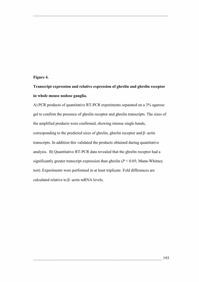
___________________________________________________________________
________________________________________________________________ 193
Figure 4.
Transcript expression and relative expression of ghrelin and ghrelin receptor
in whole mouse nodose ganglia.
A) PCR products of quantitative RT-PCR experiments separated on a 3% agarose
gel to confirm the presence of ghrelin receptor and ghrelin transcripts. The sizes of
the amplified products were confirmed, showing intense single bands,
corresponding to the predicted sizes of ghrelin, ghrelin receptor and β–actin
transcripts. In addition this validated the products obtained during quantitative
analysis. B) Quantitative RT-PCR data revealed that the ghrelin receptor had a
significantly greater transcript expression than ghrelin (P < 0.05; Mann-Whitney
test). Experiments were performed in at least triplicate. Fold differences are
calculated relative to β–actin mRNA levels.
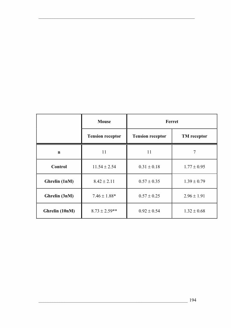
___________________________________________________________________
________________________________________________________________ 194
Mouse Ferret
Tension receptor Tension receptor TM receptor
n 11 11 7
Control 11.54 ± 2.54 0.31 ± 0.18 1.77 ± 0.95
Ghrelin (1nM) 8.42 ± 2.11 0.57 ± 0.35 1.39 ± 0.79
Ghrelin (3nM) 7.46 ± 1.88* 0.57 ± 0.25 2.96 ± 1.91
Ghrelin (10nM) 8.73 ± 2.59** 0.92 ± 0.54 1.32 ± 0.68

___________________________________________________________________
________________________________________________________________ 195
Table 1.
Spontaneous activity of gastro-oesophageal afferents sensitive to tension.
The spontaneous activity of mouse tension and ferret tension and tension/mucosal
receptors in the absence and presence of ghrelin (1, 3 and 10nM). Values are
impulses/second ± S.E. Ghrelin significantly reduced the spontaneous activity of
mouse tension sensitive receptors but did not significantly affect the spontaneous
activity of ferret tension and tension/mucosal receptors. *p<0.05, **p<0.01
compared to control (paired t-test).

___________________________________________________________________
________________________________________________________________ 196
CONCLUSIONS

___________________________________________________________________
________________________________________________________________ 197
Modulation of vagal afferent mechanoreceptor signalling to the CNS has
been well established as a novel therapeutic target for the treatment of upper
gastrointestinal tract disorders such as disordered food intake, functional dyspepsia
and most importantly to the current investigation, gastro-oesophageal reflux disease
(GORD).
GORD is one of the most common clinical problems of our society,
affecting up to 20% of the population [162]. Conventional therapies aimed at
reducing gastric acid secretion, thus limiting acid content of the refluxate have been
successful in a significant proportion of patients. However, up to 30% of patients
are refractory to this treatment, or suffer from gastro-duodenal or bile reflux which
is also damaging to the oesophagus [179, 180].
Transient lower oesophageal sphincter relaxations (TLOSRs) triggered by
activation of distension sensitive afferents in the proximal stomach and underly the
major pathophysiological mechanism of reflux disease. TLOSRs mediated by a
vagal pathway involving activation of proximal gastric tension receptors, which in
turn, activate a program generator in the brainstem. Output of this generator leads to
brief activation of vagal motorneurones connected to inhibitory pathways in the
LOS causing relaxation [168].
Intense interest in management of reflux disease has seen pharmacological
modulation of the TLOSR pathway at several points [187, 188, 193, 195, 312, 313].
In recent years it has become apparent that modulation of peripheral vagal afferent
mechanoreceptors innervating the gastro-oesophageal region correlates with a novel
therapeutic approach to reflux disease via reduction in gastric distension evoked
TLOSRs and gastro-oesophageal reflux episodes.
___________________________________________________________________
________________________________________________________________ 198
Emerging evidence has shown metabotropic receptors to the amino acids
GABA and glutamate, are able to modulate triggering of TLOSRs, corresponding to
reduction in gastro-oesophageal reflux [122, 123]. Importantly, this is corroborated
by our observations that GABAB and mGluR5 receptors are expressed in vagal
sensory ganglia [145, 148], and both GABAB agonists and mGluR5 antagonists
reduce responses to distension of gastro-oesophageal vagal afferents when applied
peripherally both in vitro and in vivo [145, 146, 191, 306]. These studies confirmed
signalling from vagal mechanoreceptors is crucial in the initiation of this motor
pattern [144]. The most accessible target for selective modulation of the neuronal
pathway mediating TLOSRs is via the vagal afferent terminal mechanoreceptors.
Such peripheral targets offer advantages by minimising interference with motor
pathways that may produce unwanted side effects elsewhere in the GI tract as well
as in other target organs of the vagus [198].
The success of GABAB agonists and mGluR agonists and antagonists to
reduce TLOSRs as well as gastro-oesophageal reflux provide the best example of
G-protein coupled receptors (GPCR) as therapeutic targets in the management of
reflux disease.
There are three families of GPCR that respond to stimuli as diverse as light,
odour, taste, calcium, and a wide range of chemical mediators, but few have been
explored in the sensory system. Whilst our interest initially was focussed on
inhibitory neuronal GPCR including galanin and ghrelin receptors given the
therapeutic success of previous GPCR, the excitatory glutamatergic counterbalance
observed in our mGluR studies highlighted the need for comprehensive evaluation
of mechanisms of glutamatergic modulation of vagal afferents and therefore,

___________________________________________________________________
________________________________________________________________ 199
excitatory coupled iGluR and mGluR were an obvious addition to those targets
mentioned above.
The effect of galanin to potently modulate vagal afferent mechanosensitivity
raised the possibility that there were other features of vagal afferents that may
provide alternative, possibly more selective G-Protein coupled receptor targets
[148].
Galanin is a 29-30 amino acid peptide found throughout the central and
enteric nervous system, known to have either inhibitory or excitatory effects on
motor function including gastric smooth muscle activity [205], and neuronal
excitability including transmission in the spinal cord [204], and signalling in central
gastric vagal pathways [206, 207].This study replicated the divergent effect of
galanin in our in vitro gastro-oesophageal vagal afferent preparation, demonstrating
galanin to exert a predominantly inhibitory effect. Three receptors for galanin have
been cloned, Gal-R1, -R2 and -R3. We confirmed using GalR1-null mutant mice,
and receptor specific ligands, that the inhibitory effect of galanin on
mechanosensation is mediated via the GalR1 receptor located on vagal afferents
innervating the upper gastrointestinal tract. In addition, GalR2 was implicated in the
mediation of excitatory effects of galanin, previously observed. A minor but
significant potentiation in mechanosensitivity by galanin was seen in GalR1-/- mice,
an effect that was reproduced by a selective GalR2 agonist. We were unable to
identify a functional role for GalR3 on gastro-oesophageal vagal afferents and as yet
a physiological role for GalR3 is still undetermined. In contrast to these findings, all
three galanin receptors were identified in vagal sensory ganglia and relative
expression of all three receptors was similar. Other findings have demonstrated

___________________________________________________________________
________________________________________________________________ 200
gastric projecting vagal neurones to contain much less GalR3 than other subtypes so
the possibility of a role for GalR3 in other organs innervated by the vagus still
exists.
The ghrelin receptor or growth hormone secretagogue receptor 1a
(GHSR1a), is also an inhibitory brain-gut GPCR which has widespread distribution
but is particularly localised in the upper GI tract [275, 279-283]. Ghrelin signalling
to the CNS is predominantly mediated via the vagus nerve and electrophysiological
recordings have demonstrated ghrelin to modulate whole vagus afferent discharge
from the stomach and jejunum [150, 277].
Other studies have identified ghrelin and its receptors located in the vagus
nerve, where they are transported peripherally [277]. We were able to isolate ghrelin
and its receptors in whole nodose ganglia and demonstrate relative expression of
ghrelin is low compared with its receptor.
We provided the first evidence that ghrelin is involved in signalling of
mechanical stimuli via vagal gastro-oesophageal afferents. In particular ghrelin is
coupled to inhibition of mechanotransduction in a subset of primary sensory
endings. Important differences between species were noted however. Ghrelin was
shown to inhibit mucosal receptors in the ferret, having no significant effect on
tension or TM receptors, whilst it was shown to inhibit tension receptors in the
mouse, having no significant effect on mucosal receptors in the mouse. These
results suggest there may be specific roles for ghrelin in specific pathways. Ghrelin
is a known satiety signal, an effect mediated peripherally, and is dependent on the
vagus nerve. The fact that ghrelin had no significant effect on ferret tension
receptors probably rules out a therapeutic role for ghrelin in terms of inhibiting

___________________________________________________________________
________________________________________________________________ 201
TLOSRs however, vagal afferents provide a means of signalling of satiety via
multiple mechanisms including mechanical and chemical stimuli. Distension of the
stomach inhibits feeding via stimulation of vagal afferent mechanoreceptors. These
results therefore implicate ghrelin to affect satiety and possibly TLOSRs via
modulation of peripheral vagal mechanoreceptors.
Glutamate exerts its effects at central synapses via ionotropic (iGluR-
NMDA, AMPA & Kainate) and metabotropic (mGluR) receptors. Group I mGluR
are coupled to excitatory, whilst group II and III mGluR are coupled to inhibitory
mechanisms. Previous in vitro work in the ferret identified a possible counter-
balance of excitatory and inhibitory glutamatergic signalling from peripheral vagal
afferents whereby glutamate acts simultaneously at iGluR and mGluR [121]. The
potent inhibitory effect of glutamate, an effect mimicked by selective group II and
III agonists, on vagal afferent mechanosensitivity was only revealed after the
addition of the non-specific iGluR antagonist Kynurenate to the preparation. In vivo
observations in the rat indicated similarly that iGluR may be involved in excitatory
modulation of vagal afferents [120]. The current data indicate AMPA, NMDA and
mGluR5 receptors to play a major role in the excitatory balance of modulation of
mechanosensitivity with kainate receptors having little if any effect under the
current conditions. Also identified was that the role of each differs according to
afferent subtypes. NMDA was more potent on mucosal receptors, and MTEP was
more potent on tension receptors, suggesting NMDA receptors may be more highly
expressed by mucosal receptor cell bodies, and mGluR5 may be expressed more by
tension receptors. These results clearly indicate a counter balance of excitatory
modulation of vagal afferent mechanosensitivity by iGluR and mGluR 5, and
___________________________________________________________________
________________________________________________________________ 202
inhibitory modulation by group II and III mGluR. The inhibitory effects of
exogenous iGluR and mGluR antagonists indicate glutamate to exert an endogenous
action on mechanosensitivity, with vagal afferents themselves, contracting smooth
muscle, luminal and circulating glutamate as candidates for an endogenous source.
In conjunction with previous findings by our group, this study further clarifies the
influence of all plausible major GluR subtypes on vagal afferent
mechanosensitivity, as well as confirming previous findings in rat and ferret, which
suggest an excitatory role for glutamate receptors on vagal afferents. There is
clearly scope for both excitatory and inhibitory modulation of afferent sensitivity by
glutamate from both exogenous and endogenous sources, providing a balance to
achieve normal vagal afferent function. These data were reinforced by the finding of
a complete complement of subtypes of all receptors located in the nodose ganglion,
therefore implicating each receptor as a possible candidate for neuromodulation.
Results from this thesis confirm further contenders in an expanding list of
modulators of vagal afferent mechanosensitivity, and thus establishing possible
future novel therapeutic targets for the treatment of disordered upper gastrointestinal
vagal afferent signalling.

___________________________________________________________________
________________________________________________________________ 203
BIBLIOGRAPHY

___________________________________________________________________
________________________________________________________________ 204
1. Furness, J.B., Novel gut afferents: Intrinsic afferent neurons and intestinofugal neurons. Auton Neurosci, 2006. 125(1-2): p. 81-5.
2. Costa, M., D. Wattchow, and S. Brookes, Neuronal control in gastrointestinal disease. Eur J Surg Suppl, 2002(587): p. 39-46.
3. Szurszewski, J.H., L.G. Ermilov, and S.M. Miller, Prevertebral ganglia and intestinofugal afferent neurones. Gut, 2002. 51 Suppl 1: p. i6-10.
4. Travagli, R.A., et al., Brainstem circuits regulating gastric function. Annu Rev Physiol, 2006. 68: p. 279-305.
5. Costa, M., S.H. Brookes, and V. Zagorodnyuk, How many kinds of visceral afferents? Gut, 2004. 53 Suppl 2: p. ii1-4.
6. Andrews, P.L., et al., The abdominal visceral innervation and the emetic reflex: pathways, pharmacology, and plasticity. Can J Physiol Pharmacol, 1990. 68(2): p. 325-45.
7. Snell, R.S., ed. Clinical anatomy for medical students. 6 ed. 2000, Lippincott Williams and Wilkins. 648-649.
8. Leslie, R.A., D.G. Gwyn, and D.A. Hopkins, The central distribution of the cervical vagus nerve and gastric afferent and efferent projections in the rat. Brain Res Bull, 1982. 8(1): p. 37-43.
9. Schemann, M. and D. Grundy, Electrophysiological identification of vagally innervated enteric neurons in guinea pig stomach. Am J Physiol, 1992. 263(5 Pt 1): p. G709-18.
10. Kunze, W.A. and J.B. Furness, The enteric nervous system and regulation of intestinal motility. Annu Rev Physiol, 1999. 61: p. 117-42.
11. Dutsch, M., et al., Vagal and spinal afferent innervation of the rat esophagus: a combined retrograde tracing and immunocytochemical study with special emphasis on calcium-binding proteins. J Comp Neurol, 1998. 398(2): p. 289-307.
12. Wank, M. and W.L. Neuhuber, Local differences in vagal afferent innervation of the rat esophagus are reflected by neurochemical differences at the level of the sensory ganglia and by different brainstem projections. J Comp Neurol, 2001. 435(1): p. 41-59.
13. Berthoud, H.R., et al., Distribution and structure of vagal afferent intraganglionic laminar endings (IGLEs) in the rat gastrointestinal tract. Anat Embryol (Berl), 1997. 195(2): p. 183-91.
14. Wang, F.B. and T.L. Powley, Topographic inventories of vagal afferents in gastrointestinal muscle. J Comp Neurol, 2000. 421(3): p. 302-24.
15. Andrews, P.L. and T.V. Taylor, An electrophysiological study of the posterior abdominal vagus nerve in man. Clin Sci (Lond), 1982. 63(2): p. 169-73.
16. Mackay, T.W. and P.L. Andrews, A comparative study of the vagal innervation of the stomach in man and the ferret. J Anat, 1983. 136(Pt 3): p. 449-81.
17. Powley, T.L. and H.R. Berthoud, A fluorescent labeling strategy for staining the enteric nervous system. J Neurosci Methods, 1991. 36(1): p. 9-15.

___________________________________________________________________
________________________________________________________________ 205
18. Sternini, C. and K. Anderson, Calcitonin gene-related peptide-containing neurons supplying the rat digestive system: differential distribution and expression pattern. Somatosens Mot Res, 1992. 9(1): p. 45-59.
19. Nonidez, J.F., Afferent nerve endings in the ganglia of intermuscular plexus of the dog's oesophagus. J. Comp. Neurol, 1946. 85: p. 177-189.
20. Rodrigo, J.F., Vegetative innervation f the esophagus. II. Intraganglionic laminar endings. Acta Anat (Basel), 1975. 92: p. 79-100.
21. Rodrigo, J., et al., Sensory vagal nature and anatomical access paths to esophagus laminar nerve endings in myenteric ganglia. Determination by surgical degeneration methods. Acta Anat (Basel), 1982. 112(1): p. 47-57.
22. Neuhuber, W.L., Sensory vagal innervation of the rat esophagus and cardia: a light and electron microscopic anterograde tracing study. J Auton Nerv Syst, 1987. 20(3): p. 243-55.
23. Berthoud, H.R. and T.L. Powley, Vagal afferent innervation of the rat fundic stomach: morphological characterization of the gastric tension receptor. J Comp Neurol, 1992. 319(2): p. 261-76.
24. Powley, T.L. and R.J. Phillips, Musings on the wanderer: what's new in our understanding of vago-vagal reflexes? I. Morphology and topography of vagal afferents innervating the GI tract. Am J Physiol Gastrointest Liver Physiol, 2002. 283(6): p. G1217-25.
25. Phillips, R.J., E.A. Baronowsky, and T.L. Powley, Afferent innervation of gastrointestinal tract smooth muscle by the hepatic branch of the vagus. J Comp Neurol, 1997. 384(2): p. 248-70.
26. Fox, E.A., et al., Vagal afferent innervation of smooth muscle in the stomach and duodenum of the mouse: morphology and topography. J Comp Neurol, 2000. 428(3): p. 558-76.
27. Zagorodnyuk, V.P. and S.J. Brookes, Transduction sites of vagal mechanoreceptors in the guinea pig esophagus. J Neurosci, 2000. 20(16): p. 6249-55.
28. Neuhuber, W.L., et al., Vagal efferent and afferent innervation of the rat esophagus as demonstrated by anterograde DiI and DiA tracing: focus on myenteric ganglia. J Auton Nerv Syst, 1998. 70(1-2): p. 92-102.
29. Zagorodnyuk, V.P., B.N. Chen, and S.J. Brookes, Intraganglionic laminar endings are mechano-transduction sites of vagal tension receptors in the guinea-pig stomach. J Physiol, 2001. 534(Pt 1): p. 255-68.
30. Zagorodnyuk, V.P., et al., Mechanotransduction by intraganglionic laminar endings of vagal tension receptors in the guinea-pig oesophagus. J Physiol, 2003. 553(Pt 2): p. 575-87.
31. Lynn, P.A., et al., Rectal intraganglionic laminar endings are transduction sites of extrinsic mechanoreceptors in the guinea pig rectum. Gastroenterology, 2003. 125(3): p. 786-94.
32. Berthoud, H.R., et al., Neuroanatomy of extrinsic afferents supplying the gastrointestinal tract. Neurogastroenterol Motil, 2004. 16 Suppl 1: p. 28-33.
33. Berthoud, H.R. and L.M. Patterson, Anatomical relationship between vagal afferent fibers and CCK-immunoreactive entero-endocrine cells in the rat small intestinal mucosa. Acta Anat (Basel), 1996. 156(2): p. 123-31.
34. Raj, P., Celiac plexus/splanchnic nerve blocks. techniques in regional anaesthesia and pain management, 2001. 5(3): p. 102-1115.

___________________________________________________________________
________________________________________________________________ 206
35. Grundy, D., Neuroanatomy of visceral nociception: vagal and splanchnic afferent. Gut, 2002. 51 Suppl 1: p. i2-5.
36. Clerc, N., Afferent innervation of the lower oesophageal sphincter of the cat. An HRP study. J Auton Nerv Syst, 1983. 9(4): p. 623-36.
37. Khurana, R.K. and J.M. Petras, Sensory innervation of the canine esophagus, stomach, and duodenum. Am J Anat, 1991. 192(3): p. 293-306.
38. Clerc, N. and C. Mazzia, Morphological relationships of choleragenoid horseradish peroxidase-labeled spinal primary afferents with myenteric ganglia and mucosal associated lymphoid tissue in the cat esophagogastric junction. J Comp Neurol, 1994. 347(2): p. 171-86.
39. Brtva, R.D., G.A. Iwamoto, and J.C. Longhurst, Distribution of cell bodies for primary afferent fibers from the stomach of the cat. Neurosci Lett, 1989. 105(3): p. 287-93.
40. Ozaki, N. and G.F. Gebhart, Characterization of mechanosensitive splanchnic nerve afferent fibers innervating the rat stomach. Am J Physiol Gastrointest Liver Physiol, 2001. 281(6): p. G1449-59.
41. Blackshaw, L.A. and G.F. Gebhart, The pharmacology of gastrointestinal nociceptive pathways. Curr Opin Pharmacol, 2002. 2(6): p. 642-9.
42. Paintal, A.S., The conduction velocities of respiratory and cardiovascular afferent fibres in the vagus nerve. J Physiol, 1953. 121: p. 341-359.
43. Iggo, A., Tension receptors in the stomach and urinary bladder. J Physiol, 1955. 128: p. 593-607.
44. Cervero, F., Sensory innervation of the viscera: peripheral basis of visceral pain. Physiol Rev, 1994. 74(1): p. 95-138.
45. Page, A.J. and L.A. Blackshaw, An in vitro study of the properties of vagal afferent fibres innervating the ferret oesophagus and stomach. J Physiol, 1998. 512 ( Pt 3): p. 907-16.
46. Page, A.J., C.M. Martin, and L.A. Blackshaw, Vagal mechanoreceptors and chemoreceptors in mouse stomach and esophagus. J Neurophysiol, 2002. 87(4): p. 2095-103.
47. Blackshaw, L.A. and D. Grundy, Effects of cholecystokinin (CCK-8) on two classes of gastroduodenal vagal afferent fibre. J Auton Nerv Syst, 1990. 31(3): p. 191-201.
48. Blackshaw, L.A. and D. Grundy, Locally and reflexly mediated effects of cholecystokinin-octapeptide on the ferret stomach. J Auton Nerv Syst, 1991. 36(2): p. 129-37.
49. Blackshaw, L.A. and D. Grundy, Effects of 5-hydroxytryptamine (5-HT) on the discharge of vagal mechanoreceptors and motility in the upper gastrointestinal tract of the ferret. J Auton Nerv Syst, 1993. 45(1): p. 51-9.
50. Iggo, A., Gastrointestinal tension receptors with unmyelinated afferent fibres in the vagus of the cat. J Physiol, 1957. 42: p. 130-143.
51. Cottrell, D.F. and A. Iggo, Tension receptors with vagal afferent fibres in the proximal duodenum and pyloric sphincter of sheep. J Physiol, 1984. 354: p. 457-75.
52. Lynn, P.A. and L.A. Blackshaw, In vitro recordings of afferent fibres with receptive fields in the serosa, muscle and mucosa of rat colon. J Physiol, 1999. 518 ( Pt 1): p. 271-82.
___________________________________________________________________
________________________________________________________________ 207
53. Davison, J.S., Response of single vagal afferent fibres to mechanical and chemical stimulation of the gastric and duodenal mucosa in cats. Q J Exp Physiol Cogn Med Sci, 1972. 57(4): p. 405-16.
54. Clarke, G.D. and J.S. Davison, Tension receptors in the oesophagus and stomach of the rat. J Physiol, 1975. 244(1): p. 41P-42P.
55. Blackshaw, L.A., D. Grundy, and T. Scratcherd, Vagal afferent discharge from gastric mechanoreceptors during contraction and relaxation of the ferret corpus. J Auton Nerv Syst, 1987. 18(1): p. 19-24.
56. Satchel, P.M., Canine oesophageal mechanoreceptors. j Physiol, 1984. 346: p. 287-300.
57. Sengupta, J.N., D. Kauvar, and R.K. Goyal, Characteristics of vagal esophageal tension-sensitive afferent fibers in the opossum. J Neurophysiol, 1989. 61(5): p. 1001-10.
58. Sengupta, J.N., J.K. Saha, and R.K. Goyal, Stimulus-response function studies of esophageal mechanosensitive nociceptors in sympathetic afferents of opossum. J Neurophysiol, 1990. 64(3): p. 796-812.
59. Cervero, F. and K.A. Sharkey, An electrophysiological and anatomical study of intestinal afferent fibres in the rat. J Physiol, 1988. 401: p. 381-97.
60. Page, A.J. and L.A. Blackshaw, Roles of gastro-oesophageal afferents in the mechanisms and symptoms of reflux disease. Handb Exp Pharmacol, 2009(194): p. 227-57.
61. Blumberg, H., et al., Encoding of visceral noxious stimuli in the discharge patterns of visceral afferent fibres from the colon. Pflugers Arch, 1983. 398(1): p. 33-40.
62. Haupt, P., W. Janig, and W. Kohler, Response pattern of visceral afferent fibres, supplying the colon, upon chemical and mechanical stimuli. Pflugers Arch, 1983. 398(1): p. 41-7.
63. Sengupta, J.N. and G.F. Gebhart, Mechanosensitive properties of pelvic nerve afferent fibers innervating the urinary bladder of the rat. J Neurophysiol, 1994. 72(5): p. 2420-30.
64. Berthoud, H.R., P.A. Lynn, and L.A. Blackshaw, Vagal and spinal mechanosensors in the rat stomach and colon have multiple receptive fields. Am J Physiol Regul Integr Comp Physiol, 2001. 280(5): p. R1371-81.
65. Brierley, S.M., et al., Splanchnic and pelvic mechanosensory afferents signal different qualities of colonic stimuli in mice. Gastroenterology, 2004. 127(1): p. 166-78.
66. Duthie, H.L. and F.W. Gairns, Sensory nerve-endings and sensation in the anal region of man. Br J Surg, 1960. 47: p. 585-95.
67. Janig, W. and M. Koltzenburg, Receptive properties of sacral primary afferent neurons supplying the colon. J Neurophysiol, 1991. 65(5): p. 1067-77.
68. Cottrell, D.F. and A. Iggo, Mucosal enteroceptors with vagal afferent fibres in the proximal duodenum of sheep. J Physiol, 1984. 354: p. 497-522.
69. Sengupta, J.N. and G.F. Gebhart, Characterization of mechanosensitive pelvic nerve afferent fibers innervating the colon of the rat. J Neurophysiol, 1994. 71(6): p. 2046-60.
70. Janig, W. and M. Koltzenburg, On the function of spinal primary afferent fibres supplying colon and urinary bladder. J Auton Nerv Syst, 1990. 30 Suppl: p. S89-96.
___________________________________________________________________
________________________________________________________________ 208
71. Ness, T.J. and G.F. Gebhart, Colorectal distension as a noxious visceral stimulus: physiologic and pharmacologic characterization of pseudaffective reflexes in the rat. Brain Res, 1988. 450(1-2): p. 153-69.
72. Ness, T.J. and G.F. Gebhart, Characterization of neuronal responses to noxious visceral and somatic stimuli in the medial lumbosacral spinal cord of the rat. J Neurophysiol, 1987. 57(6): p. 1867-92.
73. Grundy, D., What activates visceral afferents? Gut, 2004. 53 Suppl 2: p. ii5-8.
74. Robinson, P.P., Recession of sensory loss from the midline following trigeminal sensory root section: collateral sprouting from the normal side? Brain Res, 1983. 259(1): p. 177-80.
75. Morrison, J.F., Splanchnic slowly adapting mechanoreceptors with punctate receptive fields in the mesentery and gastrointestinal tract of the cat. J Physiol, 1973. 233(2): p. 349-61.
76. Hughes, P.A., et al., Post-inflammatory colonic afferent sensitisation: different subtypes, different pathways and different time courses. Gut, 2009. 58(10): p. 1333-41.
77. Longhurst, J.C., et al., Effects of bradykinin and capsaicin on endings of afferent fibers from abdominal visceral organs. Am J Physiol, 1984. 247(3 Pt 2): p. R552-9.
78. Kirkup, A.J., A.M. Brunsden, and D. Grundy, Receptors and transmission in the brain-gut axis: potential for novel therapies. I. Receptors on visceral afferents. Am J Physiol Gastrointest Liver Physiol, 2001. 280(5): p. G787-94.
79. Burnstock, G., Overview. Purinergic mechanisms. Ann N Y Acad Sci, 1990. 603: p. 1-17; discussion 18.
80. Burnstock, G., P2X receptors in sensory neurones. Br J Anaesth, 2000. 84(4): p. 476-88.
81. Galligan, J.J. and P.P. Bertrand, ATP mediates fast synaptic potentials in enteric neurons. J Neurosci, 1994. 14(12): p. 7563-71.
82. Kirkup, A.J., et al., Excitatory effect of P2X receptor activation on mesenteric afferent nerves in the anaesthetised rat. J Physiol, 1999. 520 Pt 2: p. 551-63.
83. Page, A.J., T.A. O'Donnell, and L.A. Blackshaw, P2X purinoceptor-induced sensitization of ferret vagal mechanoreceptors in oesophageal inflammation. J Physiol, 2000. 523 Pt 2: p. 403-11.
84. Fong, A.Y., et al., Immunoreactive localisation of P2Y1 receptors within the rat and human nodose ganglia and rat brainstem: comparison with [alpha 33P]deoxyadenosine 5'-triphosphate autoradiography. Neuroscience, 2002. 113(4): p. 809-23.
85. Cook, S.P., et al., Distinct ATP receptors on pain-sensing and stretch-sensing neurons. Nature, 1997. 387(6632): p. 505-8.
86. Khakh, B.S., P.P. Humphrey, and A. Surprenant, Electrophysiological properties of P2X-purinoceptors in rat superior cervical, nodose and guinea-pig coeliac neurones. J Physiol, 1995. 484 ( Pt 2): p. 385-95.
87. Dang, K., et al., Gastric ulcers evoke hyperexcitability and enhance P2X receptor function in rat gastric sensory neurons. J Neurophysiol, 2005. 93(6): p. 3112-9.
___________________________________________________________________
________________________________________________________________ 209
88. Ruan, H.Z. and G. Burnstock, Localisation of P2Y1 and P2Y4 receptors in dorsal root, nodose and trigeminal ganglia of the rat. Histochem Cell Biol, 2003. 120(5): p. 415-26.
89. Leeb-Lundberg, L.M., et al., International union of pharmacology. XLV. Classification of the kinin receptor family: from molecular mechanisms to pathophysiological consequences. Pharmacol Rev, 2005. 57(1): p. 27-77.
90. Sengupta, J.N., J.K. Saha, and R.K. Goyal, Differential sensitivity to bradykinin of esophageal distension-sensitive mechanoreceptors in vagal and sympathetic afferents of the opossum. J Neurophysiol, 1992. 68(4): p. 1053-67.
91. Mense, S., Muscular nociceptors. J Physiol (Paris), 1977. 73(3): p. 233-40. 92. Mense, S., Sensitization of group IV muscle receptors to bradykinin by 5-
hydroxytryptamine and prostaglandin E2. Brain Res, 1981. 225(1): p. 95-105.
93. Mense, S., Reduction of the bradykinin-induced activation of feline group III and IV muscle receptors by acetylsalicylic acid. J Physiol, 1982. 326: p. 269-83.
94. Lang, E., et al., Chemosensitivity of fine afferents from rat skin in vitro. J Neurophysiol, 1990. 63(4): p. 887-901.
95. Baker, D.G., et al., Search for a cardiac nociceptor: stimulation by bradykinin of sympathetic afferent nerve endings in the heart of the cat. J Physiol, 1980. 306: p. 519-36.
96. Marceau, F. and D.R. Bachvarov, Kinin receptors. Clin Rev Allergy Immunol, 1998. 16(4): p. 385-401.
97. Carr, M.J. and B.J. Undem, Pharmacology of vagal afferent nerve activity in guinea pig airways. Pulm Pharmacol Ther, 2003. 16(1): p. 45-52.
98. Cottrell, D.F. and A. Iggo, The responses of duodenal tension receptors in sheep to pentagastrin, cholecystokinin and some other drugs. J Physiol, 1984. 354: p. 477-95.
99. Kajekar, R., et al., Characterization of vagal afferent subtypes stimulated by bradykinin in guinea pig trachea. J Pharmacol Exp Ther, 1999. 289(2): p. 682-7.
100. Lee, Y.M., et al., The human brain cholecystokinin-B/gastrin receptor. Cloning and characterization. J Biol Chem, 1993. 268(11): p. 8164-9.
101. Wank, S.A., J.R. Pisegna, and A. de Weerth, Brain and gastrointestinal cholecystokinin receptor family: structure and functional expression. Proc Natl Acad Sci U S A, 1992. 89(18): p. 8691-5.
102. Liddle, R.A., Cholecystokinin cells. Annu Rev Physiol, 1997. 59: p. 221-42. 103. Broberger, C., et al., Expression and regulation of cholecystokinin and
cholecystokinin receptors in rat nodose and dorsal root ganglia. Brain Res, 2001. 903(1-2): p. 128-40.
104. Davison, J.S. and G.D. Clarke, Mechanical properties and sensitivity to CCK of vagal gastric slowly adapting mechanoreceptors. Am J Physiol, 1988. 255(1 Pt 1): p. G55-61.
105. Richards, W., et al., Sensitivity of vagal mucosal afferents to cholecystokinin and its role in afferent signal transduction in the rat. J Physiol, 1996. 497 ( Pt 2): p. 473-81.
106. Moran, T.H., et al., Transport of cholecystokinin (CCK) binding sites in subdiaphragmatic vagal branches. Brain Res, 1987. 415(1): p. 149-52.

___________________________________________________________________
________________________________________________________________ 210
107. Broberger, C., et al., Cocaine- and amphetamine-regulated transcript in the rat vagus nerve: A putative mediator of cholecystokinin-induced satiety. Proc Natl Acad Sci U S A, 1999. 96(23): p. 13506-11.
108. Ritter, R.C., L.A. Brenner, and C.S. Tamura, Endogenous CCK and the peripheral neural substrates of intestinal satiety. Ann N Y Acad Sci, 1994. 713: p. 255-67.
109. Ghilardi, J.R., et al., Trigeminal and dorsal root ganglion neurons express CCK receptor binding sites in the rat, rabbit, and monkey: possible site of opiate-CCK analgesic interactions. J Neurosci, 1992. 12(12): p. 4854-66.
110. Corp, E.S., et al., Characterization of type A and type B CCK receptor binding sites in rat vagus nerve. Brain Res, 1993. 623(1): p. 161-6.
111. Schwartz, G.J., P.R. McHugh, and T.H. Moran, Gastric loads and cholecystokinin synergistically stimulate rat gastric vagal afferents. Am J Physiol, 1993. 265(4 Pt 2): p. R872-6.
112. Schwartz, G.J., et al., Gastric loads potentiate inhibition of food intake produced by a cholecystokinin analogue. Am J Physiol, 1991. 261(5 Pt 2): p. R1141-6.
113. Zhang, L., X. Wang, and J. Han, Modification of opioid receptors and uncoupling of receptors from G proteins as possible mechanisms underlying suppression of opioid binding by cholecystokinin octapeptide. Chin Med Sci J, 1993. 8(1): p. 1-4.
114. Broberger, C., et al., Increased spinal cholecystokinin activity after systemic resiniferatoxin: electrophysiological and in situ hybridization studies. Pain, 2000. 84(1): p. 21-8.
115. Sobolevsky, A.I., insight intro structure and function of ionotropic glutamate receptor channels: starting from channel block. Biochemistry Supplement Series: Membrane and Cell Biology, 2007. 1: p. 45-56.
116. Kew, J.N. and J.A. Kemp, Ionotropic and metabotropic glutamate receptor structure and pharmacology. Psychopharmacology (Berl), 2005. 179(1): p. 4-29.
117. Tichelaar, W., et al., The Three-dimensional Structure of an Ionotropic Glutamate Receptor Reveals a Dimer-of-dimers Assembly. J Mol Biol, 2004. 344(2): p. 435-42.
118. McBain, C.J. and M.L. Mayer, N-methyl-D-aspartic acid receptor structure and function. Physiol Rev, 1994. 74(3): p. 723-60.
119. Mayer, E.A. and G.F. Gebhart, Basic and clinical aspects of visceral hyperalgesia. Gastroenterology, 1994. 107(1): p. 271-93.
120. Sengupta, J.N., et al., Response properties of antral mechanosensitive afferent fibers and effects of ionotropic glutamate receptor antagonists. Neuroscience, 2004. 125(3): p. 711-23.
121. Page, A.J., et al., Metabotropic glutamate receptors inhibit mechanosensitivity in vagal sensory neurons. Gastroenterology, 2005. 128(2): p. 402-10.
122. Frisby, C.L., et al., Inhibition of transient lower esophageal sphincter relaxation and gastroesophageal reflux by metabotropic glutamate receptor ligands. Gastroenterology, 2005. 129(3): p. 995-1004.
123. Jensen, J., et al., Transient lower esophageal sphincter relaxations in dogs are inhibited by a metabotropic glutamate receptor 5 antagonist. Eur J Pharmacol, 2005. 519(1-2): p. 154-7.
___________________________________________________________________
________________________________________________________________ 211
124. Carlton, S.M. and G.L. Hargett, Colocalization of metabotropic glutamate receptors in rat dorsal root ganglion cells. J Comp Neurol, 2007. 501(5): p. 780-9.
125. Lindstrom, E., et al., Involvement of metabotropic glutamate 5 receptor in visceral pain. Pain, 2008. 137(2): p. 295-305.
126. Laaris, N. and D. Weinreich, Prostaglandin E2 depresses solitary tract-mediated synaptic transmission in the nucleus tractus solitarius. Neuroscience, 2007. 146(2): p. 792-801.
127. Kwong, K. and L.Y. Lee, PGE(2) sensitizes cultured pulmonary vagal sensory neurons to chemical and electrical stimuli. J Appl Physiol, 2002. 93(4): p. 1419-28.
128. Kwong, K. and L.Y. Lee, Prostaglandin E2 potentiates a TTX-resistant sodium current in rat capsaicin-sensitive vagal pulmonary sensory neurones. J Physiol, 2005. 564(Pt 2): p. 437-50.
129. Ho, C.Y., et al., Prostaglandin E(2) enhances chemical and mechanical sensitivities of pulmonary C fibers in the rat. Am J Respir Crit Care Med, 2000. 162(2 Pt 1): p. 528-33.
130. Ek, M., et al., Activation of vagal afferents after intravenous injection of interleukin-1beta: role of endogenous prostaglandins. J Neurosci, 1998. 18(22): p. 9471-9.
131. Gold, M.S., S. Dastmalchi, and J.D. Levine, Co-expression of nociceptor properties in dorsal root ganglion neurons from the adult rat in vitro. Neuroscience, 1996. 71(1): p. 265-75.
132. Hicks, G.A., et al., Excitation of rat colonic afferent fibres by 5-HT(3) receptors. J Physiol, 2002. 544(Pt 3): p. 861-9.
133. Hillsley, K., A.J. Kirkup, and D. Grundy, Direct and indirect actions of 5-hydroxytryptamine on the discharge of mesenteric afferent fibres innervating the rat jejunum. J Physiol, 1998. 506 ( Pt 2): p. 551-61.
134. Saxena, P.R., Serotonin receptors: subtypes, functional responses and therapeutic relevance. Pharmacol Ther, 1995. 66(2): p. 339-68.
135. Zhu, J.X., et al., Intestinal serotonin acts as a paracrine substance to mediate vagal signal transmission evoked by luminal factors in the rat. J Physiol, 2001. 530(Pt 3): p. 431-42.
136. Blackshaw, L.A., S.M. Brierley, and P.A. Hughes, TRP channels: new targets for visceral pain. Gut. 59(1): p. 126-35.
137. Fujino, K., et al., Attenuation of acid induced oesophagitis in VR-1 deficient mice. Gut, 2006. 55(1): p. 34-40.
138. Blackshaw, L.A., A.J. Page, and E.R. Partosoedarso, Acute effects of capsaicin on gastrointestinal vagal afferents. Neuroscience, 2000. 96(2): p. 407-16.
139. Patterson, L.M., et al., Vanilloid receptor (VR1) expression in vagal afferent neurons innervating the gastrointestinal tract. Cell Tissue Res, 2003. 311(3): p. 277-87.
140. Rong, W., et al., Jejunal afferent nerve sensitivity in wild-type and TRPV1 knockout mice. J Physiol, 2004. 560(Pt 3): p. 867-81.
141. Matthews, P.J., et al., Increased capsaicin receptor TRPV1 nerve fibres in the inflamed human oesophagus. Eur J Gastroenterol Hepatol, 2004. 16(9): p. 897-902.
___________________________________________________________________
________________________________________________________________ 212
142. Bielefeldt, K., Differential effects of capsaicin on rat visceral sensory neurons. Neuroscience, 2000. 101(3): p. 727-36.
143. Blackshaw, L.A., Receptors and transmission in the brain-gut axis: potential for novel therapies. IV. GABA(B) receptors in the brain-gastroesophageal axis. Am J Physiol Gastrointest Liver Physiol, 2001. 281(2): p. G311-5.
144. Partosoedarso, E.R., R.L. Young, and L.A. Blackshaw, GABA(B) receptors on vagal afferent pathways: peripheral and central inhibition. Am J Physiol Gastrointest Liver Physiol, 2001. 280(4): p. G658-68.
145. Smid, S.D., et al., GABA(B)R expressed on vagal afferent neurones inhibit gastric mechanosensitivity in ferret proximal stomach. Am J Physiol Gastrointest Liver Physiol, 2001. 281(6): p. G1494-501.
146. Page, A.J. and L.A. Blackshaw, GABA(B) receptors inhibit mechanosensitivity of primary afferent endings. J Neurosci, 1999. 19(19): p. 8597-602.
147. Branchek, T.A., et al., Galanin receptor subtypes. Trends Pharmacol Sci, 2000. 21(3): p. 109-17.
148. Page, A.J., et al., Modulation of gastro-oesophageal vagal afferents by galanin in mouse and ferret. J Physiol, 2005. 563(Pt 3): p. 809-19.
149. Depoortere, I., Targeting the ghrelin receptor to regulate food intake. Regul Pept, 2009. 156(1-3): p. 13-23.
150. Murray, C.D., et al., Ghrelin augments afferent response to distension in rat isolated jejunum. Neurogastroenterol Motil, 2006. 18(12): p. 1112-20.
151. Eastwood, C. and D. Grundy, Opioid-receptor-mediated excitation of rat mesenteric afferent fibres supplying the rat jejunum. Neurogastroenterol Motil, 2000. 12(6): p. 517-22.
152. Page, A.J., T.A. O'Donnell, and L.A. Blackshaw, Opioid modulation of ferret vagal afferent mechanosensitivity. Am J Physiol Gastrointest Liver Physiol, 2008. 294(4): p. G963-70.
153. Gillespie, P.G. and R.G. Walker, Molecular basis of mechanosensory transduction. Nature, 2001. 413(6852): p. 194-202.
154. Ernstrom, G.G. and M. Chalfie, Genetics of sensory mechanotransduction. Annu Rev Genet, 2002. 36: p. 411-53.
155. Waldmann, R. and M. Lazdunski, H(+)-gated cation channels: neuronal acid sensors in the NaC/DEG family of ion channels. Curr Opin Neurobiol, 1998. 8(3): p. 418-24.
156. Garcia-Anoveros, J. and D.P. Corey, Touch at the molecular level. Mechanosensation. Curr Biol, 1996. 6(5): p. 541-3.
157. Page, A.J., et al., The ion channel ASIC1 contributes to visceral but not cutaneous mechanoreceptor function. Gastroenterology, 2004. 127(6): p. 1739-47.
158. Page, A.J., et al., Different contributions of ASIC channels 1a, 2, and 3 in gastrointestinal mechanosensory function. Gut, 2005. 54(10): p. 1408-15.
159. Clapham, D.E., TRP channels as cellular sensors. Nature, 2003. 426(6966): p. 517-24.
160. Spechler, S.J., GERD and its complications. Mt Sinai J Med, 2000. 67(2): p. 106-11.
161. Lee, T.J. and P.J. Kahrilas, Medical management of Barrett's esophagus. Gastrointest Endosc Clin N Am, 2003. 13(3): p. 405-18.

___________________________________________________________________
________________________________________________________________ 213
162. Dent, J., et al., Epidemiology of gastro-oesophageal reflux disease: a systematic review. Gut, 2005. 54(5): p. 710-7.
163. Cameron, A.J., et al., Gastroesophageal reflux disease in monozygotic and dizygotic twins. Gastroenterology, 2002. 122(1): p. 55-9.
164. Romero, Y., et al., Familial aggregation of gastroesophageal reflux in patients with Barrett's esophagus and esophageal adenocarcinoma. Gastroenterology, 1997. 113(5): p. 1449-56.
165. Kahrilas, P.J., GERD pathogenesis, pathophysiology, and clinical manifestations. Cleve Clin J Med, 2003. 70 Suppl 5: p. S4-19.
166. Kahrilas, P.J., GERD revisited: advances in pathogenesis. Hepatogastroenterology, 1998. 45(23): p. 1301-7.
167. McNally, E.F., J.E. Kelly, Jr., and F.J. Ingelfinger, Mechanism of Belching: Effects of Gastric Distension with Air. Gastroenterology, 1964. 46: p. 254-9.
168. Mittal, R.K., et al., Transient lower esophageal sphincter relaxation. Gastroenterology, 1995. 109(2): p. 601-10.
169. Mittal, R.K. and R.W. McCallum, Characteristics and frequency of transient relaxations of the lower esophageal sphincter in patients with reflux esophagitis. Gastroenterology, 1988. 95(3): p. 593-9.
170. Dent, J., et al., Mechanism of gastroesophageal reflux in recumbent asymptomatic human subjects. J Clin Invest, 1980. 65(2): p. 256-67.
171. Martin, C.J., J. Patrikios, and J. Dent, Abolition of gas reflux and transient lower esophageal sphincter relaxation by vagal blockade in the dog. Gastroenterology, 1986. 91(4): p. 890-6.
172. Cox, M.R., et al., Effect of general anaesthesia on transient lower oesophageal sphincter relaxations in the dog. Aust N Z J Surg, 1988. 58(10): p. 825-30.
173. Freidin, N., et al., Sleep and nocturnal acid reflux in normal subjects and patients with reflux oesophagitis. Gut, 1991. 32(11): p. 1275-9.
174. Holloway, R.H., J.B. Wyman, and J. Dent, Failure of transient lower oesophageal sphincter relaxation in response to gastric distension in patients with achalasia: evidence for neural mediation of transient lower oesophageal sphincter relaxations. Gut, 1989. 30(6): p. 762-7.
175. Franzi, S.J., et al., Response of canine lower esophageal sphincter to gastric distension. Am J Physiol, 1990. 259(3 Pt 1): p. G380-5.
176. Blackshaw, L.A., New insights in the neural regulation of the lower oesophageal sphincter. Eur Rev Med Pharmacol Sci, 2008. 12 Suppl 1: p. 33-9.
177. Chiba, N., et al., Speed of healing and symptom relief in grade II to IV gastroesophageal reflux disease: a meta-analysis. Gastroenterology, 1997. 112(6): p. 1798-810.
178. Storr, M., A. Meining, and H.D. Allescher, Pathophysiology and pharmacological treatment of gastroesophageal reflux disease. Dig Dis, 2000. 18(2): p. 93-102.
179. Fass, R., Proton-pump inhibitor therapy in patients with gastro-oesophageal reflux disease: putative mechanisms of failure. Drugs, 2007. 67(11): p. 1521-30.
180. Fass, R. and A. Gasiorowska, Refractory GERD: what is it? Curr Gastroenterol Rep, 2008. 10(3): p. 252-7.

___________________________________________________________________
________________________________________________________________ 214
181. Lidums, I., et al., Control of transient lower esophageal sphincter relaxations and reflux by the GABA(B) agonist baclofen in normal subjects. Gastroenterology, 2000. 118(1): p. 7-13.
182. Margeta-Mitrovic, M., et al., Immunohistochemical localization of GABA(B) receptors in the rat central nervous system. J Comp Neurol, 1999. 405(3): p. 299-321.
183. Smid, S.D. and L.A. Blackshaw, Vagal neurotransmission to the ferret lower oesophageal sphincter: inhibition via GABA(B) receptors. Br J Pharmacol, 2000. 131(3): p. 624-30.
184. Zhang, Q., et al., Control of transient lower oesophageal sphincter relaxations and reflux by the GABA(B) agonist baclofen in patients with gastro-oesophageal reflux disease. Gut, 2002. 50(1): p. 19-24.
185. Bowery, N.G. and G.D. Pratt, GABAB receptors as targets for drug action. Arzneimittelforschung, 1992. 42(2A): p. 215-23.
186. Lehmann, A., et al., (R)-(3-amino-2-fluoropropyl) phosphinic acid (AZD3355), a novel GABAB receptor agonist, inhibits transient lower esophageal sphincter relaxation through a peripheral mode of action. J Pharmacol Exp Ther, 2009. 331(2): p. 504-12.
187. Boulant, J., et al., Cholecystokinin and nitric oxide in transient lower esophageal sphincter relaxation to gastric distention in dogs. Gastroenterology, 1994. 107(4): p. 1059-66.
188. Boulant, J., et al., Cholecystokinin in transient lower oesophageal sphincter relaxation due to gastric distension in humans. Gut, 1997. 40(5): p. 575-81.
189. Boeckxstaens, G.E., et al., Involvement of cholecystokininA receptors in transient lower esophageal sphincter relaxations triggered by gastric distension. Am J Gastroenterol, 1998. 93(10): p. 1823-8.
190. Trudgill, N.J., et al., The effect of cholecystokinin antagonism on postprandial lower oesophageal sphincter function in asymptomatic volunteers and patients with reflux disease. Aliment Pharmacol Ther, 2001. 15(9): p. 1357-64.
191. Young, R.L., et al., Peripheral versus central modulation of gastric vagal pathways by metabotropic glutamate receptor 5. Am J Physiol Gastrointest Liver Physiol, 2007. 292(2): p. G501-11.
192. Dodds, W.J., et al., Effect of atropine on esophageal motor function in humans. Am J Physiol, 1981. 240(4): p. G290-6.
193. Mittal, R.K., R. Holloway, and J. Dent, Effect of atropine on the frequency of reflux and transient lower esophageal sphincter relaxation in normal subjects. Gastroenterology, 1995. 109(5): p. 1547-54.
194. Lidums, I., G.S. Hebbard, and R.H. Holloway, Effect of atropine on proximal gastric motor and sensory function in normal subjects. Gut, 2000. 47(1): p. 30-6.
195. Hirsch, D.P., et al., Involvement of nitric oxide in human transient lower esophageal sphincter relaxations and esophageal primary peristalsis. Gastroenterology, 1998. 115(6): p. 1374-80.
196. Penagini, R., A. Picone, and P.A. Bianchi, Effect of morphine and naloxone on motor response of the human esophagus to swallowing and distension. Am J Physiol, 1996. 271(4 Pt 1): p. G675-80.

___________________________________________________________________
________________________________________________________________ 215
197. Penagini, R. and P.A. Bianchi, Effect of morphine on gastroesophageal reflux and transient lower esophageal sphincter relaxation. Gastroenterology, 1997. 113(2): p. 409-14.
198. Lehmann, A., et al., Activation of the GABA(B) receptor inhibits transient lower esophageal sphincter relaxations in dogs. Gastroenterology, 1999. 117(5): p. 1147-54.
199. Blackshaw, L.A., et al., Mechanisms of gastro-oesophageal reflux in the ferret. Neurogastroenterol Motil, 1998. 10(1): p. 49-56.
200. Staunton, E., et al., Triggering of transient LES relaxations in ferrets: role of sympathetic pathways and effects of baclofen. Am J Physiol Gastrointest Liver Physiol, 2000. 279(1): p. G157-62.
201. Ozaki, N., J.N. Sengupta, and G.F. Gebhart, Differential effects of mu-, delta-, and kappa-opioid receptor agonists on mechanosensitive gastric vagal afferent fibers in the rat. J Neurophysiol, 2000. 83(4): p. 2209-16.
202. Blackshaw, L.A., et al., Inhibition of transient LES relaxations and reflux in ferrets by GABA receptor agonists. Am J Physiol, 1999. 277(4 Pt 1): p. G867-74.
203. Zhang, L., et al., Group I metabotropic glutamate receptor antagonists block secondary thermal hyperalgesia in rats with knee joint inflammation. J Pharmacol Exp Ther, 2002. 300(1): p. 149-56.
204. Wynick, D., S.W. Thompson, and S.B. McMahon, The role of galanin as a multi-functional neuropeptide in the nervous system. Curr Opin Pharmacol, 2001. 1(1): p. 73-7.
205. Liu, S., et al., Actions of galanin on neurotransmission in the submucous plexus of guinea pig small intestine. Eur J Pharmacol, 2003. 471(1): p. 49-58.
206. Yuan, C.S., et al., Gastric effects of galanin and its interaction with leptin on brainstem neuronal activity. J Pharmacol Exp Ther, 2002. 301(2): p. 488-93.
207. Tan, Z., et al., Galanin inhibits gut-related vagal neurons in rats. J Neurophysiol, 2004. 91(5): p. 2330-43.
208. Sternini, C., et al., Role of galanin receptor 1 in peristaltic activity in the guinea pig ileum. Neuroscience, 2004. 125(1): p. 103-12.
209. Jacoby, A.S., et al., Critical role for GALR1 galanin receptor in galanin regulation of neuroendocrine function and seizure activity. Brain Res Mol Brain Res, 2002. 107(2): p. 195-200.
210. Blakeman, K.H., et al., Hyperalgesia and increased neuropathic pain-like response in mice lacking galanin receptor 1 receptors. Neuroscience, 2003. 117(1): p. 221-7.
211. Page, A.J., T.A. O'Donnell, and L.A. Blackshaw, Inhibition of mechanosensitivity in visceral primary afferents by GABAB receptors involves calcium and potassium channels. Neuroscience, 2006. 137(2): p. 627-36.
212. Swanson, C.J., et al., Anxiolytic- and antidepressant-like profiles of the galanin-3 receptor (Gal3) antagonists SNAP 37889 and SNAP 398299. Proc Natl Acad Sci U S A, 2005. 102(48): p. 17489-94.
213. Liu, H.X., et al., Receptor subtype-specific pronociceptive and analgesic actions of galanin in the spinal cord: selective actions via GalR1 and GalR2 receptors. Proc Natl Acad Sci U S A, 2001. 98(17): p. 9960-4.

___________________________________________________________________
________________________________________________________________ 216
214. Harling, H., et al., Galanin and vasoactive intestinal polypeptide: coexistence and corelease from the vascularly perfused pig ileum during distension and chemical stimulation of the mucosa. Digestion, 1991. 50(2): p. 61-71.
215. Melander, T., et al., Distribution of galanin-like immunoreactivity in the gastro-intestinal tract of several mammalian species. Cell Tissue Res, 1985. 239(2): p. 253-70.
216. Singaram, C., et al., Nitrinergic and peptidergic innervation of the human oesophagus. Gut, 1994. 35(12): p. 1690-6.
217. Ekblad, E., et al., Galanin nerve fibers in the rat gut: distribution, origin and projections. Neuroscience, 1985. 16(2): p. 355-63.
218. Ekblad, E., et al., Galanin: neuromodulatory and direct contractile effects on smooth muscle preparations. Br J Pharmacol, 1985. 86(1): p. 241-6.
219. Furness, J.B., et al., Galanin-immunoreactive neurons in the guinea-pig small intestine: their projections and relationships to other enteric neurons. Cell Tissue Res, 1987. 250(3): p. 607-15.
220. Calingasan, N.Y. and S. Ritter, Presence of galanin in rat vagal sensory neurons: evidence from immunohistochemistry and in situ hybridization. J Auton Nerv Syst, 1992. 40(3): p. 229-38.
221. Floren, A., T. Land, and U. Langel, Galanin receptor subtypes and ligand binding. Neuropeptides, 2000. 34(6): p. 331-7.
222. Flatters, S.J., A.J. Fox, and A.H. Dickenson, In vivo and in vitro effects of peripheral galanin on nociceptive transmission in naive and neuropathic states. Neuroscience, 2003. 116(4): p. 1005-12.
223. Xu, Z.Q., et al., Evidence for galanin receptors in primary sensory neurones and effect of axotomy and inflammation. Neuroreport, 1996. 8(1): p. 237-42.
224. Xu, Z.Q., T.J. Shi, and T. Hokfelt, Expression of galanin and a galanin receptor in several sensory systems and bone anlage of rat embryos. Proc Natl Acad Sci U S A, 1996. 93(25): p. 14901-5.
225. Sten Shi, T.J., et al., Expression and regulation of galanin-R2 receptors in rat primary sensory neurons: effect of axotomy and inflammation. Neurosci Lett, 1997. 237(2-3): p. 57-60.
226. O'Donnell, D., et al., Expression of the novel galanin receptor subtype GALR2 in the adult rat CNS: distinct distribution from GALR1. J Comp Neurol, 1999. 409(3): p. 469-81.
227. Waters, S.M. and J.E. Krause, Distribution of galanin-1, -2 and -3 receptor messenger RNAs in central and peripheral rat tissues. Neuroscience, 2000. 95(1): p. 265-71.
228. Mennicken, F., et al., Restricted distribution of galanin receptor 3 (GalR3) mRNA in the adult rat central nervous system. J Chem Neuroanat, 2002. 24(4): p. 257-68.
229. Sweerts, B.W., B. Jarrott, and A.J. Lawrence, [125I]-galanin binding sites in the human nodose ganglion. Life Sci, 2000. 67(22): p. 2685-90.
230. Rattan, S., Role of galanin in the gut. Gastroenterology, 1991. 100(6): p. 1762-8.
231. Bartfai, T., G. Fisone, and U. Langel, Galanin and galanin antagonists: molecular and biochemical perspectives. Trends Pharmacol Sci, 1992. 13(8): p. 312-7.
___________________________________________________________________
________________________________________________________________ 217
232. Pham, T., et al., Distribution of galanin receptor 1 immunoreactivity in the rat stomach and small intestine. J Comp Neurol, 2002. 450(3): p. 292-302.
233. Heppelmann, B., S. Just, and M. Pawlak, Galanin influences the mechanosensitivity of sensory endings in the rat knee joint. Eur J Neurosci, 2000. 12(5): p. 1567-72.
234. Crawley, J.N., The role of galanin in feeding behavior. Neuropeptides, 1999. 33(5): p. 369-75.
235. Liu, H.X. and T. Hokfelt, The participation of galanin in pain processing at the spinal level. Trends Pharmacol Sci, 2002. 23(10): p. 468-74.
236. Fetissov, S.O., et al., Altered hippocampal expression of neuropeptides in seizure-prone GALR1 knockout mice. Epilepsia, 2003. 44(8): p. 1022-33.
237. Holmes, A., et al., Neuropeptide systems as novel therapeutic targets for depression and anxiety disorders. Trends Pharmacol Sci, 2003. 24(11): p. 580-8.
238. Holmes, A., et al., Galanin GAL-R1 receptor null mutant mice display increased anxiety-like behavior specific to the elevated plus-maze. Neuropsychopharmacology, 2003. 28(6): p. 1031-44.
239. Wrenn, C.C., et al., Learning and memory performance in mice lacking the GAL-R1 subtype of galanin receptor. Eur J Neurosci, 2004. 19(5): p. 1384-96.
240. Anselmi, L., et al., Expression of galanin receptor messenger RNAs in different regions of the rat gastrointestinal tract. Peptides, 2005. 26(5): p. 815-9.
241. Glaum, S.R. and R.J. Miller, Metabotropic glutamate receptors mediate excitatory transmission in the nucleus of the solitary tract. J Neurosci, 1992. 12(6): p. 2251-8.
242. Glaum, S.R. and R.J. Miller, Metabotropic glutamate receptors depress afferent excitatory transmission in the rat nucleus tractus solitarii. J Neurophysiol, 1993. 70(6): p. 2669-72.
243. Glaum, S.R. and R.J. Miller, Presynaptic metabotropic glutamate receptors modulate omega-conotoxin-GVIA-insensitive calcium channels in the rat medulla. Neuropharmacology, 1995. 34(8): p. 953-64.
244. Foley, C.M., et al., Glutamate in the nucleus of the solitary tract activates both ionotropic and metabotropic glutamate receptors. Am J Physiol, 1998. 275(6 Pt 2): p. R1858-66.
245. Liu, Z., C.Y. Chen, and A.C. Bonham, Metabotropic glutamate receptors depress vagal and aortic baroreceptor signal transmission in the NTS. Am J Physiol, 1998. 275(5 Pt 2): p. H1682-94.
246. Chen, C.Y., et al., Synaptic transmission in nucleus tractus solitarius is depressed by Group II and III but not Group I presynaptic metabotropic glutamate receptors in rats. J Physiol, 2002. 538(Pt 3): p. 773-86.
247. Bleakman, D. and D. Lodge, Neuropharmacology of AMPA and kainate receptors. Neuropharmacology, 1998. 37(10-11): p. 1187-204.
248. Cartmell, J. and D.D. Schoepp, Regulation of neurotransmitter release by metabotropic glutamate receptors. J Neurochem, 2000. 75(3): p. 889-907.
249. Cange, L., et al., Baclofen-mediated gastro-oesophageal acid reflux control in patients with established reflux disease. Aliment Pharmacol Ther, 2002. 16(5): p. 869-73.

___________________________________________________________________
________________________________________________________________ 218
250. van Herwaarden, M.A., et al., The effect of baclofen on gastro-oesophageal reflux, lower oesophageal sphincter function and reflux symptoms in patients with reflux disease. Aliment Pharmacol Ther, 2002. 16(9): p. 1655-62.
251. Wilding, T.J. and J.E. Huettner, Antagonist pharmacology of kainate- and alpha-amino-3-hydroxy-5-methyl-4-isoxazolepropionic acid-preferring receptors. Mol Pharmacol, 1996. 49(3): p. 540-6.
252. Bleakman, D., Kainate receptor pharmacology and physiology. Cell Mol Life Sci, 1999. 56(7-8): p. 558-66.
253. Lynch, D.R. and R.P. Guttmann, NMDA receptor pharmacology: perspectives from molecular biology. Curr Drug Targets, 2001. 2(3): p. 215-31.
254. Cosford, N.D., et al., [3H]-methoxymethyl-MTEP and [3H]-methoxy-PEPy: potent and selective radioligands for the metabotropic glutamate subtype 5 (mGlu5) receptor. Bioorg Med Chem Lett, 2003. 13(3): p. 351-4.
255. Zagorodnyuk, V.P., et al., Functional GABAB receptors are present in guinea pig nodose ganglion cell bodies but not in peripheral mechanosensitive endings. Auton Neurosci, 2002. 102(1-2): p. 20-9.
256. Cincotta, M., et al., Bidirectional transport of NMDA receptor and ionophore in the vagus nerve. Eur J Pharmacol, 1989. 160(1): p. 167-71.
257. Shigemoto, R., S. Nakanishi, and N. Mizuno, Distribution of the mRNA for a metabotropic glutamate receptor (mGluR1) in the central nervous system: an in situ hybridization study in adult and developing rat. J Comp Neurol, 1992. 322(1): p. 121-35.
258. Hay, M., et al., Activation of metabotropic glutamate receptors inhibits synapsin I phosphorylation in visceral sensory neurons. J Membr Biol, 2000. 178(3): p. 195-204.
259. Hoang, C.J. and M. Hay, Expression of metabotropic glutamate receptors in nodose ganglia and the nucleus of the solitary tract. Am J Physiol Heart Circ Physiol, 2001. 281(1): p. H457-62.
260. Chang, H.M., et al., Upregulation of NMDA receptor and neuronal NADPH-d/NOS expression in the nodose ganglion of acute hypoxic rats. J Chem Neuroanat, 2003. 25(2): p. 137-47.
261. Schaffar, N., et al., Immunohistochemical detection of glutamate in rat vagal sensory neurons. Brain Res, 1997. 778(2): p. 302-8.
262. Raab, M. and W.L. Neuhuber, Vesicular glutamate transporter 2 immunoreactivity in putative vagal mechanosensor terminals of mouse and rat esophagus: indication of a local effector function? Cell Tissue Res, 2003. 312(2): p. 141-8.
263. Kirchgessner, A.L., Glutamate in the enteric nervous system. Curr Opin Pharmacol, 2001. 1(6): p. 591-6.
264. Graham, T.E., et al., Glutamate ingestion: the plasma and muscle free amino acid pools of resting humans. Am J Physiol Endocrinol Metab, 2000. 278(1): p. E83-9.
265. Carlton, S.M., Peripheral excitatory amino acids. Curr Opin Pharmacol, 2001. 1(1): p. 52-6.
266. Marvizon, J.C., et al., Two N-methyl-D-aspartate receptors in rat dorsal root ganglia with different subunit composition and localization. J Comp Neurol, 2002. 446(4): p. 325-41.
___________________________________________________________________
________________________________________________________________ 219
267. Holloway, R.H., et al., Gastric distention: a mechanism for postprandial gastroesophageal reflux. Gastroenterology, 1985. 89(4): p. 779-84.
268. Carlton, S.M., G.L. Hargett, and R.E. Coggeshall, Localization and activation of glutamate receptors in unmyelinated axons of rat glabrous skin. Neurosci Lett, 1995. 197(1): p. 25-8.
269. Coggeshall, R.E. and S.M. Carlton, Ultrastructural analysis of NMDA, AMPA, and kainate receptors on unmyelinated and myelinated axons in the periphery. J Comp Neurol, 1998. 391(1): p. 78-86.
270. Bhave, G., et al., Peripheral group I metabotropic glutamate receptors modulate nociception in mice. Nat Neurosci, 2001. 4(4): p. 417-23.
271. McRoberts, J.A., et al., Role of peripheral N-methyl-D-aspartate (NMDA) receptors in visceral nociception in rats. Gastroenterology, 2001. 120(7): p. 1737-48.
272. Tong, Q., J. Ma, and A.L. Kirchgessner, Vesicular glutamate transporter 2 in the brain-gut axis. Neuroreport, 2001. 12(18): p. 3929-34.
273. Morris, J.L., et al., Most peptide-containing sensory neurons lack proteins for exocytotic release and vesicular transport of glutamate. J Comp Neurol, 2005. 483(1): p. 1-16.
274. Peeters, T.L., Ghrelin: a new player in the control of gastrointestinal functions. Gut, 2005. 54(11): p. 1638-49.
275. Locatelli, V., et al., Ghrelin in gastroenteric pathophysiology. J Endocrinol Invest, 2005. 28(9): p. 843-8.
276. Kojima, M., et al., Ghrelin is a growth-hormone-releasing acylated peptide from stomach. Nature, 1999. 402(6762): p. 656-60.
277. Date, Y., et al., The role of the gastric afferent vagal nerve in ghrelin-induced feeding and growth hormone secretion in rats. Gastroenterology, 2002. 123(4): p. 1120-8.
278. Lee, H.M., et al., Ghrelin, a new gastrointestinal endocrine peptide that stimulates insulin secretion: enteric distribution, ontogeny, influence of endocrine, and dietary manipulations. Endocrinology, 2002. 143(1): p. 185-90.
279. Asakawa, A., et al., A role of ghrelin in neuroendocrine and behavioral responses to stress in mice. Neuroendocrinology, 2001. 74(3): p. 143-7.
280. Nakazato, M., et al., A role for ghrelin in the central regulation of feeding. Nature, 2001. 409(6817): p. 194-8.
281. Tschop, M., D.L. Smiley, and M.L. Heiman, Ghrelin induces adiposity in rodents. Nature, 2000. 407(6806): p. 908-13.
282. Wren, A.M., et al., The novel hypothalamic peptide ghrelin stimulates food intake and growth hormone secretion. Endocrinology, 2000. 141(11): p. 4325-8.
283. Hosoda, H., et al., Ghrelin and des-acyl ghrelin: two major forms of rat ghrelin peptide in gastrointestinal tissue. Biochem Biophys Res Commun, 2000. 279(3): p. 909-13.
284. Asakawa, A., et al., Ghrelin is an appetite-stimulatory signal from stomach with structural resemblance to motilin. Gastroenterology, 2001. 120(2): p. 337-45.
285. Iggo, A., Tension receptors in the stomach and the urinary bladder. J Physiol, 1955. 128(3): p. 593-607.

___________________________________________________________________
________________________________________________________________ 220
286. Grundy, D., D. Hutson, and T. Scratcherd, A permissive role for the vagus nerves in the genesis of antro-antral reflexes in the anaesthetized ferret. J Physiol, 1986. 381: p. 377-84.
287. Gibbs, J. and G.P. Smith, [Peptides of digestive system and brain. Model of the cholecystokinin]. Ann Endocrinol (Paris), 1988. 49(2): p. 113-20.
288. Schwartz, G.J., et al., Relationships between gastric motility and gastric vagal afferent responses to CCK and GRP in rats differ. Am J Physiol, 1997. 272(6 Pt 2): p. R1726-33.
289. Sakata, I., et al., Growth hormone secretagogue receptor expression in the cells of the stomach-projected afferent nerve in the rat nodose ganglion. Neurosci Lett, 2003. 342(3): p. 183-6.
290. Geliebter, A., Gastric distension and gastric capacity in relation to food intake in humans. Physiol Behav, 1988. 44(4-5): p. 665-8.
291. Deutsch, J.A., W.G. Young, and T.J. Kalogeris, The stomach signals satiety. Science, 1978. 201(4351): p. 165-7.
292. Schick, R.R., et al., Release of hypothalamic cholecystokinin in cats: effects of nutrient and volume loading. Am J Physiol, 1989. 256(1 Pt 2): p. R248-54.
293. Grundy, D., Vagal control of gastrointestinal function. Baillieres Clin Gastroenterol, 1988. 2(1): p. 23-43.
294. Wren, A.M., et al., Ghrelin enhances appetite and increases food intake in humans. J Clin Endocrinol Metab, 2001. 86(12): p. 5992.
295. Ueno, H., et al., Ghrelin: a gastric peptide that regulates food intake and energy homeostasis. Regul Pept, 2005. 126(1-2): p. 11-9.
296. Bray, G.A., Afferent signals regulating food intake. Proc Nutr Soc, 2000. 59(3): p. 373-84.
297. Moran, T.H. and K.P. Kinzig, Gastrointestinal satiety signals II. Cholecystokinin. Am J Physiol Gastrointest Liver Physiol, 2004. 286(2): p. G183-8.
298. Berthoud, H., et al., Additive satiety-delaying effects of capsaicin-induced visceral deafferentation and NMDA receptor blockade suggest separate pathways. Pharmacol Biochem Behav, 2000. 67(2): p. 371-5.
299. Buyse, M., et al., Expression and regulation of leptin receptor proteins in afferent and efferent neurons of the vagus nerve. Eur J Neurosci, 2001. 14(1): p. 64-72.
300. Peiser, C., et al., Leptin receptor expression in nodose ganglion cells projecting to the rat gastric fundus. Neurosci Lett, 2002. 320(1-2): p. 41-4.
301. Moran, T.H., Gut peptides in the control of food intake: 30 years of ideas. Physiol Behav, 2004. 82(1): p. 175-80.
302. Schwartz, G.J., P.R. McHugh, and T.H. Moran, Integration of vagal afferent responses to gastric loads and cholecystokinin in rats. Am J Physiol, 1991. 261(1 Pt 2): p. R64-9.
303. Grundy, D., V. Bagaev, and K. Hillsley, Inhibition of gastric mechanoreceptor discharge by cholecystokinin in the rat. Am J Physiol, 1995. 268(2 Pt 1): p. G355-60.
304. Ariyasu, H., et al., Stomach is a major source of circulating ghrelin, and feeding state determines plasma ghrelin-like immunoreactivity levels in humans. J Clin Endocrinol Metab, 2001. 86(10): p. 4753-8.
___________________________________________________________________
________________________________________________________________ 221
305. Gnanapavan, S., et al., The tissue distribution of the mRNA of ghrelin and subtypes of its receptor, GHS-R, in humans. J Clin Endocrinol Metab, 2002. 87(6): p. 2988.
306. Slattery, J.A., et al., Potentiation of mouse vagal afferent mechanosensitivity by ionotropic and metabotropic glutamate receptors. J Physiol, 2006. 577(Pt 1): p. 295-306.
307. Rudd, J.A., et al., Anti-emetic activity of ghrelin in ferrets exposed to the cytotoxic anti-cancer agent cisplatin. Neurosci Lett, 2006. 392(1-2): p. 79-83.
308. Andrews, P.L. and J. Hawthorn, The neurophysiology of vomiting. Baillieres Clin Gastroenterol, 1988. 2(1): p. 141-68.
309. Boissonade, F.M., K.A. Sharkey, and J.S. Davison, Fos expression in ferret dorsal vagal complex after peripheral emetic stimuli. Am J Physiol, 1994. 266(4 Pt 2): p. R1118-26.
310. Hillsley, K. and D. Grundy, Plasticity in the mesenteric afferent response to cisplatin following vagotomy in the rat. J Auton Nerv Syst, 1999. 76(2-3): p. 93-8.
311. Hawthorn, J., K.J. Ostler, and P.L. Andrews, The role of the abdominal visceral innervation and 5-hydroxytryptamine M-receptors in vomiting induced by the cytotoxic drugs cyclophosphamide and cis-platin in the ferret. Q J Exp Physiol, 1988. 73(1): p. 7-21.
312. Mittal, R.K., et al., Atropine inhibits gastric distension and pharyngeal receptor mediated lower oesophageal sphincter relaxation. Gut, 1997. 41(3): p. 285-90.
313. Lidums, I., et al., Effect of atropine on gastro-oesophageal reflux and transient lower oesophageal sphincter relaxations in patients with gastro-oesophageal reflux disease. Gut, 1998. 43(1): p. 12-6.