molecular analysis of shower curtain biofilm microbes - biology
TRANSCRIPT

APPLIED AND ENVIRONMENTAL MICROBIOLOGY, July 2004, p. 4187–4192 Vol. 70, No. 70099-2240/04/$08.00�0 DOI: 10.1128/AEM.70.7.4187–4192.2004Copyright © 2004, American Society for Microbiology. All Rights Reserved.
Molecular Analysis of Shower Curtain Biofilm MicrobesScott T. Kelley,1† Ulrike Theisen,2† Largus T. Angenent,3
Allison St. Amand,2 and Norman R. Pace2*Department of Biology, San Diego State University, San Diego, California 921821; Department of
Molecular, Cellular and Developmental Biology, University of Colorado at Boulder, Boulder,Colorado 80309-03472; and Environmental Engineering Science Program,
Washington University in St. Louis, St. Louis, Missouri 631303
Received 10 December 2003/Accepted 25 March 2004
Households provide environments that encourage the formation of microbial communities, often as biofilms.Such biofilms constitute potential reservoirs for pathogens, particularly for immune-compromised individuals.One household environment that potentially accumulates microbial biofilms is that provided by vinyl showercurtains. Over time, vinyl shower curtains accumulate films, commonly referred to as “soap scum,” whichmicroscopy reveals are constituted of lush microbial biofilms. To determine the kinds of microbes thatconstitute shower curtain biofilms and thereby to identify potential opportunistic pathogens, we conducted ananalysis of rRNA genes obtained by PCR from four vinyl shower curtains from different households. Each ofthe shower curtain communities was highly complex. No sequence was identical to one in the databases, andno identical sequences were encountered in the different communities. However, the sequences generallyrepresented similar phylogenetic kinds of organisms. Particularly abundant sequences represented membersof the �-group of proteobacteria, mainly Sphingomonas spp. and Methylobacterium spp. Both of these genera areknown to include opportunistic pathogens, and several of the sequences obtained from the environmental DNAsamples were closely related to known pathogens. Such organisms have also been linked to biofilm formationassociated with water reservoirs and conduits. In addition, the study detected many other kinds of organismsat lower abundances. These results show that shower curtains are a potential source of opportunistic pathogensassociated with biofilms. Frequent cleaning or disposal of shower curtains is indicated, particularly in house-holds with immune-compromised individuals.
Cases of opportunistic infections in humans have increasedsteadily over the past decade, and often the source of infectionremains unidentified (6, 13, 20, 25, 27). The expanding case-loads of opportunistic infections correspond to a rising numberof immune-compromised patients, many of whom self-medi-cate (20, 25, 37, 43). Potential or adventitious pathogens inhouseholds pose a particular threat to such patients (24).Households provide many environments in which microorgan-isms can thrive, often with the formation of biofilms. Bacteriahave been cultured from many environments in and aroundhomes, particularly in moist settings such as those involvingwater pipes, toothbrushes, and spas (12, 14, 29, 30, 38). Al-though evidence of microbial growth and biofilm formation isubiquitous in households, little is known about the diversityand complexity of the organisms that make up household mi-crobial communities.
Several studies have shown that domestic water supplies canbe a source of opportunistic infectious agents, and householdplumbing accumulates numerous microorganisms (5, 55). Po-tentially infectious agents such as Mycobacterium spp. and Le-gionella spp. have been detected in water systems and mayserve as reservoirs for infection (10, 24, 32, 33). One water-related setting that may provide a persistent reservoir for
pathogenic microorganisms is shower curtain biofilms, al-though there is little information on the makeup of the micro-bial communities that compose such biofilms.
In order to identify the kinds of organisms that colonizeshower curtain biofilms, we undertook a molecular survey ofseveral such communities. In our study, we isolated microbial-community DNA from shower curtains and used a PCR-basedmolecular survey to determine the phylogenetic diversity of16S rRNA gene sequences from these communities. Numerousstudies have utilized 16S rRNA sequences to assess the natureof microbial organisms in the environment without the require-ment for culture (1, 2, 22, 39). The rRNA sequence collectionconstitutes a rough census of this particular community. Thephylogenetic identification of the constituent microbes canprovide some insight into their natures by comparison withavailable cultured organisms.
MATERIALS AND METHODS
Samples. DNA was extracted from five different samples taken from fourdifferent vinyl shower curtains, all in use for more than 6 months in Boulder,Colo. These samples consisted of (i) two samples from the same shower curtain,one from the bottom section (whitish pink flakes) of a dry shower curtain thathad been stored at 25°C for about 1 week subsequent to wetting (SC1A) and onesample from a pink film on a corner of the curtain that was folded over andconstantly wet (SC1B); (ii) one sample of white flakes from a dry curtain that hadbeen stored at 25°C for about 1 week subsequent to a previous wetting (SC2);(iii) one sample of pinkish flakes from the bottom section of a dry curtain thathad been stored at 25°C for about 1 week subsequent to wetting (SC3); and (iv)one sample of a pinkish orange biofilm from a wet shower curtain that was oftenused and essentially continuously moist (SC4). Dry biofilm flakes or moist bio-
* Corresponding author. Mailing address: Department of Molecu-lar, Cellular and Developmental Biology, University of Colorado atBoulder, Campus Box 0347, Boulder, CO 80309-0347. Phone: (303)735-1864. Fax: (303) 492-7744. E-mail: [email protected].
† S.T.K. and U.T. contributed equally to this work.
4187

films were scraped from shower curtains and stored at �80°C pending extractionof DNA.
Epifluorescence microscopy. Biofilm (“shower scum”) was hydrated with ster-ile phosphate-buffered saline (0.01 M) dispersed by vortex mixing, and a smearwas dried on a microscope slide, stained with 10 �g of 4�6-diamidino-2-phenylin-dole (DAPI; Sigma, St. Louis, Mo.)/ml, and mounted with antifadent (CitiFluorLtd., Leicester, England). Slides were examined with an epifluorescence micro-scope (Eclipse E400; Nikon Instruments Inc., Melville, N.Y.) with a SPOTcamera and software (Diagnostic Instruments Inc., Sterling Heights, Mich.).
DNA extraction. Samples from shower curtains were dispersed in sterile pu-rified water (Fluka Chemical Corp., Milwaukee, Wis.) prior to extraction. Allchemical buffers were made by using purified water to minimize contaminationwith external DNA sources. After mixing, buffers were filtered sterilized, exposedto a UV germicidal lamp for 20 min, and stored frozen. Baked (300°C) 0.1-mm-diameter zirconium-silica beads (0.4 g) were added to 2-ml screw-cap microfugetubes with 500 �l of biofilm suspension containing about 20 mg of dry biofilm. Anegative extraction control (pure water only) was processed in parallel with thesamples to test for reagent contamination. The following solutions were thenadded: 500 �l of TEN buffer (200 mM Tris HCl [pH 8.0], 20 mM EDTA, 200 mMNaCl), 200 �l of 20% sodium dodecyl sulfate, and 500 �l of phenol/chloroform/isoamyl alcohol (24:24:1). Suspensions were reciprocated with a Mini-Bead-beater (Biospec Products) at high speed for 2 min. The aqueous phase wascollected following centrifugation, and the DNA was precipitated by the additionof 90 �l of 3M NaAc and 900 �l of isopropanol, rinsed in 70% ethanol, air dried,resuspended in 50 �l of 10 mM Tris-HCl (pH 7.5)–1 mM EDTA buffer, andstored at �20°C.
PCR. The bacterium-specific primers used to amplify 16S rRNA gene frag-ments were 27F (5�-AGAGTTTGATCCTGGCTCAG-3�) and 805R (5�-GACTACCAGGGTATCTAATCC-3�). The �800-bp fragment amplified with thisprimer pair includes a region of 16S rRNA that is useful for database identifi-cation and comparisons with published sequences (52). PCR was carried out witha total reaction volume of 50 �l including 1 �l of sample DNA as template, eachdeoxynucleoside triphosphate at 200 �M, 1.5 mM MgCl2, each primer at 0.4 �M,4 �l of a 10-mg/ml concentration of bovine serum albumin, and 5 U of AmpliTaqGold (Applied-Biosystems, Foster City, Calif.). To minimize outside contamina-tion, reaction mixtures were assembled in a biological hood after all pipettes,pipette tips, and Eppendorf tubes and the PCR master mix (prior to the additionof the Taq DNA polymerase) had been irradiated with UV light for 20 min.Thirty cycles of PCR amplification were conducted. Each cycle included an initialdenaturing step at 94°C for 1 min followed by a 45-s annealing step at 55°C anda 1.5-min extension step at 72°C. The amplification cycles were preceded by aone-time denaturing step at 94°C for 2 min prior to the first cycle and includeda final 72°C extension step for 20 min to ensure complete extension for efficientcloning.
Cloning and RFLP analysis. Samples were cloned by using a pGEM T-easyvector system cloning kit (Promega Corp., Madison, Wis.) according to themanufacturer’s instructions. After cloning, colonies with inserts (96 per sample)were randomly selected and grown overnight in 1.5 ml of 2XYT broth containing1 �M ampicillin. Restriction fragment length polymorphism (RFLP) analyseswere used to identify unique clones for sequencing and analysis. In preparationfor RFLP analysis, 25 �l of the cultures were heated at 95°C for 10 min, the celldebris was pelleted, and 1 �l of the supernatant was used in PCR with primersthat flank the insertion site. The PCR products were digested simultaneouslywith HinP1I and Msp1 in NEBuffer 2 (New England Biolabs, Beverly, Mass.).The digested DNA was separated on a 3.5% 1� Tris-borate-EDTA low-meltagarose gel including ethidium bromide at 80 V for approximately 2 h (46).Fragment banding patters were visualized under UV light with a NucleoVisiondigital imaging system (NucleoTech Corp., San Carlos, Calif.).
Sequence and phylogenetic analysis. Clones with unique RFLP band patternswere sequenced on a sequencer (Licor Corp., Lincoln, Nebr.) according to themanufacturer’s instructions. The sequencing reaction mixtures were preparedwith the primers Sp6 (5�-ATTTAGGTGACACTATAG-3�) and T7 (5�-TAATACGACTCACTATA-3�). Sequences were screened for chimeras (none were de-tected in the sequences analyzed) and compared to GenBank sequences by usinga standard nucleotide basic local alignment search tool (BLAST) search. BLASTresults for all sequences analyzed are tabulated at http://pacelab.colorado.edu/Publications/publications.html. Sequences were aligned and manually refined byusing the ARB program (http://www.arb-home.de) and considering secondarystructure.
All phylogenetic analyses were performed by using PAUP* (51). Phylogenetictrees were estimated by using maximum likelihood (ML). Ten heuristic random-addition sequence searches were performed to find the highest-likelihood tree.Maximum-parsimony (MP) and neighbor-joining (NJ) analyses were performed
in addition to the ML search. MP analysis included 100 heuristic random-addition sequence searches to find the most parsimonious tree or set of trees. NJanalysis used the uncorrected distance measure to find the best tree. Bootstrapanalyses were performed with the MP and NJ criteria. (ML bootstrap analyseswere not performed because of the extensive computational time needed forsuch analyses.) MP analyses were performed with 100 resampling replicates with10 random-addition sequence searches per replicate. The NJ bootstrap analysisincluded 5,000 resampling replicates. We do not report bootstrap values lowerthan 50% for either NJ or MP analyses.
Nucleotide sequence accession numbers. Sequences obtained in this studyhave been deposited in GenBank under accession numbers AY268226 toAY268349.
RESULTS
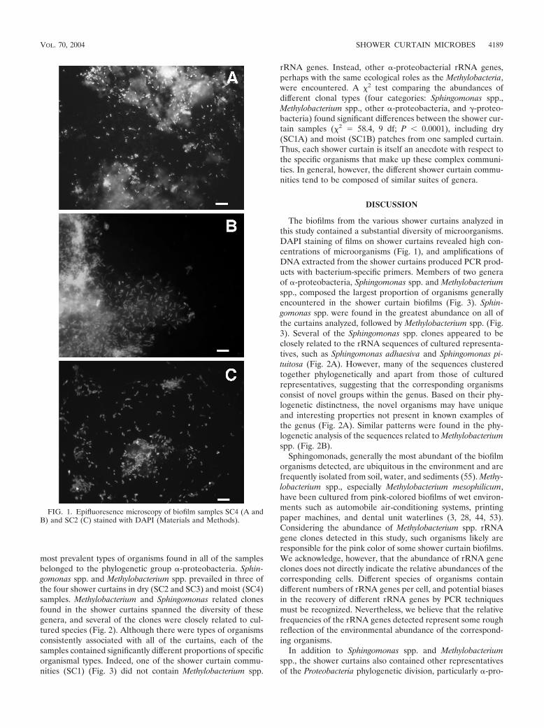
Vinyl shower curtains over time accumulate a film, flakeywhen dry, that is popularly referred to as “soap scum.” Exam-ination of this material from shower curtains by epifluores-cence microscopy, as shown in Fig. 1, revealed that the “soapscum” is in fact a lush bed of microbes, generally imbedded ina biofilm matrix. In order to survey rRNA gene sequences,DNA was purified from biofilms scraped from four differentshower curtains, including two different patches from one cur-tain, one dry and one continuously wet. All of the DNAsextracted from shower curtain samples produced PCR prod-ucts with bacteria-specific 16S rRNA gene (rDNA) primers.None of the extraction controls showed detectable amounts ofamplified rDNA. PCR products were cloned, and unique se-quences were identified by RFLP analysis and sequenced (22).In the course of the study, 337 clones were screened and 117unique rRNA sequences were determined.
The sequences obtained from the shower curtain microbeswere compared to each other and to sequences in GenBank.Results of phylogenetic analyses for the Sphingomonas- andMethylobacterium-related sequences are presented in Fig. 2.There were no identical sequences between different showercurtain samples. Instead, as generally occurs with environmen-tal samples, members of phylogenetic clusters of closely relatedsequences were seen. Several of the clone DNA sequenceswere closely related to cultured organisms. The resolution ofthe trees was fairly low in several places, likely because the dataset was limited (only 670 positions). Despite the limitations ofthe data set, the relevant relationships were supported withhigh MP and NJ bootstrap values (�70%). In relating the newsequences to known ones, we took relatedness clusters with97% or higher sequence identity to correspond to a species-level relationship and clusters with 95% or higher sequenceidentity to correspond to a genus-level relationship (50).
The pie chart diagrams in Fig. 3 illustrate the diversity andabundance of organisms found in the various shower curtainsamples. The sequences represented are only the most abun-dant of the sequences encountered. Approximately 15% of theclones analyzed on the basis of RFLP or sequence were ob-served only once, so we did not nearly exhaust the diversity oforganisms that comprise the communities. Most of the cloneswe sequenced were �95% identical to sequences already inGenBank. Only 38 out of 337 clones screened (11.2%) wereless than 95% identical (genus-level relatedness) to knownsequences in GenBank. Few clones had less than 90% identi-ties to known sequences. Although the specific sequences as-sociated with the different shower curtains differed in detail,the phylogenetic comparisons revealed common themes. The
4188 KELLEY ET AL. APPL. ENVIRON. MICROBIOL.
most prevalent types of organisms found in all of the samplesbelonged to the phylogenetic group �-proteobacteria. Sphin-gomonas spp. and Methylobacterium spp. prevailed in three ofthe four shower curtains in dry (SC2 and SC3) and moist (SC4)samples. Methylobacterium and Sphingomonas related clonesfound in the shower curtains spanned the diversity of thesegenera, and several of the clones were closely related to cul-tured species (Fig. 2). Although there were types of organismsconsistently associated with all of the curtains, each of thesamples contained significantly different proportions of specificorganismal types. Indeed, one of the shower curtain commu-nities (SC1) (Fig. 3) did not contain Methylobacterium spp.
rRNA genes. Instead, other �-proteobacterial rRNA genes,perhaps with the same ecological roles as the Methylobacteria,were encountered. A 2 test comparing the abundances ofdifferent clonal types (four categories: Sphingomonas spp.,Methylobacterium spp., other �-proteobacteria, and -proteo-bacteria) found significant differences between the shower cur-tain samples (2 � 58.4, 9 df; P � 0.0001), including dry(SC1A) and moist (SC1B) patches from one sampled curtain.Thus, each shower curtain is itself an anecdote with respect tothe specific organisms that make up these complex communi-ties. In general, however, the different shower curtain commu-nities tend to be composed of similar suites of genera.
DISCUSSION
The biofilms from the various shower curtains analyzed inthis study contained a substantial diversity of microorganisms.DAPI staining of films on shower curtains revealed high con-centrations of microorganisms (Fig. 1), and amplifications ofDNA extracted from the shower curtains produced PCR prod-ucts with bacterium-specific primers. Members of two generaof �-proteobacteria, Sphingomonas spp. and Methylobacteriumspp., composed the largest proportion of organisms generallyencountered in the shower curtain biofilms (Fig. 3). Sphin-gomonas spp. were found in the greatest abundance on all ofthe curtains analyzed, followed by Methylobacterium spp. (Fig.3). Several of the Sphingomonas spp. clones appeared to beclosely related to the rRNA sequences of cultured representa-tives, such as Sphingomonas adhaesiva and Sphingomonas pi-tuitosa (Fig. 2A). However, many of the sequences clusteredtogether phylogenetically and apart from those of culturedrepresentatives, suggesting that the corresponding organismsconsist of novel groups within the genus. Based on their phy-logenetic distinctness, the novel organisms may have uniqueand interesting properties not present in known examples ofthe genus (Fig. 2A). Similar patterns were found in the phy-logenetic analysis of the sequences related to Methylobacteriumspp. (Fig. 2B).
Sphingomonads, generally the most abundant of the biofilmorganisms detected, are ubiquitous in the environment and arefrequently isolated from soil, water, and sediments (55). Methy-lobacterium spp., especially Methylobacterium mesophilicum,have been cultured from pink-colored biofilms of wet environ-ments such as automobile air-conditioning systems, printingpaper machines, and dental unit waterlines (3, 28, 44, 53).Considering the abundance of Methylobacterium spp. rRNAgene clones detected in this study, such organisms likely areresponsible for the pink color of some shower curtain biofilms.We acknowledge, however, that the abundance of rRNA geneclones does not directly indicate the relative abundances of thecorresponding cells. Different species of organisms containdifferent numbers of rRNA genes per cell, and potential biasesin the recovery of different rRNA genes by PCR techniquesmust be recognized. Nevertheless, we believe that the relativefrequencies of the rRNA genes detected represent some roughreflection of the environmental abundance of the correspond-ing organisms.
In addition to Sphingomonas spp. and Methylobacteriumspp., the shower curtains also contained other representativesof the Proteobacteria phylogenetic division, particularly �-pro-
FIG. 1. Epifluoresence microscopy of biofilm samples SC4 (A andB) and SC2 (C) stained with DAPI (Materials and Methods).
VOL. 70, 2004 SHOWER CURTAIN MICROBES 4189

teobacteria. Proteobacteria are diverse in physiology and ubiq-uitous in water environments, ranging from deep seawater towaste and drinking water (8, 48, 49). The sources of carbon andenergy for these biofilm communities are unclear. Potentialfood resources include soap products, sloughed-off human de-bris, and bath area volatiles. Biofilms provide a supportive andprotective environment in which many kinds of metabolism canthrive. Sphingomonads, for instance, use a broad range ofcarbon compounds, including complex organics such as diben-zofuran (18) and hexachlorocyclohexane (23). Some isolatesfrom the deep subsurface have been shown to metabolize ar-omatic compounds such as toluene, naphthalene, and others,although laboratory strains have not shown these properties(17). Sphingomonads colonize new environments readily andadhere to surfaces through the production of exopolysacchar-ides, such as gellan, and welan (41). Methylobacterium spp. alsothrive on numerous different kinds of carbon sources, such assuccinate, ethanol, ethanolamine, methanol, and methylamine(26, 54).
Potential pathogens. In this limited survey, we did not en-counter any known specific pathogens. However, several sphin-gomonads are known to be opportunistic pathogens. For in-stance, Sphingomonas paucimobilis has a history of infecting
immune-compromised patients or persons with predisposingconditions (21). Infection with S. paucimobilis can lead to in-travascular catheter-related bacteremia, urinary tract infec-tions, pneumonia, cutaneous infections, and visceral abscesses(7, 21, 45). In hospitals, sources of S. paucimobilis infectionshave been traced to fluid in humidifiers and tap water (21, 31,40).
Methylobacterium spp. also are known to cause infections inimmune-compromised patients or patients with other diseasesthat render them prone to infection (15, 16, 20, 25, 35, 47). Wefound several clones representing organisms closely related toMethylobacterium extorquens and Methylobacterium zatmanii(Fig. 2B), both known to cause illness in immune-compro-mised patients (20, 25). We also encountered abundant M.mesophilicum in a 16S rDNA clone library prepared from hu-midifier filter samples (data not shown). In patients, M. meso-philicum has been detected in blood, peritoneal fluid, andascitic fluid and has been known to cause pneumonia, skinulcers, empyema, keratitis, and bacteremia (25, 35, 47). Basedon the rising number of cases, it has been proposed that Methy-lobacterium spp. infections could increase dramatically in thefuture (16, 19, 20, 42, 47).
In addition to the consistent presence of Sphingomonas spp.
FIG. 2. Results of phylogenetic analyses with Sphingomonas spp.-related (A) and Methylobacterium spp.-related (B) 16S rRNA sequencesobtained in this study. The analyses performed were based on alignments of approximately 670 nucleotide positions. The alignments includedcultured Sphingomonas and Methylobacterium spp. and outgroup -proteobacteria (Escherichia coli L10328), -proteobacteria (Nitrosospiramutiformis L35509), and �-proteobacteria (Roseobacter denitrificans M59063). The phylogenetic trees shown were estimated by using ML. MP andNJ analyses converged on very similar tree topologies. Filled circles indicate both MP and NJ bootstrap support exceeding 70%, and open circlesindicate bootstrap support exceeding 50%. (See Materials and Methods for details on the phylogenetic and bootstrap analyses.).
4190 KELLEY ET AL. APPL. ENVIRON. MICROBIOL.

and Methylobacterium spp., we also uncovered a number ofother bacterial species at low frequencies. Some of these spe-cies are closely related to known opportunistic pathogens, in-cluding Nocardia spp. and Gordonia spp. (high-G�C gram-positive bacteria). Infections with Gordonia terrae can invadewounds and result in bacteremia and brain abscess (34). Pa-tients with such infections usually have underlying diseasespredisposing them to opportunistic infections, but infections ofhealthy patients have been reported (9). There have beenreports of severe infections in immune-compromised patientsby several Nocardia species, which often are misidentified bystandard culture methods (11). Other microorganisms de-tected in samples include close relatives of the known oppor-tunistic pathogens Afipia felis (36) and Moraxella osloensis (4).
Our results suggest that shower curtains harbor potentialopportunistic pathogens that can threaten immune-compro-mised or otherwise ill patients. For immune-compromised peo-ple, consistent exposure to sources of infection, such as showercurtains, is a public health problem. Exposure can be mini-mized by regular cleaning or by changing shower curtains.
ACKNOWLEDGMENTS
We thank Mark Hernandez (Department of Civil, Environmentaland Architectural Engineering, University of Colorado at Boulder) for
contributions to this study. We thank Ruth Ley and Amy Buck forcontributing their shower curtains.
This work was supported in part by a grant from the NationalInstitutes of Health to N.R.P.
REFERENCES
1. Acinas, S. G., J. Anton, and F. Rodriguez-Valera. 1999. Diversity of free-living and attached bacteria in offshore Western Mediterranean waters asdepicted by analysis of genes encoding 16S rRNA. Appl. Environ. Microbiol.65:514–522.
2. Amann, R. I., W. Ludwig, and K. H. Schleifer. 1995. Phylogenetic identifi-cation and in situ detection of individual microbial cells without cultivation.Microbiol. Rev. 59:143–169.
3. Barbeau, J., R. Tanguay, E. Faucher, C. Avezard, L. Trudel, L. Cote, andA. P. Prevost. 1996. Multiparametric analysis of waterline contamination indental units. Appl. Environ. Microbiol. 62:3954–3959.
4. Berrocal, A. M., I. U. Scott, D. Miller, and H. W. Flynn, Jr. 2002. Endoph-thalmitis caused by Moraxella osloensis. Graefe’s Arch. Clin. Exp. Ophthal-mol. 240:329–330.
5. Boe-Hansen, R., H. J. Albrechtsen, E. Arvin, and C. Jorgensen. 2002. Bulkwater phase and biofilm growth in drinking water at low nutrient conditions.Water Res. 36:4477–4486.
6. Casadevall, A. 1996. Crisis in infectious diseases: time for a new paradigm?Clin. Infect. Dis. 23:790–794.
7. Casadevall, A., L. F. Freundlich, and L. Pirofski. 1992. Septic shock causedby Pseudomonas paucimobilis. Clin. Infect. Dis. 14:784.
8. Cottrell, M. T., and D. L. Kirchman. 2000. Community composition ofmarine bacterioplankton determined by 16S rRNA gene clone libraries andfluorescence in situ hybridization. Appl. Environ. Microbiol. 66:5116–5122.
9. Drancourt, M., J. Pelletier, A. A. Cherif, and D. Raoult. 1997. Gordonaterrae central nervous system infection in an immunocompetent patient.J. Clin. Microbiol. 35:379–382.
FIG. 3. Compositions of rRNA gene libraries obtained from shower curtain samples. Numbers of clones of each rRNA gene sequence fromlibraries are grouped as identified on the pie charts as follows: Methylobact, Methylobacterium spp.; Sphingom, Sphingomonas spp.; �-Protbact,�-proteobacteria; �-Protbact, �-proteobacteria; -Protbact, -proteobacteria; Actinomy, Actinomycetales; hGCgpbact, high-G�C gram-positivebacteria; CFBgroup, Cytophaga-Flavobacteria-Bacteroides group; Sphingobac, sphingobacteria.
VOL. 70, 2004 SHOWER CURTAIN MICROBES 4191
10. du Moulin, G. C., K. D. Stottmeier, P. A. Pelletier, A. Y. Tsang, and J. Hed-ley-Whyte. 1988. Concentration of Mycobacterium avium by hospital hotwater systems. JAMA 260:1599–1601.
11. Eggink, C. A., P. Wesseling, P. Boiron, and J. F. Meis. 1997. Severe keratitisdue to Nocardia farcinica. J. Clin. Microbiol. 35:999–1001.
12. Embil, J., P. Warren, M. Yakrus, R. Stark, S. Corne, D. Forrest, and E.Hershfield. 1997. Pulmonary illness associated with exposure to Mycobacte-rium avium-complex in hot tub water. Hypersensitivity pneumonitis or in-fection? Chest 111:813–816.
13. Excoffier, L., P. E. Smouse, and J. M. Quattro. 1992. Analysis of molecularvariance inferred from metric distances among DNA haplotypes: applicationto human mitochondrial DNA restriction data. Genetics 131:479–491.
14. Falkinham, J. O., III, C. D. Norton, and M. W. LeChevallier. 2001. Factorsinfluencing numbers of Mycobacterium avium, Mycobacterium intracellulare,and other mycobacteria in drinking water distribution systems. Appl. Envi-ron. Microbiol. 67:1225–1231.
15. Fernandez, M., Z. Dreyer, M. Hockenberry-Eaton, and C. J. Baker. 1997.Methylobacterium mesophilica as a cause of persistent bacteremia in a childwith lymphoma. Pediatr. Infect. Dis. J. 16:1007–1008.
16. Flournoy, D. J., R. L. Petrone, and D. W. Voth. 1992. A pseudo-outbreak ofMethylobacterium mesophilica isolated from patients undergoing bronchos-copy. Eur. J. Clin. Microbiol. Infect. Dis. 11:240–243.
17. Fredrickson, J. K., D. L. Balkwill, G. R. Drake, M. F. Romine, D. B. Rin-gelberg, and D. C. White. 1995. Aromatic-degrading Sphingomonas isolatesfrom the deep subsurface. Appl. Environ. Microbiol. 61:1917–1922.
18. Fukuda, K., S. Nagata, and H. Taniguchi. 2002. Isolation and characteriza-tion of dibenzofuran-degrading bacteria. FEMS Microbiol. Lett. 208:179–185.
19. Hiraishi, A., K. Furuhata, A. Matsumoto, K. A. Koike, M. Fukuyama, and K.Tabuchi. 1995. Phenotypic and genetic diversity of chlorine-resistant Methy-lobacterium strains isolated from various environments. Appl. Environ. Mi-crobiol. 61:2099–2107.
20. Hornei, B., E. Luneberg, H. Schmidt-Rotte, M. Maass, K. Weber, F. Heits,M. Frosch, and W. Solbach. 1999. Systemic infection of an immunocompro-mised patient with Methylobacterium zatmanii. J. Clin. Microbiol. 37:248–250.
21. Hsueh, P. R., L. J. Teng, P. C. Yang, Y. C. Chen, H. J. Pan, S. W. Ho, andK. T. Luh. 1998. Nosocomial infections caused by Sphingomonas paucimo-bilis: clinical features and microbiological characteristics. Clin. Infect. Dis.26:676–681.
22. Hugenholtz, P., C. Pitulle, K. L. Hershberger, and N. R. Pace. 1998. Noveldivision level bacterial diversity in a Yellowstone hot spring. J. Bacteriol.180:366–376.
23. Imai, R., Y. Nagata, M. Fukuda, M. Takagi, and K. Yano. 1991. Molecularcloning of a Pseudomonas paucimobilis gene encoding a 17-kilodaltonpolypeptide that eliminates HCl molecules from gamma-hexachlorocyclo-hexane. J. Bacteriol. 173:6811–6819.
24. Kahana, L. M., J. M. Kay, M. A. Yakrus, and S. Waserman. 1997. Myco-bacterium avium complex infection in an immunocompetent young adultrelated to hot tub exposure. Chest 111:242–245.
25. Kaye, K. M., A. Macone, and P. H. Kazanjian. 1992. Catheter infectioncaused by Methylobacterium in immunocompromised hosts: report of threecases and review of the literature. Clin. Infect. Dis. 14:1010–1014.
26. Korotkova, N., and M. E. Lidstrom. 2001. Connection between poly-beta-hydroxybutyrate biosynthesis and growth on C(1) and C(2) compounds in themethylotroph Methylobacterium extorquens AM1. J. Bacteriol. 183:1038–1046.
27. Korvick, J. A., J. D. Rihs, G. L. Gilardi, and V. L. Yu. 1989. A pink-pigmented, oxidative, nonmotile bacterium as a cause of opportunistic in-fections. Arch. Intern. Med. 149:1449–1451.
28. Kressel, A. B., and F. Kidd. 2001. Pseudo-outbreak of Mycobacterium che-lonae and Methylobacterium mesophilicum caused by contamination of anautomated endoscopy washer. Infect. Control Hosp. Epidemiol. 22:414–418.
29. Le Dantec, C., J. P. Duguet, A. Montiel, N. Dumoutier, S. Dubrou, and V.Vincent. 2002. Occurrence of mycobacteria in water treatment lines and inwater distribution systems. Appl. Environ. Microbiol. 68:5318–5325.
30. Lee, T. C., J. E. Stout, and V. L. Yu. 1988. Factors predisposing to Legionellapneumophila colonization in residential water systems. Arch. Environ.Health 43:59–62.
31. Lemaitre, D., A. Elaichouni, M. Hundhausen, G. Claeys, P. Vanhaesebrouck,M. Vaneechoutte, and G. Verschraegen. 1996. Tracheal colonization withSphingomonas paucimobilis in mechanically ventilated neonates due to con-taminated ventilator temperature probes. J. Hosp. Infect. 32:199–206.
32. Leoni, E., P. Legnani, M. T. Mucci, and R. Pirani. 1999. Prevalence ofmycobacteria in a swimming pool environment. J. Appl. Microbiol. 87:683–688.
33. Leoni, E., P. P. Legnani, M. A. Bucci Sabattini, and F. Righi. 2001. Preva-lence of Legionella spp. in swimming pool environment. Water Res. 35:3749–3753.
34. Lesens, O., Y. Hansmann, P. Riegel, R. Heller, M. Benaissa-Djellouli, M.Martinot, H. Petit, and D. Christmann. 2000. Bacteremia and endocarditiscaused by a Gordonia species in a patient with a central venous catheter.Emerg. Infect. Dis. 6:382–385.
35. Liu, J. W., J. J. Wu, H. M. Chen, A. H. Huang, W. C. Ko, and Y. C. Chuang.1997. Methylobacterium mesophilicum synovitis in an alcoholic. Clin. Infect.Dis. 24:1008–1009.
36. Luhrmann, A., K. Streker, A. Schuttfort, J. J. Daniels, and A. Haas. 2001.Afipia felis induces uptake by macrophages directly into a nonendocyticcompartment. Proc. Natl. Acad. Sci. USA 98:7271–7276.
37. Martin, D. S., P. Oray-Schrom, and Y. Amoateng-Adjepong. 2002. Emergingsignificance of Mycobacterium avium-complex infection in an inner-city hos-pital. Conn. Med. 66:323–330.
38. Nelson, F. P., S. Macari, G. Faria, S. Assed, and I. Y. Ito. 2000. Microbialcontamination of toothbrushes and their decontamination. Pediatr. Dent.22:381–384.
39. Pace, N. R. 1997. A molecular view of microbial diversity and the biosphere.Science 276:734–740.
40. Perola, O., T. Nousiainen, S. Suomalainen, S. Aukee, U. M. Karkkainen, J.Kauppinen, T. Ojanen, and M. L. Katila. 2002. Recurrent Sphingomonaspaucimobilis -bacteraemia associated with a multi-bacterial water-borne ep-idemic among neutropenic patients. J. Hosp. Infect. 50:196–201.
41. Pollock, T. J., W. A. van Workum, L. Thorne, M. J. Mikolajczak, M.Yamazaki, J. W. Kijne, and R. W. Armentrout. 1998. Assignment of bio-chemical functions to glycosyl transferase genes which are essential forbiosynthesis of exopolysaccharides in Sphingomonas strain S88 and Rhizo-bium leguminosarum. J. Bacteriol. 180:586–593.
42. Rice, E. W., D. J. Reasoner, C. H. Johnson, and L. A. DeMaria. 2000.Monitoring for methylobacteria in water systems. J. Clin. Microbiol. 38:4296–4297.
43. Rose, A. M., K. Sinka, J. M. Watson, J. Y. Mortimer, and A. Charlett. 2002.An estimate of the contribution of HIV infection to the recent rise intuberculosis in England and Wales. Thorax 57:442–445.
44. Rose, L. J., R. B. Simmons, S. A. Crow, and D. G. Ahearn. 2000. Volatileorganic compounds associated with microbial growth in automobile air con-ditioning systems. Curr. Microbiol. 41:206–209.
45. Salazar, R., R. Martino, A. Sureda, S. Brunet, M. Subira, and A. Domingo-Albos. 1995. Catheter-related bacteremia due to Pseudomonas paucimobilisin neutropenic cancer patients: report of two cases. Clin. Infect. Dis. 20:1573–1574.
46. Sambrook, J., and D. W. Russel (ed.). 2001. Molecular cloning, 3rd ed., vol.1. Cold Spring Harbor Laboratory Press, Cold Spring Harbor, N.Y.
47. Sanders, J. W., J. W. Martin, M. Hooke, and J. Hooke. 2000. Methylobac-terium mesophilicum infection: case report and literature review of an un-usual opportunistic pathogen. Clin. Infect. Dis. 30:936–938.
48. Schwartz, T., S. Hoffmann, and U. Obst. 1998. Formation and bacterialcomposition of young, natural biofilms obtained from public bank-filtereddrinking water systems. Water Res. 32:2787–2797.
49. Seviour, R. J., A. M. Maszenan, J. A. Soddell, V. Tandoi, B. K. Patel, Y.Kong, and P. Schumann. 2000. Microbiology of the �G-bacteria’ in activatedsludge. Environ. Microbiol. 2:581–593.
50. Stackebrandt, E., and B. M. Goebel. 1994. Taxonomic note: a place forDNA-DNA reassociation and 16S rRNA sequence analysis in the presentspecies definition in bacteriology. Int. J. Syst. Bacteriol. 44:846–849.
51. Swofford, D. 1998. PAUP*: phylogenetic analysis using parsimony (*andother methods), 4th ed. Sinauer Associates, Sunderland, Mass.
52. Tanner, M. A., D. Shoskes, A. Shahed, and N. R. Pace. 1999. Prevalence ofcorynebacterial 16S rRNA sequences in patients with bacterial and “non-bacterial” prostatitis. J. Clin. Microbiol. 37:1863–1870.
53. Vaisanen, O. M., A. Weber, A. Bennasar, F. A. Rainey, H. J. Busse, and M. S.Salkinoja-Salonen. 1998. Microbial communities of printing paper ma-chines. J. Appl. Microbiol. 84:1069–1084.
54. Vorholt, J. A., C. J. Marx, M. E. Lidstrom, and R. K. Thauer. 2000. Novelformaldehyde-activating enzyme in Methylobacterium extorquens AM1 re-quired for growth on methanol. J. Bacteriol. 182:6645–6650.
55. White, D. C., S. D. Sutton, and D. B. Ringelberg. 1996. The genus Sphin-gomonas: physiology and ecology. Curr. Opin. Biotechnol. 7:301–306.
4192 KELLEY ET AL. APPL. ENVIRON. MICROBIOL.