molecular and cellular mechanisms behind juvenile neuronal
TRANSCRIPT

Publications of the National Public Health Institute A 4/2006
Department of Molecular Medicine,
National Public Health Institute Helsinki, Finland
and
Department of Medical Genetics
University of Helsinki, Helsinki, Finland
Molecular and Cellular Mechanisms Behind Juvenile Neuronal Ceroid Lipofuscinosis (JNCL, Batten Disease)
Kaisu Luiro

Kaisu Luiro
MOLECULAR AND CELLULAR MECHANISMS
BEHIND
JUVENILE NEURONAL CEROID LIPOFUSCINOSIS (JNCL, BATTEN DISEASE)
A C A D E M I C D I S S E R T A T I O N
To be presented with the permission of the Medical Faculty of the University of Helsinki, for public examination in the large lecture hall, Haartman Institute, on
March 24th, 2006, at 12 noon.
Department of Molecular Medicine,
National Public Health Institute, Helsinki, Finland
Department of Medical Genetics
University of Helsinki, Helsinki, Finland
Hospital for Children and Adolescents,
Helsinki University Central Hospital, Helsinki, Finland
Helsinki 2006

H e l s i n k i U n i v e r s i t y B i o m e d i c a l D i s s e r t a t i o n s N o 7 4 ISSN 1457-8433
P u b l i c a t i o n s o f t h e N a t i o n a l P u b l i c H e a l t h I n s t i t u t e
K T L A 4 / 2 0 0 6
Copyright National Public Health Institute
Julkaisija-Utgivare-Publisher
Kansanterveyslaitos (KTL)Mannerheimintie 166 00300 Helsinki Puh. vaihde (09) 474 41, telefax (09) 4744 8408
FolkhälsoinstitutetMannerheimvägen 166 00300 Helsingfors Tel. växel (09) 474 41, telefax (09) 4744 8408
National Public Health Institute Mannerheimintie 166 FIN-00300 Helsinki, Finland Telephone +358 9 474 41, telefax +358 9 4744 8408
ISBN 951-740-604-5 ISSN 0359-3584 ISBN 951-740-604-3 (pdf) ISSN 1458-6290 (pdf)
Kannen kuva - cover graphic: Kaisu Luiro
Painopaikka Helsinki 2006

S u p e r v i s e d b y
Adjunct Professor Anu Jalanko National Public Health Institute Helsinki, Finland
R e v i e w e d b y
Adjunct Professor, Phycisian-in-Chief Helena Pihko Department of Child Neurology Hospital for Children and Adolescents Helsinki University Central Hospital (HUCH) Helsinki, Finland
Adjunct Professor Varpu Marjomäki Department of Biological and Environmental Science University of Jyväskylä Jyväskylä, Finland
O p p o n e n t
Professor Elina Ikonen Institute of Biomedicine/Anatomy University of Helsinki Helsinki, Finland


The best way to have a good idea is to have lots of ideas. Linus Pauling (1901-1994)
To my family

6
Kaisu Luiro, Molecular and Cellular Mechanisms behind Juvenile Neuronal Ceroid-Lipofuscinosis (JNCL, Batten disease)
Publications of the National Public Health Institute, A4/2006, 98 Pages ISBN 951-740-604-5; 951-740-604-3 (pdf-version) ISSN 0359-3584; 1458-6290 (pdf-version) http://www.ktl.fi/portal/4043
ABSTRACT
Neurodegenerative disorders are chronic, progressive, and often fatal disorders of the nervous system caused by dysfunction, and ultimately, death of neuronal cells. The underlying mechanisms of neurodegeneration are poorly understood, and monogenic disorders can be utilised as disease models to elucidate the pathogenesis.
Juvenile neuronal ceroid-lipofuscinosis (JNCL, Batten disease) is a recessively inherited lysosomal storage disorder with progressive neurodegeneration and accumulation of autofluorescent storage material in most tissues. It is caused by mutations in the CLN3 gene, but the exact function of the corresponding CLN3 protein, as well as the molecular mechanisms of JNCL pathogenesis have remained elusive. JNCL disease exclusively affects the central nervous system leaving other organs unaffected, and therefore it is of a particular importance to conduct studies in brain tissue and neuronal cells.
The aim of this thesis project was to elucidate the molecular and cell biological mechanisms underlying JNCL. This was the first study to describe the endogenous Cln3 protein, and it was shown that Cln3 localised to neuronal cells in the mouse brain. At a subcellular level, endogenous Cln3 was localised to the presynaptic terminals and to the synaptosome compartment, but not to the synaptic vesicles. Studies with the CLN3-deficient cells demonstrated an impaired endocytic membrane trafficking, and established an interconnection between CLN3, microtubulus-binding Hook1 and Rab proteins. This novel data was not only important in characterising the roles of CLN3 in cells, but also provided significant information delineating the versatile role of the Rab proteins. To identify affected cellular pathways in JNCL, global gene expression profiling of the knock-out mouse Cln3-/- neurons was performed and systematically analysed; this revealed a slight dysfunction of the mitochondria, cytoskeletal abnormality in the microtubule plus-end, and an impaired recovery from depolarizing stimulus when specific N-type Ca2+ channels were inhibited, thus leading to a prolonged time of higher intracellular

7
Ca2+. All these defective pathways are interrelated, and may together be sufficient to iniate the neurodegenerative process. Results of this thesis also suggest that in neuronal cells, CLN3 most likely functions at endocytic vesicles at the presynaptic terminal, potentially involved in the regulation of the calcium-mediated synaptic transmission.
Keywords: neurodegeneration, lysosomal storage disorder, neuronal ceroid lipofuscinosis, lysosome, microtubulus, endocytosis, intracellular membrane trafficking

8
Kaisu Luiro, JNCL-taudin (juveniili neuronaalinen seroidi-liposfuskinoosi, Battenin tauti) molekyyli- ja solutason tautimekanismit
Kansanterveyslaitoksen julkaisuja, A4/2006, 98 sivua ISBN 951-740-604-5; 951-740-604-3 (pdf-versio) ISSN 0359-3584; 1458-6290 (pdf-versio) http://www.ktl.fi/portal/4043
TIIVISTELMÄ
Hermoston rappeumasairaudet ovat kroonisia, eteneviä ja usein kuolemaan johtavia tauteja, jotka johtuvat hermosolujen toimintahäiriöistä ja kuolemasta. Hermosolurappeuman tarkat mekanismit ovat huonosti tunnettuja ja yhden geenivirheen aiheuttamat monogeeniset taudit voivat toimia tautimalleina patogeneesin selvittämisessä.
Juveniili neuronaalinen seroidi-lipofuskinoosi (JNCL, Battenin tauti) on peittyvästi periytyvä lysosomaalinen kertymätauti, jonka tyypillisiä piirteitä ovat etenevä keskushermoston hermosolujen rappeuma ja autofluoresoivan materiaalin kertyminen useimpiin kudoksiin. JNCL johtuu virheistä CLN3-geenissä, mutta vastaavan CLN3-proteiinin toiminta ja JNCL-taudin syntymekanismit ovat tuntemattomia. Taudin oireet keskittyvät yksinomaan keskushermostoon ja siksi on tärkeää tutkia tautia aivokudoksessa ja hermosoluissa.
Tämän väitöskirjaprojektin tavoitteena oli valottaa JNCL-taudin solu- ja molekyylitason tautimekanismeja erilaisissa solumalleissa. Työssä kuvailtiin ensimmäistä kertaa hiiren Cln3-proteiinin paikantumista aivoissa ja hermosoluissa. Cln3 paikantui kudostasolla hermosoluihin ja hermosoluissa pre-synaptiselle alueelle, synaptosomiin, mutta ei varsinaisiin synaptisiin vesikkeleihin. Kokeet CLN3-puutteisilla soluilla osoittivat solutason häiriön endosyyttisessä kalvoliikenteessä ja yhdistivät CLN3-proteiinin solun mikrotubulus-tukirankaan. Tutkimuksessa saatiin myös tärkeää tietoa solun kalvoliikenteessä toimivien Rab-proteiinien toiminnasta. JNCL-taudissa tärkeitä metaboliareittejä tutkittiin vertailemalla geeni-ilmentymistä poistogeenisen Cln3-/- hiiren hermosoluisssa ja normaaleissa kontrolleissa mikrosiruanalyysin avulla. Löydökset viittasivat lievään alentumaan mitokondrion hengitysketjun toiminnassa ja poikkeavuuteen solun tukirangassa. Lisäksi havaittiin häiriö kalsiumvälitteisen hermoimpulssin säätelyssä, mikä voi johtaa pidentyneeseen korkeaan solunsisäiseen Ca2+-tasoon. Nämä solunsisäiset metaboliareitit ovat liittyneet toisiinsa ja niiden yhtäaikainen

9
toimintahäiriö voi johtaa hermosolujen rappeumamekanismien aktivoitumiseen. Väitöskirjatyön tulokset viittaavat siihen, että CLN3 todennäköisesti toimii endosyyttisissä vesikkeleissä hermosolujen presynaptisella alueella ja mahdollisesti osallistuu kalsiumvälitteisen hermoimpulssin säätelyyn.
Avansanat: neurodegeneraatio, lysosomaaliset kertymätaudit, neuronaalinen seroidi-lipofuskinoosi, lysosomi, mikrotubulus, endosytoosi, solunsisäinen kalvoliikenne,

10
TABLE OF CONTENTS
ABSTRACT ........................................................................................................ 6
TABLE OF CONTENTS................................................................................. 10
LIST OF ORIGINAL PUBLICATIONS ....................................................... 12
ABBREVIATIONS .......................................................................................... 13
INTRODUCTION............................................................................................ 15
REVIEW OF THE LITERATURE................................................................ 17
1. Intracellular membrane trafficking............................................................ 17 1.1 Principles of intracellular membrane trafficking ................................. 17 1.2 Pathways of endocytosis...................................................................... 21 1.3 Specific features of transport in neurons ............................................. 25 1.4 Synaptic transmission .......................................................................... 27
2. Lysosomes and lysosomal storage disorders ............................................. 31 2.1 The lysosome turns fifty ...................................................................... 31 2.2 Targeting of lysosomal proteins .......................................................... 33 2.3 Lysosomal storage disorders (LSDs)................................................... 36 2.4 Mechanisms for LSDs and potential for therapy ................................. 38
3. Juvenile neuronal ceroid lipofuscinosis..................................................... 40 3.1 Neuronal ceroid lipofuscinoses (NCLs) .............................................. 40 3.2 Clinical findings in JNCL.................................................................... 43 3.3 Neuropathology of JNCL .................................................................... 43 3.4 Neuroimaging findings in JNCL ......................................................... 44 3.5 Neurophysiological findings in JNCL ................................................. 44 3.6 Intracellular storage in JNCL and its significance to pathology.......... 45 3.7 CLN3 gene and mutations ................................................................... 46 3.8 CLN3 protein....................................................................................... 47 3.9 Post-translational modifications of CLN3 ........................................... 48 3.10 Subcellular localization of CLN3 ...................................................... 50 3.11 Interactions of CLN3 protein............................................................. 51 3.12 Experimental disease models of JNCL.............................................. 51
3.12.1 Mouse models for JNCL ............................................................ 51 3.12.2 Experimental model organisms for JNCL.................................. 53
AIMS OF THE PRESENT STUDY................................................................ 55
MATERIALS AND METHODS..................................................................... 56

11
1.1 Published materials and methods ............................................................ 56 1.2 Unpublished methods............................................................................... 57
1.2.1 Dextran uptake and intracellular trafficking..................................... 57 1.3 Ethical aspects ......................................................................................... 57
RESULTS AND DISCUSSION....................................................................... 58
1. Localization of the CLN3/Cln3 to the neuronal synapses (I).................... 58 1.1 Cln3 gene and protein in mouse brain ................................................. 58 1.2 Co-localization of the CLN3/Cln3 with presynaptic proteins in neurons 59 1.3 Synaptosomal localization of CLN3.................................................... 60
2. CLN3 connected to cytoskeleton and endocytic membrane trafficking (II, unpublished).......................................................................................... 61
2.1 Effect of CLN3 on the Hook1 protein ................................................. 61 2.2 Defective endocytosis in JNCL fibroblasts ......................................... 62 2.3 Interactions of Hook1 with CLN3 and the Rab proteins ..................... 65
3. Fundamental metabolic pathways affected in the Cln3-/- neurons (III) ..... 66 3.1 Comparative gene expression profiling reveals three major pathways affected ...................................................................................................... 67 3.2 Slight mitochondrial dysfunction in Cln3-/- mice ................................ 67 3.3 Down-regulation of the dynamic microtubular plus-end component in the Cln3-/- neurons...................................................................................... 68 3.4 Evidence of dysfunction in the calcium-mediated synaptic transmission .............................................................................................................69
CONCLUSIONS AND FUTURE PROSPECTS ........................................... 72
ACKNOWLEDGEMENTS............................................................................. 75
REFERENCES ................................................................................................. 78
ORIGINAL PUBLICATIONS........................................................................ 98

12
LIST OF ORIGINAL PUBLICATIONS
This thesis is based on the following original publications, which will be referred to in the text by their Roman numerals. In addition, some unpublished data will be presented.
I Luiro Kaisu, Kopra Outi, Lehtovirta Maarit, Jalanko Anu. CLN3 Protein Is Targeted to Neuronal Synapses But Excluded From Synaptic Vesicles: New Clues to Batten Disease. Hum. Mol. Genet. 10: 2123-2131, 2001.
II Luiro Kaisu, Yliannala Kristiina, Ahtiainen Laura, Maunu Heidi, Järvelä Irma, Kyttälä Aija, Jalanko Anu. Interconnections of CLN3, Hook1 and Rab proteins link Batten disease to defects in the endocytic pathway. Hum. Mol. Genet. 13: 3017-27, 2004.
III Luiro Kaisu, Kopra Outi, Blom Tomas, Gentile Massimiliano, Mitchison Hannah, Hovatta Iiris, Törnqvist Kid, Jalanko Anu. Batten disease (JNCL) is linked to disturbances in mitochondrial, cytoskeletal and synaptic functions. Submitted.

13
ABBREVIATIONS
aa amino acid(s)
ANCL adult neuronal ceroid lipofuscinosis (CLN4)
ATP adenosine triphosphate
bp base pair
cDNA complementary DNA
COS-1 cells African green monkey kidney cells
CLN3/CLN3 human CLN3 gene/protein
Cln3/Cln3 mouse Cln3 gene/protein
CNS central nervous system
DAB diaminobenzidine
DNA deoxyribonucleic acid
DNase deoxyribonuclease
ECL enhanced chemiluminescence
EGFP enhanced GFP
EM electron microscopy
ELISA enzyme-linked immunosorbent assay
ER endoplasmic reticulum
EST expressed sequence tag
FITC fluorescein isothiocyanate
GFP green fluorescent protein
GST glutathione S-transferase
HeLa cells cervical tumour cells
Hepes N-2-hydroxyethylpiperazine-N’-2-ethane sulfonic acid
HRP horseradish peroxidase
Ig immunoglobulin

14
INCL infantile neuronal ceroid lipofuscinosis (CLN1)
IPTG isopropyl-β-D-thiogalactoside
JNCL juvenile neuronal ceroid lipofuscinosis (CLN3)
kb kilobase(s)
kD kilodalton(s)
LINCL late infantile neuronal ceroid lipofuscinosis
LSD lysosomal storage disorder
MPR mannose 6-phosphate receptor
mRNA messenger RNA
NSF N-ethyl-maleimide sensitive fusion protein
ORF open reading frame
PAGE polyacrylamide gel electrophoresis
PBS phosphate buffered saline
PCR polymerase chain reaction
PFA paraformaldehyde
RNA ribonucleic acid
RT room temperature
RT-PCR reverse transcription PCR
SDS sodium dodecyl sulphate
SFV Semliki Forest virus
SNAP soluble NSF attachment protein
SNARE SNAP receptor
TGN trans-Golgi network
TRITC tetramethylrhodamine isothiocyanate
t-/v-SNARE target membrane/vesicle membrane SNARE
vLINCL variant form of late infantile neuronal ceroid lipofuscinosis (CLN2)
vLINCLFin Finnish variant form of late infantile neuronal ceroid lipofuscinosis (CLN5)
wt wild type

15
INTRODUCTION
Neurodegenerative disorders are a diverse group of acquired and inherited diseases of the nervous system. They are chronic, progressive and cannot be curably treated; thus they are associated with substantial morbidity, mortality, and great importance medically, socially and financially. Pathologically, the neurodegenerative diseases are characterised by the death of specific neuron populations at specific regions of the central or peripheral nervous system, and the individual pattern of this deterioration creates the specific clinical characteristics of each disease. There is, however, little understanding of the underlying pathogenetic processes and mechanisms of the neuronal death. Monogenic diseases, such as the lysosomal storage disorders and the neuronal ceroid lipofuscinoses, serve as disease models for studies on neuronal death in complex neurodegenerative disorders, and thus provide novel avenues for therapeutic intervention.
Juvenile neuronal ceroid lipofuscinosis (JNCL, Batten disease, or Spielmeyer-Vogt- Sjögren disease) is the most common neurodegenerative disease of childhood (Santavuori, et al., 2000). This autosomal recessively inherited disease is caused by mutations in the CLN3 gene, identified in 1995 by the International Batten Disease Consortium (Consortium, 1995). It is particularly enriched in Finland, and is part of the Finnish disease heritage (Norio, 2003). JNCL is classified as a lysosomal storage disorder (LSD), and it also belongs to a group of at least eight inherited progressive neurodegenerative diseases called neuronal ceroid lipofuscinoses (NCLs) (Haltia, 2003). NCL diseases are marked by two histopathological findings: degeneration of nerve cells, foremost in the cerebral cortex, and accumulation of autofluorescent ceroid-lipopigment in both neural and peripheral tissues (Goebel, 1997). In JNCL, the ultrastructure of the autofluorescent inclusions resembles a fingerprint and the major storage component is identified as mitochondrial ATP synthase subunit c (Palmer, et al., 1992).
Clinical features of JNCL include visual failure, epileptic seizures and progressive psychomotor degeneration, which lead to premature death at the age of 18-30 years (Santavuori, 1988). Most patients carry a 1.0-kb deletion of the CLN3 coding region; in addition 40 mutations and 2 polymorphisms have been characterised (NCL Mutation Database). The corresponding CLN3 protein is an integral membrane protein with 5-6 transmembrane domains and two lysosomal targeting motifs (Ezaki, et al., 2003; Kyttala, et al., 2004). The function of CLN3 has

16
remained elusive, but its evolutionary conservation among species from yeast to humans indicates a fundamental role in the cell metabolism.
JNCL disease specifically affects neuronal cells and leaves other organs clinically unaffected, and hence it is of utmost importance to approach the disease mechanism by studying the expression and localisation of CLN3 in the brain and neuronal cells. The aim of this thesis was to elucidate the molecular and cell biological properties of the CLN3 protein in neuronal and patient cells and tissues, with the ultimate goal of enhancing the understanding of the vital function of CLN3, and the mechanisms of neurodegeneration. This will provide the basis for the development of novel treatment strategies for these devastating diseases.

17
REVIEW OF THE LITERATURE
1. Intracellular membrane trafficking
1.1 Principles of intracellular membrane trafficking
Intracellular membrane trafficking can be divided into two distinct pathways; the biosynthetic/secretory pathway and the endocytic pathway (Figure 1). In the secretory pathway, the newly synthesized proteins, carbohydrates and lipids are transported through the endoplasmic reticulum (ER) and Golgi to the cell surface (constitutive secretory pathway), or to the endosomes/lysosomes. Endocytic pathway accommodates the internalization of macromolecules, solutes or pathogens into the cells, and will be discussed in detail below (Chapter 1.2). Transport routes form connections between the biosynthetic/secretory and endocytic pathways at the level of the Golgi apparatus and the endocytic compartments (Gruenberg and Maxfield, 1995).
Figure 1. Schematic representation of the major pathways in intracellular membrane trafficking. The arrows represent known or presumed transport routes. The coat complexes are indicated with colours; COPII (blue), COPI (red), and clathrin (yellow). ECV/MVB, endocytic carrier vesicle/multivesicular body. Modified from Bonifacino et al., 2004, and Gruenberg, 2001.

18
Characteristic of eukaryotic cells is their ability to compartmentalize functions into membrane bound organelles. Transport between these organelles occurs via membrane-enclosed transport intermediates, or vesicles. This vesicle-transport hypothesis was formulated from early electron microscopy findings (Palade, 1975). It postulated that vesicles bud off the donor compartment (vesicle budding) in a process associated with molecular sorting (or protein sorting), which allows the selective inclusion or exclusion of individual membrane and content proteins during the formation of vesicle, and the ability to segregate the vesicular container from its cargo after vesicle fusion. The transport vesicles are subsequently targeted to their specific acceptor compartment (vesicle targeting), where they undergo fusion with the acceptor membrane (vesicle fusion), and unload their cargo. The segregated processes allow the consequent recycling or release of the components involved in the vesicle formation and targeting, which may then be returned back to the donor compartment (Bonifacino and Glick, 2004; Mellman and Warren, 2000).
The selective incorporation of cargo and the budding of transport vesicles are mediated by protein coats. These coats deform the flat membrane sites into round structures that are eventually released as coated transport vesicles. The coats also actively participate in cargo selection, and potentially function in post-budding events by recruiting accessory factors that mediate interactions with the cytoskeleton and tethering factors (Bonifacino and Lippincott-Schwartz, 2003). The first coat to be identified was clathrin (Pearce, 1975; Roth and Porter, 1964), that functions in post-Golgi locations within the cells, including the plasma membrane, trans-Golginetwork (TGN), and endosomes. Clathrin binds to its target membranes in conjunction with one of its several adaptor complexes. Arf-GTP and/or specific phosphoinositides recruit the cytosolic clathrin adaptors in a specific combination to a particular membrane, to which clathrin is consequently recruited to form the spherical, cage-like lattice (Kirchhausen, 2000; Kirchhausen and Harrison, 1981). Known adaptors of clathrin include the heterotetrameric adaptor proteins (AP), monomeric GGAs (Golgi-localized, γ-ear-containing, ADP-ribosylation factor-binding proteins), Hrs (hepatocyte growth factor-regulated tyrosine kinase substrate), Epsin 1 and Eps15 (epidermal growth factor receptor substrate 15) (Reviewed in (Bonifacino and Lippincott-Schwartz, 2003). Non-clathrin coats typically facilitate vesicular transport in the early secretory pathway (Barlowe, et al., 1994; Waters, et al., 1991). COPII is the best characterized of these, and it mediates the anterograde transport from ER to either ER-Golgi intermediate compartment (ERGIC) or the Golgi complex (Barlowe, et al., 1994). COPI functions mainly in the retrograde transport within the Golgi complex and from the Golgi to the ER (Letourneur, et al., 1994) but may also act at the level of endosomes (Oprins, et al., 1993).
19
The principle of vesicle targeting describes the process, in which the vesicles emerging from the donor compartment bear “an address tag” that permits their docking and fusion only with the appropriate acceptor compartment. The core processes seem to be similar in all intracellular fusion with the exception of fusion of mitochondria and peroxisomes, which proceed via different processes (Hermann, et al., 1998; Sesaki and Jensen, 2001; Titorenko and Rachubinski, 2000). The first step in intracellular vesicle fusion is to recognise the appropriate partner membrane, and this process is variably called membrane attachment, or tethering and docking. Central in membrane attachment are the Rab GTPases that are localised to specific intracellular compartments and may serve as identity markers for them. Other tethering factors have also been identified (Whyte and Munro, 2002). The Rab GTPase family consists of over 60 members in humans and they cycle between the active, membrane bound GTP- and the inactive, soluble GDP-bound states in response to regulating factors (Stenmark and Olkkonen, 2001; Zerial and McBride, 2001). Rab proteins act directionally; they are localised to the donor membrane in order to mediate its association to the target membrane. The GDP-bound Rabs form soluble, cytosolic complexes with GDI (GDP dissociation inhibitor) (Araki, et al., 1990). The GDP-Rab complexes are activated by the action of GEFs (guanine-nucleotide exchange factor), which exchange the GDP to GTP; this is also associated with the membrane attachment of Rabs via the hydrophobic geranylgeranylgroups (Zerial and McBride, 2001). When donor and acceptor membranes are brought to close proximity, the GTP-bound Rabs recruit their specific effector molecules to form large complexes that facilitate the membrane attachment. After the fusion, a Rab GTPase activating protein (GAP) activates the hydrolysis of GTP, and the resulting GDP-bound Rab is recognised by the GDI, which detaches it from the membrane (Dirac-Svejstrup, et al., 1997). Rab effectors, that by definition bind only the active GTP-bound Rab proteins, form a structurally heterogenous and fast-growing family. The variation and large number of the effector molecules for individual Rabs indicate a high degree of specialization in function for an individual organelle and transport process. In addition to membrane attachment, the Rab proteins have been reported to function in vesicle budding, for example Rab9 is required for the transport from late endosomes to TGN, and Rab1 may have a role in the vesicle budding from the ER (Barbero, et al., 2002; Nuoffer, et al., 1994). Recently interactions with cytoskeletal components and Rab proteins have been reported delineating the multifunctional role of the Rab protein family (e.g. (Echard, et al., 1998; Nielsen, et al., 1999; Young, et al., 2005).
After the donor and acceptor membranes are attached, the fusion reaction is initiated by interactions of the SNARE (SNAP receptor) and SM (Sec1/Munc18-like) proteins. Remarkably, both the specific targeting and the fusion reactions rely on the same class of proteins, the organelle-specific family of SNARE proteins (Rothman,

20
1994). The original SNARE hypothesis stated that transport vehicles carry a specific v-SNARE that binds to the cognate t-SNARE on the target membrane (Figure 2). The SNARE family of proteins is variable in structure and size, but all bear a homologous SNARE motif containing 60-70 aa that include typical coiled coil repeats. Most SNAREs also contain a C-terminal transmembrane domain for membrane attachment (Bock, et al., 2001). Pairing of the cognate v- and t-SNAREs produces a very stable four helix bundle, a trans-SNARE complex that bridges the fusing membranes (Hanson, et al., 1997; Lin and Scheller, 1997; Sutton, et al., 1998). After the fusion, the SNAREs are present in the same membrane in a cis-complex. The fusion complex is dissociated and the SNAREs are recycled by the action of the NSF (N-ethyl-maleimide sensitive fusion protein) ATPase and the soluble NSF-attachment proteins (SNAP) (Mayer, et al., 1996; Sollner, et al., 1993). According to the SNARE model, one -helix is contributed by the v-SNARE (VAMP/synaptobrevin or a protein related to them) and the other three -helices come from the oligomeric t-SNARE (Sutton, et al., 1998). The t-SNARE usually consists of three separate polypeptides, of which one is homologous to syntaxin, one to the N-terminus of SNAP-25, and one to the C-terminus of SNAP-25. The observation that the centre of the helix bundle is composed of four highly conserved amino acids, three glutamines (Q) and one arginine (R), led to specific renaming of the SNAREs to Q-SNAREs and R-SNAREs, which roughly correspond to the t- and v-SNAREs, respectively (Fasshauer, et al., 1998). Furthermore, it has become clear that the original v- and t-SNARE model may be misleading and too simplistic to explain all fusion reactions. For example, trans-SNARE complexes can also be formed in fusion from two pairs of transmembrane SNAREs on both fusing membranes (Cao and Barlowe, 2000). Thus, currently the SNARE components are usually referred as R-SNAREs (VAMPs), Qa-SNAREs (syntaxins), Qb-SNAREs (N-terminal SNAP-25 motif), and Qc-SNAREs (C-terminal SNAP-25 motif) (Figure 2).
Figure 2. Schematic presentation of the formation of the SNARE complex.

21
The SNARE proteins themselves are not sufficient, however, to trigger the membrane fusion reaction. Studies with knock-out mice models revealed that the deletion of the R-SNARE synaptobrevin/VAMP did not entirely abolish the synaptic exocytosis (Schoch, et al., 2001), whereas the deletion of the Munc18-1 eliminated the release completely (Verhage, et al., 2000). It therefore seems that the action of the SM family (Sec1/Munc18-like proteins) is more fundamental to membrane fusion. The SM proteins are cytosolic proteins of 650-700 residues (Jahn and Sudhof, 1999), and most of them interact with the SNARE proteins by a direct binding to their specific syntaxin (Qa-SNARE) (Toonen and Verhage, 2003). No universal principle for the action of the SM proteins seems to exist, but most evidence suggests that the SM proteins regulate the SNARE assembly in a manner coupled to the membrane attachment (Jahn, et al., 2003). It is also likely that the SM proteins function as a link between the SNARE complex and the Rab effectors and other tethering factors (Kauppi, et al., 2004).
1.2 Pathways of endocytosis
Small essential molecules, such as amino acids, carbohydrates and ions, can pass the plasma membrane through the action of their specific integral membrane channels or pumps. In contrast, larger macromolecules must be internalized into the cells through a process called endocytosis, in which the plasma membrane invaginates and pinches off as transport vesicles. Two basic forms of endocytosis exist; phagocytosis (“cell eating”) and pinocytosis (“cell drinking”, also called fluid phase endocytosis) (Conner and Schmid, 2003; Mellman, 1996).
Phagocytosis refers to the internalization of large particles (diameter >0.5 μm), usually pathogens (e.g. bacteria and yeast), or large debris (apoptotic cells, deposits of fat) by specialized cells, such as macrophages, monocytes and neutrophils (Aderem and Underhill, 1999). Phagocytosis is a cargo-stimulated process, in which the particles to be ingested must first bind to specific plasma membrane receptors that are capable of activating their own uptake in an actin-dependent manner. Rho-family of GTPases are central players in the down-stream signalling cascades and often also able to activate the cells’ own inflammatory responses (Hall and Nobes, 2000). Under most circumstances the phagocytosed vesicles, phagosomes, fuse with endosomes and/or lysosomes (Kielian and Cohn, 1980), which allows the lysosomal degradation, and the presentation of the processed pathogen peptides on the cell surface, in order to activate humoral immune response. Some pathogens are able to utilize the phagocytotic pathway to enter the cells and replicate in the phagosomes.
22
For example, mycobacteria are able to reproduce in the acidic phagosomes and escape from the degradative pathway (Steele-Mortimer, et al., 2000).
Pinocytosis is divided into four basic types based on the entry mechanism; macropinocytosis, clathrin-mediated endocytosis, caveolae-mediated endocytosis, and clathrin- and caveolae-independent endocytosis. The macropinocytic vesicles are generally over 1 μm in diameter, while the other types of pinocytic vesicles are considerably smaller (diameter <0.2 μm) (Mellman, 1996). Mechanistically, macropinocytosis resembles phagocytosis in that it is actin-dependent and involves the Rho-family of GTPases. However, the vesicle formation differs; in phagocytosis, the plasma membrane “climbs up” to surround the cargo, whereas in macropinocytosis the plasma membrane protrusions collapse and fuse back to the membrane. Macropinocytosis is an effective way to internalise large volumes of extracellular environment, however, the regulation and nature of this fusion is to a great extent unclear. It can be transiently induced and may have a role in the down-regulation of activated signalling molecules (Conner and Schmid, 2003). In addition, dendritic cells use macropinocytosis to screen large volumes of extracellular milieu (Mellman and Steinman, 2001).
Clathrin-mediated endocytosis is the best characterized of the endocytic pathways. It was earlier referred to as “receptor-mediated endocytosis”, which turned out to be a misnomer, as also other pinocytic pathways involve interactions of specific ligands and receptors. Clathrin-mediated endocytosis is constitutive and occurs in all mammalian cells. It functions in the continuous uptake of nutrients and signalling molecules and is essential for tissue and organ development, and throughout the life of an organism (Di Fiore and De Camilli, 2001; Mellman, 1996; Seto, et al., 2002). Clathrin-mediated endocytosis is initiated when ligand-transmembrane receptor complexes concentrate on the “coated pits” on the plasma membrane. The coated pits are composed of clathrin triskelions assembled into a polygonal lattice, accompanied by a heterotetrameric AP-2 adaptor complex. A multidomain GTPase dynamin mediates the fission of the coated pit leading to the release of the clathrin-coated vesicle (CCV). Dynamin is shown to be involved in a similar scission step also in phagocytosis, caveolae-mediated endocytosis as well as in some clathrin- and caveolae-independent pathways (Hinshaw, 2000; Sever, et al., 2000). Interactions with the actin cytoskeleton are important but non-essential for clathrin-mediated endocytosis in mammals (Fujimoto, et al., 2000). It has been suggested that the actin network functions in spatial organisation of the “endocytic hotspots”, from which CCVs emerge (Gaidarov, et al., 1999). Scaffolding proteins such as ampiphysin, Eps15 and intersectin, comprise multiple domains for protein and lipid interaction, and connect the endocytic machinery to the actin cytoskeleton and perhaps also to other intracellular trafficking events. In addition, the clathrin-mediated endocytic

23
pathway is regulated at least by phosphorylation, receptor signalling and lipid modifications. A plethora of accessory factors have been implicated, and they are extensively reviewed elsewhere (Brodsky, et al., 2001; Slepnev and De Camilli, 2000).
Caveolae, flask-shaped plasma membrane invaginations of 50–80 nm in diameter (Palade, 1953) have been shown to be directly involved in one type of clathrin-independent endocytosis, now referred to as caveolae-mediated endocytosis. Caveolae are rich in cholesterol and sphingolipids, the “raft lipids” (Simons and Toomre, 2000), and their major structural component is caveolin-1 (caveolin-3 in muscle cells) (Rothberg, et al., 1992). The caveolins are essential for the formation and integrity of caveolae, and caveolin knock-out animals are devoid of caveolae (Drab, et al., 2001; Galbiati, et al., 2001; Razani, et al., 2001). They are however, viable and fertile, which indicates an ability of an organism to compensate for the loss of caveolae-mediated endocytosis. Caveolae-mediated endocytosis appears to be an inducible pathway, and it can be triggered by clustering of lipid raft components and MHC class I molecules on the plasma membrane, resulting in local signal transduction pathway activation and depolymerization of the cortical actin cytoskeleton (Pelkmans, et al., 2002). Subsequently, actin- and dynamin-dependent invagination of caveolae occurs. Materials endocytosed via this pathway include extracellular ligands (folic acid, albumin, interleukin-2), membrane components (glycosphingolipids, glycosylphosphatidylinositol-anchored proteins), bacterial toxins (cholera toxin, tetanus toxin), and several nonenveloped viruses (Simian virus 40, Polyoma virus, Echovirus 1) (Reviewed in (Pelkmans and Helenius, 2002). Live cell imaging studies visualizing the cell entry of Simian Virus 40 (SV40) showed that after internalization, the primary caveolar vesicles transfer their cargo to pre-existing caveolin-1-positive organelles termed caveosomes (Pelkmans, et al., 2001). The caveosomes receive cargo exclusively from the caveolae-mediated pathway, but from caveosomes the material can be distributed to the ER and the Golgi (e.g. SV40).
Clathrin- and caveolae-independent endocytic pathways have also been discovered. For example, interleukin-2 (IL-2) receptor on lymphocytes, associated with detergent-resistant lipid domains (rafts), is internalized independent of both clathrin and caveolin, but dependent of dynamin (Lamaze, et al., 2001). Similarly, analysis of SV40 trafficking in cells devoid of caveolin-1, demonstrated a rapid, novel pathway independent of caveolae, clathrin and dynamin II but dependent of cholesterol (Damm, et al., 2005). The internalized virus bypassed the conventional endocytic organelles (see paragraph below), and was transported in apparently novel non-endosomal, cytosolic organelles to the ER. The novel pathway merged with the caveolar pathway at the level of the caveosomes. The exact mechanisms and

24
physiological functions of these novel endocytic pathways are yet to be determined; however, it is clear that they provide an important alternative to the clathrin- and caveolae-mediated pathways. It has been proposed that the different endocytic pathways have evolved due to the need to regulate and coordinate pinocytosis precisely in terms of the more complex cellular physiology, such as signal transduction, development and modulation of the cell’s responses to and interaction with its environment (Conner and Schmid, 2003).
After internalization, most of the endocytosed cargo is delivered to the early endosomes (EE), which serve as the first sorting station. From EEs they can either be recycled back (recycling endocytosis) to the PM via the recycling endosomes (RE). Classic examples of this route are the recycling of the transferrin receptor (TfR) (Bleil and Bretscher, 1982) and low-density lipoprotein receptor (LDLR) (Brown and Goldstein, 1979). Alternatively, cargo destined to degradation is delivered via the multivesicular carrier vesicles (ECVs/MVBs) and late endosomes (LE) to the lysosomes (degradative pathway), exemplified in the trafficking of the low-density lipoprotein (LDL) (Goldstein, et al., 1975) and epidermal growth factor (EGF) (Carpenter and Cohen, 1976). The transport after the actin-dependent internalization is primarily microtubule-dependent (Qualmann and Kessels, 2002).
According to the classic vesicle-transport hypothesis (Griffiths and Gruenberg, 1991; Palade, 1975), the endocytic organelles remain as stable compartments that exchange cargo while retaining their identity. Alternative maturation model depicts the organelles as mosaics of membrane domains that progressively change in composition (“mature”) as they traffic downstream the pathway (Helenius, et al., 1983; Murphy, 1991). In vivo live cell microscopy experiments have shown that Rab GTPases are central in organizing the endocytic pathway into a mosaic of biochemically and functionally distinct domains that co-operate with their effectors to create a restricted environment on the vesicle membrane. The recycling pathway is composed of three major populations of endocytic vesicles: one containing only Rab5, a second with Rab4 and Rab5, and a third containing Rab4 and Rab11 (Sonnichsen, et al., 2000). Similarly, late endosomes contain both Rab7 and Rab9 that occupy distinct and separate domains (Barbero, et al., 2002), and the cargo in the degradative pathway is first present in the Rab5-positive early endosomes and later appears in the Rab7-positive late endosomes. Some reconciliation of the two models, vesicle-transport and maturation, was brought by a recent study that showed that the progression from early endosomes to late endosomes was mechanistically achieved by a Rab replacement from Rab5 to Rab7 on the endosomal membrane, powered by a Rab7 GEF, the class C VPS/HOPS complex (Rink, et al., 2005).

25
1.3 Specific features of transport in neurons
The essence of the nervous system is to function in signalling, or information transfer both intracellularly from one part of the cell to another, and intercellularly between cells. Neuronal cells have unique, highly polarized structure to accommodate these functions (Figure 3). A typical neuron consists of a cell body (soma), several thick, narrowing dendrites that are often branched, and a thin, long axon. The length of an axon can vary from micrometers to meters before terminating at a synapse (derived from a Greek word for “connect”), a specialized structure for the intercellular communication (Levitan and Kaczmarek, 1997).
Most of the proteins needed in the axon and synaptic terminals are synthesized in the soma and transported along the axon in membranous organelles or protein complexes (Grafstein and Forman, 1980). Correct intraneural transport is fundamental to neuronal morphogenesis, function and survival, and many proteins are selectively transported either to the axons or dendrites (Hirokawa and Takemura, 2005). In addition, several specific mRNAs are transported in large protein-RNA complexes into the dendrites to support local protein synthesis (Job and Eberwine, 2001). Fast axonal transport at ~400 mm/day is utilized to transport membranous organelles, whereas cytoskeletal proteins are transported by slow axonal transport at ~0.2-2.5 mm/day (Brady, 1985). Axonal transport vesicles and macromolecular complexes are mainly transported along microtubules, long polymers of - and β-tubulin of a diameter of 25 nm that run in longitudinal orientations (Hirokawa, 1998). The microtubules in axons harbour an intrinsic polarity with the growing
Anterograde
Retrograde
Axon
Soma Presynapticterminal
Dendrites
NucleusAnterograde
Retrograde
Axon
Soma Presynapticterminal
Dendrites
Nucleus
Figure 3. Schematic drawing of the typical features of a neuronal cell. Structures and organelles that are common to all cell types, such as the ER, Golgi complex, and mitochondria are not shown.

26
“plus ends” pointing in the direction of the synapses. In contrast, the microtubules in the dendrites have mixed polarity (Baas, et al., 1988; Burton and Paige, 1981). Actin filaments are abundant near the the plasma membrane and especially in the growth cones (Dillon and Goda, 2005). In addition to the microtubules, particularly large axons contain neurofilaments, intermediate filaments with a diameter of 10 nm (Hirokawa and Takemura, 2004). Kinesin and dynein superfamily proteins function as molecular motors and facilitate the movement along the microtubules. Anterograde transport, from cell body to axons and dendrites towards the plus-end of microtubules, is mainly carried out by the kinesin superfamily of proteins (KIFs) (Aizawa, et al., 1992). In contrast, the retrograde transport, from the axonal and dendritic terminals to the cell body is mostly facilitated by the cytoplasmic minus-end directed motors of the dynein family (Harada, et al., 1998; Vallee, et al., 1988).
Neuronal polarity is dependent of selective transportation of cargoes to axons and dendrites. Several specific targeting mechanisms and signals have been identified. Palmitoylation of cysteine residues is sufficient for axonal targeting of GAP-43 (growth-associated protein 43) (El-Husseini Ael, et al., 2001) and GAD65 (glutamate decarboxylase 65), which synthesizes γ-aminobutyric acid (GABA) (Kanaani, et al., 2002). Axonal targeting of the shaker (KV1) family of voltage-gated potassium channels requires a conserved T1 tetramerization domain, also required for the formation of the central pore of the channel (Gu, et al., 2003). Transport to dendrites has been suggested to be analogous to the basolateral transport in polarized epithelial cells (Mostov, et al., 2003). For example, the tyrosine-based motifs at the cytoplasmic tails of the low-density lipoprotein (LDL) receptor and the transferrin receptor (TfR) function as signals for basolateral targeting in polarized epithelial cells as well as for dendritic targeting in cultured neurons (Burack, et al., 2000; Jareb and Banker, 1998). An evolutionarily conserved dileucine-based motif has also been identified as the dendritic targeting signal for the KV4 (Shal) family of voltage-gated potassium channels (Rivera, et al., 2003). In addition to the selective transportation, selective retention has been proposed as a means for maintaining polarity. According to this model, cargoes are transported non-selectively to both axons and dendrites, but selective endocytosis eliminates the undesired molecules. Conversely, the inhibition of the selective endocytosis keeps the desired cargo retained. For example, VAMP2 is first distributed to the surfaces of both axons and dendrites of cultured hippocampal neurons, but is then endocytosed from the dendritic membrane (Sampo, et al., 2003). The endocytotic signal at the cytoplasmic domain is responsible for the selective removal by endocytosis and the consequent localisation merely in axons, however, its exact mechanism of action is not known.

27
1.4 Synaptic transmission
Synaptic transmission is initiated when an action potential, initiated in the cell body, travels to the presynaptic nerve terminal, induces the opening of the Ca2+ channels, and the resulting transient Ca2+ influx triggers neurotransmitter release to the synaptic cleft. The neurotransmitter molecules diffuse to the post-synaptic terminal, bind to their respective receptors, and transfer the signal to the post-synaptic neuron (Sudhof, 2004). The neurotransmitters are released via exocytosis of the synaptic vesicles (SVs), which are involved in all presynaptic functions, directly or indirectly. The SVs undergo a trafficking cycle in the presynaptic nerve terminal that involves several steps (Figure 4): (1) active transport of neurotransmitters into the SVs, (2) clustering of the SVs in front of the synaptic active zone (“reserve pool”), (3) docking of the vesicles at the active zone (“readily releasable pool”), (4) priming of the vesicles (5) to make them competent for the Ca2+ triggered fusion-pore opening. After the fusion-pore opening and the exocytosis of the neurotransmitters, the synaptic vesicles are endocytosed and recycled by three alternative routes: (6) “kiss-and-stay”, (7) “kiss-and-run”, or clathrin-mediated endosomal recycling either directly (8) or via an endosomal intermediate (9).
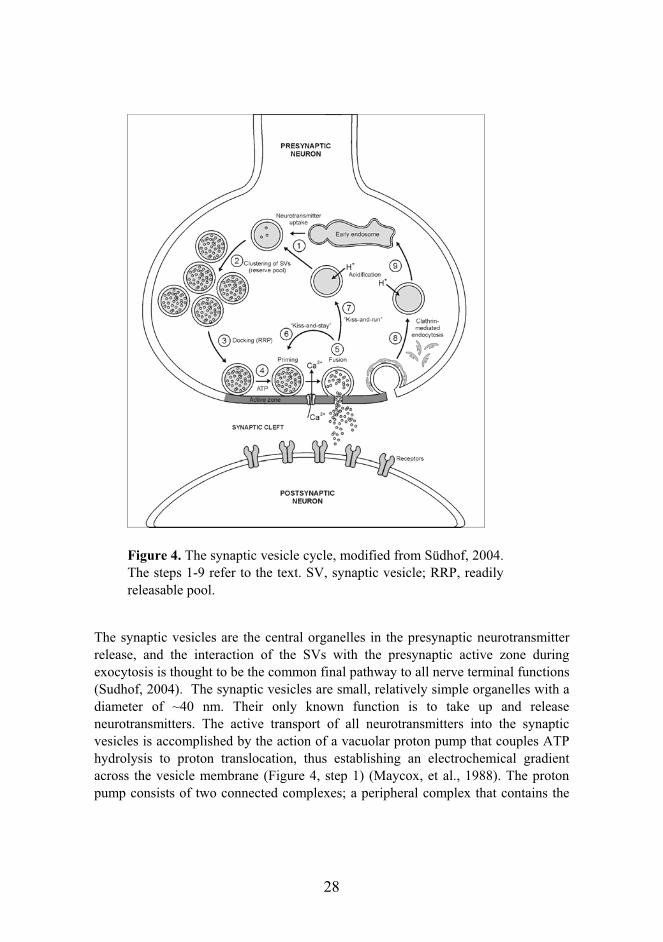
28
The synaptic vesicles are the central organelles in the presynaptic neurotransmitter release, and the interaction of the SVs with the presynaptic active zone during exocytosis is thought to be the common final pathway to all nerve terminal functions (Sudhof, 2004). The synaptic vesicles are small, relatively simple organelles with a diameter of ~40 nm. Their only known function is to take up and release neurotransmitters. The active transport of all neurotransmitters into the synaptic vesicles is accomplished by the action of a vacuolar proton pump that couples ATP hydrolysis to proton translocation, thus establishing an electrochemical gradient across the vesicle membrane (Figure 4, step 1) (Maycox, et al., 1988). The proton pump consists of two connected complexes; a peripheral complex that contains the
Figure 4. The synaptic vesicle cycle, modified from Südhof, 2004. The steps 1-9 refer to the text. SV, synaptic vesicle; RRP, readily releasable pool.

29
ATPase activity and a integral membrane complex that facilitates the proton translocation (Perin, et al., 1991). Seven transporters representing four neurotransmitter groups utilize the electrochemical gradient to mediate the neurotransmitter uptake of the synaptic vesicles; three transporters to glutamate (VGlut1-3) (Fremeau, et al., 2002; Gras, et al., 2002; Schafer, et al., 2002; Takamori, et al., 2002), two for monoamines (catecholamines, histamine, serotonine) (Erickson, et al., 1992; Liu, et al., 1992), a single transporter for both GABA and glycine (McIntire, et al., 1997; Sagne, et al., 1997), and one for acetylcholine (Alfonso, et al., 1993; Roghani, et al., 1994; Varoqui, et al., 1994). In addition to the proteins involved in the neurotransmitter uptake, the synaptic vesicles contain a complex array of trafficking proteins that take part in the synaptic exo- and endocytosis and recycling discussed below.
The membrane fusion during exocytosis follows the same principles that were reviewed in Chapter 1.1. The specific SNARE proteins involved in synaptic exocytosis are VAMP 1 and/or 2 (synaptobrevins) on synaptic vesicles, and syntaxin 1 and SNAP-25 on the presynaptic membrane (Sollner, et al., 1993). The VAMPs act as R-SNAREs/v-SNAREs, and the t-SNARE complex is composed of syntaxin 1 (Qa-SNARE motif), and SNAP-25, containing both Qb- and Qc-SNARE motifs. Small neuronal proteins called complexins bind to the synaptic SNARE core complex and are thought to stabilize it (McMahon, et al., 1995). SM-protein Munc18-1 binds to syntaxin 1 and its dissociation from it is required for the synaptic vesicle fusion with the plasma membrane (Dulubova, et al., 1999). Synaptic vesicle proteins synaptophysin 1 and 2 are also thought to regulate the SNARE function via binding to the VAMPs (Calakos and Scheller, 1994; Edelmann, et al., 1995; Johnston and Sudhof, 1990; Washbourne, et al., 1995).
After the synaptic vesicles have become docked and primed at the active zone (Figure 4 steps 3 and 4), voltage-gated Ca2+ channels open resulting in the fusion-pore opening and exocytosis of the neurotransmitter (step 5). Mostly the Ca2+ influx occurs through the P/Q (CaV2.1) or N-type (CaV2.2) Ca2+ channels (Dietrich, et al., 2003). The Ca2+ influx triggers at least two types of release; fast, synchronous and phasic (Sabatini and Regehr, 1996), and slower asynchronous release (Atluri and Regehr, 1998; Barrett and Stevens, 1972; Geppert, et al., 1994b; Goda and Stevens, 1994). The activation of the fast exocytosis requires that Ca2+ ions bind to the Ca2+
sensor proteins called synaptotagmins 1 and 2 on the synaptic vesicles (Fernandez-Chacon, et al., 2001; Geppert, et al., 1994b). The action of synaptotagmin 1 is understood in more detail; the Ca2+ binding causes synaptotagmin 1 to dissociate from the SNARE complex, to which it binds in the absence of Ca2+, and to bind to the phospholipids membranes. The insertion of synaptotagmin 1 onto the membrane and the consequent mechanical stress is hypothesised to destabilize the fusion

30
intermediate and open the fusion pore (Sudhof, 2004). Triggering of the slow, asynchronous release is mechanistically less clear, but it has been suggested that the other members of the synaptotagmin family function as Ca2+ sensors either alone or in collaboration with synaptotagmin 1. Evidence for high Ca2+ affinity exists at least for the synaptotagmins 3, 6 and 7 (Sugita, et al., 2001; Sugita, et al., 2002).
The synaptic vesicles also contain at least three different Rab proteins; Rab3 (Rab3A-D) (Schluter, et al., 2002), Rab5 (Fischer von Mollard, et al., 1994) and Rab11 (Khvotchev, et al., 2003). These small GTPases mediate membrane attachment by interacting with specific effector molecules (see Chapter 1.1). Rab3 is attached to the synaptic vesicles in the GTP-bound state and is the most abundant of the Rabs in the synaptic vesicles (Geppert, et al., 1994a). Analysis of the knock-out mouse model of Rab3A revealed that the function of Rab3A is important in the late steps of exocytosis after clustering and docking of the vesicles (Geppert, et al., 1997). Other synaptic vesicle proteins involved in the Ca2+ triggered exocytosis are the SV2s (SV2A-C) (Buckley and Kelly, 1985) that have shown to function as cation (most likely Ca2+) transporters in synaptic vesicles (Janz, et al., 1999). Integrity of the SVs is also maintained by the synapsin family of proteins (synapsins 1-3), although their precise function is unknown. They are able to bind various components of the cytoskeleton, especially actin, and may act in anchoring the SV pool to the presynaptic vesicle cluster (Greengard, et al., 1994). However, the mature SV assembly contains hardly any cytoskeletal components, and therefore the exact function of the synapsins remains unsolved (Dunaevsky and Connor, 2000; Morales, et al., 2000; Zhang and Benson, 2001).
The presynaptic active zone is located opposite to the synaptic cleft. It is an electron-dense area, composed of biochemically insoluble material (Akert, et al., 1971). The main components include six large non-membrane proteins that form a giant complex at the active zone: Munc13s (Munc13-1, -2, -3) (Brose, et al., 1995) RIMs (Rab3-interacting molecules; RIM1 , 2 / /γ/, 3γ, 4γ) (Wang, et al., 1997; Wang and Sudhof, 2003; Wang, et al., 2000), Piccolo (Cases-Langhoff, et al., 1996), Bassoon (tom Dieck, et al., 1998), ERCs (ELKS/Rab3-interacting molecule/CAST, ERC1b and ERC2) (Ohtsuka, et al., 2002), RIM-BPs (RIM-BP1-3) (Wang, et al., 2000), and -liprins (Liprin 1- 4) (Schoch, et al., 2002). The RIMs are the central elements of the presynaptic active zones as they bind directly to Munc13, ERCs, RIM-BPs and -liprins. In addition, the ERCs bind to -liprins, Piccolo and Bassoon. The RIMs are linked to the synaptic vesicles via the GTP-dependent interaction to Rab3 and Ca2+-dependent interaction to synaptotagmin 1. They are also potentially linked to the SNAREs via binding to SNAP-25 and Munc13-1 binding to syntaxin 1.

31
After exocytosis, synaptic vesicles are rapidly endocytosed and recycled (Figure 4, steps 6-9). According to the current view, three proposed endocytic pathways exist (Sudhof, 2004). The two fast pathways are clathrin-independent routes, in which the vesicles either remain at the active zone for reacidification and refilling without detachment (kiss-and-stay, step 6) (Barker, et al., 1972) or are recycled locally (kiss-and-run, step 7) (Ceccarelli, et al., 1973). The slower pathway is dependent of the clathrin-mediated internalization (Heuser and Reese, 1973) followed by recycling either directly (step 8) or via an endocytic intermediate (step 9). Low stimulation frequency leads to the utilization of the fast pathways in order to recycle synaptic vesicles fast to the readily releasable pool, whereas at high frequency stimulation, the slower clathrin-dependent pathway is used. The physiological relevance of the clathrin-mediated endosomal pathway (step 9) has been questioned, as endosomes are scarcely observed in the nerve terminals by EM studies. Recently however, strong evidence for the relevance of this endocytic pathway has been presented. Firstly, obligatory synaptic vesicle components include proteins such as the SNARE protein Vti1a , which functions in membrane fusion events at the endosomal and trans-Golgi level (Antonin, et al., 2000). Similarly, the presence of Rab5 and Rab11 on the SVs suggests that the synaptic vesicles undergo endosomal fusions during their cycle (Nielsen, et al., 2000; Nielsen, et al., 1999; Wucherpfennig, et al., 2003). The absence of the endosomes in the nerve terminal has been explained by their transient nature (Wucherpfennig, et al., 2003), and the nerve terminal has been showed to be enriched in proteins associated with clathrin-mediated endocytosis, particularly those involved in the acceleration of this pathway (Sun, et al., 2002). Moreover, inhibition of endosome fusion using phosphatidylinositol 3-kinase blockers caused a significant impairment of neurotransmitter release (Rizzoli and Betz, 2002). The presynaptic endosomes as well as the synaptic vesicles are present in the synaptosome fraction. CLN3 resides in the synaptosome fraction but is not present in the synaptic vesicles, is thus potentially involved in the endocytic pathway of the nerve terminal.
2. Lysosomes and lysosomal storage disorders
2.1 The lysosome turns fiftyFifty years ago, experiments by Christian de Duve and colleagues, aiming at characterizing hepatic glucose 6-phosphatase, led to the accidental observation of an unrelated acid phosphatase of rat liver, which in turn paved the way to the discovery of membrane-limited digestive organelles, termed lysosomes (de Duve C, 1955).

32
The term lysosome is derived from the Greek expression for a digestive body and they are defined as hydrolase-rich, acidic organelles that lack both the cation-dependent (46 kD) and cation-independent (300 kD) mannose 6-phosphate receptors (MPRs) (Reviewed in (Eskelinen, et al., 2003). Lysosomes serve as an endpoint for various intracellular pathways, at which proteins from the degradative, endocytic, autophagic and secretory pathways are degraded or recycled (Kornfeld and Mellman, 1989). Lysosomes are capable of a direct fusion with the late endosomes to form a hybrid organelle (Mullock, et al., 1998), and they have been reported to function at least in the turnover of cellular proteins, down-regulation of surface receptors, release of endocytosed nutrients, inactivation of pathogenic organisms, repair of the plasma membrane and loading of processed antigens onto the MHC class II molecules (Eskelinen, et al., 2003).
Morphology of the lysosomes is heterogenous due to the various cellular functions and variations in the intraluminal content, however, generally lysosomes are described as membranous organelles with a limiting, external membrane and intraluminal vesicles. Proteomic analyses and other studies have identified novel soluble lysosomal proteins and integral membrane proteins and the current estimation is that there are at least 50-60 soluble hydrolases (Journet, et al., 2002), and as many as 55 membrane-associated and 215 integral lysosomal membrane proteins (Bagshaw, et al., 2005). Over 50% of the lysosomal membrane mass is composed of highly glycosylated membrane proteins, called LAMP-1 (LGP-A), LAMP-2 (LGP-B), LIMP-1 (CD-63), LIMP-2 (LGP85), which were originally thought to mechanically protect the membrane from the degradative lysosomal enzymes with the help of their heavy glycosylation (Kornfeld and Mellman, 1989) (Figure 5). They do indeed form a protective glycocalyx on the surface of the inner membrane but appear to have additional functions as well. LAMP-2, for example, is proposed to function as a receptor for cytosolic proteins to be degradated via their KFERQ-related motifs (Dice and Terlecky, 1990). An important minor protein of the lysosomal membrane is a V-type H+-ATPase, a 13-subunit membrane spanning complex responsible for the acidification of the lysosomal lumen by coupling ATP hydrolysis and proton translocation. Importance of specific transporter proteins in the lysosomal membrane has been particularly well demonstrated in the form of certain lysosomal storage disorders, in which an intralysosomal accumulation of retained metabolites is observed (see Chapter 2.2).

33
Figure 5. Major and disease-associated integral membrane proteins of the lysosome. Schematic representation of the topology modified from Eskelinen et al., 2003, except for NPC1 from Davies et al., 2000 (Davies and Ioannou, 2000). LAMP, lysosome-associated membrane protein; LIMP, lysosomal integral membrane protein.
2.2 Targeting of lysosomal proteins
Targeting of most soluble lysosomal proteins is dependent of the mannose 6-phosphate residues attached to them within the Golgi apparatus, and the recognition of this signal by the mannose 6-phoshate receptors (MPR) located at the trans-Golgi network (TGN), which then mediates their delivery to the lysosomes (Kornfeld, 1990). Low levels of the MPRs are also present at the plasma membrane. Two MPRs have been identified with a general structure of a type I transmembrane glycoprotein. A large cation-independent 300-kD MPR, which also binds insulin-like growth factor II, and a smaller 46 kD cation-dependent MPR have overlapping functions (von Figura, 1991). The MPRs with the bound lysosomal enzymes are
LIMP-1
COOHNH2
COOH
NH2
LAMP-2
CLN3
COOH
COOH
NH2
LAMP-1
COOH
NH2
Cystinosin
Cystine
COOH
NH2
LIMP-2
Sialin
Sialic acidNPC1
H+ V-typeH+-ATPase
LYSOSOMECOOH
NH2
ATP
ADP+Pi
NH2
COOH
LIMP-1
COOHNH2
COOH
NH2
LAMP-2
CLN3
COOH
COOH
NH2
LAMP-1
COOH
NH2
Cystinosin
Cystine
COOH
NH2
LIMP-2
Sialin
Sialic acidNPC1
H+ V-typeH+-ATPase
LYSOSOMECOOH
NH2
COOH
NH2
COOH
NH2
ATP
ADP+Pi
NH2
COOH

34
packed into AP-1 containing, clathrin-coated vesicles (CCV) at the TGN, via the interaction of AP-1 and GGA (Golgi localized, gamma-adaptin ear homologous, ADP-ribosylation factor binding proteins), to be delivered to the early endosomal compartments. At a molecular level, the sequestration of the MPRs into the CCVs is mediated by the dileucine- and tyrosine-based motifs in their cytoplasmic segments that are recognized by GGA (Doray, et al., 2002). In the late endosomes, the acidification of the endosomal pH leads to the dissociation of the receptor from the lysosomal enzymes, and the MPRs are recycled back to the TGN by the action of TIP47 (tail-interacting protein of 47 kD) (Diaz and Pfeffer, 1998). The lysosomal enzymes are then transported to the lysosomes.
Alternative mechanisms for targeting the soluble lysosomal proteins also exist and they were explored using a knock-out mouse deficient of both the 300-kD and 46-kD MPRs (Dittmer, et al., 1999). It was found that the ability to transport Cathepsin D to the lysosomes independent of the MPRs was dependent of the cell type. Thymocytes were able to target the enzyme correctly via an alternative intracellular route, whereas hepatocytes and fibroblasts secreted the enzymes. However, hepatocytes were able to recapture a significant amount of the secreted enzymes.
Targeting of lysosomal transmembrane proteins is more complex and diverse compared to the classic MPR-route. It is mostly mediated by short, linear sequences of amino acids within the cytosolic domains of the proteins. These motifs are not precisely conserved sequences but degenerate motifs of four to seven residues, of which the two or three critical residues are often bulky and hydrophobic (Bonifacino and Traub, 2003). Cytoplasmic tyrosine-based motifs were the first to be identified, and they are composed of either NPXY or YXXØ consensus, where N is asparagine, P proline, X any amino acid, Y tyrosine, and Ø an amino acid with bulky sidechain. The NPXY signal was the first tyrosine-based sorting signal to be found (Davis, et al., 1986), but later it has been shown to mediate only rapid internalisation of a subset of type I integral membrane proteins, such as the LDL receptor, and not other intracellular events. In contrast, the YXXØ signals function in the sorting of a wide range of proteins, such as endocytic receptors (transferrin receptor), intracellular sorting receptors (mannose 6-phosphate receptors), TGN proteins (TGN38) (Canfield, et al., 1991; Jadot, et al., 1992), as well as endosomal-lysosomal transmembrane proteins (Harter and Mellman, 1992; Williams and Fukuda, 1990). YXXØ appears to be an evolutionarily conserved sorting motif, and in the sorting of the lysosomal proteins, the Y residue is essential for function, the X residues tend to be acidic, and a glycine residue often precedes the critical tyrosine (Bonifacino and Traub, 2003). Examples of lysosomal proteins harbouring the YXXØ sorting motif include the major components of the lysosomal membrane, LAMP-1 and LAMP-2,

35
as well as LIMP-2 and cystinosin transporter (Bonifacino and Traub, 2003; Cherqui, et al., 2001; Rohrer, et al., 1996).
Dileucine-based signals form the second major family of lysosomal transmembrane sorting signals. These signals match the consensus LL or LI, and substitution of either critical leucines with alanines impairs all signalling activities. The second leucine, however, can be replaced with isoleucine without a loss of activity. In addition, an acidic residue preceding the first leucine appears to be significant for lysosomal targeting (Bonifacino and Traub, 2003). Besides in lysosomal proteins such as NPC1 and LIMP-II (Sandoval, et al., 1994; Watari, et al., 1999), a dileucine-based sorting signal exists in transmembrane proteins targeted to the synaptic dense-core granules (VMAT1, VMAT2), stimulus-responsive storage vesicles (GLUT4), and melanosomes (tyrosinase) (Bonifacino and Traub, 2003).
Both the tyrosine- and dileucine-based signals are recognized by cytoplasmic coat proteins that associate with the cytosolic side of the membrane. YXXØ and LL/LI are specifically recognized by the clathrin-associated adaptor protein (AP) complexes AP-1, AP-2, AP-3, and AP-4, although it seems that AP-3 is the main adaptor complex resposible for the lysosomal membrane protein targeting to the late endosomal pathway (Dell'Angelica, et al., 1999; Le Borgne, et al., 1998; Rous, et al., 2002). Dispute over the clathrin-binding properties of AP-3 exists, but it was recently demonstrated that proteins with strong binding affinity to AP-3 are targeted directly from the TGN to the lysosomes, while most proteins trafficked via the plasma membrane and early endosomes (Ihrke, et al., 2004). On the other hand, some LL signals are recognised by GGAs, and several proteins, such as clathrin, AP-2 and Dab2, have been suggested to function in the NPXY signal recognition, but the exact mechanisms remains unclear (Bonifacino and Traub, 2003). In most cases, regulation of the targeting signal recognition is achieved by phosphorylation. In addition, ubiquitination of cytosolic lysine residues has been shownt to act as a signal for sorting at various stages of the endosomal-lysosomal pathway (Bonifacino and Traub, 2003; Strous, et al., 1996). Conjugated ubiquitin is recognized by UIM (ubiquitin-interacting motif), UBA (ubiquitin-associated motif), or UBC (ubiquitin-conjugating enzyme E2 motif) domains present within many components of the endosomal and lysosomal targeting machinery (Bonifacino and Traub, 2003). Moreover, some unconventional motifs for lysosomal membrane protein targeting have been found, e.g targeting of CLN3 requires both a conventional dileucine-based motif as well as an unconventional M(X9)G motif in its carboxyl tail (Kyttala, et al., 2004).

36
2.3 Lysosomal storage disorders (LSDs)
Lysosomal storage disorders (LSDs) are caused by a dysfunction of a lysosomal protein, which leads to the intralysosomal accumulation of undegraded metabolites. Most LSDs are present in infantile, juvenile and adult forms, of which the infantile forms are most severe, causing severe CNS symptoms and premature death. The adult forms are generally milder and peripheral symptoms play a major role in the course of the disease. Juvenile forms are often intermediate between these two (Futerman and van Meer, 2004). LSDs are classified either based on the characterization of the defective enzyme or protein, or based on the accumulated substrate(s). The latter is most often used, although it may lead to false characterization of a disease, when the accumulating substance is identified prior to the identification of the defective protein. More than 40 LSDs are known, and they are classified into (1) sphingolipidoses, (2) mucopolysaccharidoses (MPS), (3) oligosaccharidoses and glycoproteinosis, (4) lipidoses, (5) diseases caused by defects in integral membrane proteins, and (6) others.
Most mutations in classic lysosomal storage disorders cause a reduced enzymatic activity of a particular lysosomal hydrolase, or another protein that is required for an optimal enzyme activity (Futerman and van Meer, 2004). Misfolding of lysosomal proteins leading to their defective transport from the ER to the lysosomes can also underlie LSD. Defective transport from the ER to the lysosome due to an alternative mechanism is present in galactosialidosis, in which the formation of a multi-enzyme complex required for the transport of glycosidases is defective (Ostrowska, et al., 2003). A novel mechanism was identified, when the underlying defect for the multiple sulfatase deficient (MSD) was revealed. Mutations in the SUMF1 gene (sulphatase modifying factor-1) cause a production of a defective Cα-formylglycine-generating enzyme (FGE). Defective FGE is unable to convert a specific Cys-residue at the active site of the sulphatases, resulting in a delivery of multiple inactive sulphatases to the lysosomes, unable to perform their function in degrading sulphate esters (Dierks, et al., 2003). I-cell disease, on the other hand, is caused by a defective mannose 6-phosphate glycosylation in the Golgi, leading to the inability of the enzyme to bind the MPRs, which prevents their correct transport to the lysosome (Kornfeld and Sly, 2001).
The topic of this thesis, the juvenile neuronal ceroid-lipofuscinosis (JNCL), belongs to the small group of LSDs caused by gene defects affecting integral lysosomal membrane proteins (Figure 5). Summary of the basic characteristics of this group are described in Table 1. Two of these disorders, cystinosin and the infantile sialic-acid storage disease (ISSD)/Salla disease are caused by defects in transporters named cystinosin and sialin, respectively, that export soluble metabolites out of the

37
lysosome. Cystinosin transports cystine, a by-product of protein degradation, from lysosomes to the cytosol, where it is consequently reduced to the amino acid cysteine for further use. Defects in this transport result in the intra-lysosomal accumulation of cystine and wide spectrum of symptoms, whose presence is dependent on the type of mutation (Town, et al., 1998). The clinical symptoms include retinal degeneration, diabetes mellitus, hypothyreosis, and nephropathy (Attard, et al., 1999). Sialin is an anion transporter that transports free sialic acid out of the lysosomes, and consequently, defects cause an accumulation of free sialic acid in the lysosomes leading to neurodegeration manifested by an early onset developmental delay and ataxia (Verheijen, et al., 1999). Mutations in the major lysosomal protein LAMP-2 cause Danon disease (Nishino, et al., 2000), a cardioskeletal myopathy, is clinically characterized cardiomyopathy, myopathy, and variable mental retardation (Danon, et al., 1981). Defects in MCOLN1 gene encoding mucolipin-1 (MLN1) protein underlie mucolipidosis (ML) type IV, enriched in the Ashkenazi Jew population (Bargal, et al., 2000). MLN1 is a non-specific cation channel that shows topological similarities with calcium channel family called transient receptor potential (TRP) channels. The clinical symptoms include severe ophthalmologic abrnomalities (corneal opacities, retinal degeneration, strabismus), and psychomotor retardation (Berman, et al., 1974). Defects in two genes, NPC1 or NPC2, cause clinically and biochemically indistinguishable Niemann-Pick type C disease (NPC) (Carstea, et al., 1997; Naureckiene, et al., 2000). NPC1 protein is a lysosomal transmembrane protein with a sterol-sensing domain, whereas NPC2 is a soluble protein with cholesterol-binding properties (Reviewed in (Ikonen and Holtta-Vuori, 2004). NPC disease is classified as a cholesterol transport disorder; however, lysosomal accumulation of both cholesterol and sphingolipids occurs. Clinical symptoms are heterogeneous comprising of hepatic and neurodegenerative components, leading to premature death (Patterson, et al., 2001).

38
Table 1. Features of the diseases caused by defects in lysosomal membrane proteins. For references, see the text. *Defects in NPC2, a soluble lysosomal cholesterol binding protein (which is transported via the MPR-mediated route), can also cause NPC.
Diseases caused by defects in lysosomal membrane proteinsDisease Defective protein Main storage material Cystinosis Cystinosin Cystine
Danon disease LAMP-2 Cytoplasmic debris and glycogen
Infantile sialic-acid-storage disease and Salla disease Sialin Sialic acid
JNCL (Batten disease) CLN3 Subunit c of the mitochondrial ATP synthase
Mucolipidosis (ML) IV Mucolipin-1 (MLN1)
Lipids and acid mucopolysaccharides
Niemann-Pick C (NPC) NPC1 (NPC2*) Cholesterol and sphingolipids
2.4 Mechanisms for LSDs and potential for therapy
Intra-lysosomal accumulation of undegraded metabolites is characteristic to all LSDs. However, despite progress in understanding of the genetic, molecular and biochemical bases of LSDs, little is known about the pathogenetic pathways leading to the lysosomal accumulation at the cellular level. The extensive variation in disease phenotypes indicates that secondary biochemical and cellular pathways must be involved. It has been hypothesised that a general “intra-lysosomal protein response” mechanism, analogous to the unfolded protein response in the ER (resulting from accumulation of misfolded proteins in the ER, and leading to the transcriptional activation of ER chaperones and degradative enzymes), exists (Futerman and van Meer, 2004; Kaufman, 2002). This would lead to an activation of similar response pathways irrespective of the undegraded or unfolded protein. The great variability in the accumulating substances and the clinical symptoms between the different LSDs, nonetheless, disagree with this hypothesis. Impaired apoptosis due to an impaired lysosomal function has also been suggested to underlie the LSDs (Tardy, et al., 2004). This would be mediated through different potential (and so far mainly theoretical) mechanisms; accumulation of toxic compounds (due to deficient metabolism), lack of molecules acting as apoptotic suppressors (e.g. prosaposin), impaired lysosome stability, or an uncontrolled activation of lysosomal proteases and excessive proteolysis, which would all lead to the activation of the caspase

39
cascade, and consequent induction of cell death. In addition, defective intracellular trafficking, defective intracellular signalling, and altered gene expression have also been suggested to be causative for LSDs, although the exact cellular pathways leading to the lysosomal accumulations and, often to neurodegeneration, are for the most part unclear. Most of the LSDs present symptoms originating from the central nervous system, which probably indicates the lack of compensatory pathways and/or lack of neuronal cell regeneration potential (Futerman and van Meer, 2004; Tardy, et al., 2004).
First treatments for LSDs were mainly symptomatic, such as splenectomy (surgical removal of spleen), hemodialysis or renal transplantation. In addition, bone marrow transplantation has been utilized to gain some normal, lysosomal-enzyme producing cells. As the diagnosis and understanding of the cellular pathogenesis of the diseases have improved, advances for specific therapies have been made. Enzyme-replacement therapy (ERT) was the obvious form of therapy to be developed, as the defective enzymes were identified. At present, ERT is approved for Fabry disease and MPS I, but it is mainly restricted to diseases with predominating peripheral symptoms, as the enzymes used are not able to pass the blood-brain barrier (Bengtsson, et al., 2003; Desnick and Schuchman, 2002). In substrate-reduction therapy (SRT), the small molecules used act by inhibiting the synthesis of an accumulating substance, and are able to cross the blood-brain barrier. SRT has been used in sphingolipidoses such as the neuronal subtype of the Gaucher disease, in which glycolipids accumulate particularly in the brain (Futerman, et al., 2004). The long-term effects of glycolipid depletion, however, have not been established. Great promise and potential have been attributed to the development of effective gene therapy treatments for the monogenic LSDs. The preferred treatment would include the genetic modification of the patient's own cells in vitro or in vivo to constitutively express high levels of the correcting enzyme, which would then become the source of the enzyme in the patient. Both ex vivo and in vivo gene transfer methods have been experimentally explored, and several of these methods have proved efficient for the transfer of genetic material into deficient cells in culture and reconstitution of enzyme activity (D'Azzo, 2003). However, application of these methods to humans or animal models have been giving inconsistent results, and therefore broader understanding of the disease mechanisms and the animal models, as well as development of better gene transfer methods will be necessary make gene therapy a realistic option for treatment.

40
3. Juvenile neuronal ceroid lipofuscinosis
3.1 Neuronal ceroid lipofuscinoses (NCLs)
Neuronal ceroid liposfuscinoses (NCLs) comprise a group of at least eight autosomal, recessively inherited, progressive neurodegenerative diseases with onset usually in childhood. They occur worldwide and are considered to be the most common progressive brain disorders of childhood (Haltia, 2003; Mole, et al., 2005). NCL diseases are marked by two histopathological findings: degeneration of nerve cells, foremost in the cerebral cortex, and accumulation of autofluorescent ceroid-lipopigment in both neural and peripheral tissues (Goebel, 1997). Except for the newest member of the NCL family, Northern epilepsy (CLN8), typical clinical features are shared among the NCLs: visual failure leading to blindness, myoclonic epilepsy, dementia of various degrees, pyramidal/extrapyramidal symptoms, and ultimately, premature death (Santavuori, et al., 2000). NCLs are divided into four main classes based on the age of onset of the disease; infantile (CLN1), late infantile (CLN2), juvenile (CLN3) and adult (CLN4, gene not identified). NCLs are enriched in Finland and the most common types are JNCL and INCL with incidences of 1:21 000 and 1:20 000, respectively. LINCL is rare, whereas the Finnish variant LINCL is more common (Santavuori, 1988).
Infantile neuronal ceroid liposfuscinosis (Santavuori-Haltia disease, INCL) is the most severe of the NCLs, caused by a deficiency in a lysosomal depalmitoylating enzyme activity (palmitoyl protein thioesterase 1, PPT1) (Hellsten, et al., 1996; Lehtovirta, et al., 2001; Salonen, et al., 2001; Vesa, et al., 1995). INCL is characterized by a normal development to the age of 6-12 months, followed by an arrest of mental, cognitive and motor development, severe microcephaly, myoclonus and visual deterioration (Haltia, et al., 1973; Santavuori, et al., 1973). The EEG is isoelectric by the age of 3 years, and the children die between 9-11 years of age (Santavuori, 1973). Late infantile neuronal ceroid liposfuscinosis (Jansky-Bielschowsky disease, LINCL) is caused by a deficiency of a lysosomal protease (tripeptidyl peptidase, TPP1), a member of a serine-carboxyl proteinase family (Golabek, et al., 2003; Sleat, et al., 1997). In most cases, epilepsy is the first symptom of the disease, other symptoms common to all NCLs, develop slightly later (Hofmann and Peltonen, 2001). It is one of the rare lysosomal storage disorders caused by a deficiency of a lysosomal protease. In addition to the classic LINCL, three variant forms of the late infantile NCL exists: Finnish variant LINCL (vLINCLFin, CLN5), CLN6-variant LINCL, and a Turkish variant LINCL (vLINCL, CLN7). Clinical phenotype of Finnish variant LINCL (vLINCL) is an intermediate between the classic LINCL and JNCL (Santavuori 1982, 1991), and the patients

41
come exclusively from a cluster of 16 Finnish families (Isosomppi, et al., 2002; Savukoski, et al., 1998). Subsets of the Turkish variant LINCL patients have been shown to have mutations in the CLN8 and CLN6 genes (Ranta, et al., 2004; Siintola, et al., 2005), and it is yet to be determined whether the CLN7 locus, assigned for the Turkish LINCL, truly exists. The clinical and molecular characteristics of the adult NCL (CLN4, Kufs disease) (Berkovic, et al., 1988), CLN6-variant LINCL (Gao, et al., 2002; Heine, et al., 2004; Wheeler, et al., 2002), and Northern epilepsy (CLN8) (Hirvasniemi, et al., 1995; Hirvasniemi, et al., 1994; Lonka, et al., 2000; Ranta, et al., 1999) are described in detail elsewhere.
To date approximately 160 mutations causing NCL have been described (NCL Mutation Data base, http://www.ucl.ac.uk/ncl). The original hypothesis was that defects in one gene lead to a classic phenotype and ultrastructure of the storage material. However, the genotype-phenotype correlations have proven to be more complex than that; some mutations are associated with a phenotype that is of later onset, less severe or protracted in its course, or with atypical morphology (Mole, et al., 2005). A summary of the classic clinical phenotypes and their variations associated with the NCL genes, as well as the major components of the storage material and ultrastructural phenotype, are presented in Table 2.

42
Table 2. Classification of the NCLs. Defects in one gene can cause several clinical phenotypes. The encoded proteins, the major component of the storage material, and ultrastructural phenotype are also presented. GROD, granular osmiophilic deposits; FP, fingerprint bodies; RL, rectilinear complex; CL, curvilinear profiles; TM, transmembrane. For references, see the text.
Classification of the NCLs
Clinical types Gene Protein Storage material Ultrastructure
INCL, also late infantile, juvenile and adult inset CLN1
Lysosomal palmitoyl protein thioesterase PPT1
Saposins A and D GROD
LINCL, also juvenile onset CLN2
Lysosomal tripeptidyl peptidase TPP1
Subunit c of mitochondrial ATP synthase CL
JNCL CLN3 TM-protein CLN3
Subunit c of mitochondrial ATP synthase FP (CL, RL)
ANCL CLN4 Not known
Subunit c of mitochondrial ATP synthase FP, granular
LINCL, Finnish variant, also juvenile onset CLN5
Lysosomal soluble CLN5
Subunit c of mitochondrial ATPsynthase, Saposins A and D RL, CL, FP
LINCL, EJNCL, Indian/CostaRican/Czech Gypsy variant also juvenile onset CLN6
ER-resident TM-protein CLN6
Subunit c of mitochondrial ATP synthase RL, CL, FP
LINCL, Turkish variant CLN7 Not known
Subunit c of mitochondrial ATP synthase RL, CL, FP
LINCL, Northern epilepsy and Turkish variant, also juvenile onset CLN8
ER-resident TM-protein CLN8
Subunit c of mitochondrial ATP synthase
CL-like, granular

43
3.2 Clinical findings in JNCL
Juvenile neuronal ceroid-lipofuscinosis (MIM 204200), also called Spielmayer-Vogt-Sjögren disease or Batten disease, is the most common of the NCLs worldwide. The first cases were identified as early as 1826 in Norway (Stengel, 1826), and similar case reports were published independently by Batten, Spielmayer and Vogt nearly a century later in 1903 and 1923 (Haltia, 2003). Characteristic first symptom of the juvenile NCL is a progressive visual failure with an age of onset of 4-8 years (occasionally later), which leads to blindness in most cases during teenage years. Mental retardation and cognitive decline begin early but are subtle in their course and go often unnoticed until school-age. More pronounced deterioration occurs at 8-12 years of age (later in some patients). Epileptic seizures of a generalized tonic-clonic (primary or secondary) or complex partial type begin between 8-13 years of age. Parkinsonian type of extrapyramidal symptoms (ataxia, dysarthria, rigidity, difficulty of movement initiation) develop slowly, and there is great variation in the onset (Jarvela, et al., 1997). The JNCL patients become non-ambulatory at ages of 15-28 years (Santavuori, et al., 2000). Behavioural and psychiatric problems are also common, including hallucinations and psychosis (Santavuori, et al., 1993). In addition, sleep dysfunction is common in JNCL, and occurs in 50-90% of cases (Santavuori, et al., 2000).
3.3 Neuropathology of JNCL
The neuropathology of JNCL was well described in the early 20th century, and that description remains valid (reviewed in (Hofmann and Peltonen, 2001). Macroscopically, the brain is moderately or even severely decreased in weight, and the skull bones are thickened due to the brain atrophy. The cerebral cortex is thinner, and the white matter is atrophic. Loss of neurons is observed virtually in all parts of the brain, but the severity in different areas varies. Reactive astrocytosis, associated with the neuronal loss, is also observed. Ballooning and rounding of the neurons due to distension caused by the perikaryonal lipopigment, known as the Shaffer-Spielmayer process, is prominent and especially evident in the cortical neurons of the forebrain and cerebellum. Early lesions of specific layers of the cerebral cortex have also been documented; more specifically, loss of small stellate neurons in layer II, and loss of pyramidal neurons in layers IV and V are seen (Braak and Goebel, 1978). Consistent with the visual impairment, degeneration of the retinal neuroepithelium is evident. In the later stages of the disease, lipopigment-containing

44
cells invade all layers of the retina resulting in almost complete loss of rods, cones, and the inner and outer plexiform layers (Goebel, et al., 1974).
3.4 Neuroimaging findings in JNCL
Neuroimaging with computer tomography (CT) and magnetic resonance imaging (MRI) appears normal in JNCL patients under 10 years of age. Thereafter, slow progressive cerebral, and later, cerebellar atrophy appeared in a study sample of 30 Finnish JNCL patients. Even the youngest patients had a significant increase in white matter signal intensity. Reduction in size of corpus callosum and brain stem was also observed (Autti, et al., 1996). In conclusion, the MRI findings in JNCL were quite unspecific, but absence of them may be of use in differential diagnostics. Post-mortem MRI combined with histopathological analysis showed a correlation between an increased signal intensity periventricular white matter and severe loss of myelin and gliosis in these areas. In addition, reduced signal intensity in thalamus and basal ganglia areas was observed, but these did not correlate with histopathological findings (Autti, et al., 1997).
Positron emission tomography (PET) and single photon emission computed tomography (SPECT) imaging have demonstrated both pre- and postsynaptic, dopaminergic hypofunction in the nigrostriatal area that were in part related to the severity of extrapyramidal symptoms (Aberg, et al., 2000; Rinne, et al., 2002; Ruottinen, et al., 1997).
3.5 Neurophysiological findings in JNCL
The electroencephalogram (EEG) is normal at the early stages of the disease and at the age of 8-9 years shows slow, non-specific deterioration (Hofmann SL, 2001).
Progressive attenuation of amplitudes of cortical somatosensory evoked potential (SEP) is a general feature of the NCLs (Santavuori, et al., 2000). In a study sample of ten Finnish JNCL patients, the amplitude of SEP potentials was increased, as was SEF (somatosensory evoked magnetic fields) studied with magnetoencephalography (MEG) (Lauronen, et al., 1997). These results indicated increased thalamocortical excitability in the sensorimotor cortex in JNCL.
45
3.6 Intracellular storage in JNCL and its significance to pathology
The autofluorescent lipopigments accumulating in JNCL are very similar to the age-related pigments accumulating in normal tissues and their ultrastructure resembles a fingerprint (Figure 6). The lipopigment granules have an intense activity of acid phosphatase, suggesting an association with lysosomes. This impression has been later confirmed by EM studies (Reviewed in (Hofmann and Peltonen, 2001). The storage material is already detectable in the prenatal period, and inclusion bodies have been reported as early as the first trimester (Conradi, et al., 1989). Since the symptoms do not begin until school-age, this suggests, that the neuronal cells and tissues are able to tolerate the storage to some extent.
The major component of the storage material in JNCL is the subunit c of the mitochondrial ATP synthase complex (Hall, et al., 1991; Palmer, et al., 1992). ATP synthase is a large membrane-embedded enzyme complex composed of a proton-conducting unit and a catalytic unit. ATP synthase carries out the final phase of the oxidative phosphorylation in the inner mitochondrial membrane by catalyzing the formation of ATP from ADP and orthophosphate, driven by the proton flow into the mitochondrial matrix. Ten to fourteen units of the subunit c, together with a single subunit a, compose the hydrophobic proton channel of the ATP synthase, embedded into the inner mitochondrial membrane (Berg, et al., 2002).
Subunit c of the mitochondrial ATP synthase complex also accumulates in other lysosomal disorders including mucopolysaccharidosis (MPS) I, II and IIIA, multiple sulfatase deficiency, mucolipidosis I, Nieman Pick types A and C, and GM1 and GM2 gangliodoses (Hofmann and Peltonen, 2001). However, in these diseases, the accumulation is only observed in the central nervous system, whereas in the NCLs, also the peripheral cells accumulate the subunit c. This suggests that the accumulation of the mitochondrial ATP synthase subunit c is more specific for the NCLs than other lysosomal storage disorders. Vacuolated lymphocytes in peripheral blood are also a common finding in many LSDs; however, JNCL is the only NCL subtype, in which they are found.
Minor components of the lysosomal storage in JNCL are the two sphingolipid activator proteins (saposins) A and D, which activate lysosomal hydrolases involved in glycosphingolipid degradation. Saposins A and D as well as the subunit c of the ATP synthase are extremely hydrophobic molecules with a tendency to self-aggregate. It has been shown in the ovine NCL that the amounts of subunit c, its insertion into the mitochondrial membrane, and the rate of oxidative phosphorylation are normal, suggesting that the specific failure in the NCLs is in the degradation of the hydrophobic subunit in the lysosomes (Palmer, et al., 1992). This

46
hypothesis was supported by a human study using LINCL and JNCL brain samples (Kominami, et al., 1992). Nonetheless, fundamental question in terms of the lysosomal storage still remains to be answered; further studies on the mechanisms of cell death are needed to understand whether the neuronal cell death is directly caused by the storage, and why are the nerve cells exclusively affected.
Figure 6. Electron micrograph of the fingerprint type storage material of a JNCL patient. Figure courtesy of Dr Juhani Rapola, University of Helsinki.
3.7 CLN3 gene and mutations
The CLN3 gene spans a 15-kb region in the chromosome 16p11.2-12.1, consisting of 15 exons, which form an open reading frame (ORF) of 1314 bp (Consortium, 1995). The length of the exons is 47-356 bp, and they conform to the conventional AG/GT rule (Breathnach, et al., 1978). Numerous repetitive Alu elements are present within the introns and 5'- and 3'-untranslated regions. The 5' region of the CLN3 gene contains several potential transcription regulatory elements but no consensus TATA-1 box was identified, thereby suggesting that CLN3 is a continuously expressed gene (Mitchison, et al., 1997).
To date, forty disease causing mutations and five polymorphisms of CLN3 have been identified and they are fully listed in the NCL Mutation Database (http://www.ucl.ac.uk/ncl). The spectrum of mutations include 4 large deletions, 6 small deletions, 5 small insertions, 10 missense mutations, 9 nonsense mutations, 5 mutations affecting splice sites and 1 intron change. The most common 1.0-kb

47
deletion that is present on approximately 85% of the disease chromosomes, removes exons seven and eight (CLN3Δex7-8), resulting in a frameshift and a premature STOP codon. This truncated polypeptide consists of 181 aa that including 28 novel aa in addition to the original 153 aa.
In all cases of NCL caused by mutations in CLN3, visual failure has occurred by ten years. However, depending on the mutation type, other symptoms (seizures, motor disabilities, mental retardation) may be significantly delayed. In the classic JNCL, caused by the 1.0-kb major mutation or by other mutations leading to a truncated CLN3 protein, significant mental retardation is evident before the age of ten years, extrapyramidal symptoms develop before the age of fifteen, and death occurs usually around twenty years of age and no later than twenty-five years. The delayed classic JNCL, associated with patients with a compound heterozygous genotype, has slower progression of symptoms: mental retardation takes place soon after ten years of age, extrapyramidal symptoms after fifteen years of age, and death usually after twenty-five years of age (Jarvela, et al., 1997). Protracted JNCL is associated with missense mutations (best studied E295K, R334H) that presumably affect less important residues of the CLN3 protein, and is markedly milder in phenotype: visual impairment and blindness are prominent at an early age but mental and motor symptoms are manifested considerably later, usually in the third or fourth decade of life.
3.8 CLN3 protein
The CLN3 gene encodes a 438-aa transmembrane CLN3 protein with an approximate molecular weight of 43 kD (Figure 7). CLN3 is evolutionarily conserved from yeast to humans, and it has no homologies to any known proteins. Consequently, its function cannot be predicted from the amino acid sequence. The topology of CLN3 was long elusive, and prediction programs estimated 5-11 transmembrane segments. Recently, two experimentally derived topology models have emerged. Mao et al. utilized flag-tagged CLN3 and glycosylation mutagenesis and postulated that CLN3 has five membrane spanning domains, an extracellular/intraluminal N-terminus and a cytoplasmic C-terminus (Mao, et al., 2003). In contrast, Kyttälä and Ezaki argued that the N-terminus, the large loop and the C-terminus all face the cytoplasm, indicating (five or) six transmembrane domains (Figure 7) (Ezaki, et al., 2003; Kyttala, et al., 2004). The latter model is supported by stronger evidence, as the experiments were carried out in vivo

48
3.9 Post-translational modifications of CLN3
CLN3 protein contains several putative post-translational modification sequences, which will be discussed below (Figure 7). Lysosomal proteins are often heavily glycosylated in order to avoid proteolysis in the lysosomal lumen. CLN3 protein has been predicted to have four putative N-glycosylation sites, two putative O-glycosylation sites, and two putative glycosaminoglycan sites (Golabek, et al., 1999). N-linked glycosylation has been demonstrated by Järvelä et al. (Jarvela, et al., 1998) and Golabek et al. (Golabek, et al., 1999). The former group showed that the molecular weight of an unmodified, in vitro translated CLN3 was ~43 kD, and
Figure 7. Topology of CLN3 protein according to Ezaki (2003) and Kyttälä (2004), with 5-6 transmembrane segments and both N- and C-termini residing in the cytoplasm. The reported lysosomal targeting signals, the three putative glycosylation sites (antennas) and the eight putative phosphorylation sites (spheres) are indicated. The localisations of the most common mutations in terms of the topology are marked with the indicated colours. Figure courtesy of Dr Jared Benedict, University of Rochester.

49
expression for longer periods of time in COS-1 cells resulted in an apparent maturation of the protein, and an appearance of a doublet band of 43- and 45-kD. Golabek et al. reported significantly larger band size of 66-kD and 100-kD for CLN3 in a study using GFP-tagged CLN3 in Chinese hamster ovary (CHO) cells. The discrepancy in terms of the molecular weight of CLN3 may be reflective of the different cell lines used in the studies. Moreover, N-linked glycosylation was also shown in a study using endogenous mouse Cln3, which also has four putative N-glycosylation sites (Ezaki, et al., 2003). Treatment of GFP-CLN3 expressing CHO cells with tunicamycin, which prevents glycosylation, did not alter the subcellular localization of CLN3 (Kida, et al., 1999). The current topology model illustrated in Figure 7 supports the existence of three N-glycosylation sites.
CLN3 has ten serines and three threonines that could potentially act as sites for phosphorylation. Evidence on the CLN3 phosphorylation was provided by a study, in which GFP-CLN3 overexpressing CHO cells were exposed to a radiolabeled phosphate, and incorporation of phosphates was seen (Michalewski, et al., 1999). When the cells were further treated with alkaline phosphatase, the isotopic labelling was removed, indicating phosphorylation of some of the potential sites. It was also shown that the level of CLN3 phosphorylation can be regulated by several kinases (cAMP-dependent kinase, cGMP-dependent kinase, casein kinase II) and phosphateases (protein phosphatases I and II), which however, are non-specific invitro. Further in vivo studies are needed to determine the exact phosphorylation status and sites of CLN3, and the relevance to its function.
Mannose 6-phosphate residues are essential lysosomal targeting motifs for most soluble lysosomal proteins (see chapter 1.2). As would be expected for a lysosomal transmembrane protein, CLN3 does not contain any mannose 6-phosphate residues (Michalewski, et al., 1999).
Original predictions of the CLN3 protein sequence suggested two potential lipid modification sites: N-myristoylation at the amino terminus and prenylation (farnesylation) at the carboxy-terminus (Kaczmarski, et al., 1999; Pullarkat and Morris, 1997). Indirect evidence from studies by Ezaki et al. (Ezaki, et al., 2003) suggested a modification, possibly myristoylation in the amino terminus, as they were unable to sequence the N-terminus by Edman degradation. The authors also pointed out that this blockage may well be due to other secondary modifications, such as acetylation. The originally predicted prenylation motif (CAAX) at the carboxy-terminus (435CQLS438) is undefined (Figure 7) as the current programs are no longer able identify it (Phillips 2005). Some experimental evidence, however, exists, as in vitro produced GST-tagged CLN3 was observed to be prenylated (Kaczmarski, et al., 1999). At a functional level the evidence is controversial; mutation of this motif did not affect cell growth or apoptosis, as the native CLN3 did

50
(Persaud-Sawin, et al., 2002), but it did inhibit the complementing action of the CLN3 in the functional studies of the knock-out yeast homologue Btn1p (Haskell, et al., 2000).
3.10 Subcellular localization of CLN3
Endosomal/lysosomal compartment is considered the most likely subcellular localization for CLN3/Cln3 in non-neuronal cells, demonstrated by several studies (Ezaki, et al., 2003; Golabek, et al., 1999; Haskell, et al., 2000; Jarvela, et al., 1999; Jarvela, et al., 1998; Kida, et al., 1999; Kyttala, et al., 2004; Mao, et al., 2003). However, several other intracellular localizations have also been proposed for CLN3; endoplasmic reticulum (Kida, et al., 1999), Golgi (Kremmidiotis, et al., 1999), cell membranes and nucleus (Margraf, et al., 1999), and mitochondria (Katz, et al., 1997). Prior to this study, neuronal localization of CLN3 had been just tentatively examined and it seemed that the protein is trafficked along the neural extensions (Jarvela, et al., 1999). There are several potential explanations for the controversial reports on the localization of CLN3. Firstly, production of antibodies against the full-length CLN3 has been unsuccessful due to its toxicity to bacterial expression systems, which has led to the use of peptide antibodies with or without affinity purification, which in turn may result in a poor specificity or cross-reactivity. Secondly, some of the studies did not utilize double labeling techniques with subcellular organelle markers, and thus relied on morphological features of the cells (Katz, et al., 1997; Margraf, et al., 1999). Thirdly, due to the low endogenous level of CLN3, all studies in non-neuronal cells have been conducted using over-expression systems. Therefore, it should be expected that the consequent, unphysiologically high expression levels overload the ER-Golgi biosynthetic route leading to the observation of the protein in the ER and Golgi, in addition to its final destination. Finally, the diversity of the reports on the intracellular localization of CLN3 protein may also suggest that CLN3 has different intracellular localization in different cell types.
Confirmation for the endosomal/lysosomal localization of CLN3 in somatic cells was obtained from studies demonstrating two lysosomal targeting motifs within the cytoplasmic segments (Kyttala, et al., 2004). A conventional dileucine motif (LI) was found in the large cytoplasmic loop, preceded by an acidic patch. A novel motif consisting of methionine and glycine, separated by nine amino acids, in the carboxy-terminal tail is also required for the correct targeting. The localization of these motifs is shown in Figure 7. On the contrary, it has been also reported that the cytoplasmic dileucine targeting motif alone was sufficient to target CLN3 to the

51
lysosomes (Storch, et al., 2004). Furthermore, it has been shown that the lysosomal targeting of CLN3 is facilitated by the adaptor complexes AP-1 and AP-3 via its dileucine motif (Kyttala, et al., 2005).
It has also been suggested that CLN3 is localised to the lipid rafts (detergent-resistant lipid domains) together with cholesterol, palmitic acid and Flotillin-1 (Rakheja, et al., 2004).
3.11 Interactions of CLN3 protein
Since the function of CLN3 is presently unknown, interaction partners would be essential in defining the biochemical and cellular pathways at which CLN3 operates. Due to the hydrophobic nature of the CLN3, and its toxicity to expression systems, it has been very challenging to find true interaction partners. The first yeast-two hybrid assays utilizing the full-length CLN3 resulted in no interaction partners (Leung, et al., 2001). Currently, after the topology of the CLN3 protein has been experimentally determined, yeast two-hybrid screens using the different cytoplasmic or lumenal segments have been initiated in two separate research groups and results are soon to be expected (Yliannala et al. and Benedict et al., personal communications).
Two NCL proteins are resident of the ER (CLN6, CLN8) and four are trafficked through the ER and localise to the endosomal-lysosomal compartments (CLN1, CLN2, CLN3, CLN5), and it has been suggested that they are involved in a common biochemical and cellular process or pathway. The phenotypic similarities of the NCLs further support this notion. The first evidence of such a shared pathway was obtained from a report that demonstrated molecular interactions of CLN5 with CLN3 and CLN2 (Vesa, et al., 2002). However, the exact function(s) of these interactions are currently not clear.
3.12 Experimental disease models of JNCL
3.12.1 Mouse models for JNCL
The mouse Cln3 and the human CLN3 genes are 82% and 85% identical at nucleic acid and amino acid levels, respectively. Similar to the human CLN3, the mouse Cln3 also contains an open reading frame of 1314 bp encoding a predicted protein product of 438 amino acids, providing a good basis for the generation of gene

52
targeted disease models (Lee, et al., 1996). Two knock-out mouse models and one knock-in mouse model have been developed for JNCL. The null mutant knock-out mice (Cln3-/-) have been developed by targeted disruption of the Cln3 gene by an insertion of a Neo cassette (Katz, et al., 1999; Mitchison, et al., 1999). The Cln3-/-
mice by Katz et al. had JNCL-like autofluorescent accumulations in neuronal and other cells from 11 weeks of age; however, no further reports of this mouse model exist.
The Cln3-/- mouse developed by Mitchison et al., and also utilized in this thesis, presented classic, autofluorescent JNCL accumulations with multilamellar membranous fingerprint profiles, that contained mitochondrial ATP synthase subunit c, from one month of age. Delayed neurodegeneration manifesting cortical atrophy and thinning, progressive loss of certain hippocampal and cortical interneuron populations, as well as presymptomatic gliosis have also been reported (Mitchison, et al., 1999; Pontikis, et al., 2004). The specific feature of the Cln3-/-
mouse as well as other NCL mouse models appears to be a selective loss of GABAergic inhibitory interneurons, which occurs many months after the accumulation of the autofluorescent storage material (Pontikis, et al., 2004). Interestingly, an autoantibody for the glutamic adic decarboxylase (GAD65), involved in the synthesis of glutamate, was detected in the sera of the Cln3-/- mice (Chattopadhyay, et al., 2002). The inhibitory action of this autoantibody leads to increased levels of glutamate, and possibly to increased excitatory stimulus, suggested to contribute to the death of the interneurons. In addition, mild photoreceptor apoptosis and optic nerve degeneration have been observed (Sappington, et al., 2003; Seigel, et al., 2002). High resolution magnetic resolution imaging demonstrated a decrease in signal intensity ratio in grey matter regions including cortex, hippocampus and cerebellum, tissues where neuronal storage accumulation and cell loss have been observed (Greene, et al., 2001). The Cln3-/-
mice performed poorly in a recent behavioral assessment involving an associative learning task (light-cued T-maze test), in spite of normal retinal electrophysiological responses, echoing the characteristic learning impairment of the human patients (Wendt, et al., 2005).
To identify affected metabolic pathways, 10-week old Cln3-/- mice have been used for three gene expression profiling analyses using the whole brain, cerebellum and whole eye. In the whole brain, hundreds of differentially expressed genes were found with apparent down-regulation of neurotransmitter metabolism (Elshatory, et al., 2003). In the cerebellum, 756 genes were differentially expressed two-fold or more with association to neurotransmission, neuronal cell structure and development, immune response and inflammation (Brooks, et al., 2003). In the whole eye array, 285 genes with altered expression, of which 18 specific to the eye,

53
were observed and further classified into 12 functional classes (Chattopadhyay, et al., 2004). In terms of energy metabolism, a shift towards gluconeogenesis and down-regulation of genes associated with energy production in the mitochondria were observed in the cerebellum and the eye studies, respectively.
A knock-in strategy was utilized to generate a mouse model harbouring the most common patient mutation removing exons 7 and 8, Cln3Δex7-8 (Cotman, et al., 2002). This mouse apparently had a more pronounced phenotype with prenatal JNCL-like accumulations, degenerative changes in retina, cerebral cortex and cerebellum, followed by neurological symptoms and premature death. Studies using cerebellar precursor cell lines showed a defect in membrane trafficking, demonstrated with a fluorescent dextran uptake experiment, and elongated and dispersedly located mitochondria (Fossale, et al., 2004). Stereological analysis of the Cln3Δex7-8 brains showed evidence of thalamocortical neuron loss, and profound, localised gliosis in the neocortical and thalamic regions (Pontikis, et al., 2005).
3.12.2 Experimental model organisms for JNCL
Conservation of the CLN3 gene from yeast to humans has led to the development of several experimental model organisms for JNCL. There are two yeast models defective of the CLN3 orthologue named BTN1p, in Saccharomyces cerevisiae(budding yeast) and in Schizosaccharomyces pombe (fission yeast) (Gachet, et al., 2005; Pearce and Sherman, 1997). The S. cerevisiae Btn1p protein is 39% identical and 59% similar to the human CLN3, and localised to the vacuole, analogous to the mammalian lysosome. An absence of BTN1p causes an acidification of the yeast vacuolar pH at an early growth phase, but CLN3 is able to complement this change (Pearce and Sherman, 1998). A pH change in the opposite, alkaline direction has been observed in the human patients, indicating that mutations in the CLN3 result in disruption of the lysosomal pH (Holopainen, et al., 2001). Gene expression profiling of the btn1-Δ strain lacking the BTN1 gene showed an up-regulation in two genes; HSP70, involved in the vacuolar pH regulation, and endocytic Btn2p/HOOK1 protein (Pearce, et al., 1999). A defect in arginine transport out of the vacuole has also been demonstrated, and this phenomenon was also complemented by CLN3 (Kim, et al., 2003). The authors suggested a role for CLN3 in arginine transport out of the lysosomes, which however, is not known to be performed by a specific transporter in mammalian cells. Nonetheless, the data may suggest a role for CLN3 in the regulation of arginine transport and/or other small molecules across the lysosomal membrane.

54
The CLN3 homologue Btn1p in S. pombe is 30% identical and 48% similar to its human counterpart (Gachet, et al., 2005). The btn1-Δ strain cells lacking Btn1p were viable but had enlarged and more alkaline vacuoles, which were complemented by the overexpression of Btn1p and human CLN3. Btn1p was trafficked via the plasma membrane through the early endocytic and prevacuolar compartments to the vacuolar membrane in a Ypt7-dependent (homologue of human Rab7) manner. This study also suggested a role for the elevated pH in terms of the pathogenesis, as the severity of the mutations in btn1 correlated with the pH elevation. In addition, a role for Btn1p in the prevacuolar compartments as well as in cytokinesis, the last step of the yeast cell cycle, was proposed.
Caenorhabditis elegans (nematode worm) has three gene homologues to the CLN3,named cln-3.1, cln-3.2 and cln-3.3. A triple deletion mutant showed decreased lifespan but no aberrant lipopigment distribution or accumulation was detected (de Voer, et al., 2005). In addition, Drosophila melanogaster (fly) and zebrafish models for JNCL are under development (NCL Models Consortium, personal communications), and due to the relatively easy and fast genetic manipulation ability and phenotype analysis of these model organisms, they are expected to provide an essential insight for the analysis of the CLN3 function and interaction partners.

55
AIMS OF THE PRESENT STUDY
Prior to this study, the CLN3 gene defective in the JNCL had been identified, localization of the corresponding CLN3 protein had been studied in somatic cell types, and investigation of the protein in the neuronal cells had been initiated. The following aims were addressed in this study:
• To analyse the mouse Cln3 gene expression and the Cln3 protein localization at a tissue level in mouse brain
• To investigate the subcellular localization of human CLN3 and mouse Cln3 in primary neuronal cells and tissue fractions
• To study the intracellular trafficking pathways in the CLN3-deficient patient cells
• To examine the potential interaction between human CLN3 and microtubule-binding Hook1, and to study their relation to membrane trafficking
• To characterize the Cln3-/- mouse neurons and to investigate the mechanisms of the early pathogenesis in the JNCL disease

56
MATERIALS AND METHODS
1.1 Published materials and methods
Materials and methods used in this thesis are described in the original publications.
Table 3. Published materials and methods used in this study.
Material or method Original publication
Calcium imaging IIICloning of cDNAs and other plasmid constructs I, II Co-immunoprecipitation assay II Confocal microscopy I, II, III Detection of mRNA by in situ hybridization I Dissection and culturing of primary retinal and cortical neurons I, III DNA extraction I, II, III Gene expression profiling and data analysis III Immunoprecipitation II In vitro binding assay (GST pull down) II Lactate concentration of blood III Mitochondrial oxygen consumption III Mitochondrial respiratory enzyme chain activity III Mouse brain tissue preparation I, III Polyclonal antibody production against synthetic peptide I, II Primary fibroblast cultures II Protein detection by immunohistochemistry I, III Protein detection by direct and indirect immunofluorescence I, II, III Protein detection by Western analysis I, II, III Protein production by in vitro translation II Protein production in E. coli II Quantitation of mRNA by real-time reverse transcription-PCR III Receptor-mediated endocytosis assay II RNA extraction III Semliki Forest virus –mediated gene expression I Tissue/Organelle fractionation I Transferrin endocytosis and recycling assay II Transmission electron microscopy III Transient transfections II

57
1.2 Unpublished methods
1.2.1 Dextran uptake and intracellular trafficking
To examine fluid phase endocytosis, JNCL and wild type fibroblasts and were preincubated in their maintaining medium for 15 min on ice followed by an internalisation of fluorescein, lysine fixable dextran (Molecular Probes, 5mg/ml) for 10 min at 37 °C. Cells were then washed with PBS and chased for 10, 20, 40 min or 2 h at 37°C in their maintaining medium, and fixed with 4% paraformaldehyde, 75 mM lysine, 10 mM sodium periodate in 0,0375 M phosphate buffer pH 6.2 for 90 min. Then the cells were either mounted in GelMount directly or stained with immunofluorescent markers.
1.3 Ethical aspects
The study has been approved by the Laboratory Animal Care and Use Committee of the National Public Health Institute, Helsinki. The study has been carried out following good practise in laboratory animal handling and the regulations for handling genetically modified organisms.

58
RESULTS AND DISCUSSION
1. Localization of the CLN3/Cln3 to the neuronal synapses (I)
The drastic symptoms of the JNCL disease are derived from the dysfunction, and ultimately degeneration, of the central nervous system, and therefore it is of utmost importance to study the CLN3 gene and protein in brain and neuronal cells. Prior to this study the endosomal/lysosomal localisation of the CLN3 protein had been demonstrated in somatic cells using overexpression systems (Jarvela, et al., 1998). In preliminary neuronal studies, the overexpressed CLN3 appeared to localise in the neuronal extensions (Jarvela, et al., 1999). The aim of this study was to elucidate the endogenous Cln3 gene expression and Cln3 protein localisation at tissue level in mouse brain (I, (Luiro, et al., 2001). In addition, we utilised a novel primary neuronal system, the retinal neurons, to investigate the endogenous localisation of Cln3. Finally, the subcellular localisation of Cln3 was examined with tissue fractionation techniques.
1.1 Cln3 gene and protein in mouse brain
To study the endogenous expression and localisation of the Cln3 gene and Cln3 protein in adult mouse brain (2.5- and 6-month-old animals), non-radioactive in situhybridization and immunohistochemistry were performed. For the protein analyses, a novel polyclonal, mouse-specific peptide antibody against the largest cytoplasmic loop of Cln3 was generated (m385/Cln3). Both methods resulted in an essentially similar distribution pattern showing that most expression was concentrated on the neuronal cells, although not all neurons were labelled. No difference between the 2.5- and 6-month-old mice was detected in terms of the intensities or distribution of Cln3 expression (results only for the 2.5-month-old mice shown). Cln3 mRNA and protein were detected throughout the brain, and particularly, in the cerebral cortex, hippocampus, cerebellum and several different cerebral nuclei (I, Fig. 1 and 2). High expression of Cln3 was seen in the cerebral cortex (CTX) in all cortical layers (I, Fig. 1A and 2A). In the hippocampus (HC), Cln3 was expressed in the principal cells, pyramidal cells (PC) and granular cells (GC) (I, Fig. 1B). Cln3 immunopositivity was particularly evident in the PC (I, Fig. 2A-B). In the cerebellum, Cln3/Cln3 expression and immunopositivity were strong in the Purkinje cell layer (PCL) and in the granular layer (GL), and also detectable in the molecular layer (ML). Intracellular localization of Cln3 was tentatively examined and Cln3

59
immunostaining was seen throughout the cell soma and in the neuronal extensions, and also at the plasma membrane (I, Fig. 2D)
1.2 Co-localization of the CLN3/Cln3 with presynaptic proteins in neurons
To analyse the intracellular localization of endogenous Cln3 in primary neurons, the novel mouse m385/Cln3 antibody was utilized. The antibody was able to detect endogenous Cln3 in the retinal neurons prepared from embryonic day (E)14.5–16.5 mice, but not in the corresponding cortical neurons. This may indicate a higher expression level of Cln3 in the retinal neurons and coincides well with the fact that blindess is the first symptom in JNCL and common to all patients regardless of their mutation type (Munroe, et al., 1997; Santavuori, 1988). Using confocal microscopy, localisation of endogenous Cln3 specifically to the neuronal cells was confirmed by a co-localisation with neuron-specific mouse anti-tubulin ß III isoform antibody (I, Fig. 3A). Cln3 was seen to traffic along the neuronal extensions and was also present at the synaptic terminals. This observation was supported by a significant co-localisation with presynaptic markers SV2 (I, Fig. 3B) and synaptophysin (SYP). Partial co-localisation with the growth-associated protein, GAP-43, (I, Fig. 3D–F) showed that Cln3 is associated with axons and neuronal growth cones. However, the horizontal XZ-projection of the confocal image indicated that Cln3 and GAP-43 are not localized in the exactly same subcellular compartment (I, Fig. 3F, insert). Quite unexpectedly, no obvious co-localisation of Cln3 and the lysosomal marker Lamp1 was observed. Lamp1 was retained in the cell soma whereas CLN3 was also trafficked along the neural extensions (I, Fig. 3C).
Corresponding analysis of overexpressed human CLN3 was performed using retinal cultures and SFV-CLN3-mediated expression. Double immunostaining analysis with the human CLN3-specific peptide antibody (Jarvela, et al., 1998) and specific organelle markers gave similar results to those performed with the endogenous mouse Cln3 antibody. Overexpressed CLN3 protein was present in the neurites in beads-on-a-string fashion in contrast to uniform ß-tubulin staining (I, Fig. 4A–C). Partial co-localization of the human CLN3 with presynaptic SV2 was very similar to that of the endogenous Cln3 (I, Fig. 4D–F), although the incomplete overlapping suggests that the proteins are do not reside in the entirely same subcellular compartment. In line with the endogenous studies, no significant co-localisation with the lysosomal marker Lamp1 was found (I, Fig. 4G–I).

60
1.3 Synaptosomal localization of CLN3
To further analyse the subcellular localization of neuronal Cln3, a systematic subcellular fractionation of mouse brain tissue was performed. Firstly, the following fractions were prepared: 1000 g post-nuclear supernatant (PNS), 14 500 gsupernatant (S1) and pellet (P1), and 100 000 g supernatant (S2) and pellet (P2). Cln3 protein was detected quite uniformly in all fractions. Synaptophysin (SYP) had a similar distribution with an emphasis on fractions PNS, P1 and P2, whereas lysosomal Lamp1 was mostly detected in P1, as expected (I, Figure 5A). Interestingly, both Cln3 and SYP were found as a monomeric as well as a high-molecular weight complex in P2. This may indicate that Cln3, like synaptophysin,forms SDS-resistant, insoluble complexes.
The crude synaptosomal fraction (P1) was purified by a discontinuous sucrose gradient to obtain the synaptosomal fraction (S) and the synaptic vesicle (SV) fraction. Comparative Western blot analysis of Cln3 and SYP showed that both are localised to the purified synaptosomal fraction. However, virtually no Cln3 protein was detected in the synaptic vesicle fraction, where SYP was enriched (I, Fig. 5B). The lysosomal membrane protein Lamp1 was found enriched in the Percoll step gradient for lysosomes (L). As expected from the immunofluorescence analyses, no enrichment of Cln3 protein into the lysosomal fraction (L) was found, even though it was detected in the L fraction to some extent (I, Fig. 5C).
This study was the first to describe endogenous Cln3 gene and protein. Cln3 was extensively characterised at pathogenetically relevant samples; in mouse brain, primary neuronal cells and subcellular fractions. Blindess is the first symptom in JNCL and common to all patients regardless of their mutation type (Munroe, et al., 1997; Santavuori, 1988), and therefore retinal neurons provided an excellent research model. Central nervous system is most affected in JNCL disease and correspondingly, Cln3 was shown to localise specifically to the neuronal cells in brain tissue and primary neuron cultures. In somatic cells Cln3 has been localised to the lysosomes (Jarvela, et al., 1998), and two lysosomal sorting signals have been identified (Kyttala, et al., 2004). Confocal immunofluorescence analysis and subcellular fractionation showed that in neuronal cells and tissues, Cln3 is primarily localised to the presynaptic terminals, and more specifically, to the synaptosomes. In contrast to PPT1, defective in the infantile NCL (INCL) (Lehtovirta, et al., 2001), and presynaptic marker SYP, Cln3 was not detected in the actual synaptic vesicles (SVs). The absence of CLN3 protein in the SVs but its presence in the synaptosomes and microvesicles in the S2 fraction may indicate a circulating role, which is further supported by the previous findings of CLN3 residing in several different subcellular compartments in non-neuronal cells. After this study it has been reported that

61
transfected CLN3 localises to the early endosomes in primary rat neurons (Kyttala, et al., 2004), and this may indicate a role in the presynaptic endocytosis and/or exocytosis. Taken together, our findings indicate that Cln3 is not solely a lysosomal protein but has a yet unknown function at the neuronal synapses.
2. CLN3 connected to cytoskeleton and endocytic membrane trafficking (II, unpublished)
This study was initiated in response to the observations in the yeast knock-out model for CLN3, btn1-Δ, showing that deletion of BTN1, the yeast orthologue of CLN3,leads to increased expression of BTN2 (Pearce, et al., 1999). BTN2 encodes Btn2p, a proposed homologue to a human Hook1 and Drosophila hook. At the starting stages of this work, hook was known to regulate endocytosis in Drosophila, more specifically, the maturation of the multivesicular bodies and the transport of cargo to the late endosomes and lysosomes (Kramer and Phistry, 1996; Kramer and Phistry, 1999; Sunio, et al., 1999). During the course of this study, it was reported that the human Hook1 belongs to a novel microtubule-binding family of Hook-proteins (Hook1-3), and has an unspecific cytosolic localisation (Walenta, et al., 2001). The aim of the study was to examine the proposed interaction of CLN3 and Hook1 in the mammalian system. Moreover, endocytosis and intracellular membrane trafficking in the JNCL patient cells were explored (II, (Luiro, et al., 2004).
2.1 Effect of CLN3 on the Hook1 protein
Analyses of the putative interaction of CLN3 and Hook1 were begun using double-immunofluorescence stainings and confocal microscopy. In transfected Hela cells, Hook1 was distributed uniformly throughout the cytoplasm in small punctuate structures (II, Fig. 1G) that did not co-localize with any of the tested immunological markers for various subcellular compartments (early endosomes, late endosomes, lysosomes, ERGIC, Golgi). EGFP-CLN3 fusion protein generated for this study co-localised substantially with the lysosomal Lamp1 antibody, similarly to the native protein analysed in previous studies (II, Fig. 1A-C) (Jarvela, et al., 1998). However, co-expression of Hook1 and CLN3 caused a dramatic change in the Hook1 subcellular localisation; it appeared in large dot-like structures (II, Fig. 1D-F). The formation of the dotted structures was also observed using another cells line, COS-1 cells, and when native CLN3 was co-expressed with Hook1. However, co-expression of another lysosomal membrane protein sialin, was not sufficient to cause

62
this phenomenon, implicating that the effect is specific to the CLN3 overexpession (II, Fig. 1G-I).
Pulse-chase and immunoprecipitation analysis clarified our observations, as Hook1 no longer was immunoprecitable upon co-expression with CLN3 (II, Fig 2A). The same phenomenon was observed when Hook1 was co-expressed with the deletion mutant form of the CLN3 (CLN3 ex7–8) or with a point mutant form causing milder phenotype, CLN3E295K (II, Fig. 2B). In contrast, when Hook1 was co-transfected with an empty vector or sialin, it immunopricipitated normally as a ~85 kDa protein. This was observed with three different Hook1 antibodies and it suggests that upon co-expression with CLN3, Hook1 is found in aggregates in which the antigenic epitope is hidden or lost. It has been shown that Hook1 binds microtubules with its conserved amino terminal domain (Walenta, et al., 2001). We were able to demonstrate that microtubule-depolymerizing agent nocodazole induced similar dot-like Hook1 aggregates as did CLN3, suggesting that CLN3 overexpression detaches Hook1 from the microtubules (II, Fig. 2C).
2.2 Defective endocytosis in JNCL fibroblasts
Due to the dramatic influence of CLN3 overexpression on the Hook1 protein and the proposed function of Drosophila hook in endocytosis, fluid phase endocytosis (macropinocytosis) and two different types of receptor-mediated endocytosis were examined in the CLN3-deficient JNCL patient cells.
We first examined the fluid phase endocytosis by analysing the uptake and transport of fluorescent dextran in JNCL and wild type (wt) fibroblasts (Figure 8). The cells were allowed to internalize fluorescein (FITC) dextran for 10 minutes and then either fixed directly or chased for 10, 20, 40 or 120 minutes followed by fixation (20-minute chase shown). After internalisation period, dextran was detected in the early endosomes in the wt cells. After 20- and 40-minute chase periods, the dextran was predominantly seen in the late endosomes and lysosomes (Figure 8). In contrast, in the JNCL cells dextran did not co-localize with the early endosomal marker EEA1 or the lysosomal marker Lamp1 despite extended incubation periods up to two hours, indicating a mislocalization and dysfunction of the fluid phase endocytic pathway. To investigate whether the FITC-dextran was mislocalized to the recycling endocytic route, the dextran assay was performed concurrently with a transient transfection of pEGFP-Rab11, which served as a marker for recycling endosomes. However, no co-localization of FITC-dextran with the Rab11 was observed, and therefore, with the recycling endosomes either (data not shown). These unpublished findings were later supported by studies using immortalised cerebellar cells from the

63
Cln3Δex7-8 knock-in mouse (Fossale, et al., 2004). No double-immunofluorescence analysis was performed by Fossale et al., but it was observed that after 15-minute incubation, FITC-dextran localised to small, scattered vesicles throughout the cytoplasm in the Cln3-deficient cells. In contrast, the wt and heterozygote cells displayed brightly stained, large endocytic vesicles that were clustered in the perinuclear region. Recently, novel endocytic pathways and organelles have been identified (Chapter 1.2), and it would be interesting to examine whether these routes are involved in the observed mislocalization of dextran in the JNCL cells.
Figure 8. Analysis of the fluid phase endocytosis by internalizing fluorescent dextran (green) for 10 min, and chased for 20 min. (A-B) In the wt cells, the dextran had already passed the early endosomes identified with the EEA1 antibody (red), and co-localised predominantly with the Lamp1-marker (red) for the the lysosomes. (C-D) In contrast, in the JNCL cells, the dextran co-localized neither with the EEA1 (red) nor with the Lamp1 (red), indicating a mislocalisation to an unknown compartment.

64
Receptor-mediated endocytosis was examined with two marker molecules, biotinylated transferrin and fluorescent low-density lipoprotein (BODIPY FL-LDL). To investigate the receptor-mediated endocytosis and the recycling endocytic pathway, we analysed the uptake and recycling of transferrin using biotinylated transferrin and a solid phase assay. For the uptake studies, the biotin–transferrin wasbound to the cell surfaces on ice and the internalization at 37°C at two-minute intervals up to 10 min was analysed. No difference in terms of the transferrin endocytosis between the JNCL cells and the WT cells was observed (II, Fig. 3A). For the recycling assay, the endosomes were first loaded with the biotin–transferrinat 17°C, and then transferred to 37°C at which the consequent recycling at indicatedtime points was measured. Results from three independent experiments showed that the recycling of biotinylated transferrin was increased in the JNCL cells (II, Fig. 3B).
To examine receptor-mediated endocytosis and the degradative endocytic pathway,the trafficking of fluorescent low-density lipoprotein (BODIPY FL-LDL) was analysed. To up-regulate the LDL receptor expression on the cell surface, the JNCL and WT fibroblasts were starved in lipoprotein-free medium overnight. After lipoprotein starvation the cells were allowed to internalize BODIPY FL-LDL for 30 min and either fixed immediately (time point 0) or chased for 40 min or 2 h and analysed by confocal microscopy. It was observed that the trafficking of LDL was markedly delayed in the JNCL cells as compared to the WT. In the WT cells, LDL had passed the EEA1-positive early endosomes (II, Fig. 4A), and predominantly co-localized with the late endosomal (LE)/lysosomal marker LBPA already after the 30-min uptake period (II, Fig. 4B). In comparison, in the JNCL cells LDL was found exclusively in the EEA1-positive early endosomes at this time point and no co-localization of the LDL and LBPA was detected (II, Fig. 4C-D), indicating a delay in the degradative endocytic pathway. However, after 2 h of chase, BODIPY FL-LDL had partially reached the LE/lysosomes also in the JNCL cells (II, Fig. 4E-F).
Defects in endocytosis and membrane trafficking have been reported to underlie numerous human diseases (reviewed in (Aridor and Hannan, 2000; Aridor and Hannan, 2002). Defect in the late steps of endocytosis and an increased transport of cargo into the lysosomes has been demonstrated in another lysosomal storage disease, mucolipidosis IV (Chen, et al., 1998), and a delay in the late steps of endocytosis was illustrated in infantile NCL (Ahtiainen, et al., 2006). Additionally, a defect in the lysosomal degradation of arylsulfatase A was demonstrated in the CLN6-variant form of LINCL (Heine, et al., 2004). The observed defects in the membrane transport in the JNCL cells may be directly due to the CLN3-deficiency, or alternatively it may be connected to the potentially impaired function of Hook1 and the microtubule cytoskeleton. We have also observed that the position of

65
lysosomes is more perinuclear in the JNCL cells, compared to the wt, which provides additional evidence of dysfunctional microtubule cytoskeleton (Luiro et al.,unpublished observations). Defective membrane trafficking undoubtedly affects the neurodegenerative course of these disorders, but the exact mechanisms of action remain to be solved.
2.3 Interactions of Hook1 with CLN3 and the Rab proteins
Potential interactions of the CLN3, Hook1 and the endocytic machinery were further elucidated by biochemical assays. A weak interaction between the cytoplasmic, invitro translated Hook1 and two of the cytoplasmic domains of the CLN3 (aminoacids 1–33, 232–280), produced as GST fusion proteins, was demonstrated (II, Fig. 5A). Lumenal domain of CLN3 (amino acids 56–97) and bare GST protein were used as negative controls. Densitometric analysis of three separate autoradiograms showed that when compared with the GST vector alone, the binding of the cytoplasmic domains to Hook1 was significant for both the N-terminal region (amino acids 1–33; P=0.03) and the cytoplasmic loop (amino acids 232–280; P=0.03) (Fig. 5B). Due to the insoluble nature of GST-Hook1 fusion protein, wewere unable to confirm the interaction vice versa. Although the observed interaction of CLN3 and Hook1 was weak, and a possibility of a third party involved remains, the findings of this study altogether linked CLN3 to the microtubule cytoskeleton. It was later reported that the Hook1 interacts with Ankyrin G, an adaptor involved in the actin-spectrin cytoskeleton (Weimer, et al., 2005), and this further strengthens the cytoskeletal link for CLN3.
As Btn2p, the Hook1 homologue in yeast, has been genetically linked to the Ypt/Rab proteins (Chattopadhyay, et al., 2003), we explored putative interactions of Hook1 and CLN3 with the major endocytic Rab proteins (Rab7, Rab9 and Rab11) in mammalian cells. The non-endocytic Rab24 was used as a negative control. Hook1 or CLN3 was co-expressed with the individual GFP-tagged Rab proteins in COS-1 cells, which were then subjected to immunoprecipitation with a monoclonal GFPantibody conjugated to agarose beads, and subsequently analysed by western blotting with an antibody specific for Hook1 or CLN3. Hook1 was found to specifically interact with Rab7, Rab9 and Rab11 but not with Rab24 (II, Fig. 6A and B). In contrast, we were not able to demonstrate interactions between CLN3 and the Rab proteins using this method (data not shown). To visualize these interactions, a confocal immunofluorescence microscopy analysis of the co-transfected proteins was performed. Co-localisation of Rab7 and Hook1 was observed upon co-expression, and their subcellular distribution appeared altered (II, Fig 7A-B left). In

66
contrast, when co-expressed with Rab9, Hook1 appeared to retain its normalcytoplasmic localization and Rab9 distribution appeared unaltered when co-expressed with Hook1 (II, Fig. 7A-B, right). Similarly, there was no change in the subcellular distribution of Hook1 and Rab11 when expressed together (data not shown). The novel interactions of Hook1 and the endocytic Rabs, demonstrated here, are of high cell biological significance delineating the versatile role of the Rab proteins. Rab proteins, originally thought to function primarily in the tethering and docking of transport vesicles to their target membranes, have an increasingly diversifying role in the cell, including interactions with cytoskeletal elements (Chapter 1.1), and this was reinforced by this study. Our findings linking the microtubule-binding Hook1 to membrane trafficking and fusion events were supported by a nearly simultaneously published study demonstrating Hook1 interaction with mammalian Vps18, a component of the mammalian HOPS complex involved in membrane tethering and fusion (Richardson, et al., 2004). Recently, a novel Hook-related protein family (HkRP) has been discovered, from which the authors suggest the Hook proteins have evolved (Simpson, et al., 2005). The HkRP family shares the general structure of the Hook proteins; a coiled-coil central region, a microtubule-binding N-terminus and a potentially membrane-associated C-terminus. HkRP1 was proposed to have a role in endocytosis at the level of the early endosomes, potentially in the process of tubulation (Simpson, et al., 2005).
This study established a connection between CLN3, the microtubule cytoskeleton and endocytic membrane trafficking. It demonstrated that CLN3 overexpression has profound effects on Hook1 localisation and its ability to be immunoprecipated. A weak interaction between the two cytoplasmic segments of CLN3 and the microtubule-binding Hook1 was also shown, which accounts for the first interaction partner for CLN3 outside the NCL protein family. The mechanistic basis for this interaction remained to be solved, but it was demonstrated that microtubule-depolymerizing drug nocodazole causes a similar effect on the Hook1 localisation, suggesting that CLN3 overexpression may also dissociate Hook1 from the microtubules. The role of the observed defects in the endocytic membrane trafficking in the central nervous system will be of great interest to investigate, and will provide new clues of the pathogenesis of JNCL.
3. Fundamental metabolic pathways affected in the Cln3-/-
neurons (III)
The genetic background of JNCL disease was discovered already in 1995 (Consortium, 1995), however, the affected cellular and biochemical downstream processes have remained elusive. Global gene expression profiling combined with

67
statistical and pathway analysis is a powerful tool to analyse these. For this purpose, three microarray studies of the whole brain, cerebellum and retina of the Cln3-/- mice had been previously performed (Brooks, et al., 2003; Chattopadhyay, et al., 2004; Elshatory, et al., 2003). These studies had utilized adult mice and whole tissues, which resulted in a massive number of differentially expressed genes, of which a large proportion clearly described the full-scale phenotype. In order to find the early affected metabolic pathways, we utilized embryonic Cln3-/- cortical neurons and wild type control neurons as our specific disease model (III, Luiro et al. 2005, submitted).
3.1 Comparative gene expression profiling reveals three major pathways affected
Gene expression analysis of approximately 14,000 well-characterized mouse genes in the Cln3-/- cortical neurons and wild type controls identified 88 statistically significant, differentially expressed genes at a false discovery rate of 3.6%. Of these, 70 genes were significantly up-regulated genes and 18 genes were significantly down-regulated (III, Table 1). The most up-regulated genes were Gnb1, G protein (guanine nucleotide binding) beta 1 subunit, and synuclein α, with fold changes (FC) of +12.88 and +9.85, respectively. The most down-regulated gene was dynactin 5 (FC -4.22), a relatively uncharacterized subunit of the dynactin complex that interacts with cytoskeletal components such as dynein and the microtubules. These findings were confirmed by quantitative real-time RT-PCT (III, Fig.2)
Further categorization using the publicly available Gene Ontology Tree Machine tool-kit according to Gene Ontology (GO) classifications (Ashburner, et al., 2000) yielded three major pathways or structural components with significantly more genes observed than expected: mitochondrial glucose metabolism, the cytoskeleton and synaptosomal compartment (P<0.01; III, Fig. 1). These pathways were systematically analysed as described below.
3.2 Slight mitochondrial dysfunction in Cln3-/- mice
The mitochondrial function and glucose energy metabolism in the Cln3-/- mice was analysed with three methods. Firstly, the activities of the mitochondrial respiratory chain enzymes I-IV were slightly decreased in the Cln3-deficent mice (III, Fig. 3B). Correspondingly, the oxygen consumption of freshly isolated mitochondria from

68
two Cln3-/- mouse brains was also slightly lower compared to the wt control brains (III, Figure 3A). This was determined at baseline and in response to the following components or inhibitors of the mitochondrial energy production: ADP, (1) pyruvate and malate, (2) succinate and rotenone, (3) ascorbate and TMPD, (4) palmitoyl CoA, and (5) α-ketoglutarate. Thirdly, dysfunction of the mitochondrial respiratory chain could lead to an increased anaerobic energy production. This would lead to a consequent increase in the blood lactate concentration, thus providing indirect evidence of abnormal mitochondrial function. Therefore, we measured the lactate concentration in blood of six Cln3-/- mice and wt controls. No difference was observed, which indicates that at a systemic level, the Cln3-deficient mice are able to compensate the observed up-regulation of glycolysis of the Cln3-/- neurons. Finally we examined the distribution and morphology of the mitochondria in the Cln3-/- neurons using confocal and transmission electron microscopy (TEM). Light microscopy of fluorescently labelled mitochondria showed no gross differences in the intraneural distibution between the Cln3-/- neurons and wt controls (III, Figure 4A-B). However, at an ultrastructural level, the size of the mitochondria in the CLN3-deficient neurons appeared larger, although some normally sized mitochondria were also present. To further analyze this, we performed transmission electron microscopy and quantified the area and length of the mitochondria using point and intersection counting method (Griffiths and Hoppeler, 1986), which demonstrated that the average size of mitochondria was larger in the Cln3-/- neurons than in the wt neurons (P=0,016) (III, Fig. 4C-E).
3.3 Down-regulation of the dynamic microtubular plus-end component in the Cln3-/- neurons
CLN3 has been linked to the microtubule cytoskeleton via the interconnection to the microtubule-binding Hook1 protein (II), and the connection to cytoskeleton was enhanced by the results from the neuronal gene expression study. The general morphology and ultrastructure of the microtubule and actin cytoskeletons appeared unaffected, examined by confocal immunofluorescence microscopy and TEM (III, Fig. 6A-G). However, immunohistochemical analysis of the most down-regulated gene complex, dynactin, using an antibody for its microtubule-binding part p150Glued, showed that the down-regulation was evident also at protein level (III, Figure 5 A-C). Dynactin plays a major role in the plus-ends of the microtubular networks (Schroer, 2004), and the importance of the plus-end function in Cln3-/-
neurons was further emphasized by the significant up-regulation of the Clasp2 gene,
69
encoding CLIP (cytoplasmic linker protein) associating protein 2. Clasp 2 encodes a conserved microtubular plus-end tracking protein (+TIP) that is involved in stabilizing a subset of microtubules at highly specific intracellular sites in response to signalling indicators, for instance at the neuronal growth cones (Galjart, 2005)
3.4 Evidence of dysfunction in the calcium-mediated synaptic transmission
The pathway analysis highlighted the importance of the synaptosome compartment in Cln3-/- neurons. This coincides well with our previous fractionation studies, in which Cln3 was enriched in the synaptosomes but not in the synaptic vesicles (I) The most up-regulated gene in the gene expression profiling was Gnb1, a beta 1 subunit (Gbeta1) of the G protein complex, which is also localised in the presynaptic areas of neuronal cells. It forms a stable complex Gβγ with the gamma subunit and functions as the most important negative regulator of the N-type voltage-gated Ca2+
channels at the presynaptic termini (Clapham and Neer, 1997; De Waard, et al., 1997; Garcia, et al., 1998).
Immunohistochemical analysis of the G beta 1 in the Cln3-deficient brain, relative to the wt brain, showed an abundant expression throughout the brain with an increased signal intensity in the cortical layers I and II in the Cln3-/- mice (III, Figure 7 A-C). To explore the potential functional consequences of this up-regulation to the synaptic function, calcium imaging techniques were utilized. It was observed that the depolarization activity of the Cln3-/- neurons was indistinguishable from the wt, indicating that they form functional synapses in cultures. The depolarization with selective blockers for the N-type or L-type Ca2+ channels also appeared to be similar in both cell types. However, when the N-type calcium channels were selectively blocked with ω-conotoxin GVIA, the recovery from the depolarization was significantly slower in the Cln3-deficient neurons, indicating a prolonged period of higher intracellular Ca2+ (III, Figure 8 A-C).
Animal models are especially valuable in studying neurological disorders, as the supply of the relevant patient tissue material is understandably limited. When the pathogenesis is unknown, gene expression profiling is a powerful tool in identifying and analysing the biochemical and cellular pathways leading to the observed phenotype. The type and age of the samples must, however, be carefully determined, as even small spatial and temporal differences may divert the results. In a complex structure such as the brain, spatially unspecific sample (e.g. the whole brain) may

70
hide some small but significant variation. Similarly, in a progressive disease, if the age of the sample is too old, gene expression profiling may become mainly descriptive of the phenotype. To identify pathways significant in the early pathogenesis, we used embryonic, primary neuronal cultures as a disease model, and found three affected pathways or cellular compartments.
Prior to this study, mitochondrial dysfunction (Das and Kohlschutter, 1996; Das, et al., 2001; Dawson, et al., 1996) and abnormal morphology (Zeman, 1970) had been reported in the JNCL patients. In addition, elongated mitochondria had been shown in the Cln3Δex7-8 knock-in mice (Fossale, et al., 2004). Mitochondrial dysfunction may predispose to neurodegeneration per se (Brini, 2003). However, it has been shown that the mitochondrial trafficking and function are highly dependent of the microtubules (Morris and Hollenbeck, 1995; Rappaport, et al., 1998), and that disruption the dynactin-dynein complex leads to branched, elongated mitochondria (Varadi, et al., 2004). Therefore, it may well be that the observed abnormalities in the microtubule cytoskeleton precede the mitochondrial damage in the Cln3-/-
neurons.
The dynein-dynactin complex also has an essential role in neuronal synapses. It is primarily responsible for the axonal retrograde movement (LaMonte, et al., 2002), and it is also required for synapse stabilization in Drosophila (Eaton, et al., 2002). Hypothetically abnormalities in the dynactin function may underlie the synaptic disturbances in Cln3-/- neurons. It was observed by the calcium imaging experiments that Cln3-deficiency may lead to dysfunctional regulation of calcium-mediated synaptic transmission. This may also be due to the direct inhibitory effect of Gβγ,up-regulated in Cln3-/- neurons. Gβγ has a specific and direct inhibitory effect on the N-type Ca2+ channels and it also regulates synaptic vesicle fusion events via binding to the t-SNARE component SNAP-25 (Blackmer, et al., 2005; Gerachshenko, et al., 2005). Impaired membrane trafficking has been demonstrated in the JNCL patient cells (II), and the synaptosomal membrane fraction, in which Cln3 is localised (I) contains the endosomes involved in the SV recycling. Therefore Cln3 may be involved in the synaptic endocytosis or exocytosis. In vivo microscopy and electrophysiological experiments on Cln3-/- neurons or brain slices will be crucial in examining this.
In conclusion, a systematic analysis of the indicated pathways revealed a slight dysfunction of the mitochondria, cytoskeletal abnormality in the microtubule plus-end compartment, and an impaired recovery from depolarizing stimulus when specific N-type Ca2+ channels were inhibited, thus leading to a prolonged time of higher intracellular Ca2+. All these pathways are interrelated, but the primary pathway to be affected remains to be determined. It has been shown that axon degeneration precedes, and sometimes causes neuronal death (Ferri, et al., 2003;

71
Fischer, et al., 2004; Li, et al., 2001; Stokin, et al., 2005). A recent study of the slow Wallerian degeneration mutant (Wlds) mouse showed that impaired axonal transport from the cell body, mitochondrial dysfunction, and an increase in intra-axonal calcium concentration converged into a common pathway that led to calpain-mediated axonal degeneration (Coleman, 2005). The exact mechanisms of neuronal death in JNCL are not clear, and apoptotic and autophagocytotic mechanisms have been proposed (reviewed (Mitchison, et al., 2004). Further studies of the animal models should reveal whether axonal degeneration mechanism plays a role in the neurodegeneration in JNCL.

72
CONCLUSIONS AND FUTURE PROSPECTS
This study was initiated during the flourishing era of genetics, culminating in the completion of the Human Genome Project (HUGO) in February 2001 (Lander, et al., 2001; Venter, et al., 2001). Due to the advances in molecular genetics, nearly all the genes behind the NCL diseases have been discovered and numerous mutations have been identified. The focus of research has shifted towards cell biological processes and proteomics, which this thesis also addressed. However, despite the years of study concerning the genetic, molecular and biochemical bases of NCLs and lysosomal storage disorders, the exact pathological mechanisms and down-stream events that lead to the intra-lysosomal pathology and neurodegeneration remain to be identified.
Evolutionary conservation of the CLN3 protein across species has enabled the generation of mouse models and experimental model organisms for JNCL, and these will be at the core of solving the pathogenesis of JNCL. Development of systems biology approaches and high-throughput methods in cell biology, metabolomics and proteomics will contribute to our knowledge of the affected cellular routes, and pinpoint pathways for further analysis using precise methods of cell biology.
The major findings of this thesis include the synaptosomal localisation of CLN3/Cln3 in neuronal cells, the connection of CLN3 to the microtubule cytoskeleton, and the demonstration of defects in the endocytic membrane trafficking in the JNCL patient cells. These findings were confirmed, and novel affected pathways, relevant to the JNCL disease, were discovered by modern high-throughput methods. Studies from other neurodegenerative diseases suggest that dysfunctional mitochondrial energy production, impaired axonal trafficking, and an increased intra-neuronal Ca2+, all potentially present in the Cln3-/- mouse neurons, may together be sufficient to initiate a neurodegenerative process (Coleman, 2005). It is not currently clear whether this is the mechanism of neuronal cell death in the JNCL disease, and more primary, defective intracellular pathway(s) may be present. Regardless, further studies utilizing neuronal tissues and cells, central to the pathology, will be needed. Logical continuation for these studies will be the use of in vivo methods in live neurons, such as videomicroscopy and electrophysiological methods, some of which have already been initiated.
The exact function of CLN3 protein remains yet to be solved, although some clues have been obtained. Proposed functions for CLN3 include lysosomal acidification, degradation of proteins, small molecule transport, organelle fusion and apoptosis (reviewed in (Phillips, et al., 2005). Identification of interaction partners will greatly

73
enhance our knowledge, although it is clear that not all interaction partners and/or functions of CLN3 will be equally relevant for the development of JNCL. In the light of the current data, CLN3 is localized to microvesicles of the endosomal pathway at the presynaptic terminals of nerve cells (Figure 9). The synaptosomal localisation of CLN3 as well as the demonstrated impairment of the endocytic membrane trafficking in the patient cells suggests a role for CLN3 in the clathrin-mediated endocytosis that occurs via an endocytic intermediate (step 9). The details of this function are elusive, but both yeast and human studies have suggested that CLN3 functions in the acidification of the vacuoles. On the other hand, movement of these vesicles occurs along the microtubules, and dysfunction of CLN3 and its interaction partners may affect the vesicle motility, and thereby impair synaptic vesicle endocytosis. Abundance of tightly regulated ion channels and pumps is also present at the nerve terminal, and a transmembrane protein such as CLN3 could, in principle, function in the transport of ions or solutes across the cell membrane. This model gained support from a reported phenotypic similarity in between the Cln3 andthe CLC-7 knock-out mice, defective in a chloride ion channel CLC-7 in late endosomes and lysosomes (Kasper, et al., 2005). The CLC-7 mice dislayed a similar lysosomal accumulation and neurodegeneration, which were more pronounced than in the previously reported CLC-3 knock-out mice (Kasper, et al., 2005; Stobrawa, et al., 2001). CLN3 does not, however, have any homology to the chloride channels, and thus it is more likely that CLN3 would function perhaps in the regulation of the Cl- ion transport rather than be a transporter per se. Finally, calcium is the key mediator of nerve cell signal transduction, and this thesis provided some evidence that CLN3 may directly or indirectly have a role in Ca2+-mediated synaptic transmission. Analysis of CLN3 with respect to the synaptic proteins, in particular those with calcium binding capability, and the details of synapse function will provide essential information of its vital action. This will not only gradually create a detailed picture of the progressive neuronal degeneration underlying JNCL but will be applicable to other neurodegenerative disorders as well. The present experimental advances will later become the future therapeutic leads.
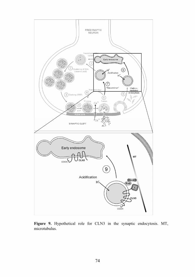
74
Figure 9. Hypothetical role for CLN3 in the synaptic endocytosis. MT, microtubulus.

75
ACKNOWLEDGEMENTS
This thesis work has been carried out at the Department of Molecular Medicine, National Public Health Institute during the years 1999-2006. The former and present Directors of the Institute, Jussi Huttunen and Pekka Puska are acknowledged for providing such excellent research facilities. This thesis has been done as a part of the M.D./Ph.D. program of the Helsinki Biomedical Graduate School. I wish to acknowledge the Dean Tomi Mäkelä, and the heart of the administration Aija Kaitera for the support and education.
I wish to express my gratitude to professor Leena Peltonen-Palotie for accepting the role of kustos at my thesis defence and for creating the international and inspiring scientific atmosphere at the department. She is truly a role model for any young scientist. I wish to thank Adjunct Professors Helena Pihko and Varpu Marjomäki for carefully reviewing my thesis and for the constructive comments on my work. Professors Tomi Mäkelä and Vesa Olkkonen are warmly thanked for their participation in my thesis committee. Our meetings over the past years have been valuable and educational, and I especially value your support in the final stages of this thesis. I also wish to express my appreciation to Professor Elina Ikonen for accepting the role of opponent at my thesis dissertation.
This thesis has been supervised by Adjunct Professor Anu Jalanko. I joined her lab and the CLN3-project as a third-year medical student and it has been nothing but a fascinating journey throughout the years. I thank you, Anu, for all the advice and mentoring, all the opportunities to educate myself at home and abroad, and the forever positive outlook on science and life. I especially value that you have from the very beginning given me freedom and responsibility in the project, and treated me as an equal partner in terms of scientific thinking, even though it has sometimes led to unnecessary experiments.
I wish to thank all the co-authors and collaborators that have contributed to this thesis work. Outi Kopra has had an essential role in my work involving neuronal cells and tissues in the first and third publication. I thank you for teaching me all the neuron preparation skills that I now have and for those long and dark hours at the EM unit, and for sharing the enthusiasm in yoga. Maarit Lehtovirta is thanked for the first lessons in immunofluorescence techniques and tissue fractionation. Irma Järvelä is acknowledged for her pioneering work in the CLN3 project as well as for initiating the Hook1 studies. Heidi Maunu is thanked for her work during her Master’s thesis that included the initial observations of CLN3 and Hook1 interconnection. Aija Kyttälä is thanked for the painstaking determination of the CLN3 topology and targeting, and the critical comments on the second publication.
76
Max Gentile and Iiris Hovatta are thanked for their expert guidance in the statistical analysis of the microarray data. Hannah Mitchison (University College London) is gratefully acknowledged for generating and providing us with the Cln3-/- mouse. Tomas Blom and professor Kid Törnqvist (Åbo Akademi) are warmly thanked for the smooth and efficient collaboration in the calcium imaging studies that not only involved primary neurons but also the national railway company (VR).
I wish to thank the past and present members of the AGU-NCL group for sharing the ups and downs of the scientific work. My lonely years with the CLN3 project ended when Kristiina joined the project. Her straight-forward and hard-working Southern Ostrobothnian attitude has made our work together pleasant, and I know I could not leave the project in more careful hands. I also wish to thank Laura, Tintti, Annina, Tarja, Liina, Salli, Minna, Kai, Juha, Ville and Jani, who have been valuable members of the team and the experience. Laura, Kristiina, Tintti and Annina are thanked for sharing the office for the last years. Our room is by far the noisiest and most fun, from which you can find solutions to a bad hair day and cloning problems, just to name a few. I thank you for tolerating my messy office table and all the memorable conference trips in the name of the NCLs. I would also like to thank all the skilful technicians that have worked in our group and made things go more smoothly in the lab; Kaija, Seija, Tuula, Anne, Auli and Marja.
Our coffee group has been crucial in my survival and the completion of this thesis. Maria, you have been a dear friend ever since the legendary two-week summer course in the Greek island of Spetses. We have gone through medical school, the M.D./Ph.D. program, and explored astanga yoga, and yet I believe the best years are still awaiting. From the first day at KTL, I have been working side by side with Laura. We have suffered through the endocytosis experiments and shared the frustration over the apparent health and well-being of our knock-out models, but it has been a delight. Tanja is the kindest and most helpful person in the lab, and literally, this thesis would not have come to an existence without you and your graphic designer skills (Figures 1, 2, 4 and 9). In addition, I would like to give the warmest hugs and thanks to the other members of our coffee group; Taina (we know the importance of horses, cats and dogs in one’s life!), Anna and Nora. I would also like to thank Heidi for all the years in the same boat, all the encouragement and great company. Finally, the prize for the best conference trip company goes to Laura and Liina, I will never forget Nizza, Chicago, DC or London!
Over the years, dozens of exceptional people have worked at our lab. In addition to those already mentioned, I would like to thank senior scientists of the lab Ismo Ulmanen, Marjo Kestilä, Markus Perola and Teppo Varilo for interesting discussions and advice. The “big lab people” Juha P, Niklas, Petra, Mira, Annika, Heli, Kaisu, Markus, Nina, Joni, Kipa and everyone else are also thanked for making

77
the lab what it is. Our secretaries, Sari K, Sari M, Sami and Tuija and the computer wizard Jari are also thanked for their friendly help in numerous practical matters.
I wish to thank Cursus Metamorphosis for all the fun years in Medical School, especially the two incredible class trips to Pesaro and Torre. I especially want to thank Saara for the shared experience, your amazing support and encouragement, and the friendship that I value most dearly. I also want to thank all my friends in Finland and far away who have provided me with a perspective of the “real world” outside of the M.D./Ph.D. The two years at the United World College of the American West (New Mexico, USA) sparked my interest towards science and making the world a better place, and I wish to acknowledge the class of ’95 for changing my life forever, and for believing that change is possible. My oldest Savukoski friends, Anu, Hanna, Minna and Suvi, I thank you for the decades of friendship and never-failing support that can beat the oceans and the long domestic distances.
I would like to thank my parents-in-law, Liisa and Eero, and sisters-in-law Maija and Salla for support and interest towards my work, and for warmly taking me into your family. I also want to thank my extended family and relatives, especially Elsi and her family, for all the support and encouragement. My parents Enni and Mikko, and my brother Jukka Pekka, I thank you from the bottom of my heart for always supporting and believing in me and loving me unconditionally. Most importantly, I thank my husband Jaakko for your the continuous support, love and happiness that you have brought to my life. Over the years I have not heard a single complaint, even though this thesis has often been done at the cost of our time together. One could not have a better companion. Our love and life together make everything worthwhile.
This study has been financially supported by the Helsinki Biomedical Graduate School, Rinnekoti Research Foundation, Finnish Cultural Foundation, Arvo and Lea Ylppö Foundation, Finnish Medical Association, Sigrid Juselius Foundation, Neurologiasäätiö, and Biomedicum Helsinki Foundation, which are all gratefully acknowledged.
In Helsinki, February 2006
Kaisu Luiro

78
REFERENCES
Aberg L, Liewendahl K, Nikkinen P, Autti T, Rinne JO, Santavuori P. 2000. Decreased striatal dopamine transporter density in JNCL patients with parkinsonian symptoms. Neurology 54(5):1069-74.
Aderem A, Underhill DM. 1999. Mechanisms of phagocytosis in macrophages. Annu Rev Immunol 17:593-623.
Ahtiainen L, Luiro K, Kauppi M, Tyynela J, Kopra O, Jalanko A. 2006. Palmitoyl protein thioesterase 1 (PPT1) deficiency causes endocytic defects connected to abnormal saposin processing. Exp Cell Res in press.
Aizawa H, Sekine Y, Takemura R, Zhang Z, Nangaku M, Hirokawa N. 1992. Kinesin family in murine central nervous system. J Cell Biol 119(5):1287-96.
Akert K, Moor H, Pfenninger K. 1971. Synaptic fine structure. Adv Cytopharmacol 1:273-90.
Alfonso A, Grundahl K, Duerr JS, Han HP, Rand JB. 1993. The Caenorhabditis elegans unc-17 gene: a putative vesicular acetylcholine transporter. Science 261(5121):617-9.
Antonin W, Riedel D, von Mollard GF. 2000. The SNARE Vti1a-beta is localized to small synaptic vesicles and participates in a novel SNARE complex. J Neurosci 20(15):5724-32.
Araki S, Kikuchi A, Hata Y, Isomura M, Takai Y. 1990. Regulation of reversible binding of smg p25A, a ras p21-like GTP-binding protein, to synaptic plasma membranes and vesicles by its specific regulatory protein, GDP dissociation inhibitor. J Biol Chem 265(22):13007-15.
Aridor M, Hannan LA. 2000. Traffic jam: a compendium of human diseases that affect intracellular transport processes. Traffic 1(11):836-51.
Aridor M, Hannan LA. 2002. Traffic jams II: an update of diseases of intracellular transport. Traffic 3(11):781-90.
Ashburner M, Ball CA, Blake JA, Botstein D, Butler H, Cherry JM, Davis AP, Dolinski K, Dwight SS, Eppig JT and others. 2000. Gene ontology: tool for the unification of biology. The Gene Ontology Consortium. Nat Genet 25(1):25-9.
Atluri PP, Regehr WG. 1998. Delayed release of neurotransmitter from cerebellar granule cells. J Neurosci 18(20):8214-27.
Attard M, Jean G, Forestier L, Cherqui S, van't Hoff W, Broyer M, Antignac C, Town M. 1999. Severity of phenotype in cystinosis varies with mutations in the CTNS gene: predicted effect on the model of cystinosin. Hum Mol Genet 8(13):2507-14.
Autti T, Raininko R, Santavuori P, Vanhanen SL, Poutanen VP, Haltia M. 1997. MRI of neuronal ceroid lipofuscinosis. II. Postmortem MRI and histopathological study of the brain in 16 cases of neuronal ceroid lipofuscinosis of juvenile or late infantile type. Neuroradiology 39(5):371-7.
Autti T, Raininko R, Vanhanen SL, Santavuori P. 1996. MRI of neuronal ceroid lipofuscinosis. I. Cranial MRI of 30 patients with juvenile neuronal ceroid lipofuscinosis. Neuroradiology 38(5):476-82.

79
Baas PW, Deitch JS, Black MM, Banker GA. 1988. Polarity orientation of microtubules in hippocampal neurons: uniformity in the axon and nonuniformity in the dendrite. Proc Natl Acad Sci U S A 85(21):8335-9.
Bagshaw RD, Mahuran DJ, Callahan JW. 2005. A proteomic analysis of lysosomal integral membrane proteins reveals the diverse composition of the organelle. Mol Cell Proteomics 4(2):133-43.
Barbero P, Bittova L, Pfeffer SR. 2002. Visualization of Rab9-mediated vesicle transport from endosomes to the trans-Golgi in living cells. J Cell Biol 156(3):511-8.
Bargal R, Avidan N, Ben-Asher E, Olender Z, Zeigler M, Frumkin A, Raas-Rothschild A, Glusman G, Lancet D, Bach G. 2000. Identification of the gene causing mucolipidosis type IV. Nat Genet 26(1):118-23.
Barker LA, Dowdall MJ, Whittaker VP. 1972. Choline metabolism in the cerebral cortex of guinea pigs. Stable-bound acetylcholine. Biochem J 130(4):1063-75.
Barlowe C, Orci L, Yeung T, Hosobuchi M, Hamamoto S, Salama N, Rexach MF, Ravazzola M, Amherdt M, Schekman R. 1994. COPII: a membrane coat formed by Sec proteins that drive vesicle budding from the endoplasmic reticulum. Cell 77(6):895-907.
Barrett EF, Stevens CF. 1972. The kinetics of transmitter release at the frog neuromuscular junction. J Physiol 227(3):691-708.
Bengtsson BA, Johansson JO, Hollak C, Linthorst G, FeldtRasmussen U. 2003. Enzyme replacement in Anderson-Fabry disease. Lancet 361(9354):352.
Berg JM, Tymoczko JL, Stryer L. 2002. Biochemistry. New York: W.H. Freeman and Company.
Berkovic SF, Carpenter S, Andermann F, Andermann E, Wolfe LS. 1988. Kufs' disease: a critical reappraisal. Brain 111 ( Pt 1):27-62.
Berman ER, Livni N, Shapira E, Merin S, Levij IS. 1974. Congenital corneal clouding with abnormal systemic storage bodies: a new variant of mucolipidosis. J Pediatr 84(4):519-26.
Blackmer T, Larsen EC, Bartleson C, Kowalchyk JA, Yoon EJ, Preininger AM, Alford S, Hamm HE, Martin TF. 2005. G protein betagamma directly regulates SNARE protein fusion machinery for secretory granule exocytosis. Nat Neurosci 8(4):421-5.
Bleil JD, Bretscher MS. 1982. Transferrin receptor and its recycling in HeLa cells. Embo J 1(3):351-5.
Bock JB, Matern HT, Peden AA, Scheller RH. 2001. A genomic perspective on membrane compartment organization. Nature 409(6822):839-41.
Bonifacino JS, Glick BS. 2004. The mechanisms of vesicle budding and fusion. Cell 116(2):153-66.
Bonifacino JS, Lippincott-Schwartz J. 2003. Coat proteins: shaping membrane transport. Nat Rev Mol Cell Biol 4(5):409-14.
Bonifacino JS, Traub LM. 2003. Signals for sorting of transmembrane proteins to endosomes and lysosomes. Annu Rev Biochem 72:395-447.
Braak H, Goebel HH. 1978. Loss of pigment-laden stellate cells: a severe alteration of the isocortex in juvenile neuronal ceroid-lipofuscinosis. Acta Neuropathol (Berl) 42(1):53-7.

80
Brady ST. 1985. A novel brain ATPase with properties expected for the fast axonal transport motor. Nature 317(6032):73-5.
Breathnach R, Benoist C, O'Hare K, Gannon F, Chambon P. 1978. Ovalbumin gene: evidence for a leader sequence in mRNA and DNA sequences at the exon-intron boundaries. Proc Natl Acad Sci U S A 75(10):4853-7.
Brini M. 2003. Ca(2+) signalling in mitochondria: mechanism and role in physiology and pathology. Cell Calcium 34(4-5):399-405.
Brodsky FM, Chen CY, Knuehl C, Towler MC, Wakeham DE. 2001. Biological basket weaving: formation and function of clathrin-coated vesicles. Annu Rev Cell Dev Biol 17:517-68.
Brooks AI, Chattopadhyay S, Mitchison HM, Nussbaum RL, Pearce DA. 2003. Functional categorization of gene expression changes in the cerebellum of a Cln3-knockout mouse model for Batten disease. Mol Genet Metab 78(1):17-30.
Brose N, Hofmann K, Hata Y, Sudhof TC. 1995. Mammalian homologues of Caenorhabditis elegans unc-13 gene define novel family of C2-domain proteins. J Biol Chem 270(42):25273-80.
Brown MS, Goldstein JL. 1979. Receptor-mediated endocytosis: insight from the lipoprotein receptor system. Proc Natl Acad Sci U S A 76:3330-37.
Buckley K, Kelly RB. 1985. Identification of a transmembrane glycoprotein specific for secretory vesicles of neural and endocrine cells. J Cell Biol 100(4):1284-94.
Burack MA, Silverman MA, Banker G. 2000. The role of selective transport in neuronal protein sorting. Neuron 26(2):465-72.
Burton PR, Paige JL. 1981. Polarity of axoplasmic microtubules in the olfactory nerve of the frog. Proc Natl Acad Sci U S A 78(5):3269-73.
Calakos N, Scheller RH. 1994. Vesicle-associated membrane protein and synaptophysin are associated on the synaptic vesicle. J Biol Chem 269(40):24534-7.
Canfield WM, Johnson KF, Ye RD, Gregory W, Kornfeld S. 1991. Localization of the signal for rapid internalization of the bovine cation-independent mannose 6-phosphate/insulin-like growth factor-II receptor to amino acids 24-29 of the cytoplasmic tail. J Biol Chem 266(9):5682-8.
Cao X, Barlowe C. 2000. Asymmetric requirements for a Rab GTPase and SNARE proteins in fusion of COPII vesicles with acceptor membranes. J Cell Biol 149(1):55-66.
Carpenter G, Cohen S. 1976. 125I-labeled human epidermal growth factor. Binding, internalization, and degradation in human fibroblasts. J Cell Biol 71(1):159-71.
Carstea ED, Morris JA, Coleman KG, Loftus SK, Zhang D, Cummings C, Gu J, Rosenfeld MA, Pavan WJ, Krizman DB and others. 1997. Niemann-Pick C1 disease gene: homology to mediators of cholesterol homeostasis. Science 277(5323):228-31.
Cases-Langhoff C, Voss B, Garner AM, Appeltauer U, Takei K, Kindler S, Veh RW, De Camilli P, Gundelfinger ED, Garner CC. 1996. Piccolo, a novel 420 kDa protein associated with the presynaptic cytomatrix. Eur J Cell Biol 69(3):214-23.
Ceccarelli B, Hurlbut WP, Mauro A. 1973. Turnover of transmitter and synaptic vesicles at the frog neuromuscular junction. J Cell Biol 57(2):499-524.

81
Chattopadhyay S, Ito M, Cooper JD, Brooks AI, Curran TM, Powers JM, Pearce DA. 2002. An autoantibody inhibitory to glutamic acid decarboxylase in the neurodegenerative disorder Batten disease. Hum Mol Genet 11(12):1421-31.
Chattopadhyay S, Kingsley E, Serour A, Curran TM, Brooks AI, Pearce DA. 2004. Altered gene expression in the eye of a mouse model for batten disease. Invest Ophthalmol Vis Sci 45(9):2893-905.
Chattopadhyay S, Roberts PM, Pearce DA. 2003. The yeast model for Batten disease: a role for Btn2p in the trafficking of the Golgi-associated vesicular targeting protein, Yif1p. Biochem Biophys Res Commun 302(3):534-8.
Chen CS, Bach G, Pagano RE. 1998. Abnormal transport along the lysosomal pathway in mucolipidosis, type IV disease. Proc Natl Acad Sci U S A 95(11):6373-8.
Cherqui S, Kalatzis V, Trugnan G, Antignac C. 2001. The targeting of cystinosin to the lysosomal membrane requires a tyrosine-based signal and a novel sorting motif. J Biol Chem 276(16):13314-21.
Clapham DE, Neer EJ. 1997. G protein beta gamma subunits. Annu Rev Pharmacol Toxicol 37:167-203.
Coleman M. 2005. Axon degeneration mechanisms: commonality amid diversity. Nat Rev Neurosci 6(11):889-98.
Conner SD, Schmid SL. 2003. Regulated portals of entry into the cell. Nature 422(6927):37-44.
Conradi NG, Uvebrant P, Hokegard KH, Wahlstrom J, Mellqvist L. 1989. First-trimester diagnosis of juvenile neuronal ceroid lipofuscinosis by demonstration of fingerprint inclusions in chorionic villi. Prenat Diagn 9(4):283-7.
Consortium. The International Batten Disease Consortium. 1995. Isolation of a novel gene underlying Batten disease, CLN3. Cell 82(6):949-57.
Cotman SL, Vrbanac V, Lebel LA, Lee RL, Johnson KA, Donahue LR, Teed AM, Antonellis K, Bronson RT, Lerner TJ and others. 2002. Cln3(Deltaex7/8) knock-in mice with the common JNCL mutation exhibit progressive neurologic disease that begins before birth. Hum Mol Genet 11(22):2709-21.
D'Azzo A. 2003. Gene transfer strategies for correction of lysosomal storage disorders. Acta Haematol 110(2-3):71-85.
Damm EM, Pelkmans L, Kartenbeck J, Mezzacasa A, Kurzchalia T, Helenius A. 2005. Clathrin- and caveolin-1-independent endocytosis: entry of simian virus 40 into cells devoid of caveolae. J Cell Biol 168(3):477-88.
Danon MJ, Oh SJ, DiMauro S, Manaligod JR, Eastwood A, Naidu S, Schliselfeld LH. 1981. Lysosomal glycogen storage disease with normal acid maltase. Neurology 31(1):51-7.
Das AM, Kohlschutter A. 1996. Decreased activity of the mitochondrial ATP-synthase in fibroblasts from children with late-infantile and juvenile neuronal ceroid lipofuscinosis. J Inherit Metab Dis 19(2):130-2.
Das AM, von Harlem R, Feist M, Lucke T, Kohlschutter A. 2001. Altered levels of high-energy phosphate compounds in fibroblasts from different forms of neuronal ceroid lipofuscinoses: further evidence for mitochondrial involvement. Eur J Paediatr Neurol 5 Suppl A:143-6.

82
Davies JP, Ioannou YA. 2000. Topological analysis of Niemann-Pick C1 protein reveals that the membrane orientation of the putative sterol-sensing domain is identical to those of 3-hydroxy-3-methylglutaryl-CoA reductase and sterol regulatory element binding protein cleavage-activating protein. J Biol Chem 275(32):24367-74.
Davis CG, Lehrman MA, Russell DW, Anderson RG, Brown MS, Goldstein JL. 1986. The J.D. mutation in familial hypercholesterolemia: amino acid substitution in cytoplasmic domain impedes internalization of LDL receptors. Cell 45(1):15-24.
Dawson G, Kilkus J, Siakotos AN, Singh I. 1996. Mitochondrial abnormalities in CLN2 and CLN3 forms of Batten disease. Mol Chem Neuropathol 29(2-3):227-35.
de Duve C PB, Gianetto R, Wattiaux R, Appelmans F 1955. Tissue fractionation studies. 6. Intracellular distribution patterns of enzymes in rat-liver tissue. Biochem J 60:604-617.
De Waard M, Liu H, Walker D, Scott VE, Gurnett CA, Campbell KP. 1997. Direct binding of G-protein betagamma complex to voltage-dependent calcium channels. Nature 385(6615):446-50.
de Voer G, der Bent P, Rodrigues AJ, van Ommen GJ, Peters DJ, Taschner PE. 2005. Deletion of the Caenorhabditis elegans homologues of the CLN3 gene, involved in human juvenile neuronal ceroid lipofuscinosis, causes a mild progeric phenotype. J Inherit Metab Dis 28(6):1065-80.
Dell'Angelica EC, Shotelersuk V, Aguilar RC, Gahl WA, Bonifacino JS. 1999. Altered trafficking of lysosomal proteins in Hermansky-Pudlak syndrome due to mutations in the beta 3A subunit of the AP-3 adaptor. Mol Cell 3(1):11-21.
Desnick RJ, Schuchman EH. 2002. Enzyme replacement and enhancement therapies: lessons from lysosomal disorders. Nat Rev Genet 3(12):954-66.
Di Fiore PP, De Camilli P. 2001. Endocytosis and signaling. an inseparable partnership. Cell 106(1):1-4.
Diaz E, Pfeffer SR. 1998. TIP47: a cargo selection device for mannose 6-phosphate receptor trafficking. Cell 93(3):433-43.
Dice JF, Terlecky SR. 1990. Targeting of cytosolic proteins to lysosomes for degradation. Crit Rev Ther Drug Carrier Syst 7(3):211-33.
Dierks T, Schmidt B, Borissenko LV, Peng J, Preusser A, Mariappan M, von Figura K. 2003. Multiple sulfatase deficiency is caused by mutations in the gene encoding the human C(alpha)-formylglycine generating enzyme. Cell 113(4):435-44.
Dietrich D, Kirschstein T, Kukley M, Pereverzev A, von der Brelie C, Schneider T, Beck H. 2003. Functional specialization of presynaptic Cav2.3 Ca2+ channels. Neuron 39(3):483-96.
Dillon C, Goda Y. 2005. The actin cytoskeleton: integrating form and function at the synapse. Annu Rev Neurosci 28:25-55.
Dirac-Svejstrup AB, Sumizawa T, Pfeffer SR. 1997. Identification of a GDI displacement factor that releases endosomal Rab GTPases from Rab-GDI. Embo J 16(3):465-72.
Dittmer F, Ulbrich EJ, Hafner A, Schmahl W, Meister T, Pohlmann R, von Figura K. 1999. Alternative mechanisms for trafficking of lysosomal enzymes in mannose

83
6-phosphate receptor-deficient mice are cell type-specific. J Cell Sci 112 ( Pt 10):1591-7.
Doray B, Ghosh P, Griffith J, Geuze HJ, Kornfeld S. 2002. Cooperation of GGAs and AP-1 in packaging MPRs at the trans-Golgi network. Science 297(5587):1700-3.
Drab M, Verkade P, Elger M, Kasper M, Lohn M, Lauterbach B, Menne J, Lindschau C, Mende F, Luft FC and others. 2001. Loss of caveolae, vascular dysfunction, and pulmonary defects in caveolin-1 gene-disrupted mice. Science 293(5539):2449-52.
Dulubova I, Sugita S, Hill S, Hosaka M, Fernandez I, Sudhof TC, Rizo J. 1999. A conformational switch in syntaxin during exocytosis: role of munc18. Embo J 18(16):4372-82.
Dunaevsky A, Connor EA. 2000. F-actin is concentrated in nonrelease domains at frog neuromuscular junctions. J Neurosci 20(16):6007-12.
Eaton BA, Fetter RD, Davis GW. 2002. Dynactin is necessary for synapse stabilization. Neuron 34(5):729-41.
Echard A, Jollivet F, Martinez O, Lacapere JJ, Rousselet A, Janoueix-Lerosey I, Goud B. 1998. Interaction of a Golgi-associated kinesin-like protein with Rab6. Science 279(5350):580-5.
Edelmann L, Hanson PI, Chapman ER, Jahn R. 1995. Synaptobrevin binding to synaptophysin: a potential mechanism for controlling the exocytotic fusion machine. Embo J 14(2):224-31.
El-Husseini Ael D, Craven SE, Brock SC, Bredt DS. 2001. Polarized targeting of peripheral membrane proteins in neurons. J Biol Chem 276(48):44984-92.
Elshatory Y, Brooks AI, Chattopadhyay S, Curran TM, Gupta P, Ramalingam V, Hofmann SL, Pearce DA. 2003. Early changes in gene expression in two models of Batten disease. FEBS Lett 538(1-3):207-12.
Erickson JD, Eiden LE, Hoffman BJ. 1992. Expression cloning of a reserpine-sensitive vesicular monoamine transporter. Proc Natl Acad Sci U S A 89(22):10993-7.
Eskelinen EL, Tanaka Y, Saftig P. 2003. At the acidic edge: emerging functions for lysosomal membrane proteins. Trends Cell Biol 13(3):137-45.
Ezaki J, Takeda-Ezaki M, Koike M, Ohsawa Y, Taka H, Mineki R, Murayama K, Uchiyama Y, Ueno T, Kominami E. 2003. Characterization of Cln3p, the gene product responsible for juvenile neuronal ceroid lipofuscinosis, as a lysosomal integral membrane glycoprotein. J Neurochem 87(5):1296-308.
Fasshauer D, Sutton RB, Brunger AT, Jahn R. 1998. Conserved structural features of the synaptic fusion complex: SNARE proteins reclassified as Q- and R-SNAREs. Proc Natl Acad Sci U S A 95(26):15781-6.
Fernandez-Chacon R, Konigstorfer A, Gerber SH, Garcia J, Matos MF, Stevens CF, Brose N, Rizo J, Rosenmund C, Sudhof TC. 2001. Synaptotagmin I functions as a calcium regulator of release probability. Nature 410(6824):41-9.
Ferri A, Sanes JR, Coleman MP, Cunningham JM, Kato AC. 2003. Inhibiting axon degeneration and synapse loss attenuates apoptosis and disease progression in a mouse model of motoneuron disease. Curr Biol 13(8):669-73.

84
Fischer LR, Culver DG, Tennant P, Davis AA, Wang M, Castellano-Sanchez A, Khan J, Polak MA, Glass JD. 2004. Amyotrophic lateral sclerosis is a distal axonopathy: evidence in mice and man. Exp Neurol 185(2):232-40.
Fischer von Mollard G, Stahl B, Walch-Solimena C, Takei K, Daniels L, Khoklatchev A, De Camilli P, Sudhof TC, Jahn R. 1994. Localization of Rab5 to synaptic vesicles identifies endosomal intermediate in synaptic vesicle recycling pathway. Eur J Cell Biol 65(2):319-26.
Fossale E, Wolf P, Espinola JA, Lubicz-Nawrocka T, Teed AM, Gao H, Rigamonti D, Cattaneo E, MacDonald ME, Cotman SL. 2004. Membrane trafficking and mitochondrial abnormalities precede subunit c deposition in a cerebellar cell model of juvenile neuronal ceroid lipofuscinosis. BMC Neurosci 5(1):57.
Fremeau RT, Jr., Burman J, Qureshi T, Tran CH, Proctor J, Johnson J, Zhang H, Sulzer D, Copenhagen DR, Storm-Mathisen J and others. 2002. The identification of vesicular glutamate transporter 3 suggests novel modes of signaling by glutamate. Proc Natl Acad Sci U S A 99(22):14488-93.
Fujimoto LM, Roth R, Heuser JE, Schmid SL. 2000. Actin assembly plays a variable, but not obligatory role in receptor-mediated endocytosis in mammalian cells. Traffic 1(2):161-71.
Futerman AH, Sussman JL, Horowitz M, Silman I, Zimran A. 2004. New directions in the treatment of Gaucher disease. Trends Pharmacol Sci 25(3):147-51.
Futerman AH, van Meer G. 2004. The cell biology of lysosomal storage disorders. Nat Rev Mol Cell Biol 5(7):554-65.
Gachet Y, Codlin S, Hyams JS, Mole SE. 2005. btn1, the Schizosaccharomyces pombe homologue of the human Batten disease gene CLN3, regulates vacuole homeostasis. J Cell Sci 118(Pt 23):5525-36.
Gaidarov I, Santini F, Warren RA, Keen JH. 1999. Spatial control of coated-pit dynamics in living cells. Nat Cell Biol 1(1):1-7.
Galbiati F, Engelman JA, Volonte D, Zhang XL, Minetti C, Li M, Hou H, Jr., Kneitz B, Edelmann W, Lisanti MP. 2001. Caveolin-3 null mice show a loss of caveolae, changes in the microdomain distribution of the dystrophin-glycoprotein complex, and t-tubule abnormalities. J Biol Chem 276(24):21425-33.
Galjart N. 2005. CLIPs and CLASPs and cellular dynamics. Nat Rev Mol Cell Biol 6(6):487-98.
Gao H, Boustany RM, Espinola JA, Cotman SL, Srinidhi L, Antonellis KA, Gillis T, Qin X, Liu S, Donahue LR and others. 2002. Mutations in a novel CLN6-encoded transmembrane protein cause variant neuronal ceroid lipofuscinosis in man and mouse. Am J Hum Genet 70(2):324-35.
Garcia DE, Li B, Garcia-Ferreiro RE, Hernandez-Ochoa EO, Yan K, Gautam N, Catterall WA, Mackie K, Hille B. 1998. G-protein beta-subunit specificity in the fast membrane-delimited inhibition of Ca2+ channels. J Neurosci 18(22):9163-70.
Geppert M, Bolshakov VY, Siegelbaum SA, Takei K, De Camilli P, Hammer RE, Sudhof TC. 1994a. The role of Rab3A in neurotransmitter release. Nature 369(6480):493-7.
85
Geppert M, Goda Y, Hammer RE, Li C, Rosahl TW, Stevens CF, Sudhof TC. 1994b. Synaptotagmin I: a major Ca2+ sensor for transmitter release at a central synapse. Cell 79(4):717-27.
Geppert M, Goda Y, Stevens CF, Sudhof TC. 1997. The small GTP-binding protein Rab3A regulates a late step in synaptic vesicle fusion. Nature 387(6635):810-4.
Gerachshenko T, Blackmer T, Yoon EJ, Bartleson C, Hamm HE, Alford S. 2005. Gbetagamma acts at the C terminus of SNAP-25 to mediate presynaptic inhibition. Nat Neurosci 8(5):597-605.
Goda Y, Stevens CF. 1994. Two components of transmitter release at a central synapse. Proc Natl Acad Sci U S A 91(26):12942-6.
Goebel HH. 1997. Morphologic diagnosis in neuronal ceroid lipofuscinosis. Neuropediatrics 28(1):67-9.
Goebel HH, Fix JD, Zeman W. 1974. The fine structure of the retina in neuronal ceroid-lipofuscinosis. Am J Ophthalmol 77(1):25-39.
Golabek AA, Kaczmarski W, Kida E, Kaczmarski A, Michalewski MP, Wisniewski KE. 1999. Expression studies of CLN3 protein (battenin) in fusion with the green fluorescent protein in mammalian cells in vitro. Mol Genet Metab 66(4):277-82.
Golabek AA, Kida E, Walus M, Wujek P, Mehta P, Wisniewski KE. 2003. Biosynthesis, glycosylation, and enzymatic processing in vivo of human tripeptidyl-peptidase I. J Biol Chem 278(9):7135-45.
Goldstein JL, Brunschede GY, Brown MS. 1975. Inhibition of proteolytic degradation of low density lipoprotein in human fibroblasts by chloroquine, concanavalin A, and Triton WR 1339. J Biol Chem 250(19):7854-62.
Grafstein B, Forman DS. 1980. Intracellular transport in neurons. Physiol Rev 60(4):1167-283.
Gras C, Herzog E, Bellenchi GC, Bernard V, Ravassard P, Pohl M, Gasnier B, Giros B, El Mestikawy S. 2002. A third vesicular glutamate transporter expressed by cholinergic and serotoninergic neurons. J Neurosci 22(13):5442-51.
Greene ND, Lythgoe MF, Thomas DL, Nussbaum RL, Bernard DJ, Mitchison HM. 2001. High resolution MRI reveals global changes in brains of Cln3 mutant mice. Eur J Paediatr Neurol 5 Suppl A:103-7.
Greengard P, Benfenati F, Valtorta F. 1994. Synapsin I, an actin-binding protein regulating synaptic vesicle traffic in the nerve terminal. Adv Second Messenger Phosphoprotein Res 29:31-45.
Griffiths G, Gruenberg J. 1991. The arguments for pre-existing early and late endosomes. Trends Cell Biol 1(1):5-9.
Griffiths G, Hoppeler H. 1986. Quantitation in immunocytochemistry: correlation of immunogold labeling to absolute number of membrane antigens. J Histochem Cytochem 34(11):1389-98.
Gruenberg J, Maxfield FR. 1995. Membrane transport in the endocytic pathway. Curr Opin Cell Biol 7(4):552-63.
Gu C, Jan YN, Jan LY. 2003. A conserved domain in axonal targeting of Kv1 (Shaker) voltage-gated potassium channels. Science 301(5633):646-9.

86
Hall A, Nobes CD. 2000. Rho GTPases: molecular switches that control the organization and dynamics of the actin cytoskeleton. Philos Trans R Soc Lond B Biol Sci 355(1399):965-70.
Hall NA, Lake BD, Dewji NN, Patrick AD. 1991. Lysosomal storage of subunit c of mitochondrial ATP synthase in Batten's disease (ceroid-lipofuscinosis). Biochem J 275 ( Pt 1):269-72.
Haltia M. 2003. The neuronal ceroid-lipofuscinoses. J Neuropathol Exp Neurol 62(1):1-13. Haltia M, Rapola J, Santavuori P, Keranen A. 1973. Infantile type of so-called neuronal
ceroid-lipofuscinosis. 2. Morphological and biochemical studies. J Neurol Sci 18(3):269-85.
Hanson PI, Roth R, Morisaki H, Jahn R, Heuser JE. 1997. Structure and conformational changes in NSF and its membrane receptor complexes visualized by quick-freeze/deep-etch electron microscopy. Cell 90(3):523-35.
Harada A, Takei Y, Kanai Y, Tanaka Y, Nonaka S, Hirokawa N. 1998. Golgi vesiculation and lysosome dispersion in cells lacking cytoplasmic dynein. J Cell Biol 141(1):51-9.
Harter C, Mellman I. 1992. Transport of the lysosomal membrane glycoprotein lgp120 (lgp-A) to lysosomes does not require appearance on the plasma membrane. J Cell Biol 117(2):311-25.
Haskell RE, Carr CJ, Pearce DA, Bennett MJ, Davidson BL. 2000. Batten disease: evaluation of CLN3 mutations on protein localization and function. Hum Mol Genet 9(5):735-44.
Heine C, Koch B, Storch S, Kohlschutter A, Palmer DN, Braulke T. 2004. Defective endoplasmic reticulum-resident membrane protein CLN6 affects lysosomal degradation of endocytosed arylsulfatase A. J Biol Chem 279(21):22347-52.
Helenius A, Mellman I, Wall D, Hubbard A. 1983. Endosomes. Trends Biochem Sci 8(7):245-250.
Hellsten E, Vesa J, Olkkonen VM, Jalanko A, Peltonen L. 1996. Human palmitoyl protein thioesterase: evidence for lysosomal targeting of the enzyme and disturbed cellular routing in infantile neuronal ceroid lipofuscinosis. Embo J 15(19):5240-5.
Hermann GJ, Thatcher JW, Mills JP, Hales KG, Fuller MT, Nunnari J, Shaw JM. 1998. Mitochondrial fusion in yeast requires the transmembrane GTPase Fzo1p. J Cell Biol 143(2):359-73.
Heuser JE, Reese TS. 1973. Evidence for recycling of synaptic vesicle membrane during transmitter release at the frog neuromuscular junction. J Cell Biol 57(2):315-44.
Hinshaw JE. 2000. Dynamin and its role in membrane fission. Annu Rev Cell Dev Biol 16:483-519.
Hirokawa N. 1998. Kinesin and dynein superfamily proteins and the mechanism of organelle transport. Science 279(5350):519-26.
Hirokawa N, Takemura R. 2004. Molecular motors in neuronal development, intracellular transport and diseases. Curr Opin Neurobiol 14(5):564-73.
Hirokawa N, Takemura R. 2005. Molecular motors and mechanisms of directional transport in neurons. Nat Rev Neurosci 6(3):201-14.

87
Hirvasniemi A, Herrala P, Leisti J. 1995. Northern epilepsy syndrome: clinical course and the effect of medication on seizures. Epilepsia 36(8):792-7.
Hirvasniemi A, Lang H, Lehesjoki AE, Leisti J. 1994. Northern epilepsy syndrome: an inherited childhood onset epilepsy with associated mental deterioration. J Med Genet 31(3):177-82.
Hofmann SL, Peltonen L. 2001. The neuronal ceroid lipofuscinoses. New York: McGraw-Hill.
Hofmann SL PL. 2001. The neuronal ceroid lipofuscinoses, 8th edition. New York: McGraw-Hill.
Holopainen JM, Saarikoski J, Kinnunen PK, Jarvela I. 2001. Elevated lysosomal pH in neuronal ceroid lipofuscinoses (NCLs). Eur J Biochem 268(22):5851-6.
Ihrke G, Kyttala A, Russell MR, Rous BA, Luzio JP. 2004. Differential use of two AP-3-mediated pathways by lysosomal membrane proteins. Traffic 5(12):946-62.
Ikonen E, Holtta-Vuori M. 2004. Cellular pathology of Niemann-Pick type C disease. Semin Cell Dev Biol 15(4):445-54.
Isosomppi J, Vesa J, Jalanko A, Peltonen L. 2002. Lysosomal localization of the neuronal ceroid lipofuscinosis CLN5 protein. Hum Mol Genet 11(8):885-91.
Jadot M, Canfield WM, Gregory W, Kornfeld S. 1992. Characterization of the signal for rapid internalization of the bovine mannose 6-phosphate/insulin-like growth factor-II receptor. J Biol Chem 267(16):11069-77.
Jahn R, Lang T, Sudhof TC. 2003. Membrane fusion. Cell 112(4):519-33. Jahn R, Sudhof TC. 1999. Membrane fusion and exocytosis. Annu Rev Biochem
68:863-911. Janz R, Goda Y, Geppert M, Missler M, Sudhof TC. 1999. SV2A and SV2B function as
redundant Ca2+ regulators in neurotransmitter release. Neuron 24(4):1003-16. Jareb M, Banker G. 1998. The polarized sorting of membrane proteins expressed in
cultured hippocampal neurons using viral vectors. Neuron 20(5):855-67. Jarvela I, Autti T, Lamminranta S, Aberg L, Raininko R, Santavuori P. 1997. Clinical
and magnetic resonance imaging findings in Batten disease: analysis of the major mutation (1.02-kb deletion). Ann Neurol 42(5):799-802.
Jarvela I, Lehtovirta M, Tikkanen R, Kyttala A, Jalanko A. 1999. Defective intracellular transport of CLN3 is the molecular basis of Batten disease (JNCL). Hum Mol Genet 8(6):1091-8.
Jarvela I, Sainio M, Rantamaki T, Olkkonen VM, Carpen O, Peltonen L, Jalanko A. 1998. Biosynthesis and intracellular targeting of the CLN3 protein defective in Batten disease. Hum Mol Genet 7(1):85-90.
Job C, Eberwine J. 2001. Localization and translation of mRNA in dendrites and axons. Nat Rev Neurosci 2(12):889-98.
Johnston PA, Sudhof TC. 1990. The multisubunit structure of synaptophysin. Relationship between disulfide bonding and homo-oligomerization. J Biol Chem 265(15):8869-73.
Journet A, Chapel A, Kieffer S, Roux F, Garin J. 2002. Proteomic analysis of human lysosomes: application to monocytic and breast cancer cells. Proteomics 2(8):1026-40.

88
Kaczmarski W, Wisniewski KE, Golabek A, Kaczmarski A, Kida E, Michalewski M. 1999. Studies of membrane association of CLN3 protein. Mol Genet Metab 66(4):261-4.
Kanaani J, el-Husseini Ael D, Aguilera-Moreno A, Diacovo JM, Bredt DS, Baekkeskov S. 2002. A combination of three distinct trafficking signals mediates axonal targeting and presynaptic clustering of GAD65. J Cell Biol 158(7):1229-38.
Kasper D, Planells-Cases R, Fuhrmann JC, Scheel O, Zeitz O, Ruether K, Schmitt A, Poet M, Steinfeld R, Schweizer M and others. 2005. Loss of the chloride channel ClC-7 leads to lysosomal storage disease and neurodegeneration. Embo J 24(5):1079-91.
Katz ML, Gao CL, Prabhakaram M, Shibuya H, Liu PC, Johnson GS. 1997. Immunochemical localization of the Batten disease (CLN3) protein in retina. Invest Ophthalmol Vis Sci 38(11):2375-86.
Katz ML, Shibuya H, Liu PC, Kaur S, Gao CL, Johnson GS. 1999. A mouse gene knockout model for juvenile ceroid-lipofuscinosis (Batten disease). J Neurosci Res 57(4):551-6.
Kaufman RJ. 2002. Orchestrating the unfolded protein response in health and disease. J Clin Invest 110(10):1389-98.
Kauppi M, Jäntti J, Olkkonen VM. 2004. The function of Sec1/Munc18 proteins – Solution of the mystery in sight? Berlin: Springer-Verlag GmbH.
Khvotchev MV, Ren M, Takamori S, Jahn R, Sudhof TC. 2003. Divergent functions of neuronal Rab11b in Ca2+-regulated versus constitutive exocytosis. J Neurosci 23(33):10531-9.
Kida E, Kaczmarski W, Golabek AA, Kaczmarski A, Michalewski M, Wisniewski KE. 1999. Analysis of intracellular distribution and trafficking of the CLN3 protein in fusion with the green fluorescent protein in vitro. Mol Genet Metab 66(4):265-71.
Kielian MC, Cohn ZA. 1980. Phagosome-lysosome fusion. Characterization of intracellular membrane fusion in mouse macrophages. J Cell Biol 85(3):754-65.
Kim Y, Ramirez-Montealegre D, Pearce DA. 2003. A role in vacuolar arginine transport for yeast Btn1p and for human CLN3, the protein defective in Batten disease. Proc Natl Acad Sci U S A 100(26):15458-62.
Kirchhausen T. 2000. Clathrin. Annu Rev Biochem 69:699-727. Kirchhausen T, Harrison SC. 1981. Protein organization in clathrin trimers. Cell
23(3):755-61. Kominami E, Ezaki J, Muno D, Ishido K, Ueno T, Wolfe LS. 1992. Specific storage of
subunit c of mitochondrial ATP synthase in lysosomes of neuronal ceroid lipofuscinosis (Batten's disease). J Biochem (Tokyo) 111(2):278-82.
Kornfeld S. 1990. Lysosomal enzyme targeting. Biochem Soc Trans 18(3):367-74. Kornfeld S, Mellman I. 1989. The biogenesis of lysosomes. Annu Rev Cell Biol 5:483-525. Kornfeld S, Sly WS. 2001. I-Cell Disease and Pseudo-Hurler Dystrophy: Disorders of
Lysosomal Enzyme Phosphorylation and Localization. New York: McGraw. Kramer H, Phistry M. 1996. Mutations in the Drosophila hook gene inhibit endocytosis
of the boss transmembrane ligand into multivesicular bodies. J Cell Biol 133(6):1205-15.

89
Kramer H, Phistry M. 1999. Genetic analysis of hook, a gene required for endocytic trafficking in drosophila. Genetics 151(2):675-84.
Kremmidiotis G, Lensink IL, Bilton RL, Woollatt E, Chataway TK, Sutherland GR, Callen DF. 1999. The Batten disease gene product (CLN3p) is a Golgi integral membrane protein. Hum Mol Genet 8(3):523-31.
Kyttala A, Ihrke G, Vesa J, Schell MJ, Luzio JP. 2004. Two Motifs Target Batten Disease Protein CLN3 to Lysosomes in Transfected Nonneuronal and Neuronal Cells. Mol Biol Cell 15(3):1313-23.
Kyttala A, Yliannala K, Schu P, Jalanko A, Luzio JP. 2005. AP-1 and AP-3 facilitate lysosomal targeting of Batten disease protein CLN3 via its dileucine motif. J Biol Chem 280(11):10277-83.
Lamaze C, Dujeancourt A, Baba T, Lo CG, Benmerah A, Dautry-Varsat A. 2001. Interleukin 2 receptors and detergent-resistant membrane domains define a clathrin-independent endocytic pathway. Mol Cell 7(3):661-71.
LaMonte BH, Wallace KE, Holloway BA, Shelly SS, Ascano J, Tokito M, Van Winkle T, Howland DS, Holzbaur EL. 2002. Disruption of dynein/dynactin inhibits axonal transport in motor neurons causing late-onset progressive degeneration. Neuron 34(5):715-27.
Lander ES, Linton LM, Birren B, Nusbaum C, Zody MC, Baldwin J, Devon K, Dewar K, Doyle M, FitzHugh W and others. 2001. Initial sequencing and analysis of the human genome. Nature 409(6822):860-921.
Lauronen L, Heikkila E, Autti T, Sainio K, Huttunen J, Aronen HJ, Korvenoja A, Ilmoniemi RJ, Santavuori P. 1997. Somatosensory evoked magnetic fields from primary sensorimotor cortex in juvenile neuronal ceroid lipofuscinosis. J Child Neurol 12(6):355-60.
Le Borgne R, Alconada A, Bauer U, Hoflack B. 1998. The mammalian AP-3 adaptor-like complex mediates the intracellular transport of lysosomal membrane glycoproteins. J Biol Chem 273(45):29451-61.
Lee RL, Johnson KR, Lerner TJ. 1996. Isolation and chromosomal mapping of a mouse homolog of the Batten disease gene CLN3. Genomics 35(3):617-9.
Lehtovirta M, Kyttala A, Eskelinen EL, Hess M, Heinonen O, Jalanko A. 2001. Palmitoyl protein thioesterase (PPT) localizes into synaptosomes and synaptic vesicles in neurons: implications for infantile neuronal ceroid lipofuscinosis (INCL). Hum Mol Genet 10(1):69-75.
Letourneur F, Gaynor EC, Hennecke S, Demolliere C, Duden R, Emr SD, Riezman H, Cosson P. 1994. Coatomer is essential for retrieval of dilysine-tagged proteins to the endoplasmic reticulum. Cell 79(7):1199-207.
Leung KY, Greene ND, Munroe PB, Mole SE. 2001. Analysis of CLN3-protein interactions using the yeast two-hybrid system. Eur J Paediatr Neurol 5 Suppl A:89-93.
Levitan IB, Kaczmarek LK. 1997. The Neuron. Cell and Molecular Biology. . New York: Oxford University Press.
Li H, Li SH, Yu ZX, Shelbourne P, Li XJ. 2001. Huntingtin aggregate-associated axonal degeneration is an early pathological event in Huntington's disease mice. J Neurosci 21(21):8473-81.

90
Lin RC, Scheller RH. 1997. Structural organization of the synaptic exocytosis core complex. Neuron 19(5):1087-94.
Liu Y, Peter D, Roghani A, Schuldiner S, Prive GG, Eisenberg D, Brecha N, Edwards RH. 1992. A cDNA that suppresses MPP+ toxicity encodes a vesicular amine transporter. Cell 70(4):539-51.
Lonka L, Kyttala A, Ranta S, Jalanko A, Lehesjoki AE. 2000. The neuronal ceroid lipofuscinosis CLN8 membrane protein is a resident of the endoplasmic reticulum. Hum Mol Genet 9(11):1691-7.
Luiro K, Kopra O, Lehtovirta M, Jalanko A. 2001. CLN3 protein is targeted to neuronal synapses but excluded from synaptic vesicles: new clues to Batten disease. Hum Mol Genet 10(19):2123-31.
Luiro K, Yliannala K, Ahtiainen L, Maunu H, Jarvela I, Kyttala A, Jalanko A. 2004. Interconnections of CLN3, Hook1 and Rab proteins link Batten disease to defects in the endocytic pathway. Hum Mol Genet 13(23):3017-27.
Mao Q, Foster BJ, Xia H, Davidson BL. 2003. Membrane topology of CLN3, the protein underlying Batten disease. FEBS Lett 541(1-3):40-6.
Margraf LR, Boriack RL, Routheut AA, Cuppen I, Alhilali L, Bennett CJ, Bennett MJ. 1999. Tissue expression and subcellular localization of CLN3, the Batten disease protein. Mol Genet Metab 66(4):283-9.
Maycox PR, Deckwerth T, Hell JW, Jahn R. 1988. Glutamate uptake by brain synaptic vesicles. Energy dependence of transport and functional reconstitution in proteoliposomes. J Biol Chem 263(30):15423-8.
Mayer A, Wickner W, Haas A. 1996. Sec18p (NSF)-driven release of Sec17p (alpha-SNAP) can precede docking and fusion of yeast vacuoles. Cell 85(1):83-94.
McIntire SL, Reimer RJ, Schuske K, Edwards RH, Jorgensen EM. 1997. Identification and characterization of the vesicular GABA transporter. Nature 389(6653):870-6.
McMahon HT, Missler M, Li C, Sudhof TC. 1995. Complexins: cytosolic proteins that regulate SNAP receptor function. Cell 83(1):111-9.
Mellman I. 1996. Endocytosis and molecular sorting. Annu Rev Cell Dev Biol 12:575-625. Mellman I, Steinman RM. 2001. Dendritic cells: specialized and regulated antigen
processing machines. Cell 106(3):255-8. Mellman I, Warren G. 2000. The road taken: past and future foundations of membrane
traffic. Cell 100(1):99-112. Michalewski MP, Kaczmarski W, Golabek AA, Kida E, Kaczmarski A, Wisniewski KE.
1999. Posttranslational modification of CLN3 protein and its possible functional implication. Mol Genet Metab 66(4):272-6.
Mitchison HM, Bernard DJ, Greene ND, Cooper JD, Junaid MA, Pullarkat RK, de Vos N, Breuning MH, Owens JW, Mobley WC and others. 1999. Targeted disruption of the Cln3 gene provides a mouse model for Batten disease. The Batten Mouse Model Consortium [corrected]. Neurobiol Dis 6(5):321-34.
Mitchison HM, Lim MJ, Cooper JD. 2004. Selectivity and types of cell death in the neuronal ceroid lipofuscinoses. Brain Pathol 14(1):86-96.
Mitchison HM, Munroe PB, O'Rawe AM, Taschner PE, de Vos N, Kremmidiotis G, Lensink I, Munk AC, D'Arigo KL, Anderson JW and others. 1997. Genomic

91
structure and complete nucleotide sequence of the Batten disease gene, CLN3. Genomics 40(2):346-50.
Mole SE, Williams RE, Goebel HH. 2005. Correlations between genotype, ultrastructural morphology and clinical phenotype in the neuronal ceroid lipofuscinoses. Neurogenetics 6(3):107-26.
Morales M, Colicos MA, Goda Y. 2000. Actin-dependent regulation of neurotransmitter release at central synapses. Neuron 27(3):539-50.
Morris RL, Hollenbeck PJ. 1995. Axonal transport of mitochondria along microtubules and F-actin in living vertebrate neurons. J Cell Biol 131(5):1315-26.
Mostov K, Su T, ter Beest M. 2003. Polarized epithelial membrane traffic: conservation and plasticity. Nat Cell Biol 5(4):287-93.
Mullock BM, Bright NA, Fearon CW, Gray SR, Luzio JP. 1998. Fusion of lysosomes with late endosomes produces a hybrid organelle of intermediate density and is NSF dependent. J Cell Biol 140(3):591-601.
Munroe PB, Mitchison HM, O'Rawe AM, Anderson JW, Boustany RM, Lerner TJ, Taschner PE, de Vos N, Breuning MH, Gardiner RM and others. 1997. Spectrum of mutations in the Batten disease gene, CLN3. Am J Hum Genet 61(2):310-6.
Murphy RF. 1991. Maturation models for endosome and lysosome biogenesis. Trends Cell Biol 1(4):77-82.
Naureckiene S, Sleat DE, Lackland H, Fensom A, Vanier MT, Wattiaux R, Jadot M, Lobel P. 2000. Identification of HE1 as the second gene of Niemann-Pick C disease. Science 290(5500):2298-301.
Nielsen E, Christoforidis S, Uttenweiler-Joseph S, Miaczynska M, Dewitte F, Wilm M, Hoflack B, Zerial M. 2000. Rabenosyn-5, a novel Rab5 effector, is complexed with hVPS45 and recruited to endosomes through a FYVE finger domain. J Cell Biol 151(3):601-12.
Nielsen E, Severin F, Backer JM, Hyman AA, Zerial M. 1999. Rab5 regulates motility of early endosomes on microtubules. Nat Cell Biol 1(6):376-82.
Nishino I, Fu J, Tanji K, Yamada T, Shimojo S, Koori T, Mora M, Riggs JE, Oh SJ, Koga Y and others. 2000. Primary LAMP-2 deficiency causes X-linked vacuolar cardiomyopathy and myopathy (Danon disease). Nature 406(6798):906-10.
Norio R. 2003. Finnish Disease Heritage I: characteristics, causes, background. Hum Genet 112(5-6):441-56.
Nuoffer C, Davidson HW, Matteson J, Meinkoth J, Balch WE. 1994. A GDP-bound of rab1 inhibits protein export from the endoplasmic reticulum and transport between Golgi compartments. J Cell Biol 125(2):225-37.
Ohtsuka T, Takao-Rikitsu E, Inoue E, Inoue M, Takeuchi M, Matsubara K, Deguchi-Tawarada M, Satoh K, Morimoto K, Nakanishi H and others. 2002. Cast: a novel protein of the cytomatrix at the active zone of synapses that forms a ternary complex with RIM1 and munc13-1. J Cell Biol 158(3):577-90.
Oprins A, Duden R, Kreis TE, Geuze HJ, Slot JW. 1993. Beta-COP localizes mainly to the cis-Golgi side in exocrine pancreas. J Cell Biol 121(1):49-59.

92
Ostrowska H, Krukowska K, Kalinowska J, Orlowska M, Lengiewicz I. 2003. Lysosomal high molecular weight multienzyme complex. Cell Mol Biol Lett 8(1):19-24.
Palade G. 1975. Intracellular aspects of the process of protein synthesis. Science 189(4200):347-58.
Palade GE. 1953. Fine structure of blood capillaries. J Appl Physiol 24:1424. Palmer DN, Fearnley IM, Walker JE, Hall NA, Lake BD, Wolfe LS, Haltia M, Martinus
RD, Jolly RD. 1992. Mitochondrial ATP synthase subunit c storage in the ceroid- lipofuscinoses (Batten disease). Am J Med Genet 42(4):561-7.
Patterson MC, Vanier MT, Suzuki K, Morris JA, Carstea ED, Neufeld EB. 2001. Neumann-Pick disease C: a lipid trafficking disorder. New York: McGraw-Hill.
Pearce BM. 1975. Coated vesicles from pig brain: purification and biochemical characterization. J Mol Biol 97:93-98.
Pearce DA, Ferea T, Nosel SA, Das B, Sherman F. 1999. Action of BTN1, the yeast orthologue of the gene mutated in Batten disease. Nat Genet 22(1):55-8.
Pearce DA, Sherman F. 1997. BTN1, a yeast gene corresponding to the human gene responsible for Batten's disease, is not essential for viability, mitochondrial function, or degradation of mitochondrial ATP synthase. Yeast 13(8):691-7.
Pearce DA, Sherman F. 1998. A yeast model for the study of Batten disease. Proc Natl Acad Sci U S A 95(12):6915-8.
Pelkmans L, Helenius A. 2002. Endocytosis via caveolae. Traffic 3(5):311-20. Pelkmans L, Kartenbeck J, Helenius A. 2001. Caveolar endocytosis of simian virus 40
reveals a new two-step vesicular-transport pathway to the ER. Nat Cell Biol 3(5):473-83.
Pelkmans L, Puntener D, Helenius A. 2002. Local actin polymerization and dynamin recruitment in SV40-induced internalization of caveolae. Science 296(5567):535-9.
Perin MS, Fried VA, Stone DK, Xie XS, Sudhof TC. 1991. Structure of the 116-kDa polypeptide of the clathrin-coated vesicle/synaptic vesicle proton pump. J Biol Chem 266(6):3877-81.
Persaud-Sawin DA, VanDongen A, Boustany RM. 2002. Motifs within the CLN3 protein: modulation of cell growth rates and apoptosis. Hum Mol Genet 11(18):2129-42.
Phillips SN, Benedict JW, Weimer JM, Pearce DA. 2005. CLN3, the protein associated with batten disease: structure, function and localization. J Neurosci Res 79(5):573-83.
Pontikis CC, Cella CV, Parihar N, Lim MJ, Chakrabarti S, Mitchison HM, Mobley WC, Rezaie P, Pearce DA, Cooper JD. 2004. Late onset neurodegeneration in the Cln3-/- mouse model of juvenile neuronal ceroid lipofuscinosis is preceded by low level glial activation. Brain Res 1023(2):231-42.
Pontikis CC, Cotman SL, MacDonald ME, Cooper JD. 2005. Thalamocortical neuron loss and localized astrocytosis in the Cln3Deltaex7/8 knock-in mouse model of Batten disease. Neurobiol Dis 20(3):823-36.
Pullarkat RK, Morris GN. 1997. Farnesylation of Batten disease CLN3 protein. Neuropediatrics 28(1):42-4.

93
Qualmann B, Kessels MM. 2002. Endocytosis and the cytoskeleton. Int Rev Cytol 220:93-144.
Rakheja D, Narayan SB, Pastor JV, Bennett MJ. 2004. CLN3P, the Batten disease protein, localizes to membrane lipid rafts (detergent-resistant membranes). Biochem Biophys Res Commun 317(4):988-91.
Ranta S, Topcu M, Tegelberg S, Tan H, Ustubutun A, Saatci I, Dufke A, Enders H, Pohl K, Alembik Y and others. 2004. Variant late infantile neuronal ceroid lipofuscinosis in a subset of Turkish patients is allelic to Northern epilepsy. Hum Mutat 23(4):300-5.
Ranta S, Zhang Y, Ross B, Lonka L, Takkunen E, Messer A, Sharp J, Wheeler R, Kusumi K, Mole S and others. 1999. The neuronal ceroid lipofuscinoses in human EPMR and mnd mutant mice are associated with mutations in CLN8. Nat Genet 23(2):233-6.
Rappaport L, Oliviero P, Samuel JL. 1998. Cytoskeleton and mitochondrial morphology and function. Mol Cell Biochem 184(1-2):101-5.
Razani B, Engelman JA, Wang XB, Schubert W, Zhang XL, Marks CB, Macaluso F, Russell RG, Li M, Pestell RG and others. 2001. Caveolin-1 null mice are viable but show evidence of hyperproliferative and vascular abnormalities. J Biol Chem 276(41):38121-38.
Richardson SC, Winistorfer SC, Poupon V, Luzio JP, Piper RC. 2004. Mammalian late vacuole protein sorting orthologues participate in early endosomal fusion and interact with the cytoskeleton. Mol Biol Cell 15(3):1197-210.
Rink J, Ghigo E, Kalaidzidis Y, Zerial M. 2005. Rab conversion as a mechanism of progression from early to late endosomes. Cell 122(5):735-49.
Rinne JO, Ruottinen HM, Nagren K, Aberg LE, Santavuori P. 2002. Positron emission tomography shows reduced striatal dopamine D1 but not D2 receptors in juvenile neuronal ceroid lipofuscinosis. Neuropediatrics 33(3):138-41.
Rivera JF, Ahmad S, Quick MW, Liman ER, Arnold DB. 2003. An evolutionarily conserved dileucine motif in Shal K+ channels mediates dendritic targeting. Nat Neurosci 6(3):243-50.
Rizzoli SO, Betz WJ. 2002. Effects of 2-(4-morpholinyl)-8-phenyl-4H-1-benzopyran-4-one on synaptic vesicle cycling at the frog neuromuscular junction. J Neurosci 22(24):10680-9.
Roghani A, Feldman J, Kohan SA, Shirzadi A, Gundersen CB, Brecha N, Edwards RH. 1994. Molecular cloning of a putative vesicular transporter for acetylcholine. Proc Natl Acad Sci U S A 91(22):10620-4.
Rohrer J, Schweizer A, Russell D, Kornfeld S. 1996. The targeting of Lamp1 to lysosomes is dependent on the spacing of its cytoplasmic tail tyrosine sorting motif relative to the membrane. J Cell Biol 132(4):565-76.
Roth TF, Porter KR. 1964. Yolk Protein Uptake in the Oocyte of the Mosquito Aedes Aegypti. L. J Cell Biol 20:313-32.
Rothberg KG, Heuser JE, Donzell WC, Ying YS, Glenney JR, Anderson RG. 1992. Caveolin, a protein component of caveolae membrane coats. Cell 68(4):673-82.
Rothman JE. 1994. Mechanisms of intracellular protein transport. Nature 372(6501):55-63.

94
Rous BA, Reaves BJ, Ihrke G, Briggs JA, Gray SR, Stephens DJ, Banting G, Luzio JP. 2002. Role of adaptor complex AP-3 in targeting wild-type and mutated CD63 to lysosomes. Mol Biol Cell 13(3):1071-82.
Ruottinen HM, Rinne JO, Haaparanta M, Solin O, Bergman J, Oikonen VJ, Jarvela I, Santavuori P. 1997. [18F]fluorodopa PET shows striatal dopaminergic dysfunction in juvenile neuronal ceroid lipofuscinosis. J Neurol Neurosurg Psychiatry 62(6):622-5.
Sabatini BL, Regehr WG. 1996. Timing of neurotransmission at fast synapses in the mammalian brain. Nature 384(6605):170-2.
Sagne C, El Mestikawy S, Isambert MF, Hamon M, Henry JP, Giros B, Gasnier B. 1997. Cloning of a functional vesicular GABA and glycine transporter by screening of genome databases. FEBS Lett 417(2):177-83.
Salonen T, Heinonen-Kopra O, Vesa J, Jalanko A. 2001. Neuronal trafficking of palmitoyl protein thioesterase provides an excellent model to study the effects of different mutations which cause infantile neuronal ceroid lipofuscinocis. Mol Cell Neurosci 18(2):131-40.
Sampo B, Kaech S, Kunz S, Banker G. 2003. Two distinct mechanisms target membrane proteins to the axonal surface. Neuron 37(4):611-24.
Sandoval IV, Arredondo JJ, Alcalde J, Gonzalez Noriega A, Vandekerckhove J, Jimenez MA, Rico M. 1994. The residues Leu(Ile)475-Ile(Leu, Val, Ala)476, contained in the extended carboxyl cytoplasmic tail, are critical for targeting of the resident lysosomal membrane protein LIMP II to lysosomes. J Biol Chem 269(9):6622-31.
Santavuori P. 1973. EEG in the infantile type of so-called neuronal ceroid-lipofuscinosis. Neuropadiatrie 4(4):375-87.
Santavuori P. 1988. Neuronal ceroid-lipofuscinoses in childhood. Brain Dev 10(2):80-3. Santavuori P, Haltia M, Rapola J, Raitta C. 1973. Infantile type of so-called neuronal
ceroid-lipofuscinosis. 1. A clinical study of 15 patients. J Neurol Sci 18(3):257-67.
Santavuori P, Lauronen L, Kirveskari E, Aberg L, Sainio K, Autti T. 2000. Neuronal ceroid lipofuscinoses in childhood. Neurol Sci 21(3):S35-41.
Santavuori P, Linnankivi T, Jaeken J, Vanhanen SL, Telakivi T, Heiskala H. 1993. Psychological symptoms and sleep disturbances in neuronal ceroid-lipofuscinoses (NCL). J Inherit Metab Dis 16(2):245-8.
Sappington RM, Pearce DA, Calkins DJ. 2003. Optic nerve degeneration in a murine model of juvenile ceroid lipofuscinosis. Invest Ophthalmol Vis Sci 44(9):3725-31.
Savukoski M, Klockars T, Holmberg V, Santavuori P, Lander ES, Peltonen L. 1998. CLN5, a novel gene encoding a putative transmembrane protein mutated in Finnish variant late infantile neuronal ceroid lipofuscinosis. Nat Genet 19(3):286-8.
Schafer MK, Varoqui H, Defamie N, Weihe E, Erickson JD. 2002. Molecular cloning and functional identification of mouse vesicular glutamate transporter 3 and its expression in subsets of novel excitatory neurons. J Biol Chem 277(52):50734-48.

95
Schluter OM, Khvotchev M, Jahn R, Sudhof TC. 2002. Localization versus function of Rab3 proteins. Evidence for a common regulatory role in controlling fusion. J Biol Chem 277(43):40919-29.
Schoch S, Castillo PE, Jo T, Mukherjee K, Geppert M, Wang Y, Schmitz F, Malenka RC, Sudhof TC. 2002. RIM1alpha forms a protein scaffold for regulating neurotransmitter release at the active zone. Nature 415(6869):321-6.
Schoch S, Deak F, Konigstorfer A, Mozhayeva M, Sara Y, Sudhof TC, Kavalali ET. 2001. SNARE function analyzed in synaptobrevin/VAMP knockout mice. Science 294(5544):1117-22.
Schroer TA. 2004. Dynactin. Annu Rev Cell Dev Biol 20:759-79. Seigel GM, Lotery A, Kummer A, Bernard DJ, Greene ND, Turmaine M, Derksen T,
Nussbaum RL, Davidson B, Wagner J and others. 2002. Retinal pathology and function in a Cln3 knockout mouse model of juvenile Neuronal Ceroid Lipofuscinosis (batten disease). Mol Cell Neurosci 19(4):515-27.
Sesaki H, Jensen RE. 2001. UGO1 encodes an outer membrane protein required for mitochondrial fusion. J Cell Biol 152(6):1123-34.
Seto ES, Bellen HJ, Lloyd TE. 2002. When cell biology meets development: endocytic regulation of signaling pathways. Genes Dev 16(11):1314-36.
Sever S, Damke H, Schmid SL. 2000. Garrotes, springs, ratchets, and whips: putting dynamin models to the test. Traffic 1(5):385-92.
Siintola E, Topcu M, Kohlschutter A, Salonen T, Joensuu T, Anttonen AK, Lehesjoki AE. 2005. Two novel CLN6 mutations in variant late-infantile neuronal ceroid lipofuscinosis patients of Turkish origin. Clin Genet 68(2):167-73.
Simons K, Toomre D. 2000. Lipid rafts and signal transduction. Nat Rev Mol Cell Biol 1(1):31-9.
Simpson F, Martin S, Evans TM, Kerr M, James DE, Parton RG, Teasdale RD, Wicking C. 2005. A novel hook-related protein family and the characterization of hook-related protein 1. Traffic 6(6):442-58.
Sleat DE, Donnelly RJ, Lackland H, Liu CG, Sohar I, Pullarkat RK, Lobel P. 1997. Association of mutations in a lysosomal protein with classical late-infantile neuronal ceroid lipofuscinosis. Science 277(5333):1802-5.
Slepnev VI, De Camilli P. 2000. Accessory factors in clathrin-dependent synaptic vesicle endocytosis. Nat Rev Neurosci 1(3):161-72.
Sollner T, Whiteheart SW, Brunner M, Erdjument-Bromage H, Geromanos S, Tempst P, Rothman JE. 1993. SNAP receptors implicated in vesicle targeting and fusion. Nature 362(6418):318-24.
Sonnichsen B, De Renzis S, Nielsen E, Rietdorf J, Zerial M. 2000. Distinct membrane domains on endosomes in the recycling pathway visualized by multicolor imaging of Rab4, Rab5, and Rab11. J Cell Biol 149(4):901-14.
Steele-Mortimer O, Knodler LA, Finlay BB. 2000. Poisons, ruffles and rockets: bacterial pathogens and the host cell cytoskeleton. Traffic 1(2):107-18.
Stengel C. 1826. Beretning om at maerkeligt Sygdomstilfaelde hos fire Soedskende i Naerheden af Roeraas. Medicinsk Tidskirf 1:347-52.
Stenmark H, Olkkonen VM. 2001. The Rab GTPase family. Genome Biol 2(5):REVIEWS3007.

96
Stobrawa SM, Breiderhoff T, Takamori S, Engel D, Schweizer M, Zdebik AA, Bosl MR, Ruether K, Jahn H, Draguhn A and others. 2001. Disruption of ClC-3, a chloride channel expressed on synaptic vesicles, leads to a loss of the hippocampus. Neuron 29(1):185-96.
Stokin GB, Lillo C, Falzone TL, Brusch RG, Rockenstein E, Mount SL, Raman R, Davies P, Masliah E, Williams DS and others. 2005. Axonopathy and transport deficits early in the pathogenesis of Alzheimer's disease. Science 307(5713):1282-8.
Storch S, Pohl S, Braulke T. 2004. A dileucine motif and a cluster of acidic amino acids in the second cytoplasmic domain of the batten disease-related CLN3 protein are required for efficient lysosomal targeting. J Biol Chem 279(51):53625-34.
Strous GJ, van Kerkhof P, Govers R, Ciechanover A, Schwartz AL. 1996. The ubiquitin conjugation system is required for ligand-induced endocytosis and degradation of the growth hormone receptor. Embo J 15(15):3806-12.
Sudhof TC. 2004. The synaptic vesicle cycle. Annu Rev Neurosci 27:509-47. Sugita S, Han W, Butz S, Liu X, Fernandez-Chacon R, Lao Y, Sudhof TC. 2001.
Synaptotagmin VII as a plasma membrane Ca(2+) sensor in exocytosis. Neuron 30(2):459-73.
Sugita S, Shin OH, Han W, Lao Y, Sudhof TC. 2002. Synaptotagmins form a hierarchy of exocytotic Ca(2+) sensors with distinct Ca(2+) affinities. Embo J 21(3):270-80.
Sun JY, Wu XS, Wu LG. 2002. Single and multiple vesicle fusion induce different rates of endocytosis at a central synapse. Nature 417(6888):555-9.
Sunio A, Metcalf AB, Kramer H. 1999. Genetic dissection of endocytic trafficking in Drosophila using a horseradish peroxidase-bride of sevenless chimera: hook is required for normal maturation of multivesicular endosomes. Mol Biol Cell 10(4):847-59.
Sutton RB, Fasshauer D, Jahn R, Brunger AT. 1998. Crystal structure of a SNARE complex involved in synaptic exocytosis at 2.4 A resolution. Nature 395(6700):347-53.
Takamori S, Malherbe P, Broger C, Jahn R. 2002. Molecular cloning and functional characterization of human vesicular glutamate transporter 3. EMBO Rep 3(8):798-803.
Tardy C, Andrieu-Abadie N, Salvayre R, Levade T. 2004. Lysosomal storage diseases: is impaired apoptosis a pathogenic mechanism? Neurochem Res 29(5):871-80.
Titorenko VI, Rachubinski RA. 2000. Peroxisomal membrane fusion requires two AAA family ATPases, Pex1p and Pex6p. J Cell Biol 150(4):881-6.
tom Dieck S, Sanmarti-Vila L, Langnaese K, Richter K, Kindler S, Soyke A, Wex H, Smalla KH, Kampf U, Franzer JT and others. 1998. Bassoon, a novel zinc-finger CAG/glutamine-repeat protein selectively localized at the active zone of presynaptic nerve terminals. J Cell Biol 142(2):499-509.
Toonen RF, Verhage M. 2003. Vesicle trafficking: pleasure and pain from SM genes. Trends Cell Biol 13(4):177-86.
Town M, Jean G, Cherqui S, Attard M, Forestier L, Whitmore SA, Callen DF, Gribouval O, Broyer M, Bates GP and others. 1998. A novel gene encoding an integral membrane protein is mutated in nephropathic cystinosis. Nat Genet 18(4):319-24.

97
Walenta JH, Didier AJ, Liu X, Kramer H. 2001. The Golgi-associated hook3 protein is a member of a novel family of microtubule-binding proteins. J Cell Biol 152(5):923-34.
Vallee RB, Wall JS, Paschal BM, Shpetner HS. 1988. Microtubule-associated protein 1C from brain is a two-headed cytosolic dynein. Nature 332(6164):561-3.
Wang Y, Okamoto M, Schmitz F, Hofmann K, Sudhof TC. 1997. Rim is a putative Rab3 effector in regulating synaptic-vesicle fusion. Nature 388(6642):593-8.
Wang Y, Sudhof TC. 2003. Genomic definition of RIM proteins: evolutionary amplification of a family of synaptic regulatory proteins( small star, filled ). Genomics 81(2):126-37.
Wang Y, Sugita S, Sudhof TC. 2000. The RIM/NIM family of neuronal C2 domain proteins. Interactions with Rab3 and a new class of Src homology 3 domain proteins. J Biol Chem 275(26):20033-44.
Varadi A, Johnson-Cadwell LI, Cirulli V, Yoon Y, Allan VJ, Rutter GA. 2004. Cytoplasmic dynein regulates the subcellular distribution of mitochondria by controlling the recruitment of the fission factor dynamin-related protein-1. J Cell Sci 117(Pt 19):4389-400.
Varoqui H, Diebler MF, Meunier FM, Rand JB, Usdin TB, Bonner TI, Eiden LE, Erickson JD. 1994. Cloning and expression of the vesamicol binding protein from the marine ray Torpedo. Homology with the putative vesicular acetylcholine transporter UNC-17 from Caenorhabditis elegans. FEBS Lett 342(1):97-102.
Washbourne P, Schiavo G, Montecucco C. 1995. Vesicle-associated membrane protein-2 (synaptobrevin-2) forms a complex with synaptophysin. Biochem J 305 ( Pt 3):721-4.
Watari H, Blanchette-Mackie EJ, Dwyer NK, Glick JM, Patel S, Neufeld EB, Brady RO, Pentchev PG, Strauss JF, 3rd. 1999. Niemann-Pick C1 protein: obligatory roles for N-terminal domains and lysosomal targeting in cholesterol mobilization. Proc Natl Acad Sci U S A 96(3):805-10.
Waters MG, Serafini T, Rothman JE. 1991. 'Coatomer': a cytosolic protein complex containing subunits of non-clathrin-coated Golgi transport vesicles. Nature 349(6306):248-51.
Weimer JM, Chattopadhyay S, Custer AW, Pearce DA. 2005. Elevation of Hook1 in a disease model of Batten disease does not affect a novel interaction between Ankyrin G and Hook1. Biochem Biophys Res Commun 330(4):1176-81.
Wendt KD, Lei B, Schachtman TR, Tullis GE, Ibe ME, Katz ML. 2005. Behavioral assessment in mouse models of neuronal ceroid lipofuscinosis using a light-cued T-maze. Behav Brain Res 161(2):175-82.
Venter JC, Adams MD, Myers EW, Li PW, Mural RJ, Sutton GG, Smith HO, Yandell M, Evans CA, Holt RA and others. 2001. The sequence of the human genome. Science 291(5507):1304-51.
Verhage M, Maia AS, Plomp JJ, Brussaard AB, Heeroma JH, Vermeer H, Toonen RF, Hammer RE, van den Berg TK, Missler M and others. 2000. Synaptic assembly of the brain in the absence of neurotransmitter secretion. Science 287(5454):864-9.

98
Verheijen FW, Verbeek E, Aula N, Beerens CE, Havelaar AC, Joosse M, Peltonen L, Aula P, Galjaard H, van der Spek PJ and others. 1999. A new gene, encoding an anion transporter, is mutated in sialic acid storage diseases. Nat Genet 23(4):462-5.
Vesa J, Chin MH, Oelgeschlager K, Isosomppi J, DellAngelica EC, Jalanko A, Peltonen L. 2002. Neuronal Ceroid Lipofuscinoses Are Connected at Molecular Level: Interaction of CLN5 Protein with CLN2 and CLN3. Mol Biol Cell 13(7):2410-20.
Vesa J, Hellsten E, Verkruyse LA, Camp LA, Rapola J, Santavuori P, Hofmann SL, Peltonen L. 1995. Mutations in the palmitoyl protein thioesterase gene causing infantile neuronal ceroid lipofuscinosis. Nature 376(6541):584-7.
Wheeler RB, Sharp JD, Schultz RA, Joslin JM, Williams RE, Mole SE. 2002. The gene mutated in variant late-infantile neuronal ceroid lipofuscinosis (CLN6) and in nclf mutant mice encodes a novel predicted transmembrane protein. Am J Hum Genet 70(2):537-42.
Whyte JR, Munro S. 2002. Vesicle tethering complexes in membrane traffic. J Cell Sci 115(Pt 13):2627-37.
Williams MA, Fukuda M. 1990. Accumulation of membrane glycoproteins in lysosomes requires a tyrosine residue at a particular position in the cytoplasmic tail. J Cell Biol 111(3):955-66.
von Figura K. 1991. Molecular recognition and targeting of lysosomal proteins. Curr Opin Cell Biol 3(4):642-6.
Wucherpfennig T, Wilsch-Brauninger M, Gonzalez-Gaitan M. 2003. Role of Drosophila Rab5 during endosomal trafficking at the synapse and evoked neurotransmitter release. J Cell Biol 161(3):609-24.
Young J, Stauber T, del Nery E, Vernos I, Pepperkok R, Nilsson T. 2005. Regulation of microtubule-dependent recycling at the trans-Golgi network by Rab6A and Rab6A'. Mol Biol Cell 16(1):162-77.
Zeman W, Donahue S, Dyken P, Green J. 1970. The neuronal ceroid lipofuscinosis (Batten-Vogt syndrome). Amsterdam.
Zerial M, McBride H. 2001. Rab proteins as membrane organizers. Nat Rev Mol Cell Biol 2(2):107-17.
Zhang W, Benson DL. 2001. Stages of synapse development defined by dependence on F-actin. J Neurosci 21(14):5169-81.
ORIGINAL PUBLICATIONS