molecular and phenotypic variability among …scortichi)_659.pdf · genomospecie 8, sensu gardan et...
TRANSCRIPT
SUMMARY
Genomospecie 8, sensu Gardan et al. (1999), in-cludes Pseudomonas avellanae, P. syringae pv. theae andP. s. pv. actinidiae. To further characterize this genomo-species, 14 P. avellanae, three P. s. pv. theae and 18 P. s.pv. actinidiae strains were analysed by multilocus se-quence typing (MLST) using gapA, gltA, gyrB and rpoDgene fragments. These strains were also checked for thepresence/absence of 38 effector protein genes based onthe corresponding sequences of P. syringae. pv. tomatoDC3000 and P. syringae pv. phaseolicola 1448A. Nutri-tional tests and a comparison of the 16S rDNA gene se-quences deposited at NCBI database were also done todetect possible differences. MLST analysis, based on 2.5kb sequences, revealed that P. s. pv. theae and P. s. pv.actinidiae are more closely-related to one another thanto P. avellanae. This technique clearly revealed that theP. s. pv. actinidiae strains causing the current severe epi-demics in Italy are different from those of past out-breaks in Japan and central Italy. Nine effector proteingenes were displayed by all strains of genomospecies 8.However, each pathogen of this genomospecies displayssome distinctive effector protein genes. HopA1 andhopH1 are unique to P. s. pv. actinidiae strains of the re-cent epidemics of bacterial canker on Actinidia chinen-sis and A. deliciosa in Italy. A triplet, in position 461-463of the 16S rDNA gene, is different in P. s. pv. actinidiae,namely GAT, and in P. avellanae and P. s. pv. theae,namely ATC. Contrarily to P. s. pv. theae and P. s. pv. ac-tinidiae, P. avellanae did not utilize sorbitol.
Key words: Multilocus sequence typing, effector pro-teins, bacterial canker and decline of hazelnut, bacterialcanker of kiwifruit, bacterial shoot blight of tea.
Corresponding author: M. ScortichiniFax: +39.06.79348102E-mail: [email protected]
INTRODUCTION
Based on DNA-DNA hybridization analysis and ri-botyping Gardan et al. (1999) circumscribed ninegenomospecies among 48 pathovars of Pseudomonas sy-ringae and eight related species of Pseudomonas. How-ever, these nine genetically distinct groups showed noclear distinguishing phenotypic traits that could enabletheir differentiation. Consequently, and according to therecommendations of a taxonomic committee (Wayne etal., 1987), they could not be formally elevated to thespecies level. Genomospecies 8 included Pseudomonasavellanae and Pseudomonas syringae pv. theae. P. avel-lanae is the causal agent of bacterial canker and declineof hazelnut (Corylus avellana), an economically impor-tant disease so far found solely in northern Greece andcentral Italy (Scortichini, 2002). P. s. pv. theae causesbacterial shoot blight of tea (Camellia sinensis) and ithas been reported only from Japan (Takikawa et al.,1989; Tomihama et al., 2009).
Subsequently, it was shown that also Pseudomonas sy-ringae pv. actinidiae belongs to genomospecies 8 (Scorti-chini et al., 2002; Manceau and Brin, 2003). This bacteri-um is the causal agent of bacterial canker of kiwifruit (Ac-tinidia deliciosa) and yellow kiwifruit (A. chinensis) re-ported as a destructive pathogen from Japan, South Ko-rea and Italy (Takikawa et al., 1989; Koh et al., 2003; Fer-rante and Scortichini, 2010). Studies on the genetic struc-ture of these pathogens revealed two distinct lineages ofP. avellanae in Greece and Italy (Scortichini et al., 1998,2006), which may represent a case of pathogenic conver-gence to the same host plant (Wang et al., 2007). More-over, different P. s. pv. actinidiae populations causing mildand severe losses to A. deliciosa in Italy have been found(Ferrante and Scortichini, 2009, 2010).
The three bacteria belonging to genomospecies 8 aredistinguishable either by repetitive-sequence PCR withBOX and ERIC primer sets or by means of nutritionaland biochemical tests (Scortichini et al., 2002). Howev-er, a rapid detection method enabling their effective dis-crimination is still lacking. In fact, primers targeting the16S rDNA gene of P. avellanae detect also P. s. pv. ac-tinidiae either with conventional PCR or TaqMan real-time PCR (Scortichini and Marchesi, 2001; Gervasi and
Journal of Plant Pathology (2011), 93 (3), 659-666 Edizioni ETS Pisa, 2011 659
MOLECULAR AND PHENOTYPIC VARIABILITY AMONG PSEUDOMONAS AVELLANAE, P. SYRINGAE pv. ACTINIDIAE
AND P. SYRINGAE pv. THEAE: THE GENOMOSPECIES 8 SENSU GARDAN et al. (1999)
P. Ferrante and M. Scortichini
CRA, Centro di Ricerca per la Frutticoltura, Via di Fioranello 52, 00134 Roma, Italy
011_JPP762RP(Scortichini)_659 16-11-2011 9:13 Pagina 659

660 Variability within bacterial genomospecies 8 Journal of Plant Pathology (2011), 93 (3), 659-666
Scortichini, 2009), whereas primers directed to genefragments of 16S-23S internal transcribed spacer region(ITS), gyrB, acnB, rpoD, pgi, cts or to a putative lipopro-tein, are unable to distinguish P. s. pv. actinidiae from P.s. pv. theae (Rees-George et al., 2010).
To further characterize this genomospecies and todetermine differences among these bacteria linked totheir pathogenic life style, we performed multilocus se-quence typing (MLST) with four housekeeping genesand a thorough screening of 39 effector proteins pres-ence on 35 strains of the genomospecies 8. In addition,we compared the sequences of gene fragments (i.e. 16SrDNA) of the genomospecies 8 bacteria deposited atNCBI for identitifying significant differences for design-ing specific primers to be utilized in the detection of the
three phytopathogens. Finally, further nutritional testswere performed to provide phenotypic traits that canhelp with the identification procedures.
MATERIALS AND METHODS
Bacterial strains. The P. avellanae, P. s. pv. actinidiaeand P. s. pv. theae strains used in this study are listed inTable 1. They were previously identified or receivedfrom international culture collections or individuals sci-entists. P. s. pv. tomato DC3000 was used as positivecontrol for the effector detection. All strains were main-tained on nutrient agar with 3% sucrose (NSA) at 25-26°C.
Table 1. Strains belonging to genomospecies 8 sensu Gardan et al. (1999) (Pseudomonas avellanae, P. syringae pv.theae, P. s. pv. actinidiae) used in this study.
Species/Pathovar Strain name Origin Year of isolation Host
P. avellanae CRA-PAV 2058 Italy-Viterbo 1994 Corylus avellanaP. avellanae CRA-PAV 037 Italy-Viterbo 1992 Corylus avellanaP. avellanae CRA-PAV 040 Italy-Viterbo 1993 Corylus avellanaP. avellanae CRA-PAV 1113 Italy-Viterbo 1998 Corylus avellanaP. avellanae CRA-PAV 1267 Italy-Viterbo 2003 Corylus avellanaP. avellanae CRA-PAV 011 Italy-Rome 1991 Corylus avellanaP. avellanae CRA-FRU VIVTGR1 Italy-Rome 2007 Corylus avellanaP. avellanae BPIC 631 T Greece 1976 Corylus avellanaP. avellanae BPIC 710 Greece 1987 Corylus avellanaP. avellanae BPIC 665 Greece 1976 Corylus avellanaP. avellanae BPIC 641 Greece 1976 Corylus avellanaP. avellanae BPIC 715 Greece 1987 Corylus avellanaP. avellanae BPIC 1422 Greece 1987 Corylus avellanaP. avellanae BPIC 1435 Greece 1990 Corylus avellanaP. s. pv. theae SUPP 2709PT1 T Japan 1983 Camellia sinensisP. s. pv. theae SUPP68 Japan 1983 Camellia sinensisP. s. pv. theae CFBP 4092 Japan 1983 Camellia sinensisP. s. pv. actinidiae CRA-FRU 8.52 Italy-Latina 2008 Actinidia chinensis-CK3P. s .pv. actinidiae CRA-FRU 8.43 Italy-Latina 2008 Actinidia chinensis-Hort16AP. s. pv. actinidiae CRA-FRU 8.57 Italy-Latina 2009 Actinidia chinensis-Hort16AP. s. pv. actinidiae CRA-FRU 1.2 Italy-Latina 2009 Actinidia chinensis-CK3P. s. pv. actinidiae CRA-FRU 3.1 Italy-Latina 2009 Actinidia chinensis-Hort16AP. s. pv. actinidiae CRA-FRU 10.14 Italy-Latina 2009 Actinidia chinensis-Jin TaoP. s .pv. actinidiae CRA-FRU 10.15 Italy-Latina 2009 Actinidia chinensis-Jin TaoP. s. pv. actinidiae CRA-FRU 10.16 Italy-Latina 2009 Actinidia chinensis-Jin TaoP. s. pv. actinidiae CRA-FRU 5.1 Italy-Latina 2009 Actinidia deliciosa-HaywardP. s. pv. actinidiae CRA-FRU 8.69 Italy-Latina 2009 Actinidia deliciosa-HaywardP. s. pv. actinidiae CRA-FRU 8.75 Italy-Latina 2009 Actinidia deliciosa-HaywardP. s. pv. actinidiae 4252 A.1 Italy-Ravenna 2009 Actinidia chinensis-Hort16AP. s. pv. actinidiae 4649.1 Italy-Ravenna 2009 Actinidia chinensis-Hort16AP. s. pv. actinidiae NCPPB 3871 Italy-Rome 1992 Actinidia deliciosa-HaywardP. s. pv. actinidiae NCPPB 3873 Italy-Rome 1992 Actinidia deliciosa-HaywardP. s. pv. actinidiae KW11 T=NCPPB 3739 Japan 1984 Actinidia deliciosa-HaywardP. s. pv. actinidiae KW31 Japan 1984 Actinidia deliciosa-HaywardP. s. pv. actinidiae KW30=NCPPB 3740 Japan 1984 Actinidia deliciosa-Hayward
T: type-strain
011_JPP762RP(Scortichini)_659 16-11-2011 9:13 Pagina 660
Journal of Plant Pathology (2011), 93 (3), 659-666 Ferrante and Scortichini 661
Multilocus sequence typing. For multilocus sequencetyping (MLST), fragments of gapA, gltA, gyrB, and rpoDgenes coding for glyceraldehyde-3-phosphate dehydroge-nase, citrate synthase, DNA gyrase B and sigma factor 70,respectively, were amplified from genomic DNA of P.avellanae, P. s. pv. actinidiae and P. s. pv. theae strains list-ed in Table 1. DNA was extracted as already described(Ferrante and Scortichini, 2010). Gene fragments wereamplified and sequenced with primers described bySarkar and Guttman (2004). All PCR reactions were per-formed in a Bio-Rad MJ Mini thermal cycler. The anneal-ing temperature used for gapA, gltA, gyrB and rpoD was54°C, 72°C, 62°C, and 76°C, respectively and the PCR-amplified products were custom sequenced (Primm,Italy). A neighbor-joining dendrogram was generatedwith the concatenated data of the four housekeepinggenes using the SplitsTree4 software (Huson and Bryant,2006) to infer the genetic relationships among thegenomospecies 8 strains. P. s. pv. tomato DC3000(genomospecies 3), indicated as the closest genomo-species by Gardan et al. (1999), was used as outgroup.Bootstrap values were obtained using the PhyMLmethod by means of the TOPALi program version 2.5(Milne et al., 2008), available at http://www. topali.org/.
Effector protein genes detection. The presence of 38
effector genes in P. avellanae, P. s. pv. actinidiae and P. s.pv. theae strains was evaluated by PCR using theprimers listed in Table 2. Two P. s. pv. syringae strains,Pss P14a and CRA-FRU 10.31, obtained from C. avel-lana and A. chinensis, respectively, were also analysedfor comparison. Effector primers were designed usingthe Primer3 program based on the sequences of P. s. pv.tomato DC3000 and P. s. pv. phaseolicola 1448A effectorgenes, and available at Pseudomonas-PlantInteractionwebsite (www.pseudomonas-syringae.org). For hrpK1,hopAF1 and hopAN1 effectors, the primers describedby Ferrante et al. (2009) were used. PCR was carriedout in a total volume of 25 µl containing 1X PCR buffer(10 mM Tris-HCl, 50 mM KCl, 0.1% TritonX-100, pH9), 50 pmol of each primer, 1.25 U GoTaq DNA Poly-merase (Promega, USA), 0.2 mM each dNTPs(Promega, USA), 2 mM MgCl2 and 1 µl of 50 ng DNAextracted by alkaline lysis. All PCR reactions were per-formed in the above mentioned thermal cycler at thefollowing condition: denaturation at 95°C for 5 min, 30sec of annealing at 58°C, extension at 72°C for 1 minfor 35 cycles, followed by 5 min final extension at 72°C.For hrpK1 and hopAF1 and for hopAN1 amplificationannealing temperatures of 64°C and 68°C, respectively,was used. Amplification products were separated on1% agarose gels, in Tris-acetate-EDTA (TAE) buffer
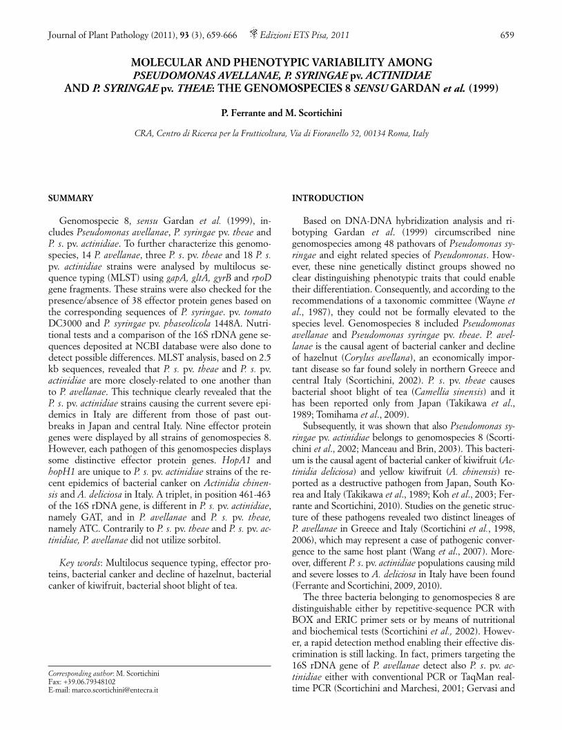
Fig. 1. Dendrogram of relationships using concatenated data and the neighbour-joining algorithm obtained with gapA, gltA, gyrB,and rpoD nucleotidic gene sequences among strains of genomospecies 8. Bootstrap values are reported at the main nodes.
011_JPP762RP(Scortichini)_659 16-11-2011 9:13 Pagina 661
662 Variability within bacterial genomospecies 8 Journal of Plant Pathology (2011), 93 (3), 659-666
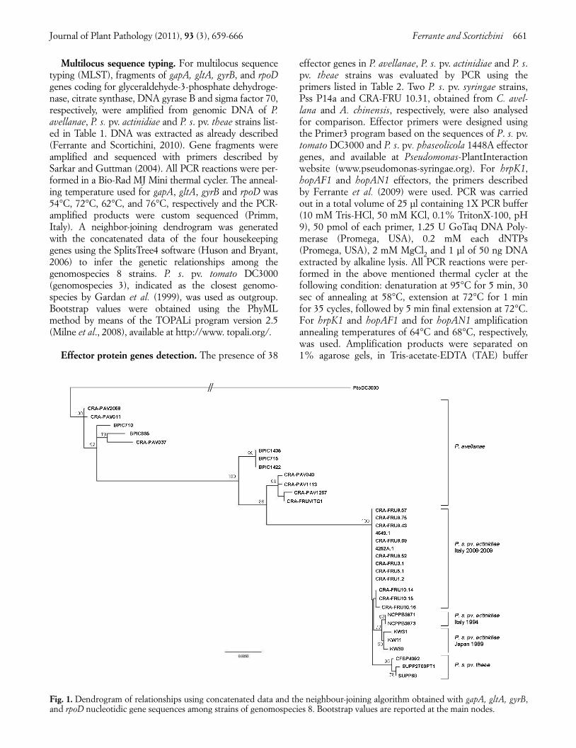
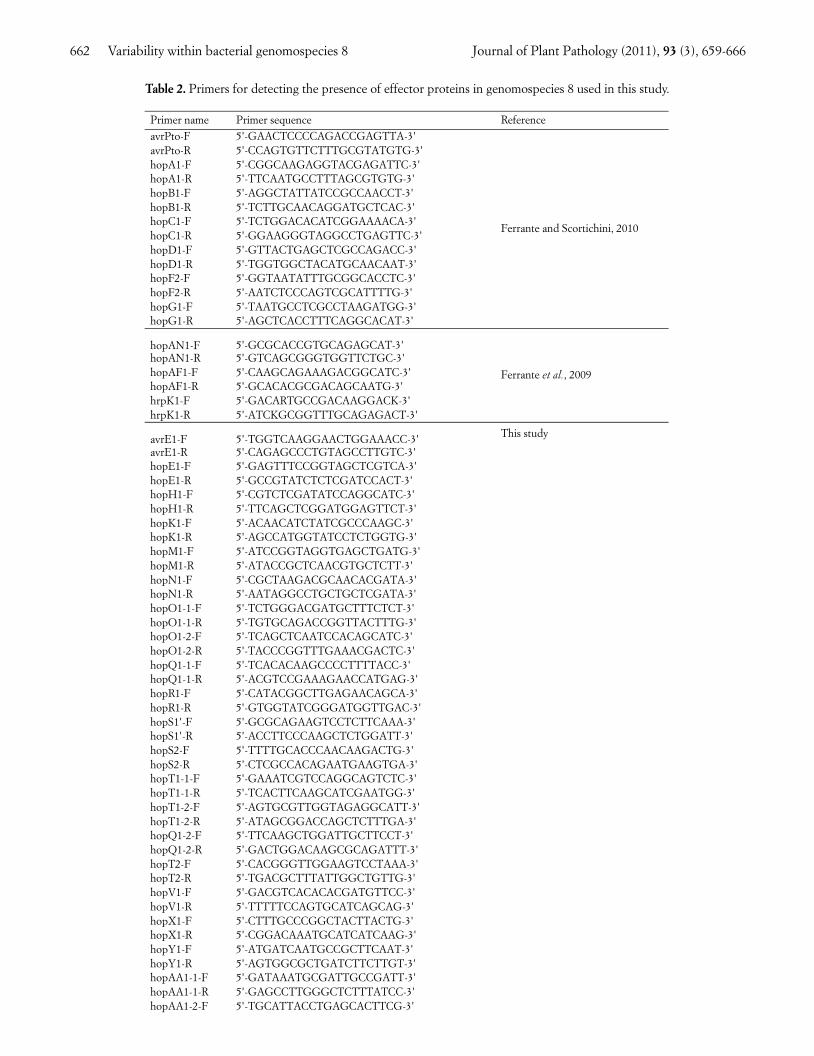
Table 2. Primers for detecting the presence of effector proteins in genomospecies 8 used in this study.
Primer name Primer sequence Reference
avrPto-F 5'-GAACTCCCCAGACCGAGTTA-3'avrPto-R 5'-CCAGTGTTCTTTGCGTATGTG-3'hopA1-F 5'-CGGCAAGAGGTACGAGATTC-3'hopA1-R 5'-TTCAATGCCTTTAGCGTGTG-3'hopB1-F 5'-AGGCTATTATCCGCCAACCT-3'hopB1-R 5'-TCTTGCAACAGGATGCTCAC-3'hopC1-F 5'-TCTGGACACATCGGAAAACA-3'hopC1-R 5'-GGAAGGGTAGGCCTGAGTTC-3'hopD1-F 5'-GTTACTGAGCTCGCCAGACC-3'hopD1-R 5'-TGGTGGCTACATGCAACAAT-3'hopF2-F 5'-GGTAATATTTGCGGCACCTC-3'hopF2-R 5'-AATCTCCCAGTCGCATTTTG-3'hopG1-F 5'-TAATGCCTCGCCTAAGATGG-3'hopG1-R 5'-AGCTCACCTTTCAGGCACAT-3'
Ferrante and Scortichini, 2010
hopAN1-F 5'-GCGCACCGTGCAGAGCAT-3'hopAN1-R 5'-GTCAGCGGGTGGTTCTGC-3'hopAF1-F 5'-CAAGCAGAAAGACGGCATC-3'hopAF1-R 5'-GCACACGCGACAGCAATG-3'hrpK1-F 5'-GACARTGCCGACAAGGACK-3'hrpK1-R 5'-ATCKGCGGTTTGCAGAGACT-3'
Ferrante et al., 2009
avrE1-F 5'-TGGTCAAGGAACTGGAAACC-3'avrE1-R 5'-CAGAGCCCTGTAGCCTTGTC-3'hopE1-F 5'-GAGTTTCCGGTAGCTCGTCA-3'hopE1-R 5'-GCCGTATCTCTCGATCCACT-3'hopH1-F 5'-CGTCTCGATATCCAGGCATC-3'hopH1-R 5'-TTCAGCTCGGATGGAGTTCT-3'hopK1-F 5'-ACAACATCTATCGCCCAAGC-3'hopK1-R 5'-AGCCATGGTATCCTCTGGTG-3'hopM1-F 5'-ATCCGGTAGGTGAGCTGATG-3'hopM1-R 5'-ATACCGCTCAACGTGCTCTT-3'hopN1-F 5'-CGCTAAGACGCAACACGATA-3'hopN1-R 5'-AATAGGCCTGCTGCTCGATA-3'hopO1-1-F 5'-TCTGGGACGATGCTTTCTCT-3'hopO1-1-R 5'-TGTGCAGACCGGTTACTTTG-3'hopO1-2-F 5'-TCAGCTCAATCCACAGCATC-3'hopO1-2-R 5'-TACCCGGTTTGAAACGACTC-3'hopQ1-1-F 5'-TCACACAAGCCCCTTTTACC-3'hopQ1-1-R 5'-ACGTCCGAAAGAACCATGAG-3'hopR1-F 5'-CATACGGCTTGAGAACAGCA-3'hopR1-R 5'-GTGGTATCGGGATGGTTGAC-3'hopS1'-F 5'-GCGCAGAAGTCCTCTTCAAA-3'hopS1'-R 5'-ACCTTCCCAAGCTCTGGATT-3'hopS2-F 5'-TTTTGCACCCAACAAGACTG-3'hopS2-R 5'-CTCGCCACAGAATGAAGTGA-3'hopT1-1-F 5'-GAAATCGTCCAGGCAGTCTC-3'hopT1-1-R 5'-TCACTTCAAGCATCGAATGG-3'hopT1-2-F 5'-AGTGCGTTGGTAGAGGCATT-3'hopT1-2-R 5'-ATAGCGGACCAGCTCTTTGA-3'hopQ1-2-F 5'-TTCAAGCTGGATTGCTTCCT-3'hopQ1-2-R 5'-GACTGGACAAGCGCAGATTT-3'hopT2-F 5'-CACGGGTTGGAAGTCCTAAA-3'hopT2-R 5'-TGACGCTTTATTGGCTGTTG-3'hopV1-F 5'-GACGTCACACACGATGTTCC-3'hopV1-R 5'-TTTTTCCAGTGCATCAGCAG-3'hopX1-F 5'-CTTTGCCCGGCTACTTACTG-3'hopX1-R 5'-CGGACAAATGCATCATCAAG-3'hopY1-F 5'-ATGATCAATGCCGCTTCAAT-3'hopY1-R 5'-AGTGGCGCTGATCTTCTTGT-3'hopAA1-1-F 5'-GATAAATGCGATTGCCGATT-3'hopAA1-1-R 5'-GAGCCTTGGGCTCTTTATCC-3'hopAA1-2-F 5'-TGCATTACCTGAGCACTTCG-3'
This study
011_JPP762RP(Scortichini)_659 16-11-2011 9:13 Pagina 662
Journal of Plant Pathology (2011), 93 (3), 659-666 Ferrante and Scortichini 663
0.5X, and visualized by a Bio-Rad Gel Logic 100 UVtransilluminator. Presence/absence of a band of the ex-pected size was taken as indication of the presence orabsence of the gene in the tested strain. P. s. pv. tomatoDC3000 and P. s. pv. phaseolicola 1448A were used aspositive controls. The analysis was performed twice.
Comparison of 16S rDNA gene. Comparisons wereperformed with sequences of 16S rDNA (1.434 bp) geneof genomospecies 8 strains deposited at NCBI databank.The type-strains of species and pathovars belonging togenomospecies 8 were included. The following acces-sions were compared. 16S rDNA: X95745, AJ889838,and AJ889839 for P. avellanae; AB001450 for P. s. pv.theae; AJ889840, EU906856, GQ914994, and D86357for P. s. pv. actinidiae. All ambiguous and terminal se-quences were edited before data analysis with Geneious4.7.4. (http://www.geneious.com) and aligned usingClustalW 1.83 (http://www.ebi.ac.uk/tools/ clustalw2/).
Nutritional tests. For determining possible discrimi-native nutritional tests within genomospecies 8, the uti-lization of the following compounds was assessed, as de-scribed by Lelliott and Stead (1987): L-alanine, dulcitol,erythritol, D-fructose, glycerol, D-glucose, mannitol, D-mannose, maltose, melibiose, myoinositol, L-rhamnose,D-ribose, sorbitol, D-sucrose, D-threalose.
RESULTS
Multilocus sequence typing (MLST). MLST ofgapA, gltA, gyrB, and rpoD gene fragments provided 2.5kb of sequences. MLST analysis based on the concate-nated data and using the NJ algorithm is shown in Fig.1. P. avellanae strains clustered separately from P. s. pv.actinidiae and P. s. pv. theae strains. Within this species,two subgroups were found. One subgroup includes on-ly strains from Greece, whereas another only strainsfrom Italy. In addition, other five strains showed singlesequence types. P. s. pv. actinidiae and P. s. pv. theae
formed a tighter cluster. The P. s. pv. actinidiae strainsfrom Japan and from past bacterial canker infections inItaly grouped in subclusters separate from the otherstrains causing recent outbreaks in Italy. The three P. s.pv. theae strains clustered separately. High bootstrapvalues supported dendrogram branching. P. s. pv. toma-to DC3000, used as outgroup, was located away fromthe genomospecies 8.
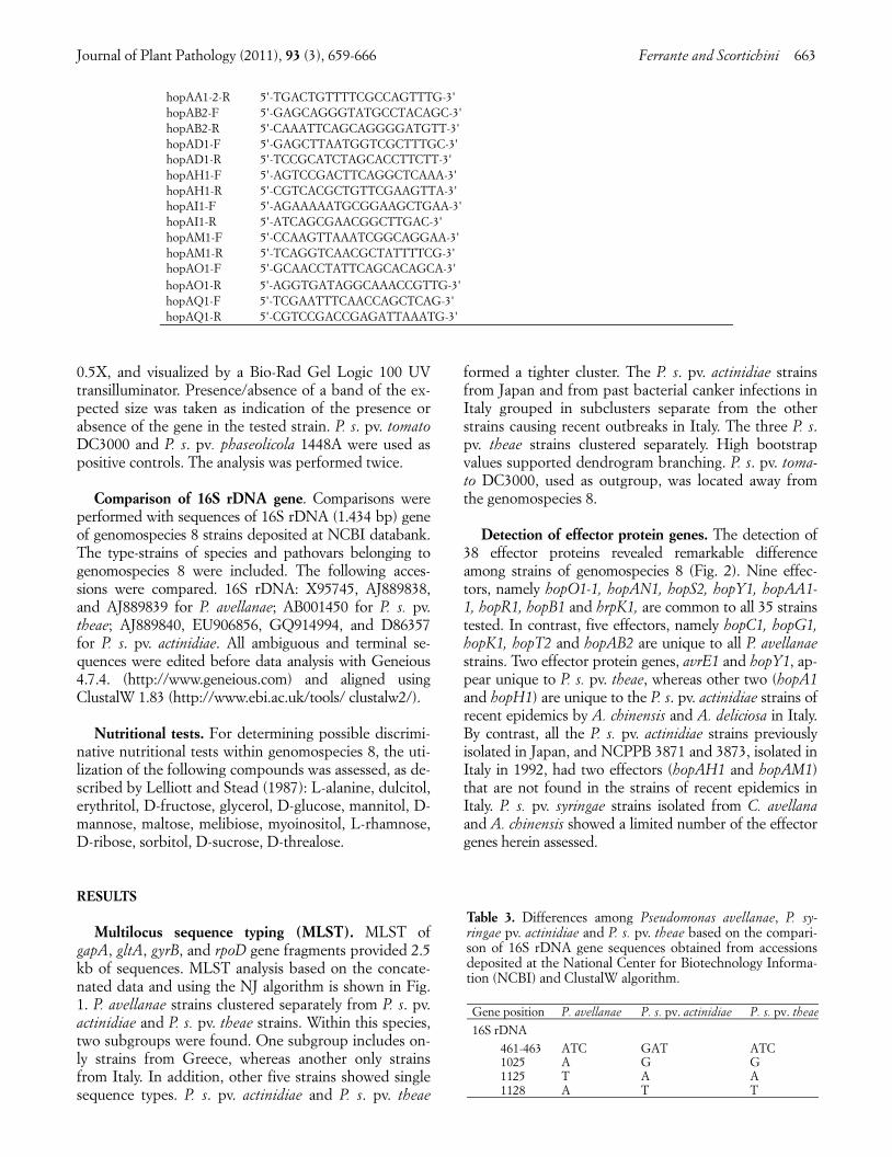
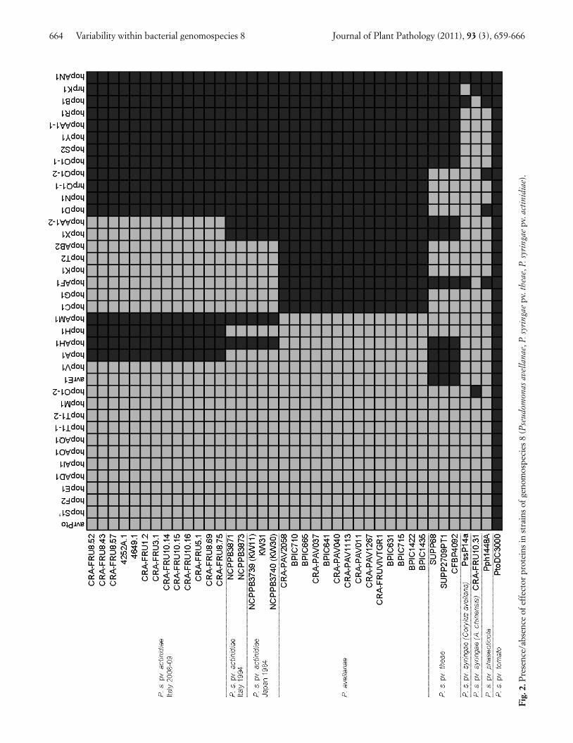
Detection of effector protein genes. The detection of38 effector proteins revealed remarkable differenceamong strains of genomospecies 8 (Fig. 2). Nine effec-tors, namely hopO1-1, hopAN1, hopS2, hopY1, hopAA1-1, hopR1, hopB1 and hrpK1, are common to all 35 strainstested. In contrast, five effectors, namely hopC1, hopG1,hopK1, hopT2 and hopAB2 are unique to all P. avellanaestrains. Two effector protein genes, avrE1 and hopY1, ap-pear unique to P. s. pv. theae, whereas other two (hopA1and hopH1) are unique to the P. s. pv. actinidiae strains ofrecent epidemics by A. chinensis and A. deliciosa in Italy.By contrast, all the P. s. pv. actinidiae strains previouslyisolated in Japan, and NCPPB 3871 and 3873, isolated inItaly in 1992, had two effectors (hopAH1 and hopAM1)that are not found in the strains of recent epidemics inItaly. P. s. pv. syringae strains isolated from C. avellanaand A. chinensis showed a limited number of the effectorgenes herein assessed.
hopAO1-R 5'-AGGTGATAGGCAAACCGTTG-3'hopAQ1-F 5'-TCGAATTTCAACCAGCTCAG-3'hopAQ1-R 5'-CGTCCGACCGAGATTAAATG-3'
Table 3. Differences among Pseudomonas avellanae, P. sy-ringae pv. actinidiae and P. s. pv. theae based on the compari-son of 16S rDNA gene sequences obtained from accessionsdeposited at the National Center for Biotechnology Informa-tion (NCBI) and ClustalW algorithm.
Gene position P. avellanae P. s. pv. actinidiae P. s. pv. theae
16S rDNA 461-463 ATC GAT ATC 1025 A G G 1125 T A A 1128 A T T
hopAA1-2-R 5'-TGACTGTTTTCGCCAGTTTG-3'hopAB2-F 5'-GAGCAGGGTATGCCTACAGC-3'hopAB2-R 5'-CAAATTCAGCAGGGGATGTT-3'hopAD1-F 5'-GAGCTTAATGGTCGCTTTGC-3'hopAD1-R 5'-TCCGCATCTAGCACCTTCTT-3'hopAH1-F 5'-AGTCCGACTTCAGGCTCAAA-3'hopAH1-R 5'-CGTCACGCTGTTCGAAGTTA-3'hopAI1-F 5'-AGAAAAATGCGGAAGCTGAA-3'hopAI1-R 5'-ATCAGCGAACGGCTTGAC-3'hopAM1-F 5'-CCAAGTTAAATCGGCAGGAA-3'hopAM1-R 5'-TCAGGTCAACGCTATTTTCG-3'hopAO1-F 5'-GCAACCTATTCAGCACAGCA-3'
011_JPP762RP(Scortichini)_659 16-11-2011 9:13 Pagina 663
664 Variability within bacterial genomospecies 8 Journal of Plant Pathology (2011), 93 (3), 659-666
Fig.
2.P
rese
nce/
abse
nce
of e
ffec
tor
prot
eins
in s
trai
ns o
f gen
omos
peci
es 8
(Pse
udom
onas
ave
llana
e, P
. syr
inga
epv
. the
ae, P
. syr
inga
epv
. act
inid
iae)
.
011_JPP762RP(Scortichini)_659 16-11-2011 9:13 Pagina 664
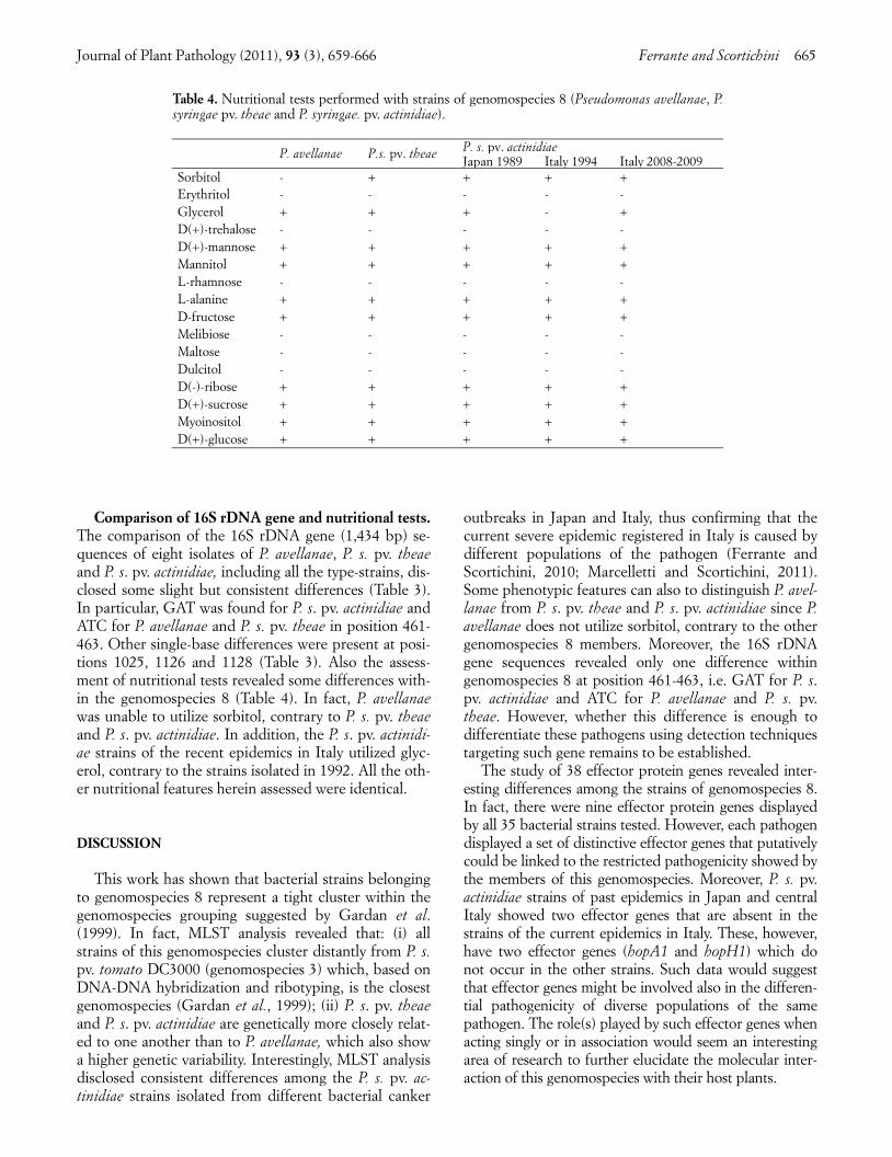
Comparison of 16S rDNA gene and nutritional tests.The comparison of the 16S rDNA gene (1,434 bp) se-quences of eight isolates of P. avellanae, P. s. pv. theaeand P. s. pv. actinidiae, including all the type-strains, dis-closed some slight but consistent differences (Table 3).In particular, GAT was found for P. s. pv. actinidiae andATC for P. avellanae and P. s. pv. theae in position 461-463. Other single-base differences were present at posi-tions 1025, 1126 and 1128 (Table 3). Also the assess-ment of nutritional tests revealed some differences with-in the genomospecies 8 (Table 4). In fact, P. avellanaewas unable to utilize sorbitol, contrary to P. s. pv. theaeand P. s. pv. actinidiae. In addition, the P. s. pv. actinidi-ae strains of the recent epidemics in Italy utilized glyc-erol, contrary to the strains isolated in 1992. All the oth-er nutritional features herein assessed were identical.
DISCUSSION
This work has shown that bacterial strains belongingto genomospecies 8 represent a tight cluster within thegenomospecies grouping suggested by Gardan et al.(1999). In fact, MLST analysis revealed that: (i) allstrains of this genomospecies cluster distantly from P. s.pv. tomato DC3000 (genomospecies 3) which, based onDNA-DNA hybridization and ribotyping, is the closestgenomospecies (Gardan et al., 1999); (ii) P. s. pv. theaeand P. s. pv. actinidiae are genetically more closely relat-ed to one another than to P. avellanae, which also showa higher genetic variability. Interestingly, MLST analysisdisclosed consistent differences among the P. s. pv. ac-tinidiae strains isolated from different bacterial canker
outbreaks in Japan and Italy, thus confirming that thecurrent severe epidemic registered in Italy is caused bydifferent populations of the pathogen (Ferrante andScortichini, 2010; Marcelletti and Scortichini, 2011).Some phenotypic features can also to distinguish P. avel-lanae from P. s. pv. theae and P. s. pv. actinidiae since P.avellanae does not utilize sorbitol, contrary to the othergenomospecies 8 members. Moreover, the 16S rDNAgene sequences revealed only one difference withingenomospecies 8 at position 461-463, i.e. GAT for P. s.pv. actinidiae and ATC for P. avellanae and P. s. pv.theae. However, whether this difference is enough todifferentiate these pathogens using detection techniquestargeting such gene remains to be established.
The study of 38 effector protein genes revealed inter-esting differences among the strains of genomospecies 8.In fact, there were nine effector protein genes displayedby all 35 bacterial strains tested. However, each pathogendisplayed a set of distinctive effector genes that putativelycould be linked to the restricted pathogenicity showed bythe members of this genomospecies. Moreover, P. s. pv.actinidiae strains of past epidemics in Japan and centralItaly showed two effector genes that are absent in thestrains of the current epidemics in Italy. These, however,have two effector genes (hopA1 and hopH1) which donot occur in the other strains. Such data would suggestthat effector genes might be involved also in the differen-tial pathogenicity of diverse populations of the samepathogen. The role(s) played by such effector genes whenacting singly or in association would seem an interestingarea of research to further elucidate the molecular inter-action of this genomospecies with their host plants.
Journal of Plant Pathology (2011), 93 (3), 659-666 Ferrante and Scortichini 665
Table 4. Nutritional tests performed with strains of genomospecies 8 (Pseudomonas avellanae, P.syringae pv. theae and P. syringae. pv. actinidiae).
P. s. pv. actinidiaeP. avellanae P.s. pv. theaeJapan 1989 Italy 1994 Italy 2008-2009
Sorbitol - + + + +Erythritol - - - - -Glycerol + + + - +D(+)-trehalose - - - - -D(+)-mannose + + + + +Mannitol + + + + +L-rhamnose - - - - -L-alanine + + + + +D-fructose + + + + +Melibiose - - - - -Maltose - - - - -Dulcitol - - - - -D(-)-ribose + + + + +D(+)-sucrose + + + + +Myoinositol + + + + +D(+)-glucose + + + + +
011_JPP762RP(Scortichini)_659 16-11-2011 9:13 Pagina 665
666 Variability within bacterial genomospecies 8 Journal of Plant Pathology (2011), 93 (3), 659-666
ACKNOWLEDGEMENTS
The authors wish to thank Dr. Yuichi Takikawa(Shizuoka University, Shizuoka, Japan) for supplyingPseudomonas syringae pv. actinidiae and P. s. pv. theaestrains from Japan.
REFERENCES
Ferrante P., Scortichini M., 2009. Identification of Pseudo-monas syringae pv. actinidiae as causal agent of bacterialcanker of yellow kiwifruit (Actinidia chinensis Planchon) incentral Italy. Journal of Phytopathology 157: 768-770.
Ferrante P., Clarke C.R., Cavanaugh K.A., Michelmore R.W.,Buonaurio R., Vinatzer B., 2009. Contributions of the ef-fector gene hopQ1-1 to differences in host range betweenPseudomonas syringae pv. phaseolicola and P. syringae pv.tabaci. Molecular Plant Pathology 10: 837-842.
Ferrante P., Scortichini M., 2010. Molecular and phenotypicfeatures of Pseudomonas syringae pv. actinidiae isolatedduring recent epidemics of bacterial canker on yellow ki-wifruit (Actinidia chinensis) in central Italy. Plant Pathology69: 954-962.
Gardan L., Shafik H., Belouin S., Brosch R., Grimont F., Gri-mont P.A.D., 1999. DNA relatedeness among the patho-vars of Pseudomonas syringae and description ofPseudomonas tremae sp. nov. and Pseudomonas cannabinasp. nov. (ex Sutic and Dowson). International Journal ofSystematic Bacteriology 49: 469-473.
Gervasi F., Scortichini M., 2009. Detection of Pseudomonasavellanae from hazelnut twigs by TaqMan real-time PCR.Journal of Plant Pathology 91: 561-566.
Huson D.H., Bryant D., 2006. Application of phylogeneticnetworks in evolutionary studies. Molecular Biology andEvolution 23: 254-267.
Koh Y.J., Jung J.-S., Hur J.-S., 2003. Current status of occur-rence of major diseases on kiwifruit and their control inKorea. Acta Horticulturae 610: 437-443.
Lelliott R.A., Stead D.E., 1987. Methods for the Diagnosis ofBacterial Diseases of Plants. Blackwell Scientific Publica-tion for the British Society for Plant Pathology, Oxford,UK.
Manceau C., Brin C., 2003. Pathovars of Pseudomonas sy-ringae are structured in genetic populations allowing theselection of specific markers for their detection in plantsamples. In: Iacobellis N.S. (ed.). Pseudomonas syringaeand Related Pathogens, pp. 503-512. Kluwer AcademicPublishers, Dordrecht, The Netherlands.
Marcelletti S., Scortichini M., 2011. Clonal outbreaks of bac-terial canker caused by Pseudomonas syringae pv. actinidiae
on Actinidia chinensis and A. deliciosa in Italy. Journal ofPlant Pathology 93: 479-483.
Milne I., Lindner D., Bayer M., Husmeier D., Mc Guire G.,Marshall D.F., Wright F., 2008. TOPALi v2: a rich graphi-cal interface for evolutionary analyses of multiple align-ments on HPC clusters and multi-core desktops. Bioinfor-matics 25: 126-127.
Rees-George J., Vanneste J.L., Cornish D.A., PushparajahI.P.S., Yu J., Templeton M.D., Everett K.R., 2010. Detec-tion of Pseudomonas syringae pv. actinidiae using poly-merase chain reaction (PCR) primers based on the 16S-23SrDNA intertranscribed spacer region and comparison withPCR primers based on other gene regions. Plant Pathology59: 453-464.
Sarkar S.F., Guttman D.S., 2004. The evolution of the coregenome of Pseudomonas syringae, a highly clonal, endemicplant pathogen. Applied and Environmental Microbiology70: 1999-2012.
Scortichini M., Dettori M.T., Marchesi U., Palombi M.A.,Rossi M.P., 1998. Differentitation of Pseudomonas avel-lanae strains from Greece and Italy by rep-PCR genomicfingerprinting. Journal of Phytopathology 146: 417-420.
Scortichini M., Marchesi U., 2001. Sensitive and specific de-tection of Pseudomonas avellanae using primers based on16S rRNA gene sequences. Journal of Phytopathology 149:527-532.
Scortichini M., 2002. Bacterial canker and decline of Euro-pean hazelnut. Plant Disease 86: 704-709
Scortichini M., Marchesi U., Di Prospero P., 2002. Genetic re-latedeness among Pseudomonas avellanae, P. syringae pv.theae and P. s. pv. actinidiae, and their identification. Euro-pean Journal of Plant Pathology 108: 269-278.
Scortichini M., Natalini E., Marchesi U., 2006. Evidence forseparate origins of the two Pseudomonas avellanae lineages.Plant Pathology 55: 451-457
Takikawa Y., Serizawa S., Ichikawa T., Tsuyumu S., Goto M.,1989. Pseudomonas syringae pv. actinidiae pv. nov.: thecausal bacterium of canker of kiwifruit in Japan. Annals ofthe Phytopathological Society of Japan 55: 437-444.
Tomihama T., Nonaka T., Nishi Y., Arai K., 2009. Environmen-tal control in tea fields to reduce infections by Pseudomonassyringae pv. theae. Phytopathology 99: 209-216.
Wang P.W., Morgan R.L., Scortichini M., Guttman D.S.,2007. Convergent evolution of phytopathogenic pseudo-monads onto hazelnut. Microbiology 153: 2067-2073.
Wayne L.G., Brenner D.J., Colwell R.R., Grimont P.A.D.,Kandler O., Krichevsky L., Moore L.H., Moore W.C.,Murray R.G.E., Stackebrandt E., Starr M.P., Truper H.G.,1987. Report of the ad hoc committee on reconciliation ofapproaches to bacterial systematics. International Journalof Systematic Bacteriology 37: 463-464.
Received April 26, 2011Accepted June 16, 2011
011_JPP762RP(Scortichini)_659 16-11-2011 9:13 Pagina 666