molecular architectures of benzoic acid-specific type iii...
TRANSCRIPT
research papers
Acta Cryst. (2017). D73, 1007–1019 https://doi.org/10.1107/S2059798317016618 1007
Received 11 July 2017
Accepted 17 November 2017
Edited by A. Berghuis, McGill University,
Canada
Keywords: chalcone synthase; biphenyl
synthase; benzophenone synthase; polyketide
synthase; thiolase; benzoyl-CoA.
PDB references: benzophenone synthase, 5uco;
chalcone synthase, 5uc5; biphenyl synthase,
5w8q; biphenyl synthase, complex with
benzoyl-CoA, 5wc4
Supporting information: this article has
supporting information at journals.iucr.org/d
Molecular architectures of benzoic acid-specifictype III polyketide synthases
Charles Stewart Jr,a,b* Kate Woods,a Greg Macias,a Andrew C. Allan,c,d Roger P.
Hellensc,e and Joseph P. Noela
aHoward Hughes Medical Institute, The Salk Institute for Biological Studies, La Jolla, CA 92037, USA, bMacromolecular
X-ray Crystallography Facility, Office of Biotechnology, Iowa State University, 0202 Molecular Biology Building, 2437
Pammel Drive, Ames, IA 50011, USA, cThe New Zealand Institute for Plant and Food Research Limited (PFR), Auckland,
New Zealand, dSchool of Biological Sciences, University of Auckland, Auckland, New Zealand, and eQueensland
University of Technology, Brisbane, Queensland 4001, Australia. *Correspondence e-mail: [email protected]
Biphenyl synthase and benzophenone synthase constitute an evolutionarily
distinct clade of type III polyketide synthases (PKSs) that use benzoic acid-
derived substrates to produce defense metabolites in plants. The use of benzoyl-
CoA as an endogenous substrate is unusual for type III PKSs. Moreover,
sequence analyses indicate that the residues responsible for the functional
diversification of type III PKSs are mutated in benzoic acid-specific type III
PKSs. In order to gain a better understanding of structure–function relationships
within the type III PKS family, the crystal structures of biphenyl synthase from
Malus � domestica and benzophenone synthase from Hypericum androsaemum
were compared with the structure of an archetypal type III PKS: chalcone
synthase from Malus � domestica. Both biphenyl synthase and benzophenone
synthase contain mutations that reshape their active-site cavities to prevent the
binding of 4-coumaroyl-CoA and to favor the binding of small hydrophobic
substrates. The active-site cavities of biphenyl synthase and benzophenone
synthase also contain a novel pocket associated with their chain-elongation and
cyclization reactions. Collectively, these results illuminate structural determi-
nants of benzoic acid-specific type III PKSs and expand the understanding of the
evolution of specialized metabolic pathways in plants.
1. Introduction
Benzoic acid-specific type III polyketide synthases (PKSs)
constitute a distinct clade within the type III PKS family that
use benzoic acid-derived substrates (for example benzoyl-
CoA, 3-hydroxybenzoyl-CoA and salicoyl-CoA) to produce
phytoalexins and pharmacologically active compounds
(Beerhues & Liu, 2009; Fig. 1). Biphenyl synthase (BIS)
generates the core chemical scaffolds of biphenyl and dibenzo-
furan phytoalexins commonly found in the Pyrinae subtribe
(Rosaceae; Liu et al., 2007; Khalil et al., 2013). The Pyrinae
contain several economically important species, including
apple (Malus � domestica), pear (Pyrus communis) and
mountain ash (Sorbus aucuparia). Apples increased their
expression of BIS after inoculation with the fireblight
bacterium Erwinia amylovora (Chizzali et al., 2011). Further-
more, BIS transcripts as well as biphenyl and dibenzofuran
compounds have been isolated from the transition zone
between necrotic and healthy tissues in both apples and pears
after inoculation with the fireblight bacterium (Chizzali et al.,
2012, 2016). Additionally, when challenged with Venturia
inaequalis, the causative fungus of apple scab, cell cultures of
S. aucuparis and a scab-resistant M. domestica cultivar
produced biphenyl and dibenzofuran metabolites (Huttner et
al., 2010; Khalil et al., 2013; Hrazdina & Borejsza-Wysocki,
ISSN 2059-7983
2003). Additionally, the promiscuous in vitro activity of BIS
with salicoyl-CoA was exploited to develop an artificial
metabolic system in Escherichia coli that is capable of
producing 4-hydroxycoumarin, an immediate precursor to
synthetic anticoagulants (for example warfarin; Liu et al., 2010;
Lin et al., 2013). In contrast to BIS, benzophenone synthase
(BPS) generates the core chemical scaffolds of xanthones,
guttiferones and sampsoniones that are prominently found in
the closely related Hypericaceae and Clusiaceae families (Liu
et al., 2003; Nualkaew et al., 2012). Xanthones are associated
with diverse biological functions in Hypericum spp., including
antimicrobials, UV pigments and antioxidants (Gronquist et
al., 2001). Cell cultures of H. calycinum produced xanthones in
response to yeast elicitation (Gaid et al., 2012). Similarly,
elicitation of H. perforatum cell cultures with Agrobacterium
tumefaciens led to an increase in BPS transcripts and xanthone
accumulation (Franklin et al., 2009). Lastly, polyisoprenylated
benzophenone derivatives are pharmacologically active and
have served as lead compounds for drug development (Acuna
et al., 2009; Wang et al., 2016).
Type III PKS catalysis involves the loading of a CoA-
tethered substrate onto an active-site cysteine followed by
chain elongation via iterative
decarboxylative Claisen conden-
sations of malonyl-CoA. Linear
polyketide intermediates
undergo a terminal intramole-
cular cyclization reaction to
produce the final cyclic polyke-
tide product (Austin & Noel,
2003). Changes in steric effects
and electrostatic interactions
within the active-site cavities of
type III PKSs underlie biochem-
ical variations in substrate
preference, chain-elongation and
cyclization patterns during cata-
lysis (Jez, Austin et al., 2000;
Austin, Izumikawa et al., 2004;
Austin, Bowman et al., 2004). The
reactions catalyzed by benzoic
acid-specific type III PKSs are
biochemically and evolutionarily
distinct from those catalyzed by
the type III PKS archetypes
chalcone synthase (CHS) and
stilbene synthase (STS). Firstly, as
stated above, the substrate
preferences of BIS and BPS for
benzoyl-CoA are unusual in the
type III PKS family. No other
type III PKSs are known to use
benzoyl-CoA in planta. Secondly,
sequence comparisons indicate
that the molecular determinants
of cyclization specificity in BIS
and BPS are not conserved in
CHSs and STSs (Liu et al., 2007). Thirdly, molecular phylo-
genetic analyses indicate that BIS and BPS form a distinct
clade within the type III PKS family, separate from CHS
homologs from the same species and other functionally
diverse type III PKSs (Liu et al., 2007). This evolutionary
pattern differs from the relationship of STSs to CHSs (Tropf et
al., 1994).
The lack of structural data for benzoic acid-specific type III
PKSs limits our understanding of the enzymology of type III
PKSs as well as the evolution of associated metabolic path-
ways. Our goal for this study was to identify structural idio-
syncrasies of benzoic acid-specific type III PKSs via
comparative analyses of the crystal structures of CHS, BIS and
BPS.
2. Methods
2.1. Chemicals
Benzoyl-CoA, malonyl-CoA, naringenin and 4-hydroxy-
coumarin were all purchased from Sigma (USA);
4-coumaroyl-CoA was purchased from TransMIT (Germany).
research papers
1008 Stewart et al. � Benzoic acid-specific type III polyketide synthases Acta Cryst. (2017). D73, 1007–1019
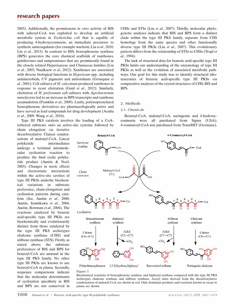
Figure 1Biochemical reactions of benzophenone synthase and biphenyl synthase compared with the type III PKSarchetypes chalcone synthase and stilbene synthase. Acetyl units derived from the decarboxylativecondensation of malonyl-CoA are shown in red. Only dominant products and reactions known to occur inplanta are shown.
3,5-Dihydroxybiphenyl was prepared as described previously
by Nilsson & Norin (1963) and was purified with a Waters
preparative LCMS FractionLynx system using mass and UV-
directed fractionation on an XBridge Prep OBD C18 5 mm
30 � 100 mm column, with a gradient from 5:95:0.1(v:v:v)
water:acetonitrile:formic acid to acetonitrile (0.1% formic
acid) over 8 min with a flow rate of 30 ml min�1. Its identity
was confirmed by comparison of its 1H NMR spectrum in
MeOD-d4 and m/z values with those reported in the literature
(Liu et al., 2004). Salicoyl-CoA (2-hydroxybenzoyl-CoA) was
prepared as described elsewhere (Sidenius et al., 2004) and was
purified via preparative LCMS as described above. The
identity of salicoyl-CoA was confirmed by comparison of its1H NMR spectrum in MeOD-d4 and m/z values with those
reported in the literature (Guo et al., 2009).
2.2. Cloning and recombinant protein expression andpurification
cDNAs for MdCHS2, MdBIS1 and MdBIS3 were derived
from a cDNA library of M. domestica and inserted into the
pBluescript SK+ vector in the laboratory of A. C. Allan and
R. P. Hellens at The New Zealand Institute for Plant and Food
Research Ltd (Newcomb et al., 2006). For recombinant
expression, the MdCHS2, MdBIS1 and MdBIS3 cDNAs were
inserted between the NcoI and EcoRI restriction sites of the
expression vector pHIS8 which, under the control of a T7
promoter, yields target proteins fused to an N-terminal octa-
histidine tag (Jez, Ferrer et al., 2000). Subcloning occurred as
follows. Firstly, NcoI digestion sites were added to each
pBluescript template using the QuikChange (Agilent) PCR
method following the manufacturer’s protocol. The primers
used were MdCHS2-forward, 50-CTCGACGGTCACCATGG
TTTTTATATCCGATCGTCGAGAAAGATC-30; MdCHS2-
reverse, 50-GATCTTTCTCGACGATCGGATATAAAAAC
CATGGTGACCGTCGAG-30; MdBIS1-forward, 50-TAACCA
AAGGCGCCATGGGGCAAGTTGAAGAGC-30; MdBIS1-
reverse, 50-GCTCTTCAACTTGCCCCATGGCGCCTTTGG
TTA-30; MdBIS3-forward, 50-CTCATTCTTAACCAAAG
GCGCCATGGAGCTAGTTAAAGAGCAGATATAAA-30;
MdBIS3-reverse, 50-TTTATATCTGCTCTTTAACTAGCT
CCATGGCGCCTTTGGTTAAGAATGAG-30 (NcoI sites
are underlined and the translation start sites are shown in
bold). Plasmid preparations (Qiagen) of NcoI-mutated
pBluescript constructs were digested with NcoI and EcoRI.
The approximately 1.2 kb digestion fragments were gel-
purified and ligated with NcoI/EcoRI-digested pHIS8. The
correct reading frames for the pHIS8 constructs were obtained
using the QuikChange PCR method and the following sets of
primers: MdCHS2-forward, 50-TCCGCGTGGTTCCATGGT
GACCGTCG-30; MdCHS2-reverse, 50-CGACGGTCACCAT
GGAACCACGCGGA-30; MdBIS1 and MdBIS3-forward, 50-
TCCGCGTGGTTCCATGGCGCCTTTGG-30; MdBIS1 and
MdBIS3-reverse, 50-CCAAAGGCGCCATGGAACCACGC
GGA-30 (NcoI sites are underlined and the translation start
sites are shown in bold). All primers were purchased from
Integrated DNA Technologies. The fidelity of all constructs
was confirmed by sequencing (Eton Biosciences, USA).
Expression constructs were heterologously overexpressed
in E. coli BL21 (DE3) (Novagen) expression hosts. E. coli
cultures were grown at 37�C in TB medium (Invitrogen) to an
optical density (600 nm) of 1.0, induced with 0.5 mM isopropyl
�-d-thiogalactopyranoside and allowed to grow overnight at
22�C. Bacterial cells were harvested by centrifugation, resus-
pended in lysis buffer [50 mM Tris–HCl pH 8.0, 500 mM NaCl,
20 mM imidazole, 1%(v/v) Tween 20, 10%(v/v) glycerol,
10 mM �-mercaptoethanol] and lysed by sonication. Recom-
binant proteins were isolated from the E. coli lysate by affinity
chromatography with nickel–nitrilotriacetic acid-coupled
agarose (Qiagen) and eluted with buffer (lysis buffer without
detergent) containing 250 mM imidazole. During the first
dialysis (50 mM Tris–HCl pH 8.0, 500 mM NaCl, 10 mM
�-mercaptoethanol), the recombinant proteins were treated
with thrombin for cleavage of the octahistidine tag. The final
purification step consisted of gel filtration of the protein
extracts on a Superdex 200 FPLC column equilibrated with
50 mM Tris pH 8.0, 500 mM NaCl, 10 mM �-mercaptoethanol.
Protein fractions were pooled, dialyzed into 12.5 mM Tris pH
8.0, 125 mM NaCl, 5 mM DTT, concentrated to at least
10 mg ml�1 and finally stored at �80�C. SDS–PAGE analysis
was used to confirm the protein purity. Protein concentrations
were determined using the absorbance at 280 nm.
A codon-optimized synthetic gene for benzophenone
synthase from Hypericum androsaemum (HaBPS), GenBank
accession AAL79808, as reported by Liu et al. (2003) was
purchased from GenScript (Piscataway, New Jersey, USA).
The initial HaBPS construct (pUC57 vector) was digested with
NcoI and BamHI, gel-purified and ligated with NcoI/BamHI-
digested pHIS8. The correct reading frame of the HaBPS
pHIS8 construct was achieved using QuikChange PCR and
the primers HaBPS-forward, 50-CTGGTTCCGCGTGGTT
CCATGGCCAATTC-30, and HaBPS-reverse, 50-GAATTG
GCCATGGAACCACGCGGAACCAG-30 (NcoI sites are
underlined and the translation start sites are shown in bold).
Primers were purchased from Integrated DNA Technologies
(USA) and the sequence of the final pHIS8 construct was
confirmed by sequencing (Eton Biosciences, USA).
2.3. qPCR analysis of MdCHS2, MdBIS1 and MdBIS3 geneexpression
Tissues from M. domestica ‘Royal Gala’ were collected and
qPCR analyses of gene expression were performed as
described previously (Henry-Kirk et al., 2012; Dare et al.,
2013). In brief, all reactions were performed using the Light-
Cycler 480 SYBR Green I Master Mix (Roche Diagnostics)
according to the procedure described by the manufacturer.
qPCR reactions were performed four times using 5 ml SYBR
Green Master Mix, 0.5 ml forward primer, 0.5 ml reverse
primer, 1 ml water and 3 ml cDNA template (Table 1). A
negative water control was included in each run. Fluorescence
was measured at the end of each annealing step. Amplification
was followed by a melting-curve analysis with continual
research papers
Acta Cryst. (2017). D73, 1007–1019 Stewart et al. � Benzoic acid-specific type III polyketide synthases 1009
fluorescence data acquisition during the 65–95�C melt. Actin
from M. domestica (GenBank accession CN938023) was
selected as the reference gene because of its consistent tran-
scription level throughout fruit tissues and leaves. The results
were interpreted using the LightCycler480 software platform.
2.4. Crystallization of MdCHS2, MdBIS3 and HaBPS
Crystals of MdCHS2, MdBIS3 and HaBPS were grown by
the hanging-drop vapor-diffusion method using buffered
protein solutions (10–15 mg ml�1) mixed with equal volumes
of the reservoir solution and incubated at 4�C. Typical reser-
voir solutions were 14% PEG 8000, 0.3 M ammonium acetate,
0.1 M sodium MOPSO pH 7.0 for MdCHS2 and 14% PEG
8000, 0.3 M ammonium acetate, 0.1 M sodium HEPES pH 7.5
for MdBIS3. Crystals of HaBPS grew from reservoirs
consisting of 18%(w/v) PEG 8000, 0.1 M PIPES buffer pH 6.5.
Crystal-soaking experiments were conducted by transferring
apo MdBIS3 crystals into fresh drops containing reservoir plus
15 mM benzoyl-CoA and incubating overnight at 4�C.
MdCHS2 and MdBIS3 crystals grew within 48 h, while HaBPS
crystals typically grew within one week.
2.5. X-ray diffraction data collection
Crystals were transferred to a cryoprotectant solution
consisting of reservoir solution supplemented with 20%(v/v)
racemic 1,3-butanediol, prior to immersion in liquid nitrogen.
X-ray diffraction data were measured from cooled crystals
using ADSC Quantum 315 CCD detectors on beamlines 8.2.1
and 8.2.2 of the Advanced Light Source, Lawrence Berkeley
National Laboratory. Diffraction intensities were indexed and
integrated with MOSFLM (Leslie, 2006) and SCALA (Evans,
2006) in the CCP4 software suite (Collaborative Computa-
tional Project, Number 4, 1994; Potterton et al., 2003; Winn et
al., 2011).
2.6. X-ray structure determination of MdCHS2, MdBIS3 andHaBPS
Initial crystallographic phases for apo crystals were deter-
mined by molecular replacement with Phaser (McCoy et al.,
2007). Homology models of MdCHS2 and MdBIS3 were
constructed with MODELLER (Sali & Blundell, 1993) based
on the structure of chalcone synthase from Medicago sativa
(PDB entry 1bi5; Ferrer et al., 1999). A homology model of
HaBPS was generated using the crystallographic model of
MdBIS3 as a template. These homology models served as the
starting models for molecular replacement. The PHENIX
suite of programs (Adams et al., 2010) was used to rebuild
these initial structural models and for subsequent structural
refinements. Coot (Emsley & Cowtan, 2004) was used for
graphical map inspection and manual rebuilding of atomic
models. Programs from the CCP4 suite were employed for all
other crystallographic calculations. Molecular visualizations
were generated with PyMOL (Schrodinger).
2.7. In vitro enzyme assays
Steady-state kinetic analyses were performed for chalcone
synthase (EC 2.3.1.74), benzophenone synthase (EC 2.3.1.220)
and biphenyl synthase (EC 2.3.1.177 and EC 2.3.1.208) activ-
ities. Standard enzyme assays for MdCHS2 consisted of 0.1 M
potassium phosphate buffer pH 7.0, 0.5 mM protein, 60 mM
coumaroyl-CoA and 100 mM malonyl-CoA in a total volume
of 500 ml. Standard enzyme assays for MdBIS1 and MdBIS3
consisted of 0.1 M potassium phosphate buffer pH 7.0, 0.1 mM
protein, 15 mM starter substrate, 60 mM malonyl-CoA in a
total volume of 500 ml. Assays for HaBPS consisted of 0.1 M
potassium phosphate buffer pH 7.0, 0.1 mM protein, 20 mM
starter substrate and 80 mM malonyl-CoA in a total volume of
500 ml. The reactions were initiated by the addition of protein
and were quenched with acetic acid [5%(v/v)] after 10 min
incubations at 37�C, except for MdCHS2, which was incubated
for 5 min at 37�C. Kinetic constants were determined using six
substrate concentrations with the concentration of the second
substrate held constant. Product formation for each protein
was linear with respect to incubation time and protein
concentration. After quenching, samples were extracted with
ethyl acetate, dried under vacuum and then resuspended in
40 ml methanol. Each substrate–enzyme series was assayed in
triplicate, and kinetic constants were calculated using a
product standard curve and nonlinear regression analyses in
GraphPad Prism (GraphPad Software).
Product formation was analyzed by LC-MS with an Agilent
1100 series LC-MSD instrument containing a Gemini (4.6 �
150 mm, 5 mm particle size) reversed-phase column. Chro-
matographic separations employed a flow rate of 0.5 ml min�1
and a linear gradient with the initial and final mobile phases
consisting of 95%(v/v) water, 5%(v/v) acetonitrile, 0.1%(v/v)
formic acid and 5%(v/v) water, 95%(v/v) acetonitrile,
0.1%(v/v) formic acid, respectively. The detection wavelengths
were 228 nm (3,5-dihydroxybiphenyl), 288 nm (4-hydroxy-
coumarin and naringenin) and 306 nm (phlorobenzo-
phenone). Products were confirmed by mass determination
and reference to the chromatographic elution of authentic
standards.
2.8. Molecular phylogenetics
Amino-acid sequences were aligned using the
PROMALS3D web server (Pei et al., 2008) and phylogenetic
analysis used the Jones–Taylor–Thornton substitution model
(Jones et al., 1992) within the MEGA7 software package
(Kumar et al., 2016).
research papers
1010 Stewart et al. � Benzoic acid-specific type III polyketide synthases Acta Cryst. (2017). D73, 1007–1019
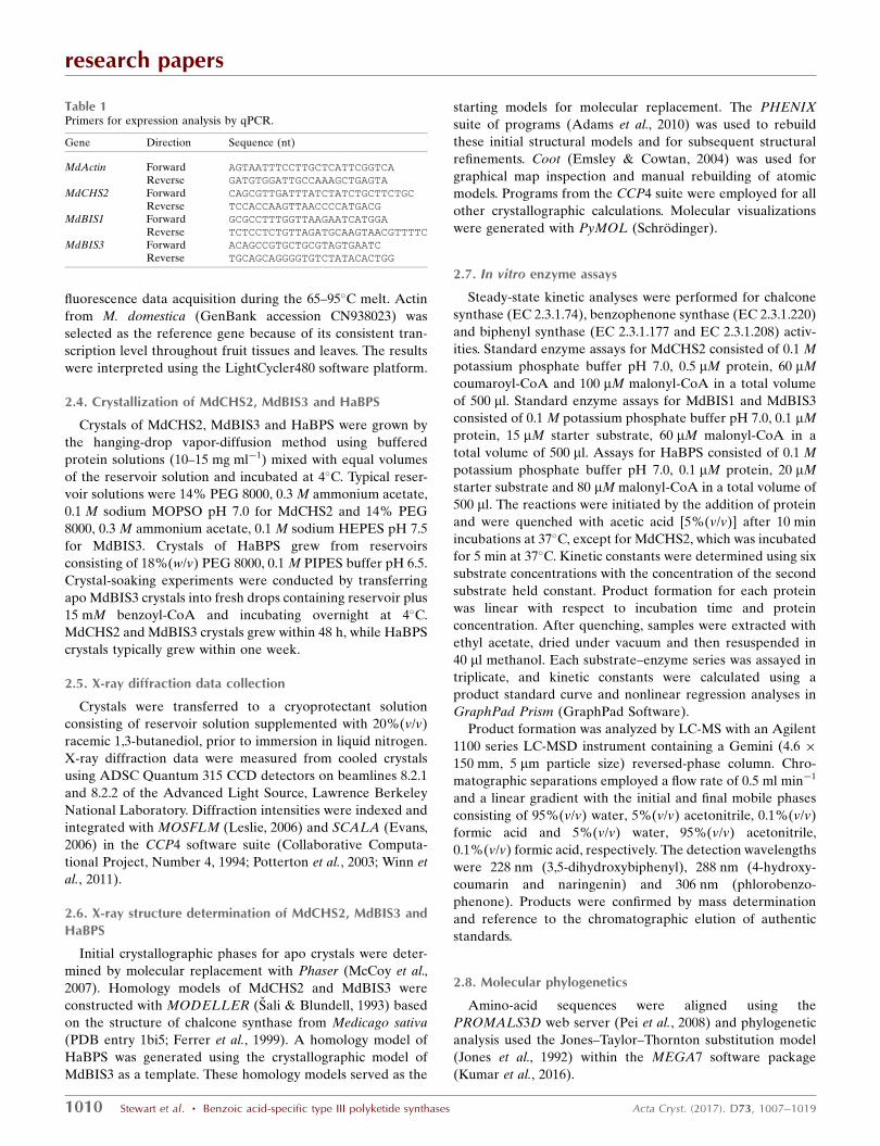
Table 1Primers for expression analysis by qPCR.
Gene Direction Sequence (nt)
MdActin Forward AGTAATTTCCTTGCTCATTCGGTCA
Reverse GATGTGGATTGCCAAAGCTGAGTA
MdCHS2 Forward CAGCGTTGATTTATCTATCTGCTTCTGC
Reverse TCCACCAAGTTAACCCCATGACG
MdBIS1 Forward GCGCCTTTGGTTAAGAATCATGGA
Reverse TCTCCTCTGTTAGATGCAAGTAACGTTTTC
MdBIS3 Forward ACAGCCGTGCTGCGTAGTGAATC
Reverse TGCAGCAGGGGTGTCTATACACTGG
2.9. Data deposition
The cDNA sequences have been deposited in GenBank
with accession numbers JQ582624 for MdCHS2, JQ582625 for
MdBIS1 and JQ582626 for MdBIS3. Crystallographic models
have been deposited in the PDB with the accession codes
listed in Table 2.
3. Results
3.1. Functional characterization of CHS, BIS and BPS
Initially, we isolated three cDNAs from an apple cDNA
library (M. domestica ‘Royal Gala’) that showed similarity to
plant type III PKSs. BLAST searches and analysis of the apple
genome showed that one of the cDNAs was >96% identical in
amino-acid sequence to chalcone synthases from Malus (Dare
et al., 2013). The other two cDNAs were 92–99% identical in
amino-acid sequence to biphenyl synthases from Sorbus
aucuparia and M. domestica ‘Holsteiner Cox’ (Liu et al., 2007,
2010; Chizzali et al., 2011). Based on sequence similarities,
functional studies and phylogenetic analyses (see below), we
named the above genes identified in this study MdCHS2,
MdBIS1.1 and MdBIS3.1. For the sake of brevity, MdBIS1.1
and MdBIS3.1 will be referred to as MdBIS1 and MdBIS3 in
this manuscript.
In vitro assays confirmed the biochemical activity of
MdCHS2 as a chalcone synthase and of MdBIS1 and MdBIS3
as biphenyl synthases (Table 2). MdCHS2 was active with
4-coumaroyl-CoA; however, neither biphenyl or benzo-
phenone products (see below) could be detected when
benzoyl-CoA was used as the starter substrate. Both MdBIS1
and MdBIS3 were active with benzoyl-CoA and salicoyl-CoA,
producing 3,5-dihydroxybiphenyl and 4-hydroxycoumarin,
respectively. We did not detect significant differences between
MdBIS1 and MdBIS3 in their catalytic efficiencies for
benzoyl-CoA compared with salicoyl-CoA. In contrast to the
observations by Chizzali et al. (2011) that the MdBIS1
homolog in ‘Holsteiner Cox’ occurred as a nonfunctional
allele, our biochemical analyses showed that MdBIS1 from
‘Royal Gala’ is a functional enzyme.
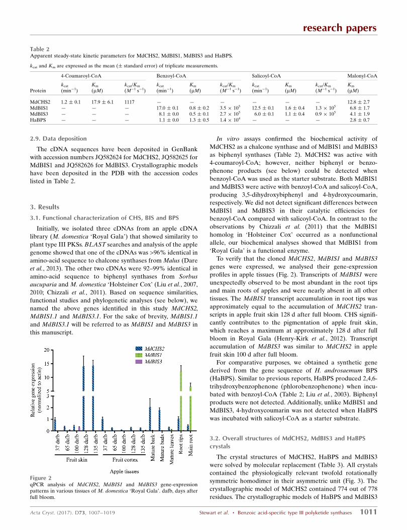
To verify that the cloned MdCHS2, MdBIS1 and MdBIS3
genes were expressed, we analysed their gene-expression
profiles in apple tissues (Fig. 2). Transcripts of MdBIS1 were
unexpectedly observed to be most abundant in the root tips
and main roots of apples and were nearly absent in all other
tissues. The MdBIS1 transcript accumulation in root tips was
approximately equal to the accumulation of MdCHS2 tran-
scripts in apple fruit skin 128 d after full bloom. CHS signifi-
cantly contributes to the pigmentation of apple fruit skin,
which reaches a maximum at approximately 128 d after full
bloom in Royal Gala (Henry-Kirk et al., 2012). Transcript
accumulation of MdBIS3 was similar to MdCHS2 in apple
fruit skin 100 d after full bloom.
For comparative purposes, we obtained a synthetic gene
derived from the gene sequence of H. androsaemum BPS
(HaBPS). Similar to previous reports, HaBPS produced 2,4,6-
trihydroxybenzophenone (phlorobenzophenone) when incu-
bated with benzoyl-CoA (Table 2; Liu et al., 2003). Biphenyl
products were not detected. Additionally, unlike MdBIS1 and
MdBIS3, 4-hydroxycoumarin was not detected when HaBPS
was incubated with salicoyl-CoA as a starter substrate.
3.2. Overall structures of MdCHS2, MdBIS3 and HaBPScrystals
The crystal structures of MdCHS2, HaBPS and MdBIS3
were solved by molecular replacement (Table 3). All crystals
contained the physiologically relevant twofold rotationally
symmetric homodimer in their asymmetric unit (Fig. 3). The
crystallographic model of MdCHS2 contained 774 out of 778
residues. The crystallographic models of HaBPS and MdBIS3
research papers
Acta Cryst. (2017). D73, 1007–1019 Stewart et al. � Benzoic acid-specific type III polyketide synthases 1011
Table 2Apparent steady-state kinetic parameters for MdCHS2, MdBIS1, MdBIS3 and HaBPS.
kcat and Km are expressed as the mean (� standard error) of triplicate measurements.
4-Coumaroyl-CoA Benzoyl-CoA Salicoyl-CoA Malonyl-CoA
Proteinkcat
(min�1)Km
(mM)kcat/Km
(M�1 s�1)kcat
(min�1)Km
(mM)kcat/Km
(M�1 s�1)kcat
(min�1)Km
(mM)kcat/Km
(M�1 s�1)Km
(mM)
MdCHS2 1.2 � 0.1 17.9 � 6.1 1117 — — — — — — 12.8 � 2.7MdBIS1 — — — 17.0 � 0.1 0.8 � 0.2 3.5 � 105 12.5 � 0.1 1.6 � 0.4 1.3 � 105 6.8 � 1.7MdBIS3 — — — 8.1 � 0.0 0.5 � 0.1 2.7 � 105 6.0 � 0.1 1.1 � 0.4 0.9 � 105 4.1 � 1.9HaBPS — — — 1.1 � 0.0 1.3 � 0.5 1.4 � 104 — — — 2.8 � 0.7
Figure 2qPCR analysis of MdCHS2, MdBIS1 and MdBIS3 gene-expressionpatterns in various tissues of M. domestica ‘Royal Gala’. dafb, days afterfull bloom.
contained 772 out of 790 residues
and 757 out of 776 residues,
respectively. Lastly, the crystallo-
graphic model of MdBIS3
complexed with benzoyl-CoA
contained 756 out of 780 residues.
The missing residues of all
crystallographic models were at
the N- and C-termini of each
monomeric subunit. Additionally,
each monomer of MdBIS3
contained electron density
consistent with 1,3-butanediol,
the cryoprotectant. When super-
posed, the polypeptide backbones
of MdCHS2 and MdBIS3 have an
r.m.s.d. of 0.9 A, those of
MdCHS2 and HaBPS have an
r.m.s.d. of 0.7 A and those of
MdBIS3 and HaBPS have an
r.m.s.d. of 0.9 A. Our final crys-
tallographic models of MdCHS2
and HaBPS were refined at 2.1 A
resolution (R = 16.6%; Rfree =
20.9%) and 2.85 A (R = 20.1%;
Rfree = 26.1%), respectively. The
final crystallographic models of
apo MdBIS3 and MdBIS3
complexed with benzoyl-CoA
research papers
1012 Stewart et al. � Benzoic acid-specific type III polyketide synthases Acta Cryst. (2017). D73, 1007–1019
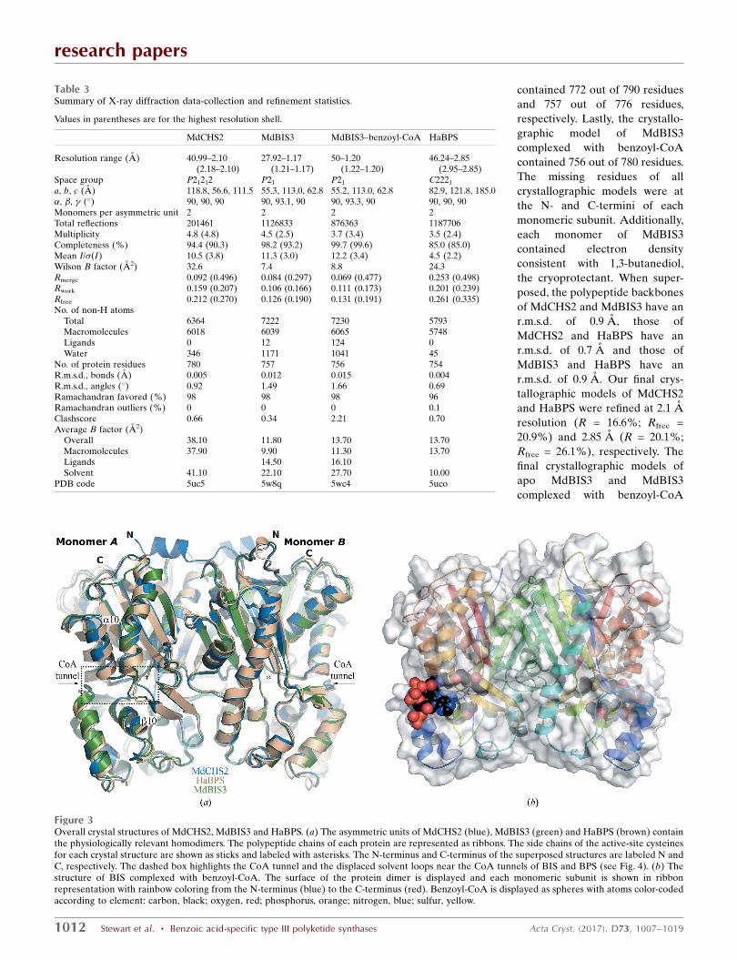
Table 3Summary of X-ray diffraction data-collection and refinement statistics.
Values in parentheses are for the highest resolution shell.
MdCHS2 MdBIS3 MdBIS3–benzoyl-CoA HaBPS
Resolution range (A) 40.99–2.10(2.18–2.10)
27.92–1.17(1.21–1.17)
50–1.20(1.22–1.20)
46.24–2.85(2.95–2.85)
Space group P21212 P21 P21 C2221
a, b, c (A) 118.8, 56.6, 111.5 55.3, 113.0, 62.8 55.2, 113.0, 62.8 82.9, 121.8, 185.0�, �, � (�) 90, 90, 90 90, 93.1, 90 90, 93.3, 90 90, 90, 90Monomers per asymmetric unit 2 2 2 2Total reflections 201461 1126833 876363 1187706Multiplicity 4.8 (4.8) 4.5 (2.5) 3.7 (3.4) 3.5 (2.4)Completeness (%) 94.4 (90.3) 98.2 (93.2) 99.7 (99.6) 85.0 (85.0)Mean I/�(I) 10.5 (3.8) 11.3 (3.0) 12.2 (3.4) 4.5 (2.2)Wilson B factor (A2) 32.6 7.4 8.8 24.3Rmerge 0.092 (0.496) 0.084 (0.297) 0.069 (0.477) 0.253 (0.498)Rwork 0.159 (0.207) 0.106 (0.166) 0.111 (0.173) 0.201 (0.239)Rfree 0.212 (0.270) 0.126 (0.190) 0.131 (0.191) 0.261 (0.335)No. of non-H atoms
Total 6364 7222 7230 5793Macromolecules 6018 6039 6065 5748Ligands 0 12 124 0Water 346 1171 1041 45
No. of protein residues 780 757 756 754R.m.s.d., bonds (A) 0.005 0.012 0.015 0.004R.m.s.d., angles (�) 0.92 1.49 1.66 0.69Ramachandran favored (%) 98 98 98 96Ramachandran outliers (%) 0 0 0 0.1Clashscore 0.66 0.34 2.21 0.70Average B factor (A2)
Overall 38.10 11.80 13.70 13.70Macromolecules 37.90 9.90 11.30 13.70Ligands 14.50 16.10Solvent 41.10 22.10 27.70 10.00
PDB code 5uc5 5w8q 5wc4 5uco
Figure 3Overall crystal structures of MdCHS2, MdBIS3 and HaBPS. (a) The asymmetric units of MdCHS2 (blue), MdBIS3 (green) and HaBPS (brown) containthe physiologically relevant homodimers. The polypeptide chains of each protein are represented as ribbons. The side chains of the active-site cysteinesfor each crystal structure are shown as sticks and labeled with asterisks. The N-terminus and C-terminus of the superposed structures are labeled N andC, respectively. The dashed box highlights the CoA tunnel and the displaced solvent loops near the CoA tunnels of BIS and BPS (see Fig. 4). (b) Thestructure of BIS complexed with benzoyl-CoA. The surface of the protein dimer is displayed and each monomeric subunit is shown in ribbonrepresentation with rainbow coloring from the N-terminus (blue) to the C-terminus (red). Benzoyl-CoA is displayed as spheres with atoms color-codedaccording to element: carbon, black; oxygen, red; phosphorus, orange; nitrogen, blue; sulfur, yellow.

were refined at 1.17 A (R = 10.6%; Rfree = 12.6%) and 1.20 A
(R = 11.1%; Rfree = 13.1%), respectively, the highest resolution
structures obtained to date for a type III PKS.
Crystallographic analysis of secondary-structural elements
between MdCHS2, HaBPS and MdBIS3 revealed only minor
differences in topology, none of which immediately explained
the differences in substrate specificity and terminal cyclization
preferences during catalysis. The five-layered ����� topology
of secondary-structural elements of each monomer is consis-
tent with the ‘thiolase fold’ originally observed in thiolase and
present in all known structures of type III PKSs (Austin &
Noel, 2003; Mathieu et al., 1994). Each monomer of MdCHS2,
MdBIS3 and HaBPS contains two cis-peptide bonds at the
dimer interfaces which are well defined by the electron
densities of each structure and are conserved in all known type
III PKSs. Structural differences between the superposed C�
traces of MdCHS2, HaBPS and MdBIS3 occur primarily at the
solvent interface. In particular, a three-residue solvent-
exposed loop in MdBIS3 and HaBPS is displaced approxi-
mately 5 A toward the CoA tunnel compared with MdCHS2
(Fig. 3a).
3.2.1. CoA tunnel. A conserved arrangement of hydrogen
bonds assists in the binding of CoA substrates in the CoA
tunnels of MdBIS3, HaBPS and MdCHS2. Despite the use of
reducing agents in all buffers and crystallization solutions, the
electron-density maps indicated that the active-site cysteines
of the MdBIS3 crystals were oxidized to their sulfinic acid
derivatives (–SO2H). Oxidation of active-site cysteines has
been observed in other type III PKS structures (PDB entries
1bi5 and 1qlv) and prevents the transfer of substrates onto the
active-site cysteines (Jez, Austin et al., 2000; Ferrer et al.,
1999). Nonetheless, insight into substrate binding was
provided by co-crystallization of MdBIS3 with benzoyl-CoA,
resulting in intact benzoyl-CoA molecules with partial occu-
pancies in both monomers of MdBIS3 (Figs. 3b and 4a). In
MdBIS3, the side chains of residues Lys50 and Arg53 as well
as the backbones of Ala305 and Gly302 form hydrogen bonds
to the phosphates and pantetheine subunits of benzoyl-CoA,
respectively. These hydrogen bonds are conserved in other
type III PKS structures containing bound CoA substrates
(PDB entries 1bq6 and 1ee0; Ferrer et al., 1999; Jez, Austin et
al., 2000). However, as mentioned above, both MdBIS3 and
HaBPS contain a three-residue solvent-exposed loop that is
displaced significantly toward the CoA tunnel (Fig. 4b). The
displaced loop in the MdBIS3–benzoyl-CoA complex gener-
ates an additional hydrogen bond between the adenine unit of
benzoyl-CoA and the backbone carbonyl of Leu262. The
displaced loops found in MdBIS3 and HaBPS appear to
correlate with the rotameric state of one of the gatekeeper
residues (Fig. 4c). The gatekeeper residues, Phe215 and
Phe265 in MdCHS2, separate the CoA tunnel from the
internal active-site cavity and affect substrate selectivity by
acting as a steric gate during substrate binding (Ferrer et al.,
1999; Jez et al., 2002). Phe265 of CHS is replaced with tyrosine
in both MdBIS3 (Tyr260) and HaBPS (Tyr269). Differences in
the side-chain torsion angles of Tyr260 in MdBIS3 (�1 = 61�)
and Tyr269 in HaBPS (�1 = 50�) compared with Phe265 of
CHS (�1 = 170�) significantly reorient the tyrosine side chains
and increase the conformational flexibility of the nearby
Leu262 in MdBIS3 and Leu271 in HaBPS. Lastly, the side-
chain conformation of Tyr260 in MdBIS3 is stabilized via the
presence of a water-mediated hydrogen bond to the backbone
carbonyl of Phe192. The F265Y mutation in CHS was one of
three mutations needed to shift the substrate specificity of
CHS towards benzoyl-CoA (Liu et al., 2003). Presumably, a
similar water-mediated hydrogen bond stabilizes Tyr269 of
research papers
Acta Cryst. (2017). D73, 1007–1019 Stewart et al. � Benzoic acid-specific type III polyketide synthases 1013
Figure 4Binding interactions of benzoyl-CoA. (a) Co-crystallization of MdBIS3 with benzoyl-CoA. MdBIS3 is displayed as a green cartoon; benzoyl-CoA isdisplayed in ball-and-stick representation. Hydrogen bonds are shown as dashed lines and waters are shown as spheres. Atoms are color-coded accordingto element: carbon, black; oxygen, red; nitrogen, blue; sulfur, yellow. The 2Fo� Fc OMIT electron-density difference map surrounding the benzoyl-CoAligand is displayed as blue-colored cages and contoured at � = 1.0 with carve = 1.3. (b) Overlay of the MdBIS3–benzoyl-CoA complex with MdCHS2 andHaBPS. Residues associated with the displaced solvent-exposed loop near the CoA tunnel and mutations near the benzoyl moiety of benzoyl-CoA aredisplayed as sticks. (c) Overlay of the MdBIS3–benzoyl-CoA complex with MdCHS2 and HaBPS rotated approximately 120� counterclockwise from thatshown in (b).
HaBPS; however, the lower resolution of the diffraction data
for the HaBPS crystals prevented the analysis of waters.
3.3. Active-site architectures of CHS, BIS and BPS
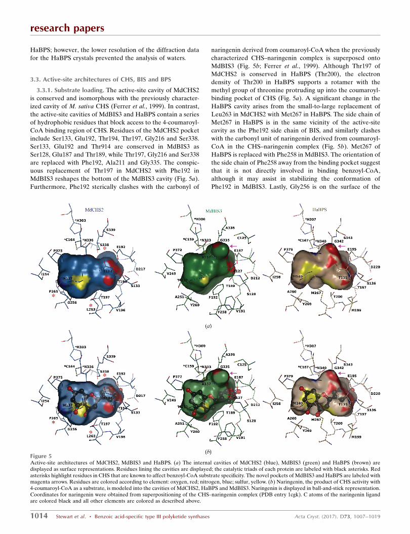
3.3.1. Substrate loading. The active-site cavity of MdCHS2
is conserved and isomorphous with the previously character-
ized cavity of M. sativa CHS (Ferrer et al., 1999). In contrast,
the active-site cavities of MdBIS3 and HaBPS contain a series
of hydrophobic residues that block access to the 4-coumaroyl-
CoA binding region of CHS. Residues of the MdCHS2 pocket
include Ser133, Glu192, Thr194, Thr197, Gly216 and Ser338.
Ser133, Glu192 and Thr914 are conserved in MdBIS3 as
Ser128, Glu187 and Thr189, while Thr197, Gly216 and Ser338
are replaced with Phe192, Ala211 and Gly335. The conspic-
uous replacement of Thr197 in MdCHS2 with Phe192 in
MdBIS3 reshapes the bottom of the MdBIS3 cavity (Fig. 5a).
Furthermore, Phe192 sterically clashes with the carbonyl of
naringenin derived from coumaroyl-CoA when the previously
characterized CHS–naringenin complex is superposed onto
MdBIS3 (Fig. 5b; Ferrer et al., 1999). Although Thr197 of
MdCHS2 is conserved in HaBPS (Thr200), the electron
density of Thr200 in HaBPS supports a rotamer with the
methyl group of threonine protruding up into the coumaroyl-
binding pocket of CHS (Fig. 5a). A significant change in the
HaBPS cavity arises from the small-to-large replacement of
Leu263 in MdCHS2 with Met267 in HaBPS. The side chain of
Met267 in HaBPS is in the same vicinity of the active-site
cavity as the Phe192 side chain of BIS, and similarly clashes
with the carbonyl unit of naringenin derived from coumaroyl-
CoA in the CHS–naringenin complex (Fig. 5b). Met267 of
HaBPS is replaced with Phe258 in MdBIS3. The orientation of
the side chain of Phe258 away from the binding pocket suggest
that it is not directly involved in binding benzoyl-CoA,
although it may assist in stabilizing the conformation of
Phe192 in MdBIS3. Lastly, Gly256 is on the surface of the
research papers
1014 Stewart et al. � Benzoic acid-specific type III polyketide synthases Acta Cryst. (2017). D73, 1007–1019
Figure 5Active-site architectures of MdCHS2, MdBIS3 and HaBPS. (a) The internal cavities of MdCHS2 (blue), MdBIS3 (green) and HaBPS (brown) aredisplayed as surface representations. Residues lining the cavities are displayed; the catalytic triads of each protein are labeled with black asterisks. Redasterisks highlight residues in CHS that are known to affect benzoyl-CoA substrate specificity. The novel pockets of MdBIS3 and HaBPS are labeled withmagenta arrows. Residues are colored according to element: oxygen, red; nitrogen, blue; sulfur, yellow. (b) Naringenin, the product of CHS activity with4-coumaroyl-CoA as a substrate, is modeled into the cavities of MdCHS2, HaBPS and MdBIS3. Naringenin is displayed in ball-and-stick representation.Coordinates for naringenin were obtained from superpositioning of the CHS–naringenin complex (PDB entry 1cgk). C atoms of the naringenin ligandare colored black and all other elements are colored as described above.
MdCHS2 cavity near the CoA tunnel; in MdBIS3 and HaBPS
this position contains the slightly bulkier Ala. Mutations that
increase the bulkiness of the side chain of position 256,
including G256A, have previously been shown to shift the
substrate specificity of CHS towards smaller substrates (Jez,
Austin et al., 2000). Collectively, hydrophobic mutations in the
active sites of MdBIS3 and HaBPS provide hydrophobic
surfaces for interacting with small hydrophobic substrates
such as benzoyl-CoA.
3.3.2. Elongation and cyclization within benzoic acid-specific type III PKSs. The replacement of Ser338 in MdCHS2
with Gly generates a novel pocket in the active-site cavities of
MdBIS3 and HaBPS associated with their elongation and
cyclization reactions. Ser338 of MdCHS2 resides at the inter-
section of the coumaroyl-binding pocket and elongation/
cyclization cavity and stabilizes the conformation of elongated
polyketide intermediates (Jez, Austin et al., 2000). The
replacement of Ser338 in CHS with Gly in both MdBIS3
(Gly335) and HaBPS (Gly342) allows the internal cavities of
MdBIS3 and HaBPS to expand behind their respective active-
site cysteines to generate novel pockets (Fig. 5a). The sides of
this novel pocket in MdBIS3 are formed from the side chains
of Cys125, Ala127, Ala157, Glu187 and Ala336 as well as the
main-chain atoms of Gly158, Ala157, Gly335 and Ala336.
Similarly, the side chains of Thr135 and Glu195 and the main-
chain atoms of Gly166, Gly342 and Ser343 line the sides of this
novel pocket in HaBPS. Notably, Klundt et al. (2009) observed
that mutagenesis of Thr135 in HaBPS disrupted chain elon-
gation and cyclization specificity.
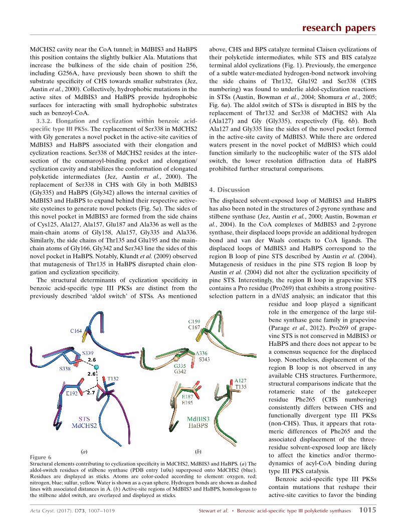
The structural determinants of cyclization specificity in
benzoic acid-specific type III PKSs are distinct from the
previously described ‘aldol switch’ of STSs. As mentioned
above, CHS and BPS catalyze terminal Claisen cyclizations of
their polyketide intermediates, while STS and BIS catalyze
terminal aldol cyclizations (Fig. 1). Previously, the emergence
of a subtle water-mediated hydrogen-bond network involving
the side chains of Thr132, Glu192 and Ser338 (CHS
numbering) was found to underlie aldol-cyclization reactions
in STSs (Austin, Bowman et al., 2004; Shomura et al., 2005;
Fig. 6a). The aldol switch of STSs is disrupted in BIS by the
replacement of Thr132 and Ser338 of MdCHS2 with Ala
(Ala127) and Gly (Gly335), respectively (Fig. 6b). Both
Ala127 and Gly335 line the sides of the novel pocket formed
in the active-site cavity of MdBIS3. While there are ordered
waters present in the novel pocket of MdBIS3 which could
function similarly to the nucleophilic water of the STS aldol
switch, the lower resolution diffraction data of HaBPS
prohibited further structural comparisons.
4. Discussion
The displaced solvent-exposed loop of MdBIS3 and HaBPS
has also been noted in the structures of 2-pyrone synthase and
stilbene synthase (Jez, Austin et al., 2000; Austin, Bowman et
al., 2004). In the CoA complexes of MdBIS3 and 2-pyrone
synthase, their displaced loops provide an additional hydrogen
bond and van der Waals contacts to CoA ligands. The
displaced loops of MdBIS3 and HaBPS correspond to the
region B loop of pine STS described by Austin et al. (2004).
Mutagenesis of residues in the pine STS region B loop by
Austin et al. (2004) did not alter the cyclization specificity of
pine STS. Interestingly, the region B loop in grapevine STS
contains a Pro residue (Pro269) that exhibits a strong positive-
selection pattern in a dN/dS analysis; an indicator that this
residue and loop played a significant
role in the emergence of the large stil-
bene synthase gene family in grapevine
(Parage et al., 2012). Pro269 of grape-
vine STS is not conserved in MdBIS3 or
HaBPS and there does not appear to be
a consensus sequence for the displaced
loop. Nonetheless, displacement of the
region B loop is not observed in any
available CHS structures. Furthermore,
structural comparisons indicate that the
rotameric state of the gatekeeper
residue Phe265 (CHS numbering)
consistently differs between CHS and
functionally divergent type III PKSs
(non-CHS). Thus, it appears that rota-
meric differences of Phe265 and the
associated displacement of the three-
residue solvent-exposed loop are likely
to affect the kinetics and/or thermo-
dynamics of acyl-CoA binding during
type III PKS catalysis.
Benzoic acid-specific type III PKSs
contain mutations that reshape their
active-site cavities to favor the binding
research papers
Acta Cryst. (2017). D73, 1007–1019 Stewart et al. � Benzoic acid-specific type III polyketide synthases 1015
Figure 6Structural elements contributing to cyclization specificity in MdCHS2, MdBIS3 and HaBPS. (a) Thealdol-switch residues of stilbene synthase (PDB entry 1u0u) superposed onto MdCHS2 (blue).Residues are displayed as sticks. Atoms are color-coded according to element: oxygen, red;nitrogen, blue; sulfur, yellow. Water is shown as a cyan sphere. Hydrogen bonds are shown as dashedlines with associated distances in A. (b) Active-site regions of MdBIS3 and HaBPS, homologous tothe stilbene aldol switch, are overlayed and displayed as sticks.
of small hydrophobic substrates. Replacement of Thr197 in
CHS with Phe in MdBIS3 (Phe192) appears to be responsible
for closing off access to the coumaroyl-binding region of CHS.
Similarly, the side chain of Met267 of HaBPS replaces Leu263
of CHS and blocks access to the coumaroyl-binding area of the
CHS cavity. Phe192 in MdBIS3 and Met267 in HaBPS also
provide hydrophobic surfaces that would favorably interact
with small hydrophobic substrates and their polyketide inter-
mediates. Met267 of HaBPS is substituted with a Phe in
MdBIS3 (Phe258) and seems to primarily function in stabi-
lizing the conformation of nearby residues including Phe192.
A triple mutant of CHS, L263M/F265Y/S338G, preferred
benzoyl-CoA over 4-coumaroyl-CoA as a substrate (Liu et al.,
2003). The mutations of Phe265 in CHS to Tyr and of Ser338
to Gly are present in all known benzoic acid-specific type III
PKSs. Conversely, the mutations of Thr197 in CHS to Phe and
of Leu263 in CHS to Met appear to be specific to BPS and BIS,
respectively (Supplementary Fig. S1).
A novel pocket in the active-site cavity of MdBIS3 and
HaBPS, in a region known to affect elongation and cyclization,
arises from the replacement of Ser338 of CHS with Gly. The
side chains of Ala127 in MdBIS3 and Thr135 in HaBPS line
the sides of the novel pocket. Mutagenesis of Thr135 in
HaBPS to Leu resulted in a reduction in chain-elongation
steps from three to two and a shift in the terminal cyclization
mechanism from Claisen to lactonization (Klundt et al., 2009).
As a result of changes in the number of chain-elongation steps
and cyclization specificity, the T135L mutant of HaBPS
produced phenylpyrone (6-phenyl-4-hydroxy-2H-pyran-2-one)
as the dominant product. Interestingly, homology modeling of
HaBPS by Klundt et al. (2009) suggested that the mutation
T135L generated a novel pocket that sterically favored the
generation of phenylpyrone over phlorobenzophenone.
Further structural and functional analyses are needed to
deconvolute steric versus electronic effects on catalysis within
this novel pocket of benzoic acid-specific type III PKS.
The structural basis of the terminal aldol cyclization of BIS
is different from the aldol switch of STSs, yet the chemical
mechanisms are likely to be similar. The replacement of
Thr132 and Ser338 of STS with Ala127 and Gly335 prohibits
the water-mediated hydrogen-bond networks underlying the
terminal aldol cyclizations of STSs from forming in BIS.
Sequence differences between the residues lining the walls of
the novel pockets in BPS (T135, G342SAC) and BIS (A127,
G335APT/S) are a useful starting point for further exploration
of cyclization specificity using mutagenesis. Additionally, the
novel pocket of MdBIS3 contains ordered waters that could
function similar to the nucleophilic water that is essential for
the aldol switch of stilbene
synthases. However, higher reso-
lution structures of BPS are
needed for comparisons of subtle
changes in backbone, side chain
and ordered waters between BPS
and BIS. Interestingly, benzal-
acetone synthase, another type
III PKS that contains mutations
that prohibit the formation of the
STS aldol switch, relies on a
nucleophilic water for the term-
inal aldol-like decarboxylation of
its diketide intermediate (Morita
et al., 2010). Although the specific
nucleophilic waters of STSs and
benzalacetone synthase are not
present in our MdBIS3 structures,
we speculate that a nucleophilic
water in the novel pocket of
BIS is a critical element for
the structure-based cyclization
mechanism of BISs.
The functional and structural
diversity of benzoic acid-specific
type III PKSs is likely to be
broader than those of BIS and
BPS. Unexpectedly, in our survey
of the literature we found a
quinolone synthase from Citrus
microcarpa (CmQNS) which
grouped with BIS and BPS in a
molecular phylogenetic analysis
research papers
1016 Stewart et al. � Benzoic acid-specific type III polyketide synthases Acta Cryst. (2017). D73, 1007–1019
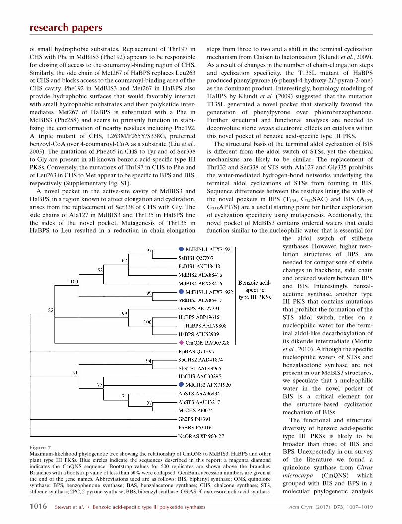
Figure 7Maximum-likelihood phylogenetic tree showing the relationship of CmQNS to MdBIS3, HaBPS and otherplant type III PKSs. Blue circles indicate the sequences described in this report; a magenta diamondindicates the CmQNS sequence. Bootstrap values for 500 replicates are shown above the branches.Branches with a bootstrap value of less than 50% were collapsed. GenBank accession numbers are given atthe end of the gene names. Abbreviations used are as follows: BIS, biphenyl synthase; QNS, quinolonesynthase; BPS, benzophenone synthase; BAS, benzalacetone synthase; CHS, chalcone synthase; STS,stilbene synthase; 2PC, 2-pyrone synthase; BBS, bibenzyl synthase; ORAS, 30-oxoresorcinolic acid synthase.
(Mori et al., 2013). Our independent analysis of MdBIS3 and
HaBPS phylogeny support the observations of Mori et al.
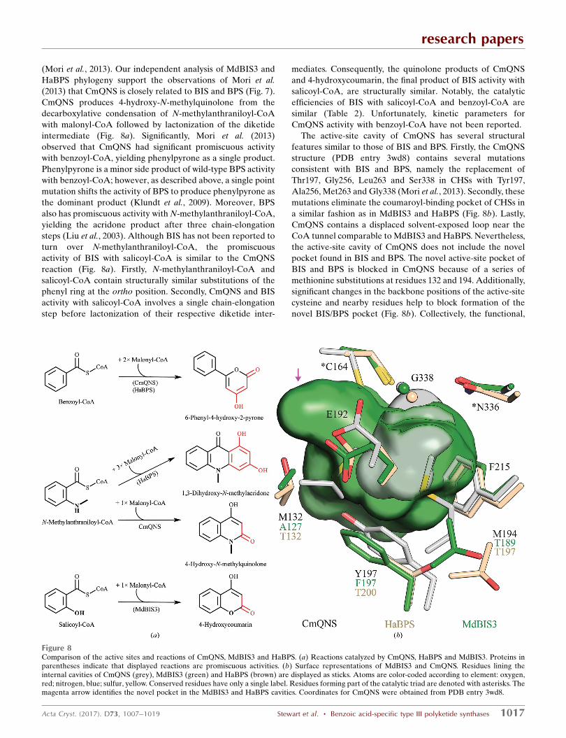
(2013) that CmQNS is closely related to BIS and BPS (Fig. 7).
CmQNS produces 4-hydroxy-N-methylquinolone from the
decarboxylative condensation of N-methylanthraniloyl-CoA
with malonyl-CoA followed by lactonization of the diketide
intermediate (Fig. 8a). Significantly, Mori et al. (2013)
observed that CmQNS had significant promiscuous activity
with benzoyl-CoA, yielding phenylpyrone as a single product.
Phenylpyrone is a minor side product of wild-type BPS activity
with benzoyl-CoA; however, as described above, a single point
mutation shifts the activity of BPS to produce phenylpyrone as
the dominant product (Klundt et al., 2009). Moreover, BPS
also has promiscuous activity with N-methylanthraniloyl-CoA,
yielding the acridone product after three chain-elongation
steps (Liu et al., 2003). Although BIS has not been reported to
turn over N-methylanthraniloyl-CoA, the promiscuous
activity of BIS with salicoyl-CoA is similar to the CmQNS
reaction (Fig. 8a). Firstly, N-methylanthraniloyl-CoA and
salicoyl-CoA contain structurally similar substitutions of the
phenyl ring at the ortho position. Secondly, CmQNS and BIS
activity with salicoyl-CoA involves a single chain-elongation
step before lactonization of their respective diketide inter-
mediates. Consequently, the quinolone products of CmQNS
and 4-hydroxycoumarin, the final product of BIS activity with
salicoyl-CoA, are structurally similar. Notably, the catalytic
efficiencies of BIS with salicoyl-CoA and benzoyl-CoA are
similar (Table 2). Unfortunately, kinetic parameters for
CmQNS activity with benzoyl-CoA have not been reported.
The active-site cavity of CmQNS has several structural
features similar to those of BIS and BPS. Firstly, the CmQNS
structure (PDB entry 3wd8) contains several mutations
consistent with BIS and BPS, namely the replacement of
Thr197, Gly256, Leu263 and Ser338 in CHSs with Tyr197,
Ala256, Met263 and Gly338 (Mori et al., 2013). Secondly, these
mutations eliminate the coumaroyl-binding pocket of CHSs in
a similar fashion as in MdBIS3 and HaBPS (Fig. 8b). Lastly,
CmQNS contains a displaced solvent-exposed loop near the
CoA tunnel comparable to MdBIS3 and HaBPS. Nevertheless,
the active-site cavity of CmQNS does not include the novel
pocket found in BIS and BPS. The novel active-site pocket of
BIS and BPS is blocked in CmQNS because of a series of
methionine substitutions at residues 132 and 194. Additionally,
significant changes in the backbone positions of the active-site
cysteine and nearby residues help to block formation of the
novel BIS/BPS pocket (Fig. 8b). Collectively, the functional,
research papers
Acta Cryst. (2017). D73, 1007–1019 Stewart et al. � Benzoic acid-specific type III polyketide synthases 1017
Figure 8Comparison of the active sites and reactions of CmQNS, MdBIS3 and HaBPS. (a) Reactions catalyzed by CmQNS, HaBPS and MdBIS3. Proteins inparentheses indicate that displayed reactions are promiscuous activities. (b) Surface representations of MdBIS3 and CmQNS. Residues lining theinternal cavities of CmQNS (grey), MdBIS3 (green) and HaBPS (brown) are displayed as sticks. Atoms are color-coded according to element: oxygen,red; nitrogen, blue; sulfur, yellow. Conserved residues have only a single label. Residues forming part of the catalytic triad are denoted with asterisks. Themagenta arrow identifies the novel pocket in the MdBIS3 and HaBPS cavities. Coordinates for CmQNS were obtained from PDB entry 3wd8.
structural and phylogenetic data discussed above lead us to
hypothesize that CmQNS is a descendent of a benzoic acid-
specific type III PKS recruited into alkaloid metabolism.
5. Conclusions
In conclusion, we report here the first structural analyses of
benzoic acid-specific type III PKSs. Benzoic acid-specific type
III PKSs catalyse the committed steps in the biosynthesis of
benzophenone and biphenyl metabolites in plants. The
products of BIS and BPS, 3,5-dihydroxybiphenyl and phloro-
benzophenone, respectively, are subsequently modified via an
assortment of tailoring reactions (for example, hydroxylation,
O-methylation and prenylation) to yield the final repertoire of
biphenyl, dibenzofuran and phlorobenzophenone metabolites
found in plants (Khalil et al., 2015; El-Awaad et al., 2016; Sircar
et al., 2015; Fiesel et al., 2015). BIS and BPS contain several
small-to-large mutations which block the binding of
4-coumaroyl-CoA and provide hydrophobic surfaces for
smaller hydrophobic starter CoAs and intermediates. Signifi-
cantly, BIS and BPS contain a novel pocket in their active-site
cavities associated with their chain-elongation and cyclization
reactions. These structural idiosyncrasies underlie the prefer-
ence of BIS and BPS for benzoic acid-derived substrates.
Furthermore, the promiscuous nature of BIS and BPS may
have contributed to the emergence of a type III PKS involved
in alkaloid biosynthesis. The renowned promiscuity of type III
PKSs is likely to underpin their ability to evolve as their host
organisms interacted with constantly changing environments
(Weng & Noel, 2012). The structural snapshots presented in
this paper deepen our understanding of structure–function
relationships in the type III PKS family and lay a foundation
for ongoing efforts to exploit the biosynthetic potential of
plant metabolic enzymes.
Acknowledgements
We thank the staff of the Advanced Light Source, a national
user facility operated by Lawrence Berkeley National
Laboratory, for assistance during data collection on beamlines
8.2.1 and 8.2.2. Additionally, we would like to thank Gordon V.
Louie for assistance and helpful discussions concerning X-ray
diffraction analyses. Finally, we would like to pay tribute to the
memory of Greg Macias: a labmate, collaborator and friend
whose efforts made this work possible. JPN is an investigator
with the Howard Hughes Medical Institute.
Funding information
This research was supported by an NSF Minority Postdoctoral
Research Fellowship (Award No. 0805691) to CS and NSF
grants EEC-0813570 and MCB-0645794 to JPN.
References
Acuna, U. M., Jancovski, N. & Kennelly, E. J. (2009). Curr. Top. Med.Chem. 9, 1560–1580.
Adams, P. D. et al. (2010). Acta Cryst. D66, 213–221.
Austin, M. B., Bowman, M. E., Ferrer, J.-L., Schroder, J. & Noel, J. P.(2004). Chem. Biol. 11, 1179–1194.
Austin, M. B., Izumikawa, M., Bowman, M. E., Udwary, D. W., Ferrer,J.-L., Moore, B. S. & Noel, J. P. (2004). J. Biol. Chem. 279, 45162–45174.
Austin, M. B. & Noel, J. P. (2003). Nat. Prod. Rep. 20, 79–110.Beerhues, L. & Liu, B. (2009). Phytochemistry, 70, 1719–1727.Chizzali, C., Gaid, M. M., Belkheir, A. K., Hansch, R., Richter, K.,
Flachowsky, H., Peil, A., Hanke, M.-V., Liu, B. & Beerhues, L.(2011). Plant Physiol. 158, 864–875.
Chizzali, C., Khalil, M. N. A., Beuerle, T., Schuehly, W., Richter, K.,Flachowsky, H., Peil, A., Hanke, M.-V., Liu, B. & Beerhues, L.(2012). Phytochemistry, 77, 179–185.
Chizzali, C., Swiddan, A. K., Abdelaziz, S., Gaid, M., Richter, K.,Fischer, T. C., Liu, B. & Beerhues, L. (2016). PLoS One, 11,e0158713.
Collaborative Computational Project, Number 4 (1994). Acta Cryst.D50, 760–763.
Dare, A. P., Tomes, S., Jones, M., McGhie, T. K., Stevenson, D. E.,Johnson, R. A., Greenwood, D. R. & Hellens, R. P. (2013). Plant J.74, 398–410.
El-Awaad, I., Bocola, M., Beuerle, T., Liu, B. & Beerhues, L. (2016).Nature Commun. 7, 11472.
Emsley, P. & Cowtan, K. (2004). Acta Cryst. D60, 2126–2132.Evans, P. (2006). Acta Cryst. D62, 72–82.Ferrer, J.-L., Jez, J. M., Bowman, M. E., Dixon, R. A. & Noel, J. P.
(1999). Nature Struct. Biol. 6, 775–784.Fiesel, T., Gaid, M., Muller, A., Bartels, J., El-Awaad, I., Beuerle, T.,
Ernst, L., Behrends, S. & Beerhues, L. (2015). Molecules, 20, 15616–15630.
Franklin, G., Conceicao, L. F. R., Kombrink, E. & Dias, A. C. P.(2009). Phytochemistry, 70, 60–68.
Gaid, M. M., Sircar, D., Muller, A., Beuerle, T., Liu, B., Ernst, L.,Hansch, R. & Beerhues, L. (2012). Plant Physiol. 160, 1267–1280.
Gronquist, M., Bezzerides, A., Attygalle, A., Meinwald, J., Eisner, M.& Eisner, T. (2001). Proc. Natl Acad. Sci. USA, 98, 13745–13750.
Guo, Z.-F., Sun, Y., Zheng, S. & Guo, Z. (2009). Biochemistry, 48,1712–1722.
Henry-Kirk, R., McGhie, T., Andre, C., Hellens, R. & Allan, A.(2012). J. Exp. Bot. 63, 5437–5450.
Hrazdina, G. & Borejsza-Wysocki, W. (2003). Phytochemistry, 64,485–492.
Huttner, C., Beuerle, T., Scharnhop, H., Ernst, L. & Beerhues, L.(2010). J. Agric. Food Chem. 58, 11977–11984.
Jez, J. M., Austin, M. B., Ferrer, J.-L., Bowman, M. E., Schroder, J. &Noel, J. P. (2000). Chem. Biol. 7, 919–930.
Jez, J. M., Bowman, M. E. & Noel, J. P. (2002). Proc. Natl Acad. Sci.USA, 99, 5319–5324.
Jez, J. M., Ferrer, J.-L., Bowman, M. E., Dixon, R. A. & Noel, J. P.(2000). Biochemistry, 39, 890–902.
Jones, D. T., Taylor, W. R. & Thornton, J. M. (1992). Comput. Appl.Biosci. 8, 275–282.
Khalil, M. N. A., Beuerle, T., Muller, A., Ernst, L., Bhavanam,V. B. R., Liu, B. & Beerhues, L. (2013). Phytochemistry, 96, 101–109.
Khalil, M. N. A., Brandt, W., Beuerle, T., Reckwell, D., Groeneveld, J.,Hansch, R., Gaid, M. M., Liu, B. & Beerhues, L. (2015). Plant J. 83,263–276.
Klundt, T., Bocola, M., Lutge, M., Beuerle, T., Liu, B. & Beerhues, L.(2009). J. Biol. Chem. 284, 30957–30964.
Kumar, S., Stecher, G. & Tamura, K. (2016). Mol. Biol. Evol. 33, 1870–1874.
Leslie, A. G. W. (2006). Acta Cryst. D62, 48–57.Lin, Y., Shen, X., Yuan, Q. & Yan, Y. (2013). Nature Commun. 4,
2603.Liu, B., Beuerle, T., Klundt, T. & Beerhues, L. (2004). Planta, 218,
492–496.
research papers
1018 Stewart et al. � Benzoic acid-specific type III polyketide synthases Acta Cryst. (2017). D73, 1007–1019
Liu, B., Falkenstein-Paul, H., Schmidt, W. & Beerhues, L. (2003).Plant. J. Cell. Mol. Biol. 34, 847–855.
Liu, B., Raeth, T., Beuerle, T. & Beerhues, L. (2007). Planta, 225,1495–1503.
Liu, B., Raeth, T., Beuerle, T. & Beerhues, L. (2010). Plant Mol. Biol.72, 17–25.
Mathieu, M., Zeelen, J. P., Pauptit, R. A., Erdmann, R., Kunau, W. H.& Wierenga, R. K. (1994). Structure, 2, 797–808.
McCoy, A. J., Grosse-Kunstleve, R. W., Adams, P. D., Winn, M. D.,Storoni, L. C. & Read, R. J. (2007). J. Appl. Cryst. 40, 658–674.
Mori, T., Shimokawa, Y., Matsui, T., Kinjo, K., Kato, R., Noguchi, H.,Sugio, S., Morita, H. & Abe, I. (2013). J. Biol. Chem. 288, 28845–28858.
Morita, H., Shimokawa, Y., Tanio, M., Kato, R., Noguchi, H., Sugio,S., Kohno, T. & Abe, I. (2010). Proc. Natl Acad. Sci. USA, 107, 669–673.
Newcomb, R. D. et al. (2006). Plant Physiol. 141, 147–166.Nilsson, M. & Norin, T. (1963). Acta Chem. Scand. 17, 1157–
1159.Nualkaew, N., Morita, H., Shimokawa, Y., Kinjo, K., Kushiro, T.,
De-Eknamkul, W., Ebizuka, Y. & Abe, I. (2012). Phytochemistry,77, 60–69.
Parage, C., Tavares, R., Rety, S., Baltenweck-Guyot, R., Poutaraud,A., Renault, L., Heintz, D., Lugan, R., Marais, G. A. B., Aubourg, S.& Hugueney, P. (2012). Plant Physiol. 160, 1407–1419.
Pei, J., Tang, M. & Grishin, N. V. (2008). Nucleic Acids Res. 36, W30–W34.
Potterton, E., Briggs, P., Turkenburg, M. & Dodson, E. (2003). ActaCryst. D59, 1131–1137.
Sali, A. & Blundell, T. L. (1993). J. Mol. Biol. 234, 779–815.Shomura, Y., Torayama, I., Suh, D., Xiang, T., Kita, A., Sankawa, U. &
Miki, K. (2005). Proteins, 60, 803–806.Sidenius, U., Skonberg, C., Olsen, J. & Hansen, S. H. (2004). Chem.
Res. Toxicol. 17, 75–81.Sircar, D., Gaid, M. M., Chizzali, C., Reckwell, D., Kaufholdt, D.,
Beuerle, T., Broggini, G. A. L., Flachowsky, H., Liu, B., Hansch, R.& Beerhues, L. (2015). Plant Physiol. 168, 428–442.
Tropf, S., Lanz, T., Rensing, S. A., Schroder, J. & Schroder, G. (1994).J. Mol. Evol. 38, 610–618.
Wang, R., Chen, R., Li, J., Liu, X., Xie, K., Chen, D., Peng, Y. & Dai, J.(2016). J. Nat. Prod. 79, 2143–2147.
Weng, J.-K. & Noel, J. P. (2012). Cold Spring Harb. Symp. Quant. Biol.77, 309–320.
Winn, M. D. et al. (2011). Acta Cryst. D67, 235–242.
research papers
Acta Cryst. (2017). D73, 1007–1019 Stewart et al. � Benzoic acid-specific type III polyketide synthases 1019