molecular assays for genetic testing. human genetic testing serves four main purposes: a.prenatal...
TRANSCRIPT

MOLECULAR ASSAYS FOR MOLECULAR ASSAYS FOR GENETIC TESTING GENETIC TESTING

Human genetic testing serves four main purposes:
a. Prenatal diagnosis
b. Newborn screening
c.Carrier (heterozygote) detection
d.Disease predisposition
Human Genetic TestingHuman Genetic Testing

Karyotyping of fetal cells from amniotic fluid or chorionic villi –gross chromosomal defects, trisomies etc
KARYOTYPINGKARYOTYPING

Trisomy 21 karyotypeTrisomy 21 karyotype

I - Molecular HybridizationI - Molecular Hybridization
For the detection of a specific DNA sequence in a heterogeneous
mixture of DNA
(e.g. whole genomic DNA etc)

ProbesProbes
Labeled, single stranded DNA complementary to the target DNA
The label may be radioactive (P32) or non radioactive (fluorescent
chromophores or biotin)

General scheme for General scheme for hybridization assayshybridization assays
Heterogeneous mix of DNA
Labeled probe
Probe binds target DNA

ProbesProbes
•Target DNA sequence knownShort oligonucleotide probes,15-30 bases,
complementary to part of the target sequence
•Protein sequence knownSeveral oligonucleotide probes constructed
using the genetic code, to locate the target sequence

Denature target DNA
Immobilize on insoluble matrix
Expose to labeled probes
Allow time for hybridization
Wash away unbound probe
Detect for the presence of label
Hybridization- the flow Hybridization- the flow chartchart
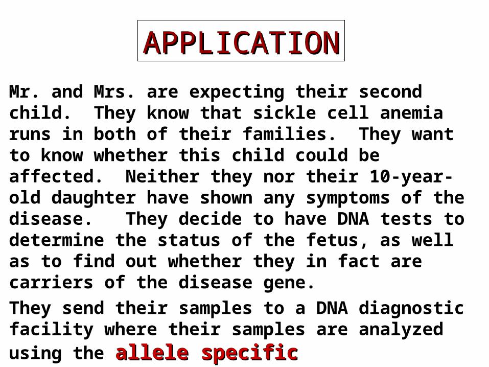
Mr. and Mrs. are expecting their second child. They know that sickle cell anemia runs in both of their families. They want to know whether this child could be affected. Neither they nor their 10-year-old daughter have shown any symptoms of the disease. They decide to have DNA tests to determine the status of the fetus, as well as to find out whether they in fact are carriers of the disease gene.
They send their samples to a DNA diagnostic facility where their samples are analyzed using the allele allele specific oligonucleotide (ASO)specific oligonucleotide (ASO) probe technique.
APPLICATIONAPPLICATION

SICKLE CELL ANEMIA
AT mutation in the -globin gene converts glutamate valine at position 6 of the protein
Normal CT CCT GAG GAG AAG TCT GCGA GGA CTC CTC TTC AGA CG
MutantCT CCT GTG GAG AAG TCT GC GA GGA CAC CTC TTC AGA CG
Allele specific oligonucleotide probe - Allele specific oligonucleotide probe - detection of detection of s-globin mutations-globin mutation

CT CCT GTG GAG AAG TCT GCGA GGA CAC CTC TTC AGA CG
One complementary to the normal DNA sequence
GA GGA CTC CTC TTC AGA CGCT CCT GAG GAG AAG TCT GC
Two probes are designed
The other complementary to the mutant DNA sequence
Normal probe
Mutant probe
Allele specific oligonucleotide probe - Allele specific oligonucleotide probe - detection of detection of s-globin mutations-globin mutation

Normal Probe
Mutant Probe
Homozygous normal AA
Heterozygous carrier AS
Homozygous mutant SS
= PROBE HYBRIDIZES
= PROBE DOES NOT HYBRIDIZE
Allele specific oligonucleotide probe - Allele specific oligonucleotide probe - detection of detection of s-globin mutations-globin mutation

Allele-specific PCRAllele-specific PCR
Normal CTCCTGAGGAGAAGTCTGCNNNNNNNNNN
Mutant CTCCTGTGGAGAAGTCTGCNNNNNNNNNN
Template DNA + dNTPs+MgCl2+Taq Polymerase, primers
Template DNA + dNTPs+MgCl2+Taq Polymerase, primers
If Normal (A) amplification takes placeIf Mutant (T) amplification does not take place
If Normal (A) amplification does not take placeIf Mutant (T) amplification takes place


A 32-year-old female presents to your clinic with concerns over a recently detected right breast lump. A biopsy is performed and reveals an intraductal carcinoma. She is invited to participate in an experimental study that is being carried out to help direct future treatment protocols and define new drug targets for breast cancer. The researcher explains to her that DNA microarrays (DNA chips)DNA microarrays (DNA chips) will be used to study the differences in the gene expression profiles of tumor versus normal cells. After considering all the pros and cons she gives her informed consent and allows her tissue samples to be used.
APPLICATIONAPPLICATION

Microarrays/DNA chips
small, solid supports onto which DNA sequences from thousands of different genes are immobilized
Microarrays/DNA chipsMicroarrays/DNA chips
• Expression analysis
• GenotypingSNP analysisMutation
detection

Expression analysis using Expression analysis using microarraysmicroarrays

Applications of DNA Applications of DNA microarraysmicroarrays
• Cell specific expression
• Gene regulation
• Tumor profiling
• Genetic variation
• Microbial strain identification
• Drug testing

Mr. and Mrs. JD are expecting their first child. Mr. JD’s uncle had died of cystic fibrosis (CF) and they recently learnt that a distant cousin of Mrs. JD has also been diagnosed with CF. They are worried that they might be carriers for the disease. Their doctor suggests an amniocentesis to detect if their unborn child has CF or is a carrier. They feel that an amniocentesis is an invasive and risky procedure and decide that they first want to be tested themselves to see if they are carriers for the disease. If they learn that they both are carriers, they would like to go through with the amniocentesis to see if their child is affected.The most common mutation, accounting for about 75% of CF cases, is called delta F508 and can be screened using the
AFLP (amplified fragment length polymorphism)AFLP (amplified fragment length polymorphism) technique.
APPLICATIONAPPLICATION
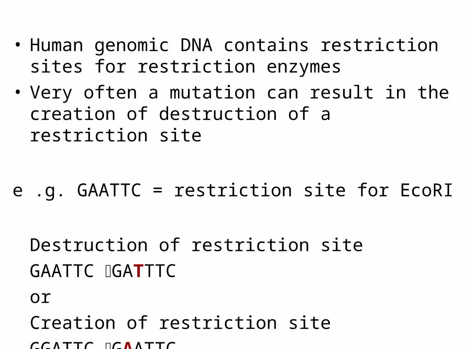
• Human genomic DNA contains restriction sites for restriction enzymes
• Very often a mutation can result in the creation of destruction of a restriction site
e .g. GAATTC = restriction site for EcoRI
Destruction of restriction siteGAATTC GATTTC orCreation of restriction siteGGATTC GAATTC

RFLPS typed using PCRRFLPS typed using PCRMspI
Forward primer
Reverse primer
PCR Amplification
Restriction digestion with MspI(Δ508F lead to loss of MspI site)
Run on agarose gel

Forward primer
Reverse primer
400 bp
300 bp 100 bp

Mr. and Mrs. SZ just had their first child. The phenylketonuria (PKU) blood test performed at birth indicated a high level of phenylalanine in the blood. The physician suggests a follow-up DNA test immediately to confirm the PKU diagnosis. None of the mutations known to cause PKU in the phenyalanine hydroxylase gene is picked up by the standard testing methods. The lab therefore decides to carry out DNA sequencingDNA sequencing of the child’s sample to check for the presence of a novel gene mutation
APPLICATIONAPPLICATION

DNA sequencingDNA sequencing
Characterization of DNA sequence is through dideoxy DNA sequencing (Sanger method)

Automated Automated DNA DNA
sequencingsequencing

A DNA sequenceA DNA sequence

The body of an unidentified young woman is found stuffed in a sack in a forest. She has multiple stab wounds and her face has been mutilated beyond recognition.
The parents of a girl, who had reported their daughter missing a few days ago, are asked to provide blood samples for DNA Profiling to establish if the body may be of their daughter.
The sack is which the girl was found is found to have several hair on it which do not belong to the girl. They are collected as forensic evidence.
APPLICATIONAPPLICATION

DNA Fingerprinting by RFLPs (Restriction Fragment Length Polymorphisms) was developed in the early 1980s by Sir Alec Jeffreys
It made use of genetic variation in the distance between restriction enzyme sites
- Due to the presence of VNTRs (Variable Number of Tandem Repeats)
Power of discrimination was in the range of 106-108 for a six probe analysis
DNA Fingerprinting by RFLPs DNA Fingerprinting by RFLPs (1987-mid 1990s)(1987-mid 1990s)

Variable Number of tandem Variable Number of tandem repeats/ Short tandem repeats repeats/ Short tandem repeats
(STRs)(STRs)
7 repeats
8 repeats
9 repeats
Individual 1
Individual 2
Individual 3

restriction fragments
Person 1 Person 2
1 2
DNA FingerprintingDNA Fingerprinting

Cut with restriction enzymes
Restriction fragments separated using gel electrophoresis
Exposed to radiolabelled probe that binds to its complimentary DNA
fragments
Photographic image obtained
The Steps: DNA FingerprintingThe Steps: DNA Fingerprinting
Transferred to a nylon membrane
DNA prepared (from crime scene samples and suspects)

Two young women were raped and murdered in Narborough, England (1983 and then in 1987)
Police contacted Alec Jefferys for DNA fingerprinting
The Colin Pitchfork CaseThe Colin Pitchfork CaseFIRST EXONERATION AND CONVICTION
BASED ON DNA EVIDENCE
The first suspect (who had confessed) was excluded 5,000 local men were then asked to provide blood/
saliva samples CP convicted in 1988

Human Identity TestingHuman Identity Testing Crime scene investigation -- matching
suspect with evidence Paternity testing -- identifying father Missing persons investigations --whose
body Mass disasters -- putting pieces back
together Inheritance Claims – who gets the money Historical investigations Military DNA “dog tag”

DNA profiling requires a DNA profiling requires a reference samplereference sample
A DNA profile on its own has NO context
DNA profiling works by comparison
Crime scene evidence compared to suspect (forensics)
Child compared to alleged father (paternity)
Victim’s remains compared to a biological relative (mass disaster ID)
Soldier’s remains compared to a reference sample (Armed Forces ID)

Sources of Biological Evidence
Blood Semen Saliva Urine Hair Teeth Bone Tissue

DNA fingerprinting by RFLPs: DNA fingerprinting by RFLPs: the downsidethe downside
RFLP testing requires a relatively large amount of HMW DNA (50-250ng = thousands of cells)
Not ideal for forensic evidence, in which small, degraded samples are common

PCR to PCR to thethe Rescue!! Rescue!!
Polymerase Chain Reaction = Molecular Xeroxing
Series of cycles of three successive steps, carried out in a Thermal Cycler, “amplify” the desired DNA fragment(s)
5 cycles of PCR = 64 copies of DNA
40 cycles of PCR = 1.099 x 10¹² copies of DNA!!
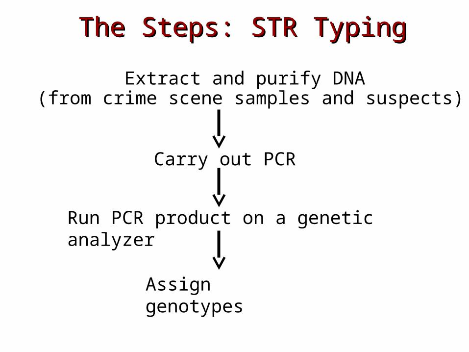
The Steps: STR TypingThe Steps: STR Typing
Extract and purify DNA (from crime scene samples and suspects)
Carry out PCR
Run PCR product on a genetic analyzer
Assign genotypes

Singleplex PCRSingleplex PCRForward primer
Reverse primerDNA ++ dNTPs+MgCl2+Taq Polymerase

DNA + Primers
+ dNTPs+MgCl2+Taq Polymerase
Locus 1
Locus 2
Locus 3
Locus 4
Multiplex PCRMultiplex PCR
Simultaneous amplification of four locations on a DNA template

ABI 310 Genetic Analyzerseparates amplified DNADNA sequencerDNA sequencer

ABI 310 Genetic Analyzer: Capillary Electrophoresis
Amplified STR DNA injected Electric current applied
DNA separated out by size: Large STRs travel
slower Small STRs travel
faster
DNA pulled towards the positive electrode
Color of STR detected and recorded as it passes the detector
DetectorWindow

FATHER
GIRL
MOTHER
PATERNITY TESTINGPATERNITY TESTING
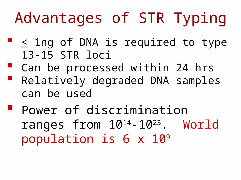
Advantages of STR Typing
< 1ng of DNA is required to type 13-15 STR loci
Can be processed within 24 hrs Relatively degraded DNA samples can be
used
Power of discrimination ranges from 1014-1023. World population is 6 x 10⁹

APPLICATION• A 60 year old heroine addict presents at the
OPD with a history of repeated episodes of flu, fever, malaise, and maculopapular rash. He reveals that has been sharing needles indiscrinately with other addicts.
Laboratory investigations include ELISA for HIV.
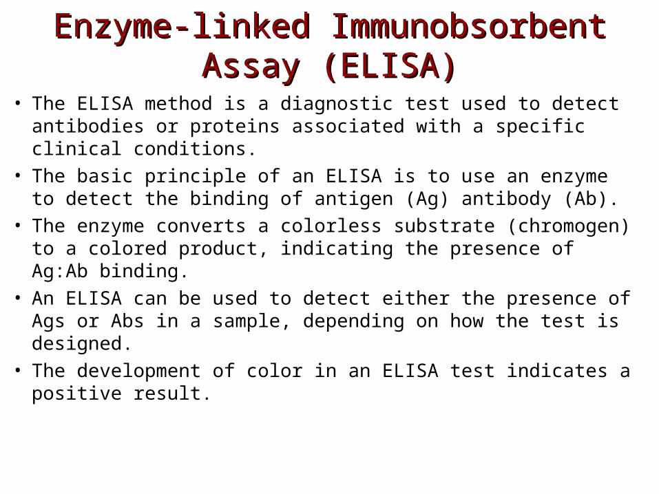
Enzyme-linked Immunobsorbent Enzyme-linked Immunobsorbent Assay (ELISA)Assay (ELISA)
• The ELISA method is a diagnostic test used to detect antibodies or proteins associated with a specific clinical conditions.
• The basic principle of an ELISA is to use an enzyme to detect the binding of antigen (Ag) antibody (Ab).
• The enzyme converts a colorless substrate (chromogen) to a colored product, indicating the presence of Ag:Ab binding.
• An ELISA can be used to detect either the presence of Ags or Abs in a sample, depending on how the test is designed.
• The development of color in an ELISA test indicates a positive result.
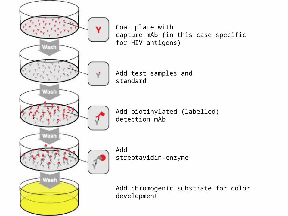
Coat plate with capture mAb (in this case specific for HIV antigens)
Add test samples andstandard
Add biotinylated (labelled) detection mAb
Add streptavidin-enzyme
Add chromogenic substrate for colordevelopment

Indirect ELISAIndirect ELISAEnzyme-linked immunobsorbent assay (ELISA)
Colorless substrate
Colored product


Northern blottingmRNAs (rather than DNA) are isolated, electrophoresed, blotted on a membrane and hybridized using a cDNA labeled probe
Western blottingProteins (rather than DNA or RNA) are isolated,
electrophoresed, blotted on a membrane and hybridized using a labeled antibodies
Analysis of gene expressionAnalysis of gene expression

THE END!