molecular characterization of clinical and environmental strains … · 2010-06-29 · serotype a...
TRANSCRIPT

Journal of Bacteriology and Virology 2010. Vol. 40, No. 2 p.91 – 98 DOI 10.4167/jbv.2010.40.2.91
Molecular Characterization of Clinical and Environmental Strains of Cryptococcus neoformans Isolated from Busan, Korea
Soo Myung Hwang*
Department of Clinical Laboratory Science, Catholic University of Pusan, Busan, Korea
Cryptocococcus neoformans is an encapsulated yeast that can cause life-threatening infections in immunocompromised patients. In this study, the genetic variability and epidemiological relationships of clinical and environmental isolates of
C. neoformans from Busan, Korea, 2000~2005 were investigated. A total of 12 strains of C. neoformans, 7 clinical and
5 environmental isolates were analyzed by random amplified polymorphic DNA (RAPD) using three different primers and PCR-fingerprinting with a minisatellite-specific core sequence of phage M13. All strains belonged to C. neoformans
serotype A and mating type MATa. Two different RAPD profiles (I and II) and a single pattern by M13 PCR-
fingerprinting were identified. The major RAPD profile was pattern I (8 of 12 strains) and pattern II was identified from 2 clinical and 2 environmental strains, which clearly distinguished among isolates. Clinical strains with pattern II were
isolated from the patients with HIV positive. Taken together, molecular patterns provide a good characterization of strains
of C. neoformans as a heterogeneous group and epidemiological relationships in clinical and environmental strains.
Key Words: Cryptocococcus neoformans, Random amplified polymorphic DNA, PCR-fingerprinting
서 론
Cryptococcus neoformans는 협막을 보유한 basidiomycetes
효모양 진균으로 사람과 동물의 폐, 중추신경계 및 전신
에 침범하여 생명을 위협하는 cryptococcosis를 일으킨다
(1).
최근 연구에 의하면 C. neoformans는 다당체 협막성분
의 항원성과 생태학적, 분자생물학적 특성 차이에 의하
여 C. neoformans (혈청형 A, D 및 AD)와 C. gattii (혈청
형 B와 C)의 두 종 (species)으로 재분류되었으며, C.
neoformans는 두 변이종 var. grubii (혈청형 A)와 var.
neoformans (혈청형 D) 그리고 hybrid 형 AD로 나누어진
다 (2, 3). C. neoformans 혈청형 A와 D는 전 세계적으로
널리 분포되어 있으며, 특히 비둘기 배설물을 비롯하여
조류 분변에 오염된 토양, 그리고 썩은 나무 등이 이 균
종의 자연 서식처로 잘 알려져 있다 (4, 5). AIDS 환자를
비롯하여 면역기능에 장애가 있는 환자에서 주로 기회감
염의 원인균이며, 인체감염의 대부분은 C. neoformans var.
grubii (혈청형 A)이다. 이에 비교하여 C. gattii (혈청형 B
와 C)는 열대와 아열대 지역에서만 제한적으로 분리되
며, 자연 서식처는 오스트레일리아 원산 Eucalyptus 나
무로 알려져 있으며 (6~8), 일부 열대 지역을 제외하고
AIDS 환자에서 C. gattii에 의한 감염증은 거의 없는 것으
로 보고되고 있다 (9, 10). Cryptococcus species의 인체감염
은 자연환경에서 basidiospore를 흡입함으로써 감염되는
것으로 알려져 있으나, 생태학적, 역학적 관련성은 여전
히 의문으로 남아있다 (11).
C. neoformans는 heterothallic basidiomycetes로 두 종류
의 mating type MATa와 MATa가 존재한다. 일반적으로
환경이나 임상검체에서 분리되는 C. neoformans mating
type은 MATa가 대부분이며, 드물게 MATa/a인 diploid 균
주도 분리된다 (1, 12).
91
Received: May 10, 2010/ Revised: May 26, 2010 Accepted: May 28, 2010
*Corresponding author: Soo Myung Hwang. Department of ClinicalLaboratory Science, Catholic University of Pusan, Busan 609-757, Korea.Phone: +82-51-0563, Fax: +82-51-510-0568 e-mail: [email protected]
Original Article

92 SM Hwang
전 세계적으로 C. neoformans에 관한 분자역학적인
연구방법으로 M13 primer 및 (GACA)4 반복서열에 의한
PCR fingerprint 분석법 (13, 14), amplified fragment length
polymorphisms (15), rDNA intergenic space regions I과 II 분
석 (16), multilocus gene seqeunce typing (MLST) 등이 (17,
18) 이용되고 있으며, 이들 분석법에 의하여 8가지의 주
요 유전자형이 알려져 있다. 우리나라에서는 Cryptococcus
균종의 분리율이 저조하며, 이 균종에 관한 몇몇 의학적
인 보고만 있을 뿐, 생태적 특성을 비롯하여 역학 연구는
거의 없는 실정이다 (19, 20). 본 연구에서는 2000년에서
2005년 사이에 부산지역 임상검체와 환경에서 분리된
환경균주, 특히 비둘기 분변에서 분리된 C. neoformans
균주를 이용하여, 이들의 혈청형, mating type, random
amplified polymorphic DNA (RAPD)와 M13 PCR fingerprint
법에 의한 분자생물학적 특성을 분석하여 비교함으로써
지역에서 분리된 C. neoformans의 생태학적, 역학적 기초
자료를 얻고자 하였다.
재료 및 방법
균주의 분리 및 동정
부산지역 종합병원 4곳에서 2000년에서 2005년 사이
분리된 임상균주 7주와 2002년 비둘기 분변에서 분리된
환경균주 5주를 포함한 총 12균주를 사용하였다 (Table 1).
임상검체에서 분리된 7주는 각 병원 미생물 검사실에
서 C. neoformans로 동정된 균주로써, 묵즙염색에 의한
협막관찰, API 20C AUX system (bioMerieux, Marcy-l'Etoile,
France)을 이용한 당 동화시험과 Christensen's urea broth
(Difco, Detroit, MI, USA)를 이용한 urease 시험 양성, nitrate
환원시험 음성, 37℃ 성장능 양성인지를 재확인하였고,
bird seed media (BSM)에서 phenol oxidase 생성에 의한 갈
색집락을 확인하여 동정하였다 (19).
환경균주는 Oh 등 (20)에 의하여 2002년 부산시내
비둘기 서식처 12곳의 비둘기 분변으로부터 분리된 C.
neoformans 균주를 사용하였으며, 균주 분리방법을 간단
히 소개하면 다음과 같다. 매우 건조된 비둘기 둥지에서
얻은 약 2.0 g의 비둘기 분변에 10 ml의 멸균식염수를
가하고, 용액이 균질해질 수 있도록 20분간 진탕한 후
1500 ×g에서 5분간 원침하였다. 상층액 100 μl를 균일하
게 BSM에 접종하고, 30℃에서 2~10일간 배양한 후에,
균주의 phenol oxidase 활성의 결과로 나타나는 멜라닌 색
소생성, 즉 갈색집락만을 취하여 Sabouraud dextrose agar
(SDA)로 옮긴 다음 다시 재배양한 후에 임상분리균주와
동일한 방법에 의하여 최종 동정하였다.
혈청형과 변종확인시험
C. neoformans 변종의 확인은 협막성 다당체 항혈청을
이용한 혈청형 분석과 두 가지 생화학적 반응시험을 실
Table 1. Sources, serotypes, and mating types of C. neoformans isolates in Busan, Korea
No. Strain Type of isolates Source Year of isolation Serotype Mating type
1 C21 aC CSF 2000 A a
2 C22 C CSF 2001 A a
3 C28 C Blood 2002 A a
4 C36 C Blood 2003 A a
5 C40 C cCSF 2003 A a
6 C41 C Blood 2005 A a
7 C58 C CSF 2004 A a
8 C23 bE Pigeon excreta 2002 A a
9 C24 E Pigeon excreta 2002 A a
10 C25 E Pigeon excreta 2002 A a
11 C26 E Pigeon excreta 2002 A a
12 C27 E Pigeon excreta 2002 A a a C; clincal source, b E; environmental source, c CSF; cerebrospinal fluid

Molecular Characterization of C.neoformans 93
시하여 결정하였다. 혈청형의 감별은 협막성 다당체 항
원을 확인하는 slide agglutination test (Crypto Check Iatron
RM 304-K kit; Iatron Laboratories, Tokyo, Japan)를 이용하였
다. 분리균주들은 30℃에서 yeast extract malt extract agar
(YMA, Difco)에 배양하였고, 48시간 후에 McFarland scale
pattern 2 (약 6 × 10⁸ CFU/ml)에 맞춰 멸균식염수에 부유
하였다. 각 항혈청 5종 (F1, F5, F6, F7, F8) 한 방울씩을
agglutination glass slide에 먼저 떨어뜨리고, 각 항혈청 당
50 μl의 C. neoformans 부유액을 떨어뜨려 혼합하였다. 혼
합액이 균질화될 수 있도록 2분간 교반하면서 슬라이드
에서 작은 응집이 생기는 것을 직접 관찰하여 다음과 같
이 판정하였다.: 혈청형 A는 F1과 F7에 응집, 혈청형 B
는 F1과 F5에 응집, 혈청형 C는 F1과 F6에 응집, 혈청
형 D는 F1과 F8에 응집 그리고 혈청형 AD는 F1, F7, F8
에 응집을 확인하여 결정하였다.
변종확인 생화학적 검사법으로, 첫 번째 시험은
canavanine-glycine-bromthymol blue (CGB)배지에서 glycine
을 탄소원으로 이용할 수 있는 glycine decarboxylase의 생
성유무에 성장능을 관찰하는 것으로, 최종 동정된 균주
들을 CGB 배지에 접종하고 30℃에서 24~48시간 배양한
다음, CGB 배지의 색깔이 원래 배지의 색깔인 녹황색에서
청색으로 색깔 변화를 보이면 양성으로 판정하였다 (21).
두 번째 시험은 Kwon 등 (22)의 EDTA를 이용한 urease
억제 시험법으로, 최종 동정된 균주들을 yeast extract-
glucose-peptone agar (YEPG)에 100 μM EDTA를 첨가한 배
지인 YEPGE에 접종하고 30℃에서 48시간 배양한 후, 자
란 집락을 멸균 증류수 2 ml에 spectrophotometer (Jasco
Co., Ishiawa-cho, Japan)를 이용하여 흡광도 (A600) 0.8~1.0
이 되도록 맞춘 다음 (균주 수, 약 1 × 10⁸ ~ 2 × 10⁸/
ml), 진탕 혼합한 뒤 혼합액 1 ml를 취해 rapid urea broth
(RUH broth, Difco)를 2배 되도록 분주한 뒤 ice bath에
서 적당 시간 방치하였다. 다시 이 RUH broth를 37℃
shaking water bath에 넣고 매 시간 마다 자홍색 (magenta
red color)이 나타나면 음성, 그리고 황색 (yellow color)이
나타나면 양성으로 판정하였다.
위 두 가지 시험 모두에 양성이면 C. gattii (혈청형 B
또는 C)이며, 모두 음성이면 C. neoformans var. grubii 또
는 var. neoformans (혈청형 A, D 또는 AD hybrid)로 변종
을 확인하였다.
DNA 분리 및 mating typing
균주의 genomic DNA는 Yamamoto (23) 방법을 일부 변
형하여 분리하였다. Brain heart infusion broth (BHI)에서
각 실험균주를 30℃, 48시간 배양한 후 원심분리하여 약
100 μl pellet를 수집하여 10 mM TE buffer (pH 8.0, 1 mM
EDTA)로 두 번 세척하였다. 각 검체에 250 μl 100 mM
TE (pH 9.0, 40 mM EDTA), 50 μl 10% sodium dodecyl sulfate
와 200 μl benzyl chloride를 가하여 혼합한 후 50℃에서
30분 동안 가볍게 진탕하면서 방치한 다음, 10000 rpm에
서 10분간 원심분리한 후 상층액을 분리하였다. 상층액에
3 M sodium acetate 50 μl를 가한 후 0℃에서 10분간 방치
하였다. DNA 침전물은 250 μl isopropanol를 가하여 -70℃
냉동고에서 1시간 방치하여 얻었으며, 침전된 DNA를
70% ethanol로 세척하여 건조한 후 40 μl TE로 용해하여
사용하였다.
Mating type은 Chaturvedi 법 (12)에 따라 C. neoformans
혈청형 A의 특이적 primer, MATa (5'-CTTCACTGCC-
ATCTTCACCA-3'과 5'-GACACAAAGGGTCATGCCA-3'),
MATa (5'-CGCCTTCACTGCTACCTTCT-3'과 5'-AACGC-
AAGAGTAAGTCGGGC-3')를 이용하여 PCR법을 실시
하였다. PCR 반응 혼합액은 AccuPower PCR premix kit
(Bioneer Co. Ltd, Daejeon, Korea)를 사용하여, DNA 시료
(75 ng) 1 μl, primer (10 pmol)를 각각 1 μl 넣고 증류수로
총 20 μl로 최종부피를 맞추었다. PCR 반응조건은 95℃에
서 3분간 초기반응, 94℃ 1분, 57.5℃ 1분, 72℃ 1분의 과
정으로 30회 반복하였고, 마지막으로 72℃에서 7분 연장
으로 PCR을 종결하였다. 증폭된 DNA 시료는 2% agarose
gel을 통하여 전기영동을 실시하였고, 증폭산물의 크기에
따라 mating type, MATa (101 bp)와 MATa (117 bp)를 확인
하였다.
RAPD 및 PCR-fingerprinting
RAPD 분석은 3종의 primer, OPH-02 (5'-TCGGACGTGA
-3'), OPH-12 (5'-ACGCGCATGT-3'), OPH-20 (5'-GGGAG-
ACATC-3')를 사용하여 실시하였다. PCR 반응 혼합액은
AccuPower PCR premix kit를 사용하여, DNA 시료 (75 ng)
1 μl, 한 종류의 primer (10 pmol)를 1 μl 넣고 증류수로
총 20 μl로 최종부피를 맞추었다. PCR 반응조건은 94℃에
서 4분간 초기반응, 92℃ 30초, 34℃ 1분, 72℃ 1분의 과정
으로 35회 반복하였고, 마지막으로 72℃에서 5분 연장으
94 SM Hwang
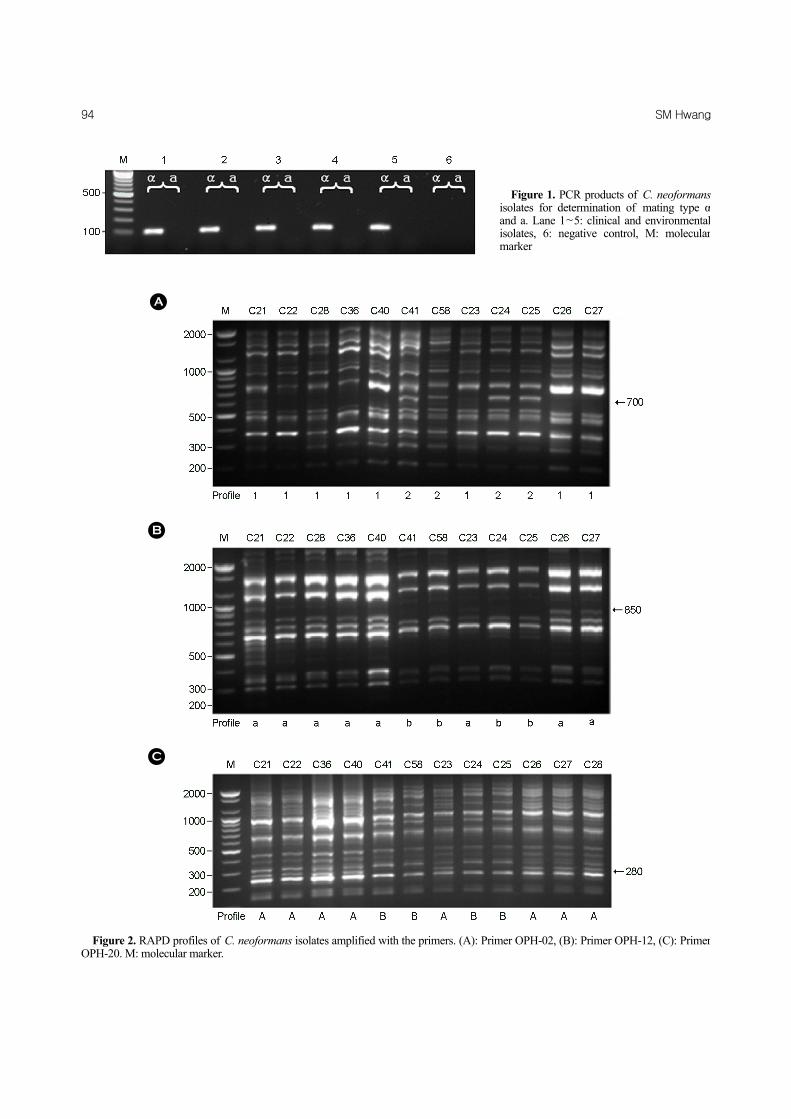
Figure 1. PCR products of C. neoformansisolates for determination of mating type αand a. Lane 1~5: clinical and environmental isolates, 6: negative control, M: molecular marker
A
B
C
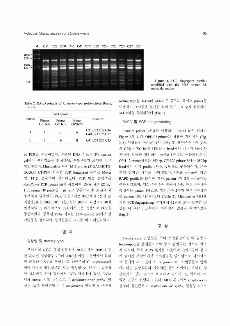
Figure 2. RAPD profiles of C. neoformans isolates amplified with the primers. (A): Primer OPH-02, (B): Primer OPH-12, (C): Primer OPH-20. M: molecular marker.
Molecular Characterization of C.neoformans 95
로 PCR을 종결하였다. 증폭된 DNA 시료는 2% agarose
gel에서 전기영동을 실시하여, 증폭산물의 크기를 비교
확인하였다. Minisatellite 특이 M13 primer (5'-GAGGGTG-
GCGGTTCT-3')를 이용한 PCR fingerprint 분석은 Meyer
법 (14)을 응용하여 실시하였다. PCR 반응 혼합액은
AccuPower PCR premix kit를 사용하여, DNA 시료 (25 ng)
1 μl, primer (10 pmol)를 1 μl 넣고 증류수로 총 20 μl로 최
종부피를 맞추었다. PCR 반응조건은 94℃에서 5분간 초
기반응, 93℃ 20초, 50℃ 1분, 72℃ 20초의 과정으로 40회
반복하였고, 마지막으로 72℃에서 5분 연장으로 PCR을
종결하였다. 증폭된 DNA 시료는 1.5% agarose gel에서 전
기영동을 실시하여, 증폭산물의 크기를 비교 확인하였다.
결 과
혈청형 및 mating type
부산지역 4곳의 종합병원에서 2000년에서 2005년 동
안 분리된 임상균주 7주와 2002년 비둘기 분변에서 분리
된 환경균주 5주를 포함한 총 12균주의 C. neoformans의
협막 다당체 항원성분은 모두 혈청형 A이었으며, 변종확
인 생화학적 검사 결과에서 CGB 배지에서 음성, EDTA
억제 urease 시험 음성으로 C. neoformans var. grubii (혈
청형 A)로 확인되었다. C. neoformans 혈청형 A 균주의
mating type을 MATa와 MATa 두 종류의 특이적 primer를
이용하여 PCR법을 실시한 결과 모두 101 bp가 검출되어
MATa임을 확인하였다 (Fig. 1).
RAPD 및 PCR-fingerprinting
Random primer 3종류를 이용하여 RAPD 분석 결과는
Figure 2와 같다. OPH-02 primer를 이용한 결과에서 (Fig.
2-A) 임상균주 2주 (C41와 C58) 및 환경균주 2주 (C24
와 C25)는 700 bp에 해당하는 band에서 나머지 8균주와
차이가 있음을 확인하여 profile 1과 2로 구분되었으며,
OPH-12 primer에서는 850 bp, OPH-20 primer에서는 280 bp
band에서 각각 profile a와 b, A와 B로 구분되어져, 균주
간의 현저한 차이를 나타내었다. 3종류 primer에 의한
RAPD profile을 분석한 결과, pattern I과 II의 두 종류로
분류되었으며, 임상균주 7주 중에서 5주, 환경균주 5주
중 3주는 pattern I이었고, 임상균주 2주와 환경균주 2주
는 pattern II를 나타내었다 (Table 2). Minisatellite M13에
의한 PCR-fingerprinting 결과에서 12균주 모두 동일한 양
상을 나타내어, 균주간의 차이점이 없음을 확인하였다
(Fig. 3).
고 찰
Cryptococcus 감염증은 주위 자연환경에서 이 균종의
basidiospore를 흡입함으로써 주로 감염되는 것으로 알려
져 있으며, 특히 AIDS 환자를 비롯하여 면역기능이 현저
히 떨어진 사람에게서 기회감염을 일으킴으로 사회적으
로 문제가 되고 있다. C. neoformans와 그 변종들로 인해
야기되는 임상질환과 지역적인 분포 사이에는 유의한 상
관관계가 있는 것으로 보고되고 있으며, 전 세계적으로
많은 연구가 진행되고 있다. AIDS 환자에서 Cryptococcus
감염의 윈인균은 C. neoformans var. grubii, 혈청형 A으로
Table 2. RAPD patterns of C. neoformans isolates from Busan,Korea
RAPD profile
Pattern Primer OPH-02
Primer OPH-12
Primer OPH-20
Strain No.
I 1 a A C21 C22 C28 C36C40 C23 C26 C27
II 2 b B C41 C58 C24 C25
Figure 3. PCR fingerprint profiles amplified with the M13 primer. M: molecular marker.

96 SM Hwang
알려져 있으나, 브라질에서는 C. neoformans var. grubii와
C. gattii 모두 그 원인균으로 보고되었다 (24).
우리나라의 경우에 1986년 Kim 등 (25)이 한국의 임상
에서 분리된 10균주 모두가 C. neoformans 혈청형 A로
보고되었으며, Hwang 등 (26)은 1993년에서 2005년 사이
분리된 임상균주 51주와 환경균주 7주의 혈청형을 분
석한 결과 혈청형 A형이 55주, B형 2주, D형 1주이었
으며, 환경균주는 모두 A형으로 보고하였다. 일본의 경
우에 Ikeda 등 (27)은 임상에서 분리된 62주 모두가 C.
neoformans 혈청형 A임을 보고하였다. 이와 같이 C.
neoformans 혈청형 A는 세계 각지에서 가장 많이 분리되
고 있는 반면에, 혈청형 B의 경우에는 대만, 베트남, 호
주, 브라질 그리고 아프리카 중부 등의 열대 또는 아열
대 지방에서 분리율이 높다고 알려져 있고, 혈청형 D 균
주는 유럽에서 다른 지역에 비해 더 많이 분리되는 것으
로 알려져 있다 (4, 6). 본 연구에서 분리된 12균주는 모
두 C. neoformans var. grubii 혈청형 A로 혈청형 A 균주의
높은 분리율을 확인하였다.
C. neoformans의 mating type은 이 균종의 생태학적, 병
원성 관련 연구에 중요하다. 임상균주나 환경균주에서
MATa type은 MATa type 보다 더 많이 존재하며, mating
에 관계없이 적절한 환경에서 균사를 만들거나 눈으로
볼 수 있는 basidiospore를 형성하는 것으로 알려져 있다.
Kwon-Chung 등 (28)은 MATa type 균주가 MATa type보다
병원성이 더 강한 것으로 보고하였다. Ohkusu 등 (24)은
임상균주 75주의 mating type 분석 결과 73주는 MATa이
었고 2주는 type이 결정되지 않았음을 보고하였다. 본 실
험에서 임상균주와 환경균주 모두 MATa type을 나타냄으
로써 MATa Cryptococcus 균종이 우세함을 재확인하였다.
Cryptococcus 균종의 분자유전학적, 역학적 분석방법
으로 여러 가지 분석법이 이용되고 있는데, 이중에서
RAPD, M13 primer 및 (GACA)4 반복서열에 의한 PCR-
fingerprint법 등이 널리 이용되고 있다. 특정 primer를 이
용한 RAPD fingerprint법은 C. neoformans의 역학적 연구
에 보다 쉽게 응용할 수 있는 방법으로 감염경로 등을
추정하는데 도움이 되고 있다 (29, 30). 본 실험에서 사
용된 Cryptococcus 12주의 M13 primer에 의한 분석 결과
는 모두 동일한 PCR fingerprint pattern를 나타냄으로서 C.
neoformans var. grubii (혈청형 A)의 유전자형의 차이는
없었으나, 3종류의 primer에 의한 RAPD 분석 결과에서는
두 종류의 pattern I과 II을 나타냄으로서, 균주간의 차이
가 있음을 확인하였다. Pattern I을 나타낸 균주는 총 8주
로 임상균주 (5주)와 환경균주 (3주)에서 우세하였으며,
나머지 임상균주 (2주)와 환경균주 (2주)는 pattern II를
나타내었다. 부산지역에 국한되었고 검체균주 수가 적었
지만, 임상균주와 환경균주 사이에 상관성이 있음을 확
인할 수 있었다. 특히 pattern II를 나타낸 임상균주는 모
두 HIV 양성 환자에서 분리된 균주로서 흥미로운 결과
를 얻었다. Yamamoto 등 (23)은 나가사키 지역에서 분리
된 총 21주의 Cryptococcus를 이용하여 RAPD 분석을
한 결과 4종류의 pattern의 특성을 보고한 바 있다.
균종의 유전적 변이는 숙주의 면역반응에 중요한 역할
을 하며, 유전자 기능변화는 병원성에 중요한 영향을 미
치게 된다. 또한 미생물의 진화, 군집의 구조변화를 인
식하는데 변이균주의 유전적 분석은 필수적이라 하겠다.
Cryptococcus 균종은 자연환경인 조류의 분변이나 썩은
나무 등에 서식하면서 인체와 많은 접촉을 할 기회가 많
으며, 노령인구의 증가, 면역기능저하 환자의 증가 등에
인한 기회감염균으로 중요시하고 있는 진균이다. 그러므
로 전 세계적으로 Cryptococcus 균종에 관련하여 다양한
연구가 이루어지고 있는 반면에, 우리나라에서는 이 균
종에 관하여 분자역학적인 연구가 거의 없는 실정이다.
이 분야의 연구가 저조한 이유로서는 여러 가지 요인이
있을 것으로 사려되나, 의진균성 감염에 관심과 임상에서
진균의 분리율이 저조한 까닭이 아닐까 생각한다.
부산지역 임상과 환경검체에서 분리된 Cryptococcus
종은 모두 혈청형 A형인 C. neoformans var. grubii이었으
며, M13 primer에 의한 유전자형도 동일하였다. 그러나
RAPD 분석에 의한 유전자형은 두 pattern으로 구분되었
으며, 임상균주와 환경균주 모두에서 분리됨을 확인하였
다. 비록 지역과 균주 수가 제한적이었지만, 비둘기 분변
에 서식하는 Cryptococcus 환경균주는 환자에서 분리된
균주와 무관하지 않으며, 역학적 상관성이 있는 것으로
추정된다. 그러므로 우리나라 전 지역에서 분리되는 C.
neoformans 변이균주의 생태적 특성, 병원성 및 분자역
학 분야에 지속적인 연구가 활발히 이루어져, 글로벌 균
주와의 상관성을 규명해야 될 것으로 사려된다.
참 고 문 헌
1) Matsumoto MT, Fusco-Almeida AM, Baeza LC, Melhem
Mde S, Medes-Giannini MJ. Genotyping, serotyping and

Molecular Characterization of C.neoformans 97
determination of mating-type of Cryptococcus neoformans
clinical isolates from São Paulo State, Brazil. Rev Inst Med
Trop Sao Paulo 2007;49:41-7.
2) Chen J, Varma A, Diaz MR, Litvintseva AP, Wollenberg KK,
Kwon-Chung KJ. Cryptococcus neoformans strains and
infection in apparently immunocompetent patients, China.
Emerg Infect Dis 2008;14:755-62.
3) Feng X, Yao Z, Ren D, Liao W, Wu J. Genotype and mating
type analysis of Cryptococcus neoformans and Cryptococcus
gattii isolates from China that mainly originated from non-
HIV-infected patients. FEMS Yeast Res 2008;8:930-8.
4) Benett JE, Kwon-Chung KJ, Howard DH. Epidemiologic
differences among serotypes of Cryptococcus neoformans.
Am J Epidermiol 1977;105:582-6.
5) Walter JE, Coffee EG. Distribution and epidemiologic
significance of the serotypes of Cryptococcus neoformans.
Am J Epidemiol 1968;87:167-72.
6) Franzot SP, Hamdan JS, Currie BP, Casadevall A. Molecular
epidemiology of Cryptococcus neoformans in Brazil and the
United States: Evidence for both local genetic differences and
a global clonal population structure. J Clin Microbiol 1997;
35:2243-51.
7) Pfeiffer TJ, Ellis DH. Environmental isolation of Cryptococcus
neoformans var. gattii from Eucalyptus tereticornis. J Med
Vet Mycol 1992;30:407-8.
8) Ellis DH, Pfeiffer TJ. Natural habitat of Cryptococcus
neoformans var. gattii. J Clin Microbiol 1990;28:1642-4.
9) Kuroki M, Phichaichumpon C, Yasuoka A, Chiranairadul P,
Chosa T, Sirinirund P, et al. Environmental isolation of
Cryptococcus neoformans from endemic region of HIV-
associated cryptococcosis in Thailand. Yeast 2004;21:809-12.
10) Ito-Kuwa S, Nagamura K, Aoki S, Ninimiya K, Kato J,
Vidotto V. Serotyping of Cryptococccus neoformans isolated
from AIDS patients. Shigaku (Odontology) 1994;82:360-4.
11) Trilles L, Lazéra Mdos S, Wanke B, Oliveira RV, Barbosa GG,
Nishikawa MM, et al. Regional pattern of the molecular types
of Cryptococcus neoformans and Cryptococcus gattii in Brazil.
Mem Inst Oswaldo Cruz 2008;103:455-62.
12) Chaturvedi S, Rodeghier B, Fan J, McClelland CM, Wickes
BL, Chaturvedi V. Direct PCR of Cryptococcus neoformans
MATalpha and MATa pheromones to determine mating type,
ploidy, and variety: a tool for epidemiological and molecular
pathogenesis studies. J Clin Microbiol 2000;38:2007-9.
13) Jain N, Wickes BL, Keller SM, Fu J, Casadevall A, Jain P, et
al. Molecular epidemiology of clinical Cryptococcus
neoformans strains from India. J Clin Microbiol 2005;43:5733
-42.
14) Meyer W, Marszewska K, Amirmostofian M, Igreja RP,
Hardtke C, Methling K, et al. Molecular typing of global
isolates of Cryptococcus neoformans var. neoformans by
polymerase chain reaction fingerprinting and randomly
amplified polymorphic DNA - a pilot study to standardize
techniques on which to base a detailed epidemiological survey.
Electrophoresis 1999;20:1790-9.
15) Boekhout T, Theelen B, Diaz M, Fell JW, Hop WC, Abeln EC,
et al. Hybrid genotypes in the pathogenic yeast Cryptococcus
neoformans. Microbiology 2001;147:891-907.
16) Diaz MR, Boekhout T, Kiesling T, Fell JW. Comparative
analysis of the intergenic spacer regions and population
structure of the species complex of the pathogenic yeast
Cryptococcus neoformans. FEMS Yeast Res 2005;5:1129-40.
17) Brandt ME, Hutwagner LC, Kuykendall RJ, Pinner RW.
Comparison of multilocus enzyme electrophoresis and random
amplified polymorphic DNA analysis for molecular subtyping
of Cryptococcus neoformans. The Cryplococcal Disease Active
Surveillance Group. J Clin Microbiol 1995;33:1890-5.
18) Litvintseva AP, Thakur R, Vilgalys R, Mitchell TG. Multilocus
sequence typing reveals three genetic subpopulations of
Cryptococcus neoformans var. grubii (serotype A), including
a unique population in Botswana. Genetics 2006;172:2223-38.
19) Chung SM, Lee EY, Lee CK, Eom DW, Yoo B, Moon HB. A
case of cryptococcal tenosynovitis in a patient with Wegener's
granulomatosis. J Kor Rheumatism Ass 2004;11:66-71.
20) Oh KS, Hwang SM. Isolation and characterization of
Cryptococcus neoformans from environmental sources in
Busan. Mycobiology 2005;33:188-93.
21) Kwon-Chung KJ, Polacheck I, Bennett JE. Improved diagnostic
medium for separation of Cryptococcus neoformans var.
neoformans (serotypes A and D) and Cryptococcus neoformans
var. gattii (serotype B and C). J Clin Microbiol 1982;15:
535-7.
22) Kwon-Chung KJ, Wickes BL, Booth JL, Vishniac HS, Bennett
JE. Urease inhibition by EDTA in the two varieties of
Cryptococcus neoformans. Infect Immun 1987;55:1751-4.
23) Yamamoto Y, Kohno S, Koga H, Kakeya H, Tomono K, Kaku
M, et al. Random amplified polymorphic DNA analysis of
clinically and environmentally isolated Cryptococcus
neoformans in Nagasaki. J Clin Microbiol 1995;33:3328-32.

98 SM Hwang
24) Ohkusu M, Tangonan N, Takeo K, Kishida E, Ohkubo M, Aoki
S, et al. Serotype, mating type and ploidy of Cryptococcus
neoformans strains isolated from patients in Brazil. Rev Inst
Med Trop Sao Paulo 2002;44:299-302.
25) Kim SJ, Kim SO, Lee SH, Chong YS, Suk JS. A study on the
mating types and serotypes of clinical isolates of Cryptococcus
neoformans and production of serodiagnostic antigen and
antiserum for cryptococcosis. Korean J Microbiol 1986;21:
127-31.
26) Hwang SM, Oh KS, Lee KW. Serotype and enzymatic profile
of Cryptococcus neoformans isolates from clinical and
environmental sources in Korea. Korean J Microbiol 2006;
42:257-64.
27) Ikeda R, Shinoda T, Fukazawa Y, Kaufman L. Antigenic
characterization of Cryptococcus neoformans serotypes and
its application to serotyping of clinical isolates. J Clin Microbiol
1982;16:22-9.
28) Kwon-Chung, KJ, Bennett JF. Distribution of alpha and alpha
mating types of Cryptococcus neoformans among natural and
clinical isolates. Am J Epidemiol 1978;108:337-40.
29) Meyer W, Castañeda A, Jackson S, Huynh M, Castañeda E.
Molecular typing of IberoAmerican Cryptococcus neoformans
isolates. Emerg Infect Dis 2003;9:189-95.
30) Boekhout, T, van Belkum A. Variability of karyotypes and
RAPD types in genetically related strains of Cryptococcus
neoformans. Curr Genet 1997;32:203-8.