molecular characterization of dengue virus host and
TRANSCRIPT

MOLECULAR CHARACTERIZATION OF DENGUE VIRUS HOST AND RESTRICTION FACTORS IN
AEDES AEGYPTI MOSQUITOES
by
Natapong Jupatanakul
A dissertation submitted to the Johns Hopkins University in conformity with the requirements for the degree of Doctor of Philosophy
Baltimore, Maryland
February 2016
©2016 Natapong Jupatanakul All Rights Reserved

ii
ABSTRACT
Despite decades of attempts at disease control, dengue remains one of the most
significant mosquito-borne arboviral diseases, causing an estimated 390 million
infections annually. While studies of molecular interactions between DENV and Ae.
aegypti have paved a way for the development of alternative DENV control strategies,
this field is still relatively understudied. Here, we used multiple molecular tools to study
interactions between the virus and Ae. aegypti, as well as to identify DENV host and
restriction factors. First, we have developed genetically modified mosquitoes with
increased activity of the JAK/STAT pathway, and showed that these transgenic
mosquitoes could inhibit DENV infection. Through microarray-based transcriptomic
comparisons, we identified candidate DENV host and restriction factors and confirmed
their function through RNAi. Second, we compared transcriptomic profiles of a panel of
field-derived and laboratory Ae. aegypti strains with different DENV susceptibility.
Through RNAi-mediated gene silencing, we have shown that basal level of immune
activity, and expression level of host factors are important determinants for DENV
susceptibility. Lastly, through a study of transcriptomic datasets comparing DENV-
infected and uninfected Ae. aegypti, we identified and characterized lipid binding protein
families, ML and NPC1, as host factors for DENV replication in Ae. aegypti.

iii
ACKNOWLEDGMENTS
Ph.D. study is a long learning process and it would not be possible to complete
this without the help of following people. I would like to express my deepest gratitude to
all people who have assisted me throughout my Ph.D.
First, and most grateful to Dr. George Dimopoulos who inspired me to come to
this school and for the opportunities to work on interesting research projects. I would like
to thank for all his expert advices, support and discussions. Also my sincere gratitude
towards my thesis advisory committee members: Dr. Douglas Norris, Dr. Anna Durbin,
Dr. Christopher Potter, Dr. Marcelo Jacobs-Lorena, and Dr. Daniela Drummond-Barbosa
for their time and invaluable suggestions that have been a great help through out my
Ph.D. study.
I would also like to extend my gratitude to all past and present members of the
Dimopoulos lab: Shuzhen, Jose, Yessy, Yuemei, Simone, Jayme,. Ana, Octavio, for all
the support, discussions, and for their help during these years. Especially for Jose and
Shuzhen whom I started training with and always been my great help and supports. I
cannot thank you guys enough.
I would like to thank Christopher Kizito from the JHMRI insect core facility as
well as Anne, and Amanda from JHAPH microarray core facility for their help
throughout the research projects.
I would like to extend my gratitude to the Royal Thai Government Scholarship,
whose provide me financial support and an opportunity to do my graduate research
overseas.

iv
I would like to thank all my friend I have earned during this Ph.D., both inside
and outside of the Johns Hopkins university, especially the Thai community in Baltimore
for all the support, and enjoyable moments in Baltimore.
Last but not least, to my family, especially my parents, for their love and support
since the beginning of my life. Although they don’t want me to be far away from home,
but they have always still support my decisions.
Thesis Advisor:
Dr. George Dimopoulos
Thesis advisory committee:
Dr. Douglas Norris
Dr. Anna Durbin
Dr. Christopher Potter
Alternates:
Dr. Marcelo Jacobs-Lorena
Dr. Daniela Drummond-Barbosa

v
Table of Contents
Abstract................................................................................................................. ii
Acknowledgments................................................................................................iii
Table of Contents.................................................................................................. v
List of Figures.………………………………………………………………….. x
List of Tables……………………………………………….…………………..xii
CHAPTER 1
Introduction……………………………………………….. 1
Global burden of dengue………………………………………………..............2
Dengue replication and tissue tropism in Ae. aegypti…………………………4
Mosquito immune responses to DENV infection…………………………....... 5
The Toll pathway………………………………………………………… 7
The JAK/STAT pathway………………………………………………… 9
The IMD pathway………………………………...……………………. 10
RNA interference………………………………………………………..11
Arbovirus interactions with host cell processes and host factors………….. 13
The Vacuolar ATPase Complex………………………...……………… 14
The Myeloid Differentiation 2-Related and Niemann-Pick Type C1
Proteins………………………………………………………….……....15
Study Objectives………………………………………………………...15
vi
CHAPTER 2
Engineered Aedes aegypti JAK/STAT pathway-mediated immunity to
dengue virus…………………………………………….……..…...17
ABSTRACT……………………………………………………………………17
INTRODUCTION……………………………………………………………..18
MATERIALS AND METHODS
Generation of transformation vector constructs……………….…………...20
Generation of transgenic Ae. aegypti………………………………………20
Cell culture and DENV strains……………………………………….........21
Oral DENV infections in Ae. aegypti and virus titration ………………….21
Genome-wide oligonucleotide microarray transcriptomic analyses……….22
RNA interference (RNAi)-mediated gene silencing …………………........22
Mosquito fitness assays………………………………………………........23
Bacterial challenge……………………………………………………........23
RESULTS
Generation of JAK/STAT pathway transgenic Ae. aegypti………………..24
Transgenic activation of the JAK/STAT pathway inhibits virus replication
throughout the mosquito’s body………………………………………..…..29
Fitness impact of transgenic Dome and Hop mediated JAK/STAT pathway
activation………………………………………………………………….. 32
Immune-related transcripts are enriched upon JAK/STAT activation……. 34
Transcript abundances of potential DENV host factors were depleted upon
JAK/STAT activation……………………………………………………... 40

vii
Functional analysis of JAK/STAT pathway-regulated putative DENV restriction
factors and DENV host factors using RNA interference………………….. 42
DISCUSSION…………………………………………………………………..46
CHAPTER 3
Identification of putative host factors and restriction factors that
contributes to refractoriness to DENV infection among laboratory and
field-derived Aedes aegypti mosquitoes……………………… 50
ABSTRACT……………………………………………………………….……51
INTRODUCTION……………………………………………………………...52
MATERIALS AND METHODS
Mosquito rearing and cell culture conditions………………………….......52
Oral DENV infections in Ae. aegypti…………….…………………..........53
Gene silencing assays……………..…………………………………….....53
DENV titration by plaque assay………...………………………………....53
RESULTS
Laboratory and field-derived Ae. aegypti strains have different degrees of DENV2
susceptibility……………………………………………………................ 55
Mosquito immune signaling pathways and the RNAi pathway control DENV2
infection to different degrees in various Ae. aegypti strains……................57
Transcriptomic comparison between refractory and susceptible Ae. aegypti reveals
candidate DENV restriction and host factors……..……..…….................. 60

viii
Functional characterization of selected candidate DENV host and restriction
factors…………………………………………………………………….. 61
vATP synthase subunits are important DENV host factors in Ae. aegypti..65
DISCUSSION………………………………………………………………….. 67
CHAPTER 4
Molecular characterization of Aedes aegypti ML and Niemann-Pick
type C family members as dengue virus host factors……………71
ABSTRACT……………………………………………………………………72
INTRODUCTION…………………………………………………………….. 73
MATERIALS AND METHODS
Bioinformatics analyses and genes selection………………………………75
Mosquito strains and mosquito maintenance………………………………75
Cell culture…………………………………………………………………75
Genes silencing by RNA interference…………………………………...…76
DENV propagation and viral infection in the mosquito…….……...………76
DENV titration by plaque assay…………………………….………..….…77
Gene expression and silencing efficiency analysis by quantitative PCR…..77
Statistical analysis of midgut DENV titer and gene expression level……...78
RESULTS
ML and NPC1 gene families are distinct and expanded in Ae. aegypti……78
Ae. aegypti ML and NPC1 family members facilitate DENV infection…...81

ix
Functions of AaegNPC1b and AaegML33 as DENV host factors are conserved in
field-derived strain of Ae. aegypti………………………..………………... 82
Ae. aegypti NPC1 and ML genes may influence DENV infection through the same
mechanism or pathway………………………………………..……………83
AaegNPC1b and AaegML33 may regulate Ae. aegypti immune pathways..86
Expression patterns and tissue tropisms of AaegNPC1b and ML33……….89
DISCUSSION………………………………………………………………….. 92
CHAPTER 5
Conclusions and General Discussion……………………………. ..…...99
REFERENCES……………………………………………….……………….104
APPENDICES………………………………………………………………...129
CURRICULUM VITAE……………………………………………………...156

x
List of Figures
Figure 1.1 Global distribution of dengue…………………………………………….. 2
Figure 1.2 Temporal tissue tropism of dengue virus type 2 in the Ae. aegypti
mosquito………………………………………………………………………………. 5
Figure 1.3 Mosquito immune signaling and RNAi pathways………………………... 7
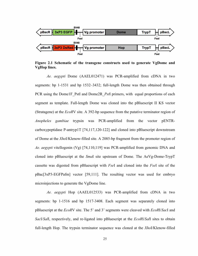
Figure 2.1 Schematic of the transgene constructs used to generate VgDome and VgHop
lines……………………………………………………………………...................... 25
Figure 2.2 Fluorescence screening of VgDome, VgHop, and hybrid VgDomexVgHop
transgenic lines.……………………………………………………………………… 27
Figure 2.3 PCR confirmation of the transgenic Ae. aegypti VgDome and
VgHop lines…………………………………………………………………………. 27
Figure 2.4 Transcript abundance of transgenes and effector genes in the fat body of
VgDome and VgHop lines from before blood feeding (0 hr) up to 48 hpbm..………29
Figure 2.5 Effect of JAK/STAT pathway activation on DENV infection in transgenic Ae.
aegypti……………………………………………………………….…………......... 31
Figure 2.6 Effect of transgenes introduction and expression on mosquito longevity. 33
Figure 2.7 Fecundity of the WT and transgenic Ae. Aegypti………………….......... 33
Figure 2.8 Expression of vitellogenin in the transgenic lines as compared to WT…. 34
Figure 2.9 Transcriptomic profiles of the VgDome and VgHop mosquitoes………. 36
Figure 2.10 Mortality of the VgDome and VgHop lines from bacterial infection…..40
Figure 2.11 Phylogenetic tree of orthologs of AAEL007703 gene obtained from
Vectorbase.…………………………………………………………………………... 44
Figure 2.12 Silencing efficiencies for candidate RFs and HFs.……………….......... 44

xi
Figure 2.13 Effect of host and restriction factor silencing on DENV susceptibility...46
Figure 3.1 Susceptibilities of Ae. aegypti strains to DENV2 infection……………...56
Figure 3.2 Contributions of the Toll, Imd, JAK-STAT, and RNAi pathways to the
control of DENV2 in refractory and susceptible mosquito strains………………….. 59
Figure 3.3 Identification of novel candidate DENV host and restriction factors through
hierarchical clustering…………………...................................................................... 60
Figure 3.4 Effect of candidate DENV host factor knockdown on midgut DENV2 titers in
susceptible Ae. aegypti strains.…………………....………………….........................63
Figure 3.5 Effect of candidate DENV host factor selected from high-througput screen
knockdown on midgut DENV2 titers in susceptible Ae. aegypti strains.…………… 64
Figure 3.6 Effect of vATPase subunits knockdown on midgut DENV2 titers in
susceptible Ae. aegypti strains………………………………………………………..66
Figure 4.1 Phylogenetic tree of the ML and NPC1 gene family in insects…………. 80
Figure 4.2 Effect of Ae. aegypti ML and NPC1 knockdown on midgut DENV…….82
Figure 4.3 AaegNPC1b and AaegML33 silencing resulted in lower midgut DENV titers
in field-derived mosquitoes from Saint Kitts………………………………….. 84
Figure 4.4 Silencing efficiency for AaegNPC1b and ML33 in Rock and Kitts strains Ae.
aegypti……………………………………………………………………………….. 85
Figure 4.5 Ae. aegypti NPC1b and ML33 may influence DENV infection through the
same mechanisms……………………………………………………………………. 85
Figure 4.6 Immune-related gene expression changes after AaegNPC1b and AaegML33
silencing………………………………………………………………………………88

xii
Figure 4.7 Transcript abundance of AaegNPC1b and AaegML33 in the midgut, and fat
body in uninfected mosquitos……………………………………………………….. 89
Figure 4.8 AaegNPC1b and AaegML33 gene expression in the midgut, and fat body over
the time course of DENV infection…………………………………………………. 91
List of tables
Table 2.1 List of top ten enriched and depleted transcripts shared between VgDome and
VgHop compared to WT ……………………………………………………………. 37
Table 2.2 List of candidate RFs and HFs for functional confirmation by RNAi…… 43
Table 3.1 Origins and name abbreviations of laboratory and field-derived Ae. aegypti
strains………………………………………………………………………………... 56
Table 3.2 Candidate DENV host factors selected for functional characterization via
RNAi-mediated gene knockdown…………………………………………………… 61
Table 4.1 Expression pattern of the selected immune genes from previous microarray
datasets………………………………………………………………………………. 87
1
CHAPTER 1 Introduction
Parts of this Chapter have been published in:
Jupatanakul N, Sim S, Dimopoulos G. The insect microbiome modulates vector competence for arboviruses. Viruses. 2014;6: 4294–4313. doi:10.3390/v6114294
Sim S, Jupatanakul N, Dimopoulos G. Mosquito immunity against arboviruses. Viruses. 2014;6: 4479–4504. doi:10.3390/v6114479
Dennison NJ, Jupatanakul N, Dimopoulos G. The mosquito microbiota influences vector competence for human pathogens. Current Opinion in Insect Science. 2014;3: 6–13. doi:10.1016/j.cois.2014.07.004
Jupatanakul N, and Dimopoulos G. 2016. Chapter 8. “Molecular Interactions Between Arboviruses and Insect Vectors: Insects' Immune Responses to Virus Infection” In: Arboviruses: Molecular Biology, Evolution and Control, Caister Academic Press, D. Gubler and N. Vasilakis (eds), in press. ISBN 978-1-910190-21-0

2
Global burden of dengue
Dengue is the most important arthropod-borne viral disease, with an estimated
390 million infections annually across over a hundred countries in tropical and
subtropical areas (Figure 1.1) [1]. The disease has become major global public health
concerns, with increasing incidence in recent decades as a result of the geographical
expansion of its primary vector, Aedes aegypti, and secondary vector, Aedes albopictus,
as well as global transport, unplanned urbanization, and climate change [2-10].
Figure 1.1. Global distribution of dengue. Cartogram of the annual number of infections for all ages. Figure was obtained from [1]. Data were from the year 2010.
Dengue is caused by any of the four serologically distinct dengue viruses (DENV
serotype 1 to 4) [11]. The genomes of the four serotypes of DENV share only 65%
similarity at the nucleotide level, yet they have similar life cycles and clinical
manifestations in humans [9,10,12]. The infection by one serotype of DENV is thought to
yield life-long antibody protection against symptomatic disease with that serotype;
however, these neutralizing antibodies do not provide life-long protection from infections
by other serotypes [11,13]. Conversely, epidemiological studies have demonstrated that

3
the highest risk for developing severe dengue is previous infection with a different
dengue serotype, thought to be due to the antibody-dependent enhancement (ADE)
[14,15]. This problem made vaccine development for DENV challenging as a balanced
antibody response to all four serotypes is required for protection and to reduce the risk of
severe disease due to ADE. Although these pathogens can cause serious diseases in
humans, they rarely cause mosquito pathology and can persistently infect the mosquito
vector for life [16].
Due to difficulties in developing dengue vaccines, only Dengvaxia (developed by
Sanofi Pasteur), has been approved for use in three countries: Mexico, the Philippines,
and Brazil. However, this vaccine has not been approved for use in children under 9 years
of age, a group that is most vulnerable to severe disease [17]. Clinical studies have
shown that efficacies of Dengvaxia was estimated at 65.6% for participants who were 9
years of age or older, and only 44.6% in participants under the age of 9 years [18].
Because Dengvaxia was approved for use in limited group of population, the mosquito
vector control remains an essential strategy to reduce disease burden in the general
population. However, conventional vector control methods such as insecticide spraying
and the removal of mosquito breeding sites have in many cases proven to be
unsustainable solutions for a variety of reasons, including lack of adequate funds to
sustain the vector control program, ecological concerns, as well as the development of
insecticide resistance [11,19]. In addition, vectors such as Ae. aegypti are extremely well
adapted to urban environments, laying their eggs in clean water in artificial containers,
and displaying a preference for staying indoors. For this reason, the development of novel

4
vector and disease control strategies is essential, and a molecular understanding of
mosquito immune responses against these viruses is necessary.
DENV replication and tissue tropism in Ae. aegypti
Human and non-human primates are hosts for DENV; however, the virus does not
require an enzootic cycle (replication in non-human hosts) to sustain epidemic
transmission in humans [9]. DENV are maintained in a human population through
horizontal transmission cycle between Ae. aegypti mosquitoes and humans. Vertical
transmission of DENV from infected female mosquitoes to their offspring has also been
reported in the laboratory and in the field with efficiencies around 1-4% [20], which is
not an important factor for long-term virus persistence in an endemic situation according
to mathematical model [21].
After the mosquito ingests an infectious blood meal, the DENV must pass through
various infection barriers [2]. First, they have to infect and replicate in the midgut
epithelium (midgut infection barrier), then escape from the midgut to spread throughout
the insect body and infect other tissues (midgut escape barrier). In order to transmit
dengue, the viruses then have to infect and replicate in the salivary glands, where they
disseminate into mosquito saliva (salivary gland infection and escape barriers) [2]. The
extrinsic incubation period (EIP), i.e., the time from virus ingestion until its
dissemination in mosquito saliva where it can be transmitted to naïve humans, can vary
depending on conditions such as mosquito strain, virus strain, and temperature but it
generally ranges from 10-14 days [16]. Virus levels in the salivary glands will remain
high throughout the infection which means that once the mosquito salivary glands get
infected by the virus, it can be transmitted for life of the mosquito [16].

5
Figure 1.2. Temporal tissue tropism of dengue virus type 2 in the Ae. aegypti mosquito. The infection rate in the respective tissues is shown in grey scale [16].
Mosquito immune responses to DENV infection
Mosquitoes, like other organisms, are exposed to a wide range of microbes from
their environments, and also during blood feeding. Knowledge of the mosquito immune
responses has been largely based on research in the insect model organism, Drosophila
melanogaster, which in contrast to vertebrate immunity, do not have adaptive immunity
and rely mainly on their innate immune system. The insect innate immune system is
comprised of cellular and humoral components [22]. Mechanisms involved in cellular
immune responses include phagocytosis, encapsulation, and nodule formation, and they
are mediated by hemocytes [23-26]. Humoral immune responses are mechanisms to
prevent systemic infection, which include systemic immune signaling, melanization, and
the production of anti-microbial peptides (AMPs). Innate immune signaling is triggered
by specific pathogen recognition receptors (PRRs) that recognize conserved molecular

6
patterns among microbes, termed Pathogen-associated molecular patterns (PAMPs).
These include lipopolysaccharides, peptidoglycans, mannans, and dsRNA [22,27]. Upon
pathogen recognition, PRRs activate different signaling cascades, which regulate the
transcription of effector molecules [28,29].
The mosquito’s innate immune system mounts potent immune responses against
microbial challenge and is capable of distinguishing among broad classes of
microorganisms. The availability of the Ae. aegypti genome in 2007 [30,31] has
facilitated the study of mosquito immunity in response to DENV infection. In this
section, we focus on the major mosquito immune signaling pathways that have been
implicated in the antiviral defense, namely the Toll, immune deficiency (IMD), and Janus
kinase/signal transducers and activators of transcription (JAK-STAT) pathways. In
addition, we will consider the RNA interference (RNAi) pathway; though not a classical
innate immune pathway, it also plays a key role in antiviral defense. A summary of these
immune pathways is presented in Figure 1.3.

7
Figure 1.3 Mosquito immune signaling and RNAi pathways [32]. Mosquitoes use classical innate immune pathways such as the Toll, Imd, and JAK/STAT pathway to deal with pathogen infections. The RNAi pathway, eventhough not a classical immune pathway, is also important for controlling viral infections.
The Toll pathway
The Toll pathway is an NF-kB signaling pathway, which was first characterized
for its role in Drosophila development and subsequently shown to play a role in insect
immune responses against Gram-positive bacteria, fungi, and virus [25,33-37]. Unlike the
vertebrate TLR, the insect Toll pathway does not directly interact with PAMPs. Instead,
the recognition of pathogens by peptidoglycan recognition proteins (PGRPs), such as
peptidoglycan recognition proteins (PGRP)-SA and -SD, triggers a proteolytic cascade
that cleaves a cytokine Spätzle (Spz) [35]. Activated Spz bound to the Toll receptor and
8
triggers signaling through the associated adaptor proteins MyD88 and Tube and the
kinase Pelle [35]. This leads to the degradation of the negative regulator, Cactus, which
binds the NF-kB transcription factor Dorsal (Rel1 in mosquitoes). After being released
from Cactus, Dorsal is translocated to the nucleus and binds to cis-acting elements of the
promoters of antimicrobial peptides and other immune effector genes [35,38,39].
The Toll pathway is conserved in mosquitoes and also plays a key role in antiviral
defense in these insects. DENV infection of the Ae. aegypti midgut, carcass, and salivary
gland activates the transcription of Toll pathway components and putative effectors such
as Späetzle, Toll, Rel1A, and multiple AMPs [12,40,41]. The activation of the Toll
pathway through the RNAi-mediated gene silencing of Cactus resulted in a reduction of
midgut DENV titers, while inactivation of Toll pathway signaling by silencing the
adaptor protein MyD88 resulted in higher midgut DENV titers [12,42]. The DENV-
infected mosquito transcriptome and that of Cactus-silenced (or Rel1-activated)
mosquitoes also overlap considerably in terms of the magnitude and direction of gene
regulation [12,43]. Subsequent experiments revealed that the role of the Toll pathway in
controlling DENV was conserved in field-derived Ae. aegypti, and against different
DENV serotypes [44,45].
Stable transinfection of Ae. aegypti with the endosymbiont bacterium Wolbachia
greatly limits infection of the mosquito vector with a range of human pathogens,
including DENV and Chikungunya virus (CHIKV) [46-51]. The inhibition occur via
several mechanisms [30,52,53], one of which is the induction by Wolbachia of reactive
oxygen species (ROS) production by the mosquito, resulting in Toll pathway activation

9
and the subsequent production of the AMPs cecropin and defensin, which hinder DENV
replication [42,44,54-56].
The JAK/STAT pathway
The Janus kinase-signal transduction and activation of transcription (JAK/STAT)
pathway was discovered in a vertebrate model as an interferon (IFN)-induced signaling
pathway important for development [46,48,57], and was later found to be important for
anti-viral immunity [52,53,58]. In Drosophila, the JAK/STAT pathway plays a crucial
role as a signaling pathway in insect development and in the immune response against
pathogenic bacteria and viruses [42,54-56].
The canonical Drosophila JAK-STAT pathway is triggered by the binding of the
activated cytokine-like Unpaired ligand (Upd) to the extracellular domain of the
Domeless receptor (Dome) [42,57]. The binding of Upd to the receptor triggers a
conformational change and dimerization of the Dome receptor [43,58]. This dimerization
then triggers the Janus kinase Hopscotch (Hop) to phosphorylate the cytosolic tail of the
Dome receptor, which in turn activates STAT [55,59]. The activated STAT is dimerized
and translocated to the nucleus and triggers the transcription of JAK/STAT pathway-
regulated genes [42,59]. The JAK/STAT pathway is negatively regulated by the protein
inhibitors of activated STAT (PIAS), and suppressors of cytokine signalling (SOCS)
repressor proteins to prevent its over-activation [43,60-62].
The antiviral role of the JAK-STAT pathway is conserved in the Ae. aegypti
defense against DENV. DENV replication in the mosquito midgut is significantly
increased when the pathway is transiently suppressed by RNAi-mediated depletion of the
receptor Dome or the JAK ortholog Hop, and the opposite effect on virus replication is

10
observed when the pathway is activated by silencing of protein inhibitor of activated
STAT (PIAS), a negative regulator [59,63]. However, JAK-STAT pathway-activated
anti-DENV mechanisms are poorly understood. Two DENV-induced, JAK-STAT-
regulated putative effector genes that restrict DENV replication in midgut tissues have
been identified but remain uncharacterized [59,60,64,65]. These genes were named as
dengue virus restriction factors (DVRFs) 1 and 2. DVRF1 is a predicted transmembrane
protein, which potentially function as a pathway receptor. DVRF2 contains antifreeze and
allergen domains and might have a function in virus recognition.
The IMD Pathway
The immune deficiency (IMD) pathway is well known to play crucial roles in
insect defense against bacteria [60-62,66,67]. In Drosophila, activation of the IMD
pathway, like that of the Toll pathway, is initiated by PRR-mediated recognition of
microbial PAMPs (reviewed in [61-63]). Intracellular signaling through the adaptor IMD
protein and various caspase-like proteins and kinases then leads to a functional split in the
pathway into two downstream branches [60,64,65,68,69]. One branch, similar to the
mammalian c-Jun/JNK pathway, activates the transcription factor AP-1 via JNK
signaling [66,67,70], while the other branch culminates in the processing and activation
of the NF-kB transcription factor Relish (Rel2 in mosquitoes) via caspase-mediated
cleavage of its carboxy-terminal end [61,62,71-74]. Activated Relish is then translocated
to the nucleus to promote the transcription of anti-microbial effectors [68,69,75,76]. The
human Fas-associated factor 1 ortholog Caspar negatively regulates Relish activation,
possibly by interfering with the enzymes involved in its cleavage [12,70]. In mosquitoes,

11
the IMD pathway also plays important roles in the antibacterial defense, and it also
directs immune responses against Plasmodium parasites [71-74,77].
The antiviral role of the IMD pathway has more recently been investigated, and in
flies it has been found to be active against SINV and cricket paralysis virus (CrPV)
[75,76,78]. In mosquitoes, up-regulation of IMD components and effectors in response to
DENV and SINV infection has been observed [24,34], but transient activation of the
pathway by RNAi-mediated gene silencing of Caspar has no effect on midgut DENV
titers [12,79,80].
RNA interference
The RNAi antiviral mechanism is not a classical pathogen-stimulated immune
response, but plays an important role in insects’ antiviral responses. RNAi is a
mechanism that can target foreign RNA for degradation, and it has long been recognized
to be a key player among the mechanisms of anti-viral immunity in insects. This process
relies on the Dicer2 (Dcr2) enzyme, which contains the DExD/H-Box RNA helicase
domain and acts as a pattern recognition receptor in RNAi’s recognition of exogenous
dsRNAs [77,81]. Once they are recognized, Dcr2 cleaves long exogenous dsRNAs to
generate 21–22 basepair small-interfering RNAs (siRNAs). The siRNAs, together with
Dcr2, can be loaded onto the RNA-induced silencing complex (RISC). During the
effector stage of the pathway, RISC unwinds the siRNAs, degrades one of the RNA
strands, and then guides it to the complementary RNA. Argonaut 2 (Ago2), a protein in
the RISC complex that contains endonuclease activity, then degrades the target RNA
strand [78,82,83]. RNAi was previously characterized as an antiviral mechanism, but
recent studies have shown that it can also function as a PRR for immune signaling

12
pathways. In the Drosophila system, in addition to degrading target RNA, RNAi can also
induce the expression of antiviral effectors, in a manner similar to RIG-I in mammals
[79,80,84]; for example, recognition of DCV by the DExD/H-Box RNA helicase domain
of Dcr2 can induce the expression of the anti-viral effector Vago [77,81,84].
Recent studies have demonstrated that the RNAi pathway also serves as an anti-
DENV mechanism in Ae. aegypti. The very first evidence of a role for RNAi in
modulating DENV infection was obtained with the transformation of the plasmid
expressing inverted repeat DENV RNA (irRNA) in mosquito cells [82,83,85]. Later,
transgenic mosquitoes expressing inducible irRNA were also used to confirm the
importance of RNAi [84,85]. These transgenic mosquitoes had lower DENV titers when
compared with wild-type mosquitoes, suggesting a role for siRNA in anti-DENV
responses. Knockdown of Ago2 in the transgenic mosquitoes negated the protective
effect of the irRNA, confirming the importance of the RNAi mechanism [84,85].
However, the role of the RNAi mechanism in the anti-DENV defense in wildtype
mosquitoes was not confirmed until 2009 [79,85]. DENV infection in mosquito cell lines
and adult female mosquitoes resulted in the production of siRNAs that could inhibit virus
replication [81,85]. On the other hand, transient silencing of the RNAi pathway
components (Dcr2, R2D2, and Ago2) resulted in an increase in DENV titres and a
reduction in the DENV extrinsic incubation period in mosquitoes [82,85].
Characterization of the role of RNAi in the systemic immune response is also
important for our understanding of how the mosquito systemically controls virus
infection. Previous studies of Drosophila C virus (DCV) in Drosophila have found a
systemic spread of RNAi through the uptake of dsRNA from the cellular environment
13
[79,86,87]. However, a similar mechanism has not yet been identified in the mosquito’s
anti-DENV response. It is complicated to confirm this phenomenon in mosquitoes
because systemic RNAi was originally discovered in a Drosophila mutant that is
deficient in the dsRNA uptake pathway, and no such mutant is available in the mosquito
system.
Another study from the Drosophila model suggests a role for insect-encoded
reverse transcriptase (RT) enzymes and the RNAi machinery in maintaining the
persistence of RNA viruses. Here, viral genome fragments are reverse-transcribed and
inserted into the insect genome by retrotransposon elements; these insertions later serve
as templates for RNAi responses against the virus [81,88-90]. Given that the Ae. aegypti
genome also contains RTs and transposable elements [82,91], and that flavivirus and
rhabdovirus sequence fragments have been detected in the genomes of Aedes species
[86,87,92], it would be intriguing to study this phenomenon in mosquitoes.
Arbovirus interactions with host cell processes and host factors
Arboviruses are obligate intracellular pathogens that exploit the host’s cellular
machinery in order to replicate. The intracellular replication cycle for DENV has been
well studied and is likely to be similar in insects and vertebrates. DENV enters cells via
clathrin-dependent receptor-mediated endocytosis, and uncoating of the positive-strand
RNA viral genome requires trafficking through an acidic endosomal compartment [13-
15,88-90]. The receptors and proteins of the mosquito midgut that interact with the virus
during early infection stages (reviewed in [91,93,94]) are poorly characterized.
Translation of viral RNA (vRNA) occurs on endoplasmic reticulum (ER)-derived

14
membranes, producing a single polypeptide that is then processed into individual
structural and non-structural proteins. vRNA replication occurs through the production of
a negative-strand intermediate that serves as a template for the synthesis of multiple
copies of positive-sense vRNA. The structural proteins C, prM, and E are then produced
in large quantities through successive rounds of translation and assembled with vRNA in
the ER. Virions mature in the Golgi and exit the cell via the host’s secretory pathway.
Host genes that facilitate DENV replication and infection are called DENV host factors.
The Vacuolar ATPase Complex
The vacuolar ATPase (vATPase) is a multisubunit enzyme located in the
membranes of endosomes, lysosomes, and secretory vesicles. The vATPase complex
brings about the acidification of these organelles via an ATP-dependent rotary
mechanism that drives proton transport [92,95]. This process is important for DENV
replication, since an acidic pH in the late endosome is required for DENV membrane
fusion and RNA genome entry into cells [13-15,88,96,97]. Bafilomycin, a specific
inhibitor of vATPases, has been reported to inhibit flaviviruses in both mammalian and
insect cells [92-94], and a recent study found that chemical inhibition of vATPase by
injecting or feeding adult Ae. aegypti with bafilomycin also restricts DENV replication in
the midgut and salivary glands [95,98-100]. Various vATPase subunits have been found to
be transcriptionally upregulated in DENV-susceptible strains of Ae. aegypti, when
compared to refractory strains [16,96,97,101,102]. In yeast, individual deletion of all of
the subunit genes results in either a complete loss of assembly of the complex or an
inactive vATPase [92,103]. Taken together, these pieces of evidence indicate the
importance of a functional vATPase complex for DENV replication in mosquitoes,

15
making this complex a promising target for chemical interventions such as treatment with
small-molecule inhibitors of DENV replication.
The Myeloid Differentiation 2-Related and Niemann-Pick Type C1 Proteins
The myeloid differentiation 2-related lipid recognition (ML) and Niemann-Pick type
C1 (NPC1) gene families encode proteins with diverse roles related to their lipid-binding
domains. ML proteins are involved in processes such as lipid trafficking and metabolism,
pheromone perception, and pathogen recognition [98-100,104]: mammalian MD2, for
example, is a co-receptor for Toll-like receptor 4 (TLR4) binding to bacterial
lipopolysaccharide [16,101,102,105,106], and silencing of An. gambiae AgMDL1
significantly increases midgut Plasmodium falciparum infection levels [1,103]. NPC1
proteins are involved in cholesterol transport and homeostasis in the late endosome, and
function together with NPC2, a member of ML family [2,100,104]. NPC1 proteins also
play roles in host–pathogen interactions, for example, Ebola virus requires mammalian
NPC1 for membrane fusion and escape from the endosome [16,97,105-109]. The roles of
these protein families in DENV infection in Ae. aegypti; however, has yet to be studied.
Study Objectives
The main aim of this thesis research is to study molecular interactions between
DENV and Ae. aegypti mosquito, specifically to identify and characterize genes that play
roles in DENV infection in the insect vector. We can classify genes that play roles in
DENV infection into two categories; DENV restriction factors which are genes that
inhibit virus replication in the vector, and DENV host factors which are genes that
facilitate or required for virus to replicate in the insect vector. In this thesis research,
16
different approaches and tools were used to identify such factors, and can be summarized
into following specific aims:
• Aim1 (Chapter 2): To characterize anti-DENV mechanisms of the JAK/STAT
pathway in Ae. aegypti using transgenic approach.
• Aim2 (Chapter 3): To identify and characterize candidate DENV host and restriction
factors from a panel of field-derived and laboratory strains Ae. aegypti with different
degrees of susceptibility.
• Aim3 (Chapter 4): To use previously published microarray dataset to identify and
characterize host factor functions of two lipid binding protein families, ML and
NPC1.

17
CHAPTER 2
Engineered Aedes aegypti JAK/STAT pathway-
mediated immunity to dengue virus
ABSTRACT
The JAK/STAT pathway is an evolutionary conserved pathway involved in anti-
dengue defense in Ae. aegypti mosquitoes. Here, our data have shown that we can induce
activation of the JAK/STAT pathway through over-expression of the JAK/STAT
pathway receptor Dome, as well as the Janus kinase Hop, under the control of a blood
meal-inducible fat body-specific Vg promoter. Activation of the JAK/STAT pathway
prior to exposure to dengue virus (DENV) inhibited DENV replication in the midguts and
limited the spread of the virus from the midgut to other parts of mosquito body, including
the salivary glands. The JAK/STAT pathway could inhibit different dengue serotypes,
suggesting a conserved function of the pathway. These transgenic VgDome and VgHop
lines had only a minimal longevity disadvantage, but their fecundity was compromised,
partly as a result of their lower expression level of the vitellogenin gene. We also used
these transgenic mosquitoes to dissect the molecular interactions between the DENV and
its mosquito vector and found that the greater resistance to DENV in the transgenic lines
was the result of a combination of a higher transcript abundance of DENV restriction
factors and a lower transcript abundance of DENV host factors.

18
INTRODUCTION
Despite decades of attempts at disease control, dengue remains a major mosquito-
borne arboviral disease, causing an estimated 390 million infections annually [1,59].
With vaccine recently licensed for use only in three countries and only among people
from the age of 9 to 45 years, vector control has remained the most important way to
reduce disease transmission in the general population.
Dengue virus (DENV) is maintained in a population through a horizontal
transmission cycle between Aedes mosquitoes and humans. The DENV replication cycle
begins when mosquitoes take an blood meal from a dengue-infected individual. DENV
in the blood meal infects the mosquito and propagates in its midgut epithelial cells, then
disseminates to other organs. DENV eventually infects the salivary glands, from which
the virus can be injected into a human host through the mosquito’s saliva, thus resulting
in virus transmission [2,59]. The replication cycle of DENV from midgut to salivary
glands in Aedes mosquitoes takes 10-14 days but can vary depending on different factors
such as the mosquito, the virus strains, and the temperature [16,54,97,107-109].
The Janus kinase/signal transducer and activator of transcription (JAK/STAT)
pathway is a conserved immune signaling pathway that regulates developmental
processes and antiviral immunity in both mammals and insects. We have previously
shown that the JAK/STAT pathway controls DENV infection in Ae. aegypti [43,59].
Transient activation of the JAK/STAT pathway through RNAi-mediated gene silencing
of the protein inhibitor of activated STAT (PIAS) renders mosquitoes more resistant to
DENV infection of the mosquito midgut, whereas silencing of the receptor Dome or the
Janus kinase Hop renders the mosquitoes more susceptible to DENV infection [59,110].

19
The JAK/STAT pathway controls DENV infection as early as 3 days post-infectious
blood meal (dpibm) ingestion, suggesting that genetic engineering of the pathway for
earlier activation after a blood meal may result in a DENV resistance phenotype, and may
therefore be a likely strategy for dengue control. Activation of the JAK/STAT pathway is
triggered by cytokine binding to the extracellular domain of the receptor, Dome. The
binding changes the conformation of Dome, resulting in a dimerization of the receptor
and self-phosphorylation of the Janus kinase Hop. Activated Hop then phosphorylates the
cytoplasmic tail of Dome to generate a docking site for the transcription factor STAT.
Once STAT is recruited to the receptor, it is phosphorylated, which leads to dimerization.
Dimerized STAT is then translocated to thenucleus to activate the transcription of
JAK/STAT pathway-regulated genes [54,111]. The JAK/STAT pathway is also
negatively regulated at different steps by the suppressor of cytokine signalling (SOCS)
and PIAS proteins [43,97].
We hypothesized that if we activated the JAK/STAT pathway prior to or
immediately upon DENV infection, the infection would be significantly limited, perhaps
to a degree that could adversely affect DENV transmission. To modify the expression
pattern of the JAK/STAT pathway, we generated genetically modified Ae. aegypti that
expressed Dome or Hop under the control of the blood meal-inducible, fat body-specific
vitellogenin (Vg) promoter. These transgenic Ae. aegypti showed greater resistance to
DENV infection than did wild-type (WT) mosquitoes, and they have enabled the further
characterization of the molecular interactions between DENV and Ae. aegypti.

20
MATERIALS AND METHODS
Generation of transformation vector constructs
A schematic of the gene constructs used to generate the VgDome and VgHop
transgenic Ae. aegypti lines is shown in Figure 2.1. Ae. aegypti Dome and Hop genes
were PCR-amplified from Ae. aegypti cDNA using the primers listed in Appendix1, and
cloned downstream of the vitellogenin promoter [12,59,110]. The terminator sequence
from the An. gambiae trypsin gene was cloned downstream of Dome or Hop. The gene
cassettes were then cloned into the piggy-Bac-based transformation vectors using either
the EGFP or DsRed selection marker driven by the eye-specific 3xP3 promoter [97,111],
pBac-3xP3-EGFPafm and pBac-3xP3-DsRedafm.
Generation of transgenic Ae. aegypti
Embryo microinjections and initial screening for transformants were performed
by the Insect Transformation Facility at the University of Maryland Biotechnology
Institiute. Two transgenic Ae. aegypti lines expressing Dome or Hop under the control of
the blood meal-inducible and fat body-specific vitellogenin (Vg) promoter were
generated in the background of the Orlando (Orl) laboratory strain of Ae. aegypti. PCR
confirmation of the inserts was performed using the primers in Appendix1.
Because the Orl strain of Ae. aegypti used to generate transgenic mosquitoes was
highly refractory to DENV infection [97], the VgDome and VgHop lines were
subsequently introgressed to the DENV-susceptible Rockefeller/UGAL (Rock) strain Ae.
aegypti for five generations. After crossing to Rock, each line was mated within the same
strain for another five generations to ensure homogeneity.

21
In an attempt to increase the induction of the JAK/STAT pathway, we crossed
homozygous transgenic VgDome male mosquitoes with homozygous transgenic VgHop
female mosquitoes in a ratio of 1:5 to generate a heterozygous hybrid VgDomexVgHop
line overexpressing both Dome and Hop after a blood feeding. All adult mosquitoes were
maintained on 10% sucrose solution in a controlled environment at 27°C and 80%
humidity with a 12 h light/dark cycle.
Cell culture and DENV strains
The Ae. albopictus C6/36 cells were maintained in MEM media (Gibco, USA)
supplemented with 10% heat-inactivated FBS, 1% L-glutamine, 1% penicillin-
streptomycin, and 1% MEM non-essential amino acids at 32°C and 5% CO2.
The Baby hamster kidney cells (BHK-21) were maintained on DMEM media
supplemented with 10% FBS, 1% penicillin-streptomycin, and 5 ug/ml Plasmocin at
37°C and 5% CO2.
DENV serotype 2 New Guinea C strain (DENV2), and DENV serotype 4 strain
Dominica/814669 (DENV4) were propagated in C6/36 cells as previously described
[12,74].
Oral DENV infections in Ae. aegypti and virus titration
Mosquitoes were orally infected with DENV via artificial membrane feeding, as
previously described [12,59,112]. Briefly, DENV2 was infected to C6/36 cells seeded to
80% confluence at a multiplicity of infection (MOI) of 3.5 and incubated at 32°C and 5%
CO2 for 6 days. The infected cells were then harvested and lysed through 3 cycles of
freezing and thawing between dry ice and 37°C water bath. The propagation yielded a
virus titers of between 106 and 107 PFU/ml. Then DENV was mixed 1:1 v/v with
22
commercial human blood and supplemented with 10% human serum and 1 mM ATP.
The bloodmeal was then offered to mosquitoes via an artificial membrane feeding
system. Each experiment was performed using at least three biological replicates.
DENV2 titers were determined by plaque assay using the BHK cell line, and plaques
were visualized by staining with 1% crystal violet. Because DENV4 cannot lyse and form
plaque in BHK cells, DENV4 titers were determined by focus-forming assay (FFA) in
C6/36 cells and visualized using peroxidase immunostaining with monoclonal antibody
4G2 as a primary, and a goat anti-mouse horseradish peroxidase (HRP) conjugate as a
secondary antibody. All procedures involving DENV infections were performed in a
BSL2 environment.
Genome-wide oligonucleotide microarray transcriptomic analyses
Fat body transcriptomes of transgenic lines were compared to the WT at 24 hpbm
using Agilent-based oligonucleotide microarrays, as previously described [97,113]. In
brief, pools of abdominal fat body tissue from 10-15 WT or transgenic mosquitoes were
collected at 24 hours post-naïve blood meal. We used 200 ng of total RNA from each
pool to generate cy3- and cy5-labeled dCTP probes. Hybridizations were performed
according to the manufacturer’s instructions, and the arrays were scanned with an Agilent
SureScan microarray scanner; spot intensity was extracted using Agilent Feature
extraction software. The expression data were processed and analyzed as described
previously [97,114-116]. Numeric gene expression data are presented in Table S1.
RNA interference (RNAi)-mediated gene silencing
We used RNAi to study the function of candidate host factors (HFs) and
restriction factors (RFs) in WT mosquitoes as previously described [12,74,117], and the

23
primers used to generate the dsRNAs are listed in Table S2. GFP dsRNA was used as a
negative control for all experiments, and gene silencing efficiency was determined three
days after dsRNA injection by using real-time PCR with gene specific primers presented
in Appendix1.
Mosquito fitness assays
Mosquito longevity and fecundity assays were performed in three biological
replicates as previously described [74,110]. Because male and female mosquitoes have a
different life span, longevity assays were performed with three- to four-day-old adult
male or female mosquitoes maintained on 10% sucrose solution. For the longevity assays
involving JAK/STAT pathway activation, mosquitoes were provided a single naïve
human blood meal, followed by maintenance on 10% sucrose solution. The number of
dead mosquitoes was then monitored daily.
For the fecundity assays, three- to four-day-old adult female mosquitoes were fed
on human blood via an artificial membrane feeding. The fed mosquitoes were
individually transferred to oviposition tubes, and the number of eggs laid was monitored
until five days post-blood meal.
Bacterial challenge
Pantoea spp. and Bacillus cereus isolated from a field site in Zambia [111,112]
were used to represent Gram-negative and Gram-positive bacteria, respectively. Bacteria
were cultured in Luria-Bertani (LB) medium at 30°C at 250 rpm for 12-14 h . Overnight
cultures were washed twice with 1xPBS buffer, then resuspended in 1xPBS buffer to
OD600=0.01. For bacterial challenge, we blood-fed mosquitoes with naïve blood meal to

24
activate the JAK/STAT pathway, then injected 69 nl of resuspended bacteria
(approximately 400 bacteria per injection) into the thorax of each cold-anesthetized
mosquito. Mosquitoes were also injected with 1xPBS as the negative control for this
experiment.
RESULTS
Generation of JAK/STAT pathway transgenic Ae. aegypti
To conditionally activate the JAK/STAT pathway when the female Ae. aegypti
acquires the virus through an infected blood meal, we generated the homozygous
transgenic Ae. aegypti lines VgDome and VgHop, which over-express the pathway
receptor Dome or Janus kinase Hop under the control of the bloodmeal-inducible, fat
body-specific vitellogenin promoter (Figure 2.1). The Vg promoter has been shown to be
activated after a blood meal and to reach its highest level of promotion at 24-48 h after
blood ingestion [59,113]. Aedes mosquitoes usually take multiple blood meals during
their gonadotropic cycle, especially when blood feeding is interrupted by a physical
response from the host or probing in a non-optimal skin area [114-116,118,119], and we
therefore hypothesized that transgene-mediated activation of the immune pathway by the
selected promoter would likely prime the mosquito’s JAK/STAT-mediated anti-DENV
defense for the next potentially infectious blood meal.
25
Figure 2.1 Schematic of the transgene constructs used to generate VgDome and VgHop lines.
Ae. aegypti Dome (AAEL012471) was PCR-amplified from cDNA in two
segments: bp 1-1531 and bp 1532–3432; full-length Dome was then obtained through
PCR using the Dome1F_PstI and Dome2R_PstI primers, with equal proportions of each
segment as template. Full-length Dome was cloned into the pBluescript II KS vector
(Stratagene) at the EcoRV site. A 392-bp sequence from the putative terminator region of
Anopheles gambiae trypsin was PCR-amplified from the vector pENTR-
carboxypeptidase P-antryp1T [74,117,120-122] and cloned into pBluescript downstream
of Dome at the XhoI/Klenow-filled site. A 2085-bp fragment from the promoter region of
Ae. aegypti vitellogenin (Vg) [74,110,119] was PCR-amplified from genomic DNA and
cloned into pBluescript at the SmaI site upstream of Dome. The AeVg-Dome-TrypT
cassette was digested from pBluescript with FseI and cloned into the FseI site of the
pBac[3xP3-EGFPafm] vector [59,111]. The resulting vector was used for embryo
microinjections to generate the VgDome line.
Ae. aegypti Hop (AAEL012533) was PCR-amplified from cDNA in two
segments: bp 1-1516 and bp 1517-3408. Each segment was separately cloned into
pBluescript at the EcoRV site. The 5’ and 3’ segments were cleaved with EcoRI/SacI and
SacI/SalI, respectively, and re-ligated into pBluescript at the EcoRI/SalI sites to obtain
full-length Hop. The trypsin terminator sequence was cloned at the XhoI/Klenow-filled

26
site downstream of Hop, and the AeVg promoter sequence was cloned at the
XbaI/Klenow-filled site upstream of Hop. The AeVg-Hop-TrypT cassette was digested
from pBluescript with FseI and cloned into the FseI site of the pBac[3xP3-DsRedafm]
vector. The resulting vector was used for embryo microinjections to generate the VgHop
line.
To generate the VgDome transgenic line, 565 embryos were injected with the
transformation vector and the phsp-pBac helper plasmid. Of these, 279 survived to
adulthood and were backcrossed to WT Orl adults in 19 pools. G1 larvae were screened
for GFP eye fluorescence (Figure 2.2), and one pool was found to contain positives.
To generate the VgHop transgenic line, 613 embryos were injected with the
transformation vector and the phsp-pBac helper plasmid. Of these, 132 survived to
adulthood and were backcrossed to WT Orl adults in 10 pools. G1 larvae were screened
for DsRed eye fluorescence (Figure 2.2), and one pool was found to contain positives.
Positive larvae were reared to adulthood and then intercrossed to G5 to ensure
homozygosity of the transgene. PCR confirmation of each line was performed with the
VgPro R and ITRR2’ primers for the VgDome line and the AeVgPro R and DsRed S
primers for the VgHop line (Figure 2.3).
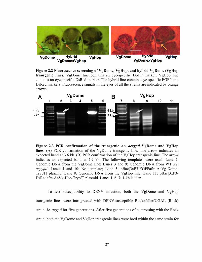
27
Figure 2.2 Fluorescence screening of VgDome, VgHop, and hybrid VgDomexVgHop transgenic lines. VgDome line contains an eye-specific EGFP marker. VgHop line contains an eye-specific DsRed marker. The hybrid line contains eye-specific EGFP and DsRed markers. Fluorescence signals in the eyes of all the strains are indicated by orange arrows.
Figure 2.3 PCR confirmation of the transgenic Ae. aegypti VgDome and VgHop lines. (A) PCR confirmation of the VgDome transgenic line. The arrow indicates an expected band at 3.6 kb. (B) PCR confirmation of the VgHop transgenic line. The arrow indicates an expected band at 2.9 kb. The following templates were used: Lane 2: Genomic DNA from the VgDome line; Lanes 3 and 9: Genomic DNA from WT Ae. aegypti; Lanes 4 and 10: No template; Lane 5: pBac[3xP3-EGFPafm-AeVg-Dome-TrypT] plasmid; Lane 8: Genomic DNA from the VgHop line; Lane 11: pBac[3xP3-DsRedafm-AeVg-Hop-TrypT] plasmid. Lanes 1, 6, 7: 1-kb ladder.
To test susceptibility to DENV infection, both the VgDome and VgHop
transgenic lines were introgressed with DENV-susceptible Rockefeller/UGAL (Rock)
strain Ae. agypti for five generations. After five generations of outcrossing with the Rock
strain, both the VgDome and VgHop transgenic lines were bred within the same strain for
3 kb 3 k kb4 kb 4 kb
3 kb

28
another five generations to ensure homogeneity. The WT Orl strain was mated with the
Rock strain in parallel to serve as a control.
To generate a hybrid transgenic line over-expressing Dome and Hop
simultaneously, male homozygous VgDome and female homozygous VgHop were mated
in a ratio of 1:5. The offspring were then screened for the expression of both GFP and
DsRed (Figure 2.2) and used for subsequent experiments to test their susceptibility to
DENV.
In the VgDome line, fat body expression of Dome was rapidly induced relative to
WT, peaking as early as 6 hours post bloodmeal (hpbm) and again at 48 hpbm. Dome
induction in the hybrid line followed a similar pattern, albeit with an approximately two-
fold higher peak at 6 and 24 hpbm (Figure 2.4). In the VgHop line, Hop expression was
induced more gradually, peaking at 24 hpbm. Hop induction in the hybrid line followed a
similar pattern, but with an earlier peak at 12 hpbm (Figure 2.4).
Dengue virus restriction factor 1 (DVRF1; AAEL008492) is transcriptionally
regulated by the JAK/STAT pathway, and encodes a putative anti-DENV effector
molecule [12,59]. In the VgDome and VgHop lines, DVRF1 expression relative to WT
peaked at 24 hpbm (Figure 2.4), indicating pathway activation. Interestingly, DVRF1
expression was induced to similar levels in the hybrid line (Figure 2.4), suggesting that
there might be limiting factors acting downstream of Dome and Hop.

29
Figure 2.4 Transcript abundance of transgenes and effector genes in the fat body of VgDome and VgHop lines from before blood feeding (0 hr) up to 48 hpbm. Each bar represents relative fold change of Dome, Hop or DVRF1 gene compared between transgenic lines and WT Ae. aegypti. The S7 ribosomal gene was used to normalize cDNA templates. Error bars indicate standard error of the mean. Statistical analyses were performed using t-test using Prism software, *: p < 0.05, **: p < 0.01 compared to WT before blood feeding.
Transgenic activation of the JAK/STAT pathway inhibits virus
replication throughout the mosquito’s body
We next investigated the effect of the transgene-mediated activation of the
JAK/STAT pathway on DENV infection in the transgenic lines. Mosquitoes were first
fed a naïve bloodmeal to activate the JAK/STAT pathway; two days later, they were
orally infected with DENV2 via a second (infectious) bloodmeal. We determined
DENV2 titers in the midguts at 7 day-post infectious blood meal (dpibm) (Figure 2.5A),
in the carcasses (whole mosquito except midgut) at 14 dpibm (Figure 2.5B), and in the
salivary glands at 21 dpibm (Figure 2.5C). VgDome and VgHop mosquitoes showed
significantly lower midgut DENV2 titers than did the WT mosquitoes (a 78.18% and
83.63% reduction in median titers for VgDome and VgHop, respectively). The VgDome
and VgHop lines displayed a 87.37% and 94.21% reduction in median carcass DENV2
titers, and more importantly, the transgenic mosquitoes also had a lower DENV2 titers in
*
*
** * *
* *

30
the salivary glands (100% reduction in median salivary gland DENV2 titers for both lines
when compared to WT).
We orally infected VgDome and VgHop mosquitoes without prior activation of
the JAK/STAT pathway in order to determine whether pathway activation at the time of
infection was enough to grant systemic resistance. We found that the VgDome
mosquitoes had comparable median midgut DENV2 titers, whereas the VgHop strain
showed a 42.86% reduction in median midgut DENV2 titers (Figure 2.5D). We saw
DENV2 resistance in the VgHop strain but not VgDome, suggesting that overexpression
of the downstream component of the pathway can provide earlier protection against
DENV infection. However, reduction of DENV titers in VgHop strain was not as strong
as when compared to the VgHop mosquitoes that were given naïve blood meal before
DENV infection. This result suggested that a naïve blood meal is required before the
infectious blood meal to provide higher resistance to DENV infection, further suggesting
that systemic immune activation by the JAK/STAT pathway is delayed.
Although the hybrid line also displayed significantly lower DENV2 titers in the
carcass compared to WT, these were not significantly different from the VgDome and
VgHop lines (Figure 2.5E). Since no difference in DENV2 susceptibility was seen
between the hybrid and the VgDome and VgHop transgenic lines, we chose to use only
the VgDome or VgHop lines for subsequent experiments.
To confirm that the inhibitory activity of the JAK/STAT pathway on DENV
infection is conserved among different DENV serotypes, we also challenged the VgDome
and VgHop lines with DENV4 as we did with DENV2 (Figure 2.5F). Both lines were
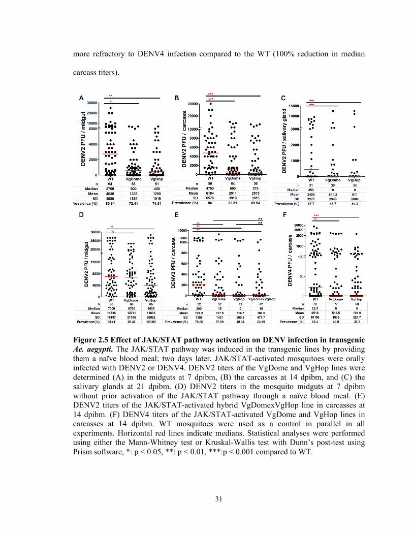
31
more refractory to DENV4 infection compared to the WT (100% reduction in median
carcass titers).
Figure 2.5 Effect of JAK/STAT pathway activation on DENV infection in transgenic Ae. aegypti. The JAK/STAT pathway was induced in the transgenic lines by providing them a naïve blood meal; two days later, JAK/STAT-activated mosquitoes were orally infected with DENV2 or DENV4. DENV2 titers of the VgDome and VgHop lines were determined (A) in the midguts at 7 dpibm, (B) the carcasses at 14 dpibm, and (C) the salivary glands at 21 dpibm. (D) DENV2 titers in the mosquito midguts at 7 dpibm without prior activation of the JAK/STAT pathway through a naïve blood meal. (E) DENV2 titers of the JAK/STAT-activated hybrid VgDomexVgHop line in carcasses at 14 dpibm. (F) DENV4 titers of the JAK/STAT-activated VgDome and VgHop lines in carcasses at 14 dpibm. WT mosquitoes were used as a control in parallel in all experiments. Horizontal red lines indicate medians. Statistical analyses were performed using either the Mann-Whitney test or Kruskal-Wallis test with Dunn’s post-test using Prism software, *: p < 0.05, **: p < 0.01, ***:p < 0.001 compared to WT.
32
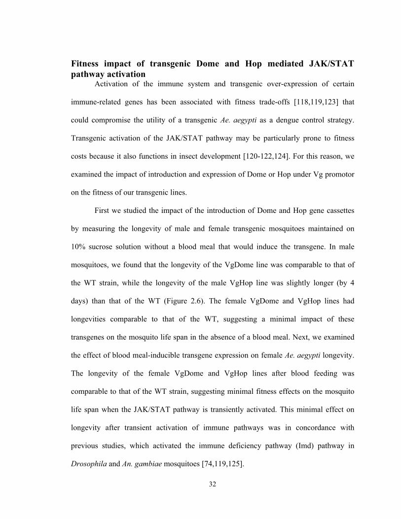
Fitness impact of transgenic Dome and Hop mediated JAK/STAT pathway activation
Activation of the immune system and transgenic over-expression of certain
immune-related genes has been associated with fitness trade-offs [118,119,123] that
could compromise the utility of a transgenic Ae. aegypti as a dengue control strategy.
Transgenic activation of the JAK/STAT pathway may be particularly prone to fitness
costs because it also functions in insect development [120-122,124]. For this reason, we
examined the impact of introduction and expression of Dome or Hop under Vg promotor
on the fitness of our transgenic lines.
First we studied the impact of the introduction of Dome and Hop gene cassettes
by measuring the longevity of male and female transgenic mosquitoes maintained on
10% sucrose solution without a blood meal that would induce the transgene. In male
mosquitoes, we found that the longevity of the VgDome line was comparable to that of
the WT strain, while the longevity of the male VgHop line was slightly longer (by 4
days) than that of the WT (Figure 2.6). The female VgDome and VgHop lines had
longevities comparable to that of the WT, suggesting a minimal impact of these
transgenes on the mosquito life span in the absence of a blood meal. Next, we examined
the effect of blood meal-inducible transgene expression on female Ae. aegypti longevity.
The longevity of the female VgDome and VgHop lines after blood feeding was
comparable to that of the WT strain, suggesting minimal fitness effects on the mosquito
life span when the JAK/STAT pathway is transiently activated. This minimal effect on
longevity after transient activation of immune pathways was in concordance with
previous studies, which activated the immune deficiency pathway (Imd) pathway in
Drosophila and An. gambiae mosquitoes [74,119,125].
33
Figure 2.6 Effect of transgenes introduction and expression on mosquito longevity. Lifespans of male and female mosquitoes maintained on 10% sucrose solution or of female mosquitoes that were provided a blood meal to induce transgene expression. Statistical analyses of survival curve was performed using Log rank test with Prism software. ***: p < 0.001.
Both VgDome and VgHop lines produced significantly fewer eggs compared to
WT (Figure 2.7), suggesting that transgene introduction or expression compromises
fecundity. The lower egg production is likely, at least in part, due to the competition
between the vitellogenin promoter of the transgenes and the endogenous vitellogenin
gene, as indicated by the lower expression level of the vitellogenin gene after blood
feeding in the transgenic mosquitoes when compared to WT (Figure 2.8).
Figure 2.7 Fecundity of the WT and transgenic Ae. Aegypti, as represented by number of eggs produced by each female mosquito. Statistical analyses were performed using the Mann-Whitney test with Prism software **: p < 0.01 as compared to WT

34
Figure 2.8 Expression of vitellogenin in the transgenic lines as compared to WT. mRNA levels were measured by real-time PCR, with ribosomal gene S7 as the normalization control. Error bars indicate standard error of the mean. Statistical analyses were performed using t-test using Prism software, *: p < 0.05 compared to vitellogenin gene expression in WT at 24 hours post blood meal.
Immune-related transcripts are enriched upon JAK/STAT activation
The JAK/STAT pathway-regulated antiviral effectors responsible for suppressing
DENV infection are largely unknown, except for two genes, DVRF1 and DVRF2, that
encode putative secreted and membrane-bound proteins, respectively, of unknown
function [59,125]. To identify possible JAK/STAT pathway-regulated antiviral effectors
and to assess the impact of transgenic JAK/STAT pathway activation on mosquito
physiology at the molecular level, we used whole-genome oligonucleotide microarrays to
compare the fat body transcriptomes of WT, VgDome, and VgHop lines at 24 hpbm
(17,346 genes in the Ae. aegypti transcriptome). We selected the 24-h post-blood meal
time point because of the DVRF1 peak expression at that time, suggesting the peak
activity of the JAK/STAT. Genes that showed at least 1.68-fold (0.75 on a log2-scale)
compared to WT were considered to be significantly differentially regulated. The log2-
fold difference in transcript abundance for each gene between VgDome or VgHop and
WT mosquitoes is listed in Appendix 2, and the number and percentage of transcripts
significantly regulated in each category are presented in Figure 2.9A and 2.9B. Genes
* *

35
commonly- or differentially-regulated in VgDome and Hop was represented in figure
2.9C. As expected, DVRF1 transcripts were enriched in both lines relative to WT (see
Appendix 2), an indication of pathway activation. In VgDome, 130 transcripts (0.75% of
the whole transcriptome) were enriched compared to WT, and 71 (0.47%) were depleted.
In VgHop, 254 transcripts (1.46%) were enriched compared to WT, and 204 (1.18%)
were depleted.
In both lines, IMM transcripts made up the largest specific class of enriched
transcripts (excluding those with diverse (DIV) and unknown (UKN) functions). Of the
659 immune-related genes (IMM) in the Ae. aegypti transcriptome, 17 genes (2.58% of
the total IMM genes) had a higher transcript abundance and 10 genes (1.52% of total
IMM) had a lower transcript abundance in the VgDome line. In the VgHop line, 29 genes
(4.40% of the total IMM) had a higher transcript abundance, and 15 genes (2.28% of total
IMM) had a lower transcript abundance. The IMM had at least a 3-fold higher percentage
of genes with higher transcript abundance when compared to the average percentage of
regulated genes in the whole transcriptome. These results emphasize the importance of
the JAK/STAT pathway in mosquito immune regulation and corroborate the fact that the
VgDome and VgHop lines had higher immune activity than did the WT. IMM transcripts
that are enriched upon JAK/STAT activation may encode potential DENV restriction
factors (RFs) - gene products that inhibit DENV replication in the mosquito.
Fifty transcripts were enriched and 18 were depleted in both VgDome and VgHop
compared to WT (Figure 2.9 A, and C). Again, the IMM category was the largest specific
class of transcripts that were enriched in both lines (9 genes, 1.37% of the total IMM).
These were: three C-type lectins (CLECs; AAEL005482, AAEL011610, and
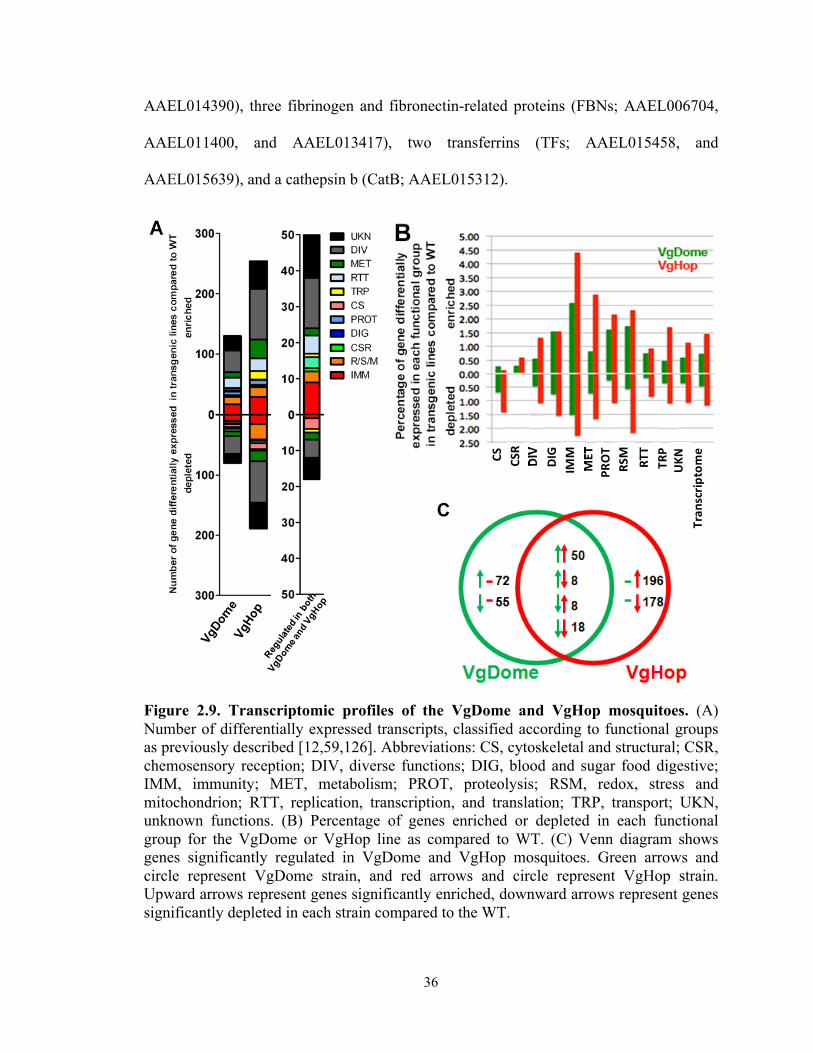
36
AAEL014390), three fibrinogen and fibronectin-related proteins (FBNs; AAEL006704,
AAEL011400, and AAEL013417), two transferrins (TFs; AAEL015458, and
AAEL015639), and a cathepsin b (CatB; AAEL015312).
Figure 2.9. Transcriptomic profiles of the VgDome and VgHop mosquitoes. (A) Number of differentially expressed transcripts, classified according to functional groups as previously described [12,59,126]. Abbreviations: CS, cytoskeletal and structural; CSR, chemosensory reception; DIV, diverse functions; DIG, blood and sugar food digestive; IMM, immunity; MET, metabolism; PROT, proteolysis; RSM, redox, stress and mitochondrion; RTT, replication, transcription, and translation; TRP, transport; UKN, unknown functions. (B) Percentage of genes enriched or depleted in each functional group for the VgDome or VgHop line as compared to WT. (C) Venn diagram shows genes significantly regulated in VgDome and VgHop mosquitoes. Green arrows and circle represent VgDome strain, and red arrows and circle represent VgHop strain. Upward arrows represent genes significantly enriched, downward arrows represent genes significantly depleted in each strain compared to the WT.
MET
PROT
RSM
RTT
TRP
UKN
Tran
scrip
tome
DIV
DIG
MET
IMM
CS
DIVCSR
C
37
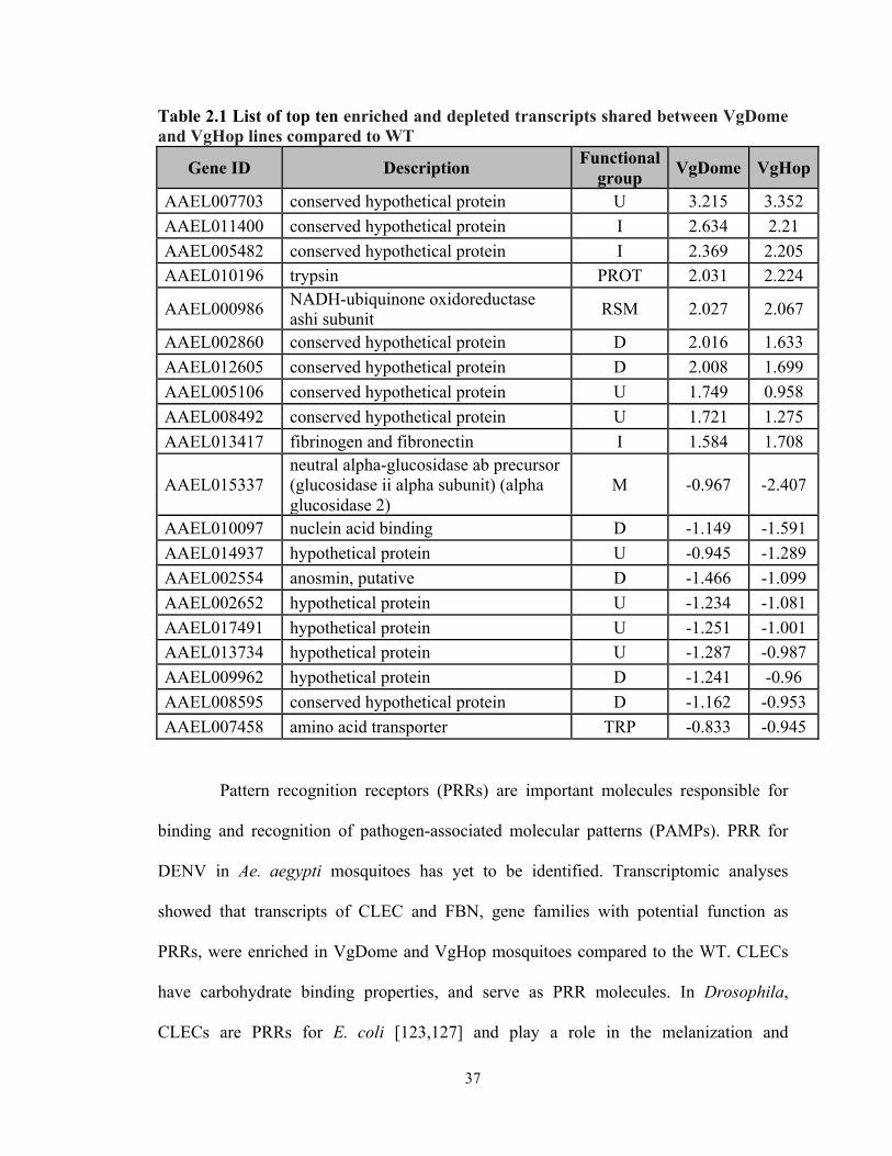
Table 2.1 List of top ten enriched and depleted transcripts shared between VgDome and VgHop lines compared to WT
Gene ID Description Functional group VgDome VgHop
AAEL007703 conserved hypothetical protein U 3.215 3.352 AAEL011400 conserved hypothetical protein I 2.634 2.21 AAEL005482 conserved hypothetical protein I 2.369 2.205 AAEL010196 trypsin PROT 2.031 2.224
AAEL000986 NADH-ubiquinone oxidoreductase ashi subunit RSM 2.027 2.067
AAEL002860 conserved hypothetical protein D 2.016 1.633 AAEL012605 conserved hypothetical protein D 2.008 1.699 AAEL005106 conserved hypothetical protein U 1.749 0.958 AAEL008492 conserved hypothetical protein U 1.721 1.275 AAEL013417 fibrinogen and fibronectin I 1.584 1.708
AAEL015337 neutral alpha-glucosidase ab precursor (glucosidase ii alpha subunit) (alpha glucosidase 2)
M -0.967 -2.407
AAEL010097 nuclein acid binding D -1.149 -1.591 AAEL014937 hypothetical protein U -0.945 -1.289 AAEL002554 anosmin, putative D -1.466 -1.099 AAEL002652 hypothetical protein U -1.234 -1.081 AAEL017491 hypothetical protein U -1.251 -1.001 AAEL013734 hypothetical protein U -1.287 -0.987 AAEL009962 hypothetical protein D -1.241 -0.96 AAEL008595 conserved hypothetical protein D -1.162 -0.953 AAEL007458 amino acid transporter TRP -0.833 -0.945
Pattern recognition receptors (PRRs) are important molecules responsible for
binding and recognition of pathogen-associated molecular patterns (PAMPs). PRR for
DENV in Ae. aegypti mosquitoes has yet to be identified. Transcriptomic analyses
showed that transcripts of CLEC and FBN, gene families with potential function as
PRRs, were enriched in VgDome and VgHop mosquitoes compared to the WT. CLECs
have carbohydrate binding properties, and serve as PRR molecules. In Drosophila,
CLECs are PRRs for E. coli [123,127] and play a role in the melanization and

38
encapsulation processes [124,128]. In Ae. aegypti, several CLEC have been reported to
function as receptors for DENV entry into cells [125,129]. However, none of the CLEC
identified in our study were reported as a receptor for virus entry. Silencing of
AAEL005482 had no effect on DENV infection, whereas silencing of a homolog of
AAEL014390 resulted in a non-significant increase in DENV loads, by 1.48-fold
[40,125]. AAEL011610 has not been tested for its role in DENV replication, but has been
reported to be up-regulated in transgenic Ae. aegypti over-expressing Rel2, transcription
factor for the Imd pathway, under the control of the vitellogenin promoter [126,130].
FBNs are thought to serve as PRRs in Drosophila and in Anopheles mosquitoes
[127,131] [126,128]. but their function in Ae. aegypti has yet to be elucidated.
CatB is a family of lysosomal cysteine proteases with functions in TLR signaling
as well as T and B cell apoptosis [12,59,129]. One of the CatBs regulated in our study
(AAEL007585) has been reported to facilitate DENV infection in Ae. aegypti salivary
glands [40,41,44]; it was hypothesized that CatB-mediated apoptosis may facilitate cell-
to-cell spread of the virus.
Over-expression of Dome and Hop also regulated specific subsets of IMM
transcripts (Appendix2). Eight IMM genes were enriched in the VgDome but not in the
VgHop, including three serine proteases (AAEL003279, AAEL000030, and
AAEL006434), two Niemann-Pick Type C2 molecules (AAEL012064, and
AAEL004120), a cathepsin b (AAEL007599), and a lysozyme C (AAEL017132).
Twenty IMM transcripts were enriched in VgHop but not in VgDome. These included
four cathepsin b genes (AAEL009637, AAEL009642, AAEL007585, and AAEL012216),
four serine proteases (AAEL007969, AAEL007006, AAEL015430, and AAEL003625), a

39
thioester-containing protein (TEP22; AAEL000087), and several anti-microbial peptides
(AMPs) such as cecropins (AAEL000621, AAEL000625), defensins (AAEL003832,
AAEL003841), a gambicin (AAEL004522), and a lysozyme P (AAEL003723). These
line-specific transcripts suggest complexities in JAK/STAT pathway regulation, and
different as-yet unknown branches of the pathway and fine-tuning mechanisms may
come into play to regulate different subsets of genes.
TEPs, which encode complement factor-like proteins belonging to the alpha-2-
macroglobulin family, play important roles in insect immunity [12,130]. In Ae. aegypti,
TEP22 was previously reported to be regulated by the CTL CLSP2 (AAEL011616), and
to be involved in the mosquito's anti-fungal response [54,131-133]. TEP22 was also up-
regulated in transgenic Ae. aegypti over-expressing the Toll pathway transcription factor
Rel1 [93,126], suggesting that there may be crosstalk between these two immune
pathways, both of which both play an important role in anti-DENV responses [12,59,97].
In further support of this, several AMPs belonging to the defensin and cecropin families
were up-regulated in our JAK/STAT transgenic strains and have been previously studied
with regard to their anti-DENV properties [41,44,134]. Gambicin was previously
described to be regulated by the Toll pathway [12,134,135]; however, it has never been
tested for anti-DENV activity.
Because we observed regulation of several AMPs, in both the VgDome and
VgHop lines, that might provide protection against bacterial infection, we challenged
these mosquitoes with a Gram-negative bacterium, Pantoea spp., and a Gram-positive
bacterium, Bacillus cereus. We found no resulting differences in mortality between the
VgDome or VgHop lines and WT (Figure 2.10).

40
Figure 2.10. Mortality of the VgDome and VgHop lines from bacterial infection. Mosquitoes were challenged with Pantoea spp. or Bacillus cereus, with PBS as a negative control. Survival analysis was performed using Data are from three independent replicates.
Transcript abundances of potential DENV host factors were depleted
upon JAK/STAT activation
Other than the IMM genes, hundreds of transcripts in other functional classes
were differentially expressed in VgDome and VgHop compared to the WT. (Figure
2.9A). This result is not unexpected since the JAK/STAT pathway plays important roles
in other biological processes such as cell development and homeostasis, as well as lipid
metabolism [54,95,132,133]. We also found that genes with unknown function
contributed to a large proportion of top shared enriched and depleted genes shared
between VgDome and VgHop compared to WT (3 out of 10 enriched transcripts, and 4
out of 10 depleted transcripts). These data show that our knowledge is still limited on the
function of the JAK/STAT pathway in Ae. aegypti.
The transcript abundances of several previously reported putative DENV host
factors (HFs; genes that facilitate virus replication in the host) were significantly depleted
in the transgenic lines compared to WT; a pattern that could potentially result in a
reduced virus replication in mosquito.

41
Glucosidase 2 alpha subunit (aGluc; AAEL015337) transcripts were depleted in
VgDome and VgHop (0.967- and 2.407-fold in log2 scale, respectively). aGluc has been
identified in a high-throughput screen as a DENV HF in both HuH-7 human and D.Mel-2
Drosophila cells [93,136,137]. We previously identified aGluc as putative HF from
transcriptomic comparisons between DENV-refractory and -susceptible strains, but
silencing of aGluc had no effect on DENV infection in Ae. aegypti [97,132].
Nevertheless, previous studies have suggested that aGluc is required for proper
glycosylation of the viral glycoproteins PrM and E [134,138], and chemical inhibition of
aGluc results in lower virus production from infected cells [134,135,139]. It is possible
that the transient silencing of aGluc by RNAi in the previous study was not sufficient to
deplete aGluc at the protein level, and therefore no effect was observed.
Transcripts of vacuolar ATP synthase subunit ac39 (vATPase-ac39;
AAEL0011025) were depleted in VgHop compared to WT (2.707-fold in log2 scale). Cell
entry, a crucial step in DENV infection, requires acidification of endosomes by vATPase
enzymes. Knockdown of vATPase-ac39 and several other vATPase subunits, as well as
chemical inhibition of vATPase activity with bafilomycin, have been shown to inhibit
DENV replication in Ae. aegypti [95,140,141].
Lipid homeostasis and trafficking play important roles in the replication of
DENV, an enveloped virus. DENV is thought to facilitate its replication by altering the
expression of lipid binding proteins and enzymes involved in lipid biosynthesis, such as
fatty acid synthases and Niemann-Pick type C protein family members
[136,137,142,143]. The JAK/STAT pathway has previously been shown to influence
lipid metabolism in mammals, and it may have a similar function in mosquitoes
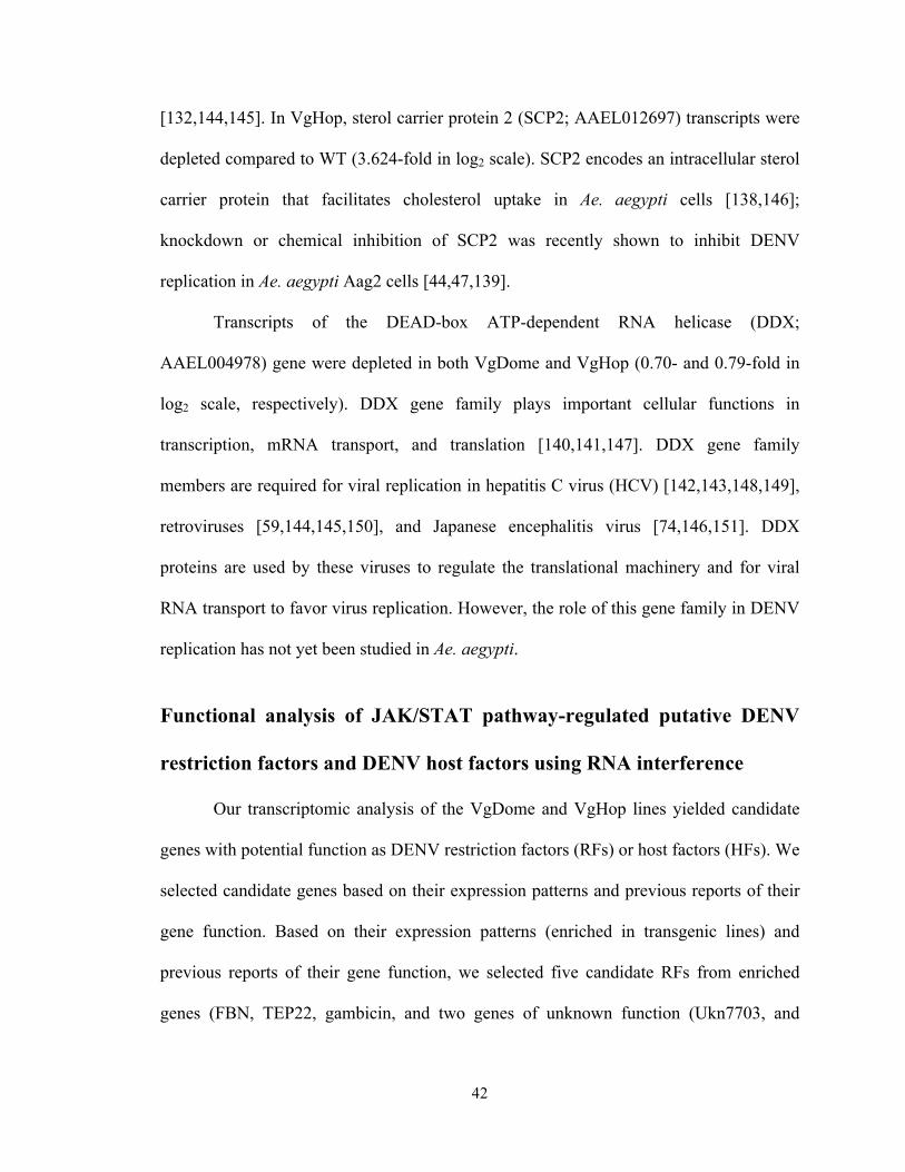
42
[132,144,145]. In VgHop, sterol carrier protein 2 (SCP2; AAEL012697) transcripts were
depleted compared to WT (3.624-fold in log2 scale). SCP2 encodes an intracellular sterol
carrier protein that facilitates cholesterol uptake in Ae. aegypti cells [138,146];
knockdown or chemical inhibition of SCP2 was recently shown to inhibit DENV
replication in Ae. aegypti Aag2 cells [44,47,139].
Transcripts of the DEAD-box ATP-dependent RNA helicase (DDX;
AAEL004978) gene were depleted in both VgDome and VgHop (0.70- and 0.79-fold in
log2 scale, respectively). DDX gene family plays important cellular functions in
transcription, mRNA transport, and translation [140,141,147]. DDX gene family
members are required for viral replication in hepatitis C virus (HCV) [142,143,148,149],
retroviruses [59,144,145,150], and Japanese encephalitis virus [74,146,151]. DDX
proteins are used by these viruses to regulate the translational machinery and for viral
RNA transport to favor virus replication. However, the role of this gene family in DENV
replication has not yet been studied in Ae. aegypti.
Functional analysis of JAK/STAT pathway-regulated putative DENV
restriction factors and DENV host factors using RNA interference
Our transcriptomic analysis of the VgDome and VgHop lines yielded candidate
genes with potential function as DENV restriction factors (RFs) or host factors (HFs). We
selected candidate genes based on their expression patterns and previous reports of their
gene function. Based on their expression patterns (enriched in transgenic lines) and
previous reports of their gene function, we selected five candidate RFs from enriched
genes (FBN, TEP22, gambicin, and two genes of unknown function (Ukn7703, and
43
Ukn566)) and two candidate HFs from depleted genes (DDX and SCP2) for further
characterization using RNAi-mediated gene silencing assays (Table 2.2).
Table 2.2. List of candidate RFs and HFs for functional confirmation by RNAi
Ukn7703 (AAEL007703) transcripts were very highly enriched in the transgenic
lines compared to WT (3.215-, and 3.352-log2 fold higher in VgDome and VgHop
respectively). The gene has also been reported to be induced in two strains of Wolbachia-
infected Ae. aegypti [44,47,59,152], and is conserved among Aedes, Culex, and
Anopheles mosquitoes (Figure 2.11), suggesting an important role in mosquito biology.
Ukn7703 encodes a putative secreted protein with a C-terminal beta-propeller domain
distantly related to WD-40 repeats (predicted using NCBI conserved domain search
[147,153]). WD-40 domains are involved in protein-protein interactions in several
biological processes, including signal transduction [148,149,154].

44
Figure 2.11. Phylogenetic tree of orthologs of AAEL007703 gene obtained from Vector base. URL: https://www.vectorbase.org/Multi/GeneTree/Image?gt=VBGT00190000016830
Figure 2.12 Silencing efficiencies for candidate RFs and HFs.
Ukn566 (AAEL000566) transcripts were enriched in both VgDome and VgHop
compared to WT (0.946- and 1.748-log2 fold, respectively); this gene was also induced in
Wolbachia-infected Ae. aegypti [59,148,150]. Ukn566 is predicted to be a transmembrane

45
protein using TMHMM software [59,74,151]. While no predicted protein domains were
detected, Ukn566 and its orthologs share conserved cysteine positions. Cysteine repeats
have previously been reported to be important for the three-dimensional structure and
function of receptor proteins such as LDL [59,74,152] and scavenger receptors [59,153].
Potential functions of the remaining candidate genes have been discussed in the previous
section.
Across the candidate genes, silencing efficiencies varied from 22% to 85%, as
shown in Figure 2.12. Our screen confirmed Ukn7703 as a putative RF (31.82% increase
in median DENV titer when compared to the GFP dsRNA-injected group), and SCP2 as a
putative HF (85.71% decrease in DENV titer when compared to the GFP dsRNA-injected
group) (Figure 2.13). Silencing of DDX also reduced the DENV2 titers in the carcass by
61.43%, although this result was not statistically significant by a small margin
(p=0.0555). The lack of statistical significance may be a result of the lower silencing
efficiency (22%) achieved for this gene. Because of the limitation of RNAi in failing to
completely knock down a gene of interest, the lack of effects on DENV infection for the
other candidate RFs does not necessarily exclude their potential involvement in anti-
dengue defense

46
Figure 2.13 Effect of putative host and restriction factor silencing on DENV susceptibility. DENV2 titers at 14 dpibm in the carcasses (whole mosquito body except midgut) of the WT Ae. aegypti after silencing of the putative HFs or RFs, compared to GFP dsRNA-injected control. Data are a pool of three biological replicates, and statistical analyses were performed using the Mann-Whitney test, **: p < 0.01, ***: p < 0.001 vs.WT.
DISCUSSION
We have demonstrated that we can induce activation of the JAK/STAT pathway
by over-expressing JAK/STAT pathway receptor Dome and the Janus kinase Hop under
the control of Vg promoter. Overexpression of Dome and Hop prior to exposure to
DENV systemically controls DENV infection. Since Dome and Hop were over-expressed
specifically in the fat body, the reduction of DENV titers in the midgut suggested that the
JAK/STAT pathway functions as a systemic anti-viral defense system in Ae. agypti, and
may possibly prime uninfected cells and other immune cells to assume an antiviral state.
This type of systemic activation in the mosquito is analogous to the mammalian
JAK/STAT pathway’s role as a Type I interferon-induced immune pathway [154,155], or
47
the systemic immune response function of the JAK/STAT pathway in Drosophila
[97,148].
A comparable level of DENV infection was found in the hybrid line compared to
VgDome or VgHop, suggesting the existence of a limiting factor downstream of Dome
and Hop, a possibility that is in agreement with the results concerning the induction of the
DVRF1. It will be an interesting attempt to further reduce the vector competence for
DENV by generating transgene constructs expressing both receptor and downstream
pathway molecules such as STAT. Availability of JAK/STAT pathway downstream
components might not be the only limiting factor of pathway activation. The JAK/STAT
pathway is negatively regulated by PIAS or suppressor of cytokine signaling (SOCS)
proteins to prevent overactivation of the pathway [54], and we have previously shown
that silencing PIAS activates the JAK/STAT pathway and inhibits DENV infection
[59,156]. To improve JAK/STAT pathway activation in transgenic mosquitoes, we can
generate a transgene construct that contains the receptor gene together with a hairpin
double-stranded RNA sequence complementary to PIAS or SOCS to reduce the level of
negative regulator molecules.
We also showed that both transgenic lines were refractory to DENV2 and DENV4
infection compared to the WT, which suggests that the anti-viral function of the
JAK/STAT pathway is conserved across different DENV serotypes. It will be interesting
to determine if JAK/STAT pathway activation is also effective against other mosquito-
borne flaviviruses such as the West Nile, zika, and yellow fever virus, as well as against
alphaviruses such as chikungunya virus.

48
Analyses of the fitness impact from transgene expression in the transgenic lines
showed that there was a minimal disadvantage to the longevity of these mosquitoes, but
there was a negative impact on fecundity for both transgenic lines. It should be noted that
these mosquitoes were maintained under laboratory conditions with an abundant food
supply and minimal environmental stress. The assays may therefore not reflect the effect
of transient JAK/STAT pathway activation on mosquito longevity in a natural setting
where a myriad of stressors can apply. Additional experiments will be necessary to fully
evaluate the effect of transient JAK/STAT pathway activation on longevity in natural
settings. Reduced egg production and lower expression of vitellogenin gene have also
been observed in transgenic An. stephensi lines using the Vg promoter to drive gene
expression of Imd pathway component [74,97]. This phenomenon; however, was not
observed in transgenic An. stephensi that overexpressed Imd pathway component under a
regulation of bloodmeal inducible, gut-specific carboxypeptidase promoter. These results
suggest that the use of Vg promoter compromises mosquito’s fecundity, which suggests
that alternative fat body-specific promoters may be required to help to minimize fitness
disadvantages.
Transcriptomic comparisons between the fat body of blood-fed transgenic
mosquitoes and WT allowed us to identify genes that influence DENV infection in Ae.
Aegypti. The greater resistance to DENV infection displayed by the transgenic lines was a
result of a combination of a higher transcript abundance of RFs and a lower transcript
abundance of HFs. These multifactorial factors for DENV resistance make it harder for
DENV to develop counter-measures to be able to efficiently replicate in our transgenic
mosquitoes.
49
Even though several AMPs were enriched in the transgenic lines compared to the
WT, we did not observe differences in mosquitoes’ mortality from bacterial infection
This result was similar to what we observed in a previous study: that the transient
silencing of PIAS, a negative regulator of the JAK/STAT pathway, has no effect on
mosquito mortality resulting from bacterial infection [59,97]. It is also possible that the
regulated AMPs may have more specialized anti-DENV function or may not have anti-
microbial activity against the particular bacteria used in our study. A previous study of
defensins from humans has also suggested that the anti-bacterial activity of certain AMPs
is highly specific [85,155]. Future extensive study of the anti-bacterial function of these
AMPs will require recombinant expression of individual AMPs so they can be tested with
a wide panel of bacteria.
In summary, our study is the first to provide a proof-of-concept that genetic
engineering of the mosquitoes’ JAK/STAT immune pathway can be used to render the
insect more resistant to dengue virus infection and possibly to block transmission of the
disease.

50
CHAPTER 3
Identification of putative host and restriction factors that
contributes to refractoriness to DENV infection among
laboratory and field-derived Aedes aegypti mosquitoes
Parts of this work were published in:
Sim S, Jupatanakul N, Ramirez JL, Kang S, Romero-Vivas CM, Mohammed H, et al. Transcriptomic profiling of diverse Aedes aegypti strains reveals increased basal-level immune activation in dengue virus-refractory populations and identifies novel virus-vector molecular interactions. Plos Neglect Trop D. 2013;7: e2295–e2295. doi:10.1371/journal.pntd.0002295
Kang S, Shields AR, Jupatanakul N, Dimopoulos G. Suppressing dengue-2 infection by chemical inhibition of Aedes aegypti host factors. Plos Neglect Trop D. 2014;8: e3084. doi:10.1371/journal.pntd.0003084

51
ABSTRACT
Our lab has previously established a panel of laboratory and field-derived Ae.
agypti strains from different geographical origins. We have shown that these mosquito
strains vary in susceptibility to DENV infection, and comparative genome-wide gene
expression microarray-based analysis revealed higher basal levels of numerous
immunity-related gene transcripts in DENV-refractory mosquito strains compared to
susceptible strains. Here we used RNA interference-mediated gene silencing assays to
further confirm functions of the Toll, Imd, JAK/STAT, and RNAi-pathway in
contribution to refractoriness in different strains. By correlating transcript abundance
patterns with DENV susceptibility, we also identified new candidate modulators of
DENV infection in the mosquito, and we provide functional evidence for vATPase
subunits as DENV host factors. Our comparative transcriptome dataset thus not only
provides valuable information about immune gene regulation and usage in natural
refractoriness of mosquito populations to dengue virus but also allows us to identify new
molecular interactions between the virus and its mosquito vector.

52
INTRODUCTION
Mosquitoes, like other organisms, are exposed to a variety of microbes in their
natural habitats and possess an innate immune system that is capable of mounting a
potent response against microbial challenge. However, studies of mosquito immune
responses to DENV and other human pathogens have largely been performed in
laboratory strains of Ae. aegypti, which have been maintained under insectary conditions
for many generations. As compared to natural mosquito populations, laboratory mosquito
strains are exposed to lower doses and a much narrower range of microbes, constant
temperature and humidity; together with the genetic bottleneck of a small initial parental
population size, this often results in a loss of genetic variability. However, differences
between laboratory-maintained mosquitoes and field populations have been poorly
studied.
In this study, we collected Ae. aegypti from different DENV-endemic
geographical locations as well as laboratory-maintained Ae. aegypti strains [20,97], and
showed that they have a wide range of DENV susceptibility and different transcriptomic
profiles. These mosquito strains and transcriptomic dataset served as powerful tools to
dissect interactions between DENV and to identify novel modulators of DENV infection
in Ae. aegypti.
MATERIALS AND METHODS
Mosquito rearing and cell culture conditions
Mosquitoes were maintained on a 10% sucrose solution in insectary condition at
27°C and 95% humidity with a 12 h light/dark cycle. The C6/36 Aedes albopictus cell

53
line was maintained in MEM (Gibco) supplemented with 10% heat-inactivated FBS, 1%
L-glutamine, 1% non-essential amino acids, and 1% penicillin-streptomycin at at 32°C
and 5% CO2. BHK-21 hamster kidney cells were maintained on DMEM (Gibco)
supplemented with 10% FBS, 1% L-glutamine, 1% penicillin-streptomycin, and 5 ug/ml
Plasmocin (Invivogen) at 37°C and 5% CO2.
Oral DENV infections in Ae. aegypti
Mosquito infections with DENV were carried out by oral infection as previously
described [97,156]. The New Guinea C strain of DENV2 was propagated in C6/36 cells
as previously described [12]. Briefly, DENV2 was infected to C6/36 cells seeded to 80%
confluence at a multiplicity of infection (MOI) of 3.5 and incubated at 32°C and 5% CO2
for 6 days. The infected cells were then harvested and lysed through 3 cycles of freezing
and thawing between dry ice and 37°C water bath. The propagation yielded a virus titer
of between 106 and 107 PFU/ml. Then DENV was mixed 1:1 v/v with commercial human
blood and supplemented with 10% human serum and 1 mM ATP. The bloodmeal was
then offered to mosquitoes via an artificial membrane feeding system. Midguts were
dissected at 7 days post-blood meal (dpbm) and stored individually in DMEM at -80°C
until titrated by plaque assay.
Gene silencing assays
Silencing of candidate hotst and restriction factor genes was performed through
RNA interference (RNAi)-mediated gene silencing as previously described. Field-derived
mosquitoes used in these assays were from generations F5 to F13, depending on the
strain. Each experiment was performed with mosquitoes from the same generation to
ensure valid comparison between experimental and control groups. In brief, three-day-old

54
female mosquitoes were cold-anesthetized and individually injected with 200 ng of
dsRNA specific for the target gene of interest, and GFP dsRNA was used as control in all
experiments. The dsRNA-injected mosquitoes were then orally infected with DENV2-
supplemented blood at 3-4 days post-dsRNA injection. Midguts were dissected at 7 dpbm
for DENV titration by plaque assay. dsRNA was synthesized using the HiScribe T7 in
vitro transcription kit (New England Biolabs). Primer sequences used for dsRNA
synthesis and to confirm gene silencing by real-time PCR are presented in the
Appendix1. Due to high sequence identity, it was unavoidable that dsRNA against
AAEL010429 also targeted AAEL013577 and AAEL010436 (also putative insect
allergen family members), and dsRNA against AAEL015337 also targeted AAEL010599
(also a neutral alpha-glucosidase ab precursor). We cannot however exclude the
possibility that these seemingly different transcripts represent the same gene, due to
possible genome sequence annotation errors.
DENV titration by plaque assay
DENV2 titers in the mosquito midguts were determined by plaque assay on BHK-
21 cells. Individual midguts were homogenized in DMEM using glass beads with a Bullet
Blender homogenizer (NextAdvance), 10-fold serially diluted, and then inoculated onto
BHK cells seeded to 80% confluence in 24-well plates. Plates were rocked for 15 min at
room temperature, and then incubated for 45 min at 37°C and 5% CO2. Each well was
then overlayed with 1 ml of DMEM containing 2% FBS and 0.8% methylcellulose and
plates were incubated for 5 days at 37°C and 5% CO2. Plates were fixed with a methanol
/ acetone mixture (1:1 volume/volume) for at least 1 h at 4°C, and plaque-forming units

55
were visualized by staining with 1% crystal violet solution for at least 10 min at room
temperature.
RESULTS
Laboratory and field-derived Ae. aegypti strains have different degrees
of DENV2 susceptibility
Our lab has established a panel of field-derived and laboratory strains Ae. aegypti
strains obtained either through generous contributions from collaborators or our own field
collections (Table 3.1). Field locations were selected to represent geographically distinct
dengue-endemic regions spanning South America, the Caribbean, and Southeast Asia,
and laboratory strains were included to allow us to compare DENV susceptibility
between laboratory maintained and natural mosquito populations.
To assess their susceptibility for DENV, we orally infected each mosquito strain
with DENV2 and assessed midgut virus titers at 7 dpbm. The highly susceptible
Rockefeller (Rock) laboratory strain typically used in our group’s experiments served as
a basis for comparison. To compare susceptibility to DENV2 infection, we calculated the
relative infection level of each strain by calculating its median DENV2 titer as a
percentage of the median titer of its respective Rock control. We found that Orl, Waco,
PFin, PR, PTri, and BKK had significantly lower midgut DENV2 titers as compared to
Rock, and median virus titers in Kitts were four-fold lower than in Rock (Figure 3.1).
SIN was the only strain with midgut DENV2 titer comparable to Rock strain. Among
these Ae. aegypti strains, the SIN and PTri strains were the most susceptible, while BKK
and Orl were the most refractory to DENV2 infection.

56
Table 3.1 Origins and name abbreviations of laboratory and field-derived Ae. aegypti strains [93,97].
Figure 3.1 Susceptibilities of Ae. aegypti strains to DENV2 infection. Relative median DENV2 midgut infection levels at 7 days post-bloodmeal (dpbm) for each Ae. aegypti strain, compared to the Rockefeller strain. Statistical analyses performed by using Mann-Whitney test using Prism software, ***:p < 0.001, ****: p < 0.0001 compared to Rock.
57
Mosquito immune signaling pathways and the RNAi pathway control DENV2 infection to different degrees in various Ae. aegypti strains Transcriptomic analyses by whole genome microarray assays revealed that
differences in basal levels of immune related gene (IMM) transcript abundance among
mosquito strains is an important factor in determining susceptibility to DENV [93,97].
These IMM are likely to be regulated by major immune signaling pathways, the Toll,
IMD, and JAK/STAT pathways, which regulate the mosquito immune response to a
variety of pathogens [32]. We examined the contributions of these pathways to
refractoriness in these strains. In addition, since the RNAi pathway is an important
controller of DENV infection in Ae. aegypti [85,92], we also examined its role in this
regard. Each pathway was inhibited or activated through the RNAi-mediated knockdown
of a key pathway component or regulator, and the effect of this manipulation on midgut
DENV2 titers was assessed.
In the Orl strain, individually compromising the Toll, IMD, JAK/STAT, and
RNAi pathways via knockdown of MyD88, IMD, Dome, and Dcr2, respectively, resulted
in a significant and dramatic increase in midgut DENV2 titers to a level that was
comparable to what typically seen in Rock (Figure 3.2A), suggesting that each of these
pathways is a major contributor to the refractoriness seen in Orl. In the BKK strain,
compromising the Toll, IMD, JAK/STAT or RNAi pathways only resulted in a non-
significant 3- to 5-fold increase in midgut virus titers (Figure 3.2A), suggesting the action
of BKK strain-specific DENV restriction factor(s) that operate independently of these
pathways (Figure 3.2A).
Conversely, activating immune signaling pathways by silencing pathway negative
regulators should render the susceptible Rock and SIN strains more refractory to DENV

58
infection (Figure 3.2B). In agreement with our previous studies [20,93,94], silencing the
Toll and JAK/STAT pathway negative regulators Cactus and PIAS in the Rock strain
resulted in a significant decrease in midgut DENV titers, while silencing the IMD
pathway negative regulator Caspar resulted in a non-significant decrease (Figure 3.2B).
As expected, silencing Cactus in the SIN strain also resulted in a significant decrease in
virus titers, but no effect was seen for PIAS or Caspar. It is possible that the JAK/STAT
and IMD pathways are already operating at maximum capacity, especially given the high
viral load observed in this strain, or that the SIN strain possesses factors acting
independently of these pathways that facilitate DENV infection. From these data, we
speculate that the basal activation levels of immune pathways may be higher in refractory
mosquitoes, and that these elevated levels in refractory strains may contribute to
refractoriness by increasing the transcript abundance of various immune effectors.

59
Figure 3.2 Contributions of the Toll, Imd, JAK-STAT, and RNAi pathways to the control of DENV2 in refractory and susceptible mosquito strains. Midgut DENV2 titers at 7 dpbm in (A) MyD88, Imd, Dome, and Dcr2-silenced Orl and BKK mosquitoes, and (B) Cactus, Caspar, and PIAS-silenced Rock and SIN mosquitoes. Data are a pool of at least three independent biological replicates. ****, p<0.0001; ***, p<0.001; **, p<0.01 *, p<0.05 compared to dsGFP-treated mosquitoes in Dunn’s post-test after Kruskal-Wallis test.
60
Transcriptomic comparison between refractory and susceptible Ae.
aegypti reveals candidate DENV restriction and host factors
Other than immune-related transcripts, genes in other functional groups can also
influence DENV infection in different fashion. DENV host factors (HFs) are host genes
that the virus uses to facilitate its infection or are required to complete its replication
cycle. To identify putative HFs, hierarchical cluster analyses of the transcriptomes were
performed between the two most refractory (Orl, BKK) and the two most susceptible
(PTri, SIN) mosquito strains (Figure 3.3). In addition to expanding our limited
knowledge of molecular interactions between DENV and Ae. aegypti mosquitoes, this
analysis also offered the potential to identify molecular determinants that affect vector
competence in field mosquitoes.
Figure 3.3 Identification of novel candidate DENV host factors through hierarchical clustering. Selected gene clusters obtained through hierarchical clustering of the midgut transcriptomes of the two most refractory (Orl, BKK) and two most susceptible (PTri, SIN) Ae. aegypti strains. Genes that displayed differentially abundant transcripts in at least two of the four strains were included in the analysis. Red and green indicate enriched and depleted transcript abundance, respectively, compared to the Rock strain. [97]
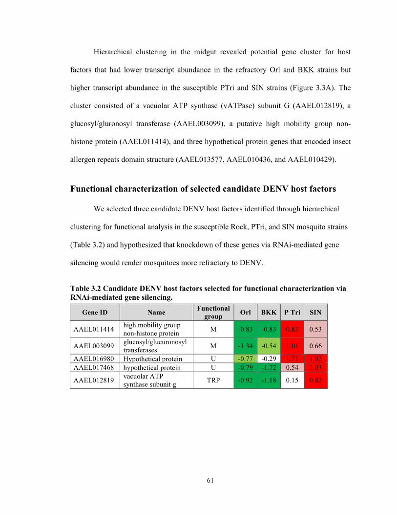
61
Hierarchical clustering in the midgut revealed potential gene cluster for host
factors that had lower transcript abundance in the refractory Orl and BKK strains but
higher transcript abundance in the susceptible PTri and SIN strains (Figure 3.3A). The
cluster consisted of a vacuolar ATP synthase (vATPase) subunit G (AAEL012819), a
glucosyl/gluronosyl transferase (AAEL003099), a putative high mobility group non-
histone protein (AAEL011414), and three hypothetical protein genes that encoded insect
allergen repeats domain structure (AAEL013577, AAEL010436, and AAEL010429).
Functional characterization of selected candidate DENV host factors
We selected three candidate DENV host factors identified through hierarchical
clustering for functional analysis in the susceptible Rock, PTri, and SIN mosquito strains
(Table 3.2) and hypothesized that knockdown of these genes via RNAi-mediated gene
silencing would render mosquitoes more refractory to DENV.
Table 3.2 Candidate DENV host factors selected for functional characterization via RNAi-mediated gene silencing.
Gene ID Name Functional group Orl BKK P Tri SIN
AAEL011414 high mobility group non-histone protein M -0.83 -0.83 0.82 0.53
AAEL003099 glucosyl/glucuronosyl transferases M -1.34 -0.54 1.01 0.66
AAEL016980 Hypothetical protein U -0.77 -0.29 1.71 1.92 AAEL017468 hypothetical protein U -0.79 -1.72 0.54 1.03
AAEL012819 vacuolar ATP synthase subunit g TRP -0.92 -1.18 0.15 0.82

62
Knockdown of the vATPase subunit G gene (AAEL012819) resulted in
significantly reduced midgut DENV titers in both the Rock and PTri strains and a non-
significant decrease in the SIN strain, suggesting that it does indeed function as a DENV
host factor (Figure 3.4). Surprisingly, knockdown of the gene encoding a HMGB protein
(AAEL011414) resulted in a significant increase in SIN strain DENV titers, but it had no
effect in the other two mosquito strains (Figure 3.4). Knockdown of the putative insect
allergen had no effect in any of the strains.
Our panel of Ae. aegypti strains is also an excellent platform for characterizing or
validating candidate DENV host factors identified through other screening
methodologies. Sessions et al. (2009) identified numerous candidate DENV host factors
by performing a high-throughput RNAi screen in Drosophila cells [12,59,93]. We
selected the Ae. aegypti orthologs of three hits from this study for functional
characterization in our susceptible mosquito strains. Only the knockdown of
AAEL002430, which codes for an N-acetylglucosamine-6-phosphate deacetylase,
resulted in significantly decreased DENV titers in the Rock strain but not in the SIN or
PTri strains (Figure 3.5).
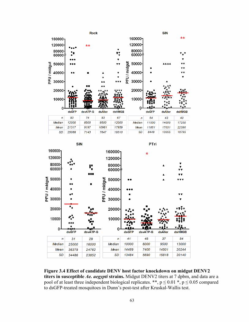
63
Figure 3.4 Effect of candidate DENV host factor knockdown on midgut DENV2 titers in susceptible Ae. aegypti strains. Midgut DENV2 titers at 7 dpbm, and data are a pool of at least three independent biological replicates. **, p ≤ 0.01 *, p ≤ 0.05 compared to dsGFP-treated mosquitoes in Dunn’s post-test after Kruskal-Wallis test.

64
Figure 3.5 Effect of candidate DENV host factor selected from high-througput screen knockdown on midgut DENV2 titers in susceptible Ae. aegypti strains. Data are a pool of at least three independent biological replicates. **, p ≤ 0.01 *, p ≤ 0.05 compared to dsGFP-treated mosquitoes in Dunn’s post-test after Kruskal-Wallis test.

65
vATPase subunits are important DENV host factors in Ae. aegypti
In addition to our results, previous high-throughput screening for insect and
human DENV host factor suggested that several vATPase subunits might serve as DENV
host factors in the mosquitoes [93,97,157]. In the previous section, vATPase subunit g
(vATP-g: AAEL012819) was experimentally confirmed as a host factor; however, the
requirement of other subunits on DENV replication was unknown. We performed RNA-
mediated gene silencing on more vATPase subunits to confirm that the function of the
whole vATPase enzyme complex, not just the vATP-g as DENV host factors. The knock
down of vATPase subunits ac39 (vATP-ac39), and vATPase subunits V0B (vATP-V0B)
resulted in a reduction in midgut DENV titers in all the mosquito strains tested ranging
from 61-98% (Figure 3.6).

66
Figure 3.6 Effect of vATPase subunits knockdown on midgut DENV2 titers in susceptible Ae. aegypti strains. Data are a pool of at least three independent biological replicates. **, p ≤ 0.01 *, p ≤ 0.05 compared to dsGFP-treated mosquitoes in Dunn’s post-test after Kruskal-Wallis test.

67
DISCUSSION
A major finding of this study is that we confirmed the role of the Toll, JAK-
STAT, and IMD pathways in controlling DENV infection in refractory Ae. aegypti
strains. Our results indicate that basal levels of mosquito immunity influence vector
competence, in addition to other factors, such as the availability of receptors or host
factors required for virus infection and replication. While these may seem to be obvious
conclusions, determinants of vector competence in natural mosquito populations have
been very poorly studied, and the idea of a basal level of immunity is not necessarily a
given.
The importance of classical immune pathways varies among strains as we
observed that activation of the JAK/STAT pathway resulted in a decreased midgut
DENV2 titers in Rock but not SIN strain. Similarly, inhibition of immune pathways in
BKK strain also results in a weaker increase of DENV2 midgut titers when compared to
the inhibition of the immune pathways in Orl strain. These results emphasized that there
is a natural variation in how each immune pathway contributes to DENV susceptibility.
Our data also suggest that while the major immune signaling pathways play a key
role in determining DENV susceptibility in both laboratory- and field-derived
mosquitoes, strain-specific factors acting independently of these pathways are also likely
to make important contributions. This panel of Ae. aegypti strains also allowed us to
functionally characterize several candidate DENV host factors, which were selected
because they displayed increased transcript abundance in susceptible strains but
decreased transcript abundance in refractory strains.

68
RNAi-mediated knockdown of the vATPase subunit G gene (vATP-g,
AAEL012819) rendered susceptible mosquito strains more refractory to midgut DENV2
infection, suggesting that it does indeed function as a DENV host factor. In the vATPase
complex, subunit G is part of a peripheral stalk connecting the peripheral domain (which
catalyzes ATP hydrolysis) and the integral domain (through which protons are
translocated), and it may play the role of stator in the rotary machinery. In yeast, deletion
of subunit G leads to complete loss of assembly of the complex [92]. In our dataset,
vATP-G was the only subunit to show a clear pattern of enriched basal-level transcript
abundance in susceptible strains. However, functional assays of other vATPase subunits
as DENV host factors by RNAi-mediated gene silencing suggested that the function of
vATPase enzyme as a whole complex is required for efficient DENV infection in Ae.
aegypti. Moreover, we observed the effect across laboratory-adapted and field-derived
mosquito strains emphasizing a crucial role of this enzyme complex in facilitating DENV
infection. vATPases are multisubunit enzymes found in the membranes of endosomes,
lysosomes, and secretory vesicles that bring about the acidification of these organelles via
an ATP-dependent rotary mechanism that drives proton transport [92,98,99,158,159].
This is an important step in the DENV replication cycle, since an acidic pH in the late
endosome is required for DENV fusion and entry. Although bafilomycin, a specific
inhibitor of vATPases, has been reported to inhibit flaviviruses in both mammalian and
insect cells [93,94,101], this is to our knowledge the first functional evidence in adult Ae.
aegypti for the role of the vATPase complex as a DENV host factor, and it suggests that
this class of enzyme could be a promising target for chemical interventions, such as the
development of small-molecule inhibitors of DENV replication.
69
The knockdown of a candidate DENV host factor, HMGB gene (AAEL011414),
unexpectedly resulted in a significant increase in SIN midgut DENV titers, suggesting
that it may function as a restriction factor instead in this strain. This is in agreement with
the relatively well-studied role of this gene family in mammalian cells, in which human
HMGB1 translocates out of the nucleus and is released from DENV-infected epithelial
and dendritic cells, triggering a pro-inflammatory antiviral response [160-162]. While our
functional data suggest that Ae. aegypti HMGB may play a similar antiviral role at least
in the SIN strain, the transcript abundance pattern of this gene across our panel runs
counter to this possibility. Since HMGB family members are also abundant in the
nucleus, where they regulate chromatin structure, transcription, and DNA repair and
replication[163], the transcription pattern we observed may have more to do with one or
more of these functions than with a response to DENV. A recent study characterizing a
separate Ae. aegypti HMGB family member (AAEL011380) confirmed that it, like
human HMGB1, effectively binds and alters the topology of DNA. The authors suggest
diverse regulatory roles for mosquito HMGB family members, for example in
vitellogenesis and molting, in addition to innate immunity[164]. This example illustrates
the idea that the transcriptome is shaped by multiple environmental factors, and it
underscores the importance of performing functional assays to validate any predictions
drawn from transcriptomic data.
High-throughput RNAi screens have proved to be a powerful method for
identifying candidate flavivirus host and restriction factors in vertebrate and invertebrate
systems [93]. Our panel of laboratory and field-derived Ae. aegypti strains with a range of
DENV susceptibilities is a valuable tool for functionally characterizing these candidates.
70
We tested the Ae. aegypti orthologs of three candidate DENV host factors identified
through an RNAi screen in Drosophila cells [93] for ability to modulate resistance to
infection by silencing them in three susceptible mosquito strains. However, only the
knockdown of a gene encoding an N-acetylglucosa- mine-6-phosphate deacetylase (N-
Gluc, AAEL002430) significantly decreased midgut DENV2 titers, and this effect was
only seen in the Rock strain and not in the SIN or PTri strains. This result was somewhat
unexpected, given the strength of these hits in the initial high-throughput RNAi screen
[93], but it again illustrates the utility of performing functional assays in adult
mosquitoes.

71
CHAPTER 4
Molecular characterization of Aedes aegypti ML and
Niemann-Pick type C family members as dengue virus
host factors
Parts of this work has been published in:
Jupatanakul N, Sim S, Dimopoulos G. Aedes aegypti ML and Niemann-Pick type C family members are agonists of dengue virus infection. Developmental and Comparative Immunology. 2014;43: 1–9. doi:10.1016/j.dci.2013.10.002 Jupatanakul N. Aedes aegypti ML and Niemann-Pick type C family members are agonists of dengue virus infection. Master thesis. 2012.

72
ABSTRACT
Upon exposure to dengue virus, the Aedes aegypti mosquito vector mounts an
anti-viral immune defense by activating the Toll, JAK/STAT, and RNAi pathways,
thereby limiting infection. While these pathways and several other factors have been
identified as dengue virus antagonists, our knowledge of factors that facilitate dengue
virus infection is limited. Previous dengue virus infection-responsive transcriptome
analyses have revealed an increased mRNA abundance of members of the myeloid
differentiation 2-related lipid recognition protein (ML) and the Niemann Pick-type C1
(NPC1) families upon dengue virus infection. These genes encode lipid-binding proteins
that have been shown to play a role in host-pathogen interactions in other organisms.
RNAi-mediated gene silencing of a ML and a NPC1 gene family member in both
laboratory strain and field-derived Ae. aegypti mosquitoes resulted in significantly
elevated resistance to dengue virus in mosquito midguts, suggesting that these genes play
roles as dengue virus agonists. In addition to their possible roles in virus cell entry and
replication, gene expression analyses suggested that ML and NPC1 family members also
facilitate viral infection by modulating the mosquito’s immune competence. Our study
suggests that the dengue virus influences the expression of these genes to facilitate its
infection of the mosquito host.

73
INTRODUCTION
Through genome-wide transcriptomic analyses, in conjunction with RNAi-
mediated gene silencing, we have identified the Toll and JAK-STAT pathways as key
DENV antagonists that act by controlling virus restriction factors [12,59,103]. DENV
infection-responsive transcriptome analyses have revealed that the transcript abundance
of five members of two lipid-binding protein gene families, the myeloid differentiation 2-
related lipid recognition protein (ML) and Niemann Pick-type C1 (NPC1) families, is
increased in response to DENV infection. Since DENV is an enveloped virus and its
outer shell is lipid-based, these lipid-binding proteins are likely to play a role(s) in
mosquito-virus interactions.
The ML domain is a lipid recognition protein domain found in several proteins
with lipid-binding properties [100,157,158]. Members of this family have diverse
functions associated with lipid recognition, including pathogen recognition, lipid
trafficking and metabolism, and pheromone perception [98,99,105,106,158,159]. A role
for the ML domain in immune recognition has been described for the vertebrate MD2
protein and its insect homologs. MD2 is a secreted glycoprotein that mediates the
activation of the vertebrate Toll-like receptor 4 (TLR4) upon exposure to bacterial
lipopolysaccharide (LPS) [101,165,166]. Drosophila MD2 homologs have been shown to
mediate the activation of the immune deficiency (IMD) immune signaling pathway upon
exposure to lipopolysaccharide (LPS) [160,167]. The An. gambiae homolog of ML,
AgMDL1, is involved in the mosquito’s immune defense against Plasmodium falciparum
infection [103,168]. Niemann-Pick disease type C1 (NPC1) is another class of lipid-
binding proteins that is responsible for cholesterol transport and homeostasis; these

74
proteins function together with the NPC2 proteins in the late endosomal/lysosomal
system [12,40,100,158]. NPC1 has been shown to be required for the Ebola virus to
escape from the vesicular compartment [97,105,106], but the function of the NPC1
family in DENV infection in mosquito has yet not been investigated.
Although these lipid-binding protein families have been shown to be involved in
virus-host interaction and immune responses in various systems, little is known about
their function in the Ae. aegypti-DENV interaction. Here, we investigated the role of Ae.
aegypti ML and NPC1 gene family members in modulating DENV infection in the
mosquito by conducting RNAi-mediated gene silencing and gene expression studies. Our
results suggest roles for the ML and a NPC1 proteins as agonists of DENV in the
mosquito. Furthermore, our data suggest that the virus might influence the expression of
these genes to facilitate its infection, emphasizing the importance of lipid-binding
proteins in viral infection of insects.
75
MATERIALS AND METHODS
Bioinformatics analyses and genes selection
The gene sequences and gene annotations for the insect ML and NPC gene
families were obtained from the ImmunoDB (http://cegg.unige.ch/Insecta/immunodb)
and Vectorbase (http://aaegypti.vectorbase.org/) databases [12,165,166]. To compare
sequence similarity, a multiple sequence alignment (MSA) was generated using t-coffee
software (http://www.tcoffee.org/) [156,167]. The MSA was then used to generate a
phylogenetic tree using MEGA 5.05 software [168,169]. ML and NPC genes that
potentially play a role in DENV infection were suggested from the transcriptional
changes of these genes in previous microarray-based transcriptome studies [12,40,169],
Dimopoulos group, unpublished data).
Mosquito strains and mosquito maintenance
The mosquitoes used for most of the experiments were of the Ae. aegypti
Rockefeller/UGAL strain. A second mosquito strain was used to confirm that the results
were common among different mosquito strains. This strain was a recently colonized Ae.
aegypti population obtained from the Caribbean island of Saint Kitts (sixth to seventh
generation) [97]. The mosquitoes were maintained on a 10% sugar solution at 27oC and
95% humidity with a 12-hr light/dark cycle, following the protocol described previously
[12].
Cell culture
The Ae. albopictus C6/36 cell line was used to propagate DENV. It was
maintained in complete minimum essential medium (MEM) supplemented with 10%

76
heat-inactivated FBS, 1% L-glutamine, 1% MEM non-essential amino acids, 10 unit/mL
penicillin, and 10 µg/mL streptomycin at 32oC and 5% CO2.
The baby hamster kidney (BHK) cell line was used for plaque assays to determine
DENV titter. BHK cells were maintained in complete Dulbecco’s modified Eagle’s
(DMEM) medium supplemented with 10% heat-inactivated FBS, 1% L-glutamine, 10
unit/mL penicillin, 10 µg/mL streptomycin, and 5 µg/mL plasmocin at 37oC and 5% CO2.
Genes silencing by RNA interference
The role of the ML and NPC1 genes in DENV infection in Ae. aegypti was
assessed using RNA interference-mediated gene silencing as described previously [156].
In brief, dsRNAs were constructed using in vitro transcription with the HiScribe™ T7 In
Vitro Transcription Kit (New England Biolabs). Approximately 200 ng of dsRNA was
injected into the thorax of cold-anesthetized 3- to 4-day-old female mosquitoes using a
nano-injector. The dsRNA-injected mosquitoes were kept in the insectary under the
conditions mentioned above. Gene silencing efficiency, evaluated using real-time PCR,
was determined by comparison to the GFP dsRNA-injected group at 3 days after dsRNA
injection, the time when we infected mosquito with DENV.
DENV propagation and viral infection in the mosquito
The DENV strain used in these experiments was DENV serotype 2 (New Guinea
C strain, DENV-2). The virus was propagated in the C6/36 cell line according to a
protocol previously described [169]. In brief, DENV stock was added to a 75-cm2 flask of
C6/36 cells at 80% confluence to yield a multiplicity of infection of 1. The virus-infected
cells were harvested 6 days after infection. The virus was extracted from the cells by
freezing and thawing for two cycles in dry CO2 and a 37°C water bath, centrifuged at
77
800g for 10 min, and mixed 1:1 with commercial human blood. The infectious blood
meal (~106-107 colony-forming units [cfu]/ml) was maintained at 37°C for 30 min prior
to membrane feeding to 5- to 7-day-old or 3-day post-dsRNA-injected mosquitoes.
Blood-fed mosquitoes were separated on ice and maintained under the conditions
mentioned above.
DENV titration by plaque assay
Virus titers in the midguts were determined at 7 dpbm according to an established
protocol [12,169]. Mosquito midguts were dissected in sterile 1XPBS and stored in
complete DMEM medium at -80°C until used. Midgut samples were homogenized using
a homogenizer (Bullet Blender, Next Advance) with 0.5-mm glass beads. The virus-
containing homogenates were 10-fold serially diluted in DMEM media, then inoculated
onto 80% confluent BHK cells in 24-well plates. The cell monolayer was then overlaid
with 0.8% methylcellulose in complete DMEM medium, and kept for 5–7 days at 37 °C
and 5% CO2. Plaque forming units (PFUs) were visualized by crystal violet staining. All
procedures involving DENV-2 infections were carried out in a Biological Safety Level 2
laboratory.
Gene expression and silencing efficiency analysis by quantitative PCR
Gene silencing efficiency and expression of the genes of interest were assayed
using real-time PCR. Whole mosquito samples were used for the silencing efficiency
analysis while different mosquito organs were used for gene transcript abundance
analyses. Mosquito samples were dissected in 1XPBS, collected in RLT buffer
(QIAGEN), and then stored at -80°C until extraction. Total RNA was extracted from
tissue samples using the RNeasy Mini Kit (QIAGEN). To construct cDNA, RNA samples
78
were treated with Turbo DNase (Ambion) before reverse transcription with a MMLV
Reverse Transcriptase kit (Promega) according to the manufacturer’s instructions. The
cDNA was then used to determine gene expression by quantitative PCR using SYBR®
Green PCR Master Mix (Applied Biosystem). Transcript abundance of genes was
compared to the expression of the ribosomal protein gene S7 as a normalization control.
The primers specific for each gene are presented in Appendix 1.
Statistical analysis of midgut DENV titer and gene expression level
The DENV midgut titers of GFP dsRNA and experimental groups were compared
using the plaque assay results from at least two biological replicates, with the elimination
of outliers (data outside the median plus 2SD). The significance values were determined
using either the Kruskal-Wallis test with Dunn’s post-test or the Mann-Whitney test (*
p<0.05, ** p<0.01).
RESULTS
ML and NPC1 gene families are distinct and expanded in Ae. aegypti
The Ae. aegypti genome contains 26 ML-like genes, suggesting an expansion of
this gene family when compared to other insects (19 in Culex pipiens quinquefasciatus,
15 in Anopheles gambiae, and 8 in Drosophila melanogaster) (immunoDB database
(http://cegg.unige.ch/Insecta/immunodb)) [166,170]. Of these 26 members, 24 contain
the ML domains and are annotated as Niemann-Pick C 2 (NPC2) genes, while the
remaining two are annotated as Niemann-Pick C1 (NPC1) proteins. However, further
sequence analysis revealed that the putative NPC1 does not contain the MD2 domain.
Thus, the Ae. aegypti NPC2 genes will be considered as ML gene family genes (as per

79
the annotation in immunoDB database). All six cysteine residues are conserved in all the
ML genes used for analysis, suggesting their importance for protein structure and
function across organisms. Phylogenetic analysis of the ML genes suggests four groups
of Ae. aegypti ML genes, with groups 2 being unique to Ae. aegypti (Figure 4.1A). The
amino acid sequence of AaegML13 is the most conserved when compared to the human
NPC2 gene. The conserved cholesterol-binding pocket amino acids of the mouse NPC2
proteins, F66 V96 and Y100 [171], were mutated in some Aedes ML genes (Appendix 4).
While the human genome contains only one NPC1, the mosquito genomes of C.
pipiens quinquefasciatus, Ae. gambiae, and Ae. aegypti contain two NPC1 genes (NPC1a
and NPC1b), as in Drosophila (Figure 4.1B) [172]. The alignment revealed the presence
of all 13 conserved transmembrane domains and a sterol-sensing domain in NPC1b; in
contrast, NPC1a has approximately 150 amino acids truncated at N-terminus.
80
Figure 4.1 Phylogenetic tree of the ML and NPC1 gene family in insects. (A) All the Ae. aegypti ML family members were compared to all eight D. melanogaster ML genes (DmNPC2 A–F), An. gambiae ML1 (AgMDL1), Homo sapiens MD2 (HsMD2), H. sapiens NPC2 (HsNPC2), and Mus musculus NPC2 (MmNPC2). (B) The Ae. aegypti NPC1 genes were compared to D. melanogaster (Dm), Anopheles gambiae (Ag), Culex

81
pipiens quinquefasciatus (Cp), and H. sapiens (Hs). The genes selected for further study are indicated by filled circles.
Ae. aegypti ML and NPC1 family members facilitate DENV infection
Using genome-wide transcriptome analyses, we have previously shown that the
mRNA abundance of members of the Ae. aegypti NPC1 and ML gene families (3 of 24
members) were found to be modulated by DENV infection in the mosquito midgut and
carcass tissue compartments [12,40], suggesting possible roles for these proteins as
DENV host or restriction factors (data is presented in Appendix3). Upon DENV
infection, the transcript abundance of AaegML13 and AaegML33 increased in the
mosquito midgut (2.2 fold) and carcass (2.8 fold), respectively, and the mRNA
abundance of AaegNPC1b increased in the midgut compartment (1.9 fold). We further
investigated whether any of these genes is involved in modulating DENV infection in the
midgut by using RNAi-mediated gene silencing in laboratory Rockefeller/UGAL strain
Ae. Aegypti, followed by infection with DENV via blood feeding (Figure 4.2). Silencing
of AaegNPC1b and AaegML33 resulted in a significant reduction in the median DENV
titer in mosquito midguts by 81.1% and 43.24%, respectively, at 7 dpbm. This result
suggests that these genes represent potential DENV agonists that could facilitate virus
infection of the mosquito midgut. The silencing of AaegML13, which is closely related to
the human ML gene, had no significant effect on midgut DENV infection. The following
experiments will focus only on the characterization of the AaegNPC1b and AaegML33
since the silencing of these genes resulted in phenotypic change.
82
Figure 4.2 Effect of Ae. aegypti ML and NPC1 gene knockdown on midgut DENV titers. Members of the ML and NPC1 gene families were silenced in Ae. aegypti mosquitoes, and midgut DENV titers at 7 dpbm were determined by plaque assay. Data represent a pool of 2–3 biological replicates with the elimination of outliers (data outside the median plus 2SD). p-values were determined by comparing the experimental group with the dsGFP-treated group in the Mann–Whitney test (*p < 0.05, **p < 0.01).
Functions of AaegNPC1b and AaegML33 as DENV host factors are
conserved in field-derived strain of Ae. aegypti
Since the introduction and maintenance of mosquitoes into a laboratory
environment can impose a selection bias that can lead to genetic differences between
laboratory-adapted mosquitoes and the natural mosquito population, we investigated
whether the influence of AaegNPC1b and AaegML33 on DENV infection is conserved in
field-derived mosquitoes. Silencing these genes in the recently colonized Ae. aegypti

83
strain from Saint Kitts, [97] prior to feeding on DENV-infected blood, resulted in
significantly lower midgut DENV titers than in the controls, just as we had observed for
the laboratory strain (Figure 4.3).
Ae. aegypti NPC1 and ML genes may influence DENV infection through
the same mechanism or pathway
Although silencing of some ML and NPC1 genes significantly reduced DENV
infection in the mosquito midgut, how these genes function as DENV agonists was still
unknown. To investigate whether these genes were acting as DENV agonists through
independent mechanisms or the same mechanism/pathway, we compared the effects of
their independent and combined gene silencing on mosquito susceptibility to virus
infection. The silencing efficiency of each gene was similar in both single- and double-
silencing experiments (Figure 4.4). The double-silencing of these genes lowered DENV
infection of the midgut to the same level as did independent silencing of each gene,
without an apparent synergistic effect (Figure 4.5). This result suggested that these two
genes influence DENV infection through a similar or same mechanism, as no additive
effect was observed upon double-silencing.

84
Figure 4.3 AaegNPC1b and AaegML33 silencing resulted in lower midgut DENV titers in field-derived mosquitoes from Saint Kitts. Data represent a pool of four biological replicates with the elimination of outliers (data outside the median plus 2SD). p-values were determined by comparing the experimental group with the dsGFP- treated group in the Mann–Whitney test (*p < 0.05, **p < 0.01

85
Figure 4.4 Silencing efficiency for AaegNPC1b and ML33 in Rock and Kitts strains Ae. aegypti. (A) Silencing efficiency in Rockefeller/UGAL strain. (B) Silencing efficiency in Kitts strain Figure 4.5 Ae. aegypti NPC1b and ML33 may influence DENV infection through the same mechanisms. Midgut DENV titers was assayed upon single- and double-silencing of AaegNPC1b and AaegML33 at 7 dpbm. Data represent a pool of three biological replicates with the elimination of outliers (data outside the median plus 2SD). p-values were determined by comparing the experimental group with dsGFP-treated group in the Kruskal–Wallis one-way ANOVA test (*p < 0.05, **p < 0.01).

86
AaegNPC1b and AaegML33 may regulate Ae. aegypti immune
pathways
Since the transient silencing of AaegNPC1b and AaegML33 resulted in a lower
midgut DENV level, we wanted to investigate whether this resistance could have resulted
from altered Toll, Imd, and/or JAK/STAT pathway activity in response to AaegNPC1b
and AaegML33 gene silencing. Activation of the Toll, Imd, and JAK/STAT pathways
results in increased antimicrobial peptide (AMP) gene expression [12,59,160]. We
hypothesized that if AaegNPC1b and AaegML33 play a role in regulating these immune
pathways (as potential negative regulators), silencing these genes would result in changes
in the transcript abundance of AMPs and other effector genes. To test this hypothesis, we
assayed the transcript abundance of AMPs and other effector molecules after silencing
AaegNPC1b and AaegML33. We measured transcript abundance of immune effector
genes in the midgut and fat body since they represent major immune tissues and the
midgut is the first mosquito immune-barrier that the virus encounters [12]. The fat body
is mainly responsible for humoral and systemic immune responses. Mosquito midguts
and fat bodies were collected at 1 and 3 days post-dsRNA injection to assay the transcript
abundance of AMPs and other effector molecules (Figure 4.6). The AMPs analyzed in
this study were defensin E, cecropin E, lysozyme C, diptericin, and gambicin. Although
they do not belonging to the AMP class, we also assayed SOCS36E and DVRF1 because
they are regulated by the anti-dengue JAK/STAT pathway [12,45,59,173] and can be
used as markers of JAK/STAT activation. The transcript abundance patterns of these
proteins upon immune pathway activation, as determined by previous studies, are
presented in Table 4.1.

87
Overall, the transcript abundance of several AMP genes was influenced by
AaegNPC1b and AaegML33 silencing, either in the midgut or fat body, and the
differential transcript abundances were greater at 3 days than at 1 day post-gene
silencing. The silencing of AaegNPC1b resulted in an increase in cecropin E and
lysozyme C transcript abundance, suggesting a possible activation of the Toll pathway,
since these genes have been shown in previous studies [12] to be induced after Toll
pathway activation. The up-regulation of DVRF1 transcripts after AaegNPC1b silencing
also suggested the possible activation of the JAK/STAT pathway [59]. The silencing of
AaegML33 resulted in an increase in SOCS transcript abundance at 1 day post-dsRNA
injection, as has been seen for activation of the JAK/STAT pathway [59]. The transcript
abundance of both SOCS36E and DVRF1 in fat body tissue was decreased at 3 days
post-silencing of AaegML33 (Figure 4.6D). Defensin E transcripts were enriched in the
midgut tissue after the silencing of both AaegNPC1b and AaegML33, suggesting an
activation of the Imd pathway, since this AMP was induced by silencing of the Imd
pathway negative regulator Caspar [12].
Table 4.1 Expression pattern of the selected immune genes from previous microarray datasets in dengue virus infected midgut (DV MG), dengue virus infected carcass (DV Car), Cactus silenced (Toll activated), Caspar silenced (Imd activated), and PIAS silenced (JAK/STAT activated). Name Accession# DV MG DV Car Cactus Caspar PIAS Defensin E AAEL003849 0.824 -0.811 1.697 -0.525 Diptericin AAEL004833 -0.546 Cecropin E AAEL000611 1.33 -1.127 Lysozyme C7B AAEL015404 0.935 1.007 1.105 Gambicin AAEL004522 1.118 0.851 -1.406 0.85 SOCS36E AAEL000393 0.909 0.539 DVRF1 AAEL008492 0.559 2.517 0.79
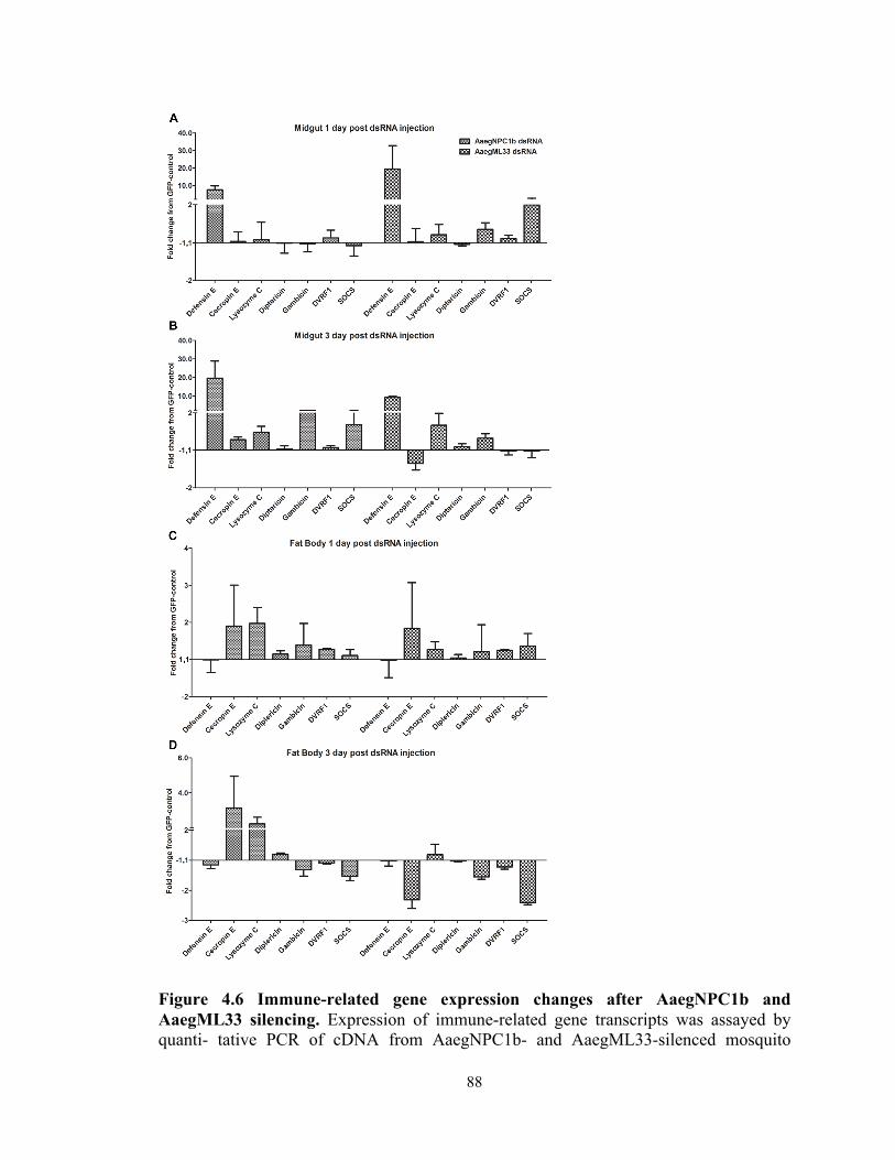
88
Figure 4.6 Immune-related gene expression changes after AaegNPC1b and AaegML33 silencing. Expression of immune-related gene transcripts was assayed by quanti- tative PCR of cDNA from AaegNPC1b- and AaegML33-silenced mosquito

89
tissues. Gene expression levels were assayed from three biological replicates. Error bars represent the standard error of the mean (SEM)
Expression patterns and tissue tropisms of AaegNPC1b and AaegML33
Tissue-specific infection-responsive gene expression patterns can provide
information about the putative functions of genes during the course of infection. We used
real-time PCR to compare the transcript abundance of AaegNPC1b and AaegML33 in the
midgut and fat body. AaegNPC1b was highly expressed almost exclusively in the midgut
tissue, whereas AaegML33 was expressed at similar levels in both tissues (Figure 4.7).
Figure 4.7 Transcript abundance of AaegNPC1b and AaegML33 in the midgut, and fat body in uninfected mosquitos. The graphs show a transcript abundance of AaegNPC1b and AaegML33 relative to the transcript abundance of ribosomal S7 gene as a normalization control. The data was an average of three pools of 14-day old mosquitoes. Transcript abundances were assayed from three biological replicates. Error bars represent the standard error of the mean (SEM).
We then investigated the infection-responsive changes in expression of these
genes by comparing their transcript abundance between DENV-infected and naïve blood-
fed mosquitoes at 1, 3, 7, 10, and 14 dpbm. After DENV bloodmeal, at 1 dpbm,

90
AaegNPC1b transcript abundance in the midgut was decreased in comparison to
uninfected controls (a 1.59-fold decrease). The abundance of AaegNPC1b transcripts in
the midgut increased at 3 dpbm and peaked at 7 dpbm (a 2.15-fold increase), then
decreased at the later time points (Figure 4.8A). AaegML33 transcripts were enriched in
the midgut throughout the infection (a 1.99- to 18.85-fold increase) (Figure 4.8B). The
AaegML33 transcripts in the fat body were also up-regulated at 1, 7, 10, and 14 dpbm
(3.23-, 6.40-, 1.82-, and 3.99-fold increases, respectively) (Figure 4.8C).

91
Figure 4.8 AaegNPC1b and AaegML33 gene expression in the midgut, and fat body over the time course of DENV infection. Relative expression of these two genes was compared between DENV-bloodfed and naïve-bloodfed mosquitoes. The gene expression
AaegNPC1b-midgut
1dpbm 3dpbm 7dpbm 10dpbm 14dpbm-3
-2
-1
0
1
2
3
Days post-bloodmeal
Rel
ativ
e fo
ld c
hang
e fr
om n
aive
-blo
odm
eal
AaegML33-midgut
1dpbm 3dpbm 7dpbm 10dpbm 14dpbm1
2
3
4
510
20
30
40
Days post-bloodmeal
Rel
ativ
e fo
ld c
hang
e fro
m n
aive
-blo
odm
eal
AaegML33-fat body
1dpbm 3dpbm 7dpbm 10dpbm 14dpbm
-4
-3
-2
-1
0
1
2
4
8
12
Days post-infection
Rel
ativ
e fo
ld c
hang
e fr
om n
aive
-blo
odm
eal

92
was compared relative to the ribosomal S7 gene, and was assayed from three biological replicates. Error bars represent the standard error of the mean (SEM).
DISCUSSION
The ML family comprises of proteins that contain the MD2-related lipid-binding
(ML) domain, which has been extensively studied in the humansfor its LPS-binding
property and its role as a TLR4 co-receptor [101]. The immune-related function of the
ML gene family was recently characterized in the insect model organism, D.
melanogaster [160]. Three of the eight Drosophila ML genes (DmNPC2a, DmNPC2e,
and DmNPC2h) were shown to bind to LPS, peptidoglycan, and lipoteichoic acid. Over
expression of recombinant DmNPC2a and DmNPC2e in the Drosophila S2 cell line
activates the promoter of the anti-microbial peptide diptericin, which is controlled by the
IMD immune signaling pathway [160,170]. The An. gambiae ML protein AgMDL1 is
also implicated in the anti-Plasmodium response [103]. Here we investigated the
function of the ML and NPC genes in the Ae. aegypti response to DENV infection. Since
DENV is an enveloped virus, these lipid-binding proteins might be involved in
interactions with the DENV and modulate immune responses.
According to the immunoDB database, the Ae. aegypti ML gene family consists
of 26 members [166]. Our analysis of Ae. aegypti amino acid sequences, however,
suggested that only 24 genes actually contain the ML domain. The other two ML genes
reported in the database belong to the Nieman-Pick C1 protein family. This discrepancy
in annotation might be due to the similar names of NPC1 and NPC2, but in fact, these
two protein families have different protein structures. NPC2 is a soluble protein
containing an ML domain, whereas NPC1 is a transmembrane protein containing 13

93
transmembrane domains, with a sterol-sensing domain located between the third and
seventh transmembrane domains [158]. Both the NPC1 and NPC2 proteins reside and
work closely together in the late endosomal/lysosomal compartment of the cells and
facilitate cholesterol trafficking and metabolism [158,172,174].
Multiple sequence alignment of the Ae. aegypti ML family revealed variations in
ML conserved amino acids, as reported for the mouse ML gene [171]. This variation
implies that the insect ML family, and especially Ae. aegypti, is under high selective
pressure to expand its spectrum of biological functions and lipid-binding properties.
According to the phylogenetic analysis, as well as previous classification of the DmNPC2
[170], Ae. aegypti ML genes can be classified into four different groups. There are eight
ML genes in group 2 that are unique to Ae. aegypti and lack Drosophila homologs,
suggesting an expansion of these ML genes during evolution (Figure 4.1A). The
Drosophila NPC2 a-h genes were grouped as in previous studies, indicating the accuracy
of the tree [160,170]. The DmNPC2a mutant has been shown to possess physiological
defects and disordered cholesterol metabolism, similar to that observed with the human
NPC2 mutant [170,174]. Thus, AaegML13, which clustered together with DmNPC2a and
human NPC2, may have a similar function, but this relationship was not the focus of our
study and still needs to be experimentally confirmed. AaegML1 clustered together with
AgMDL1, an anti-Plasmodium ML gene [103], but its transcript abundance was not
influenced by DENV infection in any tissue compartment, suggesting that it might not
play a role in DENV infection. This result is not entirely unexpected, since the mosquito
uses different mechanisms to combat different pathogens.
94
The data from previous genome-wide transcriptomic analyses revealed that the
transcript abundance of several ML genes was influenced by DENV infection in the
midgut and carcass tissue compartments. The differences in transcript abundance suggest
that the Ae. aegypti ML genes might have diverse functions in different tissue
compartments. Functional screening by RNAi-mediated gene silencing of one of the two
selected ML genes (AaegML33) resulted in lower DENV midgut titers (Figure 4.2),
suggesting that they facilitate DENV infection, underscoring the potential role of the ML
gene family members as DENV agonists. AaegML33 was also of particular interest, since
it belongs to the distinct Ae. aegypti ML group 3, suggesting that it might have a
specialized DENV-related function in Ae. aegypti.
Studies of the Drosophila NPC1 gene revealed two NPC1 genes in its genome,
NPC1a and NPC1b [172,175,176]. All the genomes of insects used for phylogenetic
analysis also contain two NPC1 genes, suggesting a duplication of the gene to NPC1a
and NPC1b in a common insect ancestor (Figure 4.1B). NPC1a is an insect NPC1 gene
more closely related to human NPC1 and is required for molting and sterol homeostasis
in Drosophila [158,172,174]. NPC1a mutant larvae have abnormally high levels of
accumulated sterol in cells and are unable to molt [172]. Hence, the Ae. aegypti NPC1a
might have a similar lipid-related function. The other NPC1 gene, NPC1b, has also been
studied in Drosophila and found to be involved in sterol absorption in the midgut
epithelium. The study of DmNPC1a and DmNPC1b revealed that they have non-
redundant roles in sterol homeostasis and are not interchangeable; DmNPC1a is
important for sterol trafficking, while DmNPC1b is important for sterol absorption [176].
However, the function(s) of Ae. aegypti NPC1 in both lipid homeostasis and immunity
95
are yet to be determined. In Ae. aegypti, the transcript abundance of AaegNPC1 genes
has been shown to be altered by DENV infection in both midgut and carcass tissue
compartments, suggesting a possible role for Ae. aegypti NPC1 genes in DENV infection.
AaegNPC1b was of particular interest to us because it is involved in lipid absorption in
the midgut and might interact with DENV during that cellular process. The role of NPC1
as a host factor for virus infection has also been suggested in mammals, since Ebola virus
can hijack human NPC1 for viral entry into the cytosol [105,106,177]. Silencing of
AaegNPC1b resulted in lower midgut DENV titers, emphasizing its potential role in
DENV infection (Figure 4.2).
The functions of AaegNPC1b and AaegML33 with regard to DENV infection are
likely to be conserved across Ae. aegypti strains and populations, since a similar level of
resistance was observed after silencing of these genes in a strain of recently colonized
field mosquitoes (Figure 4.3). Simultaneous silencing of AaegNPC1b and AaegML33
was conducted to provide an indication as to whether these genes might affect DENV
infection through a similar mechanism, and indeed, we saw comparable levels of
resistance when they were silenced individually (Figure 4.5).
Based on the infection data and previous studies of ML genes, we hypothesized
that AaegNPC1b or AaegML33 could be involved in modulating mosquito immune
responses to DENV infection. The silencing of these genes resulted in the activation of
immune pathways, of which the Toll and JAK/STAT pathways are of particular interest,
since they have been reported to control DENV infection in the mosquito
[12,45,59,173,178]. To investigate this hypothesis, we measured the expression of
immune pathway-regulated gene transcripts after transient silencing of AaegNPC1b and

96
AaegML33. Our study showed that the transcript abundance of AMPs and other
JAK/STAT-regulated genes was influenced by AaegNPC1b/AaegML33 silencing,
suggesting that they playing a role as negative regulators of these anti-DENV innate
immune pathways (Figure 4.6).
The expression pattern of the pathway-regulated genes upon AaegNPC1b and
AaegML33 gene silencing was similar to the pattern obtained when the Toll, Imd, and
JAK/STAT pathways were activated (Table 4.1) [12,59,176]. We observed an increase of
SOCS gene transcript abundance, which suggested an activation of the JAK/STAT
pathway. The dynamic changes in SOCS gene expression likely reflects feedback loops
in the respective JAK/STAT pathway transcription circuits, as has been shown for the
Drosophila SOCS36E [31,54]. A prominent up-regulation of defensin E in the midgut
occurred after the silencing of both AaegNPC1b and AaegML33 (Figure 4.6A and B).
This increased transcript abundance of defensin E in the midgut suggests that the
activation of the Imd pathway, or some other unknown immune pathway, might also
influence DENV infection, as has been shown in Ae. aegypti salivary glands [41,179].
Different expression pattern of Defensin E and other immune effector genes in the
midgut and fat body also suggested that transcription of these genes is controlled
differently in different mosquito tissues and cell types, as has been shown previously
[12,59,84,180]. Immune genes can be controlled by multiple immune pathways, and the
transcriptional regulation by an immune signaling pathway can also be fine-tuned by
different factors. AaegNPC1b and AaegML33 might serve as alternative regulators of the
immune signaling pathways. A previous study has shown that DENV can suppress
immune responses in a mosquito cell line, but the mechanism was not investigated

97
[97,177]. Changes in AMP expression after silencing AaegNPC1b and AaegML33
emphasize their role as immune pathway antagonists, and the up-regulation of the
AaegNPC1b and AaegML33 by DENV infection is likely reflecting one of the
mechanisms the virus uses to suppress mosquito immune responses. The function of the
ML gene as negative regulator was also shown in humans; a splice variance of the MD2
protein (MD2s) can inhibit TLR4 signaling by competing with normal MD2 for binding
to TLR4 [12,40,178].
Tissue-specific transcript abundance analysis revealed that in uninfected
mosquitoes, AaegML33 was constitutively expressed in the midgut and fat body-
containing carcass (Figure 4.7). In contrast, AaegNPC1b expression was limited mainly
to the mosquito midgut, which corresponds to the expression of NPC1b in Drosophila
[97,176], suggesting that the biological role of the NPC1b gene may be conserved among
insects. The increase in the transcript abundance of the AaegNPC1b and AaegML33
genes after the ingestion of a DENV-infected blood meal (Figure 4.8) suggests that
DENV may up-regulate the expression of both genes to prevent immune activation or
suppress mosquito immune responses. The dynamic temporal changes of their expression
patterns likely reflect variations in virus titer and propagation of virus infection through
different host cells and tissues during the course of infection.
This is the first report to show that two lipid-binding protein families, ML and
NPC1, play a role as DENV agonists in Ae. aegypti. The silencing of these genes resulted
in higher resistance to DENV in the mosquito midgut, likely through altered regulation of
immune pathways. These effects were conserved between laboratory-adapted and field-
derived Ae. aegypti. Moreover, our study suggests that DENV might influence the

98
expression of these genes to facilitate efficient virus infection. The direct interaction of
the ML and NPC1 genes with DENV and how these molecules modulate immune
signaling pathways are yet to be investigated.
99
CHAPTER 5
Conclusion and General Discussion
Dengue has remained a significant public health concern for a number of decades;
unfortunately, current dengue controls strategies, such as vector population reduction
using insecticides, have been shown to be both inefficient and costly, and novel disease
control strategies are needed to reduce burden of dengue. Dengue transmission-blocking
through self-propagating genetically-modified mosquitoes has been deemed as a
logistically simpler and likely cheaper disease control strategy compared to the use of
vcaccines and drugs that, nevertheless, still under development. However, little is known
about the interactions between the dengue virus and its insect vector for the development
of transgenic strategies, and there is therefore a great need for further research in this
area.
The recent availability of the Ae. aegypti genome [31,95] together with genomic
analyses tools, such as Vectorbase [136,179], ave accelerated research on dissecting
interactions between DENV and mosquito vector. Previous studies identified the Toll,
JAK/STAT, and RNAi pathways as anti-DENV defense systems in Ae. aegypti
[12,59,84,139]; however, molecular the mechanisms of how these pathways inhibit
DENV were still largely unknown. It was also unknown whether these pathways could be
used for DENV transmission control. To identify and characterize DENV restriction
factors and host factors in Ae. aegypti, this thesis research has used different approaches
and tools, such as genetically modified mosquitoes with inducible JAK/STAT pathway
activation, a panel of field-derived and laboratory strains of Ae. aegypti with different

100
DENV susceptibility [97], as well as previous microarray-based transcriptomic datasets
[12,40].
First we studied the interactions between DENV and Ae. aegypti by using a
transgenic approach to manipulate the activation of the mosquitoes’ immune pathway,
specifically the JAK/STAT pathway. We generated transgenic Ae. aegypti
overexpressing the JAK/STAT pathway components, Dome and Hop, under the control
of a blood meal-inducible fat body-specific vitellogenin promoter. These genetically
modified mosquitoes showed an increased resistance to DENV infection, perhaps
because they had higher expression of dengue virus restriction factors and lower
expression of DENV host factors than did wild-type mosquitoes. DENV, as RNA virus,
is prone to mutations which allows the virus to evade control strategies. The multiple
factors contributing to DENV resistance in our transgenic lines reduce the chance of
DENV to mutate to overcome the resistance.
We also showed that these transgenic mosquitoes were useful for studying the
molecular interactions between DENV and Ae. aegypti. Through transcriptomic
comparisons by microarray-based methodology together with functional confirmation by
RNAi, we identified a gene with unknown function (AAEL007703) as a putative DENV
restriction factor, and the SCP2 and DDX genes as putative DENV host factors.
This study is the first to provide a proof-of-concept that genetic engineering of the
mosquitoes’ JAK/STAT immune pathway can be used to render the insect more resistant
to DENV infection and possibly to block transmission of the disease through further
development of the technology. Mosquito transgenesis technique has been employed as a
Ae. aegypti population suppression approach through the sterile insect technique (SIT) in

101
several countries including Brazil, Panama, Malaysia, and the Cayman islands [181-183].
A biotechnology company, Oxitec, has released genetically modified sterile male Ae.
aegypti in the field and genetically modified Ae. aegypti carries a lethal gene that
prevents offsprings of the cross between the genetically modified Ae. aegypti and wild
mosquitoes to survive to adulthood. One advantage that a transgenic Ae. aegypti with
increased DENV resistance has over Oxitec’s technology is that it will not require
constant production and release of transgenic mosquitoes into the field., but function as a
logistically simpler and more cost-effective self-propagating system. Offsprings the
transgenic mosquitoes can survive to adulthood and spread the transgene to next
generations of mosquitoes through a genetic drive mechanism.
Our lab has previously established a panel of laboratory and field derived strains
with various degrees of DENV susceptibility, and transciptomic profiles of these
mosquitoes suggested that basal level of immune activity might contribute to the
differences in DENV susceptibility [97]. In this study we confirmed this hypothesis by
using RNAi-mediated gene silencing to activate or inhibit the Toll, Imd, JAK/STAT, and
RNAi pathways, and confirm that these pathway contribute to the natural differences in
DENV susceptibility for different strains to certain degrees. Transciptomic profiles of
these mosquitoes also showed differential expression of hundreds of genes belongoing to
various functional groups which play potential roles in DENV infection in Ae. aegypti.
Hierarchical cluster analyses revealed differential transcript abundance of several DENV
host and restriction factors among DENV-susceptible and -refractory strains. Functional
confirmation by RNAi-mediated gene silencing assays emphasized an importance of the
vATPase enzyme as a DENV host factor in natural Ae. aegypti populations, suggesting it

102
may be a candidate intervention to decrease DENV transmission. The function of
vATPase genes as a DENV host factor confirmed in this study also support our findings
from Study 1 that the higher DENV resistance in VgHop mosquitoes might be a result
from lower expression of vATPase genes. Subsequent experiments by our group applied
this knowledge in a more translational fashion using a chemical inhibitor of vATPase,
bafilomycin, to inhibit DENV infection in Ae. aegypti [95].
Previous studies have shown that DENV perturbs lipid homeostasis [136], and
have also shown that a lipid carrier protein is important for efficient infection of
mosquitoes with DENV [139]. This thesis used microarray-based transcriptomic datasets
of DENV infected Ae. aegypti to identify members of two lipid binding protein families,
ML and NPC1, that play important roles in DENV infection in Ae. aegypti. Transient
silencing of NPC1b and ML33 resulted in lower DENV infection in the mosquito midgut,
suggesting that they were required for efficient infection. We have also shown that the
silencing of these genes resulted in an induction of AMP gene expression, and DENV
influenced expression of these genes in a fashion that suggested it would facilitate
infection. However, the direct interaction between ML and NPC1 genes with DENV and
how these molecules modulate immune signaling pathways remain to be investigated.
In recent years, arboviruses other than DENV such as Chikungunya virus and
Zika virus have also become global public health concerns. However, knowledge of
interactions between Ae. aegypti and these arboviruses is still limited. It is possible that
the JAK/STAT pathway is involved in mosquito immunity against these arboviruses
since the pathway is an evolutionary conserved immune signaling pathway which have
been shown to control virus infection in several insects such as Drosophila and Culex

103
[184,185]. The VgDome and VgHop transgenic mosquito strains should allow further
characterization of the role of JAK/STAT pathway in arboviruses infection. Our recently
colonized and laboratory Ae. aegypti strains can also be used to study genes that can
modulate infection of other arboviruses. The natural variations in the basal level of
immune activity and expression level of host factor genes should allow identification of
factors contributing to refractoriness against specific arboviruses.
Collectively, this thesis has used multiple tools available in the lab such as the
JAK/STAT transgenic mosquitoes, a panel of field-derived and laboratory Ae. aegypti
strains with various degrees of DENV susceptibility, as well as published microarray
datasets to broaden our knowledge on the molecular interactions between DENV and Ae.
aegypti mosquito. We have identified several novel DENV host factors involved in lipid
trafficking and homeostasis such as SCP2, ML, and NPC1 genes as well as genes that
facilitate cell entry such as vATPases. Tools and knowledge generated by this thesis
research may prove to be useful for the development of alternative dengue transmission
control approaches.

104
REFERENCES: 1. Bhatt S, Gething PW, Brady OJ, Messina JP, Farlow AW, Moyes CL, et al. The global
distribution and burden of dengue. Nature. 2013;496: 504–507.
doi:10.1038/nature12060
2. Black WC, Bennett KE, Gorrochótegui-Escalante N, Barillas-Mury CV, Fernández-
Salas I, de Lourdes Muñoz M, et al. Flavivirus susceptibility in Aedes aegypti. Arch
Med Res. 2002;33: 379–388. doi:10.1016/S0188-4409(02)00373-9
3. Cleton N, Koopmans M, Reimerink J, Godeke G-J, Reusken C. Come fly with me:
review of clinically important arboviruses for global travelers. J Clin Virol.
2012;55: 191–203. doi:10.1016/j.jcv.2012.07.004
4. Reiter P. Yellow fever and dengue: a threat to Europe? Euro Surveill. 2010;15: 19509.
5. Soverow JE, Wellenius GA, Fisman DN, Mittleman MA. Infectious disease in a
warming world: how weather influenced West Nile virus in the United States
(2001-2005). Environ Health Perspect. 2009;117: 1049–1052.
doi:10.1289/ehp.0800487
6. Weaver SC, Reisen WK. Present and future arboviral threats. Antiviral Res. 2010;85:
328–345. doi:10.1016/j.antiviral.2009.10.008
7. Weaver SC. Urbanization and geographic expansion of zoonotic arboviral diseases:
mechanisms and potential strategies for prevention. Trends Microbiol. 2013;21:
360–363. doi:10.1016/j.tim.2013.03.003
8. Sutherst RW. Global change and human vulnerability to vector-borne diseases. Clin
Microbiol Rev. 2004;17: 136–173.
9. Whitehead SS, Blaney JE, Durbin AP, Murphy BR. Prospects for a dengue virus
105
vaccine. Nat Rev Microbiol. 2007;5: 518–528. doi:10.1038/nrmicro1690
10. Guzman MG, Halstead SB, Artsob H, Buchy P, Farrar J, Gubler DJ, et al. Dengue:
a continuing global threat. Nat Rev Microbiol. 2010;8: S7–16.
doi:10.1038/nrmicro2460
11. Gubler DJ. Dengue and dengue hemorrhagic fever. Clin Microbiol Rev. 1998;11:
480–496.
12. Xi Z, Ramirez JL, Dimopoulos G. The Aedes aegypti toll pathway controls dengue
virus infection. PLoS Pathogens. 2008;4: e1000098.
doi:10.1371/journal.ppat.1000098
13. Rothman AL. Dengue: defining protective versus pathologic immunity. J Clin
Invest. 2004;113: 946–951. doi:10.1172/JCI21512
14. Vaughn DW, Green S, Kalayanarooj S, Innis BL, Nimmannitya S, Suntayakorn S,
et al. Dengue viremia titer, antibody response pattern, and virus serotype correlate
with disease severity. … Infectious Diseases. 2000.
15. Littaua R, Kurane I, Ennis FA. Human IgG Fc receptor II mediates antibody-
dependent enhancement of dengue virus infection. J Immunol. 1990;144: 3183–
3186.
16. Salazar MI, Richardson JH, Sánchez-Vargas I, Olson KE, Beaty BJ. Dengue virus
type 2: replication and tropisms in orally infected Aedes aegypti mosquitoes. BMC
Microbiol. 2007;7: 9. doi:10.1186/1471-2180-7-9
17. Hammond SN, Balmaseda A, Pérez L, Tellez Y, Saborío SI, Mercado JC, et al.
Differences in dengue severity in infants, children, and adults in a 3-year hospital-
based study in Nicaragua. Am J Trop Med Hyg. 2005;73: 1063–1070.
106
18. Hadinegoro SR, Arredondo-García JL, Capeding MR, Deseda C,
Chotpitayasunondh T, Dietze R, et al. Efficacy and Long-Term Safety of a Dengue
Vaccine in Regions of Endemic Disease. N Engl J Med. 2015;373: 1195–1206.
doi:10.1056/NEJMoa1506223
19. Gratz NG, Jany WC. What role for insecticides in vector control programs? Am J
Trop Med Hyg. 1994;50: 11–20.
20. Lequime S, Lambrechts L. Vertical transmission of arboviruses in mosquitoes: a
historical perspective. Infect Genet Evol. 2014;28: 681–690.
doi:10.1016/j.meegid.2014.07.025
21. Adams B, Boots M. How important is vertical transmission in mosquitoes for the
persistence of dengue? Insights from a mathematical model. Epidemics. 2010;2: 1–
10. doi:10.1016/j.epidem.2010.01.001
22. Tsakas S, Marmaras V. Insect immunity and its signalling: an overview.
Invertebrate Surv J. 2010;7: 228–238.
23. Araújo HCR, Cavalcanti MGS, Santos SS, Alves LC, Brayner FA. Hemocytes
ultrastructure of Aedes aegypti (Diptera: Culicidae). Micron. 2008;39: 184–189.
doi:10.1016/j.micron.2007.01.003
24. Blandin SA, Levashina EA. Phagocytosis in mosquito immune responses. Immunol
Rev. 2007;219: 8–16. doi:10.1111/j.1600-065X.2007.00553.x
25. Kim T, Kim Y-J. Overview of innate immunity in Drosophila. J Biochem Mol
Biol. 2005;38: 121–127.
26. Govind S, Nehm RH. Innate immunity in fruit flies: a textbook example of genomic
recycling. PLoS Biol. 2004;2: E276. doi:10.1371/journal.pbio.0020276
107
27. Welchman DP, Aksoy S, Jiggins F, Lemaitre B. Insect immunity: from pattern
recognition to symbiont-mediated host defense. Cell Host Microbe. 2009;6: 107–
114. doi:10.1016/j.chom.2009.07.008
28. De Gregorio E, Spellman PT, Rubin GM, Lemaitre B. Genome-wide analysis of the
Drosophila immune response by using oligonucleotide microarrays. Proc Natl Acad
Sci USA. 2001;98: 12590–12595. doi:10.1073/pnas.221458698
29. Irving P, Troxler L, Heuer TS, Belvin M, Kopczynski C, Reichhart JM, et al. A
genome-wide analysis of immune responses in Drosophila. Proc Natl Acad Sci
USA. 2001;98: 15119–15124. doi:10.1073/pnas.261573998
30. Iturbe-Ormaetxe I, Walker T, O' Neill SL. Wolbachia and the biological control of
mosquito-borne disease. EMBO Rep. 2011;12: 508–518.
doi:10.1038/embor.2011.84
31. Nene V, Wortman JR, Lawson D, Haas B, Kodira C, Tu ZJ, et al. Genome
sequence of Aedes aegypti, a major arbovirus vector. Science. 2007;316: 1718–
1723. doi:10.1126/science.1138878
32. Sim S, Jupatanakul N, Dimopoulos G. Mosquito immunity against arboviruses.
Viruses. 2014;6: 4479–4504. doi:10.3390/v6114479
33. Rutschmann S, Kilinc A, Ferrandon D. Cutting edge: the toll pathway is required
for resistance to gram-positive bacterial infections in Drosophila. J Immunol.
2002;168: 1542–1546.
34. Belvin MP, Anderson KV. A conserved signaling pathway: the Drosophila toll-
dorsal pathway. Annu Rev Cell Dev Biol. 1996;12: 393–416.
doi:10.1146/annurev.cellbio.12.1.393

108
35. Lemaitre B, Nicolas E, Michaut L, Reichhart JM, Hoffmann JA. The dorsoventral
regulatory gene cassette spätzle/Toll/cactus controls the potent antifungal response
in Drosophila adults. Cell. 1996;86: 973–983.
36. Zambon RA, Nandakumar M, Vakharia VN, Wu LP. The Toll pathway is
important for an antiviral response in Drosophila. Proc Natl Acad Sci USA.
2005;102: 7257–7262. doi:10.1073/pnas.0409181102
37. Valanne S, Wang J-H, Rämet M. The Drosophila Toll signaling pathway. J
Immunol. 2011;186: 649–656. doi:10.4049/jimmunol.1002302
38. Shin SW, Kokoza V, Bian G, Cheon H-M, Kim YJ, Raikhel AS. REL1, a
homologue of Drosophila dorsal, regulates toll antifungal immune pathway in the
female mosquito Aedes aegypti. J Biol Chem. 2005;280: 16499–16507.
doi:10.1074/jbc.M500711200
39. Lemaitre B, Hoffmann J. The Host Defense of Drosophila melanogaster. Annu Rev
Immunol. 2007;25: 697–743. doi:10.1146/annurev.immunol.25.022106.141615
40. Sim S, Ramirez JL, Dimopoulos G. Dengue Virus Infection of the Aedes aegypti
Salivary Gland and Chemosensory Apparatus Induces Genes that Modulate
Infection and Blood-Feeding Behavior. PLoS Pathogens. Public Library of Science;
2012;8: e1002631.
41. Luplertlop N, Surasombatpattana P, Patramool S, Dumas E, Wasinpiyamongkol L,
Saune L, et al. Induction of a peptide with activity against a broad spectrum of
pathogens in the Aedes aegypti salivary gland, following infection with dengue
virus. PLoS Pathogens. Public Library of Science; 2011;7: e1001252.
doi:10.1371/journal.ppat.1001252
109
42. Wang L, Ligoxygakis P. Pathogen recognition and signalling in the Drosophila
innate immune response. Immunobiology. 2006;211: 251–261.
doi:10.1016/j.imbio.2006.01.001
43. Shuai K, Liu B. Regulation of JAK–STAT signalling in the immune system. Nature
Reviews Immunology. Nature Publishing Group; 2003;3: 900–911.
doi:10.1038/nri1226
44. Pan X, Zhou G, Wu J, Bian G, Lu P, Raikhel AS, et al. Wolbachia induces reactive
oxygen species (ROS)-dependent activation of the Toll pathway to control dengue
virus in the mosquito Aedes aegypti. P Natl Acad Sci Usa. 2012;109: E23–31.
doi:10.1073/pnas.1116932108
45. Ramirez JL, Dimopoulos G. The Toll immune signaling pathway control conserved
anti-dengue defenses across diverse Ae. aegypti strains and against multiple dengue
virus serotypes. Developmental and Comparative Immunology. Elsevier Ltd;
2010;34: 625–629. doi:10.1016/j.dci.2010.01.006
46. Schindler CC, Fu XYX, Improta TT, Aebersold RR, Darnell JEJ. Proteins of
transcription factor ISGF-3: one gene encodes the 91-and 84-kDa ISGF-3 proteins
that are activated by interferon alpha. Proc Natl Acad Sci USA. 1992;89: 7836–
7839.
47. Kambris Z, Cook PE, Phuc HK, Sinkins SP. Immune activation by life-shortening
Wolbachia and reduced filarial competence in mosquitoes. Science. 2009;326: 134–
136. doi:10.1126/science.1177531
48. Fu XY, Schindler C, Improta T, Aebersold R, Darnell JE. The proteins of ISGF-3,
the interferon alpha-induced transcriptional activator, define a gene family involved

110
in signal transduction. P Natl Acad Sci Usa. 1992;89: 7840–7843.
doi:10.1073/pnas.89.16.7840
49. Kambris Z, Blagborough AM, Pinto SB, Blagrove MSC, Godfray HCJ, Sinden RE,
et al. Wolbachia stimulates immune gene expression and inhibits plasmodium
development in Anopheles gambiae. PLoS Pathogens. Public Library of Science;
2010;6: e1001143. doi:10.1371/journal.ppat.1001143
50. Moreira LA, Iturbe-Ormaetxe I, Jeffery JA, Lu G, Pyke AT, Hedges LM, et al. A
Wolbachia symbiont in Aedes aegypti limits infection with dengue, Chikungunya,
and Plasmodium. Cell. 2009;139: 1268–1278. doi:10.1016/j.cell.2009.11.042
51. Bian G, Xu Y, Lu P, Xie Y, Xi Z. The endosymbiotic bacterium Wolbachia induces
resistance to dengue virus in Aedes aegypti. PLoS Pathogens. 2010;6: e1000833–
e1000833. doi:10.1371/journal.ppat.1000833
52. Dupuis S, Jouanguy E, Al-Hajjar S, Fieschi C, Al-Mohsen IZ, Al-Jumaah S, et al.
Impaired response to interferon-α/β and lethal viral disease in human STAT1
deficiency. Nature Genetics. Nature Publishing Group; 2003;33: 388–391.
doi:10.1038/ng1097
53. Karst SM, Wobus CE, Lay M, Davidson J, Virgin HW. STAT1-dependent innate
immunity to a Norwalk-like virus. Science. 2003;299: 1575–1578.
doi:10.1126/science.1077905
54. Arbouzova NI, Zeidler MP. JAK/STAT signalling in Drosophila: insights into
conserved regulatory and cellular functions. Development. 2006;133: 2605–2616.
doi:10.1242/dev.02411
55. Bina S, Zeidler M. JAK/STAT pathway signalling in Drosophila melanogaster. In:
111
Stephanou A, editor. JAK-STAT Pathway in Disease. Austin, TX: Landes
Bioscience; 2009. pp. 24–42.
56. Cronin SJF, Nehme NT, Limmer S, Liegeois S, Pospisilik JA, Schramek D, et al.
Genome-wide RNAi screen identifies genes involved in intestinal pathogenic
bacterial infection. Science. 2009;325: 340–343. doi:10.1126/science.1173164
57. Harrison DA, McCoon PE, Binari R, Gilman M, Perrimon N. Drosophila unpaired
encodes a secreted protein that activates the JAK signaling pathway. Genes Dev.
1998;12: 3252–3263.
58. Brown S, Hu N, Hombría JC-G. Novel level of signalling control in the JAK/STAT
pathway revealed by in situ visualisation of protein-protein interaction during
Drosophila development. Development. 2003;130: 3077–3084.
59. Souza-Neto JA, Sim S, Dimopoulos G. An evolutionary conserved function of the
JAK-STAT pathway in anti-dengue defense. P Natl Acad Sci Usa. 2009;106:
17841–17846. doi:10.1073/pnas.0905006106
60. Georgel P, Naitza S, Kappler C, Ferrandon D, Zachary D, Swimmer C, et al.
Drosophila immune deficiency (IMD) is a death domain protein that activates
antibacterial defense and can promote apoptosis. Dev Cell. 2001;1: 503–514.
61. Rutschmann S, Jung AC, Zhou R, Silverman N, Hoffmann JA, Ferrandon D. Role
of Drosophila IKK gamma in a toll-independent antibacterial immune response.
Nat Immunol. 2000;1: 342–347. doi:10.1038/79801
62. Lu Y, Wu LP, Anderson KV. The antibacterial arm of the Drosophila innate
immune response requires an IkappaB kinase. Genes Dev. 2001;15: 104–110.
63. Aggarwal K, Silverman N. Positive and negative regulation of the Drosophila

112
immune response. BMB Rep. 2008;41: 267–277.
64. Choe KM, Lee H, Anderson KV. Drosophila peptidoglycan recognition protein LC
(PGRP-LC) acts as a signal-transducing innate immune receptor. 2005.
65. Silverman N, Zhou R, Erlich RL, Hunter M. Immune activation of NF-κB and JNK
requires Drosophila TAK1. Journal of Biological …. 2003.
66. Chen W, White MA, Cobb MH. Stimulus-specific requirements for MAP3 kinases
in activating the JNK pathway. J Biol Chem. 2002;277: 49105–49110.
doi:10.1074/jbc.M204934200
67. Sluss HK, Han Z, Barrett T, Goberdhan DC, Wilson C, Davis RJ, et al. A JNK
signal transduction pathway that mediates morphogenesis and an immune response
in Drosophila. Genes Dev. 1996;10: 2745–2758.
68. Stöven S, Silverman N, Junell A, Hedengren-Olcott M, Erturk D, Engström Y, et
al. Caspase-mediated processing of the Drosophila NF-kappaB factor Relish. Proc
Natl Acad Sci USA. 2003;100: 5991–5996. doi:10.1073/pnas.1035902100
69. Leulier F, Rodriguez A, Khush RS, Abrams JM, Lemaitre B. The Drosophila
caspase Dredd is required to resist gram-negative bacterial infection. EMBO Rep.
2000;1: 353–358. doi:10.1093/embo-reports/kvd073
70. Kim M, Lee JH, Lee SY, Kim E, Chung J. Caspar, a suppressor of antibacterial
immunity in Drosophila. Proc Natl Acad Sci USA. 2006;103: 16358–16363.
doi:10.1073/pnas.0603238103
71. Meister S, Kanzok SM, Zheng X-L, Luna C, Li T-R, Hoa NT, et al. Immune
signaling pathways regulating bacterial and malaria parasite infection of the
mosquito Anopheles gambiae. Proc Natl Acad Sci USA. 2005;102: 11420–11425.
113
doi:10.1073/pnas.0504950102
72. Garver LS, Dong Y, Dimopoulos G. Caspar controls resistance to Plasmodium
falciparum in diverse anopheline species. PLoS Pathogens. 2009;5: e1000335–.
doi:10.1371/journal.ppat.1000335
73. Garver LS, Bahia AC, Das S, Souza-Neto JA, Shiao J, Dong Y, et al. Anopheles
Imd pathway factors and effectors in infection intensity-dependent anti-
Plasmodium action. PLoS Pathogens. 2012;8: e1002737.
doi:10.1371/journal.ppat.1002737
74. Dong Y, Das S, Cirimotich C, Souza-Neto JA, McLean KJ, Dimopoulos G.
Engineered Anopheles immunity to Plasmodium infection. PLoS Pathogens.
2011;7: e1002458–. doi:10.1371/journal.ppat.1002458
75. Costa A, Jan E, Sarnow P, Schneider D. The Imd pathway is involved in antiviral
immune responses in Drosophila. PLoS ONE. 2009;4: e7436.
doi:10.1371/journal.pone.0007436
76. Avadhanula V, Weasner BP, Hardy GG, Kumar JP, Hardy RW. A novel system for
the launch of alphavirus RNA synthesis reveals a role for the Imd pathway in
arthropod antiviral response. PLoS Pathogens. 2009;5: e1000582.
doi:10.1371/journal.ppat.1000582
77. Deddouche S, Matt N, Budd A, Mueller S, Kemp C, Galiana-Arnoux D, et al. The
DExD/H-box helicase Dicer-2 mediates the induction of antiviral activity in
drosophila. Nat Immunol. Nature Publishing Group; 2008;9: 1425–1432.
doi:10.1038/ni.1664
78. Ding S-W, Voinnet O. Antiviral Immunity Directed by Small RNAs. Cell.

114
2007;130: 413–426. doi:10.1016/j.cell.2007.07.039
79. Saleh M-C, Tassetto M, van Rij RP, Goic B, Gausson V, Berry B, et al. Antiviral
immunity in Drosophila requires systemic RNA interference spread. Nature.
2009;458: 346–350. doi:10.1038/nature07712
80. Yoneyama M, Kikuchi M, Natsukawa T, Shinobu N, Imaizumi T, Miyagishi M, et
al. The RNA helicase RIG-I has an essential function in double-stranded RNA-
induced innate antiviral responses. Nat Immunol. 2004;5: 730–737.
doi:10.1038/ni1087
81. Goic B, Vodovar N, Mondotte JA, Monot C, Frangeul L, Blanc H, et al. RNA-
mediated interference and reverse transcription control the persistence of RNA
viruses in the insect model Drosophila. Nat Immunol. 2013;14: 396–403.
doi:10.1038/ni.2542
82. Bonizzoni M, Britton M, Marinotti O, Dunn WA, Fass J, James AA. Probing
functional polymorphisms in the dengue vector, Aedes aegypti. BMC Genomics.
2013;14: 739–739. doi:10.1186/1471-2164-14-739
83. Adelman ZN, Blair CD, Carlson JO, Beaty BJ, Olson KE. Sindbis virus-induced
silencing of dengue viruses in mosquitoes. Insect Mol Biol. 2001;10: 265–273.
84. Franz AWE, Sánchez-Vargas I, Adelman ZN, Blair CD, Beaty BJ, James AA, et al.
Engineering RNA interference-based resistance to dengue virus type 2 in
genetically modified Aedes aegypti. Proc Natl Acad Sci USA. 2006;103: 4198–
4203. doi:10.1073/pnas.0600479103
85. Sánchez-Vargas I, Scott JC, Poole-Smith BK, Franz AWE, Barbosa-Solomieu V,
Wilusz J, et al. Dengue virus type 2 infections of Aedes aegypti are modulated by

115
the mosquito's RNA interference pathway. PLoS Pathogens. 2009;5: e1000299.
doi:10.1371/journal.ppat.1000299
86. Crochu S, Cook S, Attoui H, Charrel RN, De Chesse R, Belhouchet M, et al.
Sequences of flavivirus-related RNA viruses persist in DNA form integrated in the
genome of Aedes spp. mosquitoes. J Gen Virol. 2004;85: 1971–1980.
doi:10.1099/vir.0.79850-0
87. Fort P, Albertini A, Van-Hua A, Berthomieu A, Roche S, Delsuc F, et al. Fossil
rhabdoviral sequences integrated into arthropod genomes: ontogeny, evolution, and
potential functionality. Molecular Biology and Evolution. 2012;29: 381–390.
doi:10.1093/molbev/msr226
88. Acosta EG, Castilla V, Damonte EB. Functional entry of dengue virus into Aedes
albopictus mosquito cells is dependent on clathrin-mediated endocytosis. J Gen
Virol. 2008;89: 474–484. doi:10.1099/vir.0.83357-0
89. Clyde K, Kyle JL, Harris E. Recent advances in deciphering viral and host
determinants of dengue virus replication and pathogenesis. J Virol. 2006;80:
11418–11431. doi:10.1128/JVI.01257-06
90. van der Schaar HM, Rust MJ, Waarts B-L, van der Ende-Metselaar H, Kuhn RJ,
Wilschut J, et al. Characterization of the early events in dengue virus cell entry by
biochemical assays and single-virus tracking. J Virol. 2007;81: 12019–12028.
doi:10.1128/JVI.00300-07
91. Smith D. An update on mosquito cell expressed dengue virus receptor proteins.
Insect Mol Biol. 2012.
92. Nishi T, Forgac M. The vacuolar (H+)-ATPases — nature's most versatile proton

116
pumps. Nature Reviews Molecular Cell Biology. Nature Publishing Group; 2002;3:
94–103. doi:doi:10.1038/nrm729
93. Sessions OM, Barrows NJ, Souza-Neto JA, Robinson TJ, Hershey CL, Rodgers
MA, et al. Discovery of insect and human dengue virus host factors. Nature.
2009;458: 1047–1050. doi:10.1038/nature07967
94. Nawa M. Effects of bafilomycin A1 on Japanese encephalitis virus in C6/36
mosquito cells. Arch Virol. 1998;143: 1555–1568.
95. Kang S, Shields AR, Jupatanakul N, Dimopoulos G. Suppressing dengue-2
infection by chemical inhibition of Aedes aegypti host factors. Plos Neglect Trop D.
2014;8: e3084. doi:10.1371/journal.pntd.0003084
96. Behura SK, Gomez-Machorro C, Harker BW, deBruyn B, Lovin DD, Hemme RR,
et al. Global cross-talk of genes of the mosquito Aedes aegypti in response to
dengue virus infection. Plos Neglect Trop D. 2011;5: e1385.
doi:10.1371/journal.pntd.0001385
97. Sim S, Jupatanakul N, Ramirez JL, Kang S, Romero-Vivas CM, Mohammed H, et
al. Transcriptomic profiling of diverse Aedes aegypti strains reveals increased
basal-level immune activation in dengue virus-refractory populations and identifies
novel virus-vector molecular interactions. Plos Neglect Trop D. 2013;7: e2295–
e2295. doi:10.1371/journal.pntd.0002295
98. Horácková J, Rudenko N, Golovchenko M, Grubhoffer L. Der-p2 (
Dermatophagoides pteronyssinus) allergen-like protein from the hard tick Ixodes
ricinus - a novel member of ML (MD-2-related lipid-recognition) domain protein
family. Parasitology. 2010;137: 1139–1149. doi:10.1017/S0031182009992083

117
99. Starostina E, Xu A, Lin H, Pikielny CW. A Drosophila protein family implicated in
pheromone perception is related to Tay-Sachs GM2-activator protein. J Biol Chem.
2009;284: 585–594. doi:10.1074/jbc.M806474200
100. Garver WS, Heidenreich RA. The Niemann-Pick C proteins and trafficking of
cholesterol through the late endosomal/lysosomal system. Curr Mol Med. 2002;2:
485–505.
101. Shimazu R, Akashi S, Ogata H, Nagai Y, Fukudome K, Miyake K, et al. MD-2, a
molecule that confers lipopolysaccharide responsiveness on Toll-like receptor 4. J
Exp Med. 1999;189: 1777–1782.
102. Nagai Y, Akashi S, Nagafuku M, Ogata M, Iwakura Y, Akira S, et al. Essential role
of MD-2 in LPS responsiveness and TLR4 distribution. Nat Immunol. 2002;3:
667–672. doi:10.1038/ni809
103. Dong Y, Aguilar R, Xi Z, Warr E, Mongin E, Dimopoulos G. Anopheles gambiae
immune responses to human and rodent Plasmodium parasite species. PLoS
Pathogens. 2006;2: e52–525. doi:10.1371/journal.ppat.0020052
104. Dixit SS, Jadot M, Sohar I, Sleat DE, Stock AM, Lobel P. Loss of Niemann-Pick
C1 or C2 protein results in similar biochemical changes suggesting that these
proteins function in a common lysosomal pathway. PLoS ONE. 2011;6: e23677–
11. doi:10.1371/journal.pone.0023677
105. Côté M, Misasi J, Ren T, Bruchez A, Lee K, Filone CM, et al. Small molecule
inhibitors reveal Niemann-Pick C1 is essential for Ebola virus infection. Nature.
Nature Publishing Group; 2011;477: 344–348. doi:10.1038/nature10380
106. Carette JE, Raaben M, Wong AC, Herbert AS, Obernosterer G, Mulherkar N, et al.

118
Ebola virus entry requires the cholesterol transporter Niemann-Pick C1. Nature.
Nature Publishing Group; 2011;477: 340–343. doi:10.1038/nature10348
107. Alto BW, Bettinardi D. Temperature and dengue virus infection in mosquitoes:
independent effects on the immature and adult stages. Am J Trop Med Hyg.
2013;88: 497–505. doi:10.4269/ajtmh.12-0421
108. Murdock CC, Paaijmans KP, Cox-Foster D, Read AF, Thomas MB. Rethinking
vector immunology: the role of environmental temperature in shaping resistance.
Nat Rev Microbiol. 2012;10: 869–876. doi:10.1038/nrmicro2900
109. Tjaden NB, Thomas SM, Fischer D, Beierkuhnlein C. Extrinsic incubation period
of dengue: knowledge, backlog, and applications of temperature dependence. Plos
Neglect Trop D. 2013;7: e2207. doi:10.1371/journal.pntd.0002207
110. Romans P, Tu Z, Ke Z, Hagedorn HH. Analysis of a vitellogenin gene of the
mosquito, Aedes aegypti and comparisons to vitellogenins from other organisms.
Insect Biochem Mol Biol. 1995;25: 939–958.
111. Kokoza V, Ahmed A, Wimmer EA, Raikhel AS. Efficient transformation of the
yellow fever mosquito Aedes aegypti using the piggyBac transposable element
vector pBac[3xP3-EGFP afm]. Insect Biochem Mol Biol. 2001;31: 1137–1143.
112. Cirimotich CM, Dong Y, Clayton AM, Sandiford SL, Souza-Neto JA, Mulenga M,
et al. Natural microbe-mediated refractoriness to Plasmodium infection in
Anopheles gambiae. Science. 2011;332: 855–858. doi:10.1126/science.1201618
113. Raikhel AS, Kokoza VA, Zhu J, Martin D, Wang S-F, Li C, et al. Molecular
biology of mosquito vitellogenesis: from basic studies to genetic engineering of
antipathogen immunity. Insect Biochem Mol Biol. 2002;32: 1275–1286.
119
doi:10.1016/S0965-1748(02)00090-5
114. Delatte H, Desvars A, Bouétard A, Bord S, Gimonneau G, Vourc'h G, et al. Blood-
feeding behavior of Aedes albopictus, a vector of Chikungunya on La Réunion.
Vector Borne Zoonotic Dis. 2010;10: 249–258. doi:10.1089/vbz.2009.0026
115. Klowden MJ, Briegel H. Mosquito gonotrophic cycle and multiple feeding
potential: contrasts between Anopheles and Aedes (Diptera: Culicidae). Journal of
Medical Entomology. 1994;31: 618–622.
116. Scott TW, Amerasinghe PH, Morrison AC, Lorenz LH, Clark GG, Strickman D, et
al. Longitudinal studies of Aedes aegypti (Diptera: Culicidae) in Thailand and
Puerto Rico: blood feeding frequency. Journal of Medical Entomology. 2000;37:
89–101.
117. Yoshida S, Shimada Y, Kondoh D, Kouzuma Y, Ghosh AK, Jacobs-Lorena M, et
al. Hemolytic C-type lectin CEL-III from sea cucumber expressed in transgenic
mosquitoes impairs malaria parasite development. PLoS Pathogens. 2007;3: e192.
doi:10.1371/journal.ppat.0030192
118. Moreira LA, Wang J, Collins FH, Jacobs-Lorena M. Fitness of anopheline
mosquitoes expressing transgenes that inhibit Plasmodium development. Genetics.
2004;166: 1337–1341.
119. Libert S, Chao Y, Chu X, Pletcher SD. Trade-offs between longevity and pathogen
resistance in Drosophila melanogaster are mediated by NFkappaB signaling. Aging
Cell. 2006;5: 533–543. doi:10.1111/j.1474-9726.2006.00251.x
120. Nüsslein-Volhard C, Wieschaus E. Mutations affecting segment number and
polarity in Drosophila. Nature. 1980;287: 795–801.

120
121. Luo H, Asha H, Kockel L, Parke T, Mlodzik M, Dearolf CR. The Drosophila Jak
kinase hopscotch is required for multiple developmental processes in the eye. Dev
Biol. 1999;213: 432–441. doi:10.1006/dbio.1999.9390
122. Yan R, Small S, Desplan C, Dearolf CR, Darnell JE. Identification of a Stat gene
that functions in Drosophila development. Cell. 1996;84: 421–430.
123. Tanji T, Ohashi-Kobayashi A, Natori S. Participation of a galactose-specific C-type
lectin in Drosophila immunity. Biochem J. 2006;396: 127–138.
doi:10.1042/BJ20051921
124. Ao J, Ling E, Yu X-Q. Drosophila C-type lectins enhance cellular encapsulation.
Mol Immunol. 2007;44: 2541–2548. doi:10.1016/j.molimm.2006.12.024
125. Liu Y, Zhang F, Liu J, Xiao X, Zhang S, Qin C, et al. Transmission-blocking
antibodies against mosquito C-type lectins for dengue prevention. PLoS Pathogens.
2014;10: e1003931. doi:10.1371/journal.ppat.1003931
126. Zou Z, Souza-Neto J, Xi Z, Kokoza V, Shin SW, Dimopoulos G, et al.
Transcriptome analysis of Aedes aegypti transgenic mosquitoes with altered
immunity. PLoS Pathogens. 2011;7: e1002394. doi:10.1371/journal.ppat.1002394
127. Middha S, Wang X. Evolution and potential function of fibrinogen-like domains
across twelve Drosophila species. BMC Genomics. 2008;9: 260. doi:10.1186/1471-
2164-9-260
128. Dong Y, Dimopoulos G. Anopheles fibrinogen-related proteins provide expanded
pattern recognition capacity against bacteria and malaria parasites. J Biol Chem.
2009;284: 9835–9844. doi:10.1074/jbc.M807084200
129. Conus S, Simon H-U. Cathepsins and their involvement in immune responses.

121
Swiss Med Wkly. 2010;140: w13042. doi:10.4414/smw.2010.13042
130. Blandin S, Levashina EA. Thioester-containing proteins and insect immunity. Mol
Immunol. 2004;40: 903–908.
131. Wang Y-H, Hu Y, Xing L-S, Jiang H, Hu S-N, Raikhel AS, et al. A Critical Role
for CLSP2 in the Modulation of Antifungal Immune Response in Mosquitoes.
PLoS Pathogens. 2015;11: e1004931. doi:10.1371/journal.ppat.1004931
132. Xu D, Yin C, Wang S, Xiao Y. JAK-STAT in lipid metabolism of adipocytes.
JAK-STAT. Taylor & Francis; 2014;2: e27203. doi:10.4161/jkst.27203
133. Richard AJ, Stephens JM. The role of JAK-STAT signaling in adipose tissue
function. Biochim Biophys Acta. 2014;1842: 431–439.
doi:10.1016/j.bbadis.2013.05.030
134. Courageot MP, Frenkiel MP, Santos Dos CD, Deubel V, Desprès P. Alpha-
glucosidase inhibitors reduce dengue virus production by affecting the initial steps
of virion morphogenesis in the endoplasmic reticulum. J Virol. 2000;74: 564–572.
135. Wu S-F, Lee C-J, Liao C-L, Dwek RA, Zitzmann N, Lin Y-L. Antiviral effects of
an iminosugar derivative on flavivirus infections. J Virol. 2002;76: 3596–3604.
136. Perera R, Riley C, Isaac G, Hopf-Jannasch AS, Moore RJ, Weitz KW, et al.
Dengue virus infection perturbs lipid homeostasis in infected mosquito cells. PLoS
Pathogens. 2012;8: e1002584. doi:10.1371/journal.ppat.1002584
137. Jupatanakul N, Sim S, Dimopoulos G. Aedes aegypti ML and Niemann-Pick type C
family members are agonists of dengue virus infection. Developmental and
Comparative Immunology. 2014;43: 1–9. doi:10.1016/j.dci.2013.10.002
138. Radek JT, Dyer DH, Lan Q. Effects of mutations in Aedes aegypti sterol carrier

122
protein-2 on the biological function of the protein. Biochemistry. 2010;49: 7532–
7541. doi:10.1021/bi902026v
139. Fu Q, Inankur B, Yin J, Striker R, Lan Q. Sterol Carrier Protein 2, a Critical Host
Factor for Dengue Virus Infection, Alters the Cholesterol Distribution in Mosquito
Aag2 Cells. Journal of Medical Entomology. 2015;: tjv101. doi:10.1093/jme/tjv101
140. Rocak S, Linder P. DEAD-box proteins: the driving forces behind RNA
metabolism. Nature Reviews Molecular Cell Biology. 2004;5: 232–241.
doi:10.1038/nrm1335
141. Lee C-S, Dias AP, Jedrychowski M, Patel AH, Hsu JL, Reed R. Human DDX3
functions in translation and interacts with the translation initiation factor eIF3.
Nucleic Acids Res. 2008;36: 4708–4718. doi:10.1093/nar/gkn454
142. Scheller N, Mina LB, Galão RP, Chari A, Giménez-Barcons M, Noueiry A, et al.
Translation and replication of hepatitis C virus genomic RNA depends on ancient
cellular proteins that control mRNA fates. P Natl Acad Sci Usa. 2009;106: 13517–
13522. doi:10.1073/pnas.0906413106
143. Ariumi Y, Kuroki M, Abe K-I, Dansako H, Ikeda M, Wakita T, et al. DDX3
DEAD-box RNA helicase is required for hepatitis C virus RNA replication. J Virol.
2007;81: 13922–13926. doi:10.1128/JVI.01517-07
144. Yu SF, Lujan P, Jackson DL, Emerman M, Linial ML. The DEAD-box RNA
helicase DDX6 is required for efficient encapsidation of a retroviral genome. PLoS
Pathogens. 2011;7: e1002303. doi:10.1371/journal.ppat.1002303
145. Ariumi Y. Multiple functions of DDX3 RNA helicase in gene regulation,
tumorigenesis, and viral infection. Front Genet. 2014;5: 423.

123
doi:10.3389/fgene.2014.00423
146. Li C, Ge L-L, Li P-P, Wang Y, Sun M-X, Huang L, et al. The DEAD-box RNA
helicase DDX5 acts as a positive regulator of Japanese encephalitis virus
replication by binding to viral 3' UTR. Antiviral Res. 2013;100: 487–499.
doi:10.1016/j.antiviral.2013.09.002
147. Marchler-Bauer A, Derbyshire MK, Gonzales NR, Lu S, Chitsaz F, Geer LY, et al.
CDD: NCBI's conserved domain database. Nucleic Acids Res. 2015;43: D222–6.
doi:10.1093/nar/gku1221
148. Morin-Poulard I, Vincent A, Crozatier M. The Drosophila JAK-STAT pathway in
blood cell formation and immunity. JAK-STAT. 2014;2: e25700.
doi:10.4161/jkst.25700
149. Xu C, Min J. Structure and function of WD40 domain proteins. Protein Cell.
2011;2: 202–214. doi:10.1007/s13238-011-1018-1
150. Rancès E, Ye YH, Woolfit M, McGraw EA, O'Neill SL. The relative importance of
innate immune priming in Wolbachia-mediated dengue interference. PLoS
Pathogens. 2012;8: e1002548. doi:10.1371/journal.ppat.1002548
151. Krogh A, Larsson B, Heijne von G, Sonnhammer EL. Predicting transmembrane
protein topology with a hidden Markov model: application to complete genomes. J
Mol Biol. 2001;305: 567–580. doi:10.1006/jmbi.2000.4315
152. Daly NL, Scanlon MJ, Djordjevic JT, Kroon PA, Smith R. Three-dimensional
structure of a cysteine-rich repeat from the low-density lipoprotein receptor. Proc
Natl Acad Sci USA. 1995;92: 6334–6338.
153. Hohenester E, Sasaki T, Timpl R. Crystal structure of a scavenger receptor

124
cysteine-rich domain sheds
light on an ancient superfamily. Nat Struct Biol. Nature Publishing Group; 1999;6: 228–
232. doi:10.1038/6669
154. Ivashkiv LB, Donlin LT. Regulation of type I interferon responses. Nature Reviews
Immunology. 2014;14: 36–49. doi:10.1038/nri3581
155. Ericksen B, Wu Z, Lu W, Lehrer RI. Antibacterial activity and specificity of the six
human alpha-defensins. Antimicrob Agents Chemother. 2005;49: 269–275.
doi:10.1128/AAC.49.1.269-275.2005
156. Garver L, Dimopoulos G. Protocol for RNAi assays in adult mosquitoes (A.
gambiae). J Vis Exp. 2007;: 230. doi:10.3791/230
157. Inohara N, Nunez G. ML - a conserved domain involved in innate immunity and
lipid metabolism. Trends Biochem Sci. 2002;27: 219–221.
158. Chang T-Y, Chang CCY, Ohgami N, Yamauchi Y. Cholesterol Sensing,
Trafficking, and Esterification. Annu Rev Cell Dev Biol. Annual Reviews;
2006;22: 129–157. doi:10.1146/annurev.cellbio.22.010305.104656
159. Gruber A, Mancek M, Wagner H, Kirschning CJ, Jerala R. Structural model of
MD-2 and functional role of its basic amino acid clusters involved in cellular
lipopolysaccharide recognition. J Biol Chem. 2004;279: 28475–28482.
doi:10.1074/jbc.M400993200
160. Shi X-Z, Zhong X, Yu X-Q. Drosophila melanogaster NPC2 proteins bind bacterial
cell wall components and may function in immune signal pathways. Insect
Biochem Mol Biol. 2012;42: 545–556. doi:10.1016/j.ibmb.2012.04.002
161. Kamau E, Takhampunya R, Li T, Kelly E, Peachman KK, Lynch JA, et al. Dengue

125
virus infection promotes translocation of high mobility group box 1 protein from
the nucleus to the cytosol in dendritic cells, upregulates cytokine production and
modulates virus replication. J Gen Virol. 2009;90: 1827–1835.
doi:10.1099/vir.0.009027-0
162. Chen L-C, Yeh T-M, Wu H-N, Lin Y-Y, Shyu H-W. Dengue virus infection
induces passive release of high mobility group box 1 protein by epithelial cells. J
Infect. 2008;56: 143–150. doi:10.1016/j.jinf.2007.10.008
163. Stros M. HMGB proteins: interactions with DNA and chromatin. Biochim Biophys
Acta. 2010;1799: 101–113. doi:10.1016/j.bbagrm.2009.09.008
164. Ribeiro FS, de Abreu da Silva IC, Carneiro VC, Belgrano FDS, Mohana-Borges R,
de Andrade Rosa I, et al. The dengue vector Aedes aegypti contains a functional
high mobility group box 1 (HMGB1) protein with a unique regulatory C-terminus.
PLoS ONE. 2012;7: e40192. doi:10.1371/journal.pone.0040192
165. Lawson D, Arensburger P, Atkinson P, Besansky NJ, Bruggner RV, Butler R, et al.
VectorBase: a data resource for invertebrate vector genomics. Nucleic Acids Res.
2009;37: D583–7. doi:10.1093/nar/gkn857
166. Waterhouse RM, Kriventseva EV, Meister S, Xi Z, Alvarez KS, Bartholomay LC,
et al. Evolutionary dynamics of immune-related genes and pathways in disease-
vector mosquitoes. Science. 2007;316: 1738–1743. doi:10.1126/science.1139862
167. Di Tommaso P, Moretti S, Xenarios I, Orobitg M, Montanyola A, Chang J-M, et al.
T-Coffee: a web server for the multiple sequence alignment of protein and RNA
sequences using structural information and homology extension. Nucleic Acids
Res. 2011;39: W13–7. doi:10.1093/nar/gkr245

126
168. Tamura K, Peterson D, Peterson N, Stecher G, Nei M, Kumar S. MEGA5:
Molecular Evolutionary Genetics Analysis Using Maximum Likelihood,
Evolutionary Distance, and Maximum Parsimony Methods. Molecular Biology and
Evolution. 2011;28: 2731–2739. doi:10.1093/molbev/msr121
169. Das S, Garver L, Ramirez JR, Xi Z, Dimopoulos G. Protocol for dengue infections
in mosquitoes (A. aegypti) and infection phenotype determination. J Vis Exp.
2007;: 220. doi:10.3791/220
170. Huang X, Warren JT, Buchanan J, Gilbert LI, Scott MP. Drosophila Niemann-Pick
Type C-2 genes control sterol homeostasis and steroid biosynthesis: a model of
human neurodegenerative disease. Development. 2007;134: 3733–3742.
doi:10.1242/dev.004572
171. Ko DC, Binkley J, Sidow A, Scott MP. The integrity of a cholesterol-binding
pocket in Niemann–Pick C2 protein is necessary to control lysosome cholesterol
levels. Proc Natl Acad Sci USA. National Acad Sciences; 2003;100: 2518.
172. Huang X, Suyama K, Buchanan J, Zhu AJ, Scott MP. A Drosophila model of the
Niemann-Pick type C lysosome storage disease: dnpc1a is required for molting and
sterol homeostasis. Development. 2005;132: 5115–5124. doi:10.1242/dev.02079
173. Dong Y, Morton JC, Ramirez JL, Souza-Neto JA, Dimopoulos G. The
entomopathogenic fungus Beauveria bassiana activate toll and JAK-STAT
pathway-controlled effector genes and anti-dengue activity in Aedes aegypti. Insect
Biochem Mol Biol. 2012;42: 126–132. doi:10.1016/j.ibmb.2011.11.005
174. Niwa R, Niwa YS. The Fruit Fly Drosophila melanogaster as a Model System to
Study Cholesterol Metabolism and Homeostasis. Cholesterol. 2011;2011: 176802.
127
doi:10.1155/2011/176802
175. McQuilton P, St Pierre SE, Thurmond J, FlyBase Consortium. FlyBase 101--the
basics of navigating FlyBase. Nucleic Acids Res. 2012;40: D706–14.
doi:10.1093/nar/gkr1030
176. Voght SP, Fluegel ML, Andrews LA, Pallanck LJ. Drosophila NPC1b promotes an
early step in sterol absorption from the midgut epithelium. Cell Metab. 2007;5:
195–205. doi:10.1016/j.cmet.2007.01.011
177. Sim S, Dimopoulos G. Dengue virus inhibits immune responses in Aedes aegypti
cells. PLoS ONE. 2010;5: e10678. doi:10.1371/journal.pone.0010678
178. Gray P, Michelsen KS, Sirois CM, Lowe E, Shimada K, Crother TR, et al.
Identification of a novel human MD-2 splice variant that negatively regulates
Lipopolysaccharide-induced TLR4 signaling. J Immunol. 2010;184: 6359–6366.
doi:10.4049/jimmunol.0903543
179. Megy K, Emrich SJ, Lawson D, Campbell D, Dialynas E, Hughes DST, et al.
VectorBase: improvements to a bioinformatics resource for invertebrate vector
genomics. Nucleic Acids Res. 2011;40: D729–D734. doi:10.1093/nar/gkr1089
180. Manfruelli PP, Reichhart JMJ, Steward RR, Hoffmann JAJ, Lemaitre BB. A
mosaic analysis in Drosophila fat body cells of the control of antimicrobial peptide
genes by the Rel proteins Dorsal and DIF. EMBO J. 1999;18: 3380–3391.
doi:10.1093/emboj/18.12.3380
181. Lacroix R, McKemey AR, Raduan N, Kwee Wee L, Hong Ming W, Guat Ney T, et
al. Open field release of genetically engineered sterile male Aedes aegypti in
Malaysia. PLoS ONE. 2012;7: e42771. doi:10.1371/journal.pone.0042771
128
182. Subbaraman N. Science snipes at Oxitec transgenic-mosquito trial. Nature
biotechnology. 2011.: 9–11. doi:10.1038/nbt0111-9a
183. Winskill P, Carvalho DO, Capurro ML, Alphey L, Donnelly CA, McKemey AR.
Dispersal of Engineered Male Aedes aegypti Mosquitoes. Plos Neglect Trop D.
2015;9: e0004156. doi:10.1371/journal.pntd.0004156
184. Paradkar PN, Trinidad L, Voysey R, Duchemin J-B, Walker PJ. Secreted Vago
restricts West Nile virus infection in Culex mosquito cells by activating the Jak-
STAT pathway. Proc Natl Acad Sci USA. 2012;109: 18915–18920.
doi:10.1073/pnas.1205231109
185. Dostert C, Jouanguy E, Irving P, Troxler L, Galiana-Arnoux D, Hetru C, et al. The
Jak-STAT signaling pathway is required but not sufficient for the antiviral response
of drosophila. Nat Immunol. 2005;6: 946–953. doi:10.1038/ni1237
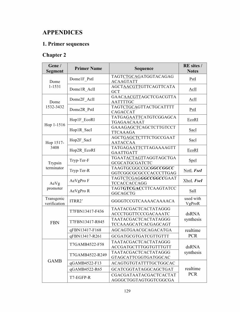
129
APPENDICES
1. Primer sequences
Chapter 2
Gene / Segment Primer Name Sequence RE sites /
Notes
Dome 1-1531
Dome1F_PstI TAGTCTGCAGATGGTACAGAGACAAGTATT PstI
Dome1R_AclI AGCTAACGTTGTTCAGTTCATAGCT AclI
Dome 1532-3432
Dome2F_AclI GAACAACGTTAGCTCGACGTTAAATTTTGC AclI
Dome2R_PstI TAGTCTGCAGTTACTGCATTTTCAGACCAT PstI
Hop 1-1516 Hop1F_EcoRI TATGAGAATTCATGTCGGAGCA
TGAGAACAAAT EcoRI
Hop1R_SacI GAAAGAGCTCAGCTCTTGTCCTTTCAAAGA SacI
Hop 1517-3408
Hop2F_SacI AGCTGAGCTCTTTCTGCCGAATAATACCAA SacI
Hop2R_EcoRI TATGAGAATTCTTAGAAAAGTTGAATTGATT EcoRI
Trypsin terminator
Tryp-Ter-F TGAATACTAGTTAGGTAGCTGAGCGCATGCGATCTC SpeI
Tryp-Ter-R TAAGTGCGGCCGCGGCCGGCCGGTCGGCGCGCCCACCCTTGAG NotI, FseI
AeVg promoter
AeVgPro F TAGTCTCGAGGGCCGGCCGAATTCCACCACCAGG XhoI, FseI
AeVgPro R TAGTGTCGACCTTCAAGTATCCGGCAGCTG SalI
Transgenic verification ITRR2’ GGGGTCCGTCAAAACAAAACA used with
VgProR
FBN
T7FBN13417-F436 TAATACGACTCACTATAGGG ACCCTGGTTCCCGACAAATC dsRNA
synthesis T7FBN13417-R845 TAATACGACTCACTATAGGG TCCAAAGCATCACGAGCAGT
qFBN13417-F168 AGCAGTGAACGCAGACATGA realtime PCR qFBN13417-R261 GCGATGCGTGATCGTTGTTT
GAMB
T7GAMB4522-F58 TAATACGACTCACTATAGGG ACCGATGCTTTGGTGTTTGTT dsRNA
synthesis T7GAMB4522-R249 TAATACGACTCACTATAGGG GTAGCATTCGGTGATGGCAC
qGAMB4522-F13 ACAGTGTGTATTTTGCTGGCAC realtime
PCR qGAMB4522-R65 GCATCGGTATAGGCAGCTGAT
T7-EGFP-R CGACGATAATACGACTCACTATAGGGCTGGTAGTGGTCGGCGA
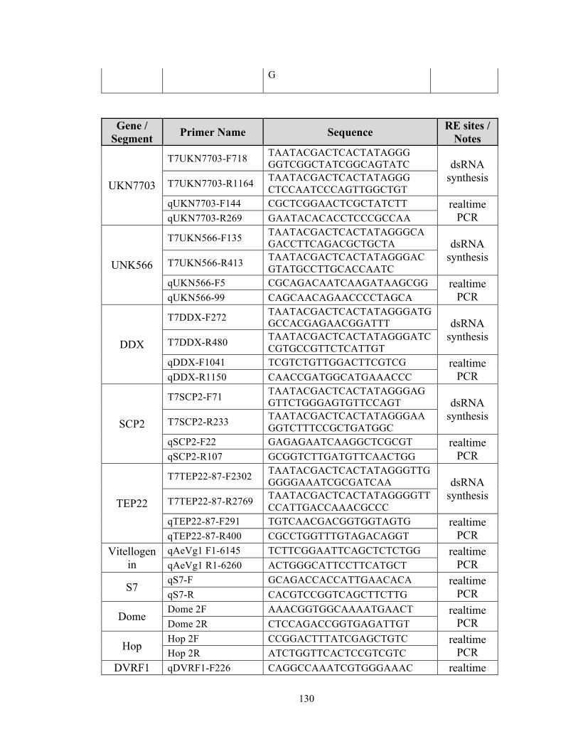
130
G
Gene / Segment Primer Name Sequence RE sites /
Notes
UKN7703
T7UKN7703-F718 TAATACGACTCACTATAGGG GGTCGGCTATCGGCAGTATC dsRNA
synthesis T7UKN7703-R1164 TAATACGACTCACTATAGGG CTCCAATCCCAGTTGGCTGT
qUKN7703-F144 CGCTCGGAACTCGCTATCTT realtime PCR qUKN7703-R269 GAATACACACCTCCCGCCAA
UNK566
T7UKN566-F135 TAATACGACTCACTATAGGGCAGACCTTCAGACGCTGCTA dsRNA
synthesis T7UKN566-R413 TAATACGACTCACTATAGGGACGTATGCCTTGCACCAATC
qUKN566-F5 CGCAGACAATCAAGATAAGCGG realtime PCR qUKN566-99 CAGCAACAGAACCCCTAGCA
DDX
T7DDX-F272 TAATACGACTCACTATAGGGATGGCCACGAGAACGGATTT dsRNA
synthesis T7DDX-R480 TAATACGACTCACTATAGGGATCCGTGCCGTTCTCATTGT
qDDX-F1041 TCGTCTGTTGGACTTCGTCG realtime PCR qDDX-R1150 CAACCGATGGCATGAAACCC
SCP2
T7SCP2-F71 TAATACGACTCACTATAGGGAGGTTCTGGGAGTGTTCCAGT dsRNA
synthesis T7SCP2-R233 TAATACGACTCACTATAGGGAAGGTCTTTCCGCTGATGGC
qSCP2-F22 GAGAGAATCAAGGCTCGCGT realtime PCR qSCP2-R107 GCGGTCTTGATGTTCAACTGG
TEP22
T7TEP22-87-F2302 TAATACGACTCACTATAGGGTTGGGGGAAATCGCGATCAA dsRNA
synthesis T7TEP22-87-R2769 TAATACGACTCACTATAGGGGTTCCATTGACCAAACGCCC
qTEP22-87-F291 TGTCAACGACGGTGGTAGTG realtime PCR qTEP22-87-R400 CGCCTGGTTTGTAGACAGGT
Vitellogenin
qAeVg1 F1-6145 TCTTCGGAATTCAGCTCTCTGG realtime PCR qAeVg1 R1-6260 ACTGGGCATTCCTTCATGCT
S7 qS7-F GCAGACCACCATTGAACACA realtime
PCR qS7-R CACGTCCGGTCAGCTTCTTG
Dome Dome 2F AAACGGTGGCAAAATGAACT realtime
PCR Dome 2R CTCCAGACCGGTGAGATTGT
Hop Hop 2F CCGGACTTTATCGAGCTGTC realtime
PCR Hop 2R ATCTGGTTCACTCCGTCGTC DVRF1 qDVRF1-F226 CAGGCCAAATCGTGGGAAAC realtime

131
qDVRF1-R319 TGGGCTGTTCATAGAATGGGG PCR
GFP T7-EGFP-F CGACGATAATACGACTCACTATA
GGGTTCATCTGCACCACCGGC dsRNA synthesis T7-EGFP-R CGACGATAATACGACTCACTATA
GGGCTGGTAGTGGTCGGCGAG
Chapter 3
Accession no. Primer Name Sequence AAEL007768 dsMyD88 F TAATACGACTCACTATAGGGGGCGATT
GGTGGTTGTTATT AAEL007768 dsMyD88 R TAATACGACTCACTATAGGGTTGAGCGC
ATTGCTAACATC AAEL010083 dsIMD F TAATACGACTCACTATAGGGACCGAAG
AAGACCGCACAAGGC AAEL010083 dsIMD R TAATACGACTCACTATAGGGTGCCGAG
CGTTGGTTCGTCG AAEL012471
dsDomeF TAATACGACTCACTATAGGGCCATCTCCACCACGAAACTT
AAEL012471
dsDome R TAATACGACTCACTATAGGGCCGGTGGTTGCCATATAATC
AAEL006794 dsDcr2 F TAATACGACTCACTATAGGGGCATTGACGACGAAATCATCGTCCGATG
AAEL006794 dsDcr2 R TAATACGACTCACTATAGGGACCATGGCATCCGCCGGTGTCTTGTCC
AAEL011414 dsHMBG F TAATACGACTCACTATAGGGCGCCAAGCGAGGTGGTGAGC
AAEL011414 dsHMBG R TAATACGACTCACTATAGGGGTCCTTGCCGCCGCCATTTC
AAEL010429 dsAller10-13 F TAATACGACTCACTATAGGGAGACCAGCCCCGAGTTCAAGG
AAEL010429 dsAller10-13 R TAATACGACTCACTATAGGGCCCCAGCCGAAGAAACCGGC
AAEL012819 dsvATP-G F TAATACGACTCACTATAGGGAAGGCCGCCGAAAAGGTCGG
AAEL012819 dsvATP-G R TAATACGACTCACTATAGGGGTGAGAGCTGAGGGCCCGGT
AAEL012092 dsLRRprot F TAATACGACTCACTATAGGGCCTGCCTCGGTTGGCACTGG
AAEL012092 dsLRRprot R TAATACGACTCACTATAGGGGACCAGCTTGCCGGACGTGA
AAEL015458 dsTsf F TAATACGACTCACTATAGGGTAGGAGGCGCCCCAGCCAAA
AAEL015458 dsTsf R TAATACGACTCACTATAGGGAGGCCGGACGGACATCACGA
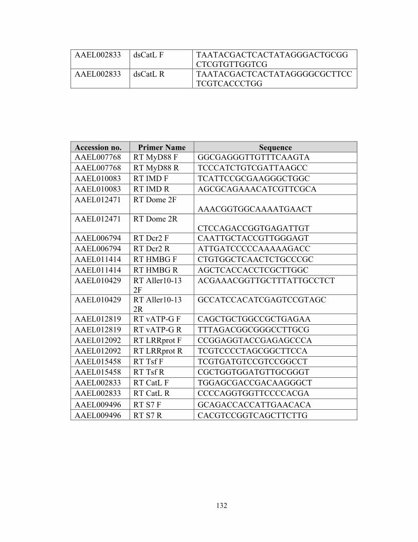
132
AAEL002833 dsCatL F TAATACGACTCACTATAGGGACTGCGGCTCGTGTTGGTCG
AAEL002833 dsCatL R TAATACGACTCACTATAGGGGCGCTTCCTCGTCACCCTGG
Accession no. Primer Name Sequence AAEL007768 RT MyD88 F GGCGAGGGTTGTTTCAAGTA AAEL007768 RT MyD88 R TCCCATCTGTCGATTAAGCC AAEL010083 RT IMD F TCATTCCGCGAAGGGCTGGC AAEL010083 RT IMD R AGCGCAGAAACATCGTTCGCA AAEL012471
RT Dome 2F AAACGGTGGCAAAATGAACT
AAEL012471
RT Dome 2R CTCCAGACCGGTGAGATTGT
AAEL006794 RT Dcr2 F CAATTGCTACCGTTGGGAGT AAEL006794 RT Dcr2 R ATTGATCCCCCAAAAAGACC AAEL011414 RT HMBG F CTGTGGCTCAACTCTGCCCGC AAEL011414 RT HMBG R AGCTCACCACCTCGCTTGGC AAEL010429 RT Aller10-13
2F ACGAAACGGTTGCTTTATTGCCTCT
AAEL010429 RT Aller10-13 2R
GCCATCCACATCGAGTCCGTAGC
AAEL012819 RT vATP-G F CAGCTGCTGGCCGCTGAGAA AAEL012819 RT vATP-G R TTTAGACGGCGGGCCTTGCG AAEL012092 RT LRRprot F CCGGAGGTACCGAGAGCCCA AAEL012092 RT LRRprot R TCGTCCCCTAGCGGCTTCCA AAEL015458 RT Tsf F TCGTGATGTCCGTCCGGCCT AAEL015458 RT Tsf R CGCTGGTGGATGTTGCGGGT AAEL002833 RT CatL F TGGAGCGACCGACAAGGGCT AAEL002833 RT CatL R CCCCAGGTGGTTCCCCACGA AAEL009496 RT S7 F GCAGACCACCATTGAACACA AAEL009496 RT S7 R CACGTCCGGTCAGCTTCTTG

133
Chapter 4
Gene name Primer name Sequence
NPC1b
T7NPC1b2-1122 TAATACGACTCACTATAGGGTCCCGAAAGCCGATCACGCG
T7NPC1b2-1558 TAATACGACTCACTATAGGGAGGGACCGAAGCAAGCCGGA
NPC1b2870F ACACCTTTTGCGAATCCTGCCC NPC1b3061R CATGGACGTTCAGATGACCGGC
ML13
T7ML13F TAATACGACTCACTATAGGGAAAATTGCGGTGAC
T7ML13R TAATACGACTCACTATAGGGAATGACGTCCTTATC
ML13-150F GGATGAACCAGCTTGCGTCCTG ML13-325R GGCATTCCAAACCGCTGTCCTT
ML33
T7ML33F TAATACGACTCACTATAGGGAACTTCCGAGTATG
T7ML33R TAATACGACTCACTATAGGGACCCAAAGCTACGC
ML33-235F ATGGACCTCGGTTTCAGGACCC ML33-385R TTTCGACCGGCATATTGACCGC
SOCS36E qAeSOCS36E F1-81 CCACTGTTTGGTGCCGGATTTGC qAeSOCS36E R1-266 GCGTGCAGCGACCGGTTGTA
Defensin E DEFE-F AACGTCGAAAGCGCATCTCA DEFE-R CGGTAGCGCCAGCTTATGG
DVRF1 DVRF1-F TCTTCATGCGGCATACTCAG DVRF1-R AGGAATGTTTCCGAGGGTTT
LYSC7B LYSC-F CCACGGCAACTGGATATGTCT LYSC-R TCTGCGTCACCTTGGTGGTAT
Cecropin E CecE-F CGAAGCCGGTGGTCTGAAG CecE-R ACTACGGGAAGTGCTTTCTCA
Gambicin GAM-F CGGACCATCAAGCATTTCTCAA GAM-R CCAGACGGTGGGTAGAACA

134
2. Log2-fold values and functional groups of transcripts that were up- or down-regulated in the fat body of VgDome or VgHop lines relative to WT
Gene ID Description Functional group VgDome VgHop
AAEL001904 arp2/3 CS
-0.773 AAEL002185 cuticle protein, putative CS -0.61 -0.853
AAEL002495 conserved hypothetical protein (mucin-like protein) CS
-1.65
AAEL002759 tropomyosin invertebrate CS
-0.991
AAEL004798 conserved hypothetical protein (mucin-like protein) CS 0.65 -1.569
AAEL005417 annexin x CS 0.194 0.964 AAEL010094 cyclin b CS 0.505 -1.224 AAEL013984 structural constituent of cuticle CS 0.389 -1.924 AAEL005146 conserved hypothetical protein CS -1.023 -0.652 AAEL005426 annexin x CS -1.442 -0.606 AAEL012644 conserved hypothetical protein CS 2.702
AAEL017334
Conserved hypothetical protein (chitin-binding domain type 2) CS 2.267
AAEL000335 lamin CS -0.971 -0.84 AAEL006726 innexin CS -1.513 -0.835 AAEL009572 cyclin B3 CS -0.784 -0.818 AAEL003593 hypothetical protein CSR
0.851
AAEL005772 Odorant-binding protein 99c, putative CSR 1.051 0.893
AAEL000005 hypothetical protein D
-0.76 AAEL000079 hypothetical protein D
-0.769
AAEL000105 beta-alanine synthase, putative D 0.322 -1.513 AAEL000115 conserved hypothetical protein D
0.864
AAEL000125 hypothetical protein D
-0.762
AAEL000147 single-stranded DNA binding protein, putative D
-0.903
AAEL000159 nipsnap D 0.715 1.073 AAEL000262 conserved hypothetical protein D
-0.848
AAEL000428 tryptophan 2,3-dioxygenase D
-1.939
AAEL000551
hypothetical protein (pacifastin light chain [Culex quinquefasciatus]) D -0.516 -1.886
AAEL000807 Tetratricopeptide repeat protein, putative D -0.165 0.829
AAEL000923 conserved hypothetical protein D -0.725 -0.829
135
Gene ID Description Functional group VgDome VgHop
AAEL001087 synaptic vesicle protein D 0.724 1.109 AAEL001100 phosphoserine phosphatase D 0.417 1.23 AAEL001287 conserved hypothetical protein D
0.861
AAEL001293 conserved hypothetical protein D -0.214 1.057 AAEL001307 SEC14, putative D
1.183
AAEL001352 scaffold attachment factor b D -0.575 -0.803 AAEL001401 conserved hypothetical protein D
1.052
AAEL001627 UDP-n-acteylglucosamine pyrophosphorylase D
1.085
AAEL001795 orfY, putative D 0.615 0.818 AAEL002048 histidyl-tRNA synthetase D 0.337 0.969 AAEL002125 conserved hypothetical protein D
-0.762
AAEL002194 uricase D -0.48 -1.086 AAEL002261 GTP cyclohydrolase i D
0.968
AAEL002501 protein disulfide isomerase D
2.191 AAEL002675 arginase D 0.186 0.775
AAEL002764
dihydrolipoamide succinyltransferase component of 2-oxoglutarate dehydrogenase D
0.764
AAEL002920 hypothetical protein D
-1.949 AAEL002948 frataxin, putative D
-0.761
AAEL003039 nonsense-mediated mrna decay protein D -0.355 1.211
AAEL003109 atlastin D
1.988
AAEL003165 low molecular weight protein-tyrosine-phosphatase D 0.221 1.078
AAEL003179 protein arginine n-methyltransferase 1, putative D
-0.857
AAEL003312 hypothetical protein D
1.425
AAEL003371 f-box and wd-40 domain protein D
-0.816
AAEL003413 f-spondin D
1.129 AAEL003509 smap1 D -0.37 -0.805
AAEL003581 amidophosphoribosyltransferase D -0.447 0.803
AAEL003599 DNA binding, sulfiredoxin activity, oxidation reduction D
0.778
AAEL003606 purine biosynthesis protein 6, pur6 D -0.542 2.35
AAEL003980 component of oligomeric golgi D -0.678 -1.022
136
Gene ID Description Functional group VgDome VgHop
complex
AAEL004023 Juvenile hormone-inducible protein, putative D
1.056
AAEL004237 vacuolar protein sorting 18 (deep orange protein) D -0.385 1.032
AAEL004278 conserved hypothetical protein D
1.592
AAEL004335 secreted ferritin G subunit precursor, putative D
1.225
AAEL004392
IAP-antagonist michelob-X-like protein, pro-apoptotic protein D
1.038
AAEL004404 HIG1 domain family member 2A, putative D
0.751
AAEL004480 cell division cycle 20 (cdc20) (fizzy) D 0.248 1.017
AAEL004503 conserved hypothetical protein D 0.3 1.134 AAEL004547 conserved hypothetical protein D -0.355 0.937 AAEL004566 myo inositol monophosphatase D
-1.415
AAEL004575 beta-galactosidase D
0.809
AAEL004613 phenylalanyl-tRNA synthetase beta chain D 0.674 1.109
AAEL004701 argininosuccinate synthase D 0.615 0.976
AAEL004813 M-phase phosphoprotein, putative D
0.885
AAEL005199 hypothetical protein D 0.607 1.41 AAEL005289 ornithine aminotransferase D
1.273
AAEL005384 phosphoribosylformylglycinamidine synthase, putative D 0.475 2.203
AAEL005457 conserved hypothetical protein D -0.656 0.937 AAEL005558 conserved hypothetical protein D -0.184 1.717 AAEL005760 hypothetical protein D 0.037 1.508
AAEL006023 Vanin-like protein 1 precursor, putative D -0.541 -0.98
AAEL006279 hypothetical protein D
-0.955 AAEL006446 trehalose-6-phosphate synthase D -0.398 -0.862 AAEL006518 cytidine deaminase, putative D
-2.892
AAEL006544 nucleoporin P54 D
-1.09 AAEL006602 hypothetical protein D 0.467 0.752 AAEL006712 serine/threonine protein kinase D -0.662 -0.897 AAEL006909 hypothetical protein D
-0.928
AAEL007072 conserved hypothetical protein D
-0.751

137
Gene ID Description Functional group VgDome VgHop
AAEL007114 conserved hypothetical protein D -0.566 -0.79 AAEL007226 nidogen D
1.601
AAEL007383 secreted ferritin G subunit precursor, putative D -0.388 -0.768
AAEL007621 conserved hypothetical protein D
2.049 AAEL007686 conserved hypothetical protein D
0.755
AAEL007701 conserved hypothetical protein D
-0.85 AAEL007767 conserved hypothetical protein D -0.624 0.977 AAEL007783 centromere protein-A, putative D -0.437 -1.078 AAEL007828 palmitoyl-protein thioesterase D
2.194
AAEL008007 conserved hypothetical protein D 0.391 0.955 AAEL008320 conserved hypothetical protein D
-0.754
AAEL008473 cysteinech venom protein, putative D
-1.328
AAEL008753 conserved hypothetical protein D 0.331 -1.126
AAEL008863 protein regulator of cytokinesis 1 prc1 D
-1.226
AAEL008953 conserved hypothetical protein D
-1.559
AAEL009037 GTP-binding protein (i) alpha subunit, gnai D -0.553 -0.792
AAEL009309 lipid depleted protein D 0.207 1.381 AAEL009508 zinc finger protein D
-0.852
AAEL009629 endoU protein, putative D 0.452 -0.818 AAEL009636 conserved hypothetical protein D
0.876
AAEL009719 conserved hypothetical protein D
-1.359
AAEL009931 arsenite inducuble RNA associated protein aip-1 D -0.473 1.379
AAEL009968 hypothetical protein D
-0.987 AAEL010028 sarcosine dehydrogenase D
0.922
AAEL010204 dihydropyrimidine dehydrogenase D -0.661 -1.28
AAEL010280 conserved hypothetical protein D 0.666 0.874 AAEL010455 cxyorf1 D
-0.827
AAEL010520 conserved hypothetical protein D 0.17 -0.778
AAEL010572 late endosomal/lysosomal MP1 interacting protein, putative D
-1.297
AAEL010656 conserved hypothetical protein D
1.414 AAEL010879 conserved hypothetical protein D 0.484 -1.093
AAEL011063 tumor endothelial marker 7 precursor D 0.567 0.817
AAEL011088 conserved hypothetical protein D 0.448 0.757
138
Gene ID Description Functional group VgDome VgHop
AAEL011135 conserved hypothetical protein D
-0.774 AAEL011159 cartilage associated protein D
-0.836
AAEL011168 GTP-binding protein (i) alpha subunit, gnai D -0.181 -0.75
AAEL011341 apyrase, putative D
-0.98
AAEL011529 late endosomal/lysosomal MP1 interacting protein, putative D
-1.047
AAEL011580 conserved hypothetical protein D
0.786 AAEL011853 conserved hypothetical protein D -0.376 -0.865
AAEL011892 receptor for activated C kinase, putative D -0.497 -1.134
AAEL011980 hypothetical protein D -0.594 -0.84 AAEL012233 hypothetical protein D
1.117
AAEL012417 conserved hypothetical protein D
1.117
AAEL012464 alanine-glyoxylate aminotransferase D -0.743 1.099
AAEL012502 conserved hypothetical protein D 0.108 -1.179 AAEL012856 hypothetical protein D
1.083
AAEL013078 glycosyltransferase D 0.595 0.764 AAEL013334 conserved hypothetical protein D 0.192 1.068
AAEL013338 lethal(2)essential for life protein, l2efl D
-0.949
AAEL013590 conserved hypothetical protein D
-0.854
AAEL013596 phosphatidylinositol 3-kinase regulatory subunit D
-0.957
AAEL013822 protein binding D
-0.927
AAEL014199 dihydropyrimidine dehydrogenase D
-1.669
AAEL014275 molybdopterin cofactor sulfurase (mosc) D 0.428 0.787
AAEL014310 hypothetical protein D
-0.852 AAEL014561 conserved hypothetical protein D -0.189 1.955 AAEL015375 serine/threonine protein kinase D
-0.905
AAEL015658 conserved hypothetical protein D
-1.281 AAEL000016 conserved hypothetical protein D 1.064 0.748 AAEL000315 pigeon protein (linotte protein) D 0.838
AAEL000442 conserved hypothetical protein D -0.931 -0.656 AAEL000776 conserved hypothetical protein D -0.846
AAEL000973 conserved hypothetical protein D -0.789 -0.431
AAEL001666 nucleic acid binding, zinc ion binding D -1.265 -0.696

139
Gene ID Description Functional group VgDome VgHop
AAEL001682 nuclear movement protein nudc D 1.58 0.221
AAEL002473 hypothetical protein D -0.777 -0.633 AAEL002559 conserved hypothetical protein D 1.526 -0.335 AAEL003213 guanine deaminase D 0.813 0.527
AAEL003237 low molecular weight protein-tyrosine-phosphatase D 0.958 0.295
AAEL003345 argininosuccinate lyase D -1.081 -0.382 AAEL003385 conserved hypothetical protein D -0.914
AAEL003877 ubiquitin D -0.916 -0.582 AAEL004860 acireductone dioxygenase D 1.969
AAEL005348 hypothetical protein D -0.928 AAEL005790 malic enzyme D -0.935
AAEL005976
adenine phosphoribosyltransferase, putative D 1.025
AAEL006353 sulfotransferase (sult) D 1.528 0.18
AAEL006972 hepatocellular carcinoma-associated antigen D 1.463 0.23
AAEL007130 leucyl-tRNA synthetase D -1.176
AAEL007477 ubiquitin-conjugating enzyme E2 i D -0.967 -0.585
AAEL007702 chaperonin D -0.868 -0.201 AAEL008076 PIWI D -2.045
AAEL008598 conserved hypothetical protein D 1.055 AAEL009652 activin receptor type ii D 0.955 AAEL009654 hypothetical protein D -1.179 -0.669
AAEL009859 nucleolar GTP-binding protein D -1.567
AAEL010065 protein disulfide-isomerase A6 precursor D 2.233
AAEL010943 conserved hypothetical protein D -0.772 0.358 AAEL011105 adducin D 1.201
AAEL011264
phosphatidylethanolamine-binding protein D 1.128 -0.242
AAEL011448 conserved hypothetical protein D 1.044 AAEL011452 conserved hypothetical protein D 0.913 AAEL011478 cytoplasmic dynein light chain D -0.928 -0.489
AAEL011849 hypothetical protein D -0.79 -0.598 AAEL012260 wdpeat protein D 1.298 -0.709 AAEL012632 hypothetical protein D 0.93

140
Gene ID Description Functional group VgDome VgHop
AAEL012939
gamma-subunit,methylmalonyl-CoA decarboxylase, putative D -1.029
AAEL013510 smaug protein D -0.972
AAEL013844 diazepam binding inhibitor, putative D 1.276 -0.564
AAEL014715 67 kDa polymerase-associated factor PAF67, putative D 0.883 0.661
AAEL014852 hypothetical protein D -0.862
AAEL001607 galactose-1-phosphate uridylyltransferase D 1.168 1.245
AAEL001667 multicopper oxidase D 1.553 1.086 AAEL002554 anosmin, putative D -1.466 -1.099 AAEL002860 conserved hypothetical protein D 2.016 1.633 AAEL005308 pyruvate dehydrogenase D 0.933 1.36 AAEL005458 carnitine o-acyltransferase D -0.783 0.776 AAEL006625 conserved hypothetical protein D -1.063 2.651 AAEL006662 hypothetical protein D 1.212 2.29 AAEL007484 protein transport protein sec23 D 0.809 -0.805 AAEL007494 calcineurin b subunit D 0.771 0.791 AAEL007557 asparagine synthetase D 1.038 1.431
AAEL007868
ubiquinol-cytochrome c reductase complex 14 kd protein D 0.816 1.358
AAEL008431 a kinase anchor protein D -1.011 -0.919 AAEL008595 conserved hypothetical protein D -1.162 -0.953 AAEL008789 apolipophorin-III, putative D -0.792 0.865 AAEL009962 hypothetical protein D -1.241 -0.96 AAEL010097 nuclein acid binding D -1.149 -1.591 AAEL011881 conserved hypothetical protein D 0.897 0.867 AAEL012605 conserved hypothetical protein D 2.008 1.699 AAEL012851 wdpeat protein D 0.903 1.017 AAEL012855 hypothetical protein D 1.019 3.375
AAEL013851
conserved hypothetical protein (acetyltransferase (GNAT) family domain) D 1.233 2.448
AAEL015631 asparagine synthetase D 1.071 1.418 AAEL002969 brain chitinase and chia DIG 0.452 1.381
AAEL003060 serine-type enodpeptidase, putative DIG
2.319
AAEL005481 alpha-glucosidase DIG -0.062 -0.964

141
Gene ID Description Functional group VgDome VgHop
AAEL006121 Trypsin, putative DIG
-0.788 AAEL008080 trypsin-eta, putative DIG 1.369
AAEL013262 conserved hypothetical protein DIG -1.001 AAEL014361 amidase DIG 0.782
AAEL000064
dopachrome-conversion enzyme (DCE) isoenzyme, putative I 0.357 0.764
AAEL000087 macroglobulin/complement I 0.414 0.752 AAEL000621 antibacterial peptide, putative I
1.547
AAEL000625 antibacterial peptide, putative I
1.659 AAEL002276 serine protease, putative I -0.461 -1.607 AAEL002592 hypothetical protein I -0.523 -0.792
AAEL003625 clip-domain serine protease, putative I 0.452 0.892
AAEL003723 lysozyme P, putative I 0.691 0.94 AAEL003832 conserved hypothetical protein I
1.168
AAEL003841 conserved hypothetical protein I
1.435 AAEL004401 peroxinectin I
-0.836
AAEL004522 gambicin I
1.796
AAEL005431 clip-domain serine protease, putative I 0.495 -1.312
AAEL006168 serine carboxypeptidase, putative I -0.372 -0.822
AAEL007006 serine protease I 0.7 0.834 AAEL007969 serine protease I 0.665 0.789 AAEL008607 tep3 I 0.43 -1.084 AAEL009637 cathepsin b I -0.462 1.513 AAEL009642 cathepsin b I -0.371 1.828
AAEL011446 galactose-specific C-type lectin, putative I -0.433 1.316
AAEL012092 leucinech repeat I 0.601 1.094
AAEL012251 low-density lipoprotein receptor (ldl) I -0.371 -0.805
AAEL012471 protein tyrosine phosphatase, putative I
-0.979
AAEL012711 trypsin, putative I
1.502
AAEL014238 aromatic amino acid decarboxylase I -0.15 -0.795
AAEL014349 serine protease I -0.357 -1.681 AAEL014755 tep2 I 0.07 -0.957 AAEL015430 serine protease, putative I 0.373 0.905

142
Gene ID Description Functional group VgDome VgHop
AAEL017325
Clip-Domain Serine Protease, family B. (Truncated Protease). [Source:Aedes_ManualAnnotation;Acc:AAEL800831] I -0.095 -0.978
AAEL000030 clip-domain serine protease, putative I 1.079 0.496
AAEL000598 antibacterial peptide, putative I -0.877 0.719 AAEL000611 antibacterial peptide, putative I -1.488
AAEL003279
clip-domain serine protease, putative I 0.953 0.686
AAEL004120 Niemann-Pick Type C-2, putative I 0.832 -0.144
AAEL006434 serine protease, putative I 0.803 -0.39 AAEL006586 serine protease I -0.976 -0.482 AAEL007599 cathepsin b I 1.229 0.703 AAEL011616 serine protease, putative I -0.956 -0.501
AAEL012064 Niemann-Pick Type C-2, putative I 0.808 0.241
AAEL014004 clip-domain serine protease, putative I -0.761 0.461
AAEL014385 conserved hypothetical protein I -1.179 -0.298
AAEL017536
Holotricin, Glycine Rich Repreat Protein (GRRP), Anti-Microbial Peptide. [Source:Aedes_ManualAnnotation;Acc:AAEL800434] I 0.801 0.186
AAEL005482 conserved hypothetical protein I 2.369 2.205 AAEL006271 superoxide dismutase I -0.853 -0.821 AAEL006704 fibrinogen and fibronectin I 0.794 1.227 AAEL006830 yellow protein precursor I -0.839 1.7 AAEL007585 cathepsin b I -0.853 1.184 AAEL011400 conserved hypothetical protein I 2.634 2.21
AAEL011610 galactose-specific C-type lectin, putative I 0.767 0.796
AAEL012216 cathepsin b I -0.776 1.181 AAEL013417 fibrinogen and fibronectin I 1.584 1.708
AAEL014390 galactose-specific C-type lectin, putative I 0.76 0.763
AAEL015312 cathepsin b I 1.105 0.823 AAEL015458 transferrin I 0.874 2.292 AAEL015639 transferrin I 1.24 1.726

143
Gene ID Description Functional group VgDome VgHop
AAEL017132
C-Type Lysozyme (Lys-C). [Source:Aedes_ManualAnnotation;Acc:AAEL800171] I 1.146 -1.13
AAEL010125 mitotic protein phosphatase 1 regulator, putative I(LRR) -0.382 -1.094
AAEL000006 phosphoenolpyruvate carboxykinase M 0.511 1.422
AAEL000059 proacrosin, putative M
0.944
AAEL000080 phosphoenolpyruvate carboxykinase M
1.135
AAEL000101 AMP dependent coa ligase M
1.032 AAEL000111 nitrilase, putative M
0.928
AAEL001423 acid phosphatase-1 M 0.676 0.76
AAEL001586 glucosyl/glucuronosyl transferases M 0.481 1.056
AAEL001593 glycerol-3-phosphate dehydrogenase M
-0.756
AAEL002304 porphobilinogen synthase M
-0.816 AAEL002964 brain chitinase and chia M -0.248 1.046 AAEL004059 cystathionine beta-lyase M
0.769
AAEL004126 sterol desaturase M 0.548 1.252 AAEL004127 acyl-coa dehydrogenase M 0.656 1 AAEL004313 fk506-binding protein M -0.434 1.016 AAEL004739 acyl-coa dehydrogenase M -0.486 1.391
AAEL004757 cleavage and polyadenylation specificity factor M -0.518 0.768
AAEL005732 acyl-coa dehydrogenase M
1.331 AAEL005740 AMP dependent ligase M
0.94
AAEL006085 methylenetetrahydrofolate dehydrogenase M -0.108 0.758
AAEL006171 n-myc downstream regulated M
0.786 AAEL006354 epoxide hydrolase M
-0.851
AAEL007201 glutamyl aminopeptidase M
-0.888 AAEL007707 malate dehydrogenase M
-1.012
AAEL007880 ornithine decarboxylase M -0.091 -0.782
AAEL008006 3-hydroxyacyl-coa dehyrogenase M -0.732 -1.513
AAEL008302 glutamine-dependent nad(+) synthetase M
-0.961
AAEL008330 hexaprenyldihydroxybenzoate methyltransferase M 0.394 1.062

144
Gene ID Description Functional group VgDome VgHop
AAEL009503 4-nitrophenylphosphatase M 0.686 1.389 AAEL009911 rotamase M 0.38 1.465
AAEL010366 glucosyl/glucuronosyl transferases M 0.592 0.918
AAEL010590 aldose-1-epimerase M
-1.259
AAEL010691 ribonucleoside-diphosphate reductase small chain M
-0.945
AAEL010938 l-asparaginase M
0.842 AAEL011126 alcohol dehydrogenase M
-0.794
AAEL011130 alcohol dehydrogenase M
-0.852
AAEL012312 proliferation-associated 2g4 (pa2g4/ebp1) M -0.613 0.841
AAEL012341 lysosomal acid lipase, putative M -0.557 -1.067 AAEL012430 AMP dependent ligase M -0.509 -1.048
AAEL012697 sterol carrier protein-2, putative M
-3.624
AAEL013245 proacrosin, putative M 0.574 1.485
AAEL013458 glutamine synthetase 1, 2 (glutamate-amonia ligase) (gs) M -0.11 -2.122
AAEL014662 AMP dependent coa ligase M
1.081
AAEL017039
Conserved hypothetical protein (alcohol dehydrogenase 2 [Culex quinquefasciatus] _ M -0.364 1.013
AAEL017299 AMP dependent coa ligase, putative M
0.973
AAEL007097 4-nitrophenylphosphatase M -1.064 -0.357 AAEL007883 fk506-binding protein M 0.798
AAEL008144 AMP dependent ligase M 0.918 -0.327 AAEL008467 cysteine synthase M 1.265
AAEL009038
prolylcarboxypeptidase, putative M -1.184 -0.387
AAEL009462 hydroxyacylglutathione hydrolase M 1.342 0.13
AAEL011624 granzyme A precursor, putative M 0.894
AAEL012179 methylthioadenosine phosphorylase M 0.834 0.444
AAEL012825 bifunctional purine biosynthesis protein M -1.089
AAEL013521 tryptophanyl-tRNA synthetase M 0.771 0.183
AAEL013967
Methylmalonyl-CoA carboxyltransferase 12S subunit, putative M -1.655

145
Gene ID Description Functional group VgDome VgHop
AAEL015143 glycine rich RNA binding protein, putative M -1.01
AAEL001548
glucosyl/glucuronosyl transferases M 0.975 1.273
AAEL002422 cytoplasmic polyadenylation element binding protein (cpeb) M -1.192 -0.861
AAEL009246 glycoside hydrolases M 0.878 1.662 AAEL014709 methionine-tRNA synthetase M -1.072 2.089
AAEL015337
neutral alpha-glucosidase ab precursor (glucosidase ii alpha subunit) (alpha glucosidase 2) M -0.967 -2.407
AAEL005638 conserved hypothetical protein PROT
1.034
AAEL006563
retinoid-inducible serine carboxypeptidase (serine carboxypeptidase PROT
1.62
AAEL008862 conserved hypothetical protein PROT -0.523 -1.014 AAEL009771 hypothetical protein PROT
0.816
AAEL014350 hypothetical protein PROT
-0.83 AAEL014353 conserved hypothetical protein PROT
-0.957
AAEL015527 conserved hypothetical protein PROT
0.768
AAEL017451
proteolysis, metallopeptidase activity, peptidyl-dipeptidase activity, membrane. PROT 0.431 0.789
AAEL009406
n(4)-(beta-n-acetylglucosaminyl)-l-asparaginase PROT 1.252
AAEL011658
plasma glutamate carboxypeptidase PROT 1.067 0.403
AAEL015432 Trypsin, putative PROT -1.16 -0.491 AAEL000252 hypothetical protein PROT 0.845 0.766 AAEL006323 hypothetical protein PROT 0.797 -3.277
AAEL006542
retinoid-inducible serine carboxypeptidase (serine carboxypeptidase PROT 0.932 1.054
AAEL010196 trypsin PROT 2.031 2.224 AAEL010634 hypothetical protein REDOX -0.295 0.938 AAEL010592 esterase, putative REDOX 1.749 0.442 AAEL000546 carboxylesterase RSM
-1.14
AAEL001960 cytochrome P450 RSM
-1.033 AAEL002046 cytochrome P450 RSM -0.741 -0.82 AAEL002886 thioredoxin reductase RSM -0.025 -0.8
146
Gene ID Description Functional group VgDome VgHop
AAEL003380 cytochrome P450 RSM
1.003
AAEL004643 mitochondrial ribosomal protein L1 RSM -0.644 -0.805
AAEL005178 juvenile hormone esterase RSM 0.616 1.172 AAEL005305 conserved hypothetical protein RSM
-0.824
AAEL005946 NADH-ubiquinone oxidoreductase subunit B14.5b RSM -0.268 0.956
AAEL006230 gonadotropin inducible transcription factor RSM
-0.758
AAEL006824 cytochrome P450 RSM -0.314 2.468
AAEL007046 mitochondrial brown fat uncoupling protein RSM 0.193 -0.854
AAEL007355 mitochondrial ribosomal protein, S18A, putative RSM 0.258 0.95
AAEL008128
mitochondrial inner membrane protein translocase, 13kD-subunit, putative RSM 0.426 0.781
AAEL008397 glutathione peroxidase RSM
-1.617 AAEL010075 oxidoreductase RSM
1.014
AAEL011016 carboxypeptidase m RSM 0.662 1.25
AAEL012427
metabolic proces, oxidoreductase activity, oxidation reduction RSM
-0.863
AAEL013066 checkpoint kinase RSM -0.473 -1.071 AAEL013555 cytochrome P450 RSM -0.246 1.358 AAEL014019 cytochrome P450 RSM
0.918
AAEL014673 NADH:ubiquinone dehydrogenase, putative RSM
1.103
AAEL014830 cytochrome P450 RSM
1.882 AAEL015578 alpha-esterase RSM 0.108 -1.038 AAEL017071 Alpha-esterase, putative RSM
-1.378
AAEL001210
NADH ubiquinone oxidoreductase subunit, putative RSM 1.049 0.578
AAEL002683 aldehyde oxidase RSM 1.058 AAEL003890 cytochrome P450 RSM -1.064 -0.396
AAEL007752 cytochrome c oxidase, subunit VIIA, putative RSM 1.135 -0.688
AAEL008757 juvenile hormone esterase RSM 0.861 0.392
AAEL009225 mitochondrial ribosome recycling factor RSM -0.807 -0.64

147
Gene ID Description Functional group VgDome VgHop
AAEL012845
mitochondrial import inner membrane translocase subunit tim44 RSM 0.777
AAEL013744
NADH:ubiquinone dehydrogenase, putative RSM -1.282
AAEL014893 cytochrome P450 RSM -1.374
AAEL015635 mitochondrial ribosomal protein, S10, putative RSM 1.038 0.216
AAEL000986 NADH-ubiquinone oxidoreductase ashi subunit RSM 2.027 2.067
AAEL003423 NADH dehydrogenase, putative RSM 1.189 -1.074
AAEL004450 cytochrome b5, putative RSM 0.84 0.826
AAEL007946 glutathione-s-transferase theta, gst RSM 0.919 -0.797
AAEL010181 mitochondrial ribosomal protein, L51, putative RSM 1.064 0.868
AAEL000032 ribosomal protein S6 RTT 0.606 0.965 AAEL000497 histone h2a RTT 0.678 -1.106 AAEL002103 histone H1, putative RTT
-0.901
AAEL002879 heterogeneous nuclear ribonucleoprotein r RTT -0.27 0.895
AAEL003352 ribosomal protein l7ae RTT 0.534 0.751 AAEL003427 ribosomal protein S9, putative RTT -0.559 -0.751 AAEL003646 conserved hypothetical protein RTT
0.867
AAEL003685 histone H3 RTT 0.449 -1.192 AAEL003818 histone h2a RTT
-1.202
AAEL003820 histone h2a RTT
-1.126 AAEL003826 histone h2a RTT
-1.197
AAEL003851 histone h2a RTT
-0.976
AAEL003942 60S ribosomal protein L44 L41, putative RTT
1.133
AAEL004978 DEAD box ATP-dependent RNA helicase RTT -0.699 -0.79
AAEL005368 transcription initiation factor TFIIB RTT
-0.92
AAEL007078 eukaryotic translation initiation factor 3, theta subunit RTT 0.186 1.249
AAEL007928 eukaryotic translation initiation factor 4 gamma RTT
-0.827
AAEL008266 hypothetical protein RTT
-0.847 AAEL008500 DEAD box ATP-dependent RTT
-0.956

148
Gene ID Description Functional group VgDome VgHop
RNA helicase AAEL009653 40S ribosomal protein S30 RTT
2.336
AAEL010085 DNA polymerase epsilon subunit, putative RTT
-1.042
AAEL010787 DEAD box ATP-dependent RNA helicase RTT -0.278 1.474
AAEL011150 RNA-binding protein precursor, putative RTT
-0.775
AAEL012185 ribosome biogenesis regulatory protein RTT 0.619 1.255
AAEL012684 conserved hypothetical protein RTT 0.006 1.378
AAEL012877 homeobox protein extradenticle, putative RTT 0.126 0.978
AAEL013221 60S ribosomal protein L10a RTT 0.505 1.023
AAEL014764 acidic ribosomal protein P1, putative RTT -0.184 0.929
AAEL014838 60S ribosomal protein L27e RTT -0.444 -0.751
AAEL017595 5S ribosomal RNA [Source: RFAM 9.0] RTT
0.76
AAEL017630 5S ribosomal RNA [Source: RFAM 9.0] RTT
0.782
AAEL017779 5S ribosomal RNA [Source: RFAM 9.0] RTT 0.435 0.757
AAEL002534 60S ribosomal protein L10 RTT -2.644 0.277
AAEL003071 tRNA pseudouridine synthase D RTT 0.986 0.527
AAEL003396 60S ribosomal protein L32 RTT -0.765 AAEL005127 ribonuclease UK114, putative RTT 0.872 0.448
AAEL006698 60S ribosomal protein L31 RTT 0.812 0.431 AAEL007005 histone h2a RTT 0.795
AAEL010821
60S acidic ribosomal protein P0 RTT 1.649 -0.269
AAEL011251 RNA binding motif protein RTT -0.934 -0.681 AAEL012074 conserved hypothetical protein RTT 2.387 0.194 AAEL013964 ribosomal protein L20, putative RTT 1.411
AAEL014106 ATP-dependent RNA helicase RTT -0.763 -0.358 AAEL015244 splicing factor 3a RTT 1.212
AAEL017685
Nuclear RNase P [Source: RFAM 9.0] RTT 1.164 0.367
AAEL000518 histone h2a RTT 0.826 -1.071 AAEL000525 histone h2a RTT 0.768 -0.969 AAEL003659 histone H3 RTT 1.049 -1.216

149
Gene ID Description Functional group VgDome VgHop
AAEL005129 40S ribosomal protein S30 RTT 0.859 2.634 AAEL011447 60S ribosomal protein L14 RTT 1.526 1.235 AAEL012686 ribosomal protein S12, putative RTT 1.388 1.085
AAEL017742 5S ribosomal RNA [Source: RFAM 9.0] RTT 0.846 0.766
AAEL017760 5S ribosomal RNA [Source: RFAM 9.0] RTT 0.888 0.76
AAEL000471 monocarboxylate transporter TRP
0.809
AAEL001308 CRAL/TRIO domain-containing protein TRP -0.269 1.065
AAEL002063 cationic amino acid transporter TRP 0.565 0.984 AAEL002576 sodium/solute symporter TRP
0.931
AAEL003548 sulfate transporter TRP -0.641 -0.859
AAEL003626 sodium/shloride dependent amino acid transporter TRP
0.832
AAEL004247 Sialin, Sodium/sialic acid cotransporter, putative TRP
-0.758
AAEL005769 glucose dehydrogenase TRP
-0.836 AAEL006138 hypothetical protein TRP
1.353
AAEL008406 cationic amino acid transporter TRP 0.647 0.955 AAEL008635 abc transporter TRP -0.293 0.907 AAEL009832 exocyst complex protein exo70 TRP -0.255 -1.481 AAEL010102 tetraspanin, putative TRP
1.164
AAEL010434 conserved hypothetical protein TRP
1.519 AAEL010481 sugar transporter TRP -0.343 -1.701 AAEL010485 sugar transporter TRP
1
AAEL010584 vesicular mannose-binding lectin TRP 0.18 -0.972
AAEL011025 vacuolar ATP synthase subunit ac39 TRP
-2.707
AAEL011244 surfeit locus protein TRP -0.425 1.09
AAEL014927 sodium/chloride dependent transporter TRP
-1.124
AAEL015549 calcineurin b subunit TRP 0.387 0.769 AAEL002555 sodium/solute symporter TRP 0.771
AAEL002726 D7 protein, putative TRP -0.869 AAEL004855 adp,atp carrier protein TRP -0.778 -0.628
AAEL005496 zinc/iron transporter TRP 0.884 0.703 AAEL012674 d-amino acid oxidase TRP 1.387
AAEL007458 amino acid transporter TRP -0.833 -0.945 AAEL009863 sodium/dicarboxylate TRP 1.445 1.056

150
Gene ID Description Functional group VgDome VgHop
cotransporter, putative AAEL000309 hypothetical protein U 0.318 -1.223 AAEL000619 conserved hypothetical protein U
1.291
AAEL001325 conserved hypothetical protein U 0.188 -1.639 AAEL001414 conserved hypothetical protein U -0.064 0.884 AAEL001718 conserved hypothetical protein U 0.397 0.776 AAEL001880 conserved hypothetical protein U
1.242
AAEL001885 conserved hypothetical protein U
1.258 AAEL001888 hypothetical protein U 0.555 1.118 AAEL001892 conserved hypothetical protein U -0.484 -1.661 AAEL001897 conserved hypothetical protein U -0.323 -1.378 AAEL002719 conserved hypothetical protein U 0.362 0.81 AAEL002758 conserved hypothetical protein U 0.286 1.46 AAEL002815 conserved hypothetical protein U
1.197
AAEL002828 hypothetical protein U
-0.805 AAEL002900 conserved hypothetical protein U
0.774
AAEL003029 hypothetical protein U -0.316 0.81 AAEL003067 conserved hypothetical protein U 0.11 0.997 AAEL003766 hypothetical protein U -0.211 0.901 AAEL003842 hypothetical protein U 0.267 0.838 AAEL003944 conserved hypothetical protein U
1.086
AAEL004498 hypothetical protein U 0.384 0.806 AAEL004670 conserved hypothetical protein U
1.172
AAEL004809 conserved hypothetical protein U 0.317 0.858 AAEL004826 conserved hypothetical protein U
-1.156
AAEL005215 conserved hypothetical protein U
-0.759 AAEL005620 conserved hypothetical protein U
1.81
AAEL005755 hypothetical protein U -0.25 1.331 AAEL006792 conserved hypothetical protein U
-0.865
AAEL006848 conserved hypothetical protein U -0.606 -0.876 AAEL006863 hypothetical protein U
3.042
AAEL006971 conserved hypothetical protein U
0.876 AAEL007259 conserved hypothetical protein U -0.382 -0.914 AAEL007342 conserved hypothetical protein U
-0.814
AAEL007847 conserved hypothetical protein U
1.119 AAEL008025 conserved hypothetical protein U 0.214 1.749 AAEL008039 conserved hypothetical protein U
-1.212
AAEL008100 conserved hypothetical protein U
-0.992 AAEL008274 conserved hypothetical protein U -0.158 -1.427

151
Gene ID Description Functional group VgDome VgHop
AAEL008286 conserved hypothetical protein U
-1.18 AAEL008365 conserved hypothetical protein U 0.262 -1.158 AAEL008485 conserved hypothetical protein U
-1.848
AAEL008771 conserved hypothetical protein U
1.584 AAEL008802 conserved hypothetical protein U -0.479 0.829 AAEL009177 conserved hypothetical protein U -0.399 -0.864 AAEL009487 hypothetical protein U 0.282 0.893 AAEL009519 hypothetical protein U
-0.785
AAEL010752 hypothetical protein U
-1.077 AAEL011010 conserved hypothetical protein U 0.325 1.191 AAEL011330 conserved hypothetical protein U
-0.807
AAEL011388 conserved hypothetical protein U -0.136 0.756 AAEL011456 conserved hypothetical protein U
1.762
AAEL011532 hypothetical protein U -0.694 -0.827 AAEL011665 hypothetical protein U
-0.762
AAEL011884 hypothetical protein U 0.395 -2.065 AAEL012208 hypothetical protein U -0.332 -1.004 AAEL012454 conserved hypothetical protein U 0.456 1.13 AAEL012858 hypothetical protein U
-0.952
AAEL012860 conserved hypothetical protein U 0.573 0.912 AAEL012867 conserved hypothetical protein U 0.632 1.242 AAEL013300 conserved hypothetical protein U
-0.786
AAEL013484 hypothetical protein U
-0.754 AAEL013486 hypothetical protein U
-1.061
AAEL013800 conserved hypothetical protein U -0.652 -0.864 AAEL014171 conserved hypothetical protein U
-1.184
AAEL014388 conserved hypothetical protein U
-0.913 AAEL014511 predicted protein U
-0.883
AAEL014565 hypothetical protein U
-0.957 AAEL017034 Hypothetical protein U
-1.584
AAEL017190 Hypothetical protein U 0.227 -1.305 AAEL017530 hypothetical protein U
-1.363
AAEL000019 conserved hypothetical protein U 1.278 AAEL001032 conserved hypothetical protein U -0.753 -0.462
AAEL001323 conserved hypothetical protein U 0.933 AAEL001511 conserved hypothetical protein U -0.769 -0.653
AAEL002889 hypothetical protein U 2.243 AAEL004100 hypothetical protein U 1.055 0.384
AAEL004591 hypothetical protein U 1.108 -0.655
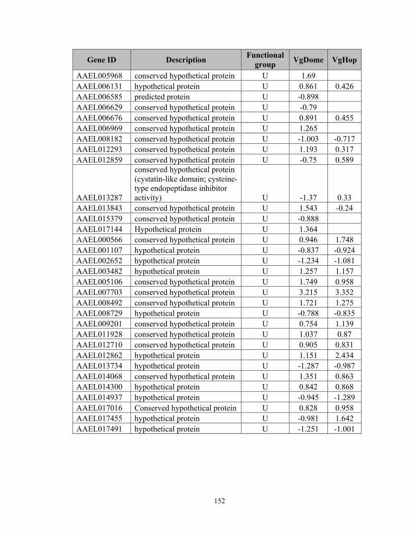
152
Gene ID Description Functional group VgDome VgHop
AAEL005968 conserved hypothetical protein U 1.69 AAEL006131 hypothetical protein U 0.861 0.426
AAEL006585 predicted protein U -0.898 AAEL006629 conserved hypothetical protein U -0.79 AAEL006676 conserved hypothetical protein U 0.891 0.455
AAEL006969 conserved hypothetical protein U 1.265 AAEL008182 conserved hypothetical protein U -1.003 -0.717
AAEL012293 conserved hypothetical protein U 1.193 0.317 AAEL012859 conserved hypothetical protein U -0.75 0.589
AAEL013287
conserved hypothetical protein (cystatin-like domain; cysteine-type endopeptidase inhibitor activity) U -1.37 0.33
AAEL013843 conserved hypothetical protein U 1.543 -0.24 AAEL015379 conserved hypothetical protein U -0.888
AAEL017144 Hypothetical protein U 1.364 AAEL000566 conserved hypothetical protein U 0.946 1.748
AAEL001107 hypothetical protein U -0.837 -0.924 AAEL002652 hypothetical protein U -1.234 -1.081 AAEL003482 hypothetical protein U 1.257 1.157 AAEL005106 conserved hypothetical protein U 1.749 0.958 AAEL007703 conserved hypothetical protein U 3.215 3.352 AAEL008492 conserved hypothetical protein U 1.721 1.275 AAEL008729 hypothetical protein U -0.788 -0.835 AAEL009201 conserved hypothetical protein U 0.754 1.139 AAEL011928 conserved hypothetical protein U 1.037 0.87 AAEL012710 conserved hypothetical protein U 0.905 0.831 AAEL012862 hypothetical protein U 1.151 2.434 AAEL013734 hypothetical protein U -1.287 -0.987 AAEL014068 conserved hypothetical protein U 1.351 0.863 AAEL014300 hypothetical protein U 0.842 0.868 AAEL014937 hypothetical protein U -0.945 -1.289 AAEL017016 Conserved hypothetical protein U 0.828 0.958 AAEL017455 hypothetical protein U -0.981 1.642 AAEL017491 hypothetical protein U -1.251 -1.001
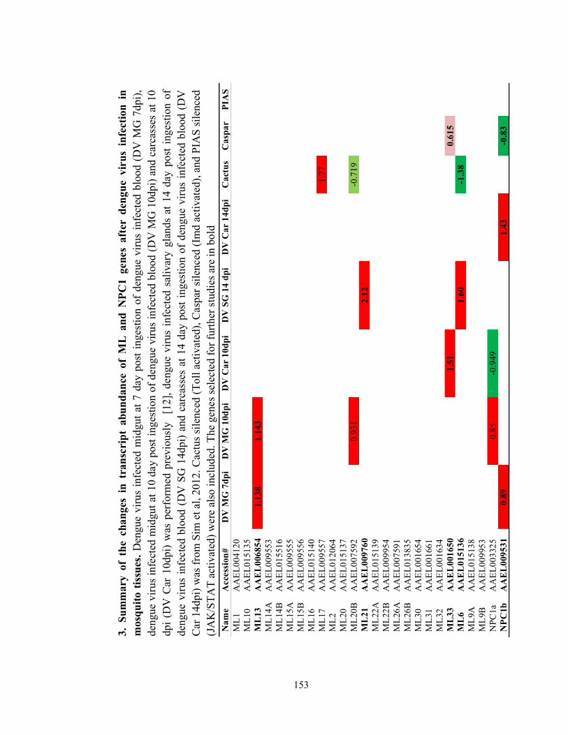
153
3.
Sum
mar
y of
the
cha
nges
in
tran
scri
pt a
bund
ance
of
ML
and
NPC
1 ge
nes
afte
r de
ngue
vir
us i
nfec
tion
in
mos
quito
tiss
ues.
Den
gue
viru
s in
fect
ed m
idgu
t at 7
day
pos
t ing
estio
n of
den
gue
viru
s in
fect
ed b
lood
(D
V M
G 7
dpi),
de
ngue
viru
s in
fect
ed m
idgu
t at 1
0 da
y po
st in
gest
ion
of d
engu
e vi
rus
infe
cted
blo
od (D
V M
G 1
0dpi
) and
car
cass
es a
t 10
dpi (
DV
Car
10d
pi)
was
per
form
ed p
revi
ousl
y [
12],
deng
ue v
irus
infe
cted
sal
ivar
y gl
ands
at 1
4 da
y po
st in
gest
ion
of
deng
ue v
irus
infe
cted
blo
od (
DV
SG
14d
pi)
and
carc
asse
s at
14
day
post
inge
stio
n of
den
gue
viru
s in
fect
ed b
lood
(D
V
Car
14d
pi) w
as fr
om S
im e
t al,
2012
. Cac
tus s
ilenc
ed (T
oll a
ctiv
ated
), C
aspa
r sile
nced
(Im
d ac
tivat
ed),
and
PIA
S si
lenc
ed
(JA
K/S
TAT
activ
ated
) wer
e al
so in
clud
ed. T
he g
enes
sele
cted
for f
urth
er st
udie
s are
in b
old
Nam
e A
cces
sion
# D
V M
G 7
dpi
DV
MG
10d
pi
DV
Car
10d
pi
DV
SG
14
dpi
DV
Car
14d
pi
Cac
tus
Cas
par
PIA
S M
L1
AA
EL00
4120
M
L10
AA
EL01
5135
M
L13
A
AE
L00
6854
1.
138
1.14
3
M
L14A
A
AEL
0095
53
ML1
4B
AA
EL01
5516
M
L15A
A
AEL
0095
55
ML1
5B
AA
EL00
9556
M
L16
AA
EL01
5140
M
L17
AA
EL00
9557
1.77
M
L2
AA
EL01
2064
M
L20
AA
EL01
5137
M
L20B
A
AEL
0075
92
0.
931
-0
.719
M
L21
A
AE
L00
9760
2.12
M
L22A
A
AEL
0151
39
ML2
2B
AA
EL00
9954
M
L26A
A
AEL
0075
91
ML2
6B
AA
EL01
3835
M
L30
AA
EL00
1654
M
L31
AA
EL00
1661
M
L32
AA
EL00
1634
M
L33
A
AE
L00
1650
1.
51
0.
615
M
L6
AA
EL
0151
36
1.
60
-1
.38
ML9
A
AA
EL01
5138
M
L9B
A
AEL
0099
53
NPC
1a
AA
EL00
3325
0.85
-0
.949
NPC
1b
AA
EL
0095
31
0.89
1.43
-0.8
3
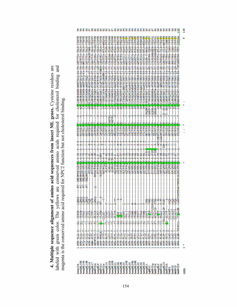
154
4. M
ultip
le s
eque
nce
alig
nmen
t of
amin
o ac
id s
eque
nces
from
inse
ct M
L ge
nes.
Cys
tein
e re
sidu
es a
re
labe
led
with
gre
en c
olor
. Th
e ye
llow
s ar
e co
nser
ved
amin
o ac
ids
requ
ired
for
chol
este
rol
bind
ing
and
mag
enta
is th
e co
nser
ved
amin
o ac
id re
quire
d fo
r NPC
2 fu
nctio
n bu
t not
cho
lest
erol
bin
ding
.

155
156
NATAPONG JUPATANAKUL PhD candidate, Laboratory of Dr. George Dimopoulos Department of Molecular Microbiology and Immunology Johns Hopkins Bloomberg School of Public Health CONTACT INFORMATION Office: 615 N. Wolfe St., Room E3209, Baltimore, MD, 21205 Email: [email protected] Telephone: (+1) 410-955-3223 EDUCATION 2012-Present Johns Hopkins Bloomberg School of Public Health, expected May, 2016 Doctor of Philosophy (Molecular Microbiology and Immunology) Advisor: George Dimopoulos 2010-2012 Johns Hopkins Bloomberg School of Public Health, May, 2012 Master of Science in Molecular Microbiology and Immunology Advisor: George Dimopoulos 2007-2009 Mahidol University Master of Science in Biotechnology, August, 2009 Advisors: Timothy W. Flegel, Boonsirm Withyachumnarnkul, Kallaya Sritunyalucksana 2003-2007 Mahidol University Bachelor of Science in Biotechnology with Second class honor, May, 2007 RESEARCH EXPERIENCE August 2012- present PhD student, Laboratory of Dr George Dimopoulos, Department of Molecular Microbiology and Immunology, Johns Hopkins Bloomberg School of Public Health December 2010- May 2012 ScM student, Laboratory of Dr George Dimopoulos, Department of Molecular Microbiology and Immunology, Johns Hopkins Bloomberg School of Public Health August 2010-2015 Research assistant, Laboratory of Dr. Fidel Zavala, Department of Molecular Microbiology and Immunology, Johns Hopkins Bloomberg School of Public Health August 2009-July 2010 Research assistant, Shrimp-Virus Interaction Laboratory, National Center for Genetic Engineering and Biotechnology (BIOTEC), National Science and Technology Development Agency (NSTDA), Thailand. Under the supervision of Dr. Kallaya Sritunyalucksana. June 2007-August 2009 Master degree student, Department of Biotechnology, Faculty of Science, Mahidol University Thailand.

157
Under the supervision of Dr.Timothy W. Flegel, Dr.Boonsirm Withyachumnarnkul, and Dr.Kallaya Sritunyalucksana. August 2006-May 2007 Senior Project research, Department of Biotechnology, Faculty of Science, Mahidol University Thailand. Under the supervision of Dr.Timothy W. Flegel, and Dr.Kallaya Sritunyalucksana. April 2006-May 2006 Research internship, Armed Forces Research Institute of Medical Sciences (AFRIMS), Thailand. Under the supervision of Dr. Thippawan Chuenchitr. TEACHING EXPERIENCE January 2015 Teaching assistant in PH. 260.613: Techniques in Molecular Biology
• Prepared materials and teach basic molecular biology techniques September 2011 Teaching assistant in PH.260.852: Molecular Biology literature
• Assisted Dr. Jelena Levitskaya in class discussion, and encouraged student participation
October 2009 Teaching assistant in SCBT 609: Biology and Pathology of shrimp
• Prepared materials for lab section in the course • Assisted students and demonstrated lab in Shrimp total hemocyte count and
Bacterial isolation and identification using biochemical and bioinformatics analysis.
November 2008-March 2009 Teaching assistant in SCBT 203: Bacteriology
• Assisted and demonstrated students in lab section of the course, held office hours to discuss the lab report and homework
October 2008 Teaching assistant in SCBT 609: Biology and Pathology of shrimp
• Prepared materials for lab section in the course • Assisted students and demonstrated lab in Shrimp total hemocyte count and
Bacterial isolation and identification using biochemical analysis. HONORS AND AWARDS 2010-present Ministry of Science and Technology , Royal Thai Government Scholarship for
Master and PhD study 2014 James Stuart Porterfield Prize in International Virology 2012-2013 Dr.Lioyd and Mae Rozeboom Scholarship for academic excellence and research
potential in molecular microbiology and immunology 2011-2012 Tuition Scholarship from Department of Molecular Microbiology and
Immunology, Johns Hopkins Bloomberg School of Public Health.

158
2009 Honorable mention: student presentation at the 11th International congress of International Society of Developmental and Comparative Immunology, Prague, Czech Republic
2009 Travel award from the Department of Biotechnology, Faculty of Science, Mahidol University.
2007-2009 Thailand Graduate Institute of Science and Technology (TGIST) scholarship from the National Science and Technology Development Agency (NSTDA), Thailand cover tuition fee and stipend
2007 Best Presentation Award from The 8th Science Project Exhibition, Faculty of Science, Mahidol University, Thailand
2003-2007 Sritang Thong Scholarship from the Faculty of Science, Mahidol University to cover tuition fee and stipend.
ORAL AND POSTER PRESENTATIONS Poster 2014 JAK/STAT super-immune Aedes aegypti as a tool for dengue disease control.
Jupatanakul N, Sim S, Anglero Y, Souza-Neto J, Dimopoulos G. The forth pan-american dengue research network meeting, Belem, Brazil.
Oral 2013 Aedes aegypti laboratory adaptation leads to global transcriptomic down-regulation and dengue vector capacity changes. Jupatanakul N, Bahia AC, Sim S, Dimopoulos G. The 62nd ASTMH annual meeting, Washington DC, USA.
Oral 2013 Laboratory adaptation leads to global transcriptomic down-regulation and dengue vector capacity changes in Aedes aegypti. Jupatanakul N, Bahia AC, Sim S, Dimopoulos G. The Third International Conference on Dengue and Dengue Hemorrhagic fever, Bangkok, Thailand.
Poster 2012 Aedes aegypti ML and Niemann-Pick type C proteins are agonists of dengue virus infection. Jupatanakul N, Sim S, Dimopoulos G. The Third pan-american dengue research network meeting, Cartagena, Colombia.
Oral 2009 PmRab7 and its interacting partners are involved in white spot syndrome virus infection in the black tiger shrimp, Penaeus monodon, a WSSV binding protein. Jupatanakul N, Wannapapho W, Ongvarrasopone C, Saksamerprome W, Senapin S, Sritunyalucksana K. The 11th International congress of International Society of Developmental and Comparative Immunology, Prague, Czech Republic.
Poster 2008 Yeast two-hybrid system identification of shrimp proteins that interacts with PmRab7, a WSSV binding protein of shrimp. Jupatanakul N, Areechon N, Wannapapho W, Senapin S, Ongvarrasopone C, Sritunyalucksana K. Poster presentation, The 7th Symposium on Diseases in Asian Aquaculture (DAA VII), Taipei, Taiwan.
Oral 2008 Recombinant VP28 and PmRab7 production and their use in protection against White spot syndrome virus. Sritunyalucksana K, Wannapapho W, Jupatanakul N, Eurwilaichitr L, Withyachumnarnkul B. Oral presentation, The 7th Symposium on Diseases in Asian Aquaculture (DAA VII), Taipei, Taiwan.
Poster 2008 Yeast feed additive for shrimp viral protection. Jupatanakul N, Wannapapho W,Sritunyalucksana K, Withyachumnarnkul B, and Flegel TW. Poster presentation, NSTDA Annual Conference, Thailand Science Park, Thailand.
Oral 2007 PmRab7 in yeast system: Protein production and protein-protein interaction.JupatanakulN, Wannapapho W, Sritunyalucksana K, and Flegel TW.

159
Oral presentation, The 8th Science Project Exhibition. Faculty of Science, Mahidol University, Thailand.
Poster 2007 Production of recombinant PmRab7 in Pichia pastoris. Jupatanakul N, Wannapapho W, Sritunyalucksana K, and Flegel TW. Poster presentation, The 6th National Symposium on Marine Shrimp, Thailand.
PUBLICATIONS Bottino-Rojas V, Talyuli OAC, Jupatanakul N, Sim S, Dimopoulos G, Venancio TM, et al.
Heme Signaling Impacts Global Gene Expression, Immunity and Dengue Virus Infectivity in Aedes aegypti. PLoS ONE. 2015;10: e0135985. doi:10.1371/journal.pone.0135985
Sim S, Jupatanakul N, Dimopoulos G. Mosquito Immunity against Arboviruses. Viruses. 2014 Nov 19;6(11):4479-4504.
Jupatanakul N, Sim S, Dimopoulos G. The Insect Microbiome Modulates Vector Competence for Arboviruses. Viruses. 2014 Nov 11;6(11):4294-4313.
Dennison NJ, Jupatanakul N, Dimopoulos G, The mosquito microbiota influences vector competence for human pathogens, Current Opinion in Insect Science, Volume 3, September 2014, Pages 6-13, ISSN 2214-5745, http://dx.doi.org/10.1016/j.cois.2014.07.004.
Kang S, Shields AR, Jupatanakul N, Dimopoulos G (2014) Suppressing Dengue-2 Infection by Chemical Inhibition of Aedes aegypti Host Factors. PLoS Negl Trop Dis 8(8): e3084. doi:10.1371/journal.pntd.0003084
Jupatanakul N, Sim S, Dimopoulos G, Aedes aegypti ML and Niemann-Pick type C family members are agonists of dengue virus infection, Developmental & Comparative Immunology, Volume 43, Issue 1, March 2014, Pages 1-9,
Sim S, Jupatanakul N, Ramirez JL, Kang S, Romero-Vivas CM, et al. (2013) Transcriptomic Profiling of Diverse Aedes aegypti Strains Reveals Increased Basal-level Immune Activation in Dengue Virus-refractory Populations and Identifies Novel Virus-vector Molecular Interactions. PLoS Negl Trop Dis 7(7): e2295. doi:10.1371/journal.pntd.0002295
Jupatanakul N, Wannapapho W, Eurwilaichitr L, Flegel TW, Sritunyalucksana K. Cloning and expression of recombinant shrimp PmRab7 (a virus-binding protein) in Pichia pastoris. Protein Expr Purif. 2011 Mar;76(1):1-6. Epub 2010 Nov 4. PubMed PMID: 21056104.
BOOK CHAPTER Jupatanakul N, and Dimopoulos G. 2016. Chapter 8. “Molecular Interactions Between
Arboviruses and Insect Vectors: Insects' Immune Responses to Virus Infection” In: Arboviruses: Molecular Biology, Evolution and Control, Caister Academic Press, D. Gubler and N. Vasilakis (eds), in press. ISBN 978-1-910190-21-0
PATENT Production of PmRab7 in yeast and its use for WSSV protection in shrimp. Sritunyalucksana K,
Jupatanakul N, Flegel TW, Eurwilaichitr L, patent filing no. 0801004402, Thailand. PROFESSIONAL MEMBERSHIPS
• Member of International Society of Developmental and Comparative Immunology • Member of American Society of Microbiology • Member of American Society of Tropical Medicine and Hygine

160
REFERENCES George Dimopoulos
Johns Hopkins Bloomberg School of Public Health. 615 North Wolfe Street, Suite E3630 Baltimore, MD 21205 Email : [email protected] Phone: (+1) 443-287-0128 Fax: (+1) 410-955-0105
Fidel Zavala
Johns Hopkins Bloomberg School of Public Health. 615 North Wolfe Street, Baltimore, MD 21205 Email : [email protected] Phone: (+1) 443-287-1769 Fax: (+1) 410-955-0105
Timothy W Flegel
Center of Excellence for Shrimp Molecular Biology and Biotechnology (Centex Shrimp). Faculty of Science, Mahidol University Rama VI Road, Bangkok 10400 Thailand E-mail : [email protected] Phone (+66 2) 201-5876 Fax. (+66 2) 354-7344
Kallaya Sritunyalucksana
Center of Excellence for Shrimp Molecular Biology and Biotechnology (Centex Shrimp). Faculty of Science, Mahidol University Rama VI Road, Bangkok 10400 Thailand E-mail : [email protected]
Phone (+66 2) 201-5869 Fax. (+66 2) 354-7344