molecular diagnosis in lung cancer treatment - oncology
TRANSCRIPT
1
Molecular Diagnosis in Lung Cancer Treatment
Ming S. Tsao, MD, FRCPCDepartment of Laboratory Medicine and Pathobiology
University of TorontoDivision of Applied Molecular Oncology
Ontario Cancer Institute/Princess Margaret Hospital
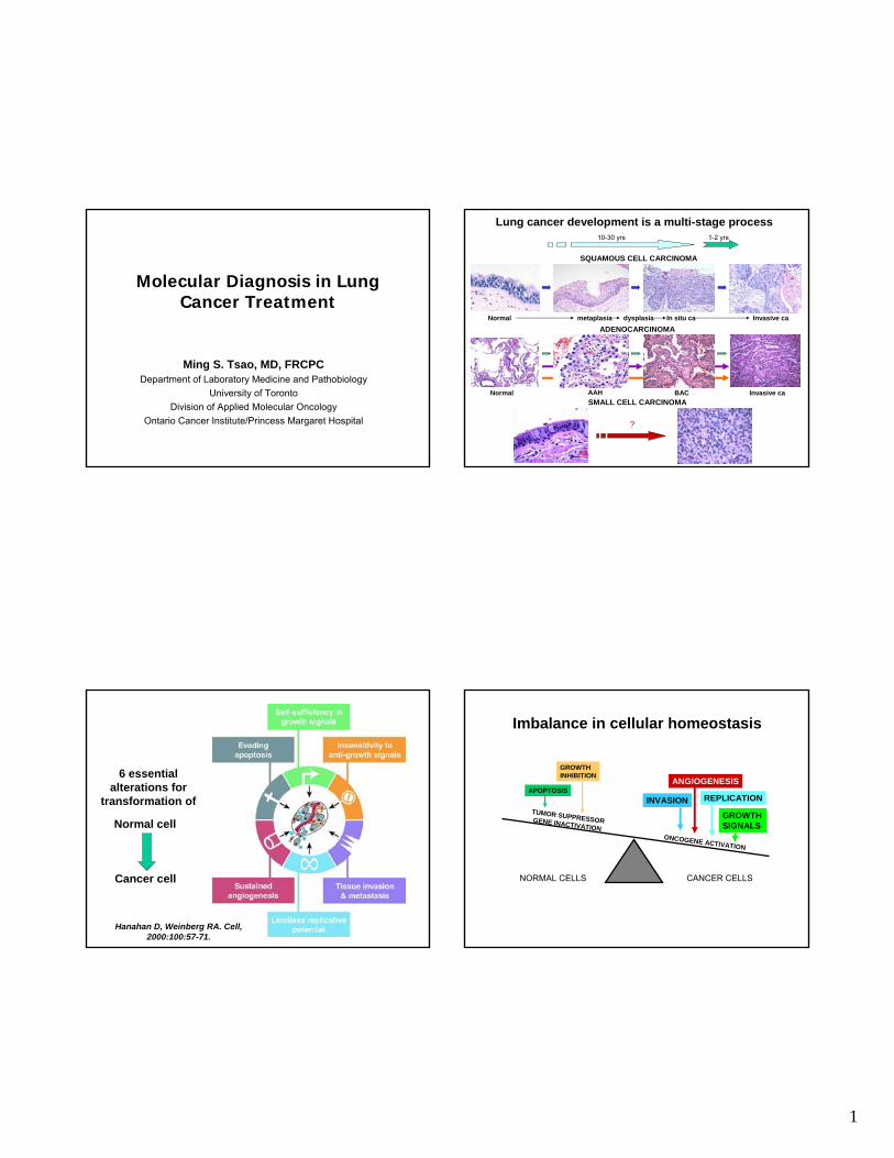
Lung cancer development is a multi-stage process
ADENOCARCINOMA
Normal AAH BAC Invasive caSMALL CELL CARCINOMA
?
10-30 yrs 1-2 yrs
SQUAMOUS CELL CARCINOMA
Normal metaplasia Invasive caIn situ cadysplasia
Normal cell
Cancer cell
6 essential alterations for
transformation of
Hanahan D, Weinberg RA. Cell, 2000:100:57-71.
Imbalance in cellular homeostasis
APOPTOSIS
GROWTH INHIBITION
ANGIOGENESIS
REPLICATION
GROWTH SIGNALS
INVASION
CANCER CELLSNORMAL CELLS
ONCOGENE ACTIVATION
TUMOR SUPPRESSOR GENE INACTIVATION

2
Potential impacts of molecular aberrations
Clinical outcome:• Promote metastasis and poor prognosis• Affect response to treatment
Opportunities:• Develop better disease/therapeutic markers • Serve as therapeutic targets
Carcinoma cells invariably demonstrate complex chromosomal abnormalities
Normal Karyotype (2n=46) NSCLC cell line (2n=70)
ABNORMALITIES OBSERVED: • aneuploidy or unbalanced gain or loss of chromosomes• marker chromosomes that cannot be classified by usual G-banding technique
Abnormal (marker) chromosomes invariably represent complex translocations
SKY: Spectral Karyotyping (Chromosomal painting)
Comparative Genomic Hybridization (CGH)
Tumor DNANormal DNA
3
CGH studies have revealed chromosomal regions that are commonly involved in
genetic gains and losses
7p12: EGFR
gain
loss
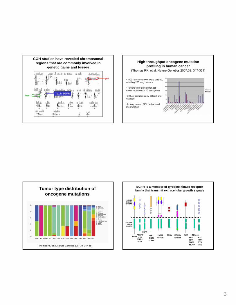
High-throughput oncogene mutation profiling in human cancer
(Thomas RK, et al. Nature Genetics 2007;39: 347-351)
• 1000 human cancers were studied, including 255 lung cancers
• Tumors were profiled for 238 known mutations in 17 oncogenes
• 30% of samples carry at least one mutation
• In lung cancer, 32% had at least one mutation
Tumor type distribution of oncogene mutations
Thomas RK, et al. Nature Genetics 2007;39: 347-351
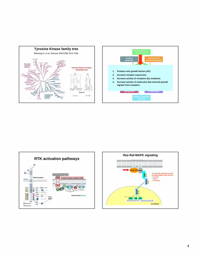
EGFR is a member of tyrosine kinase receptor family that transmit extracellular growth signals
EGFRPDGFR
c-kitCSFR1FLT3
RETMETRONc-Sea
INSRIGF1R
TRKs EPHAsEPHBs
Others:ALK AXLDDR RORROS1 RYKMUSK TIE
FGFR
TYROSINE KINASE
DOMAIN
LIGAND BINDING DOMAIN
4
Tyrosine kinase receptor signaling loops
ligands
Tyrosine Kinase family treeManning G, et al. Science 2002;298:1912-1934
1. Produce own growth factors (GF)
2. Increase receptor expression3. Increase activity of receptors (by mutation)4. Increase activity of molecules that transmit growth
signals from receptors
RTK activation pathways
Tyrosine kinase receptor (TKR)
Growth factor ligand
MAP
K
P
P
nucleus
Ras-Raf-MAPK signaling
RAS-GTP RAF
MEK P
P
MAP
K
P
P
Elk-1 Ets Myc
Functionally important protein phosphorylation sites include:• tyrosine• serine• threonine
5
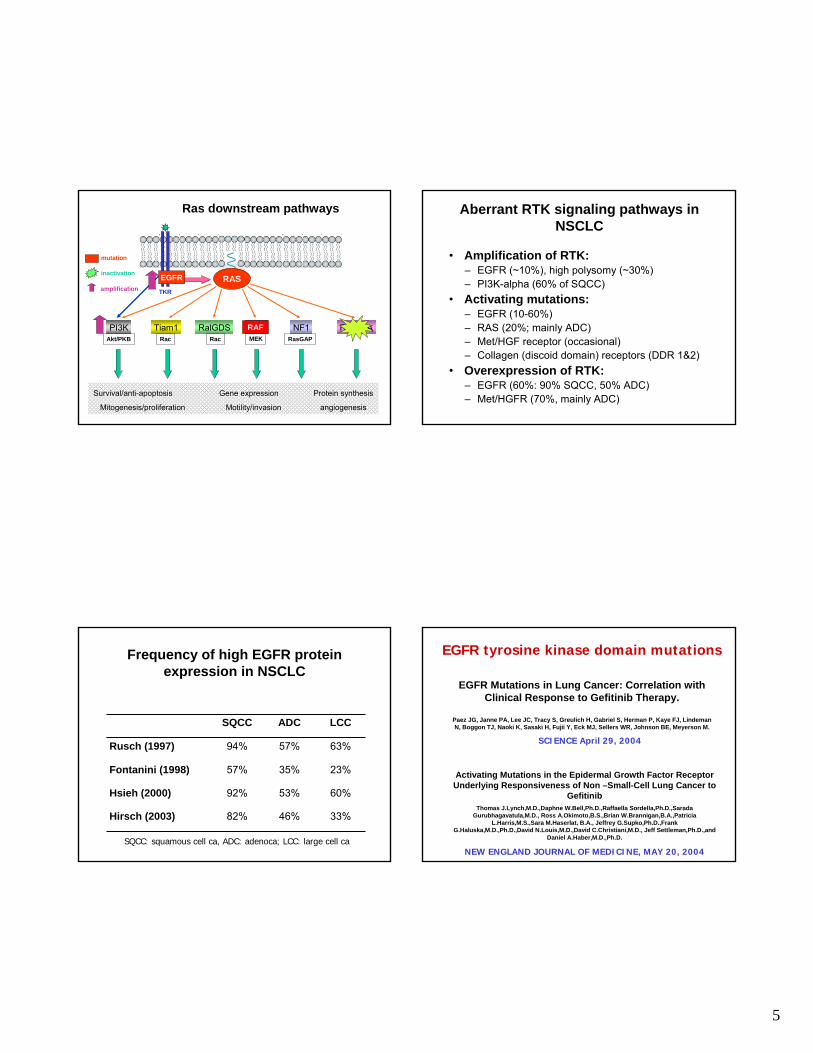
Ras downstream pathways
RAS-GTP
Rassf1ARalGDSTiam1PI3K
TKR
Akt/PKB Rac
RAFMEK
NF1RasGAPRac
Survival/anti-apoptosis Gene expression Protein synthesis
Mitogenesis/proliferation Motility/invasion angiogenesis
RAS
RAF
EGFR
mutation
inactivation
amplification
Aberrant RTK signaling pathways in NSCLC
• Amplification of RTK:– EGFR (~10%), high polysomy (~30%)– PI3K-alpha (60% of SQCC)
• Activating mutations:– EGFR (10-60%)– RAS (20%; mainly ADC)– Met/HGF receptor (occasional)– Collagen (discoid domain) receptors (DDR 1&2)
• Overexpression of RTK:– EGFR (60%: 90% SQCC, 50% ADC)– Met/HGFR (70%, mainly ADC)
Frequency of high EGFR protein expression in NSCLC
33%46%82%Hirsch (2003)
60%53%92%Hsieh (2000)
23%35%57%Fontanini (1998)
63%57%94%Rusch (1997)
LCCADCSQCC
SQCC: squamous cell ca, ADC: adenoca; LCC: large cell ca
EGFR tyrosine kinase domain mutations
EGFR Mutations in Lung Cancer: Correlation with Clinical Response to Gefitinib Therapy.
Paez JG, Janne PA, Lee JC, Tracy S, Greulich H, Gabriel S, Herman P, Kaye FJ, LindemanN, Boggon TJ, Naoki K, Sasaki H, Fujii Y, Eck MJ, Sellers WR, Johnson BE, Meyerson M.
SCIENCE April 29, 2004
Activating Mutations in the Epidermal Growth Factor Receptor Underlying Responsiveness of Non –Small-Cell Lung Cancer to
GefitinibThomas J.Lynch,M.D.,Daphne W.Bell,Ph.D.,Raffaella Sordella,Ph.D.,Sarada
Gurubhagavatula,M.D., Ross A.Okimoto,B.S.,Brian W.Brannigan,B.A.,PatriciaL.Harris,M.S.,Sara M.Haserlat, B.A., Jeffrey G.Supko,Ph.D.,Frank
G.Haluska,M.D.,Ph.D.,David N.Louis,M.D.,David C.Christiani,M.D., Jeff Settleman,Ph.D.,andDaniel A.Haber,M.D.,Ph.D.
NEW ENGLAND JOURNAL OF MEDICINE, MAY 20, 2004
6
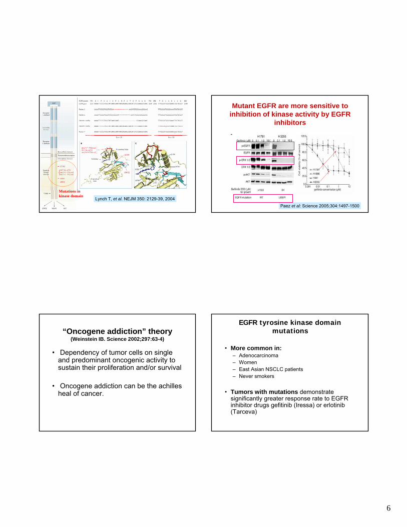
Mutations in kinase domain
Lynch T, et al. NEJM 350: 2129-39, 2004
Mutant EGFR are more sensitive to inhibition of kinase activity by EGFR
inhibitors
Paez et al: Science 2005;304:1497-1500
“Oncogene addiction” theory(Weinstein IB. Science 2002;297:63-4)
• Dependency of tumor cells on single and predominant oncogenic activity to sustain their proliferation and/or survival
• Oncogene addiction can be the achillesheal of cancer.
EGFR tyrosine kinase domain mutations
• More common in:– Adenocarcinoma– Women– East Asian NSCLC patients– Never smokers
• Tumors with mutations demonstrate significantly greater response rate to EGFR inhibitor drugs gefitinib (Iressa) or erlotinib (Tarceva)
7
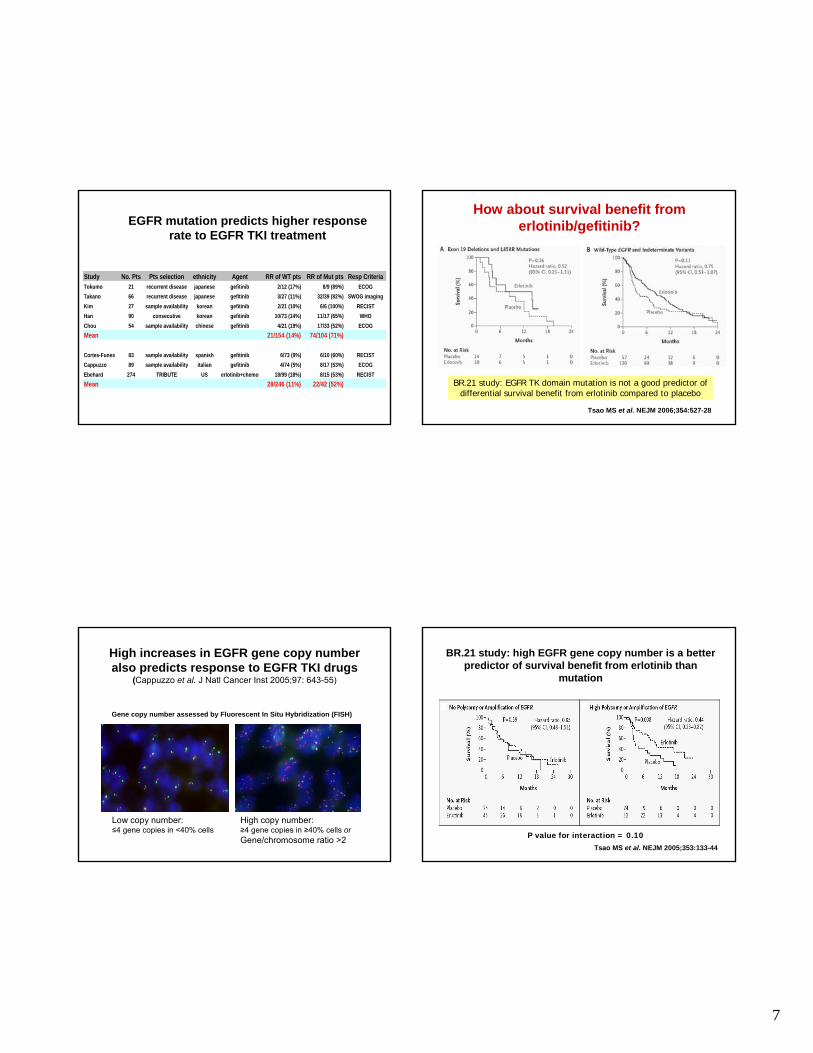
EGFR mutation predicts higher response rate to EGFR TKI treatment
Study No. Pts Pts selection ethnicity Agent RR of WT pts RR of Mut pts Resp CriteriaTokumo 21 recurrent disease japanese gefitinib 2/12 (17%) 8/9 (89%) ECOGTakano 66 recurrent disease japanese gefitinib 3/27 (11%) 32/39 (82%) SWOG imagingKim 27 sample availability korean gefitinib 2/21 (10%) 6/6 (100%) RECISTHan 90 consecutive korean gefitinib 10/73 (14%) 11/17 (65%) WHOChou 54 sample availability chinese gefitinib 4/21 (19%) 17/33 (52%) ECOGMean 21/154 (14%) 74/104 (71%)
Cortes-Funes 83 sample availability spanish gefitinib 6/73 (9%) 6/10 (60%) RECISTCappuzzo 89 sample availability italian gefitinib 4/74 (5%) 8/17 (53%) ECOGEbehard 274 TRIBUTE US erlotinib+chemo 18/99 (18%) 8/15 (53%) RECISTMean 28/246 (11%) 22/42 (52%)
How about survival benefit from erlotinib/gefitinib?
BR.21 study: EGFR TK domain mutation is not a good predictor of differential survival benefit from erlotinib compared to placebo
Tsao MS et al. NEJM 2006;354:527-28
High increases in EGFR gene copy number also predicts response to EGFR TKI drugs
(Cappuzzo et al. J Natl Cancer Inst 2005;97: 643-55)
Low copy number:≤4 gene copies in <40% cells
High copy number:≥4 gene copies in ≥40% cells orGene/chromosome ratio >2
Gene copy number assessed by Fluorescent In Situ Hybridization (FISH)
BR.21 study: high EGFR gene copy number is a better predictor of survival benefit from erlotinib than
mutation
P value for interaction = 0.10Tsao MS et al. NEJM 2005;353:133-44
8
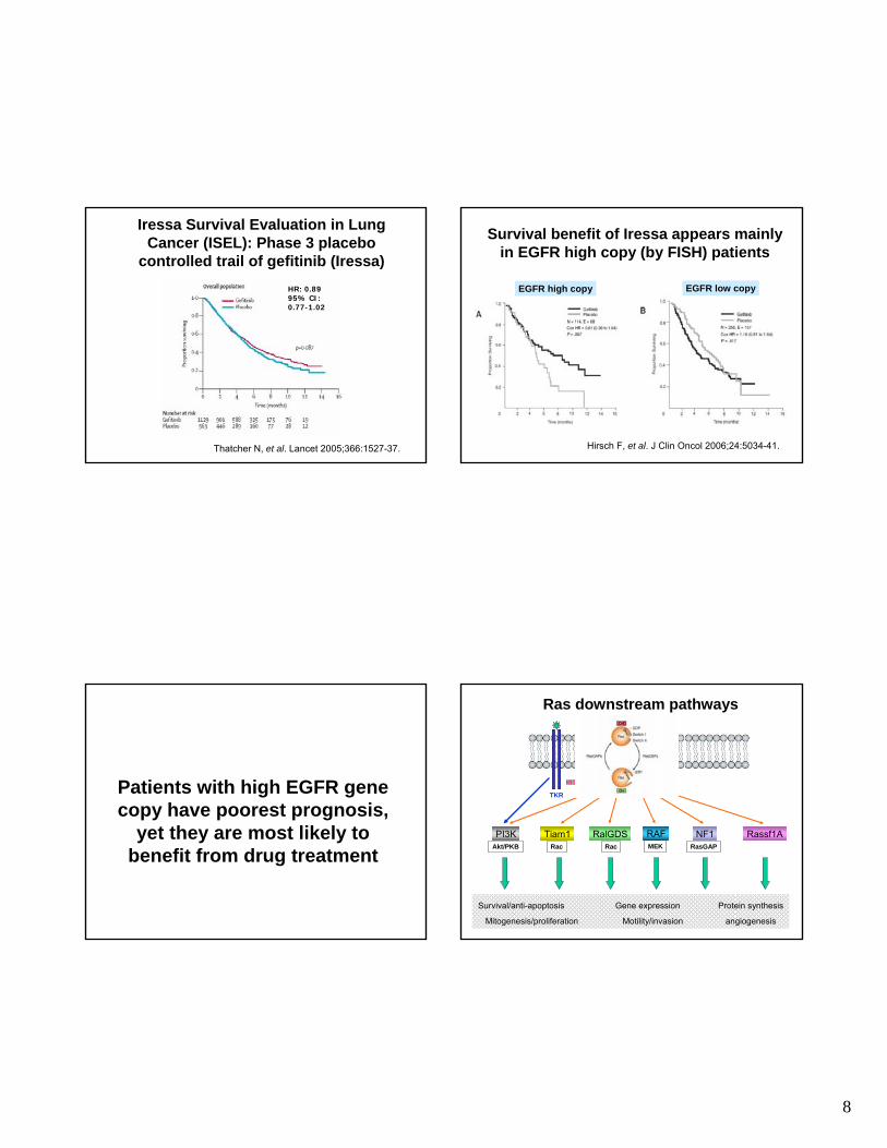
Iressa Survival Evaluation in Lung Cancer (ISEL): Phase 3 placebo
controlled trail of gefitinib (Iressa)HR: 0.8995% CI: 0.77-1.02
Thatcher N, et al. Lancet 2005;366:1527-37.
Survival benefit of Iressa appears mainly in EGFR high copy (by FISH) patients
EGFR high copy EGFR low copy
Hirsch F, et al. J Clin Oncol 2006;24:5034-41.
Patients with high EGFR gene copy have poorest prognosis,
yet they are most likely to benefit from drug treatment
Ras downstream pathways
RAS-GTP
Rassf1ARalGDSTiam1PI3K
TKR
Akt/PKB Rac
RAFMEK
NF1RasGAPRac
Survival/anti-apoptosis Gene expression Protein synthesis
Mitogenesis/proliferation Motility/invasion angiogenesis
9
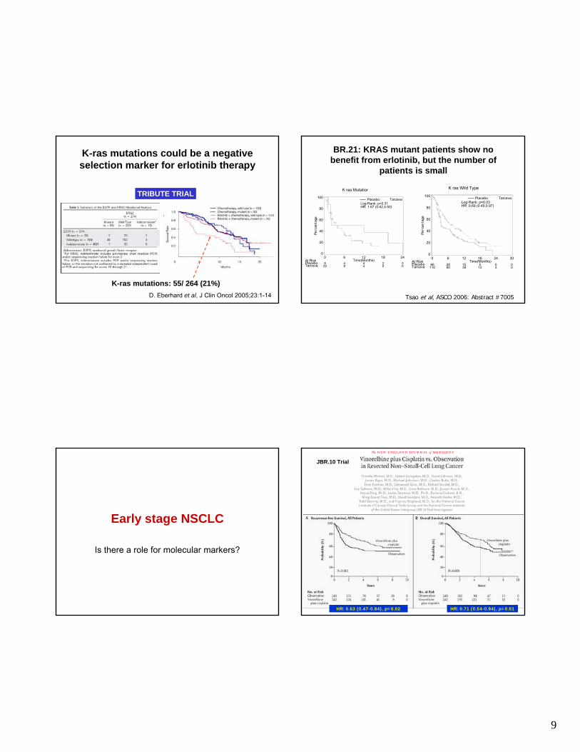
K-ras mutations could be a negative selection marker for erlotinib therapy
TRIBUTE TRIAL
K-ras mutations: 55/ 264 (21%)D. Eberhard et al, J Clin Oncol 2005;23:1-14
At RiskPlaceboTarceva
Log-Rank: p=0.03HR: 0.69 (0.49,0.97)
K ras Wild Type
Placebo Tarceva
Perc
enta
ge
0
20
40
60
80
100
0
66110
6
2860
12
1538
18Time(Months)
512
24
03
30
00
Tsao et al, ASCO 2006: Abstract #7005
At RiskPlaceboTarceva
Log-Rank: p=0.31HR: 1.67 (0.62,4.50)
K ras Mutation
Placebo Tarceva
Perc
enta
ge
0
20
40
60
80
100
0
822
6
48
12Time(Months)
44
18
20
24
00
BR.21: KRAS mutant patients show no benefit from erlotinib, but the number of
patients is small
Early stage NSCLC
Is there a role for molecular markers?
HR: 0.71 (0.54-0.94), p=0.01HR: 0.63 (0.47-0.84), p=0.02
JBR.10 Trial
10
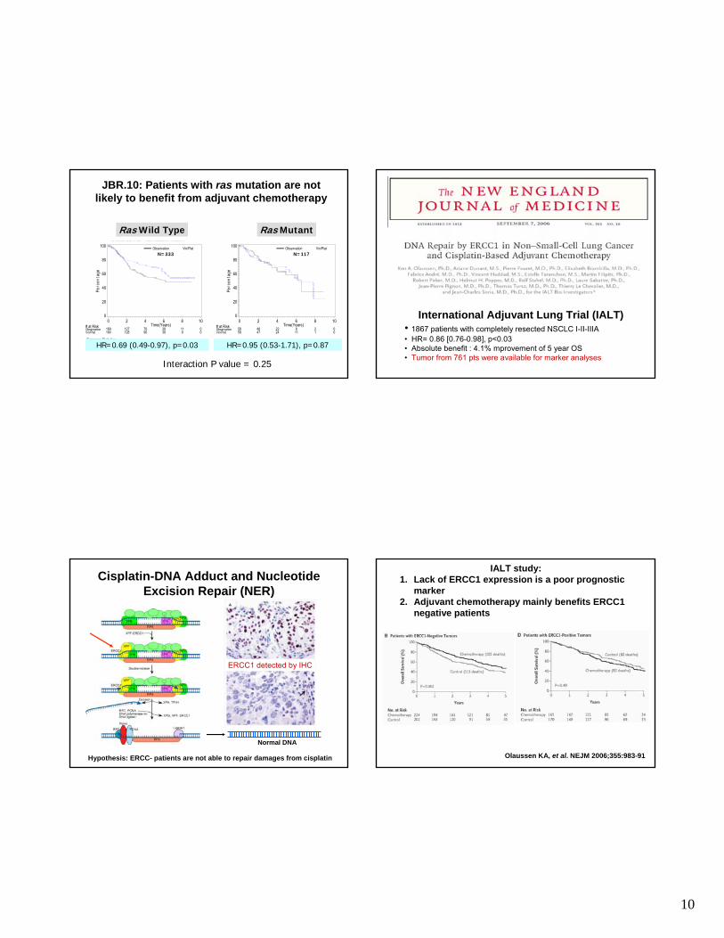
JBR.10: Patients with ras mutation are not likely to benefit from adjuvant chemotherapy
# at RiskObservationVin/Plat
NCIC CTG TRIAL BR.10 Overall Survival - Ras Mutation Absent
Hazard Ratio (95% C.I.): Vin/Plat/Observation: 0.69 (0.49,0.97) Median (95% C.I.): Observation: 6.2 (3.8,inf.), Vin/Plat: N.E. (6.3,inf.) Test for equality of groups: Log-Rank: p=0.0341 Summary Statistics:
Observation Vin/Plat
Perc
enta
ge
0
20
40
60
80
100
0
169164
2
127128
4
7090
6Time(Years)
3939
8
119
10
00
# at RiskObservationVin/Plat
NCIC CTG TRIAL BR.10 Overall Survival - Ras Mutation Present
Hazard Ratio (95% C.I.): Vin/Plat/Observation: 0.95 (0.53,1.71) Median (95% C.I.): Observation: 6.5 (4.0,inf.), Vin/Plat: 6.2 (5.3,inf.) Test for equality of groups: Log-Rank: p=0.8664 Summary Statistics:
Observation Vin/Plat
Perc
enta
ge
0
20
40
60
80
100
0
5859
2
4847
4
2224
6Time(Years)
811
8
21
10
00
N=333 N=117
HR=0.69 (0.49-0.97), p=0.03 HR=0.95 (0.53-1.71), p=0.87
Ras Wild Type Ras Mutant
Interaction P value = 0.25
International Adjuvant Lung Trial (IALT)• 1867 patients with completely resected NSCLC I-II-IIIA• HR= 0.86 [0.76-0.98], p<0.03• Absolute benefit : 4.1% mprovement of 5 year OS• Tumor from 761 pts were available for marker analyses
Cisplatin-DNA Adduct and Nucleotide Excision Repair (NER)
Normal DNA
ERCC1 detected by IHC
Hypothesis: ERCC- patients are not able to repair damages from cisplatin
IALT study: 1. Lack of ERCC1 expression is a poor prognostic
marker2. Adjuvant chemotherapy mainly benefits ERCC1
negative patients
Olaussen KA, et al. NEJM 2006;355:983-91

11
JBR.10: Patients with high class III β-tubulinexpression have poorer survival (prognosis)
JBR.10: Survival benefit from adjuvant cis/vin appears greater in patients with high
class III β-tubulin
HR: 0.78; p=0.39 HR: 0.45; p=0.002
Question: Are high tubulin expressing tumors addicted to their tubulin level?
Biomarkers for adjuvant therapy in NSCLC patients
• Poor prognostic markers: to select patients with high risk for death from recurrence and possibly benefit from adjuvant therapy.
• Predictive markers: to select patients who are most likely to benefit from a specific adjuvant therapy
What about microarrays?
• Good evidence that gene expression profiles by microarray can distinguish:– tumors of different histological types– patients with different prognosis
• But signatures for histological typing do not overlap with those for prognosis
• Prognosis signatures are more reflective of molecular pathways important for the biology of lung cancers
12
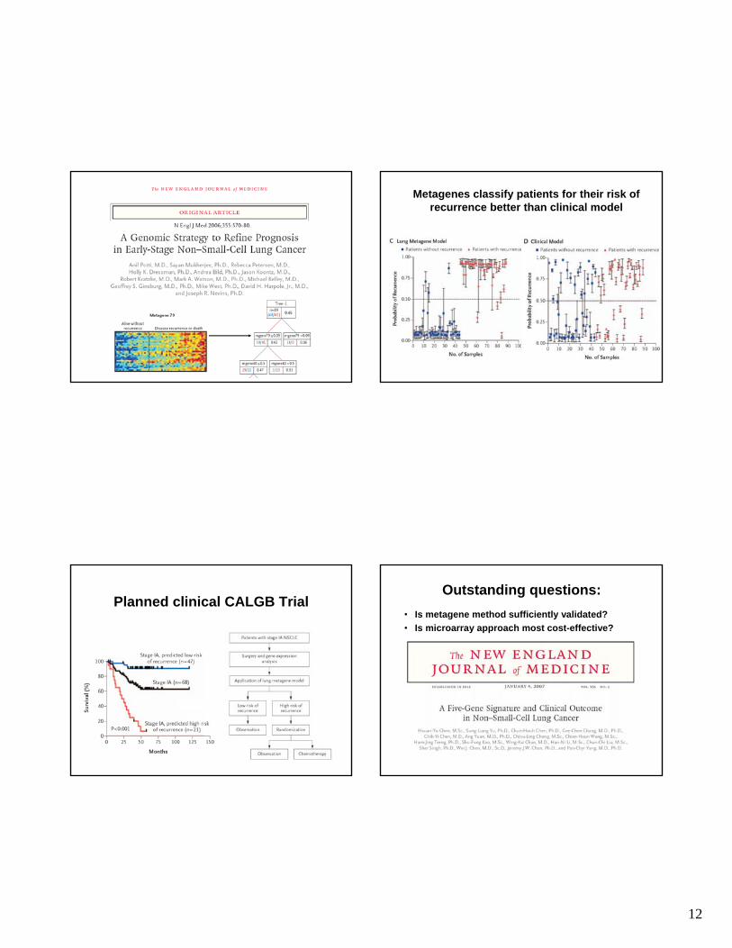
Metagenes classify patients for their risk of recurrence better than clinical model
Planned clinical CALGB TrialOutstanding questions:
• Is metagene method sufficiently validated?• Is microarray approach most cost-effective?

13
Fine mapping of chromosomal abnormalities by Array-CGH
• Chromosomes are cut into fragments that are ~100 kilobases long
• Fragments are arrayed on glass slide
• Differentially labeled DNA from tumor and normal are co-hybridized to the microarrays and signals for each type of sample are detected and compared
Chromosomal abnormalities in NSCLC cell lines detected by array-CGH
(Garnis C et al. Int J Cancer 2006;118: 1156-64)
Chromosome 5p: telomerase (hTERT)
• Role of telomerase complex is to maintain the length of telomeres
• Progressive shortening with continuous replication can trigger apoptosis or growth arrest
• Activation of telomerase is an obligate step during carcinogenesis
hTERT

14
CONCLUSIONS• Currently there are several very promising predictive
markers for selection of NSLC patients to receive targeted or adjuvant chemotherapy
• These markers still require additional validation before they can be implemented as routine clinical tests
• Best way to validate these markers are in patients involved in large phase 3 clinical trials
• The use of molecular markers for stratifying cancer patients to therapeutic options will likely come to reality during the next decade