molecular diagnosis of american … diagnosis of american foulbrood in honeybee brood in bulgaria...
TRANSCRIPT
899
MOLECULAR DIAGNOSIS OF AMERICAN FOULBROOD IN HONEYBEE BROOD IN BULGARIAM. Bonovska, k. GurGulova, s. Takova and T. savovaNational Diagnostic and Research Veterinary Medical Institute “Proof. Dr. G. Pavlov”, BG - 1606 Sofia, Bulgaria
Abstract
Bonovska, M., k. GurGulova, s. Takova and T. savova, 2014. Molecular diagnosis of american foulbrood in honeybee brood in Bulgaria. Bulg. J. Agric. Sci., 20: 899-902
The analysis of the 47 isolates for identification of Paenibacillus larvae, obtained of honeybee brood with american foul-brood (AFB) symptoms from 9 different districts of Bulgaria by conventional PCR technique was performed. Specific DNA products by size 1106 bp were obtained from the 42 isolates. Five isolates were PCr negative for aFB (P. larvae). The results showed that PCR assay is specific and sensitive method for molecular identification of Paenibacillus larvae and can provide a rapid and reliable diagnosis of infected honeybee brood.
Key words: Honeybee brood, aFB, Paenibacillus larvae, PCr
Bulgarian Journal of Agricultural Science, 20 (No 4) 2014, 899-902Agricultural Academy
E-mail: [email protected]
Introduction
american foulbrood (aFB) is a disease of bee brood caused by the spore-forming bacterium Paenibacillus larvae. This is the most serious disease of bee brood, leading to sig-nificant economic losses to beekeepers worldwide. The main host of the bacterium is the honeybee apis mellifera (lind-ström and Fries, 2005). spores of P. larvae can be isolated from honey, wax, pollen, walls of an infected beehive and debris (Gochnauer and Corner, 1987). These spores can re-main infectious for long - at least 35 years (Haseman, 1991), but there have been reports of a longer period of survival (Borchert, 1979).
The disease can be spread by spores when transporting bees, queen bees and bee hives, as well as theft, or using tools and inventory, beekeeper clothes and contaminated pollen or honey (Gochnauer and Corner, 1987). There are different culture media, developed for isolation of P. larvae, but other similar microorganisms grow on them as well, which compli-cated the timely diagnosis.
spores of Paenibacillus larvae and slow-growing vegeta-tive forms of this bacterium are difficult for primary isola-tion and its identification takes a long time (Dobbelaere еt al., 2001). The finding of spores in naturally infected bee lar-vae is very important to confirm the presence of the disease
(Piccini еt al., 2002). Therefore, a new strategy is needed in the fight against American foulbrood and early diagnosis on subclinical level.
In recent years, other methods such as ElIsa and poly-merase chain reaction (PCr) are successfully applied in the diagnosis of many bacterial, viral and parasitological diseas-es, which enable the accurate and quick detection and dif-ferentiation of pathogens (Avaniss-Aghjani еt al., 1994; Dob-belaere еt al., 2001; Piccini еt al., 2002).
The purpose of this paper is the proving of P. larvae, iso-lated from diseased brood by conventional PCr technique in order to introduce the method into routine laboratory diag-nosis of american foulbrood in Bulgaria.
Materials and Methods
Forty-seven isolates of honeycomb with brood, subspi-cious for american foulbrood (aFB), were investigated. samples were sent by 9 different regions in Bulgaria: Burgas, veliko Tarnovo, vratsa, Gabrovo, Pernik, Pleven, smolyan, Sofia and Yambol.
samples were previously studied bacteriologically on blood agar and selective MPYGP agar. For the determination of the obtained isolates as Paenibacillus larvae, they were also tested with aFB kit “vita. Europe”, based on monoclo-

M. Bonovska, K. Gurgulova, S. Takova and T. Savova900
nal antibodies. These isolates were then examined with con-ventional PCr technique using protocols recommended in the oIE Terrestrial Manual (oIE, Terrestrial Manual, 2008). The target DNA needed for PCR was prepared from the bac-terial culture by dissolving one colony in 50 μl of distilled water, heating at 95°C/15 min and centrifugation at 5000 g/5 min. The supernatant containing DNA was used in PCR (Govan еt al., 1999). The study was conducted with a pair of specific primers (AFB-F and AFB-R) with the following nucleotide sequence:
AFB-F-(5’-CTT-GTG-TTT-CTT-TCG-GGa-GaC-GCC-a-3’) andAFB-R–(5’-TCT-TaG-aGT-GCC-CaC-CTC-TGC-G-3’).
The reaction was conducted in a volume 25 μl per sample. The reaction mixture contained: primers aFB F and aFB R (x 50pM); dNTPs (x 200 μl of each dNTP); PCR buffer (x 2.50 μl of the kit); Tag Polymerase (x 0.5 U); a target DNA (x 5.0 μl of goods) and distilled water (dH2O) to 25 μl (Govan еt al., 1999).
Proving specific DNA was performed by amplification of genomic DNA of Paenibacillus larvae in a thermocycler “Techne TC-412” (uk) with the following temperature re-gime: 1 cycle denaturation at 95oC for 15 min and 30 cycles consists of: melting at 93°C for 1 min; annealing at 55°C for 30 s and extension of 72°C for 1 min. Final extension cycle (elongation) at 72°C for 5 min. The PCr products were vi-sualized by horizontal electrophoresis on a 2% agarose gel at 100 v for 30 min, stained in ethidium bromide solution (1:10000) for 10 min and photo-documented on uv-transillu-minator with camera VisiDoc-It (UVP, USA).
Results and Discussion
Most of the tested samples were with typical clinical signs of aFB – mottled brood, the capping of cell are punctured, becomes darkened and concave, the larvae become glutinous in consistency (Figure 1).
Bacteriological inoculations on blood agar and selective MPYGP agar showed growth of Paenibacillus larvae in 42 of the samples (Figure 2). When performed with aFB kit ElI-sa assay, positive results were seen in the same 42 isolates of Paenibacillus larvae (Figure 3).
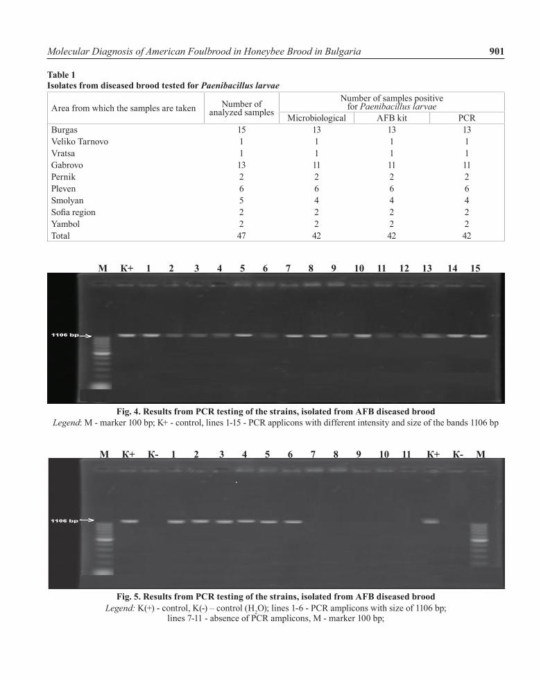
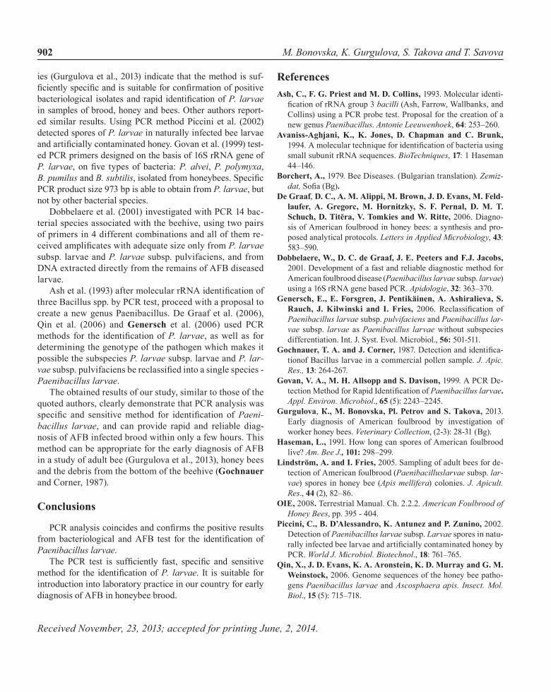
We received specific DNA product with a size of 1106 bp from 42 examined by PCR isolates (Figure 4, lines 1-15; Fig-ure 5, lines 1-6). These results confirmed the previously con-ducted bacteriological examination and testing with aFB kit (Table 1). The varying intensity of the observed bands may be due to the different numbers of the nucleotide sequences located along the length of the DNA chain of Paenibacillus
larvae. No PCR amplicons were received from five isolates from the regions Burgas (2), Gabrovo (2) and smolyan (1) (Figure 5, lines 7-11). The results of the application of our PCR method for identification of P. larvae were confirmed when carried out by the Eurl in France interlaboratory tests for bee health in april 2014. These and our previous stud-
Fig. 1. Clinical picture of AFB diseased bee brood
Fig. 2. Culture of Paenibacillus larvae on the MPYGP agar
Fig. 3. Positive AFB test
Molecular Diagnosis of American Foulbrood in Honeybee Brood in Bulgaria 901
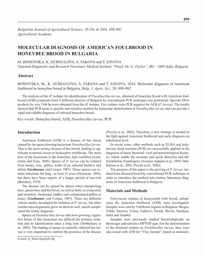
Table 1Isolates from diseased brood tested for Paenibacillus larvae
area from which the samples are taken number of analyzed samples
number of samples positive for Paenibacillus larvae
Microbiological aFB kit PCrBurgas 15 13 13 13veliko Tarnovo 1 1 1 1vratsa 1 1 1 1Gabrovo 13 11 11 11Pernik 2 2 2 2Pleven 6 6 6 6smolyan 5 4 4 4Sofia region 2 2 2 2Yambol 2 2 2 2Total 47 42 42 42
Fig. 4. Results from PCR testing of the strains, isolated from AFB diseased broodLegend: M - marker 100 bp; K+ - control, lines 1-15 - PCR applicons with different intensity and size of the bands 1106 bp
М К+ 1 2 3 4 5 6 7 8 9 10 11 12 13 14 15
Fig. 5. Results from PCR testing of the strains, isolated from AFB diseased broodLegend: K(+) - control, K(-) – control (H2O); lines 1-6 - PCR amplicons with size of 1106 bp;
lines 7-11 - absence of PCR amplicons, M - marker 100 bp;
М К+ К- 1 2 3 4 5 6 7 8 9 10 11 К+ К- М
M. Bonovska, K. Gurgulova, S. Takova and T. Savova902
ies (Gurgulova et al., 2013) indicate that the method is suf-ficiently specific and is suitable for confirmation of positive bacteriological isolates and rapid identification of P. larvae in samples of brood, honey and bees. other authors report-ed similar results. using PCr method Piccini et al. (2002) detected spores of P. larvae in naturally infected bee larvae and artificially contaminated honey. Govan et al. (1999) test-ed PCr primers designed on the basis of 16s rrna gene of P. larvae, on five types of bacteria: P. alvei, P. polymyxa, B. pumilus and B. subtilis, isolated from honeybees. Specific PCr product size 973 bp is able to obtain from P. larvae, but not by other bacterial species.
Dobbelaere et al. (2001) investigated with PCR 14 bac-terial species associated with the beehive, using two pairs of primers in 4 different combinations and all of them re-ceived amplificates with adequate size only from P. larvae subsp. larvae and P. larvae subsp. pulvifaciens, and from DNA extracted directly from the remains of AFB diseased larvae.
Ash et al. (1993) after molecular rRNA identification of three Bacillus spp. by PCr test, proceed with a proposal to create a new genus Paenibacillus. De Graaf et al. (2006), Qin et al. (2006) and Genersch et al. (2006) used PCr methods for the identification of P. larvae, as well as for determining the genotype of the pathogen which makes it possible the subspecies P. larvae subsp. larvae and P. lar-vae subsp. pulvifaciens be reclassified into a single species - Paenibacillus larvae.
The obtained results of our study, similar to those of the quoted authors, clearly demonstrate that PCr analysis was specific and sensitive method for identification of Paeni-bacillus larvae, and can provide rapid and reliable diag-nosis of aFB infected brood within only a few hours. This method can be appropriate for the early diagnosis of aFB in a study of adult bee (Gurgulova et al., 2013), honey bees and the debris from the bottom of the beehive (Gochnauer and Corner, 1987).
Conclusions
PCR analysis coincides and confirms the positive results from bacteriological and AFB test for the identification of Paenibacillus larvae.
The PCR test is sufficiently fast, specific and sensitive method for the identification of P. larvae. It is suitable for introduction into laboratory practice in our country for early diagnosis of aFB in honeybee brood.
ReferencesAsh, C., F. G. Priest and M. D. Collins, 1993. Molecular identi-
fication of rRNA group 3 bacilli (ash, Farrow, Wallbanks, and Collins) using a PCr probe test. Proposal for the creation of a new genus Paenibacillus. Antonie Leeuwenhoek, 64: 253–260.
Avaniss-Aghjani, K., K. Jones, D. Chapman and C. Brunk, 1994. A molecular technique for identification of bacteria using small subunit rrna sequences. BioTechniques, 17: 1 Haseman 44–146.
Borchert, A., 1979. Bee Diseases. (Bulgarian translation). Zemiz-dat, Sofia (Bg).
De Graaf, D. C., A. M. Alippi, M. Brown, J. D. Evans, M. Feld-laufer, A. Gregorc, M. Hornitzky, S. F. Pernal, D. M. T. Schuch, D. Titĕra, V. Tomkies and W. Ritte, 2006. Diagno-sis of american foulbrood in honey bees: a synthesis and pro-posed analytical protocols. Letters in Applied Microbiology, 43: 583–590.
Dobbelaere, W., D. C. de Graaf, J. E. Peeters and F.J. Jacobs, 2001. Development of a fast and reliable diagnostic method for american foulbrood disease (Paenibacillus larvae subsp. larvae) using a 16s rrna gene based PCr. Apidologie, 32: 363–370.
Genersch, E., E. Forsgren, J. Pentikäinen, A. Ashiralieva, S. Rauch, J. Kilwinski and I. Fries, 2006. Reclassification of Paenibacillus larvae subsp. pulvifaciens and Paenibacillus lar-vae subsp. larvae as Paenibacillus larvae without subspecies differentiation. Int. J. syst. Evol. Microbiol., 56: 501-511.
Gochnauer, T. A. and J. Corner, 1987. Detection and identifica-tionof Bacillus larvae in a commercial pollen sample. J. Apic. Res., 13: 264-267.
Govan, V. A., M. H. Allsopp and S. Davison, 1999. A PCR De-tection Method for Rapid Identification of Paenibacillus larvae. Appl. Environ. Microbiol., 65 (5): 2243–2245.
Gurgulova, К., М. Bonovska, Pl. Petrov and S. Тakova, 2013. Early diagnosis of american foulbrood by investigation of worker honey bees. Veterinary Collection, (2-3): 28-31 (Bg).
Haseman, L., 1991. How long can spores of american foulbrood live? Am. Bee J., 101: 298–299.
Lindström, A. and I. Fries, 2005. sampling of adult bees for de-tection of american foulbrood (Paenibacilluslarvae subsp. lar-vae) spores in honey bee (Apis mellifera) colonies. J. Apicult. Res., 44 (2), 82–86.
OIE, 2008. Terrestrial Manual. Ch. 2.2.2. American Foulbrood of Honey Bees, pp. 395 - 404.
Piccini, C., B. D’Alessandro, K. Antunez and P. Zunino, 2002. Detection of Paenibacillus larvae subsp. Larvae spores in natu-rally infected bee larvae and artificially contaminated honey by PCr. World J. Microbiol. Biotechnol., 18: 761–765.
Qin, X., J. D. Evans, K. A. Aronstein, K. D. Murray and G. M. Weinstock, 2006. Genome sequences of the honey bee patho-gens Paenibacillus larvae and Ascosphaera apis. Insect. Mol. Biol., 15 (5): 715–718.
Received November, 23, 2013; accepted for printing June, 2, 2014.