molecular epidemiology of dengue viruses … · nakhon pathom; 3department of statistic, faculty of...
TRANSCRIPT
SoutheaSt aSian J trop Med public health
604 Vol 49 No. 4 July 2018
Correspondence: Jundee Rabablert, Depart-ment of Biology, Faculty of Science, Silpakorn University, Nakhon Pathom 73000, Thailand.E-mail: rabablert_je.su.ac.th
MOLECULAR EPIDEMIOLOGY OF DENGUE VIRUSES ISOLATED FROM PATIENTS WITH SUSPECTED
DENGUE FEVER IN BANGKOK, THAILAND DURING 2006-2015
Sutee Yoksan2, Kumchol Chaiyo1,2, Supoth Rajakam2, Soratorn Kerdkriangkrai1, Pairoj Khawsithiwong3 and Jundee Rabablert1
1Department of Biology, Faculty of Science, Silpakorn University; 2Center for Vaccine Development, Institute of Molecular Bioscience, Salaya Campus, Mahidol University,
Nakhon Pathom; 3Department of Statistic, Faculty of Science, Silpakorn University, Nakhon Pathom, Thailand
Abstract. Dengue virus serotypes 1, 2, 3, and 4 (DENV1-DENV4), Family Flavi-viridae, genus Flavivirus cause mosquito-borne diseases, such as dengue fever (DF), dengue hemorrhagic fever (DHF) and dengue shock syndrome (DSS) in sub-tropical and tropical regions. In addition, different genotypes of each DENV serotype are involved in severity among dengue patients. This study evaluated the molecular epidemiology of patients’ sera DENV isolates grown in C6/36 cells using quantitative (q)RT-PCR, DNA sequencing and construction of phylogenetic tree. QRT-PCR revealed 75 isolates consisting of DENV1 (n = 15), DENV2 (n = 20), DENV3 (n = 28), and DENV4 (n = 12). DNA sequencing and phylogenetic tree analysis demonstrated genotype I of DENV1, genotype Asian I of DENV3 consisting of genotype II (n = 5) and genotype III (n = 23), and genotype I of DENV4. Survey of dengue in Thailand showed presence of DENV3 genotype II since 1973, and genotype III since 2008. Our study reveals genetic information, which complements current knowledge on dengue epidemiology, evolution and transmission dynamics. Understanding of dengue epidemiology at the molecular level will be of particular importance in dengue disease control and prevention.
Keywords: dengue virus, DNA sequencing, epidemiology, phylogenetic tree analysis
INTRODUCTION
Dengue virus (DENV) belongs to the genus Flavivirus, family Flaviviri-dae. There are four antigenically dis-tinct DENV serotypes, namely, DENV1, DENV2, DENV3, and DENV4 (Guzman et al, 2016). DENV genome consists of
single-stranded positive-sense RNA ap-proximately 11 kb in length that is capped at the 5’ end and lacks a 3’ polyadenylated sequence. DENV genome is translated as a single polypeptide and post-translation-ally cleaved into three structural proteins [capsid (C), premembrane (prM) and en-velope (E)] and seven nonstructural (NS) proteins (NS1, NS2A, NS2B, NS3, NS4A, NS4B, and NS5) (Guzman et al, 2016).
DENV is transmitted to humans through the bite of infected Aedes mosqui-
Phylogenetic AnAlysis of Dengue Viruses, BAngkok, thAilAnD 2006-2015
Vol 49 No. 4 July 2018 605
toes, particularly Ae. aegypti and Ae. albop-ictus (Hugo et al, 2014). Infection with any of the four DENV causes a wide spectrum of clinical features ranging from nearly asymptomatic disease, an undifferenti-ated febrile illness, dengue fever (DF), to dengue hemorrhagic fever (DHF)/dengue shock syndrome (DSS). DENV affects 50-200 million people and leads to approxi-mately 20,000 deaths annually in tropical and subtropical regions of the world. The mortality rate of patients with severe den-gue diseases is about 1-2.5% (Martina et al, 2009; Guzman et al, 2016). In Thailand, a total of 142,925 dengue cases (morbidity rate of 222.56/100,000 population) with 147 deaths (mortality rate of 0.23/100,000 population) were reported in 2015 (BOE, 2016). In endemic areas, co-circulation of multiple DENV serotypes has been shown (Holmes et al, 2009). In addition, each DENV serotype shows phylogeneti-cally distinct genotypes (Klungthong et al, 2008; Teoh et al, 2013). Genotype and clade replacements in DENV serotypes are associated with prevalence of dengue disease (Zhang et al, 2005).
In this study, we evaluated the mo-lecular epidemiology of DENV isolated from patients’ sera, obtained in Bangkok, Thailand during 2006 to 2015.
MATERIALS AND METHODS
Viruses and cell culture DENV1 (16007-strain), DENV2
(16681-strain), DENV3 (16562-strain) and DENV4 (1036-strain) passaged 50 times in C6/36 cells at a viral titer of 1×10⁵ plaque-forming units (pfus)/ml were used as sources of viral nucleic acid/positive standards.
C6/36 cells were cultured in modified Eagle medium (MEM; GIBCO, Grands Island, NY) containing 10% fetal bovine
serum (FBS; GIBCO), 2 mM L-glutamine (GIBCO), 1% sodium bicarbonate (Sigma-Aldrich, St. Louis, MO) and 1% non-essen-tial amino acid (GIBCO) at 32°C.Patients’ sera
Sera from 326 patients with sus-pected dengue and subsequently clinical diagnosed by Professor Dr Ampaiwan Juansamrit, Ramathibodi Hospital and Professor Dr Kulkanya Chokephaibulkit, Faculty of Medicine Siriraj Hospital, Ma-hidol University, Bangkok during 2006-2015 were screened by SD Bioline Dengue Duo Rapid Test (Standard Diagnostics, Gyeonggi-do, Korea) and stored at -80°C until used.Virus infection of C6/36 cells
A 100 µl aliquot of patient’s serum was added to complete medium (MEM+10% FBS) containing C6/36 cells, which were serially passaged three times over a pe-riod of 7 days at 32°C. DENV in infected C6/36 cells and culture supernatant was detected by indirect immunofluorescent assay (IFA) and quantitative (q)RT-PCR), respectively. IFA
Mouse monoclonal antibodies (mAbs) to DENV1 (15F3-1, ATCC HB-47), DENV2 (3H5-1, ATCC HB-46), DENV3 (5D4-11, ATCC HB-49) and DENV4 (1H10-6, ATCC HB-48) in tissue culture fluid were used as the primary Abs and goat FITC-conju-gated anti-mouse IgG (Thermo Scientific, Amarillo, TX) was used as secondary Ab.
Uninfected and DENV-infected cells were spotted on slides and fixed with cold acetone. Each anti-DENV mouse mAb [di-luted 1:100 in phosphate-buffered saline pH 7.4 (PBS)] was applied onto slides for 45 minutes at 37°C. Goat Ab conjugate (diluted 1:50 in PBS) then was applied and incubated for 45 minutes at 37°C. Slides were mounted with 50% buffered glycerol
SoutheaSt aSian J trop Med public health
606 Vol 49 No. 4 July 2018
Table 1Primers and probes used in the study.
Name Nucleotide sequence (5′→3′) Genome position
QRT-PCR DENV1 F CAAAAGGAAGTCGTGCAATA 8936-8955DENV1 C CTGAGTGAATTCTCTCTACTGAACC 9023-9047DENV1 probe FAM-CATGTGGTTGGGAGCACGC-BHQ1 8961-8979DENV2 F CAGGTTATGGCACTGTCACGAT 1426-1447DENV2 C CCATCTGCAGCAACACCATCTC 1482-1504DENV2 probe HEX-CTCTCCGAGAACAGGCCTCGACTTCAA-BHQ1 1454-1480DENV3 F GGACTGGACACACGCACTCA 701-720DENV3 C CATGTCTCTACCTTCTCGACTTGTCT 749-775DENV3 probe TXR-ACCTGGATGTCGGCTGAAGGAGCTTG-BHQ2 722-747DENV4 F TTGTCCTAATGATGCTGGTCG 884-904DENV4 C TCCACCTGAGACTCCTTCCA 953-992DENV4 probe Cy5-TTCCTACTCCTACGCATCGCATTCCG-BHQ3 939-960RT-PCR and DNA sequencing DG1 (E1)-F AGTAGAGACTTGGGCTCTGA 802-821DG1 (E2)-R CCAGTTGATTACACATCCCG 2424-2443DG2 (E1)-F CAGCTGTCGCTCCTTCA 914-930DG2 (E2)-R GCTCTAGATCGGCCTGCACCAT 2410-2432DG3 (E1)-F GCCCATTACATAGGCACTTCC 857-877DG3 (E2)-R ACACAYCCCATGTCAGCTTG 2408-2427DG4 (E1)-F CTCTTGGCAGGATTYATGGC 843-862DG4 (E2)-R CACTCCATGACACCACACAACC 2430-2451
Y = C or T.
and inspected under an UV microscope (BX60 model, Olympus, Tokyo, Japan).QRT-PCR
RNA was extracted from culture su-pernatant using E.Z.N.A viral RNA mini kit (Omega biotek, Norcross, GA) and a small aliquot was used for qRT-PCR with primers specific for each of the DENV (Table 1). Reaction solutions were pre-pared using KAPA PROBE FAST universal one-step qRT-PCR master mix kit (KAPA biosystems, Wilmington, MA) and were thermocycled in a Chromo4 (Bio-Rad, Hercules, CA) as previously described (Johnson et al, 2005). Positive controls were viruses extracted from cell culture,
and negative control was nuclease-free water.DENV E gene sequencing and phylogenetic tree construction
DENV genotyping was performed using E gene sequence (1,485 nt). Viral RNA was reverse-transcribed into cDNA using Maxima H minus-first strand cDNA synthesis kit (Thermo Scientific), which then was used as template for PCR ampli-fication using Phusion Flash high-fidelity PCR master mix (Thermo Scientific) and DENV E-specific primers (Table 1). Am-plicons were sequenced by First BASE Laboratories (Selangor, Malaysia), and sequences were aligned using BioEdit
Phylogenetic AnAlysis of Dengue Viruses, BAngkok, thAilAnD 2006-2015
Vol 49 No. 4 July 2018 607
sequence alignment editor (www.mbio.ncsu.edu/BioEdit/bioedit.html) and ana-lyzed using basic local alignment search tool (BLAST; https://www.ncbi.nlm.nih.gov/BLAST/). DENV E gene sequence data from GenBank were included. Sequence multiple alignments were performed using ClustalW (Thompson et al, 1994). The phylogenetic tree was generated using Molecular Evolutionary Genetics Analysis (MEGA) 6.0 software (Tamura et al, 2013). Neighbor-joining tree was con-structed with 1,000 bootstrap replicates. Genotypes of DENV were classified as previously described (Klungthong et al, 2008). The E gene sequences obtained in the study were deposited in GenBank with accession numbers MG564068-MG564138, KT026308-KT026310 and KR922405 (Table 2).
RESULTS
In order to evaluate the molecular epidemiology of DENV isolated from pa-tients’ sera obtained from two hospitals in Bangkok during 2006 to 2015, 326 dengue patients’ sera were screened using SD Bioline dengue duo rapid test, of which 143 (44%) were positive for dengue NS1 Ag, 221 (68%) positive for dengue IgM and 173 (53%) positive for IgG. When the 326 patients’ sera were directly inoculated to C6/36 cells for 3 passages, IFA revealed 75 (23.0%) DENV-positive isolates (bright green apple fluorescence in cytoplasm) and 251 (77%) DENV-negative virus isolates (dull green fluorescence in cyto-plasm) (data not shown). The 75 DENV isolates were categorized as DENV1 (n = 15, 20%), DENV2 (n = 20, 27%), DENV3 (n = 28, 37%), and DENV4 (n = 12, 16%). QRT-PCR quantified DENV titers as rang-ing from 7.75×10² to 2.26×10⁶ copies/µl cell culture supernatant (data not shown).
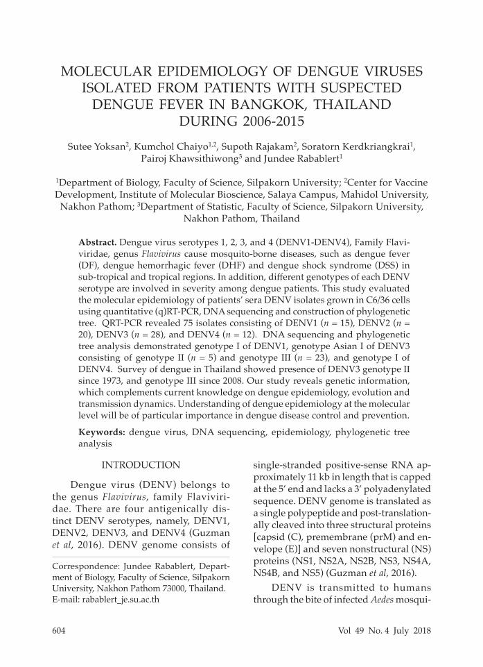
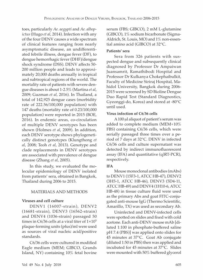
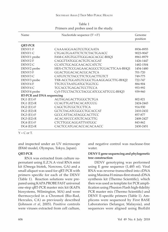
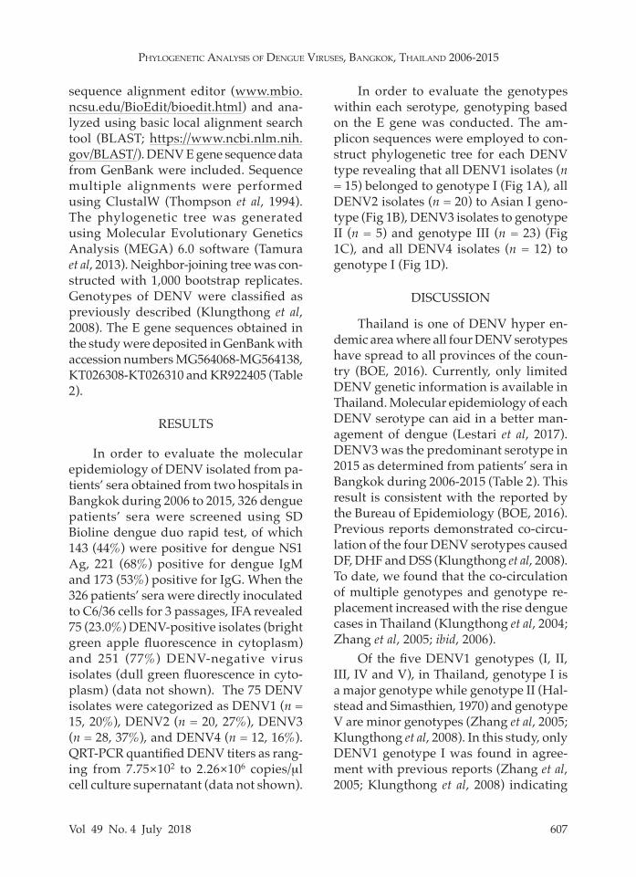
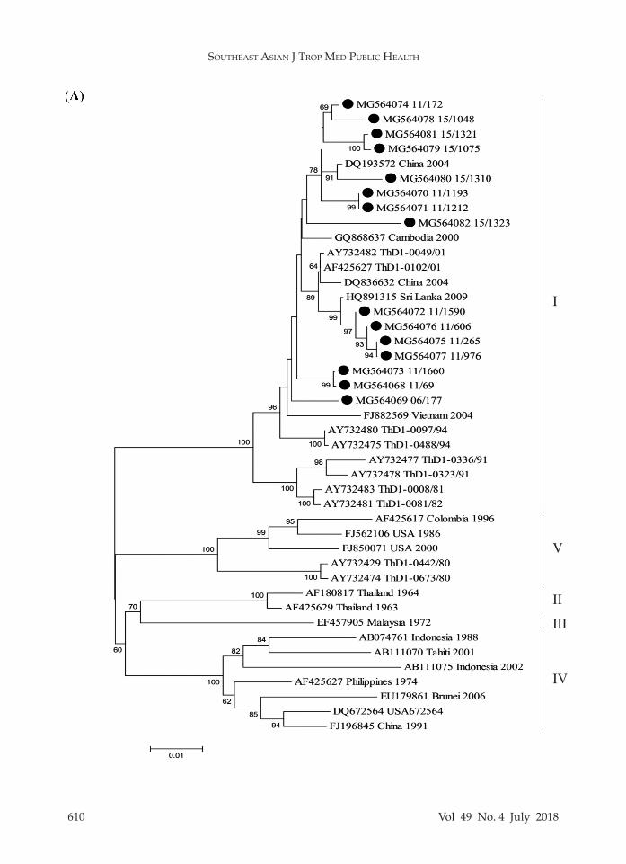
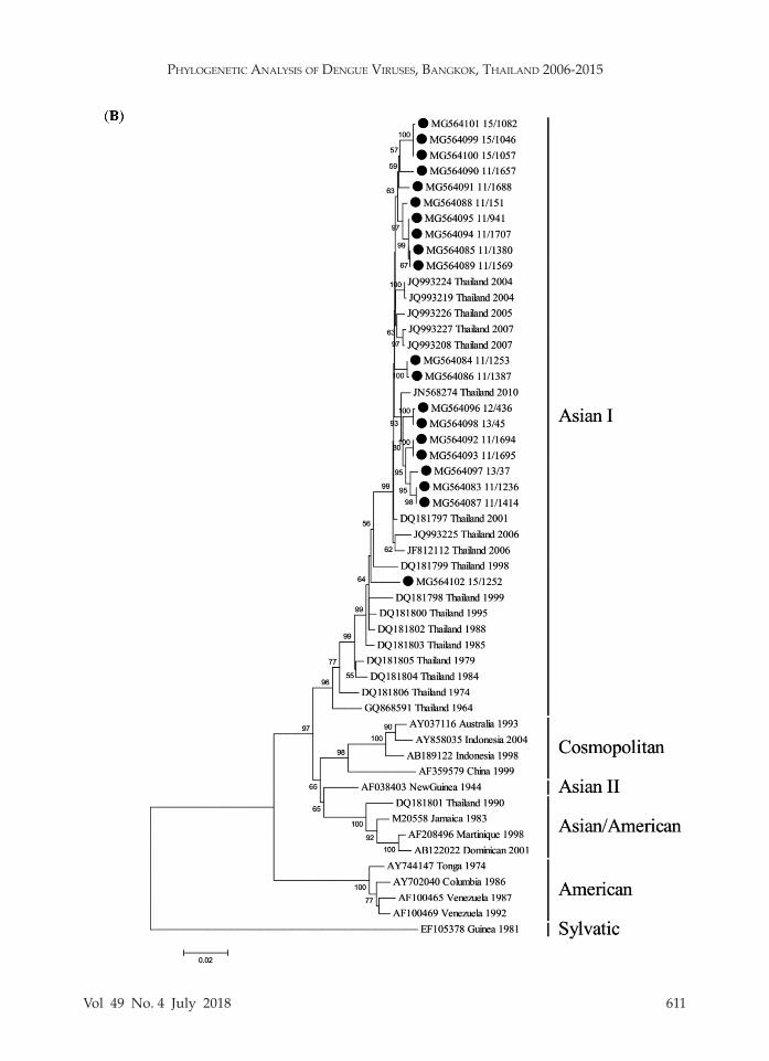
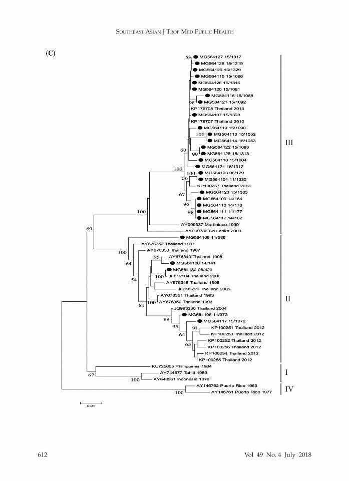
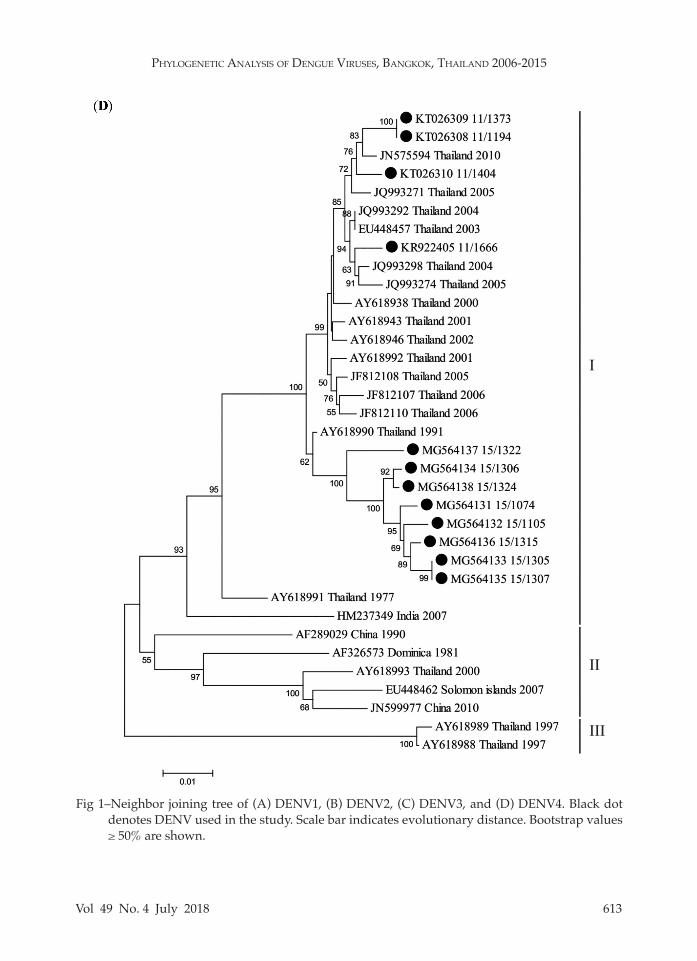
In order to evaluate the genotypes within each serotype, genotyping based on the E gene was conducted. The am-plicon sequences were employed to con-struct phylogenetic tree for each DENV type revealing that all DENV1 isolates (n = 15) belonged to genotype I (Fig 1A), all DENV2 isolates (n = 20) to Asian I geno-type (Fig 1B), DENV3 isolates to genotype II (n = 5) and genotype III (n = 23) (Fig 1C), and all DENV4 isolates (n = 12) to genotype I (Fig 1D).
DISCUSSION
Thailand is one of DENV hyper en-demic area where all four DENV serotypes have spread to all provinces of the coun-try (BOE, 2016). Currently, only limited DENV genetic information is available in Thailand. Molecular epidemiology of each DENV serotype can aid in a better man-agement of dengue (Lestari et al, 2017). DENV3 was the predominant serotype in 2015 as determined from patients’ sera in Bangkok during 2006-2015 (Table 2). This result is consistent with the reported by the Bureau of Epidemiology (BOE, 2016). Previous reports demonstrated co-circu-lation of the four DENV serotypes caused DF, DHF and DSS (Klungthong et al, 2008). To date, we found that the co-circulation of multiple genotypes and genotype re-placement increased with the rise dengue cases in Thailand (Klungthong et al, 2004; Zhang et al, 2005; ibid, 2006).
Of the five DENV1 genotypes (I, II, III, IV and V), in Thailand, genotype I is a major genotype while genotype II (Hal-stead and Simasthien, 1970) and genotype V are minor genotypes (Zhang et al, 2005; Klungthong et al, 2008). In this study, only DENV1 genotype I was found in agree-ment with previous reports (Zhang et al, 2005; Klungthong et al, 2008) indicating
SoutheaSt aSian J trop Med public health
608 Vol 49 No. 4 July 2018
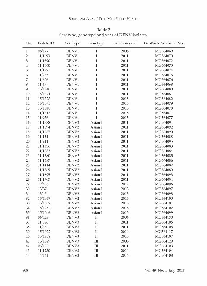
Table 2Serotype, genotype and year of DENV isolates.
No. Isolate ID Serotype Genotype Isolation year GenBank Accession No.
1 06/177 DENV1 I 2006 MG5640692 11/1193 DENV1 I 2011 MG5640703 11/1590 DENV1 I 2011 MG5640724 11/1660 DENV1 I 2011 MG5640735 11/172 DENV1 I 2011 MG5640746 11/265 DENV1 I 2011 MG5640757 11/606 DENV1 I 2011 MG5640768 11/69 DENV1 I 2011 MG5640689 15/1310 DENV1 I 2011 MG56408010 15/1321 DENV1 I 2011 MG56408111 15/1323 DENV1 I 2015 MG56408212 15/1075 DENV1 I 2015 MG56407913 15/1048 DENV1 I 2015 MG56407814 11/1212 DENV1 I 2015 MG56407115 11/976 DENV1 I 2015 MG56407716 11/1688 DENV2 Asian I 2011 MG56409117 11/1694 DENV2 Asian I 2011 MG56409218 11/1657 DENV2 Asian I 2011 MG56409019 11/151 DENV2 Asian I 2011 MG56408820 11/941 DENV2 Asian I 2011 MG56409521 11/1236 DENV2 Asian I 2011 MG56408322 11/1253 DENV2 Asian I 2011 MG56408423 11/1380 DENV2 Asian I 2011 MG56408524 11/1387 DENV2 Asian I 2011 MG56408625 11/1414 DENV2 Asian I 2011 MG56408726 11/1569 DENV2 Asian I 2011 MG56408927 11/1695 DENV2 Asian I 2011 MG56409328 11/1707 DENV2 Asian I 2011 MG56409429 12/436 DENV2 Asian I 2012 MG56409630 13/37 DENV2 Asian I 2013 MG56409731 13/45 DENV2 Asian I 2013 MG56409832 15/1057 DENV2 Asian I 2015 MG56410033 15/1082 DENV2 Asian I 2015 MG56410134 15/1252 DENV2 Asian I 2015 MG56410235 15/1046 DENV2 Asian I 2015 MG56409936 06/429 DENV3 II 2006 MG56413037 11/586 DENV3 II 2011 MG56410638 11/372 DENV3 II 2011 MG56410539 15/1072 DENV3 II 2014 MG56411740 15/1328 DENV3 II 2015 MG56410741 15/1329 DENV3 III 2006 MG56412942 06/129 DENV3 III 2011 MG56410343 11/1230 DENV3 III 2014 MG56410444 14/141 DENV3 III 2014 MG564108
Phylogenetic AnAlysis of Dengue Viruses, BAngkok, thAilAnD 2006-2015
Vol 49 No. 4 July 2018 609
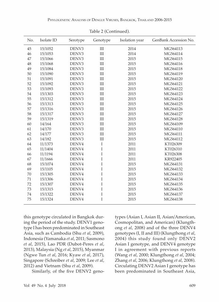
45 15/1052 DENV3 III 2014 MG56411346 15/1053 DENV3 III 2014 MG56411447 15/1066 DENV3 III 2015 MG56411548 15/1068 DENV3 III 2015 MG56411649 15/1084 DENV3 III 2015 MG56411850 15/1090 DENV3 III 2015 MG56411951 15/1091 DENV3 III 2015 MG56412052 15/1092 DENV3 III 2015 MG56412153 15/1093 DENV3 III 2015 MG56412254 15/1303 DENV3 III 2015 MG56412355 15/1312 DENV3 III 2015 MG56412456 15/1313 DENV3 III 2015 MG56412557 15/1316 DENV3 III 2015 MG56412658 15/1317 DENV3 III 2015 MG56412759 15/1319 DENV3 III 2015 MG56412860 14/164 DENV3 III 2015 MG56410961 14/170 DENV3 III 2015 MG56411062 14/177 DENV3 III 2015 MG56411163 14/182 DENV3 III 2015 MG56411264 11/1373 DENV4 I 2011 KT02630965 11/1404 DENV4 I 2011 KT02631066 11/1194 DENV4 I 2011 KT02630867 11/1666 DENV4 I 2011 KR92240568 15/1074 DENV4 I 2015 MG56413169 15/1105 DENV4 I 2015 MG56413270 15/1305 DENV4 I 2015 MG56413371 15/1306 DENV4 I 2015 MG56413472 15/1307 DENV4 I 2015 MG56413573 15/1315 DENV4 I 2015 MG56413674 15/1322 DENV4 I 2015 MG56413775 15/1324 DENV4 I 2015 MG564138
Table 2 (Continued).
No. Isolate ID Serotype Genotype Isolation year GenBank Accession No.
this genotype circulated in Bangkok dur-ing the period of the study. DENV1 geno-type I has been predominated in Southeast Asia, such as Cambodia (Shu et al, 2009), Indonesia (Yamanaka et al, 2011; Sasmono et al, 2015), Lao PDR (Dubot-Peres et al, 2013), Malaysia (Ng et al, 2015), Myanmar (Ngwe Tun et al, 2016; Kyaw et al, 2017), Singapore (Schreiber et al, 2009; Lee et al, 2012) and Vietnam (Shu et al, 2009).
Similarly, of the five DENV2 geno-
types (Asian I, Asian II, Asian/American, Cosmopolitan, and American) (Klungth-ong et al, 2008) and of the three DENV4 genotypes (I, II and III) (Klungthong et al, 2004) this study found only DENV2 Asian I genotype, and DENV4 genotype I in agreement with previous reports (Wang et al, 2000; Klungthong et al, 2004; Zhang et al, 2006; Klungthong et al, 2008). Circulating DENV2 Asian I genotype has been predominated in Southeast Asia,
SoutheaSt aSian J trop Med public health
610 Vol 49 No. 4 July 2018
V
VII
III
IV
I
Phylogenetic AnAlysis of Dengue Viruses, BAngkok, thAilAnD 2006-2015
Vol 49 No. 4 July 2018 611
SoutheaSt aSian J trop Med public health
612 Vol 49 No. 4 July 2018
II
III
IV
I
Phylogenetic AnAlysis of Dengue Viruses, BAngkok, thAilAnD 2006-2015
Vol 49 No. 4 July 2018 613
Fig 1–Neighbor joining tree of (A) DENV1, (B) DENV2, (C) DENV3, and (D) DENV4. Black dot denotes DENV used in the study. Scale bar indicates evolutionary distance. Bootstrap values ≥ 50% are shown.
II
III
I
SoutheaSt aSian J trop Med public health
614 Vol 49 No. 4 July 2018
such as Cambodia (Huang et al, 2012). Lao PDR (Huang et al, 2012; Ernst et al, 2015), Myanmar (Thant et al, 2015; Kyaw et al, 2017) and Vietnam (Vu et al, 2010). However, in 1969 and 1998, DENV2 Cos-mopolitan genotype (Twiddy et al, 2002; Zhang et al, 2006) and in 1980-1991 Asian/American genotype (Zhang et al, 2006; Klungthong et al, 2008) were reported in Bangkok. Likewise, DENV4 genotype I has been reported to be predominant in Cambodia (Tuiskunen et al, 2011), Myan-mar (Thant et al, 2015; Kyaw et al, 2017) and Vietnam (Takamatsu et al, 2015). But DENV4 genotypes II and III were reported during 1997-2001 in Bangkok (Klungth-ong et al, 2004). Genotype II was first reported in Thailand in 2012 (Kittichai et al, 2015) and is predominant in Indo-nesia (Haryanto et al, 2016), Malaysia (Holmes et al, 2009), Singapore (Lee et al, 2012).
DENV3 consists of four genotypes (I, II, III and IV) (Klungthong et al, 2008) and genotypes II and III were found in this survey. DENV3 genotype III was shown to be predominant in Latin America region (Aquino et al, 2008; Kochel et al, 2008) and South Asia (Patil et al, 2008; Koo et al, 2013) while genotype II has been predominant in Thailand since the 1970s (Lanciotti et al, 1994; Zhang et al, 2005; Klungthong et al, 2008; Chen, 2013). Genotype III has been circulating predominantly in Thailand and Lao PDR since 2008 (Huang et al, 2012; Lao et al, 2014) and also in Lao PDR (Lao et al, 2014), Myanmar (Thant et al, 2015: Kyaw et al, 2017), Singapore (Lee et al, 2012) and Vietnam (Phu Ly et al, 2015). In addition, genotype II has been circulated in Lao PDR (Lao et al, 2014), Malaysia (Fong et al, 2004), Myanmar (Shu et al, 2009; Thant et al, 2015), Singapore (Lee et al, 2012) and Vietnam (Huang et al, 2007).
In summary, this study describes the
molecular epidemiology of dengue infec-tion in Bangkok during 2006-2015. All four DENV serotypes circulated in Bangkok during this period. DENV1, DENV2 or DENV4 each have only one genotype while DENV3 has two genotypes (II and III), with DENV3 genotype III becoming predominant in recent years. The study provides genetic information that comple-ments current knowledge on dengue epidemiology, evolution and transmission dynamics. The data should prove useful in future studies on molecular epidemiology in the prevention and control of dengue in Thailand.
ACKNOWLEDGEMENTS
This study was supported by the Department of Biology, Faculty of Sci-ence, Silpakorn University (Grant No. SRF-JRG-2559-04).
REFERENCES
Aquino JD, Tang WF, Ishii R, et al. Molecular epidemiology of dengue virus serotypes 2 and 3 in Paraguay during 2001-2006: the association of viral clade introductions with shifting serotype dominance. Virus Res 2008; 137: 266-70.
Bureau of Epidemiology (BOE), Ministry of Public Health (MOPH). Current disease situation. Nonthaburi: BOE, 2016. [Cited 2018 Feb 16]. Available from: http://www.boe.moh.go.th/boedb/surdata/disease.php?ds=262766.
Chen SP. Molecular evolution and epidemio- logy of four serotypes of dengue virus in Thailand from 1973 to 2007. Epidemiol Infect 2013; 141: 419-24.
Dubot-Peres A, Vongphrachanh P, Denny J, et al. An epidemic of dengue-1 in a remote village in rural Laos. PLOS Negl Trop Dis 2013; 7: e2360.
Ernst T, McCarthy S, Chidlow G, et al. Emer-
Phylogenetic AnAlysis of Dengue Viruses, BAngkok, thAilAnD 2006-2015
Vol 49 No. 4 July 2018 615
gence of a new lineage of dengue virus type 2 identified in travelers entering Western Australia from Indonesia, 2010-2012. PLOS Negl Trop Dis 2015; 9: e0003442.
Fong MY, Yusup R, Yusof R, Lam SK. Neuro-virulence of four encephalitogenic dengue 3 virus strains isolated in Malaysia (1992-1994) is not attributed to their envelope protein. Trans R Soc Trop Med Hyg 2004; 98: 379-81.
Guzman MG, Gubler DJ, Izquierdo A, Martinez E, Halstead SB. Dengue infection. Nat Rev Dis Primers 2016; 2: 16055.
Halstead SB, Simasthien P. Observations related to the pathogenesis of dengue hemor-rhagic fever. II. Antigenic and biologic properties of dengue viruses and their association with disease response in the host. Yale J Biol Med 1970; 42: 276-92.
Haryanto S, Hayati RF, Yohan B, et al. The molecular and clinical features of dengue during outbreak in Jambi, Indonesia in 2015. Pathog Glob Health 2016; 110: 119-29.
Holmes EC, Tio PH, Perera D, Muhi J, Cardosa J. Importation and co-circulation of multiple serotypes of dengue virus in Sarawak, Malaysia. Virus Res 2009; 143: 1-5.
Huang JH, Liao TL, Chang SF, et al. Laboratory-based dengue surveillance in Taiwan, 2005: a molecular epidemiologic study. Am J Trop Med Hyg 2007; 77: 903-9.
Huang JH, Su CL, Yang CF, et al. Molecular characterization and phylogenetic analysis of dengue viruses imported into Taiwan during 2008-2010. Am J Trop Med Hyg 2012; 87: 349-58.
Hugo LE, Jeffery JA, Trewin BJ, et al. Adult survivorship of the dengue mosquito Aedes aegypti varies seasonally in central Vietnam. PLOS Negl Trop Dis 2014; 8: e2669.
Johnson BW, Russell BJ, Lanciotti RS. Serotype-specific detection of dengue viruses in a fourplex real-time reverse transcriptase PCR assay. J Clin Microbiol 2005; 43: 4977-83.
Kittichai V, Sriprapun M, Konklong P, et al.
Double dengue serotypes in asymptomatic populations living in an area of Thailand endemic for dengue hemorrhagic fever. Thai J Vet Med 2015; 45: 205-12.
Klungthong C, Putnak R, Mammen MP, Li T, Zhang C. Molecular genotyping of den-gue viruses by phylogenetic analysis of the sequences of individual genes. J Virol Methods 2008; 154: 175-81.
Klungthong C, Zhang C, Mammen MP, Jr., Ubol S, Holmes EC. The molecular epidemiol-ogy of dengue virus serotype 4 in Bangkok, Thailand. Virology 2004; 329: 168-79.
Kochel T, Aguilar P, Felices V, et al. Molecular epidemiology of dengue virus type 3 in northern South America: 2000-2005. Infect Genet Evol 2008; 8: 682-8.
Koo C, Nasir A, Hapuarachchi HC, et al. Evolution and heterogeneity of multiple serotypes of Dengue virus in Pakistan, 2006-2011. Virol J 2013; 10: 275.
Kyaw AK, Ngwe Tun MM, Moi ML, et al. Clinical, virological and epidemiological characterization of dengue outbreak in Myanmar, 2015. Epidemiol Infect 2017; 145: 1886-97.
Lanciotti RS, Lewis JG, Gubler DJ, Trent DW. Molecular evolution and epidemiology of dengue-3 viruses. J Gen Virol 1994; 75: 65-75.
Lao M, Caro V, Thiberge JM, et al. Co-circulation of dengue virus type 3 genotypes in Vien-tiane capital, Lao PDR. PLOS One 2014; 9: e115569.
Lee KS, Lo S, Tan SS, et al. Dengue virus sur-veillance in Singapore reveals high viral diversity through multiple introductions and in situ evolution. Infect Genet Evol 2012; 12: 77-85.
Lestari CSW, Yohan B, Yunita A, et al. Phyloge-netic and evolutionary analyses of dengue viruses isolated in Jakarta, Indonesia. Virus Genes 2017; 53: 778-88.
Martina BE, Koraka P, Osterhaus AD. Dengue virus pathogenesis: an integrated view. Clin Microbiol Rev 2009; 22: 564-81.
SoutheaSt aSian J trop Med public health
616 Vol 49 No. 4 July 2018
Ng LC, Chem YK, Koo C, et al. 2013 dengue outbreaks in Singapore and Malaysia caused by different viral strains. Am J Trop Med Hyg 2015; 92: 1150-5.
Ngwe Tun MM, Kyaw AK, Makki N, et al. Char-acterization of the 2013 dengue epidemic in Myanmar with dengue virus 1 as the dominant serotype. Infect Genet Evol 2016; 43: 31-7.
Patil JA, Cherian S, Walimbe AM, et al. Influ-ence of evolutionary events on the Indian subcontinent on the phylogeography of dengue type 3 and 4 viruses. Infect Genet Evol 2012; 12: 1759-69.
Phu Ly MH, Takamatsu Y, Nabeshima T, et al. Isolation of dengue serotype 3 virus from the cerebrospinal fluid of an encephalitis patient in Hai Phong, Vietnam in 2013. J Clin Virol 2015; 70: 93-6.
Sasmono RT, Wahid I, Trimarsanto H, et al. Ge-nomic analysis and growth characteristic of dengue viruses from Makassar, Indone-sia. Infect Genet Evol 2015; 32: 165-77.
Schreiber MJ, Holmes EC, Ong SH, et al. Ge-nomic epidemiology of a dengue virus epidemic in urban Singapore. J Virol 2009; 83: 4163-73.
Shu PY, Su CL, Liao TL, et al. Molecular char-acterization of dengue viruses imported into Taiwan during 2003-2007: geographic distribution and genotype shift. Am J Trop Med Hyg 2009; 80: 1039-46.
Takamatsu Y, Nabeshima T, Nguyen TT, et al. A dengue virus serotype 4-dominated outbreak in central Vietnam, 2013. J Clin Virol 2015; 66: 24-6.
Tamura K, Stecher G, Peterson D, Filipski A, Kumar S. MEGA6: Molecular evolutionary genetics analysis version 6.0. Mol Biol Evol 2013; 30: 2725-9.
Teoh BT, Sam SS, Tan KK, et al. Dengue virus type 1 clade replacement in recurring ho-motypic outbreaks. BMC Evol Biol 2013; 13: 213.
Thant KZ, Tun MM, Parquet Mdel C, et al. Mo-
lecular epidemiology of dengue viruses co-circulating in upper Myanmar in 2006. Trop Med Health 2015; 43: 21-7.
Thompson JD, Higgins DG, Gibson TJ. CLUST-AL W: improving the sensitivity of pro-gressive multiple sequence alignment through sequence weighting, position-specific gap penalties and weight ma-trix choice. Nucleic Acids Res 1994; 22: 4673-80.
Tuiskunen A, Monteil V, Plumet S, et al. Phe-notypic and genotypic characterization of dengue virus isolates differentiates dengue fever and dengue hemorrhagic fever from dengue shock syndrome. Arch Virol 2011; 156: 2023-32.
Twiddy SS, Farrar JJ, Vinh Chau N, et al. Phy-logenetic relationships and differential selection pressures among genotypes of dengue-2 virus. Virology 2002; 298: 63-72.
Vu TT, Holmes EC, Duong V, et al. Emergence of the Asian 1 genotype of dengue virus serotype 2, in Vietnam: in vivo fitness advantage and lineage replacement in South-East Asia. PLOS Negl Trop Dis 2010; 4: e757.
Wang E, Ni H, Xu R, et al. Evolutionary relation-ships of endemic/epidemic and sylvatic dengue viruses. J Virol 2000; 74: 3227-34.
Yamanaka A, Mulyatno KC, Susilowati H, et al. Displacement of the predominant dengue virus from type 2 to type 1 with a subsequent genotype shift from IV to I in Surabaya, Indonesia 2008-2010. PLOS One 2011; 6: e27322.
Zhang C, Mammen MP, Jr., Chinnawirotpisan P, et al. Clade replacements in dengue vi-rus serotypes 1 and 3 are associated with changing serotype prevalence. J Virol 2005; 79: 15123-30.
Zhang C, Mammen MP, Jr., Chinnawirotpisan P, et al. Structure and age of genetic diversity of dengue virus type 2 in Thailand. J Gen Virol 2006; 87: 873-83.