molecular genetic characterization of thyroid dyshormonogenesis
TRANSCRIPT

Molecular Genetic Characterization of Thyroid Dyshormonogenesisin a French Bulldog
S. Major, R.W. Pettigrew, and J.C. Fyfe
Background: A case of congenital hypothyroidism with goiter (CHG) in a juvenile French bulldog was identified and
hypothesized to be caused by dyshormonogenesis of genetic etiology.
Objectives: To describe case management, unusual phenotypic aspects, and a CHG-causing mutation in a French bull-
dog.
Animals: Thyroid tissue and blood from a CHG-affected French bulldog and 4 normal control dogs and buccal brush
samples of 125 French bulldogs were studied.
Methods: Standard clinical assessment and laboratory tests were applied. Thyroid peroxidase (TPO) iodide oxidation
activity was measured in vitro, and TPO protein was assessed on Western blots. Thyroid peroxidase exons and flanking splice
sites were amplified from genomic DNA and sequenced. Thyroid peroxidase cDNA was amplified from thyroid RNA and
sequenced.
Results: At 9 months of age, the affected dog had signs of cretinism, but near-normal skeletal maturation. The enlarged
thyroid glands exhibited noninflammatory fibrosis and aberrant follicular organization. Thyroid peroxidase activity and
immunocrossreactive protein were undetectable. There was a T>C mutation of the intron 12 splice donor consensus that
caused abnormally spliced mRNA, consistent with absent TPO function. The mutant allele was not observed in 125 clinically
normal French bulldogs.
Conclusions: Presumptive CHG in a French bulldog with unusual clinical presentation is described. Genetic etiology was
confirmed by identifying the underlying TPO mutation.
Key words: Goiter; Inborn error; Mutation; RNA splicing; Thyroid peroxidase.
A lthough hypothyroidism in dogs is most commonlyan adult-onset disorder,1 it sometimes occurs in the
neonatal period,2 and as in humans, early developmentof goiter signals hypothyroidism because ofdyshormonogenesis.3 Congenital hypothyroidism withgoiter (CHG) causes developmental delay and a constel-lation of signs collectively known as cretinism, in addi-tion to the metabolic abnormalities observed in adultpatients. Affected pups typically exhibit delayed openingof eyes and ear canals, poor nursing, inactivity, unre-sponsiveness to environmental stimuli, hypomyelination
of the central nervous system, disproportionate dwarf-ism caused by epiphyseal dysplasia, and macroglossia.Canine CHG most often is an inborn error of metabo-lism, but may be an acquired disorder caused by latefetal or neonatal iodine excess4,5 or deficiency6 or byexposure of the neonate or pregnant dam to a varietyof drugs.7,8
Inherited CHG is an autosomal recessive disordercaused by breed-specific mutations of the thyroid perox-idase gene (TPO) in toy fox, rat, and Tenterfield terri-ers,9–11 and in Spanish water dogs.12 An unusualpresentation of CHG in a juvenile French bulldogprecipitated a candidate gene investigation of the molec-ular basis of its disorder. We report here, unusual
From the Rancho Regional Veterinary Hospital, RanchoCucamonga, CA (Major); Southern California Veterinary SpecialtyHospital, Irvine, CA (Pettigrew); Laboratory of ComparativeMedical Genetics, Michigan State University, East Lansing, MI(Fyfe); and Department of Microbiology & Molecular Genetics,College of Veterinary Medicine, Michigan State University, EastLansing, MI (Fyfe).Clinical assessment of the affected dog was performed at RanchoRegional Veterinary Hospital, Rancho Cucamonga, CA, andSouthern California Veterinary Specialty Hospital, Irvine, CA.Biochemical and molecular genetic studies were performed in theLaboratory of Comparative Medical Genetics at Michigan StateUniversity, East Lansing, MI. There was no external grant supportfor this investigation.
Corresponding author: J.C. Fyfe, Laboratory of ComparativeMedical Genetics, Michigan State University, 567 Wilson Road, Rm2209, East Lansing, MI 48824; e-mail: [email protected]
Submitted May 4, 2015; Revised August 10, 2015; AcceptedSeptember 23, 2015.
Copyright © 2015 The Authors. Journal of Veterinary InternalMedicine published by Wiley Periodicals, Inc. on behalf of the Ameri-can College of Veterinary Internal Medicine.
This is an open access article under the terms of the CreativeCommons Attribution-NonCommercial License, which permits use,distribution and reproduction in any medium, provided the originalwork is properly cited and is not used for commercial purposes.
DOI: 10.1111/jvim.13651
Abbreviations:
CHG congenital hypothyroidism with goiter
TPO thyroid peroxidase
T4 thyroid hormone
TSH thyroid stimulating hormone
DNA deoxyribonucleic acid
cDNA complementary DNA
RNA ribonucleic acid
PCR polymerase chain reaction
RT reverse transcription
EDTA ethylenediaminetetraacetic acid
SDS sodium dodecyl sulfate
bp base pair
kDa kilodalton
DUOX2 dual oxidase 2
Standard single letter code for amino acids
A, alanine; C, cysteine; D, aspartic acid; E, glutamic acid; G, glycine;
I, isoleucine; L, leucine; N, asparagine; P proline; Q, glutamine
Standard single letter code for nucleotides
A, adenosine; C, cytosine; G guanosine; T, thymine; R, either purine
J Vet Intern Med 2015;29:1534–1540
phenotypic features of the case and a TPO splice sitemutation that abrogates TPO expression and function.
Case Report
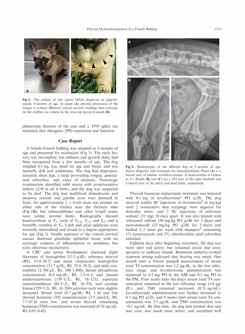
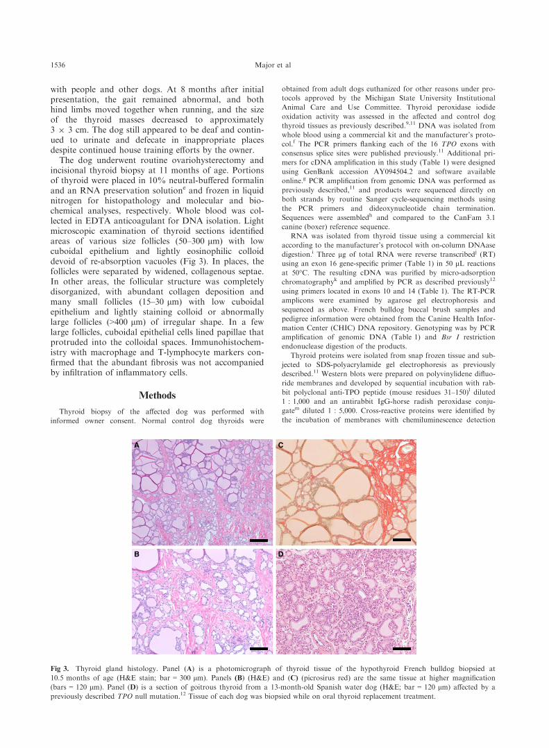
A female French bulldog was adopted at 9 months ofage and presented for evaluation (Fig 1). The early his-tory was incomplete, but dullness and growth delay hadbeen recognized from a few months of age. The dogweighed 6.5 kg, was small for age and breed, and wasmentally dull and withdrawn. The dog had dispropor-tionately short legs, a large protruding tongue, general-ized seborrhea, and areas of alopecia. Neurologicexamination identified mild ataxia with proprioceptivedeficits (2/4) in all 4 limbs, and the dog was suspectedto be deaf. The dog had multifocal demodecosis andisospora oocysts and giardia cysts were detected infeces. An approximately 3 9 6-cm mass was present oneither side of the trachea near the thoracic inlet(Fig 1B), but submandibular and other lymph nodeswere within normal limits. Radiographs showedhemivertebrae at T1, each of T4–6, T12, and L3; and abutterfly vertebra at T8. Limb and axial epiphyses werenormally mineralized and closed to a degree appropriatefor age (Fig 2). Needle aspirates of the ventral cervicalmasses disclosed glandular epithelial tissue with nocytologic evidence of inflammation or neoplasia, butwere otherwise inconclusive.
A CBC and serum biochemistry disclosed slightdecreases of hemoglobin [13.3 g/dL; reference interval(RI), 13.4–20.7] and mean corpuscular hemoglobinconcentration (31.7 g/dL; RI, 32.6–39.2), and mild eosi-nophilia (1,788/lL; RI, 700–1,490). Serum phosphorusconcentration (6.8 mg/dL; RI, 2.5–6.1) and alanineaminotransferase (158 U/L; RI, 18–121), aspartateaminotransferase (68 U/L; RI, 16–55), and creatinekinase (259 U/L; RI, 10–200) activities each were slightlyincreased. Serum total (0.7 lg/dL; RI, 1–4) and freethyroid hormone (T4) concentrations (3.9 pmol/L; RI,7.7-47.6) were low, and serum thyroid stimulatinghormone (TSH) concentration was increased (0.70 ng/mL;RI, 0.05–0.42).
Thyroid hormone replacement treatment was initiatedwith 0.1 mg of levothyroxinea PO q12h. The dogreceived weekly SC injections of doramectinb (6 mg/kg)until 2 consecutive skin scrapings were negative fordemodex mites, and 2 SC injections of cefovecinsodiumc (51 mg) 10 days apart. It was also treated withtoltrazuril sulfone (50 mg/kg PO q24h for 3 days) andmetronidazole (25 mg/kg PO q24h for 5 days) andbathed 2–3 times per week with shampood containing1% ketoconazole and 2% chlorhexidine until seborrheasubsided.
Eighteen days after beginning treatment, the dog wasmore alert and active, but remained ataxic and unre-sponsive to auditory stimuli. Brainstem auditory-evokedresponse testing indicated that hearing was intact. Onemonth later a 4-hour postpill measurement of serumtotal T4 concentration was 1.2 lg/dL, in the low refer-ence range, and levothyroxine administration wasincreased to 0.2 mg PO in the AM and 0.1 mg PO inthe PM. Four weeks later the dog’s serum total T4 con-centration remained in the low reference range (1.6 lg/dL), and TSH remained increased (0.51 ng/mL).Levothyroxine administration was further increased to0.3 mg PO q12h, and 4 weeks later serum total T4 con-centration was 5.2 lg/dL and TSH concentration was<0.1 ng/mL. By that time, the dog had normal skin andhair coat, was much more active, and socialized well
A B
Fig 1. The patient of this report before diagnosis at approxi-
mately 9 months of age. In panel (A) chronic protrusion of the
tongue is evident. Bilateral ventral cervical swellings that converge
on the midline are evident in the close-up shown in panel (B).
A
B C
Fig 2. Radiographs of the affected dog at 9 months of age,
before diagnosis and treatment for hypothyroidism. Panel (A) is a
lateral view of lumbar vertebral column. A hemivertebra is evident
at L3. Panels (B) and (C) are a VD view of the right forelimb and
a lateral view of the pelvis and hind limbs, respectively.
Thyroid Dyshormonogenesis in a French Bulldog 1535
with people and other dogs. At 8 months after initialpresentation, the gait remained abnormal, and bothhind limbs moved together when running, and the sizeof the thyroid masses decreased to approximately3 9 3 cm. The dog still appeared to be deaf and contin-ued to urinate and defecate in inappropriate placesdespite continued house training efforts by the owner.
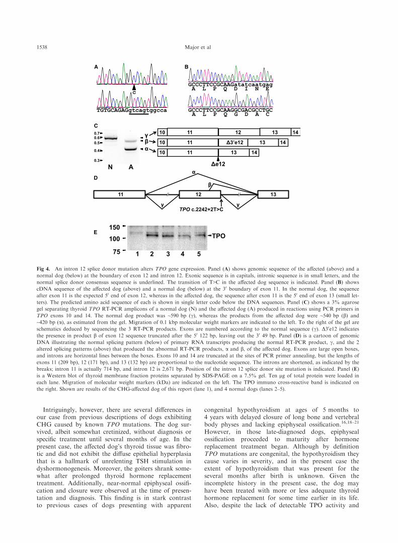
The dog underwent routine ovariohysterectomy andincisional thyroid biopsy at 11 months of age. Portionsof thyroid were placed in 10% neutral-buffered formalinand an RNA preservation solutione and frozen in liquidnitrogen for histopathology and molecular and bio-chemical analyses, respectively. Whole blood was col-lected in EDTA anticoagulant for DNA isolation. Lightmicroscopic examination of thyroid sections identifiedareas of various size follicles (50–300 lm) with lowcuboidal epithelium and lightly eosinophilic colloiddevoid of re-absorption vacuoles (Fig 3). In places, thefollicles were separated by widened, collagenous septae.In other areas, the follicular structure was completelydisorganized, with abundant collagen deposition andmany small follicles (15–30 lm) with low cuboidalepithelium and lightly staining colloid or abnormallylarge follicles (>400 lm) of irregular shape. In a fewlarge follicles, cuboidal epithelial cells lined papillae thatprotruded into the colloidal spaces. Immunohistochem-istry with macrophage and T-lymphocyte markers con-firmed that the abundant fibrosis was not accompaniedby infiltration of inflammatory cells.
Methods
Thyroid biopsy of the affected dog was performed with
informed owner consent. Normal control dog thyroids were
obtained from adult dogs euthanized for other reasons under pro-
tocols approved by the Michigan State University Institutional
Animal Care and Use Committee. Thyroid peroxidase iodide
oxidation activity was assessed in the affected and control dog
thyroid tissues as previously described.9,11 DNA was isolated from
whole blood using a commercial kit and the manufacturer’s proto-
col.f The PCR primers flanking each of the 16 TPO exons with
consensus splice sites were published previously.11 Additional pri-
mers for cDNA amplification in this study (Table 1) were designed
using GenBank accession AY094504.2 and software available
online.g PCR amplification from genomic DNA was performed as
previously described,11 and products were sequenced directly on
both strands by routine Sanger cycle-sequencing methods using
the PCR primers and dideoxynucleotide chain termination.
Sequences were assembledh and compared to the CanFam 3.1
canine (boxer) reference sequence.
RNA was isolated from thyroid tissue using a commercial kit
according to the manufacturer’s protocol with on-column DNAase
digestion.i Three lg of total RNA were reverse transcribedj (RT)
using an exon 16 gene-specific primer (Table 1) in 50 lL reactions
at 50°C. The resulting cDNA was purified by micro-adsorption
chromatographyk and amplified by PCR as described previously12
using primers located in exons 10 and 14 (Table 1). The RT-PCR
amplicons were examined by agarose gel electrophoresis and
sequenced as above. French bulldog buccal brush samples and
pedigree information were obtained from the Canine Health Infor-
mation Center (CHIC) DNA repository. Genotyping was by PCR
amplification of genomic DNA (Table 1) and Bsr I restriction
endonuclease digestion of the products.
Thyroid proteins were isolated from snap frozen tissue and sub-
jected to SDS-polyacrylamide gel electrophoresis as previously
described.11 Western blots were prepared on polyvinylidene difluo-
ride membranes and developed by sequential incubation with rab-
bit polyclonal anti-TPO peptide (mouse residues 31–150)l diluted1 : 1,000 and an antirabbit IgG-horse radish peroxidase conju-
gatem diluted 1 : 5,000. Cross-reactive proteins were identified by
the incubation of membranes with chemiluminescence detection
A
D
C
B
Fig 3. Thyroid gland histology. Panel (A) is a photomicrograph of thyroid tissue of the hypothyroid French bulldog biopsied at
10.5 months of age (H&E stain; bar = 300 lm). Panels (B) (H&E) and (C) (picrosirus red) are the same tissue at higher magnification
(bars = 120 lm). Panel (D) is a section of goitrous thyroid from a 13-month-old Spanish water dog (H&E; bar = 120 lm) affected by a
previously described TPO null mutation.12 Tissue of each dog was biopsied while on oral thyroid replacement treatment.
1536 Major et al

reagents.n Positive and negative control samples were included on
each blot, and specificity was determined from duplicate blots
incubated with irrelevant primary antibody.
Results
Thyroid peroxidase iodide oxidation activity wasundetectable in the membrane fraction of thyroid tissuehomogenates of the affected dog, and activity was2.74 � 0.45 U/mg protein (mean � SD) in 3 normalcontrol dog samples. Additionally, the expected approx-imately 105–110 kDa TPO protein13 of normal dogswas undetectable in the affected dog thyroid tissue sam-ple on Western blots (Fig 4E).
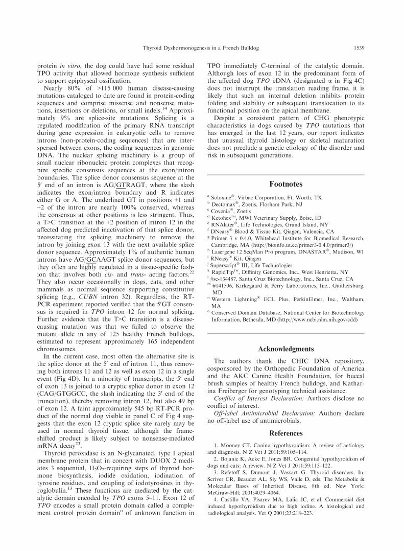
Upon amplification and sequencing the TPO exonsand flanking splice sites, we found no variant of theTPO protein-coding sequences in the affected dog, butthere was a homozygous T>C transition in the +2 posi-tion of the intron 12 splice donor site (CFA17:801,598;TPO c.2242 + 2T>C; Fig 4A). To investigate the poten-tial effect of this putative mutation, we amplified a por-tion of TPO cDNA from thyroid tissue of the affectedand a normal dog by RT-PCR using a gene-specific RTprimer in exon 16 and a PCR primer pair annealing inexons 10 and 14. The main cDNA product amplifiedfrom the affected dog was 423 bp, 171 bp smaller thanthe 594 bp predicted by the reference sequence as wellas what was amplified from normal dog thyroid cDNA(Fig 4C). In addition, a second, but, less robust amplifi-cation product of 545 bp consistently was present in theaffected dog reactions.
Sequencing of the 423 bp product identified loss ofthe 171 bp of exon 12 exactly, with no other inserted ordeleted sequence (Fig 4B). The deletion of exon 12maintained the translation reading frame, but predictedloss of 57 amino acid residues. Sequence of the 545 bpproduct demonstrated that the 50 122 bp of exon 12were included between exons 11 and 13, but the 30
49 bp of exon 12 were deleted. This longer sequencepredicted a shift of the translation reading frame at thesite of exon 12 truncation and altered the deducedamino acid sequence thereafter until a premature stop44 codons later, at the last codon of exon 13.
The T>C transition in the intron 12 splice donor sitecreated a new Bsr I restriction endonuclease recognitionsite allowing design of a convenient genotyping assay.A 216 bp portion of genomic DNA flanking the muta-tion site was amplified by PCR and subjected to Bsr Idigestion. When present, the mutant allele was cut intofragments of 156 and 60 bp. The digestion assay was
validated for heterozygous allele detection by mixingnormal and affected dog DNA 1 : 1 before PCR ampli-fication. The mutant allele was not observed in any of125 clinically healthy French bulldog samples tested.
Discussion
Congenital hypothyroidism with goiter has beenobserved in toy fox terriers,9 Tenterfield terriers,11 andSpanish water dogs12 as autosomal recessive disorders,each segregating a breed-specific null mutation of theTPO gene. The toy fox terrier TPO mutation also wasintroduced into rat terriers, apparently by cross-breed-ing, and produced at least 1 rat terrier family affectedby CHG.10 Thyroid peroxidase gene mutations also arethe most common cause of inherited CHG inhumans.14,15 Thyroid peroxidase is a multi-functionalenzyme required for thyroid hormone synthesis.13 Lackof TPO activity causes failure of iodide incorporationinto thyroglobulin, the so-called organification defectdemonstrated in radioiodine uptake and perchlorate dis-charge studies.9,16 The consequent failure of thyroidhormone synthesis, or dyshormonogenesis, leads tounrelenting TSH stimulation and diffuse thyroid follicu-lar epithelial cell hyperplasia recognized as goiter.
In each of the previously described breeds, the inher-ited disorder caused thyroid hormone deficiency in thenewborn period and life-threatening developmentaldelays that were apparent in the first few weeks of life.Survivors required intensive nursing care and assistedfeeding until diagnosis and institution of PO thyroid hor-mone replacement treatment. In the present case, a juve-nile French bulldog exhibited signs of hypothyroidismand enlarged thyroid glands. Thyroid gland tissue lackeddetectable TPO activity and immunoreactive proteinin vitro, and an intron 12 splice donor site mutation pro-duced TPO mRNA lacking all or part of exon 12.Despite an incomplete history and no pedigree informa-tion, the genetic etiology of TPO deficiency in this dogindicates that the dysfunction was present from birth.
The affected dog appeared to be deaf, and normalBAER test results suggested a central nervous systemcause. This presentation is distinct from the syndromicsensorineural deafness observed in human patients withcongenital hypothyroidism caused by mutation ofSLC26A4, an anion exchanger expressed in thyroidepithelium and the inner ear. However, hearing loss inhumans who are hypothyroid of various causes is com-mon and may be conductive, sensorineural, central, ormixed.17
Table 1. RT and PCR primers new in this study.
Location Reaction Sequence 50>30 Tm (°C)
TPO cDNA primers
Exon 16 Gene-specific RT primer CCACGTGGCTCCTACTGGATGT 61
Exon 10 PCR forward TCATCGGGAGGCAGATGAAG 63.6
Exon 14 PCR reverse ACGCGATGGAGACCAAGGA 64.1
Genotyping primers
Exon 12 PCR forward TCCCCGACAGCCTGGACAAT 67.3
Intron 12 PCR reverse CGTCTGAGGACCGCCGATTT 66.9
Thyroid Dyshormonogenesis in a French Bulldog 1537
Intriguingly, however, there are several differences inour case from previous descriptions of dogs exhibitingCHG caused by known TPO mutations. The dog sur-vived, albeit somewhat cretinized, without diagnosis orspecific treatment until several months of age. In thepresent case, the affected dog’s thyroid tissue was fibro-tic and did not exhibit the diffuse epithelial hyperplasiathat is a hallmark of unrelenting TSH stimulation indyshormonogenesis. Moreover, the goiters shrank some-what after prolonged thyroid hormone replacementtreatment. Additionally, near-normal epiphyseal ossifi-cation and closure were observed at the time of presen-tation and diagnosis. This finding is in stark contrastto previous cases of dogs presenting with apparent
congenital hypothyroidism at ages of 5 months to4 years with delayed closure of long bone and vertebralbody physes and lacking epiphyseal ossification.16,18–21
However, in those late-diagnosed dogs, epiphysealossification proceeded to maturity after hormonereplacement treatment began. Although by definitionTPO mutations are congenital, the hypothyroidism theycause varies in severity, and in the present case theextent of hypothyroidism that was present for theseveral months after birth is unknown. Given theincomplete history in the present case, the dog mayhave been treated with more or less adequate thyroidhormone replacement for some time earlier in its life.Also, despite the lack of detectable TPO activity and
A B
Δe12
10 11 13 14
10 11 Δ3’e12 13 14
10 11 12 13 14C
αβγ
N A
0.70.60.5
0.4
0.3
Dα
11 12 13
γ γ
β
TPO c.2242+2T>C
E
1 2 3 4 5
150
100
75
TPO
Fig 4. An intron 12 splice donor mutation alters TPO gene expression. Panel (A) shows genomic sequence of the affected (above) and a
normal dog (below) at the boundary of exon 12 and intron 12. Exonic sequence is in capitals, intronic sequence is in small letters, and the
normal splice donor consensus sequence is underlined. The transition of T>C in the affected dog sequence is indicated. Panel (B) shows
cDNA sequence of the affected dog (above) and a normal dog (below) at the 30 boundary of exon 11. In the normal dog, the sequence
after exon 11 is the expected 50 end of exon 12, whereas in the affected dog, the sequence after exon 11 is the 50 end of exon 13 (small let-
ters). The predicted amino acid sequence of each is shown in single letter code below the DNA sequences. Panel (C) shows a 3% agarose
gel separating thyroid TPO RT-PCR amplicons of a normal dog (N) and the affected dog (A) produced in reactions using PCR primers in
TPO exons 10 and 14. The normal dog product was ~590 bp (c), whereas the products from the affected dog were ~540 bp (b) and
~420 bp (a), as estimated from the gel. Migration of 0.1 kbp molecular weight markers are indicated to the left. To the right of the gel are
schematics deduced by sequencing the 3 RT-PCR products. Exons are numbered according to the normal sequence (c). D30e12 indicates
the presence in product b of exon 12 sequence truncated after the 50 122 bp, leaving out the 30 49 bp. Panel (D) is a cartoon of genomic
DNA illustrating the normal splicing pattern (below) of primary RNA transcripts producing the normal RT-PCR product, c, and the 2
altered splicing patterns (above) that produced the abnormal RT-PCR products, a and b, of the affected dog. Exons are large open boxes,
and introns are horizontal lines between the boxes. Exons 10 and 14 are truncated at the sites of PCR primer annealing, but the lengths of
exons 11 (209 bp), 12 (171 bp), and 13 (132 bp) are proportional to the nucleotide sequence. The introns are shortened, as indicated by the
breaks; intron 11 is actually 714 bp, and intron 12 is 2,671 bp. Position of the intron 12 splice donor site mutation is indicated. Panel (E)
is a Western blot of thyroid membrane fraction proteins separated by SDS-PAGE on a 7.5% gel. Ten lg of total protein were loaded in
each lane. Migration of molecular weight markers (kDa) are indicated on the left. The TPO immuno cross-reactive band is indicated on
the right. Shown are results of the CHG-affected dog of this report (lane 1), and 4 normal dogs (lanes 2–5).
1538 Major et al
protein in vitro, the dog could have had some residualTPO activity that allowed hormone synthesis sufficientto support epiphyseal ossification.
Nearly 80% of >115 000 human disease-causingmutations cataloged to date are found in protein-codingsequences and comprise missense and nonsense muta-tions, insertions or deletions, or small indels.14 Approxi-mately 9% are splice-site mutations. Splicing is aregulated modification of the primary RNA transcriptduring gene expression in eukaryotic cells to removeintrons (non-protein-coding sequences) that are inter-spersed between exons, the coding sequences in genomicDNA. The nuclear splicing machinery is a group ofsmall nuclear ribonucleic protein complexes that recog-nize specific consensus sequences at the exon/intronboundaries. The splice donor consensus sequence at the50 end of an intron is AG/GTRAGT, where the slashindicates the exon/intron boundary and R indicateseither G or A. The underlined GT in positions +1 and+2 of the intron are nearly 100% conserved, whereasthe consensus at other positions is less stringent. Thus,a T>C transition at the +2 position of intron 12 in theaffected dog predicted inactivation of that splice donor,necessitating the splicing machinery to remove theintron by joining exon 13 with the next available splicedonor sequence. Approximately 1% of authentic humanintrons have AG/GCAAGT splice donor sequences, butthey often are highly regulated in a tissue-specific fash-ion that involves both cis- and trans- acting factors.22
They also occur occasionally in dogs, cats, and othermammals as normal sequence supporting constitutivesplicing (e.g., CUBN intron 32). Regardless, the RT-PCR experiment reported verified that the 50GT consen-sus is required in TPO intron 12 for normal splicing.Further evidence that the T>C transition is a disease-causing mutation was that we failed to observe themutant allele in any of 125 healthy French bulldogs,estimated to represent approximately 165 independentchromosomes.
In the current case, most often the alternative site isthe splice donor at the 50 end of intron 11, thus remov-ing both introns 11 and 12 as well as exon 12 in a singleevent (Fig 4D). In a minority of transcripts, the 50 endof exon 13 is joined to a cryptic splice donor in exon 12(CAG/GTGGCC, the slash indicating the 30 end of thetruncation), thereby removing intron 12, but also 49 bpof exon 12. A faint approximately 545 bp RT-PCR pro-duct of the normal dog visible in panel C of Fig 4 sug-gests that the exon 12 cryptic splice site rarely may beused in normal thyroid tissue, although the frame-shifted product is likely subject to nonsense-mediatedmRNA decay23.
Thyroid peroxidase is an N-glycanated, type I apicalmembrane protein that in concert with DUOX 2 medi-ates 3 sequential, H2O2-requiring steps of thyroid hor-mone biosynthesis, iodide oxidation, iodination oftyrosine residues, and coupling of iodotyrosines in thy-roglobulin.13 These functions are mediated by the cat-alytic domain encoded by TPO exons 5–11. Exon 12 ofTPO encodes a small protein domain called a comple-ment control protein domaino of unknown function in
TPO immediately C-terminal of the catalytic domain.Although loss of exon 12 in the predominant form ofthe affected dog TPO cDNA (designated a in Fig 4C)does not interrupt the translation reading frame, it islikely that such an internal deletion inhibits proteinfolding and stability or subsequent translocation to itsfunctional position on the apical membrane.
Despite a consistent pattern of CHG phenotypiccharacteristics in dogs caused by TPO mutations thathas emerged in the last 12 years, our report indicatesthat unusual thyroid histology or skeletal maturationdoes not preclude a genetic etiology of the disorder andrisk in subsequent generations.
Footnotes
a Soloxine�, Virbac Corporation, Ft. Worth, TXb Dectomax�, Zoetis, Florham Park, NJc Covenia�, Zoetisd KetohexTM, MWI Veterinary Supply, Boise, IDe RNAlater�, Life Technologies, Grand Island, NYf DNeasy� Blood & Tissue Kit, Qiagen, Valencia, CAg Primer 3 v 0.4.0, Whitehead Institute for Biomedical Research,
Cambridge, MA (http://bioinfo.ut.ee/primer3-0.4.0/primer3/)h Lasergene 12 SeqMan Pro program, DNASTAR�, Madison, WIi RNeasy� Kit, Qiagenj Superscript� III, Life Technologiesk RapidTipTM, Diffinity Genomics, Inc., West Henrietta, NYl #sc-134487, Santa Cruz Biotechnology, Inc., Santa Cruz, CAm #141506, Kirkegaard & Perry Laboratories, Inc., Gaithersburg,
MDn Western Lightning� ECL Plus, PerkinElmer, Inc., Waltham,
MAo Conserved Domain Database, National Center for Biotechnology
Information, Bethesda, MD (http://www.ncbi.nlm.nih.gov/cdd)
Acknowledgments
The authors thank the CHIC DNA repository,cosponsored by the Orthopedic Foundation of Americaand the AKC Canine Health Foundation, for buccalbrush samples of healthy French bulldogs, and Kathar-ina Freiberger for genotyping technical assistance.
Conflict of Interest Declaration: Authors disclose noconflict of interest.
Off-label Antimicrobial Declaration: Authors declareno off-label use of antimicrobials.
References
1. Mooney CT. Canine hypothyroidism: A review of aetiology
and diagnosis. N Z Vet J 2011;59:105–114.2. Bojanic K, Acke E, Jones BR. Congenital hypothyroidism of
dogs and cats: A review. N Z Vet J 2011;59:115–122.3. Refetoff S, Dumont J, Vassart G. Thyroid disorders. In:
Scriver CR, Beaudet AL, Sly WS, Valle D, eds. The Metabolic &
Molecular Bases of Inherited Disease, 8th ed. New York:
McGraw-Hill; 2001:4029–4064.4. Castillo VA, Pisarev MA, Lalia JC, et al. Commercial diet
induced hypothyroidism due to high iodine. A histological and
radiological analysis. Vet Q 2001;23:218–223.
Thyroid Dyshormonogenesis in a French Bulldog 1539

5. Markou K, Georgopoulos N, Kyriazopoulou V, Vagenakis
AG. Iodine-induced hypothyroidism. Thyroid 2001;11:501–510.6. Delange F. The disorders induced by iodine deficiency. Thy-
roid 1994;4:107–128.7. Daminet S, Ferguson DC. Influences of drugs on thyroid
function in dogs. J Vet Intern Med 2003;17:463–472.8. Barbesino G. Drugs affecting thyroid function. Thyroid
2010;20:763–770.9. Fyfe JC, Kampschmidt K, Dang V, et al. Congenital
hypothyroidism with goiter in toy fox terriers. J Vet Intern Med
2003;17:50–57.10. Pettigrew R, Fyfe JC, Gregory BL, et al. CNS hypomyeli-
nation in rat terrier dogs with congenital goiter and a mutation in
the thyroid peroxidase gene. Vet Pathol 2007;44:50–56.11. Dodgson SE, Day R, Fyfe JC. Congenital hypothyroidism with
goiter in Tenterfield terriers. J Vet Intern Med 2012;26:1350–1357.12. Fyfe JC, Lynch M, Olsen J, Lou€er E. A thyroid peroxidase
(TPO) mutation in dogs reveals a canid-specific gene structure.
Mamm Genome 2013;24:127–133.13. Ruf J, Carayon P. Structural and functional aspects of thy-
roid peroxidase. Arch Biochem Biophys 2006;445:269–277.14. Stenson PD, Mort M, Ball EV, et al. The Human Gene
Mutation Database: Building a comprehensive mutation repository
for clinical and molecular genetics, diagnostic testing and personal-
ized genomic medicine. Hum Genet 2015;133:1–9.
15. Ris-Stalpers C, Bikker H. Genetics and phenomics of
hypothyroidism and goiter due to TPO mutations. Mol Cell Endo-
crinol 2010;322:38–43.16. Chastain CB, McNeel SV, Graham CL, Pezzanite SC. Con-
genital hypothyroidism in a dog due to an iodide organification
defect. Am J Vet Res 1983;44:1257–1265.17. Anand VT, Mann SB, Dah RJ, Mehra YN. Auditory inves-
tigations in hypothyroidism. Acta Otolaryngol 1989;108:83–87.18. Saunders MH, Jezyk PK. The radiographic appearance of
canine congenital hypothyroidism: Skeletal changes with delayed
treatment. Radiology 1991;32:171–177.19. Greco DS, Feldman EC, Peterson ME, et al. Congenital
hypothyroidism in a family of giant schnauzers. J Vet Intern Med
1991;5:57–65.20. Mooney CT, Anderson TJ. Congenital hypothyroidism in a
boxer dog. J Small Anim Pract 1993;34:31–35.21. Lieb AS, Grooters AM, Tyler JW, et al. Tetraparesis due
to vertebral physeal fracture in an adult dog with congenital
hypothyroidism. J Small Anim Pract 1997;38:364–367.22. Kralovicova J, Hwang G, Asplund AC, et al. Compen-
satory signals associated with the activation of human GC 50 splicesites. Nucleic Acids Res 2011;39:7077–7091.
23. Kervestin S, Jacobson A. NMD: A multifaceted response
to premature translational termination. Nat Rev Mol Cell Biol
2012;13:700–712.
1540 Major et al