molecular mechanisms of copi transport · 41 a schematic view of individual steps in copi ves-icle...
TRANSCRIPT
40
1978 Ph.D. - Ludwig-Maximilians-Universität München, Germany (Max Planck Institute of Biochemistry, Martinsried)
1978 - 1986 PostDoc and Group Leader - University of Regensburg, Germany
1986 - 1988 Visiting Scientist - Dept. of Biochemistry, Stanford University, USA
1988 - 1997 Full Professor and Chairman of Biochemistry I - University of Heidelberg, Germany
1991 - 2003 Chairman SFB 352 1997 - 2002 Director - BZHsince 2001 Managing Editor FEBS Letters
2005 - 2007 President of the GBM
since 2003 Chairman SFB 638
Goal Our research interests comprise two converging fields:i) Molecular mechanisms of coated vesicle formation and uncoating, andii) Specificity and structural basis for protein - lipid interactions within a bilayer that regulate membrane protein activity.These research interests have led us to develop and maintainiii) A platform for quantitative lipidomics.We are characterizing the components and their coordinate action that allow formation, fission and uncoating of Golgi-derived COPI-coated vesicles. This includes proteomics and lipidomics, functional in vitro assays and reconstitution of individual functional steps in a chemically defined liposomal system.
BackgroundIn the eukaryotic cell, vesicular transport repre-
sents the basic mechanism for i) maintaining the
homeostasis of the endomembrane system, ii)
biosynthetic transport of newly synthesized pro-
teins and lipids, and iii) the uptake and intracellu-
lar transport of exogenous macromolecules. The
mechanism of fusion of vesicles as well as their
role in neurotransmission has been recognized
by the 2013 Nobel Prize for Physiology and
Medicine. Three classes of coated vesicles are
well established to mediate transport in the exo-
and endocytic pathway: COPII vesicles for ER
export, COPI vesicles for retrograde Golgi-ER
and bidirectional intra-Golgi transport, and clath-
rin-coated vesicles operating in the late secretory
and endocytic pathway. Coat components are
involved in multiple tasks such as cargo selec-
tion, curvature formation at the donor membrane,
vesicle fission and initiation of uncoating.
We are interested in the molecular mechanisms
underlying vesicular transport by COPI vesicles.
In contrast to COPII and clathrin coats, the hep-
tameric large COPI coat component coatomer is
recruited en bloc to the membrane, so that both
the inner and outer shell of the vesicle are formed
at the same time.
In our view, the formation of a COPI transport
vesicle involves the following minimal set of com-
ponents: donor membranes with transmembrane
proteins acting as coat and/or cargo receptors
(e.g. members of the p24 family), cytosolic Arf1,
cytosolic coatomer and auxiliary enzymes that
serve activation on the membrane of Arf1 (GBF1)
and the activation of GTP hydrolysis by Arf1 (Arf
GAPs).
Molecular mechanisms of COPI transport
Felix Wieland
Felix Wieland / Britta Brügger
41
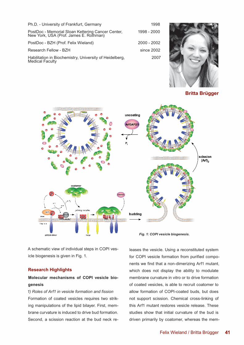
A schematic view of individual steps in COPI ves-
icle biogenesis is given in Fig. 1.
Research HighlightsMolecular mechanisms of COPI vesicle bio-genesis1) Roles of Arf1 in vesicle formation and fission
Formation of coated vesicles requires two strik-
ing manipulations of the lipid bilayer. First, mem-
brane curvature is induced to drive bud formation.
Second, a scission reaction at the bud neck re-
leases the vesicle. Using a reconstituted system
for COPI vesicle formation from purified compo-
nents we find that a non-dimerizing Arf1 mutant,
which does not display the ability to modulate
membrane curvature in vitro or to drive formation
of coated vesicles, is able to recruit coatomer to
allow formation of COPI-coated buds, but does
not support scission. Chemical cross-linking of
this Arf1 mutant restores vesicle release. These
studies show that initial curvature of the bud is
driven primarily by coatomer, whereas the mem-
Felix Wieland / Britta Brügger
Ph.D. - University of Frankfurt, Germany 1998 PostDoc - Memorial Sloan Kettering Cancer Center, 1998 - 2000New York, USA (Prof. James E. Rothman) PostDoc - BZH (Prof. Felix Wieland) 2000 - 2002 Research Fellow - BZH since 2002 Habilitation in Biochemistry, University of Heidelberg, 2007Medical Faculty
Britta Brügger
Fig. 1: COPI vesicle biogenesis.
42
brane curvature potentiating activity of dimeric
Arf1 is required for membrane scission. Using a
semi-intact cell system we have shown that dur-
ing the scission reaction Arf does not hydrolyse
its bound GTP.
2) Structures of coatomer and of the COPI coat
Together with John Briggs’ group at the EMBL
we investigate the structure of soluble coatomer
by single particle electron microscopy, and of
the coatomer shell on coated vesicles. With the
first data of a coat on a membrane, a structure
emerges that is strikingly different from those of
the COPII and the clathrin systems as delineated
from protein assemblies. The basic unit of the lat-
tice is a coatomer triade. Triades can be arranged
on the vesicular membrane in various patterns
that are defined by variable vertices at the con-
tact sites of triades.
3) Regulation of COPI transport by a unique
sphingolipid/cargo-receptor complex
We have discovered a specific binding of the
sphingomyelin molecular species SM 18:0 to
the transmembrane domain of one member of
the p24 family, p24. SM 18:0 binding favors di-
merization of p24. Dimeric p24, in turn, recruits
coatomer and triggers a conformational change
of the complex resulting in polymerization, initiat-
ing COPI bud formation. Thus, a membrane lipid
molecular species can serve as a cofactor in con-
trolling vesicle budding. We have defined steps
in vesicular transport in vivo that depend on this
specific interaction.
Structural principles of transmembrane pro-tein/membrane lipid interactions1) A signature within the p24 transmembrane do-
main for recognition of a sphingolipid molecular
species
We have discovered a peptide signature for sphin-
golipid binding within the transmembrane span of
p24. When transplanted, the signature confers
sphingolipid binding to a non-sphingolipid bind-
ing transmembrane domain. Results from a data
mining approach indicate that this signature rep-
resents a conserved binding site for sphingolipids
in several transmembrane proteins.
2) We found that a functional sphingomyelin-bind-
ing signature in the influenza neuraminidase is
necessary for optimal transport of this protein to
the host’s plasma membrane.
3) Capitalizing on fluorescent sphingomyelin spe-
cies containing pentaenyl fatty acids we have de-
veloped a simple assay to assess specific lipid-
lipid recognition in liposomal membranes.
Lipidomics platformUsing our lipidomics platform, we have elucidated
lipidoms of organisms and subcellular systems
(see lipidomics by Britta Brügger on page 52).
Felix Wieland / Britta Brügger
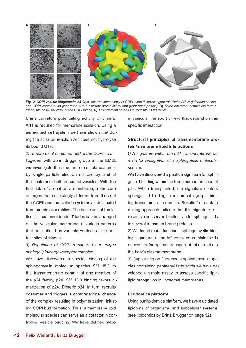
Fig. 2: COPI vesicle biogenesis. A) Cryo-electron microscopy of COPI-coated vesicles generated with Arf wt (left hand panels) and COPI-coated buds generated with a scission arrest Arf mutant (right hand panels). B) Three coatomer complexes form a triade, the basic structure of the COPI lattice, C) Arrangement of triads to form the COPI lattice.
A B C
43
Our investigations are based on a wide range of
methods, including live cell imaging (with Rainer
Pepperkok; EMBL), bioinformatics (with Gunnar
von Heijne and Arne Elofsson, Stockholm), mo-
lecular dynamics simulations (with Erik Lindahl,
Stockholm), in vivo and in vitro FRET studies,
cryo-electron microscopy (with John Briggs,
EMBL), protein chemistry, molecular biology, and
quantitative nano-mass spectrometry of lipids, as
well as chemical biology approaches.
Our research is supported by the German
Research Council (SFB 638: Dynamics of mac-
romolecular complexes in biosynthetic transport,
SFB/TRR 83: Molecular architecture and cel-
lular functions of protein/lipid assemblies, GRK
1188: Quantitative analysis of dynamic processes
in membrane transport and translocation, and
CellNetworks Heidelberg.
Selected Publications 2011 - 2013F. Adolf, A. Herrmann, A. Hellwig, R. Beck, B. Brügger and F. T. Wieland (2013) Scission of COPI and COPII vesicles is independent of GTP hydrolysis. Traffic 14(8):922-32.
A.M. Ernst, S. Zacherl, A. Herrmann, M. Hacke, W. Nickel, F.T. Wieland, B. Brügger (2013) Differential transport of Influenza A neuraminidase signal anchor peptides to the plasma membrane. FEBS Lett. 587(9):1411-7.
M. Faini, R. Beck, F.T. Wieland, J.A. Briggs (2013) Vesicle coats: structure, function, and general principles of assembly. Trends Cell Biol. Feb 12.
J.M. Duran, F. Campelo, J. van Galen, T. Sachsenheimer, J. Sot, M.V. Egorov, C. Rentero, C. Enrich, R.S. Polishchuk, F.M. Goñi, B. Brügger, F. Wieland, V. Malhotra (2012) Sphingomyelin organization is required for vesicle biogenesis at the Golgi complex. EMBO J. 12;31(24):4535-46.
M. Faini, S. Prinz, R. Beck, M. Schorb, J.D. Riches, K. Bacia, B. Brügger, F.T. Wieland* and J.A. Briggs* (shared corresponding authors) (2012) The Structures of COPI-coated vesicles reveal alternate Coatomer Conformations and Interactions. Science 336(6087):1451-4.
F.-X. Contreras, A.M. Ernst, P. Haberkant, P. Björkholm, E. Lindahl, B. Gönen, C. Tischer, A. Elofsson, G. von Heijne, C. Thiele, R. Pepperkok, F. Wieland, and B. Brügger (2012) Molecular recognition of a single sphingolipid species by a protein’s transmembrane domain. Nature 481:525-529.
R. Beck, S. Prinz, P. Diestelkötter-Bachert, S. Röhling, F. Adolf, K. Hoehner, S. Welsch, P. Ronchi, B. Brügger, J.A. Briggs, and F. Wieland (2011) Coatomer and dimeric ADP ribosylation factor 1 (Arf1) promote distinct steps in membrane scission. J Cell Biol. 194(5):765-77.
F.X. Contreras, A.M. Ernst, F. Wieland and B. Brügger (2011). Specificity of intramembrane protein-lipid interactions. Cold Spring Harb Perspect Biol. Jun 1;3(6)
M.C. Sahlmüller, J.R. Strating, R. Beck, V. Popoff, M. Haag, A. Hellwig, I. Berger, B. Brügger, and F.T. Wieland (2011) Recombinant heptameric coatomer complexes: novel tools to study isoform-specific functions. Traffic 12(6):682-692.
Awards and Honors Felix Wieland1993 Honorary Member of Charité, Medical Faculty of the Humboldt University, Berlin
since 2000 Elected EMBO Member
2001 Heinrich-Wieland Award
since 2003 Member of Deutsche Akademie der Naturforscher Leopoldina
2006 Feldberg Foundation Award
2011 Elected Member of the Academia Europea
2013 HMLS Award
Felix Wieland Phone: +49 (0)6221-54 4150 E-mail: [email protected]
Britta Brügger Phone: +49 (0)6221-54 5426 E-mail: [email protected]
Felix Wieland / Britta Brügger
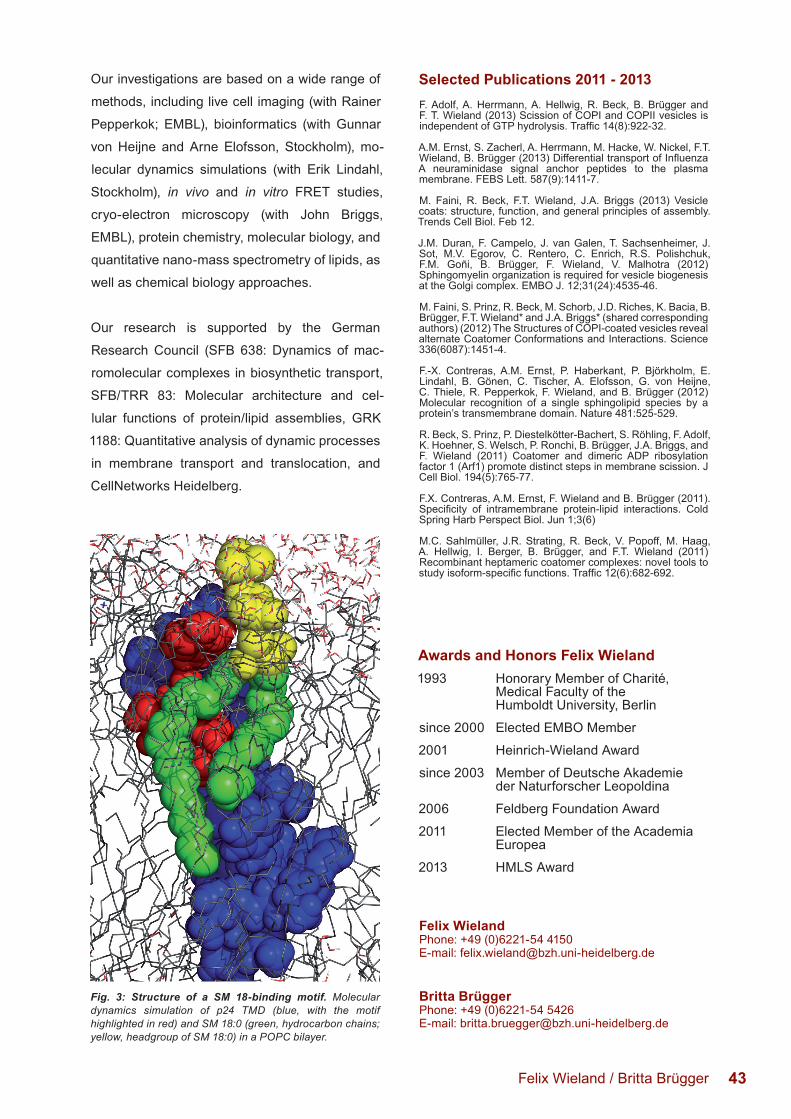
Fig. 3: Structure of a SM 18-binding motif. Molecular dynamics simulation of p24 TMD (blue, with the motif highlighted in red) and SM 18:0 (green, hydrocarbon chains; yellow, headgroup of SM 18:0) in a POPC bilayer.