molecular organization of soluble type iii secretion …106 results 107 108 the spao gene encodes...
TRANSCRIPT

1
Molecular organization of soluble type III secretion system sorting 1
platform complexes 2
Ivonne Bernala,b,†, Jonathan Börnickea,b,†, Johannes Heidemannc,†, Dmitri 3
Svergund, Anne Tuukkanend,#, Charlotte Uetrechtc,e, #, Michael Kolbea,b,f,# 4
a Center for Structural Systems Biology, Helmholtz Centre for Infection Research, Department of 5 Structural Infection Biology, Notkestraße 85, 22607 Hamburg, Germany 6 b Max Planck Institute for Infection Biology, Structural Systems Biology Group, Charitéplatz 1, 7 10117 Berlin, Germany 8 c Heinrich Pette Institute, Leibniz Institute for Experimental Virology, Martinistraße 52, 20251 9
Hamburg, Germany 10 d European Molecular Biology Laboratory, Hamburg Outstation, Notkestraße 85, 22607 11
Hamburg, Germany 12 e European XFEL GmbH, Sample Environment Group, Holzkoppel 4, 22869 Schenefeld, 13 Germany 14 f Faculty of Mathematics, Informatics and Natural Sciences, University of Hamburg, 15
Rothenbaumchaussee 19, 20148 Hamburg, Germany 16 † these authors contributed equally to this work 17 18
Running Title: Building Blocks of the Salmonella Sorting Platform 19
20
# Correspondence to: Michael Kolbe, [email protected] 21
Center for Structural Systems Biology, Department for Structural Infection Biology, Notkestraße 22
85, 22607 Hamburg, Germany 23 Tel: +49 40 8998 87550 24
Fax: +49 40 8998 2720 25
or 26
Charlotte Uetrecht, [email protected] 27
Heinrich Pette Institute, Leibniz Institute for Experimental Virology, Martinistraße 52, 20521 28 Hamburg, Germany 29 Tel: +49 40 480 51 261 30 Fax: +49 40 480 51 252 31
or 32
Anne Tuukkanen, [email protected] 33
European Molecular Biology Laboratory, Hamburg Outstation, Notkestraße 85, Hamburg 22607, 34 Germany 35 Tel: +49 40 89902 113 36 Fax: +49 40 89902 149 37 38
39
certified by peer review) is the author/funder. All rights reserved. No reuse allowed without permission. The copyright holder for this preprint (which was notthis version posted May 24, 2019. . https://doi.org/10.1101/648071doi: bioRxiv preprint

2
Abstract 40
Many medically relevant Gram-negative bacteria use the type III secretion system (T3SS) to 41
translocate effector proteins into the host for their invasion and intracellular survival. A multi-42
protein complex located at the cytosolic interface of the T3SS is proposed to act as a sorting 43
platform by selecting and targeting substrates for secretion through the system. However, the 44
precise stoichiometry and 3D organization of the sorting platform components is unknown. Here 45
we reconstitute soluble complexes of the Salmonella Typhimurium sorting platform proteins 46
including the ATPase InvC, the regulator OrgB, the protein SpaO and a recently identified 47
subunit SpaOC, which we show to be essential for the solubility of SpaO. We establish domain-48
domain interactions, determine for the first time the stoichiometry of each subunit within the 49
complexes by native mass spectrometry and gain insight into their organization using small-angle 50
X-ray scattering. Importantly, we find that in solution the assembly of SpaO/SpaOC/OrgB/InvC 51
adopts an extended L-shaped conformation resembling the sorting platform pods seen in in situ 52
cryo-electron tomography, proposing that this complex is the core building block that can be 53
conceivably assembled into higher oligomers to form the T3SS sorting platform. The determined 54
molecular arrangements of the soluble complexes of the sorting platform provide important 55
insights into its architecture and assembly. 56
certified by peer review) is the author/funder. All rights reserved. No reuse allowed without permission. The copyright holder for this preprint (which was notthis version posted May 24, 2019. . https://doi.org/10.1101/648071doi: bioRxiv preprint
3
Introduction 57
Type III secretion systems (T3SS) are protein nanomachines used by several medically relevant 58
pathogenic Gram-negative bacteria to deliver effector molecules into host cells to subvert 59
multiple cellular processes, leading to diseases such as salmonellosis, bubonic plague or sexually 60
transmitted infections (1,2). The T3SS forms a syringe-shaped macromolecular complex of 61
~3.5 MDa, whose main elements are a basal body that spans both bacterial membranes and a 62
protruding needle that forms a continuous secretion channel connecting the bacterial and host cell 63
cytoplasms (3-5). The precise assembly and function of the T3SS critically depends on the 64
hierarchical delivery of structural proteins to build the extracellular needle, followed by effector 65
proteins for translocation into the host cell (6,7). The control of this ordered process involves a 66
multi-protein complex associated with the cytoplasmic side of the T3SS that is proposed to act as 67
a sorting platform by recognizing and selecting substrates for secretion through the system (8,9). 68
In Salmonella Typhimurium, the components of the sorting platform of the SPI-1 (Salmonella 69
pathogenicity island 1) T3SS include the ATPase InvC (SctN in unified nomenclature), the 70
protein SpaO (SctQ), the ATPase regulator OrgB (SctL) and the accessory protein OrgA (SctK) 71
(8). Visualization of the SPI-1 sorting platform by cryo-electron tomographic analysis (CET) 72
indicates that it adopts a structure of six pods containing SpaO that are connected to the T3SS 73
base through OrgA and to a presumed hexameric ATPase through OrgB linkers (10,11). 74
However, this contrasts with other studies on both the S. Typhimurium SPI-1 and the Yersinia 75
enterocolitica T3SS showing the presence of ~24 and ~22 subunits, respectively, of the SctQ 76
protein at the needle base, which suggests a more extensive structure comparable to the 77
continuous cytosolic ring of flagellar T3SSs (12,13). Furthermore, the sorting platform has been 78
found to be a dynamic structure in which different components are exchanging between a T3SS-79
associated state and a cytosolic pool (12-14). 80
The probably best characterized component in the Salmonella SP1-I sorting platform is the 81
protein SpaO. SpaO contains two surface presentation of antigen domains (SPOA1 and SPOA2) 82
that can form SPOA2-SPOA2 homodimers, as well as SPOA1-SPOA2 heterodimers that are able 83
to interact with OrgB (15). Similar to the homologs of other pathogenic bacteria including 84
Yersinia and Shigella species, the gene encoding SpaO contains an internal translation initiation 85
site and produces an additional short isoform comprising the SPOA2 domain of SpaO, which we 86
certified by peer review) is the author/funder. All rights reserved. No reuse allowed without permission. The copyright holder for this preprint (which was notthis version posted May 24, 2019. . https://doi.org/10.1101/648071doi: bioRxiv preprint
4
refer to as SpaOC (16,17). This short product interacts with the full-length protein in other species 87
and thus could represent an additional structural component of the sorting platform (12,18-20). 88
However, the function of SpaOC in type III secretion is elusive, and how it interacts with the 89
other subunits of the sorting platform is unknown. Moreover, the precise protein composition and 90
spatial molecular organization of the sorting platform, as well its assembly process and 91
mechanism of action in substrate sorting remain uncertain. 92
In this study, we reconstitute and analyze for the first time the soluble assembling units of the 93
Salmonella Typhimurium SPI-1 sorting platform using purified proteins. We observe that SpaOC, 94
the second protein product of the gene spaO, is required for fully efficient type III function and 95
for the stability of the sorting platform complexes in solution. Using native mass spectrometry 96
(MS), small-angle X-ray scattering (SAXS) and multi-angle light scattering (MALS), we 97
characterize different substructures of the sorting platform, determining their stoichiometry and 98
association into SpaO/SpaOC/OrgB/InvC complexes. These complexes adopt an extended L-99
shaped conformation in solution that mirrors a segment of the sorting platform visualized by 100
CET. Our data present the most detailed assembly of the Salmonella Typhimurium SPI-1 sorting 101
platform in solution, reporting the conformation of what we propose is the core building block to 102
assemble the sorting platform at the T3SS needle base. 103
104
105
Results 106
107
The spaO gene encodes two protein products required for fully active type III secretion. 108
SpaO is a critical component of the S. Typhimurium SPI-1 sorting platform and it has recently 109
been shown that the spaO gene, similar to several of its homologs in other T3SSs, produces both 110
the full-length SpaO protein and a shorter variant that is the result of translation initiation from an 111
internal ribosome binding site (RBS) (17). When we recombinantly expressed C-terminally 112
Strep-tagged spaO and purified the protein by Strep-Tactin affinity purification, we could 113
confirm the production of this smaller protein product SpaOC (Fig. 1A). Using MALDI MS and 114
Edman sequencing we found that it begins with a methionine, rather than a valine that is encoded 115
certified by peer review) is the author/funder. All rights reserved. No reuse allowed without permission. The copyright holder for this preprint (which was notthis version posted May 24, 2019. . https://doi.org/10.1101/648071doi: bioRxiv preprint

5
at its starting position at codon 203 (Fig. S1 and Table S1), supporting the conclusion that it is the 116
product of internal translation initiation. 117
118
To examine the role of the SpaO isoforms in the infection process of S. Typhimurium, we first 119
created mutants that produce only the full-length or short variant of SpaO by introducing into the 120
chromosome either two stop codons shortly after the spaO start codon (ΔspaOFL) or silent 121
mutations in both the putative RBS and start codon of SpaOC (ΔspaOC) (Fig. 1A). We tested the 122
ability of these mutants to secrete T3SS substrate proteins into the culture supernatant, which 123
showed that loss of SpaOFL causes complete inhibition of T3SS function similar to that observed 124
for spaO gene knockout mutants (Fig. 1B). In contrast, abrogation of SpaOC translation resulted 125
in a marked reduction in secretion, which could almost completely be restored by 126
complementation with spaOC. Similarly, while the deletion of SpaO completely abolished the 127
ability of Salmonella to invade host cells, loss of SpaOC resulted in a statistically significant 128
reduction of invasiveness by about 50% compared to the wild type (Fig. 1C). However, it is 129
possible that the loss of SpaOC in the ΔspaOC mutant is incomplete, because when we expressed 130
and affinity-purified a SpaO variant in which only the start codon of SpaOC was mutated from 131
GTG to GCG (SpaOV203A), small amounts of SpaOC were still co-purified with the full-length 132
SpaO (Fig. 2A). MALDI MS of this SpaOC showed the presence of both methionine and alanine 133
in the first amino acid position (Fig. S2, Table S2), indicating that even in the absence of internal 134
translation initiation a SpaOC-like protein can still be produced by an alternative mechanism, 135
probably proteolysis. 136
137
SpaOC dimers bind to the N-terminal domain of SpaO to form SpaO-2SpaOC complexes. In 138
order to determine the molecular function of SpaOC in type III secretion, we first tested the 139
influence of SpaOC on SpaO stability. To this end, we expressed C-terminally Strep-tagged 140
SpaOV203A in a spaO knockout strain and purified it using Strep-Tactin affinity chromatography. 141
Interestingly, this mutation did not only almost completely abolish the production of soluble 142
SpaOC, but also drastically reduced the levels of soluble full-length SpaOV203A (Fig. 2A), which 143
instead formed insoluble inclusion bodies (data not shown). Complementation of the mutant with 144
spaOC restored the soluble levels of both proteins, indicating that SpaOC is required for SpaO 145
stability in solution. 146
147
certified by peer review) is the author/funder. All rights reserved. No reuse allowed without permission. The copyright holder for this preprint (which was notthis version posted May 24, 2019. . https://doi.org/10.1101/648071doi: bioRxiv preprint

6
The observed enhancement of SpaO solubility could involve interaction between SpaO and 148
SpaOC, as reported for homologous proteins (18-20). Therefore, we used size-exclusion 149
chromatography (SEC) coupled to MALS and SAXS, as well as native MS to study complex 150
formation between the two SpaO isoforms. In native MS non-covalent complexes of biological 151
samples are ionized and transferred to the gas phase under mild conditions, making it a sensitive 152
technique to take a snapshot of all non-covalent assemblies in a sample (21-23). First, we 153
observed that SpaOC exists mostly as a homodimer in solution (Fig. 2B and Fig. S3A, Table S4, 154
SASDC68), consistent with the crystal structure of the SPOA2-SPOA2 domain dimer of SpaO 155
and homolog proteins in Shigella and Yersinia (15,19,20). A very low abundance of 156
homotetramers was observed irrespective of the protein concentration tested (Fig. 2B), indicating 157
that these complexes reflect biologically relevant units and are not the result of unspecific 158
clustering during the MS ionization process. 159
160
Subsequent analysis of co-purified SpaO/SpaOC showed that both proteins interact to form 161
predominantly heterotrimers with a stoichiometry of SpaO-2SpaOC (Fig. 2C and Fig. S3B, Table 162
S4, SASDC78). Notably, no monomeric SpaO was detected in MS measurements, which 163
indicates high binding affinity within the SpaO-2SpaOC complex and further highlights the 164
critical role of SpaOC in SpaO solubility. Excess SpaOC was found to be mainly dimeric, 165
suggesting that it binds to SpaO as a pre-formed dimer. In addition to the predominant SpaO-166
2SpaOC species, we also observed the dimerization of these heterotrimers into 2(SpaO-2SpaOC) 167
heterohexamers, which was independent of both the protein concentrations and the position of the 168
Strep-tag (Fig. 2C, Fig. S3C). Higher-order oligomers could only be observed when measuring 169
highly concentrated samples that showed unspecific clustering during the ionization process and 170
were therefore considered to be non-specific assemblies. This conclusion is also supported by 171
SEC-MALS, which at a high protein concentration of 140 µM showed no evidence of species 172
larger than the 2(SpaO-2SpaOC) heterohexamer (Fig. S3B). Selected ions of the SpaO-2SpaOC 173
heterotrimers were subjected to collision-induced dissociation (CID) MS/MS experiments. The 174
observed dissociation pathways in these experiments give further insights into complex topology 175
since dissociating proteins are mostly small monomeric proteins from the periphery of protein 176
complexes, although the process of CID is not completely understood and exceptions have been 177
reported (24). Here, one SpaOC monomer was found to dissociate, leaving a residual SpaO-178
SpaOC complex (Fig. 2C inset). 179
certified by peer review) is the author/funder. All rights reserved. No reuse allowed without permission. The copyright holder for this preprint (which was notthis version posted May 24, 2019. . https://doi.org/10.1101/648071doi: bioRxiv preprint
7
180
In order to determine the domain of interaction between SpaO and SpaOC, we purified constructs 181
covering different regions of SpaO and combined them for native MS and SEC-MALS/SAXS 182
analysis (Table 1). By themselves, both the SpaO N-terminal domain (SpaO1-145) and a construct 183
containing the SPOA1 and SPOA2 domains (SpaO140-297) are mostly monomeric in solution (Fig. 184
S4A, Table S4, SASDC88 and SASDEK7). Combination of these proteins with SpaOC in both 185
SEC-MALS/SAXS and native MS subsequently showed the formation of a stable 186
SpaO1-145-2SpaOC complex resembling the SpaO:2SpaOC stoichiometry, while only very low 187
levels of complexes between SpaOC and SpaO140-297 could be detected (Fig. 3A-C, Fig. S4C, D, 188
Table S4 and SASDC98). We characterized the interaction between SpaO1-145 and the SpaOC 189
dimer by isothermal titration calorimetry (ITC) and obtained a Kd of 1.04 ± 0.21 µM, which also 190
demonstrates strong affinity between these proteins (Fig. 3D, E). Together, these results 191
demonstrate that the intermolecular interaction between the SpaO isoforms is mediated by SpaOC 192
stably binding to the N-terminal domain of SpaO. Furthermore, no significant interaction was 193
detected between the N-terminal domain and the C-terminal SPOA1-SPOA2 domain dimer of 194
SpaO (Fig. 3C, Fig. S4B), indicating that in SpaO these domains are held together largely by 195
their covalent linkage, suggesting conformational flexibility between them. 196
197
Sorting platform subcomplexes of SpaOC, SpaO, OrgB and InvC are stable in solution. 198
Next, we determined the interactions of SpaO and SpaOC with other proteins of the Salmonella 199
sorting platform in solution by co-expression with the interaction partners OrgB and InvC (8) in 200
E. coli (Table 1). While OrgB by itself was insoluble (data not shown), stable complexes of 201
SpaO/SpaOC/OrgB, SpaO/SpaOC/OrgB/InvC and OrgB/InvC were soluble and could be purified 202
for further characterization (Fig. 4A). We also tested co-expression of these proteins with OrgA, 203
but this did not yield any soluble OrgA-containing complexes, suggesting that OrgA could either 204
require the presence of needle base-forming proteins for correct folding, or stay localized to the 205
membrane or needle base and not participate in soluble sorting platform complexes. 206
In order to determine the regions of OrgB involved in interactions with SpaO/SpaOC and InvC, 207
we dissected OrgB into its N-terminal (residues 1-105) and C-terminal (residues 106-226) halves 208
and co-expressed His-tagged variants of these together with Strep-tagged InvC or SpaO/SpaOC. 209
Subsequent Strep-Tactin affinity purification showed that OrgB1-105 is pulled down by 210
certified by peer review) is the author/funder. All rights reserved. No reuse allowed without permission. The copyright holder for this preprint (which was notthis version posted May 24, 2019. . https://doi.org/10.1101/648071doi: bioRxiv preprint

8
SpaO/SpaOC, while only trace amounts co-purified with InvC (Fig. 4B). Conversely, OrgB106-226, 211
was pulled down by InvC, and even though small amounts could also be pulled down by 212
SpaO/SpaOC, the ratio between SpaO/SpaOC and OrgB106-226 indicates that the affinity between 213
them is low. We similarly dissected InvC after residue 79 and tested the ability of Strep-tagged 214
InvC1-79 and InvC80-431 to pull down OrgB. While both full-length InvC and InvC1-79 were able to 215
co-purify OrgB, this was not the case for InvC80-431 (Fig. 4C), showing that the N-terminal 79 216
amino acids of InvC are both necessary and sufficient for its interaction with OrgB. Together, 217
these results show that OrgB interacts through its C-terminus with the N-terminal 79 amino acids 218
of InvC and confirm that the binding site for SpaO is located in the N-terminus of OrgB (15). 219
220
OrgB dimers induce dimerization of SpaO-2SpaOC. We analyzed the SpaO/SpaOC/OrgB 221
complex by native MS and found that the major molecular species contains two units of SpaO-222
2SpaOC bound to two molecules of OrgB, resulting in 2(SpaO-2SpaOC)-2OrgB complexes. Less 223
abundant species, possibly representing assembly intermediates of this complex, were also 224
identified (Fig. 5). The vast majority of OrgB-containing complexes possesses two molecules of 225
OrgB, which indicates that OrgB exists mainly in dimeric form, similar to its flagellar homolog 226
FliH (25). When we subjected the 2(SpaO-2SpaOC)-2OrgB species to MS/MS analysis, a single 227
OrgB dissociated from the complex, while a second, less prominent dissociation pathway led to 228
the dissociation of a SpaO monomer (Fig. S5A). 229
230
We also attempted native MS analysis of the mutant SpaOV203A, in which the start codon of 231
SpaOC has been mutated, both alone and in complex with OrgB. While it was possible to purify 232
both SpaOV203A and SpaOV203A/OrgB complexes lacking SpaOC by affinity purification and SEC, 233
these complexes were unstable and could not be detected in native MS or successfully analyzed 234
by other methods. This highlights the importance of SpaOC not only for the stability of SpaO, but 235
also of higher-order sorting platform complexes containing OrgB. 236
237
The ATPase InvC binds to SpaO/SpaOC/OrgB complexes to form the core building block of 238
the sorting platform. The most comprehensive sorting platform subcomplexes we obtained in 239
this study contained the four proteins SpaO, SpaOC, OrgB and InvC. Native MS revealed that the 240
ATPase InvC is present in different types of complexes, with species of SpaO-2SpaOC-2OrgB-241
certified by peer review) is the author/funder. All rights reserved. No reuse allowed without permission. The copyright holder for this preprint (which was notthis version posted May 24, 2019. . https://doi.org/10.1101/648071doi: bioRxiv preprint

9
InvC and 2(SpaO-2SpaOC)-2OrgB-InvC stoichiometry being the most abundant in the spectra 242
(Fig. 6A). Complexes of 2OrgB-InvC and 2SpaO-2SpaOC-2OrgB-InvC stoichiometry were 243
detected at lower levels. Importantly, InvC was detected exclusively in complexes containing 244
OrgB dimers, which is in agreement with our findings that the OrgB C-terminal region binds to 245
the N-terminus of InvC (Fig. 4B, C) and previously reported CET maps and pull-down assays 246
(10,15). It should be noted that the signal intensity ratios of the different complex species were 247
heavily dependent on the electrospray conditions and while the presented spectrum was selected 248
for a high resolution, the majority of acquired spectra showed higher signal intensities for high 249
molecular weight complexes. However, direct translation of signal intensity ratios into complex 250
ratios in solution is not possible due to fluctuating signals and different ionization and 251
transmission efficiencies of different complex species. Nevertheless, the different observed 252
species indicate a degree of dynamic association and dissociation of subunits within the system. 253
In addition to the species identified in the presented mass spectrum, occasionally signals in the 254
higher m/z-range were observed, depending on the electrospray conditions (Fig. S6). Due to the 255
low resolution and signal intensity, charge states for these peaks could not be unambiguously 256
identified in MS or MS/MS measurements. However, the mass range and peak interval suggest 257
the presence of complexes with masses of approximately 433 kDa, possibly dimers of 2(SpaO-258
2SpaOC)-2OrgB-InvC. 259
In CID MS/MS measurements of the different identified SpaO/SpaOC/OrgB/InvC complexes the 260
dissociation of a single OrgB monomer was observed in every case (Fig. S5B, C). Because no 261
other components were lost together with the OrgB, this dissociation pattern allows us to 262
conclude that the interactions of the OrgB dimer with both SpaO/SpaOC and InvC are mediated 263
by the same OrgB molecule, while the other is less tightly integrated in the complex. 264
We further characterized the SpaO/SpaOC/OrgB/InvC complexes using SEC-MALS and SEC-265
SAXS. MALS revealed a molecular mass range over the main SEC elution peak of 266
approximately 208 to 180 kDa (Fig. 6B), which is in good agreement with the complexes 267
identified in native MS (Table S3). Since this analysis showed the later regions of the elution 268
peak to be a mixture of several molecular species, we only used the largely homogenous first half 269
of the peak in the SAXS analysis in order to generate a model with minimal averaging between 270
different species. Subsequently, bead model reconstruction from the SAXS data showed that the 271
SpaO/SpaOC/OrgB/InvC adopts an extended L-shape in solution. (Fig. 7A-C, Table S4, 272
certified by peer review) is the author/funder. All rights reserved. No reuse allowed without permission. The copyright holder for this preprint (which was notthis version posted May 24, 2019. . https://doi.org/10.1101/648071doi: bioRxiv preprint
10
SASDEJ7). By simultaneously employing the SAXS data of the SpaO/SpaOC/OrgB/InvC and the 273
SpaO-2SpaOC complexes in a multiphase bead modeling approach (26), the position of SpaO-274
2SpaOC within the larger complex could be determined. The resulting multiphase bead model 275
indicates that SpaO-2SpaOC is located in the shorter leg of the extended L-shape (Fig. 7D). 276
Unfortunately, due to the complexity of the studied system the generation of a reliable SAXS-277
based atomistic hybrid model of the SpaO/SpaOC/OrgB/InvC complex is hindered by a number 278
of uncertainties, which include the number of different subunits, flexibility of the complex in 279
solution (indicated by Kratky analysis, see SASBDB), the remaining possibility of heterogeneity 280
in the SEC peak region used for SAXS analysis and a lack of high-resolution structure for many 281
of the complex components. Nevertheless, by combining the SpaO/SpaOC/OrgB/InvC SAXS data 282
with our native MS results and the interactions between different subunit domains (Table 1), it is 283
possible to construct a schematic model of the architecture of the soluble 284
SpaO/SpaOC/OrgB/InvC complex (Fig. 7E). Thus, while SpaO-2SpaOC occupies the shorter leg 285
of the L-shape, InvC-OrgB would be placed in the longer leg with OrgB forming a linker 286
between SpaO-2SpaOC and InvC. Interestingly, even though native MS and MALS indicate the 287
presence of two SpaO-2SpaOC heterotrimers in the SpaO/SpaOC/OrgB/InvC complex (Fig. 6, 288
Table S3), both the multiphase analysis (Fig. 7D) and comparison of the SpaO/SpaOC and 289
SpaO/SpaOC/OrgB/InvC SAXS bead structures (see SASBDB for details) indicate that only a 290
single SpaO-2SpaOC heterotrimer can be accommodated in the short leg of the L-shape, 291
suggesting that the SAXS structure is that of a complex with SpaO-2SpaOC-2OrgB-InvC 292
stoichiometry. This apparent discrepancy could be due to uncertainties in the SAXS bead model 293
caused by flexibility of the complex or heterogeneity in the SEC peak region used for SAXS 294
analysis. 295
296
Because the extended SAXS shape of the SpaO-2SpaOC-2OrgB-InvC complex is reminiscent of 297
the pod densities seen in the in-situ 3D CET map of the Salmonella needle complex (10), we 298
hypothesized that this complex represents the soluble core building block from which the full 299
sorting platform is assembled. To test this hypothesis, we superimposed the ab initio SAXS bead 300
model with the CET map (Fig. 8), which shows a good correspondence between the two 301
structures and orients the SAXS shape in a way that places SpaO-2SpaOC in the outer pods, InvC 302
in the central hub and 2OrgB in the linker region between the two. This is in good agreement 303
certified by peer review) is the author/funder. All rights reserved. No reuse allowed without permission. The copyright holder for this preprint (which was notthis version posted May 24, 2019. . https://doi.org/10.1101/648071doi: bioRxiv preprint

11
with the assignments by CET using fluorescent protein tags and sorting platform protein deletions 304
(10,27). If six units of the SAXS bead model were to be placed within the CET map, steric 305
clashes would occur in the central hub region. Interestingly, Kratky analysis of the SAXS data 306
showed conformational flexibility of the building block complexes, indicating an ability to 307
undergo conformational changes upon assembly of the complete sorting platform. In fact, by 308
rotating InvC in our model upwards by 90° around its interaction site with OrgB and shifting that 309
interaction site towards the bottom of the central hub, InvC would be re-oriented into a 310
configuration parallel to the outer pods with its C-terminus pointing towards the T3SS basal 311
body. This change would both resolve the steric clashes and allow for the formation of an InvC 312
ATPase hexamer to fill the central hub region of the CET map. 313
It should, however, be noted that this in silico approach relies on the superposition of two 314
structures of low resolution, both of which are associated with their own errors, posing a limit on 315
the conclusions that can be drawn from it. Therefore, while the good agreement between our 316
SAXS structure and the CET map supports our idea that the SpaO/SpaOC/OrgB/InvC complex 317
represents the soluble core building block of the sorting platform, biochemical studies will be 318
required to show the assembly of these soluble complexes into the complete sorting platform. 319
320
Discussion 321
322
The sorting platform, together with the export apparatus complex, is still one of the less well 323
characterized components of the T3SS. In this work we present an analysis of inter-subunit 324
interactions, stoichiometry and shape of the main soluble module of the SPI-1 sorting platform in 325
S. Typhimurium. Expression and functional analysis confirm that the gene encoding the protein 326
SpaO produces an additional short protein SpaOC that comprises the C-terminus of the SpaO 327
sequence, and that SpaOFL is essential for type III secretion, while SpaOC appears non-essential 328
but required for full secretion efficiency (16,17). Interestingly, a similar phenotype has been 329
observed for the Salmonella SPI-2 orthologue protein SsaQC (18) and the remaining secretion 330
activity upon deletion of the shorter protein product appears to be unique to the two T3SSs of 331
Salmonella. This raises the possibility that cross-complementation might occur between the 332
Salmonella T3SSs, a hypothesis that could be the subject of future investigations. On the other 333
certified by peer review) is the author/funder. All rights reserved. No reuse allowed without permission. The copyright holder for this preprint (which was notthis version posted May 24, 2019. . https://doi.org/10.1101/648071doi: bioRxiv preprint
12
hand, given that MALDI MS showed that low levels of a SpaOC-like protein were still produced 334
from a spaO variant carrying a mutation in the SpaOC start codon (spaOV203A), it is possible that 335
the incomplete loss of secretion and invasion activity of the ΔspaOC mutant might be due to 336
partial complementation by such a product that appears to be produced by proteolysis even in the 337
absence of internal translation initiation (Fig. 1B, C). Overall, these data are consistent with the 338
results of previous studies in the Salmonella SPI-1 and SPI-2 systems, as well as Shigella and 339
Yersinia (12,16-20), suggesting that the alternative translation into a full-length protein and a 340
shorter product may represent a widespread strategy among the SctQ proteins of virulence-341
associated T3SSs. 342
343
The isoform SpaOC forms a homodimer that binds to the full-length SpaO to form SpaO-2SpaOC 344
complexes, similar to the 1:2 complexes observed for the Shigella Spa33 and the Yersinia YscQ 345
homologs (19,20). However, while the Spa33-2Spa33C trimers readily assembled into higher-346
order oligomers, we found only little dimerization of trimers and no further oligomerization for 347
SpaO-2SpaOC. Additionally, our analysis shows that the SpaOC dimer stably associates with the 348
N-terminal domain of SpaO (SpaO1-145). In contrast, stable interactions between SpaOC and the 349
SPOA domains of SpaO, like those observed for the homolog Spa33, could not be detected (20). 350
Interestingly, in a recent study a SpaO variant carrying a photo-activatable amino acid in the 351
SPOA2 domain (residue 289) was found to cross-link with SpaOC, indicating interaction between 352
these regions after all (16). However, given the irreversibility of cross-linking and the 353
comparatively low levels of cross-linked species in that study, these might have been the result of 354
more transient interactions such as those indicated by the low levels of SpaO140-297-SpaOC 355
complexes observed in native MS (Fig. 3C). Importantly, our newly found stable interaction 356
between the N-terminal domain of SpaO and SpaOC has implications for any model of the 357
structure of the T3SS cytosolic complex, which is currently based on interactions between the 358
small SctQ protein isoform and the SPOA1-SPOA2 domain dimer of the full-length variant. 359
360
The SpaO-2SpaOC heterotrimer interacts with the ATPase regulator OrgB to form stable 2(SpaO-361
2SpaOC)-2OrgB complexes (Fig.5) and we therefore propose that OrgB exists as a dimer 362
comparable to its flagellar homolog FliH (25). Interestingly, while it has been suggested that the 363
binding of SpaOC and OrgB to SpaO may be mutually exclusive due to overlapping binding sites 364
on the C-terminal SPOA1-SPOA2 domains of SpaO (15,16), our data shows that SpaO can 365
certified by peer review) is the author/funder. All rights reserved. No reuse allowed without permission. The copyright holder for this preprint (which was notthis version posted May 24, 2019. . https://doi.org/10.1101/648071doi: bioRxiv preprint

13
simultaneously interact with both of these proteins. This is consistent with our finding that SpaOC 366
interacts with the N-terminal domain of SpaO rather than the C-terminal SPOA domains. In 367
addition, in CID MS/MS of 2(SpaO-2SpaOC)-2OrgB complexes the dissociation of both SpaO 368
and OrgB monomers was observed, indicating that the recruitment of OrgB leads to a 369
stabilization of SpaOC within the complex. While our data does not offer a clear mechanism for 370
this stabilization, it is conceivable that it involves direct interactions between SpaOC and OrgB 371
that occur in addition to those of the extreme N-terminus of OrgB and the SpaO SPOA1-SPOA2 372
dimer (15). Furthermore, the dissociation of either OrgB or SpaO without the simultaneous loss 373
of other subunits suggests that direct interactions between the two SpaO-2SpaOC trimers are 374
promoted in these complexes. It should be noted that the observed MS/MS dissociation pattern is 375
also compatible with a complex architecture in which both SpaO-2SpaOC heterotrimers are 376
associated with the same OrgB unit. However, this arrangement seems unlikely since an 377
association of one SpaO-2SpaOC trimer to one OrgB would be expected in light of the reported 378
interaction between the N-terminus of OrgB and the SPOA1-SPOA2 domains of SpaO (15). 379
Nevertheless, it cannot be excluded given the asymmetry of OrgB units within the OrgB dimer 380
revealed by MS/MS of SpaO/SpaOC/OrgB/InvC complexes (see below). 381
382
Complexes of SpaO, SpaOC and OrgB associate with the ATPase InvC to form both SpaO-383
2SpaOC-2OrgB-InvC and 2(SpaO-2SpaOC)-2OrgB-InvC complexes, in which OrgB acts as a 384
central connector by binding of its N-terminus to SpaO-2SpaOC and its C-terminus to InvC. 385
Based on MS/MS experiments (Fig. S5), we propose that both of these interactions are formed by 386
the same OrgB subunit, while the second OrgB is less tightly integrated in the complex, possibly 387
acting to stabilize extended helical regions in the first OrgB. SAXS analysis showed that the 388
SpaO/SpaOC/OrgB/InvC complexes adopt an extended L-shaped structure in solution. Because 389
this conformation is in good agreement with the in situ cryo-electron tomography (CET) structure 390
of the Salmonella SPI-1 T3SS (10), we propose that the SpaO/SpaOC/OrgB/InvC complexes 391
identified in this study represent the main soluble building blocks of the sorting platform. These 392
complexes would bind to other T3SS proteins like the docking protein OrgA, InvI or the export 393
apparatus and undergo a conformational change in re-orienting the ATPase InvC to assemble the 394
complete sorting platform at the base of the T3SS needle complex. Adding to the similarity of the 395
SAXS and CET structures the fact that we found SpaOC in all of the sorting platform 396
subcomplexes, its importance for their stability and that SpaOC is itself stabilized in these 397
certified by peer review) is the author/funder. All rights reserved. No reuse allowed without permission. The copyright holder for this preprint (which was notthis version posted May 24, 2019. . https://doi.org/10.1101/648071doi: bioRxiv preprint
14
complexes by the presence of OrgB, it can be hypothesized that SpaOC is an integral structural 398
part of the sorting platform similar to the Yersinia homolog YscQC, which has previously been 399
shown to co-localize with YscQFL into sorting platform complexes at the bacterial membrane 400
(12). On the other hand, the in vitro nature of our study means that it cannot be excluded that 401
SpaOC plays a role in the soluble forms of the building blocks and might dissociate from the 402
complexes upon assembly of the complete sorting platform in vivo, as has been suggested by the 403
lack of additional densities in CET maps of sorting platforms from strains expressing SpaOC 404
fused to a fluorescent protein (16). 405
406
The superposition between our SAXS bead model and the CET map suggests that the individual 407
legs as seen by tomography would be of SpaO-2SpaOC-2OrgB-InvC stoichiometry, bringing the 408
assembled sorting platform to 6SpaO-12SpaOC-12OrgB-6InvC. While these numbers are 409
compatible with the stoichiometry determined by fluorescence microscopy for InvC and OrgB, 410
SpaO has been indicated to be present in the sorting platform at a higher copy number of 411
approximately 24 (13). Our findings show that the soluble building blocks can recruit an 412
additional SpaO-2SpaOC trimer and it is conceivable that further units dynamically associate with 413
the sorting platform at the T3SS needle base. In fact, the dynamic exchange of the SpaO homolog 414
YscQ in Yersinia has previously been observed by fluorescence microscopy and found to 415
increase during the active secretion process (12). Together with the observation that the diffusion 416
behavior of cytosolic populations of sorting platform components also changes upon secretion 417
activation, this indicates that soluble sorting platform complexes might play an important role in 418
the function of type 3 secretion (28). Thus, it can be hypothesized that soluble building blocks of 419
SpaO/SpaOC/OrgB/InvC could act as T3SS substrate shuttles that recruit substrate-chaperone 420
complexes in the cytosol and transfer them to the basal-body associated sorting platform for 421
subsequent secretion. Furthermore it can be speculated that the dissociation of SpaO/SpaOC/OrgB 422
from hexameric T3SS-associated InvC might act to fully activate the secretion process by 423
overcoming the inhibitory effect of OrgB on InvC ATPase activity (25,29). 424
425
Experimental Procedures 426
Cloning and mutagenesis of Salmonella genes. Genes ligated into the expression vectors 427
pASK-IBA (IBA GmbH, Göttingen, Germany), pET (Novagen, Madison, WI, USA), or 428
certified by peer review) is the author/funder. All rights reserved. No reuse allowed without permission. The copyright holder for this preprint (which was notthis version posted May 24, 2019. . https://doi.org/10.1101/648071doi: bioRxiv preprint

15
pCDFDuet-1 (Novagen, Madison, WI, USA) were derived from Salmonella Typhimurium strain 429
SL1344 using standard techniques. All PCRs were performed using Phusion polymerase (New 430
England Biolabs, Ipswich, MA, USA) and oligonucleotides synthesized by Sigma-Aldrich or 431
Eurofins Genomics. Site-directed mutagenesis of the spaO gene was performed according to the 432
QuikChange PCR site-directed mutagenesis protocol (Agilent, Santa Clara, CA, USA). All 433
primers used in this study can be found in Table S5. 434
435
Salmonella genomic spaO deletion was carried out by homologous recombination using the λ 436
Red recombinase system (30). Briefly, the λ Red recombinase plasmid pKD46 was expressed in 437
S. Typhimurium SL1344 and a kanamycin cassette flanked by two 50bp regions homologous to 438
the spaO gene was subsequently transformed into the strain for homologous recombination. The 439
ΔspaOC, ΔspaOFL, spaO-3xFLAG, ΔspaOC-3xFLAG and ΔspaOFL-3xFLAG strains were 440
generated following a similar protocol, introducing a tetracycline cassette into the spaO region as 441
described above. In a second step, the tetracycline cassette was replaced by spaO DNA carrying 442
mutations and colonies were selected on tetracycline-sensitivity selection media (31,32). To 443
generate the ΔspaOC strain, silent mutations at the internal putative Shine-Dalgarno region 444
(position 594 to 600, AGGGGGA to gGGcGGc) and start codon (position 607-609, GTG to GTt) 445
of spaO were introduced, while the ΔspaOFL strain was produced by introducing nonsense 446
mutations shortly after the start codon of spaO at amino acid position 28 and 29. For the 447
generation of the strains spaO-3xFLAG, ΔspaOC-3xFLAG and ΔspaOFL-3xFLAG, a 3xFLAG-448
tag was inserted at the C-terminus of spaO in the chromosome. Introduction of mutations was 449
verified by PCR and DNA sequencing. The Δspi-1 strain was kindly provided by the lab of 450
Arturo Zychlinsky. 451
452
Detection of SpaO and SpaOC in Salmonella cells. spaO-3xFLAG, ΔspaOC-3xFLAG and 453
ΔspaOFL-3xFLAG strains were grown in LB medium (Luria/Miller) at 37 °C to an OD600 of 1. 454
Total cell lysates were separated by SDS-PAGE and analyzed by western blot using anti-FLAG 455
M2 primary antibody (Sigma-Aldrich, St. Louis, MO, USA), horseradish peroxidase (HRP)-456
conjugated secondary antibodies (Jackson ImmunoResearch Laboratories, West Grove, PA, 457
USA) and ECL western blotting substrates (Thermo Fischer Scientific, Waltham, MA, USA) for 458
protein detection. 459
certified by peer review) is the author/funder. All rights reserved. No reuse allowed without permission. The copyright holder for this preprint (which was notthis version posted May 24, 2019. . https://doi.org/10.1101/648071doi: bioRxiv preprint
16
460
Recombinant gene expression and protein purification. Constructs used for recombinant gene 461
expression in E. coli BL21 (DE3) are listed in Table S6. Cells were grown in LB with the 462
appropriate antibiotics at 37 °C. At an OD600 of 0.5, the temperature was reduced to 20 °C and 463
gene expression induced by addition of 200 μg/l anhydrotetracycline (AHT, Sigma-Aldrich, St. 464
Louis, MO, USA) for pASK-IBA vectors and/or 0.5 mM IPTG for pET and pCDFDuet plasmids. 465
Cells were grown further for 18 h and harvested by centrifugation. 466
467
All purification steps were performed at 4 °C. To purify SpaOC, SpaO1-145, SpaO140-297, 468
SpaO1-145/SpaOC, SpaO/SpaOC, SpaO/SpaOC/OrgB/InvC and OrgB/InvC complexes, cell pellets 469
were resuspended in buffer B1 (100 mM Tris pH 7.5, 150 mM NaCl) supplemented with 470
complete EDTA-free protease inhibitor cocktail (Roche), 1 mg/ml lysozyme, 10 μg/ml DNase I 471
and 2 mM 2-mercaptoethanol (2ME). Cell lysis was achieved by French press and lysates were 472
clarified by centrifugation at 48,000 x g for 30 min. The protein complexes were purified by 473
Strep-Tactin affinity chromatography and eluted with buffer B1 supplemented with 7.5 mM 474
desthiobiotin. Affinity-purified proteins were polished by size-exclusion chromatography (SEC) 475
on Superdex 75 or Superdex 200 columns (GE Healthcare, Chicago, IL, USA) equilibrated with 476
buffer B2 (20 mM HEPES pH 7.5, 350 mM NaCl). The affinity-purified OrgB/InvC and 477
SpaO/SpaOC/OrgB/InvC complexes were further purified by SEC on a Superose 6 column 478
equilibrated with 10 mM Tris-HCl pH 8.0, 50 mM NaCl, with InvC/OrgB having been dialyzed 479
against the same buffer before the SEC. For SpaO/SpaOC/OrgB complex purification, cells were 480
resuspended in buffer B3 (20 mM sodium phosphate buffer pH 7.4, 500 mM NaCl) supplemented 481
with 40 mM imidazole, protease inhibitors, 1 mg/ml lysozyme, 10 μg/ml DNase I and 2 mM 482
2ME. The SpaO/SpaOC/OrgB complex was immobilized on HisTrap HP columns (GE 483
Healthcare, Chicago, IL, USA), washed with buffer B3 containing 3 mM ATP and 10 mM MgCl2 484
and eluted with buffer B3 containing 400 mM imidazole. Eluted proteins were diluted three-fold 485
in buffer B1, purified by Strep-Tactin affinity chromatography, followed by SEC in a Superdex 486
200 column equilibrated with buffer B2. 487
488
For solubility analysis of sorting platform proteins (Table S6), cells were lysed by sonication 489
(Sonopuls HD 2070, Bandelin, Berlin, Germany), soluble and insoluble fractions were separated 490
by centrifugation and analyzed by SDS-PAGE and western blot using anti-Strep (Qiagen, Hilden, 491
certified by peer review) is the author/funder. All rights reserved. No reuse allowed without permission. The copyright holder for this preprint (which was notthis version posted May 24, 2019. . https://doi.org/10.1101/648071doi: bioRxiv preprint

17
Germany) and anti-His (GE Healthcare, Chicago, IL, USA) primary antibodies, HRP-conjugated 492
secondary antibodies and SuperSignal West Dura Extended Duration Substrate (Thermo Fisher 493
Scientific, Waltham, MA, USA) for detection. 494
For the purification of OrgB fragments with SpaO/SpaOC and InvC, as well as InvC fragments 495
with OrgB, cells were resuspended in buffer B1 supplemented with complete EDTA-free 496
protease inhibitor cocktail (Roche, Basel, Switzerland), 1 mg/ml lysozyme, 10 μg/ml DNase I, 497
2 mM 2-ME and 1 mM MgCl2. Cells were lysed by sonication, lysates clarified by centrifugation 498
and protein from the soluble fraction purified by Strep-Tactin affinity purification. Eluted 499
proteins were analyzed by SDS-PAGE followed by Coomassie-staining or western blot using an 500
anti-His primary antibody (Thermo Fisher Scientific, Waltham, MA, USA), HRP-conjugated 501
secondary antibody (Jackson ImmunoResearch Laboratories, West Grove, PA, USA) and 502
ClarityMax ECL substrate (Bio-Rad, Hercules, CA, USA). 503
To test the effect of SpaOC on the solubility of mutant SpaO proteins, plasmid constructs (Table 504
S6) were expressed for 3 h at 37 °C in a Salmonella spaO-knockout strain (SL1344ΔspaO). 505
Harvested cells were resuspended in phosphate-buffered saline and lysed with BugBuster reagent 506
(Novagen, Madison, WI, USA). Soluble proteins were loaded onto Strep-Tactin resin and loaded 507
resin was analyzed by SDS-PAGE and Coomassie staining. 508
509
Protein secretion. Strains were grown in LB at 37 °C for 6 h to induce SPI-1 effector protein 510
secretion. Where appropriate, expression was induced with AHT at an OD600 of 0.1. Proteins 511
were precipitated from 12-13 ml of filtered culture supernatants by addition of 15% ice-cold 512
trichloroacetic acid (TCA) and centrifugation at 3.200 x g for 90 min. Pellets were washed with 513
ice-cold acetone, air-dried and resuspended in 200 mM Tris-HCl (pH 8.0) containing 200 mM 514
NaCl. Samples were loaded onto SDS-PAGE gels and analyzed by Coomassie staining and 515
western blot. Rabbit anti-SipB, anti-SipC, anti-SipD and anti-SopB polyclonal antibodies were 516
raised and applied for detection of T3SS-dependent substrates in western blot analysis. Anti-FliC 517
(kindly provided by Marc Erhardt´s lab) and anti-DnaK antibodies (Stressgen Biotechnologies, 518
San Diego, CA, USA) were used as loading control and lysis control, respectively. HRP-519
conjugated secondary antibodies and ECL western blotting substrates were used for protein 520
detection. 521
certified by peer review) is the author/funder. All rights reserved. No reuse allowed without permission. The copyright holder for this preprint (which was notthis version posted May 24, 2019. . https://doi.org/10.1101/648071doi: bioRxiv preprint
18
522
Invasion assay. The murine epithelial cell line MODE-K (33) was cultivated in Dulbecco’s 523
modified Eagle’s medium (DMEM). 5 x 10⁵ cells were seeded in 24-well plates and infected with 524
Salmonella strains at a multiplicity of infection (MOI) of 10 for 1 h at 37 °C with 5% CO2 in a 525
humidified tissue culture incubator. After treating the cells with 100 µg/ml gentamycin for 1 h, 526
cells were washed with sterile PBS three times. Infected monolayers were lysed with 1 % Triton-527
X and colony forming units (CFU) were determined by serial dilution and plating. Relative 528
invasion of each strain was calculated by comparison of the CFUs after invasion with those of the 529
inoculum. 530
531
Isothermal titration calorimetry (ITC). ITC of SpaOC binding to the SpaO1-145 was performed 532
using a MicroCal VP-ITC titration calorimeter (Malvern Panalytical, Almelo, Netherlands) 533
calibrated to 25°C. 1.4 ml of SpaO1-145 at 8 µM was placed in the sample cell, and the syringe 534
was loaded with 120 µM of SpaOC dimer. Injections of 10 µl were performed with stirring at 310 535
rpm and the heat of reaction was recorded. Data were analyzed using Origin (OriginLab, 536
Northampton, MA, USA). 537
538
Native mass spectrometry. Purified protein samples were buffer-exchanged into 50 mM 539
ammonium acetate pH 7.5 (SpaO and SpaO fragments), 300 mM ammonium acetate pH 7.5 540
(SpaOC/SpaO/OrgB) or 50 mM ammonium acetate pH 8 (SpaO/SpaOc/OrgB/InvC) using 541
Vivaspin® 500 centrifugal concentrators (Sartorius, Göttingen, Germany). SEC-purified proteins 542
were used for all samples but the SpaO/SpaOC/OrgB complex, which was affinity-purified. 543
Samples were loaded into home-made gold-coated glass capillaries (34), which were mounted 544
into the nano electrospray ionization (ESI) source of a QToF 2 mass spectrometer (Waters, 545
Manchester, UK, and MS Vision, Almere, the Netherlands) adapted for high-mass experiments 546
(35) and operated in positive ion mode. Capillary and cone voltages of 1.3 to 1.5 kV and 110 to 547
150 V were applied, respectively. The source pressure was set in the range of 6 to 10 mbar and 548
argon was used as collision gas at 1.7 to 1.9 x 10⁻² mbar. Acceleration voltages for collision-549
induced dissociation (CID) were optimized for resolution and minimal complex dissociation. CID 550
tandem mass spectrometry (MS/MS) experiments on protein complexes were performed to 551
confirm mass assignments and deduce topological information by selecting specific precursor 552
certified by peer review) is the author/funder. All rights reserved. No reuse allowed without permission. The copyright holder for this preprint (which was notthis version posted May 24, 2019. . https://doi.org/10.1101/648071doi: bioRxiv preprint

19
peaks for dissociation and ramping acceleration voltages up to 400 V or until the entire precursor 553
signal disappeared. Cesium iodide spectra (25 mg/ml) were acquired on the same day of each 554
measurement and used to calibrate raw data using MassLynx software (Waters, Manchester, UK). 555
Peak series were assigned with MassLynx and Massign (36). Average measured masses of 556
protein complexes, standard deviations of replicate measurements and average full width at half 557
maximum (FWHM) values as a measure of the mass heterogeneity and resolution are listed in 558
Table S3. 559
560
Small-angle X-ray scattering and multi-angle light scattering. Small-angle X-ray scattering 561
(SAXS) measurements were carried out at the beamline P12 (EMBL/DESY, Hamburg, Germany) 562
(37) at the PETRA III storage ring using a Pilatus 2M detector (Dectris, Baden-Dätwil, 563
Switzerland). The SAXS camera was set to a sample-detector distance of 3.1 m, covering the 564
momentum transfer range 0.008 Å-1 < s < 0.47 Å-1 s = 4π sin(θ)/λ (where 2 θ is the scattering 565
angle and λ=1.24 Å is the X-ray wavelength). For each SAXS measurement, 75-90 μl of affinity-566
purified protein sample was loaded onto a Superdex 200 Increase 10/300 GL SEC column (GE 567
Healthcare, Chicago, IL, USA) previously equilibrated with 20 mM HEPES pH 7.5, 150 mM 568
NaCl and eluted at 0.5 ml/min. In the case of the SpaO/SpaOC/OrgB/InvC complex, Superose 6 569
10/300 GL (GE Healthcare, Chicago, IL, USA) equilibrated with 10 mM Tris-HCl pH 8.0, 50 570
mM NaCl and a flow rate of 0.3 ml/min was used. The sample eluting from the SEC column was 571
split into two fractions using a mobile phase-flow splitter. One fraction was directed to the SAXS 572
flow cell and the other into a triple detector array of UV absorption, multi-angle light scattering 573
(MALS, Wyatt MiniDawn Treos), and RI detectors (Wyatt Optilab T-rEX, both Wyatt, Santa 574
Barbara, CA, USA). Only in the case of SpaO/SpaOC/OrgB/InvC, independent experiments were 575
run for SAXS and MALS data acquisition. The molecular masses of the separated sample 576
components eluting from the column were estimated by combining the results from light and X-577
ray scattering with RI and UV absorption measurements. For each sample the scattering profiles 578
over the elution peak, collected with an exposure time of 1 s each, were separated into sample 579
and buffer regions, appropriately averaged and the signal from the buffer was subtracted using 580
CHROMIXS (38). 581
582
certified by peer review) is the author/funder. All rights reserved. No reuse allowed without permission. The copyright holder for this preprint (which was notthis version posted May 24, 2019. . https://doi.org/10.1101/648071doi: bioRxiv preprint

20
SAXS model-free parameters. The radius of gyration Rg and forward scattering intensity I(0) 583
were determined using Guinier analysis (39) and an indirect Fourier transformation approach by 584
the program GNOM (40), the latter also providing maximum particle dimensions Dmax. 585
586
Structural modelling against SAXS data. Ab initio models were reconstructed from the 587
scattering data using bead modelling program DAMMIF and multiphase modelling program 588
MONSA (26,41). Ten independent reconstructions were averaged to generate a representative 589
model with the program DAMAVER (42). The average DAMMIF model was also used to 590
calculate the excluded volume of the particle, VDAM, from which an independent MW estimate 591
can be derived (empirically, MMDAM ~ VDAM/2). Resolutions of the ab initio models were 592
computed using a Fourier Shell Correlation (FSC) based approach (43). Ambiguity associated 593
with spherically averaged single-particle scattering was determined using by AMBIMETER (44). 594
595
For the comparison between SAXS data and the electron microscopy density map the program 596
Chimera (45) was used to superimpose a bead model based on the ab initio SAXS shape with the 597
Salmonella T3SS CET map (EMDB ID: EMD-8544). A contour level of 2.53 was used for the 598
CET. 599
Accession Codes. The details of the SAXS analysis and the generated models were deposited at 600
the Small-Angle Scattering Biological Data Bank (SASBDB) under the codes: SASDC68 601
(SpaOC); SASDEK7 (SPAO140-297); SASDC88 (SpaO1-145); SASDC98 (SpaO1-145/SpaOC); 602
SASDC78 (SpaO/SpaOC); SASDEJ7 (SpaO/SpaOC/OrgB/InvC). 603
604
605
Acknowledgements 606
The authors gratefully acknowledge P. Jungblut and M. Schmidt for the MALDI-TOF/TOF mass 607
spectrometry analysis, B. Jaschok-Kentner and W. Blankenfeldt for Edman sequencing, C. 608
Jeffries for the MALS analysis, M. Lunelli for CET and SAXS alignment and J. de Diego for her 609
useful comments and critical reading of the manuscript. 610
611
certified by peer review) is the author/funder. All rights reserved. No reuse allowed without permission. The copyright holder for this preprint (which was notthis version posted May 24, 2019. . https://doi.org/10.1101/648071doi: bioRxiv preprint
21
This work was funded by the European Research Council under the European Community’s 612
Seventh Framework Programme (FP7/2007–2013). The Heinrich Pette Institute, Leibniz Institute 613
for Experimental Virology is supported by the Free and Hanseatic City of Hamburg and the 614
German Federal Ministry of Health. JH and CU are funded by the Leibniz Association through 615
SAW-2014-HPI-4 grant. AT was supported by the EMBL interdisciplinary Postdoc Programme 616
under Marie Curie COFUND Actions. 617
618
Conflicts of Interest 619
The authors declare that they have no conflicts of interest with the contents of this article. 620
621
622
623
References 624
1. Hueck, C. J. (1998). Type III protein secretion systems in bacterial pathogens of animals and 625 plants. Microbiol. Mol. Biol. Rev. 62, 379-433. 626
2. Coburn, B., Sekirov, I., Finlay, B. B. (2007). Type III secretion systems and disease. Clin. Microbiol. 627 Rev. 20, 535-549. 628
3. Dohlich, K., Zumsteg, A. B., Goosmann, C., Kolbe, M. (2014). A substrate-fusion protein is trapped 629 inside the type III secretion system channel in Shigella flexneri. PLoS Pathog. 10, e1003881. 630
4. Radics, J., Königsmaier, L., Marlovits, T. C. (2014). Structure of a pathogenic type 3 secretion 631 system in action. Nat. Struct. Mol. Biol. 21, 82-87. 632
5. Galan, J. E., Lara-Tejero, M., Marlovits, T. C., Wagner, S. (2014). Bacterial type III secretion 633 systems: specialized nanomachines for protein delivery into target cells. Annu. Rev. Microbiol. 634 68, 415-438. 635
6. Deane, J. E., Abrusci, P., Johnson, S., Lea, S. M. (2010). Timing is everything: the regulation of 636 type III secretion. Cell. Mol. Life Sci. 67, 1065-1075. 637
7. Barison, N., Gupta, R., Kolbe, M. (2013). A sophisticated multi-step secretion mechanism: how 638 the type 3 secretion system is regulated. Cell. Microbiol. 15, 1809-1817. 639
8. Lara-Tejero, M., Kato, J., Wagner, S., Liu, X., Galan, J. E. (2011). A sorting platform determines the 640 order of protein secretion in bacterial type III systems. Science 331, 1188-1191. 641
9. Morita-Ishihara, T., Ogawa, M., Sagara, H., Yoshida, M., Katayama, E., Sasakawa, C. (2006). 642 Shigella Spa33 is an essential C-ring component of type III secretion machinery. J. Biol. Chem. 643 281, 599-607. 644
10. Hu, B., Lara-Tejero, M., Kong, Q., Galan, J. E., Liu, J. (2017). In situ molecular architecture of the 645 Salmonella type III secretion machine. Cell 168, 1065-1074 e1010. 646
11. Makino, F., Shen, D., Kajimura, N., Kawamoto, A., Pissaridou, P., Oswin, H., Pain, M., Murillo, I., 647 Namba, K., Blocker, A. J. (2016). The architecture of the cytoplasmic region of type III secretion 648 systems. Sci. Rep. 6, 33341. 649
12. Diepold, A., Kudryashev, M., Delalez, N. J., Berry, R. M., Armitage, J. P. (2015). Composition, 650 formation, and regulation of the cytosolic C-ring, a dynamic component of the type III secretion 651 injectisome. PLoS Biol. 13, e1002039. 652
certified by peer review) is the author/funder. All rights reserved. No reuse allowed without permission. The copyright holder for this preprint (which was notthis version posted May 24, 2019. . https://doi.org/10.1101/648071doi: bioRxiv preprint

22
13. Zhang, Y., Lara-Tejero, M., Bewersdorf, J., Galan, J. E. (2017). Visualization and characterization 653 of individual type III protein secretion machines in live bacteria. Proc. Natl. Acad. Sci. USA 114, 654 6098-6103. 655
14. Diepold, A., Sezgin, E., Huseyin, M., Mortimer, T., Eggeling, C., Armitage, J. P. (2017). A dynamic 656 and adaptive network of cytosolic interactions governs protein export by the T3SS injectisome. 657 Nat. Commun. 8, 15940. 658
15. Notti, R. Q., Bhattacharya, S., Lilic, M., Stebbins, C. E. (2015). A common assembly module in 659 injectisome and flagellar type III secretion sorting platforms. Nat. Commun. 6, 7125. 660
16. Lara-Tejero, M., Qin, Z., Hu, B., Butan, C., Liu, J., Galan, J. E. (2019). Role of SpaO in the assembly 661 of the sorting platform of a Salmonella type III secretion system. PLoS Pathog. 15, e1007565. 662
17. Song, M., Sukovich, D. J., Ciccarelli, L., Mayr, J., Fernandez-Rodriguez, J., Mirsky, E. A., Tucker, A. 663 C., Gordon, D. B., Marlovits, T. C., Voigt, C. A. (2017). Control of type III protein secretion using a 664 minimal genetic system. Nat. Commun. 8, 14737. 665
18. Yu, X. J., Liu, M., Matthews, S., Holden, D. W. (2011). Tandem translation generates a chaperone 666 for the Salmonella type III secretion system protein SsaQ. J. Biol. Chem. 286, 36098-36107. 667
19. Bzymek, K. P., Hamaoka, B. Y., Ghosh, P. (2012). Two translation products of Yersinia yscQ 668 assemble to form a complex essential to type III secretion. Biochemistry 51, 1669-1677. 669
20. McDowell, M. A., Marcoux, J., McVicker, G., Johnson, S., Fong, Y. H., Stevens, R., Bowman, L. A., 670 Degiacomi, M. T., Yan, J., Wise, A., Friede, M. E., Benesch, J. L., Deane, J. E., Tang, C. M., 671 Robinson, C. V., Lea, S. M. (2016). Characterisation of Shigella Spa33 and Thermotoga FliM/N 672 reveals a new model for C-ring assembly in T3SS. Mol. Microbiol. 99, 749-766. 673
21. Lossl, P., van de Waterbeemd, M., Heck, A. J. (2016). The diverse and expanding role of mass 674 spectrometry in structural and molecular biology. EMBO J. 35, 2634-2657. 675
22. Sharon, M. (2010). How far can we go with structural mass spectrometry of protein complexes? 676 J. Am. Soc. Mass Spectrom. 21, 487-500. 677
23. Ngounou Wetie, A. G., Sokolowska, I., Woods, A. G., Roy, U., Loo, J. A., Darie, C. C. (2013). 678 Investigation of stable and transient protein-protein interactions: Past, present, and future. 679 Proteomics 13, 538-557. 680
24. Benesch, J. L. P. (2009). Collisional Activation of Protein Complexes: Picking Up the Pieces. J. Am. 681 Soc. Mass Spectrom. 20, 341-348. 682
25. Minamino, T., MacNab, R. M. (2000). FliH, a soluble component of the type III flagellar export 683 apparatus of Salmonella, forms a complex with FliI and inhibits its ATPase activity. Mol. 684 Microbiol. 37, 1494-1503. 685
26. Svergun, D. I. (1999). Restoring low resolution structure of biological macromolecules from 686 solution scattering using simulated annealing. Biophys. J. 76, 2879-2886. 687
27. Hu, B., Morado, D. R., Margolin, W., Rohde, J. R., Arizmendi, O., Picking, W. L., Picking, W. D., Liu, 688 J. (2015). Visualization of the type III secretion sorting platform of Shigella flexneri. Proc. Natl. 689 Acad. Sci. USA 112, 1047-1052. 690
28. Rocha, J. M., Richardson, C. J., Zhang, M., Darch, C. M., Cai, E., Diepold, A., Gahlmann, A. (2018). 691 Single-molecule tracking in live Yersinia enterocolitica reveals distinct cytosolic complexes of 692 injectisome subunits. Integr. Biol. 10, 502-515. 693
29. Case, H. B., Dickenson, N. E. (2018). MxiN differentially regulates monomeric and oligomeric 694 species of the Shigella type three secretion system ATPase Spa47. Biochemistry 57, 2266-2277. 695
30. Datsenko, K. A., Wanner, B. L. (2000). One-step inactivation of chromosomal genes in Escherichia 696 coli K-12 using PCR products. Proc. Natl. Acad. Sci. USA 97, 6640-6645. 697
31. Bochner, B. R., Huang, H. C., Schieven, G. L., Ames, B. N. (1980). Positive selection for loss of 698 tetracycline resistance. J. Bacteriol. 143, 926-933. 699
certified by peer review) is the author/funder. All rights reserved. No reuse allowed without permission. The copyright holder for this preprint (which was notthis version posted May 24, 2019. . https://doi.org/10.1101/648071doi: bioRxiv preprint
23
32. Maloy, S. R., Nunn, W. D. (1981). Selection for loss of tetracycline resistance by Escherichia coli. J. 700 Bacteriol. 145, 1110-1111. 701
33. Vidal, K., Grosjean, I., evillard, J. P., Gespach, C., Kaiserlian, D. (1993). Immortalization of mouse 702 intestinal epithelial cells by the SV40-large T gene. Phenotypic and immune characterization of 703 the MODE-K cell line. J. Immunol. Methods 166, 63-73. 704
34. Dunne, M., Leicht, S., Krichel, B., Mertens, H. D., Thompson, A., Krijgsveld, J., Svergun, D. I., 705 Gomez-Torres, N., Garde, S., Uetrecht, C., Narbad, A., Mayer, M. J., Meijers, R. (2016). Crystal 706 structure of the CTP1L endolysin reveals how its activity is regulated by a secondary translation 707 product. J. Biol. Chem. 291, 4882-4893. 708
35. van den Heuvel, R. H., van Duijn, E., Mazon, H., Synowsky, S. A., Lorenzen, K., Versluis, C., Brouns, 709 S. J., Langridge, D., van der Oost, J., Hoyes, J., Heck, A. J. (2006). Improving the performance of a 710 quadrupole time-of-flight instrument for macromolecular mass spectrometry. Anal. Chem. 78, 711 7473-7483. 712
36. Morgner, N., Robinson, C. V. (2012). Massign: an assignment strategy for maximizing information 713 from the mass spectra of heterogeneous protein assemblies. Anal. Chem. 84, 2939-2948. 714
37. Blanchet, C. E., Spilotros, A., Schwemmer, F., Graewert, M. A., Kikhney, A., Jeffries, C. M., Franke, 715 D., Mark, D., Zengerle, R., Cipriani, F., Fiedler, S., Roessle, M., Svergun, D. I. (2015). Versatile 716 sample environments and automation for biological solution X-ray scattering experiments at the 717 P12 beamline (PETRA III, DESY). J. Appl. Crystallogr. 48, 431-443. 718
38. Franke, D., Petoukhov, M. V., Konarev, P. V., Panjkovich, A., Tuukkanen, A., Mertens, H. D. T., 719 Kikhney, A. G., Hajizadeh, N. R., Franklin, J. M., Jeffries, C. M., Svergun, D. I. (2017). ATSAS 2.8: a 720 comprehensive data analysis suite for small-angle scattering from macromolecular solutions. J. 721 Appl. Crystallogr. 50, 1212-1225. 722
39. Guinier, A. (1939). La diffraction des rayons X aux très petits angles : application à l'étude de 723 phénomènes ultramicroscopiques. Ann. Phys. (Paris) 11, 161-237. 724
40. Svergun, D. I. (1992). Determination of the regularization parameter in indirect-transform 725 methods using perceptual criteria. J. Appl. Crystallogr. 25, 495-503. 726
41. Franke, D., Svergun, D. I. (2009). DAMMIF, a program for rapid ab-initio shape determination in 727 small-angle scattering. J. Appl. Crystallogr. 42, 342-346. 728
42. Volkov, V. V., Svergun, D. I. (2003). Uniqueness of ab initio shape determination in small-angle 729 scattering. J. Appl. Crystallogr. 36, 860-864. 730
43. Tuukkanen, A. T., Kleywegt, G. J., Svergun, D. I. (2016). Resolution of ab initio shapes determined 731 from small-angle scattering. IUCrJ 3, 440-447. 732
44. Petoukhov, M. V., Svergun, D. I. (2015). Ambiguity assessment of small-angle scattering curves 733 from monodisperse systems. Acta Crystallogr. D Biol. Crystallogr. 71, 1051-1058. 734
45. Pettersen, E. F., Goddard, T. D., Huang, C. C., Couch, G. S., Greenblatt, D. M., Meng, E. C., Ferrin, 735 T. E. (2004). UCSF Chimera - a visualization system for exploratory research and analysis. J. 736 Comput. Chem. 25, 1605-1612. 737
738
certified by peer review) is the author/funder. All rights reserved. No reuse allowed without permission. The copyright holder for this preprint (which was notthis version posted May 24, 2019. . https://doi.org/10.1101/648071doi: bioRxiv preprint

24
Figure legends 739
Table 1. Summary of interacting proteins and domains and complex stoichiometries. 740
741
742
743
744
745
746
747
748
749
Alternative complexes detected by native mass spectrometry (MS) and SEC-MALS are indicated in 750
brackets. ND= Not determined. 751
752
753
Interactions Pull
down
Native MS SEC-
MALS
ITC
SpaO/SpaOC + 1:2 (2:4) 1:2 (2:4) ND
SpaOV203A/ SpaOC + ND ND ND
SpaOC/SpaOC ND 1:1 (2:2, 4:4) 1:1 ND
SpaO1-145/SpaOC + 1:2 (1:4) 1:2 Kd = 1.04 ± 0.21 µM
SpaO1-145/ SpaO140-297 ND - - ND
SpaO140-297/SpaOC ND (1:2) - ND
SpaO/SpaOC /OrgB + 2:4:2 (1:2:2, 1:2:1) ND ND
SpaO/SpaOC/OrgB1-105 + ND ND ND
SpaO/SpaOC/OrgB106-226 +/- ND ND ND
SpaOV203A/OrgB + Not stable ND ND
OrgB/InvC + Not stable ND ND
OrgB1-105/InvC +/- ND ND ND
OrgB106-226/InvC + ND ND ND
OrgB/InvC1-79 + ND ND ND
OrgB/InvC80-431 - ND ND ND
SpaO/SpaOC/OrgB/InvC + 1:2:2:1, 2:4:2:1 (2:2:2:1) 2:4:2:1 ND
certified by peer review) is the author/funder. All rights reserved. No reuse allowed without permission. The copyright holder for this preprint (which was notthis version posted May 24, 2019. . https://doi.org/10.1101/648071doi: bioRxiv preprint

25
754
Figure 1. SpaOC is made by internal translation initiation within the spaO gene and is required 755
for fully efficient T3 secretion. (A) Coomassie-stained SDS-PAGE of Strep-tagged SpaO and 756 SpaOC purified from E. coli recombinantly expressing spaO-Strep (p-spaO) (left panel). Western 757
blot detection of C-terminally 3xFLAG-tagged SpaO and SpaOC in whole cell lysates of 758 Salmonella wild type (WT) and strains harboring silent mutations at the spaO internal Shine-759 Dalgarno sequence and start codon (ΔspaOC) or a nonsense mutation shortly after the spaO start 760
codon (ΔspaOFL) (right panel). Molecular weight markers (M) are indicated and the result shown 761 is representative of three biological replicates. (B) Western blot analysis of proteins secreted by 762
Salmonella into culture supernatants (top panel). The proteins DnaK and FliC served as cell lysis 763 control and loading control, respectively. Expression of T3SS substrates in whole cell lysates is 764 shown in the bottom panel. Data shown are representative of three biological replicates. (C) 765
Analysis of Salmonella invasion into MODE-K cells. Relative invasion was normalized to the 766 levels of the wildtype and the results summarize three independent experiments. A Salmonella 767 strain from which the entire Salmonella pathogenicity island 1 that encodes the SPI-1 T3SS has 768 been deleted was included as a non-invasive control (Δspi-1). Error bars represent one standard 769
deviation. ***= p-value < 0.001 770
certified by peer review) is the author/funder. All rights reserved. No reuse allowed without permission. The copyright holder for this preprint (which was notthis version posted May 24, 2019. . https://doi.org/10.1101/648071doi: bioRxiv preprint

26
771
Figure 2. SpaO and SpaOC interact to form stable 1:2 complexes. (A) Coomassie-stained SDS-772
PAGE of plasmid-encoded SpaO expressed in Salmonella ΔspaO and affinity-purified using 773
Strep-Tactin. The SpaO V203A strain has a mutation at the SpaOC start codon. The solubility of 774
SpaO for the mutants was recovered by co-expression with plasmid-encoded spaOC (+). A 775 Coomassie-stained SDS-PAGE of whole cells lysates showing expression levels for SpaO and 776 SpaOC is depicted in the right panel. Molecular mass markers are included in lane M. (B) Native 777
mass spectrum of SpaOC at two different protein concentrations (black and blue spectra). SpaOC 778 monomers (dark green arrows), dimers (white arrows) and tetramers (light green arrows) are 779
indicated. The main charge state of each protein or protein complex is labeled. (C) Native mass 780 spectrum of SpaO/SpaOC complexes at two different protein concentrations (black and blue 781 spectra). The formation of SpaO-2SpaOC complexes (light green arrows) and further dimerization 782
of these heterotrimers (dark blue arrows) was observed irrespective of the protein concentration 783 (indicated by absorbance at 280 nm, A280). CID MS/MS (inset) of the +14 precursor of the 784 heterotrimer shows dissociation of SpaOC monomers and a residual SpaO-SpaOC complex. Both 785
SpaO and SpaOC carry a C-terminal Strep-tag. The precursor peak in the MS/MS spectrum has 786
been scaled down to 20% of its original size. Experimental and theoretical molecular masses are 787 given in Table S3. 788
certified by peer review) is the author/funder. All rights reserved. No reuse allowed without permission. The copyright holder for this preprint (which was notthis version posted May 24, 2019. . https://doi.org/10.1101/648071doi: bioRxiv preprint

27
789 Figure 3. Analysis of inter- and intramolecular domain interactions in SpaO-2SpaOC. (A) SEC-790 MALS analysis of co-purified SpaO1-145/SpaOC. SEC elution profiles (dRI traces) and the weight-791
averaged molar masses across the elution peaks are shown. The experimental mass is consistent 792 with the formation of SpaO1-145-2SpaOC complexes (theoretical mass of 42kDa). (B) SEC-MALS 793 analysis of combined SpaO140-297 and SpaOC. (C) Analysis of interactions between SpaO domains 794
by native MS of mixed SpaO1-145, SpaO140-297 and SpaOC. Besides monomeric components, 795
SpaO1-145-2SpaOC heterotrimers (white arrows) were found. Other species like SpaO1-145-796
2SpaOC-SpaO140-297 heterotetramers (black arrows), SpaO1-145-4SpaOc (grey arrows) and 797 2SpaOC-SpaO140-297 (light green arrows) were detected at very low levels. The used SpaOC 798 sample comprised two protein species with a difference of about 131 Da, resulting in a 799 characteristic peak fine structure with three distinct maxima for complex species containing 2 800 SpaOC. Native mass spectra of SpaOC mixed with only SpaO1-145 or SpaO140-297 can be found in 801
Fig. S4C and D. Experimental and theoretical molecular masses are given in Table S3. (D) 802 Analysis of SpaOC and SpaO1-145 interaction by isothermal titration calorimetry (ITC). Raw heat 803 signal for 10 µl injections of SpaOC dimer (120 µM) to 1.4 ml of SpaO1-145 (8 µM). (E) ITC 804
integrated heats and fits to a 1:1 binding model where SpaOC is considered a dimer. Data shown 805 is representative of two experiments. 806
certified by peer review) is the author/funder. All rights reserved. No reuse allowed without permission. The copyright holder for this preprint (which was notthis version posted May 24, 2019. . https://doi.org/10.1101/648071doi: bioRxiv preprint
28
807 808
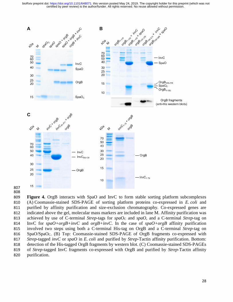
Figure 4. OrgB interacts with SpaO and InvC to form stable sorting platform subcomplexes 809 (A) Coomassie-stained SDS-PAGE of sorting platform proteins co-expressed in E. coli and 810 purified by affinity purification and size-exclusion chromatography. Co-expressed genes are 811 indicated above the gel, molecular mass markers are included in lane M. Affinity purification was 812 achieved by use of C-terminal Strep-tags for spaOC and spaO, and a C-terminal Strep-tag on 813
InvC for spaO+orgB+invC and orgB+invC. In the case of spaO+orgB affinity purification 814 involved two steps using both a C-terminal His-tag on OrgB and a C-terminal Strep-tag on 815
SpaO/SpaOC. (B) Top: Coomassie-stained SDS-PAGE of OrgB fragments co-expressed with 816 Strep-tagged invC or spaO in E. coli and purified by Strep-Tactin affinity purification. Bottom: 817 detection of the His-tagged OrgB fragments by western blot. (C) Coomassie-stained SDS-PAGEs 818 of Strep-tagged InvC fragments co-expressed with OrgB and purified by Strep-Tactin affinity 819 purification. 820
certified by peer review) is the author/funder. All rights reserved. No reuse allowed without permission. The copyright holder for this preprint (which was notthis version posted May 24, 2019. . https://doi.org/10.1101/648071doi: bioRxiv preprint

29
821 822 Figure 5. Native mass spectrum of SpaO/SpaOC/OrgB complexes. SpaO-2SpaOC heterotrimers 823
(white arrows) bind to OrgB dimers resulting in 2(SpaO-2SpaOC)-2OrgB complexes (light blue 824 arrows). 2(SpaO-2SpaOC) heterohexamers (black arrows) and a small fraction of SpaO-2SpaOC 825 complexes bound to OrgB monomers (light green arrows) and dimers (dark blue arrows) were 826
also observed. Experimental and theoretical molecular masses are given in Table S3. 827
certified by peer review) is the author/funder. All rights reserved. No reuse allowed without permission. The copyright holder for this preprint (which was notthis version posted May 24, 2019. . https://doi.org/10.1101/648071doi: bioRxiv preprint
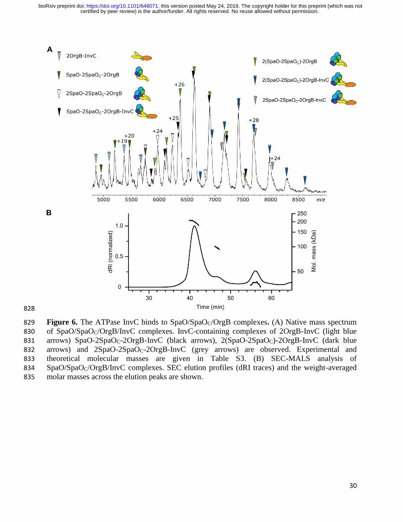
30
828
Figure 6. The ATPase InvC binds to SpaO/SpaOC/OrgB complexes. (A) Native mass spectrum 829
of SpaO/SpaOC/OrgB/InvC complexes. InvC-containing complexes of 2OrgB-InvC (light blue 830
arrows) SpaO-2SpaOC-2OrgB-InvC (black arrows), 2(SpaO-2SpaOC)-2OrgB-InvC (dark blue 831 arrows) and 2SpaO-2SpaOC-2OrgB-InvC (grey arrows) are observed. Experimental and 832 theoretical molecular masses are given in Table S3. (B) SEC-MALS analysis of 833 SpaO/SpaOC/OrgB/InvC complexes. SEC elution profiles (dRI traces) and the weight-averaged 834 molar masses across the elution peaks are shown. 835
certified by peer review) is the author/funder. All rights reserved. No reuse allowed without permission. The copyright holder for this preprint (which was notthis version posted May 24, 2019. . https://doi.org/10.1101/648071doi: bioRxiv preprint

31
836
Figure 7. Analysis of SpaO/SpaOC/OrgB/InvC complexes by small-angle X-ray scattering. (A) 837
Small-angle X-ray scattering profile of SpaO/SpaOC/OrgB/InvC. (B) Pair-distance distribution 838 function P(r) computed from the SAXS data (A). (C) SAXS-based ab initio bead model of 839
SpaO/SpaOC/OrgB/InvC. D) MONSA multi-phase modeling using SAXS data of both of 840 SpaO/SpaOC/OrgB/InvC and SpaO/SpaOC. The phase corresponding to SpaO/SpaOC is colored 841 dark blue. E) Schematic model of the SpaO/SpaOC/OrgB/InvC complex taking into account the 842
association of SpaOC with the SpaO N-terminal domain SpaO1-145 (Fig. 3), the interaction 843 between the SpaO SPOA1-SPOA2 dimer and the N-terminus of OrgB (15), and the interaction 844
between the C-terminal domain of OrgB and the N-terminal domain of InvC (Fig. 4B, C). 845
846
certified by peer review) is the author/funder. All rights reserved. No reuse allowed without permission. The copyright holder for this preprint (which was notthis version posted May 24, 2019. . https://doi.org/10.1101/648071doi: bioRxiv preprint
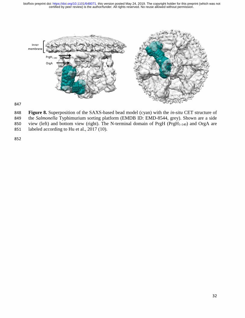
32
847
Figure 8. Superposition of the SAXS-based bead model (cyan) with the in-situ CET structure of 848
the Salmonella Typhimurium sorting platform (EMDB ID: EMD-8544, grey). Shown are a side 849 view (left) and bottom view (right). The N-terminal domain of PrgH (PrgH1-140) and OrgA are 850 labeled according to Hu et al., 2017 (10). 851
852
certified by peer review) is the author/funder. All rights reserved. No reuse allowed without permission. The copyright holder for this preprint (which was notthis version posted May 24, 2019. . https://doi.org/10.1101/648071doi: bioRxiv preprint