molecular pathology of soft tissue tumorspathology.ge/uploads/edu/soft_tissue.pdf · molecular...
TRANSCRIPT

Molecular Pathology of Soft Tissue Tumors
Ketevani Kankava, MD, MBATbilisi State Medical University
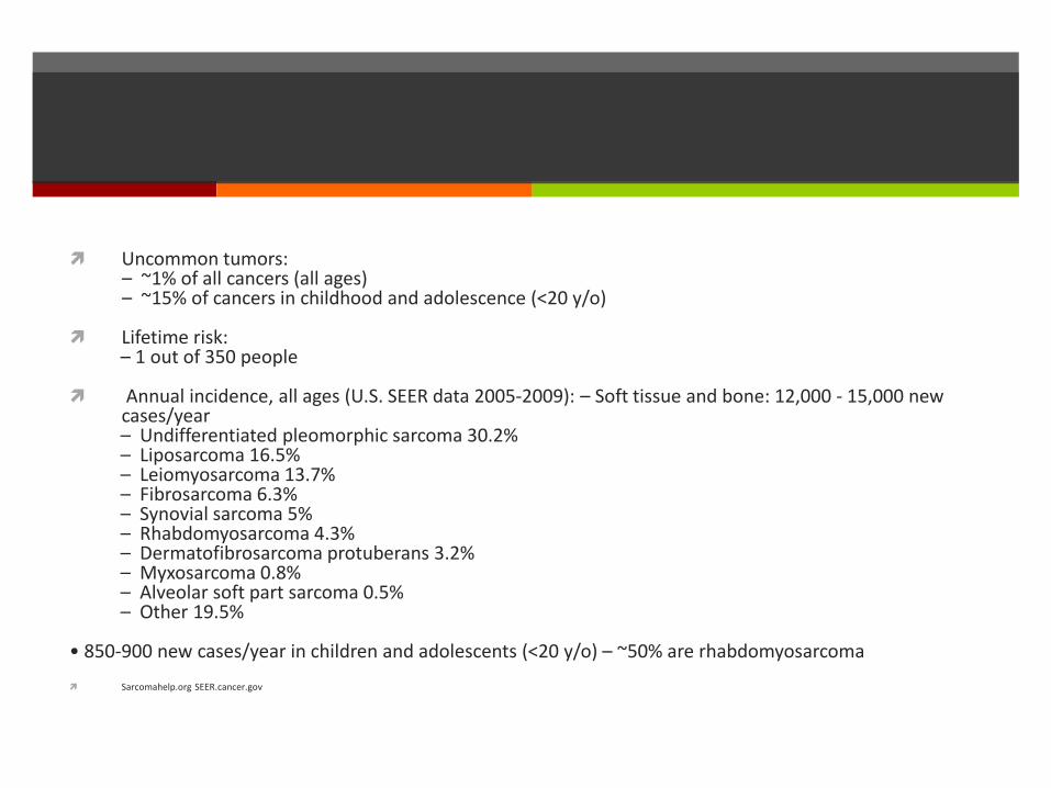
Uncommon tumors: – ~1% of all cancers (all ages) – ~15% of cancers in childhood and adolescence (<20 y/o)
Lifetime risk: – 1 out of 350 people
Annual incidence, all ages (U.S. SEER data 2005‐2009): – Soft tissue and bone: 12,000 ‐ 15,000 new cases/year – Undifferentiated pleomorphic sarcoma 30.2% – Liposarcoma 16.5% – Leiomyosarcoma 13.7% – Fibrosarcoma 6.3% – Synovial sarcoma 5% – Rhabdomyosarcoma 4.3% – Dermatofibrosarcoma protuberans 3.2% – Myxosarcoma 0.8% – Alveolar soft part sarcoma 0.5% – Other 19.5%
• 850‐900 new cases/year in children and adolescents (<20 y/o) – ~50% are rhabdomyosarcoma
Sarcomahelp.org SEER.cancer.gov

Similar “cancer” criteria for sarcoma.....but
Multistep tumorigenesis seen in epithelial neoplasia(carcinomagenesis) has been more difficult to document
– Example: translocation associated sarcomas• No pre‐neoplastic cell identified• Translocation may be the only genetic alteration; sufficient for the development of mesenchymal cancer
Cell type specificity appears to be important in sarcomagenesis
– Same translocation may be found in different tumors

Classification of cancer genes
Mechanistic classification
– Tumor suppressor genes• Deactivation: deactivating mutation, deletion, or reduced expression
– Oncogenes• Activation: activating mutation or amplification
– Caretaker genes• More likely to develop mutation(s) in tumor suppressor genes or oncogenes
Functional classification
– Protein kinases• Can function as oncogenes or tumor suppressor genes
– Transcription factors• Can function as oncogenes or tumor suppressor genes
– DNA maintenance and repair proteins• Caretaker genes

1. Variable & complex abnormal karyotype 50% – Spindle cell/pleomorphic sarcoma, NOS – Leiomyosarcoma– Myxofibrosarcoma– Pleomorphic liposarcoma– Pleomorphic rhabdomyosarcoma– Other
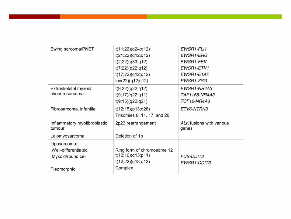
2. Reciprocal translocations 15‐30% – Ewing sarcoma (EWSR1‐FLI1) – Synovial sarcoma (SS18‐SSX) – Other
3. Specific mutations – Gastrointestinal stromal tumors (KIT & PDGFRA activating mutations) – Other
4. Amplifications – Well‐differentiated liposarcoma/atypical lipomatous tumor (12q13‐15 ring chromosome; MDM2) – Other

Why test?
Potential type(s) of information obtained:
– Diagnostic: Aid in rendering a morphologic diagnosis– Prognostic: Educated guess at a tumor’s behavior without the influence of treatment
– Predictive: Response of tumor to therapy

10/16/2014
3
Method Advantages Disadvantages
CytogeneticsDNA
Global viewPrimary and secondary abnormalities identified
Does not require knowledge of abnormality or diagnosisMay detect abnormalities not seen by FISH or PCR
Requires fresh tissue (dividing cells)Low resolution
Cryptic rearrangementsLower sensitivitySlower TAT
FISHDNA or RNA
More targeted viewRequires prior knowledge of abnormality or diagnosisDiagnostically specific and sensitiveModerate resolutionModerate analytic sensitivityMultiple tissue types can be used
FFPE, frozen, cytology or cultured cells (FISH or iFISH)Can localize abnormality to specific cellsFaster TAT
Need for fluorescence microscopeSignals fadeDoes not work on decal tissue
RT‐PCR(reverse transcriptase)RNA
High resolution (very targeted view)High sensitivity and specificity, and quantifiable (MRD)Multiplexing possibleCan use FFPE, frozen sections, cytology, or freshFaster TAT
Requires knowledge of abnormalityDoes not work on decal tissueFFPE may have degraded RNAPCR inhibitors
IHCProtein
Can use FFPE, frozen sections or cytologyMorphologic correlationRapid TATRelatively inexpensiveMutation specific antibodies available
Interlab variablility
NGSDNA or RNA
High throughput (huge multiplex capability)High resolution (individual nucleotide level)
Cost of equipment (decreasing)Need and cost of bioinformatics
MRI: Large 10.5 x 8.7 x 6.5 cm lobulated extraperitoneal mass in the posterolateral mid and upper right abdomen, showing heterogeneous enhancement and multiple enhancing septations surrounding nodular areas with variable degrees of enhancement. It displaces the adjacent liver medially and the outer layer of adjacent thoracic/abdominal wall musculature laterally. It surrounds portions of the lower right ribs, and appears to be arising from the lower aspect of the intercostal muscles, suggesting a sarcoma.
• 17 y/o female, 30 weeks pregnant, presenting with right flank pain.
CD99
NSECD45, CD20, CD3, desmin, and myogenin
Image from Dr. Mark Micale, William Beaumont Hospital, Royal Oak, MI
• Ewing sarcoma/Primitive NeuroectodermalTumor (ES/PNET)
– Lung and bone metastases present
• Delivered healthy 33‐week‐old baby
• Treated
Diagnosis:

Morphological Groups
round cell
spindle
epithelioid/polygonal
pleomorphic
adipocytic
myxoid
giant cell
others

Cytogenetics and Molecular Genetics
(1) soft-tissue neoplasms associated with complex karyotypes
(2) soft-tissue neoplasms characterized by recurrent chromosomal structural abnormalities, gene amplification, mutations, or loss of heterozygosity

Round Cell Tumors
DD in small round blue cell tumors
Most of this group are associated with specific translocations

Round cell tumors
Ewing Sarcoma t(11;22)(q24;q12)
Ewing-like sarcomas t(4:19)(q35;q13)t(10;19)(q26;q13)t(X;19)(q13;q13.3) X chromosome paracentric inversion
ARMS t(2;13)(q35;q14)t(1;13) (p36;q14)
Desmoplastic small round cell tumor t(11;22)(p13;q12
EMC t(9:22) (q22;q12)t(9;17)(q22;q11)

Ewing Sarcoma
Second most common in children and young adults
10-20% occur at extraskeletal sites
t(11;22)(q24;q12) in 85% - EWSR (22) / FL11 (11) fusion
t(21;22)(q22;12)in 5-10% - EWSR1 (22) / ERG (21)
In rare cases FUS-ERG and FUS-FEV fusion (FUS is similar to EWSR1, part of the TET family)
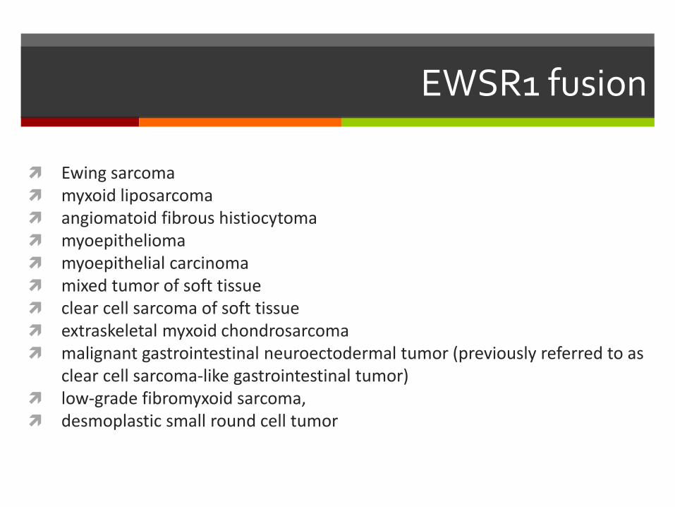
EWSR1 fusion
Ewing sarcoma myxoid liposarcoma angiomatoid fibrous histiocytoma myoepithelioma myoepithelial carcinoma mixed tumor of soft tissue clear cell sarcoma of soft tissue extraskeletal myxoid chondrosarcoma malignant gastrointestinal neuroectodermal tumor (previously referred to as
clear cell sarcoma-like gastrointestinal tumor) low-grade fibromyxoid sarcoma, desmoplastic small round cell tumor

Group of small round cell sarcomas with features similar to Ewing sarcoma
rearrangements of EWSR1 with non-ETS gene partners
No rearrangement of EWSR1 or other TET family members
In small subset - CIC-DUX4 gene fusion resulting from t(4:19)(q35;q13) or t(10;19)(q26;q13)
CIC-FOXO4 gene fusion, t(X;19)(q13;q13.3) in 2 cases
BCOR-CCNB3 fusion gene, arising from an Xchromosome paracentric inversion (biologically distinctentity within undifferentiated round cell sarcomas)

Practical approach
RT-PCR
IHC with cyclin B3
Unclear classification
Treatment similar to Ewing sarcoma

Alveolar Rabdomyosarcoma
Adolescents and young adults
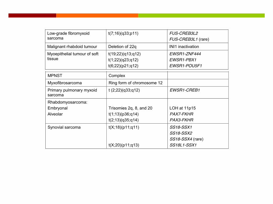
t(2;13)(q35;q14) occurs in approximately 60% of cases, t(1;13) (p36;q14) occurs in a smaller subset
Translocations involve FOXO1A (13) and PAX3 (2) or PAX7(1)
No specific chromosomal abnormality in embryonalrhabdomyosarcoma (identification of t(2;13) or t(1;13) is diagnostically valuable and prognostically significant -PAX-FOXO1A fusion status -unfavorable outcome for children with rhabdomyosarcoma)

Desmoplastic small round cell tumor
Children and young adults
Widespread abdominal serosal involvement
t(11;22)(p13;q12) - fusion of EWSR1 and WT1
identification of the partner gene is warranted for a specific diagnosis

Extraskeleral Myxoid chondrosarcoma
NR4A3 rearrangement
t(9:22) (q22;q12) or, less frequently, t(9;17)(q22;q11)
Fusion of NR4A3 (9q22) to either EWSR1 (22q12) or TAF15 (17q12)
IHC is not helpful in the diagnosis of EMC
EMC and mixed tumor of soft-tissue and myoepithelioma have overlapping histological and IHC features – different treatment
These two may share EWSR1 rearrangement
FISH for NR4A3 – ideal test platform
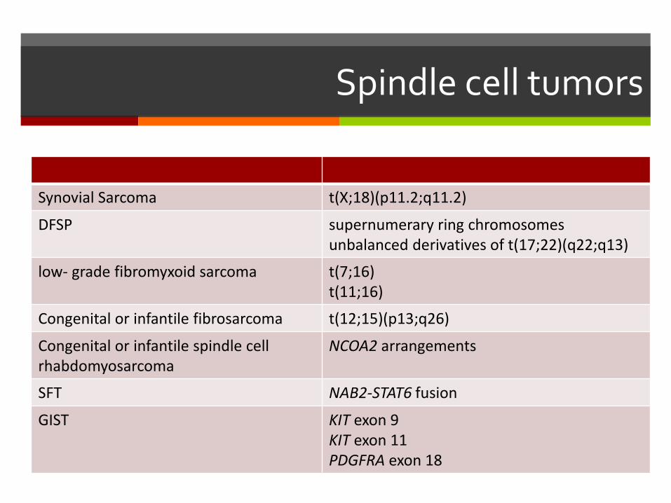
Spindle cell tumors
Synovial Sarcoma t(X;18)(p11.2;q11.2)
DFSP supernumerary ring chromosomesunbalanced derivatives of t(17;22)(q22;q13)
low- grade fibromyxoid sarcoma t(7;16)t(11;16)
Congenital or infantile fibrosarcoma t(12;15)(p13;q26)
Congenital or infantile spindle cell rhabdomyosarcoma
NCOA2 arrangements
SFT NAB2-STAT6 fusion
GIST KIT exon 9KIT exon 11PDGFRA exon 18

Synovial Sarcoma
recurrent reciprocal t(X;18)(p11.2;q11.2), which fuses SYT (18q11) to 1 of the 3 homologous genes on Xp11 (SSX1, SSX2, or SSX4)

Dermatofibrosarcoma protuberans
supernumerary ring chromosomes
unbalanced derivatives of t(17;22)(q22;q13)
chimeric gene that fuses COL1A1 with PDGFB in both cases
multiplex RT-PCR or, preferably, FISH
therapy with imatinib mesylate may be clinically useful

Low- grade fibromyxoid sarcoma
FUS-CREB3L2 gene fusion or, less frequently, FUS-CREB3L1 gene fusion
t(7;16) or t(11;16)
EWSR1-CRE- B3L1 gene fusion (in a small number)

Sclerosing epithelioid fibrosarcoma
considerable morphological overlap with low-grade fibromyxoid sarcoma
EWSR1 gene rearrangements
a minority of cases exhibits FUS-CREB3L2 fusions
Both show mucin 4 expression by IHC
perhaps part of a disease spectrum

Congenital or infantile fibrosarcoma
histologically similar to adult fibrosarcoma
t(12;15)(p13;q26) resulting in the ETV6-NTRK3 fusion
histological features may mimic other pediatric spindle cell neoplasms, such as infantile fibromatosis and infantile myofibromatosis or myofibroma
It is often difficult to cytogenetically identify the ETV6-NTRK3 fusion, so it is typically detected by FISH or PCR

Congenital or infantile spindle cell rhabdomyosarcoma
No PAX3-FOXO1 and PAX7-FOXO1 fusions
Present NCOA2 arrangements
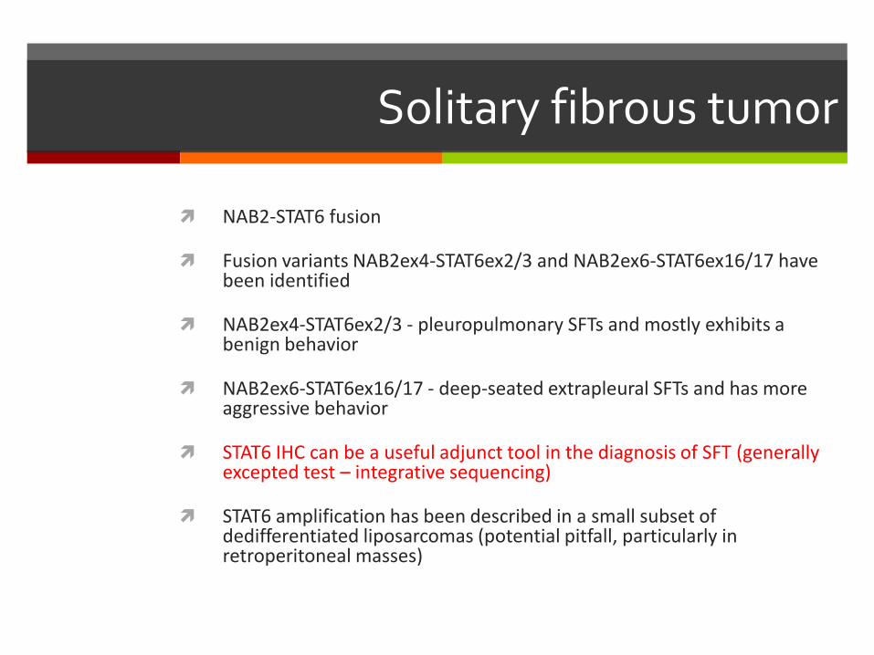
Solitary fibrous tumor
NAB2-STAT6 fusion
Fusion variants NAB2ex4-STAT6ex2/3 and NAB2ex6-STAT6ex16/17 have been identified
NAB2ex4-STAT6ex2/3 - pleuropulmonary SFTs and mostly exhibits a benign behavior
NAB2ex6-STAT6ex16/17 - deep-seated extrapleural SFTs and has more aggressive behavior
STAT6 IHC can be a useful adjunct tool in the diagnosis of SFT (generally excepted test – integrative sequencing)
STAT6 amplification has been described in a small subset of dedifferentiated liposarcomas (potential pitfall, particularly in retroperitoneal masses)

GIST
KIT exon 9
KIT exon 11
PDGFRA exon 18
BRAF V600E mutation was identified in patients with GIST lacking KIT and PDGFRA mutations
GIST are typically associated with the Carney triad and Carney–Stratakis syndrome when they show mutations in SDH-related genes

Lipomatous tumors
Myxoid/round cell liposarcoma t(12;16)(q13;p11)
ALT/WDL supernumerary ring chromosomesgiant-marker chromosomes corresponding to amplification of the 12q13-15 bandMDM2

Myxoid/round cell liposarcoma
FUS-DDIT3 chimeric gene due to reciprocal t(12;16)(q13;p11)
myxoid liposarcomas are sensitive to radiation therapy and select patients receive neoadjuvanttherapy

Lipoma VS ALT/WDL
Differently from normal fat, lipoma exhibits a HMGA2 translocation
LT/WDL is likely to recur and carries the risk of dedifferentiation, which results in a poor prognosis depending on the anatomical location
dedifferentiated liposarcoma with predominant, high-grade dedifferentiated areas may be difficult to discriminate from other high-grade pleomorphic sarcomas
ALT/WDL – supernumerary ring chromosomes and/or giant-marker chromosomes corresponding to amplification of the 12q13-15 band


Tumors of uncertain histogenesis
Clear cell sarcoma (12;22) (q13;q12)t(2;22)(q32.3;q12)
Alveolar soft-part sarcoma der(17)t(X;17) (p11;q25)
Soft tissue angiofibroma t(5;8) (p15;q13)

Clear cell sarcoma
EWSR1-ATF1 fusion in more than 90% of cases from a reciprocal t(12;22) (q13;q12)
EWSR-CREB1 fusion in small subset -t(2;22)(q32.3;q12)
Some have BRAF mutations
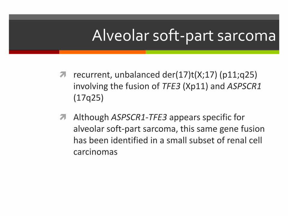
Alveolar soft-part sarcoma
recurrent, unbalanced der(17)t(X;17) (p11;q25) involving the fusion of TFE3 (Xp11) and ASPSCR1 (17q25)
Although ASPSCR1-TFE3 appears specific for alveolar soft-part sarcoma, this same gene fusion has been identified in a small subset of renal cell carcinomas

Soft tissue angiofibroma
benign, fibrovascular soft-tissue tumor of uncertain cellular origin
AHRR-NCOA2 fusion resulting from t(5;8) (p15;q13)
evaluation of NCOA2 rearrangements using FISH

Myxoid tumors


Thank you for attention
Molecular Pathology of Soft-Tissue Neoplasms and Its Role in Clinical Practice Evita B. Henderson-Jackson, MD, and Marilyn M. Bui, MD, PhD
Molecular Pathology of Soft Tissue Sarcomas Jon D. Wilson, MD [email protected] (lecture)
The royal college of pathologists - Standards and datasets for reporting cancers Dataset for histopathology reporting of soft tissue sarcomas. January 2017 Author: Professor Cyril Fisher. Consultant Histopathologist, Sarcoma Unit, Royal Marsden NHS Foundation Trust, London Professor of Tumour Pathology, Institute of Cancer Research, University of London