molecular targets of dietary agents for prevention and ...comilac.com.tr/uploads/pdf/35pomgt.pdf ·...
TRANSCRIPT

Commentary
Molecular targets of dietary agents for prevention andtherapy of cancer§
Bharat B. Aggarwal a,*, Shishir Shishodia b
aCytokine Research Laboratory, Department of Experimental Therapeutics,
The University of Texas M.D. Anderson Cancer Center, Box 143, 1515 Holcombe Boulevard, Houston, TX 77030, USAbDepartment of Biology, Texas Southern University, 3100 Cleburne Street, Houston, TX 77004, USA
b i o c h e m i c a l p h a r m a c o l o g y 7 1 ( 2 0 0 6 ) 1 3 9 7 – 1 4 2 1
a r t i c l e i n f o
Keywords:
NF-kB
AP-1
MAP kinases
Apoptosis
Cell cycle
Cancer
Dietary agents
Abbreviations:
AP-1, activator protein-1
CAPE, caffeic acid
phenethyl ester
Cdk, cyclin-dependent kinase
COX-2, cyclooxygenase-2
cPLA2, phospholipase A
CSF, colony-stimulating factors
DIM, 1,1-bis(30-indolyl)-1-
( p-substituted phenyl) methanes
DMBA, dimethyl-benz(a)
anthracene
EGF, epidermal growth factor
EGCG, epigallocatechin-3-gallate
Epo, erythropoietin
ERK, extracellular signal-
regulated kinase
FGF, fibroblast growth factor
a b s t r a c t
While fruits and vegetables are recommended for prevention of cancer and other diseases,
their active ingredients (at the molecular level) and their mechanisms of action less well
understood. Extensive research during the last half century has identified various molecular
targets that can potentially be used not only for the prevention of cancer but also for
treatment. However, lack of success with targeted monotherapy resulting from bypass
mechanisms has forced researchers to employ either combination therapy or agents that
interfere with multiple cell-signaling pathways. In this review, we present evidence that
numerous agents identified from fruits and vegetables can interfere with several cell-
signaling pathways. The agents include curcumin (turmeric), resveratrol (red grapes, pea-
nuts and berries), genistein (soybean), diallyl sulfide (allium), S-allyl cysteine (allium), allicin
(garlic), lycopene (tomato), capsaicin (red chilli), diosgenin (fenugreek), 6-gingerol (ginger),
ellagic acid (pomegranate), ursolic acid (apple, pears, prunes), silymarin (milk thistle),
anethol (anise, camphor, and fennel), catechins (green tea), eugenol (cloves), indole-3-
carbinol (cruciferous vegetables), limonene (citrus fruits), beta carotene (carrots), and diet-
ary fiber. For instance, the cell-signaling pathways inhibited by curcumin alone include NF-
kB, AP-1, STAT3, Akt, Bcl-2, Bcl-XL, caspases, PARP, IKK, EGFR, HER2, JNK, MAPK, COX2, and
5-LOX. The active principle identified in fruit and vegetables and the molecular targets
modulated may be the basis for how these dietary agents not only prevent but also treat
cancer and other diseases. This work reaffirms what Hippocrates said 25 centuries ago, let
food be thy medicine and medicine be thy food.
# 2006 Elsevier Inc. All rights reserved.
avai lab le at www.sc iencedi rect .com
journal homepage: www.e lsev ier .com/ locate /b iochempharm
§ Supported by the Clayton Foundation for Research (to BBA), Department of Defense US Army Breast Cancer Research Program grant(BC010610, to BBA), a PO1 grant (CA91844) from the National Institutes of Health on lung cancer chemoprevention (to BBA), a P50 Head andNeck Cancer SPORE grant from the National Institutes of Health (to BBA), Cancer Center Core Grant CA 16672, and Texas SouthernUniversity Seed Grant (to SS).
* Corresponding author. Tel.: +1 713 792 3503/6459; fax: +1 713 794 1613.E-mail address: [email protected] (B.B. Aggarwal).
0006-2952/$ – see front matter # 2006 Elsevier Inc. All rights reserved.doi:10.1016/j.bcp.2006.02.009

b i o c h e m i c a l p h a r m a c o l o g y 7 1 ( 2 0 0 6 ) 1 3 9 7 – 1 4 2 11398
1. Introduction
Epidemiological data accumulated over the last 50 years show
a significant decrease in the death rate within the US due to
heart, cerebrovascular, and infectious diseases; however,
cancer-related mortality has remained unaltered since 1950
[1,2]. Despite a better understanding of the disease and the
advent of modern technology and rationally targeted drugs,
the incidence and cure rate of cancer have not improved.
One secret to improving cancer statistics seems to reside in
the epidemiology of the disease. Epidemiology has revealed
that certain cancers are more common among people of some
cultures than others [3–6]. Cancers of the lung, colon, prostate
and breast are very common in Western countries; they are
not as prevalent in Eastern countries. Similarly, cancers of the
head and neck and of the cervix are most common in India,
whereas stomach cancer is most prevalent in Japan.
Studies indicate that migration from native to adopted
country, however, exposes an individual to the same cancer
risk and incidence as that of others living in the adopted
country. Because human beings are 99.1% identical in their
genetic sequence, these differences in incidence cannot be
attributed to the variation in their DNA sequence. In fact, if one
twin is identified with breast cancer, the chance that the
second twin will be diagnosed with breast cancer is 20%,
indicating that the contribution of faulty genes to the
pathogenesis of cancer is minimum [7]. Instead, it is estimated
that 75–85% of all chronic illnesses and diseases are linked to
HER2, human epidermal
growth factor receptor 2
I-3-C, indole-3-carbinol
ICAM-1, intercellular
adhesion molecule-1
IFN, interferon
IGF, insulin-like growth factor
IkBa, inhibitory kappa B alpha
IKK, IkBa kinase
IL-1, interleukin
iNOS, inducible nitric
oxide synthase
JNK, c Jun N-terminal
kinase
LPS, lipopolysaccharide
LOX, lipoxygenase
MMP, matrix
metalloproteinase
MAPK, mitogen-activated
protein kinases
NF-kB, nuclear
factor-kappa B
PDK, pyruvate
dehydrogenase kinase
PTK, protein tyrosine kinase
PKB, protein kinase B
p27KIP1, p27 kinase
inhibitor protein 1
PDGF, platelet-derived
growth factor
PARP, polyadenosine-50-
diphosphate-ribose polymerase
STAT, signal transducer and
activator of transcription
TGF, transforming growth factor
THC, tetrahydrocurcumin
TNF, tumor necrosis factor
TPA, phorbol 12-O-
tetradecanoate-13-acetate
TRE, TPA response elements
uPA, urokinase-type
plasminogen activator
VEGF, vascular endothelial
growth factor

b i o c h e m i c a l p h a r m a c o l o g y 7 1 ( 2 0 0 6 ) 1 3 9 7 – 1 4 2 1 1399
lifestyle and cannot be explained by differences in genetic
makeup [8]. For example, there is a positive association
between fat and red meat and an inverse association between
dietary fiber, fruit and vegetable intake with the development
of colorectal adenomas [9]. Epidemiological studies have
indicated that populations that consume food rich in fruits
and vegetables have a lower incidence of cancers [10–12].
Review of results from 206 human epidemiologic studies and
22 animal studies has indicated an inverse relationship
between consumption of vegetables and fruits and risk for
cancers of the stomach, esophagus, lung, oral cavity and
pharynx, endometrium, pancreas, and colon [13].
Natural dietary agents including fruits, vegetables, and
spices have drawn a great deal of attention from both the
scientific community and the general public owing to their
demonstrated ability to suppress cancers. The questions that
remain to be answered are which component of these dietary
agents is responsible for the anti-cancer effects and what is
the mechanism by which they suppress cancer? Dietary
agents consist of a wide variety of biologically active
compounds that are ubiquitous in plants, many of which
have been used in traditional medicines for thousands of
years. As early as 2500 years ago, Hippocrates recognized and
professed the importance of various foods both natural and
those derived from human skill in the primary constitution of
the person.
Fruits and vegetables are excellent sources of fiber,
vitamins, and minerals, but they also contain components
like polyphenols, terpenes, alkaloids, and phenolics that may
provide substantial health benefits beyond basic nutrition.
Research over the last decade has shown that several
micronutrients in fruits and vegetables reduce cancer
(Table 1). The active components of dietary phytochemicals
that most often appear to be protective against cancer are
curcumin, genistein, resveratrol, diallyl sulfide, S-allyl
cysteine, allicin, lycopene, capsaicin, diosgenin, 6-gingerol,
ellagic acid, ursolic acid, silymarin, anethol, catechins,
eugenol, isoeugenol, dithiolthiones, isothiocyanates, indole-
3-carbinol, isoflavones, protease inhibitors, saponins, phytos-
terols, inositol hexaphosphate, Vitamin C, D-limonene, lutein,
folic acid, beta carotene, selenium, Vitamin E, flavonoids, and
dietary fiber (Figs. 1 and 2). These dietary agents are believed to
suppress the inflammatory processes that lead to transforma-
tion, hyperproliferation, and initiation of carcinogenesis.
Their inhibitory influences may ultimately suppress the final
steps of carcinogenesis as well, namely angiogenesis and
metastasis (Fig. 3).
Tumorigenesis is a multistep process that can be activated
by any of various environmental carcinogens (such as
cigarette smoke, industrial emissions, gasoline vapors),
inflammatory agents (such as tumor necrosis factor (TNF)
and H2O2), and tumor promoters (such as phorbol esters and
okadaic acid). These carcinogens are known to modulate the
transcription factors (e.g., NF-kB, AP-1, STAT3), anti-apoptotic
proteins (e.g., Akt, Bcl-2, Bcl-XL), proapoptotic proteins (e.g.,
caspases, PARP), protein kinases (e.g., IKK, EGFR, HER2, JNK,
MAPK), cell cycle proteins (e.g., cyclins, cyclin-dependent
kinases), cell adhesion molecules, COX-2, and growth factor
signaling pathways. This article reviews the current studies
regarding the numerous pathways and molecular targets of
dietary agents for not only prevention but also for therapy of
cancers.
2. Molecular targets
2.1. Nuclear factor-kappa B (NF-kB)
NF-kB is a family of closely related protein dimers that bind to
a common sequence motif in DNA called the kB site (for
references see [14]). The identification of the p50 subunit (v-
REL) of NF-kB as a member of the reticuloendotheliosis (REL)
family of viruses provided the first evidence that NF-kB is
linked to cancer. Under resting condition, the NF-kB dimers
reside in the cytoplasm. NF-kB is activated by free radicals,
inflammatory stimuli, cytokines, carcinogens, tumor promo-
ters, endotoxins, g-radiation, ultraviolet (UV) light, and X-rays.
Upon activation, it is translocated to the nucleus, where it
induces the expression of more than 200 genes that have been
shown to suppress apoptosis and induce cellular transforma-
tion, proliferation, invasion, metastasis, chemo-resistance,
radio-resistance, and inflammation. Many of the target genes
that are activated are critical to the establishment of the early
and late stages of aggressive cancers, including expression of
cyclin D1, apoptosis suppressor proteins such as Bcl-2 and Bcl-
XL and those required for metastasis and angiogenesis, such
as matrix metalloproteases (MMP) and vascular endothelial
growth factor (VEGF).
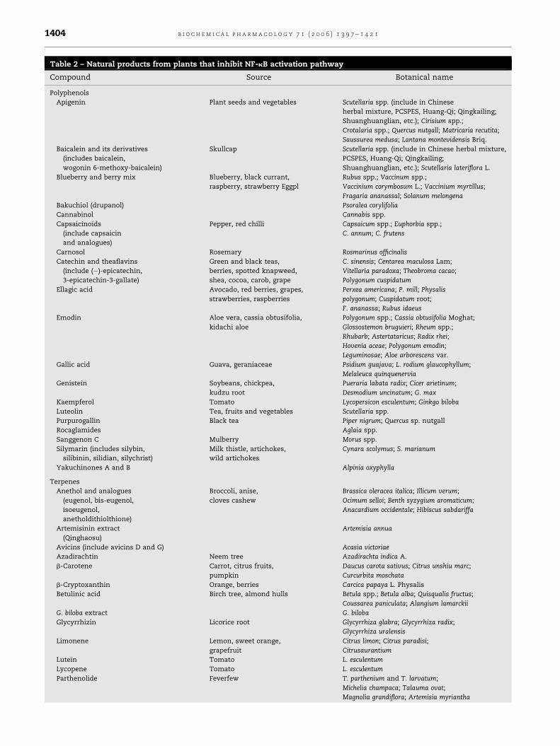
Several dietary agents like curcumin [15], resveratrol [16],
guggulsterone [17], ursolic acid [18], betulinic acid [19], emodin
[20], gingerol [21], flavopiridol [22], zerumbone [23], evodia-
mine [24], indole-3-carbinol [25], elagic acid, anethole [26],
green tea catechins [27], S-allyl cysteine [28], lycopene [29], and
diosgenin [30] are natural chemopreventive agents that have
been found to be potent inhibitors of NF-kB (Table 2). How
these agents suppress NF-kB activation is becoming increas-
ingly apparent. These inhibitors may block any one or more
steps in the NF-kB signaling pathway such as the signals that
activate the NF-kB signaling cascade, translocation of NF-kB
into the nucleus, DNA binding of the dimers, or interactions
with the basal transcriptional machinery.
A number of studies from our laboratory have shown that
the spices derived from plants exert their anti-cancer effects
through the suppression of NF-kB. Curcumin as well as various
other curcuminoids from ginger family mediate their ther-
apeutic effects by regulating the transcription factor NF-kB
and NF-kB regulated gene products COX-2, cyclin D1, adhesion
molecules, MMPs, inducible nitric oxide synthase, Bcl-2, Bcl-XL
and TNF [31]. Curcumin suppresses the TNF-induced activa-
tion of IKK that leads to the inhibition of TNF-dependent
phosphorylation and degradation of IkBa and translocation of
the p65 subunit. Curcumin also blocks phorbol ester- and
hydrogen peroxide-mediated activation of NF-kB [15]. Guggul-
sterone suppresses NF-kB activation by suppressing the
activation of IKK by interacting directly with the kinase [17].
In studies from our laboratory, resveratrol suppressed TNF-
induced phosphorylation and nuclear translocation of the p65
subunit of NF-kB and NF-kB-dependent reporter gene tran-
scription. Suppression of TNF-induced NF-kB activation by
resveratrol was not cell type specific and was observed in

b i o c h e m i c a l p h a r m a c o l o g y 7 1 ( 2 0 0 6 ) 1 3 9 7 – 1 4 2 11400
Table 1 – Molecular targets of dietary agents
Plant name Active compound Molecular target
Turmeric
(Curcuma longa)
Curcumin,
curcuminoids
#NF-kB, # AP-1, #Egr-1, #STAT1, #STAT3, #STAT5, "PPARg, #EpRE,
#CBP, #b-catenin, "Nrf2, "IKK, #EGFR, #HER2, #AKt, #Src, #JAK2,
#TYK2, #JNK, #PKA, #PKC, #VCAM-1, #Bcl-2, #Bcl-XL, #ICAM-1, #TF,
#AR/ARP, #p53, "MDR, #ELAM-1, #FTPase, "GST, "GSH-px, #uPA, "HO, #XOD,
#cyclin D1, #5-LOX, #COX-2, #INOS, #MMP-9, #TNF, #IL-6, #IL-8, #IL-12
Grapes
(Vitis vinifera)
Resveratrol #COX-2, #iNOS, #JNK, #MEK, #AP-1, #NF-kB, "P21 Cip1/WAF1,
"p53, "Bax, "caspases, #survivin, #cyclin D1, #cyclin E, #Bcl-2,
#Bcl-xL, #CIAP, #Egr-1, #PKC, #PKD, #casein kinase II, #5-LOX,
#VEGF, #IL-1, #IL-6, #IL-8, #AR, #PSA, #CYP1A1, #TypeII-Ptdlns-4kinase,
#Cdc2-tyr15a, "HO-1, "Nrf2, #endothelin-1
Guggulu
(Commiphora mukul)
Guggulsterone #NF-kB, #IAP1, #XIAP, #Bfl-1/A1, #Bcl-2, #cFLIP, #survivin, #cyclin D1,
#c-Myc, #MMP-9, #COX-2, #VEGF, #BAR, #CYP7A1, #FXR, "CYP3A, #Cyp2b10
Pinecone ginger
(Zingiber zerumbet)
Zerumbone #NF-kB, #IAP1, #XIAP, #Bfl-1/A1, #Bcl-2, #cFLIP, #survivin, #cyclin D1,
#c-Myc, #MMP-9, #COX-2, #TRAF1
Aloe (Aloe vera) Emodin #NF-kB, "HER-2/neu, "caspase-3, #AR, #MMP-9, "CYP1A1, "CYP1B1
Boswellia
(Salai guggul)
(Boswellia serrata)
Boswellic acids #NF-kB, "p42 MAPK, "p38 MAPK, #5-LOX, #survivin,
#cyclin D1, #Bcl-2, #Bcl-xL, #CIAP
Cruciferous
vegetables
(Brassica sp.)
Sulforaphane,
indole-3-carbinol
#NF-kB, #survivin, #cyclin D1, #Bcl-2, #Bcl-xL,
#CIAP #Cdc25, #Cdk1, #Bcl-2, #Bcl-xL
Quince
(Cydonia oblonga)
Caffeoylquinic acids #IFN-g, #IL-2, #ERK1/2, #AKTa, #NF-kB, #NO, #iNOS
Rohitukine (Dysoxylum
binectrariferum)
Flavopiridol #NF-kB, #COX-2, #cyclin D1, #MMP-9, #Bcl2
Coriander
(Coriandrum sativum)
Linalool,
monoterpenes
#NF-kB, #AP-1, #JNK, #MAPK
Sweet Fennel
(Foeniculum vulgare)
Anethole #NF-kB, #AP-1, #JNK, #MAPK
Ashwagandha
(Withania somnifera)
Withonalides #NF-kB, #COX-2, #cyclin D1, #MMP-9, #survivin,
#cyclin E, #Bcl-2, #Bcl-xL, #CIAP
Pomegranate
(Punica granatum)
Ellagic acid #NF-kB, #COX-2, #cyclin D1, #MMP-9, #PDGF, #VEGF, "p21/WAF1, "p53
Soyabean
(Glycine max)
Genistein #NF-kB, "caspase-12, "p21/WAF1, "glutathione peroxidase
Basil (Ocimum sanctum) Ursolic acid #NF-kB, #COX-2, #cyclin D1, #MMP-9
Parthenium
(Tanacetum parthenium)
Sesqiterpene lactones,
parthenolides
#NF-kB, "p53, "reactive oxygen species
Prunes and plums Ursolic acid,
oleanolic acid,
triterpenoids
#NF-kB, #COX-2, #cyclin D1, #MMP-9
Oleander
(Nerium oleander)
Oleandrin #NF-kB, #AP-1, #JNK, #COX-2, #cyclin D1, #MMP-9
Tea (Camellia sinensis) Flavonoids,
catechins
#NF-kB, #AP-1, #JNK, #COX-2, #cyclin D1, #MMP-9, "HO-1,
#IL-6, #VEGF, #IGF, "p53, #Bcl-2, "p21/WAF1
Silymarin
(Silybum marianum L.)
Silybinin #NF-kB, #AP-1, #JNK, #COX-2, #cyclin D1, #MMP-9
Citrus fruits,
apple (Citrus sp.)
Quercetin #NF-kB, "Bax, # Bcl-2, #cyclin D1, "caspase, "PARP, "Gadd 45
Red chilli
(Capsicum annum)
Capsaicin #NF-kB, #survivin, #cyclin D1, #Bcl-2, #Bcl-xL,
#CIAP, #Cdc25, #Cdk1, #Bcl-2, #Bcl-xL
Cloves
(Eugenia caryophyllus)
Eugenol,
isoeugenol
#NF-kB
Cardamon
(Elettaria cardamomum)
Limonene #COX-2, #iNOS
Ginger
(Zingiber officinale)
Gingerol,
paradol
#TNF, #NF-kB, #AP-1, #COX-2, #ODC, #iNOS,
#p38MAPK, #HIF, #VEGF, "caspase-3, #Bcl2
Galanga
(Alpinia officinarum)
Yakuchinone
A&B
#COX2, # iNOS, #NF-kB, #adhesion molecules, #TNF, #AP-1, #5-HETE
Kokum
(Garcinia indica)
Garcinol #NF-kB, #COX-2, #iNOS, #HAT
Licorice
(Glycyrrhiza echinata)
Dibenzoylmethane #COX2, # LOX, #HIF, #VEGF
References, please visit Pub Med (http://www.ncbi.nih.gov/entrez/query.fcgi). NF-kB, nuclear factor kappa B; NO, nitric oxide; PGE, prostaglandin; iNOS,
inducible nitric oxide synthase; COX-2, cyclooxygenase-2; IL, interleukin; MAP, mitogen-activated protein; TNF, tumor necrosis factor; BAR, bile acid
receptor; FXR, farnesoid X receptor; CYP7A1, cholesterol 7alpha-hydroxylase; CYP, cytochrome p450; HO, heme oxygenase; Nrf, NF-E2-related factor;
Ptdlns, phosphatidylinositol; IAP, inhibitor-of-apoptosis protein; PKC, protein kinase C; PKD, protein kinase D; LOX, lipoxygenase; VEGF, vascular
endothelial growth factor; AR, androgen receptor; PSA, prostate-specific antigen; ICAM, intercellular cell adhesion molecules; TF, tissue factor; MDR,
multidrug resistance; Ftase, farnesyl-protein transferase; GST, glutathione S-transferase; GST-px, glutathione peroxidase; XOD, xanthine oxidase; TNF,
tumor necrosis factor; MMP, matrix metalloprotease; STAT, signal transducers and activators of transcription.a Indicates phosphorylation.

b i o c h e m i c a l p h a r m a c o l o g y 7 1 ( 2 0 0 6 ) 1 3 9 7 – 1 4 2 1 1401
Fig. 1 – Dietary agents with anti-cancer properties.
Fig. 2 – Chemical structures of dietary compounds.

b i o c h e m i c a l p h a r m a c o l o g y 7 1 ( 2 0 0 6 ) 1 3 9 7 – 1 4 2 11402
Fig. 2. (Continued ).
myeloid (U-937), lymphoid (Jurkat) and epithelial (HeLa and
H4) cells. The molecule also blocked NF-kB activation
induced by various carcinogens and tumor promoters
including PMA, LPS, H2O2, okadaic acid, and ceramide [16].
Caffeic acid phenethyl ester (CAPE) has been shown to
suppress NF-kB activation by suppressing the binding of p50–
p65 complex directly to the DNA [32], whereas both
sanguinarine and emodin act by blocking the degradation
of IkBa. The alkaloid sanguinarine can prevent phosphoryla-
tion and degradation of IkBa in response to TNFa, phorbol
ester, IL-1 or okadaic acid stimulation [33]. Similar to
sanguinarine, emodin inhibits TNF-dependent IkBa degra-
dation [20]. Based on its ability to inhibit other kinases,
emodin may act directly on the IKK complex to block
phosphorylation of IkBa. Yang et al. found that the green tea
polyphenol, EGCG, suppresses NF-kB activation by inhibiting
IKK activity [27], as do various other chemopreventive
dietary agents. Some act by suppressing IkBa degradation
and p65 translocation or NF-kB–DNA binding activity. Thus,
one of the probable mechanisms by which dietary agents
exercise their anti-tumor properties is through the suppres-
sion of the NF-kB signaling pathway.
2.2. Activator protein-1 (AP-1)
AP-1 was originally identified by its binding to a DNA sequence
in the SV40 enhancer [34]. This complex consists of either
homo- or heterodimers of the members of the JUN and FOS
family of proteins [35]. Many stimuli, most notably serum,
growth factors, and oncoproteins, are potent inducers of AP-1
activity; it is also induced by TNF and Interleukin 1 (IL-1), as
well as by a variety of environmental stresses, such as UV
radiation [35]. AP-1 activation is linked to growth regulation,
cell transformation, inflammation, and innate immune
response. AP-1 has been implicated in regulation of genes
involved in apoptosis and proliferation and may promote cell
proliferation by activating the cyclin D1 gene, and repressing
tumor-suppressor genes, such as p53, p21cip1/waf1 and p16.
Most important, AP-1 can promote the transition of tumor
cells from an epithelial to mesenchymal morphology, which is
one of the early steps in tumor metastasis. Expression of genes
such as MMP and uPA especially promotes angiogenesis and
invasive growth of cancer cells. These oncogenic properties of
AP-1 are primarily dictated by the dimer composition of the
AP-1 family proteins and their post-transcriptional and
translational modifications [35].
Several phytochemicals such as green tea catechins [36],
quercetin [37], resveratrol [16], curcumin [38,39], capsaicin [40],
oleandrin [41], anethole [26], and beta-lapachone, [42] have
been shown to suppress the AP-1 activation process. EGCG and
theaflavins inhibit TPA- and epidermal growth factor-induced
transformation of JB6 mouse epidermal cells [36]. This finding
correlates with the inhibition of AP-1 DNA binding and
transcriptional activity. The inhibition of AP-1 activity by

b i o c h e m i c a l p h a r m a c o l o g y 7 1 ( 2 0 0 6 ) 1 3 9 7 – 1 4 2 1 1403
Fig. 3 – Molecular targets of dietary agents.
EGCG was associated with inhibition of JNK activation but not
ERK activation. Interestingly, in another study where EGCG
blocked the UVB-induced c-Fos activation in a human
keratinocyte cell line HaCaT [43], inhibition of p38 activation
was suggested as the major mechanism underlying the effects
of EGCG. The role of MAPK pathways in the regulation of AP-1
activity by EGCG has been further investigated [44]. Treatment
of Ha-ras-transformed human bronchial cells with EGCG has
been shown to inhibit c-Jun and ERK1/2 phosphorylation as
well as the phosphorylation of ELK1 and MEK1/2 [45]. In
contrast to these reports, EGCG has been shown to markedly
increase AP-1 factor-associated responses through a MAPK
signaling mechanism in normal human keratinocytes, sug-
gesting that the signaling mechanism of EGCG action could be
markedly different in different cell types [46]. Lagarrigue et al.
showed that the flavonoid quercetin could inhibit the
transformation of the rat liver epithelial cell line overexpres-
sing c-Fos, suggesting that regulation of c-Fos/AP-1 complexes
might be involved in the antitransforming mechanism of
quercetin [37]. Pretreatment of RAW 264.7 macrophages with
quercetin blocked LPS-induced TNF transcription. This effect
of quercetin was mediated by inhibiting the phosphorylation
and activation of JNK/stress-activated protein kinase, by
suppressing AP-1 DNA binding, and by down-regulating TNF
transcription [47].
Resveratrol has been shown to inhibit the activity of AP-1 as
demonstrated by several studies. We have found that
resveratrol inhibits TNF-dependent AP-1 activation in U-937
cells, and that pretreatment with resveratrol strongly attenu-
ates TNF-activated JNK and MEK kinases [16]. Curcumin has
been shown to suppress the activation of TPA-induced AP-1 in
HL-60 cells and Raji cells [38,39]. Curcumin treatment also
suppresses constitutive AP-1 activity in the prostate cancer
cell lines LNCaP, PC-3, and DU145 [48,49]. Inhibition of AP-1
transcriptional activity by curcumin also correlated with
inhibition of Lewis lung carcinoma invasion in an orthotopic
implantation model [50]. More recently, curcumin was
reported to suppress LPS-induced cyclooxygenase-2 gene
expression by inhibiting AP-1 DNA binding in BV2 microglial
cells [51]. These results suggest that chemopreventive agents
specifically targeting AP-1 or its activating kinases could be
promising agents for the treatment of several cancers.
2.3. Cell cycle
Several proteins are known to regulate the timing of the events
in the cell cycle. The loss of this regulation is the hallmark of
cancer. Major control switches of the cell cycle are the cyclins
and the cyclin-dependent kinases. Cyclin D1, a component
subunit of cyclin-dependent kinase (Cdk)-4 and Cdk6, is a
b i o c h e m i c a l p h a r m a c o l o g y 7 1 ( 2 0 0 6 ) 1 3 9 7 – 1 4 2 11404
Table 2 – Natural products from plants that inhibit NF-kB activation pathway
Compound Source Botanical name
Polyphenols
Apigenin Plant seeds and vegetables Scutellaria spp. (include in Chinese
herbal mixture, PCSPES, Huang-Qi; Qingkailing;
Shuanghuanglian, etc.); Cirisium spp.;
Crotalaria spp.; Quercus nutgall; Matricaria recutita;
Saussurea medusa; Lantana montevidensis Briq.
Baicalein and its derivatives
(includes baicalein,
wogonin 6-methoxy-baicalein)
Skullcap Scutellaria spp. (include in Chinese herbal mixture,
PCSPES, Huang-Qi; Qingkailing;
Shuanghuanglian, etc.); Scutellaria lateriflora L.
Blueberry and berry mix Blueberry, black currant,
raspberry, strawberry Eggpl
Rubus spp.; Vaccinum spp.;
Vaccinium corymbosum L.; Vaccinium myrtillus;
Fragaria ananassal; Solanum melongena
Bakuchiol (drupanol) Psoralea corylifolia
Cannabinol Cannabis spp.
Capsaicinoids
(include capsaicin
and analogues)
Pepper, red chilli Capsaicum spp.; Euphorbia spp.;
C. annum; C. frutens
Carnosol Rosemary Rosmarinus officinalis
Catechin and theaflavins
(include (�)-epicatechin,
3-epicatechin-3-gallate)
Green and black teas,
berries, spotted knapweed,
shea, cocoa, carob, grape
C. sinensis; Centarea maculosa Lam;
Vitellaria paradoxa; Theobroma cacao;
Polygonum cuspidatum
Ellagic acid Avocado, red berries, grapes,
strawberries, raspberries
Perxea americana; P. mill; Physalis
polygonum; Cuspidatum root;
F. ananassa; Rubus idaeus
Emodin Aloe vera, cassia obtusifolia,
kidachi aloe
Polygonum spp.; Cassia obtusifolia Moghat;
Glossostemon bruguieri; Rheum spp.;
Rhubarb; Astertataricus; Radix rhei;
Hovenia aceae; Polygonum emodin;
Leguminosae; Aloe arborescens var.
Gallic acid Guava, geraniaceae Psidium guajava; L. rodium glaucophyllum;
Melaleuca quinquenervia
Genistein Soybeans, chickpea,
kudzu root
Pueraria labata radix; Cicer arietinum;
Desmodium uncinatum; G. max
Kaempferol Tomato Lycopersicon esculentum; Ginkgo biloba
Luteolin Tea, fruits and vegetables Scutellaria spp.
Purpurogallin Black tea Piper nigrum; Quercus sp. nutgall
Rocaglamides Aglaia spp.
Sanggenon C Mulberry Morus spp.
Silymarin (includes silybin,
silibinin, silidian, silychrist)
Milk thistle, artichokes,
wild artichokes
Cynara scolymus; S. marianum
Yakuchinones A and B Alpinia oxyphylla
Terpenes
Anethol and analogues
(eugenol, bis-eugenol,
isoeugenol,
anetholdithiolthione)
Broccoli, anise,
cloves cashew
Brassica oleracea italica; Illicum verum;
Ocimum selloi; Benth syzygium aromaticum;
Anacardium occidentale; Hibiscus sabdariffa
Artemisinin extract
(Qinghaosu)
Artemisia annua
Avicins (include avicins D and G) Acasia victoriae
Azadirachtin Neem tree Azadirachta indica A.
b-Carotene Carrot, citrus fruits,
pumpkin
Daucus carota sativus; Citrus unshiu marc;
Curcurbita moschata
b-Cryptoxanthin Orange, berries Carcica papaya L. Physalis
Betulinic acid Birch tree, almond hulls Betula spp.; Betula alba; Quisqualis fructus;
Coussarea paniculata; Alangium lamarckii
G. biloba extract G. biloba
Glycyrrhizin Licorice root Glycyrrhiza glabra; Glycyrrhiza radix;
Glycyrrhiza uralensis
Limonene Lemon, sweet orange,
grapefruit
Citrus limon; Citrus paradisi;
Citrusaurantium
Lutein Tomato L. esculentum
Lycopene Tomato L. esculentum
Parthenolide Feverfew T. parthenium and T. larvatum;
Michelia champaca; Talauma ovat;
Magnolia grandiflora; Artemisia myriantha

b i o c h e m i c a l p h a r m a c o l o g y 7 1 ( 2 0 0 6 ) 1 3 9 7 – 1 4 2 1 1405
Table 2 (Continued )
Compound Source Botanical name
Ursolic acid Basil, salvia, rosemary, berries Rosemainus officinalis; O. sanctum;
Aronia melanocarpa; Oxycoccus quadripetalus;
Origanum majorana; Diospyros melanoxylon;
Salvia przewalskii Maxim
Withanolides Withania sominifera
Alkaloids
Conophylline Tabernaemontana spp.; Ervatamia microphylla
Cucurbitacin Cucurbitaceae Cucurbita andreana; Trichosanthes kirilowii;
Elaeocarpus mastersii
Higenamine Ranunculaceae, lianas Aconitum japonicum; Argemone mexicana;
Gnetum parvifolium
Mahanimbine Rutaceous Murraya koenigii; Clausena dunniana;
Murraya siamensis
Mahanine Rutaceous M. koenigii; Micromelum minutum
Morphine and its analogues
(include KT 90 and sanguinarine)
Rapaver spp.; Opium poppy
Piperine Black pepper Garcinia xanthochymu; Piper longum
Flavonoids
Cirsimaritin Basil, sage, rosemary O. sanctum; Salvia officinalis; Rosemaribus officinalis
Flavopiridol Dysoxylum binectariferum
Hesperidine Oranges Gamellia sinensis O.Ktze.
Morin Almond P. guajava L.; Prunus dulcis (Mill.)
Nobiletin Citrus C. u. marc
Pycnogenol Citrus fruit Citrus retirulata; Pinus maritima
Persenone A Tomato, avocado L. esculentum; Persea americana P. Mil
Phenolics
Ethyl gallate Grapes, tea, red maple Paeonia spp.; Sophora japonica; Acerrubrum;
Haematoxylon campechianum; V. vinifera;
V. paradoxa; Camellia senensis
Gingerol Ginger Z. officinale Roscoe
Morellin Indica fruit Garcinia purpurea; Garcinia hanburyi
Rosemarinic acid Rosemary, sage R. officinalis; Saliva officinalis
Others
Allicin (allyl-thiosulfinate) Garlic Allium sativum
Allixin (phytoalexin) Garlic A. sativum Linn
Aucubin (iridoid glycoside) Algae Eucommia spp.; Veronica spp.; Vitex spp.;
Globularia spp.
Calagualine (saponin) Polypodium spp.
CAPE (caffeic acid phenethyl ester) Honey bee propolis Apis mellifera capensis
Diallyl sulfide Garlic, Chinese leek A. sativum
Flavokavine Kava kava Piper methysticum
Garcinol and its analogue
(polyisoprenylated benzophenone)
Indica fruit, African plant A. sativum Linn; Garcinia huillensis; G. purpurea
Indole-3-carbinol (indole) Onions, cabbage, Brassicaceae Allium cepa; B. o. capita; Brassica genus
Lapachone (benzo[a]phenazine) Indian ginseng, lapacha tree,
trunkwood
Tabebuia avellanedae genus; Tabebuia heptaphylla
Plumbagin (naphthoquinone) Plumbago zeylanica
Resveratrol and analogues e.g.
picetannol (stilbene)
Grapes, cranberries, etc. P. cuspidatum; Veratrum spp.
Rotenone (benzopyranone) Derris spp.
Sulforaphane (glucosinolate) Broccoli, cauliflower B. o. italica; B. o. italica
10-Acetoxychavicol acetate Ginger family Languas galanga
a-Lipoic acid Asparagus, wheat, potato
Aged garlic extract; garlic Garlic A. sativum
Cirsilineol Basil, Verbenaceae, thyme O. sanctum; L. montevidensis Briq.; Thymus vulgaris
Isothymonin Basil O. sanctum
Trans-asarone Carrot Daucus carota L.
S-Allylcysteine Allium A. sativum
Procyanidin Apple, grape, pear Braeburn cultivar; Vitis vinfera L.; Prunus perisicol
Vitamin C Fruits and vegetables
Vitamin E Plant seeds and vegetables

b i o c h e m i c a l p h a r m a c o l o g y 7 1 ( 2 0 0 6 ) 1 3 9 7 – 1 4 2 11406
rate-limiting factor in progression of cells through the first gap
(G1) phase of the cell cycle [52]. Dysregulation of the cell cycle
check points and overexpression of growth-promoting cell
cycle factors such as cyclin D1 and cyclin-dependent kinases
(CDK) are associated with tumorigenesis [53]. Several dietary
agents including curcumin [54], resveratrol [55], genistein [56],
dietary isothiocyanates [57], apigenin [58], and silibinin [59]
have been shown to block the deregulated cell cycle in cancers.
Cyclin D1 has been shown to be overexpressed in many
cancers including breast, esophagus, head and neck, and
prostate [60–64]. Curcumin has been shown to inhibit progres-
sion of the cell cycle bydown-regulatingtheexpression of cyclin
D1 at the transcriptional and posttranscriptional level [54,65].
Cyclin D1 expression is regulated by NF-kB, and suppression of
NF-kB activity by curcumin in multiple myeloma cells led to a
down regulation of cyclin D1 [65]. This resulted in a decrease in
theformation ofcyclinD1/Cdk4holoenzymecomplex, resulting
in suppression of proliferation and induction of apoptosis. In
another study, curcumin induced G0/G1 and/or G2/M phase cell
cycle arrest, up-regulated Cdk inhibitors such as p21/Cip1/waf1,
and p27Kip1, and down-regulated cyclin B1 and Cdc2 [66].
Chaudhary and Hruska recently reported that curcumin
reversibly inhibits normal mammary epithelial cell cycle
progression by down-regulating cyclin D1 expression and
blocking its association with Cdk4/Cdk6 as well as by inhibiting
phosphorylation and inactivation of retinoblastoma protein
[67].
Numerous reports indicate that resveratrol inhibits pro-
liferation of cells by inhibiting cell-cycle progression at
different stages of the cell cycle [55,68–70]. Wolter et al.
showed the down-regulation of the cyclin D1/Cdk4 complex by
resveratrol in colon cancer cell lines [71]. However, resvera-
trol-induced G2 arrest through the inhibition of Cdk7 and Cdc2
kinases in colon carcinoma HT-29 cells [70]. Similarly, the
green tea component EGCG causes cell cycle arrest and
promotes apoptosis via a dose- and time-dependent up-
regulation of p21/Cip1/Waf1, p27Kip1, and p16/INK4A, and
down-regulation of proteins such as cyclin D1, cyclin E, Cdk2,
and Cdk4 [72].
Genistein induced apoptosis and G2 arrest and inhibited
proliferation in a variety of human cancer cell lines, regardless
of p53 status [56]. Six dietary isothiocyanates (ITCs) from
cruciferous vegetables, allyl-ITC, benzyl-ITC, phenethyl-ITC,
sulforaphane, erucin, and iberin, were examined for their
effects on cell cycle progression in multidrug-resistant HL60/
ADR (MRP-1-positive) and HL60/VCR (Pgp-1-positive) cells. All
the ITCs induced time- and dose-dependent G2/M arrest, with
the allyl-ITC being most effective [57]. The dietary flavonoid
apigenin induces G2/M phase arrest in two p53-mutant cancer
cell lines, HT-29 and MG63, in parallel with a marked increase
in the production of p21/WAF1 [58].
Dietary agents also synergize with chemotherapeutic
drugs, thereby reducing the toxicity of chemotherapeutic
drugs. Silibinin strongly synergized the growth-inhibitory
effect of doxorubicin in prostate carcinoma DU145 cells that
was associated with a strong G2/M arrest in cell cycle
progression. The underlying mechanism of G2/M arrest
showed a strong inhibitory effect of the combination on
Cdc25c, Cdc2/p34, and cyclin B1 protein expression and Cdc2/
p34 kinase activity [59].
2.4. Apoptosis
Apoptosis helps to establish a natural balance between cell
death and cell renewal in mature animals by destroying
excess, damaged, or abnormal cells. However, the balance
between survival and apoptosis often tips towards the former
in cancer cells. Several reports published within the last
decade showed that activation of NF-kB promotes cell survival
and proliferation and down-regulation of NF-kB sensitizes the
cells to apoptosis induction [14]. Expression of several NF-kB-
regulated genes including Bcl-2, Bcl-XL, cIAP, survivin, TRAF1,
and TRAF2 have been reported to function primarily by
blocking the apoptosis pathway [14]. Several phytochemicals
that are known to inhibit NF-kB or AP-1 activation can
significantly suppress cell proliferation and sensitize cells to
apoptosis induction [73,74].
Most notably, phytochemicals such as curcumin, resver-
atrol, guggulsterone, flavopiridol, betulinic acid, ursolic acid,
indole-3-carbinol, zerumbone, evodiamine, and green tea
polyphenols are also known to down-regulate the expression
of apoptosis suppressor proteins, such as Bcl-2 and Bcl-XL, in
several cancer cell lines. We have found that curcumin
induces apoptosis through mitochondrial pathway involving
caspase-8, BID cleavage, cytochrome c release, and caspase-3
activation. Our results also suggest that Bcl-2 and Bcl-XL are
critical negative regulators of curcumin-induced apoptosis
[75]. Curcumin suppresses the constitutive expression of Bcl-2
and Bcl-XL in mantle cell lymphoma [76] and multiple
myeloma [65] cell lines. Curcumin also activates caspase-7
and caspase-9 and induced polyadenosine-50-diphosphate-
ribose polymerase (PARP) cleavage in both cell lines.
Numerous studies continue to report that resveratrol
exerts its anti-cancer effects by causing cell cycle arrest and
inducing apoptosis in many different human cancers (for
references see [77]). These include colon adenocarcinoma cells
(Caco-2), esophageal carcinoma cells, medulloblastoma cells,
the highly invasive and metastatic breast cancer cell line
MDA-MB-231, melanoma cells, pancreatic carcinoma cells,
esophageal adenocarcinoma cells (Seg-1 and Bic-1), human
esophageal squamous carcinoma cells (HCE7), human colon
carcinoma cells (SW480), human breast carcinoma cells,
numerous human leukemia cell lines, and lung cancer cells.
Resveratrol induction of apoptosis has repeatedly been
reported to be accompanied by increased caspase activity,
cell cycle arrest in the G1 phase, or inhibition of cell cycle
progression from S to G2 phase, decreased protein levels of
cyclin D1 and cyclin-dependent kinase (Cdk)-4, decreased Bcl-
2 and Bcl-XL levels, and increased Bax levels and induction of
the Cdk inhibitor p21WAF1/CIP.
The pregnadienedione steroid, guggulsterone induced
apoptosis in acute myeloid leukemia cell lines and primary
leukemic blast cells in culture [78]. Guggulsterone induced
externalization of phosphatidylserine and loss of mitochon-
drial membrane potential in AML cells. EGCG treatment of
human colorectal carcinoma HT-29 cells resulted in classic
signs of apoptosis including nuclear condensation, DNA
fragmentation, caspase activation, disruption of mitochon-
drial membrane potential and cytochrome c release, which all
appeared to be mediated by the JNKs pathway [79]. In human
prostate carcinoma LNCaP cells, treatment with EGCG induced

b i o c h e m i c a l p h a r m a c o l o g y 7 1 ( 2 0 0 6 ) 1 3 9 7 – 1 4 2 1 1407
apoptosis and was associated with stabilization of p53 and
also with a down-regulation of NF-kB activity, resulting in a
decreased expression of the anti-apoptotic protein Bcl-2 [80].
In liver cancer cells (HepG2), EGCG has been shown to induce
apoptosis and block cell cycle progression at G1 [81]. These
effects were accompanied by increased expression of p53 and
p21/WAF1 proteins and proapoptotic Fas and Bax proteins [82].
Evidence suggests that ginger and other related com-
pounds may act as chemopreventive agents by inducing
apoptosis. Two structurally related compounds of the ginger
family, 6-gingerol and 6-paradol, block EGF-induced cell
transformation and both can induce apoptosis [83]. Another
recent study showed that 6-paradol and other structurally
related derivatives inhibit proliferation of oral squamous
carcinoma cells and induce apoptosis through a caspase-3-
dependent mechanism [84]. Exposure of Jurkat human T cell
leukemia cells to various ginger constituents resulted in
apoptosis mediated through the mitochondrial pathway [85].
Apoptosis was accompanied by a down-regulation of anti-
apoptotic Bcl-2 protein and an enhancement of proapoptotic
Bax expression, further supporting the idea that ginger
compounds are potential anti-cancer agents.
2.5. Cell survival kinase Akt
The serine/threonine protein kinase Akt/PKB is the cellular
homologue of the viral oncogene v-Akt and is activated by
various growth and survival factors. In mammals, there are
three known isoforms of the Akt kinase, Akt1, Akt2, and Akt3.
Akt is activated by phospholipid binding and phosphorylation
at Thr308 by PDK1 or at Ser473 by PDK2 [86]. Akt plays critical
roles in mammalian cell survival signaling and has been
shown to be activated in various cancers [87,88]. Activated Akt
promotes cell survival by activating the NF-kB signaling
pathway [89,90] and by inhibiting apoptosis through inactiva-
tion of several proapoptotic factors including Bad, Forkhead
transcription factors, and caspase-9 [91–93]. This kinase has
also been considered an attractive target for cancer prevention
and treatment. Several phytochemicals including genistein
[94], indole-3-carbinol [95], diosgenin [30], curcuminoids [96],
EGCG [97] and black raspberries [98] are known to suppress the
activation of Akt.
Li and Sarkar found that genistein inhibited both Akt and
NF-kB pathways in prostate cancer cell lines [94]. Genistein
pretreatment also abrogated the activation of Akt by EGF, thus
inhibiting the NF-kB pathway. Down-regulation of NF-kB and
Akt signaling pathways by genistein may be one of the
molecular mechanisms by which genistein inhibits cancer cell
growth and induces apoptosis. Chinni and Sarkar showed that
indole-3-carbinol pretreatment also abrogated EGF-induced
Akt activation [95]. Our laboratory has shown that diosgenin, a
steroidal saponin present in fenugreek, suppresses TNF-
induced activation of Akt [30].
We have found that curcuminoids down-regulate expres-
sion of cell proliferation and antiapoptotic and metastatic
gene products through suppression of IkBa kinase and Akt
activation [96]. Several reports by other investigators also
suggest that curcumin has molecular targets within the Akt
signaling pathways, and the inhibition of Akt activity may
facilitate inhibition of proliferation and induction of apoptosis
in cancer cells [99,100]. Curcumin completely inhibited Akt
activation in the human prostate cancer cell lines LNCaP and
PC-3, but not Du-145, suggesting that one way curcumin
inhibits prostate cancer is via inhibition of Akt [67].
A recent report has shown that EGCG from green tea
inhibits VEGF-induced angiogenesis in vitro through suppres-
sion of VE-cadherin phosphorylation and inactivation of Akt
[97]. Masuda et al. also found that treatment with EGCG
inhibited the constitutive activation of the Akt, EGFR, and
Stat3 in both YCU-H891 head and neck squamous cell
carcinoma and MDA-MB-231 breast carcinoma cell lines
[101]. Black raspberries have also been shown to exert their
chemopreventive activity through the inhibition of the PI-3K/
Akt/AP-1/VEGF pathway [98]. Thus, these studies provide
evidence to suggest targeting of the Akt pathway with specific
dietary agents in order to suppress the development of
cancers.
2.6. Tumor-suppressor gene p53
p53 is a tumor-suppressor and transcription factor. It is a
critical regulator in many cellular processes including cell
signal transduction, cellular response to DNA-damage, geno-
mic stability, cell cycle control, and apoptosis. The protein
activates the transcription of downstream genes such as
p21WAF1 and Bax to induce the apoptotic process, inhibiting
the growth of cells with damaged DNA or cancer cells
[102,103]. Mutant p53 loses its ability to bind DNA effectively,
and as a consequence the p21 protein is not made available to
regulate cell division. Thus cells divide uncontrollably and
form tumors. Subjects with only one functional copy of the p53
gene are predisposed to cancer and usually develop several
independent tumors in a variety of tissues in early adulthood.
Beyond its effects in these early cancer syndromes, p53
mutants are found in most tumor types, where they contribute
to the complex network of molecular events leading to tumor
formation. Some of the dietary agents that are known to
modulate p53 activity are curcumin [104], resveratrol [105],
EGCG [72], indole-3-carbinol [106], and silibinin [107].
Curcumin is a powerful inhibitor of tumor cell prolifera-
tion. In B cells, it down-regulates the expression of the tumor-
suppressor gene p53 as well as the survival genes egr-1, c-myc,
and Bcl-XL [104]. The dietary spice is known to induce
apoptosis in eight melanoma cell lines, four with wild-type
and four with mutant p53 without inducing additional
expression of p53 [108]; however, in human breast cancer
cells curcumin induces apoptosis through p53-dependent Bax
induction [109,110]. Curcumin also inhibits cell cycle progres-
sion of immortalized human umbilical vein endothelial cells
by up-regulating the cyclin-dependent kinase inhibitors,
p21WAF1/CIP1, p27KIP1, and p53 [66]. In neuroblastoma, both
curcumin and resveratrol up-regulate p53 expression and
induce nuclear translocation of p53, followed by induction of
p21WAF1/CIP1 and Bax expression [111].
There are numerous reports about the effects of resveratrol
on p53. Huang et al. found that resveratrol-induced apoptosis
occurred only in cells expressing wild-type p53, not in p53-
deficient cells [105]. These results demonstrated for the first
time that resveratrol induces apoptosis through activation of
p53 activity. Hsieh et al. showed that resveratrol inhibited

b i o c h e m i c a l p h a r m a c o l o g y 7 1 ( 2 0 0 6 ) 1 3 9 7 – 1 4 2 11408
proliferation of pulmonary artery endothelial cells, which
correlated with suppression of cell progression through the S-
and G2-phases of the cell cycle and was accompanied by
increased expression of p53 and elevation of the level of the
Cdk inhibitor p21Cip1/WAF1 [69]. She et al. elucidated the
potential signaling components underlying resveratrol-
induced p53 activation and induction of apoptosis [112]. They
found that, in the JB6 mouse epidermal cell line, resveratrol
activated ERK1/2, JNK, and p38 MAPK and induced serine-15
phosphorylation of p53. Shih et al. also showed that
resveratrol acted via a Ras-MAPK kinase-MAPK signal trans-
duction pathway to increase p53 expression, serine phosphor-
ylation of p53, and p53-dependent apoptosis in thyroid
carcinoma cell lines [113]. Apoptosis and the expression of
the non-steroidal anti-inflammatory drug-activated gene-1
(NAG-1), a member of the TGFb superfamily that has been
associated with proapoptotic and antitumorigenic activities, is
induced by resveratrol-induced NAG-1 expression through an
increase in the expression of p53 [114].
EGCG treatment resulted in a dose-dependent increase of
p53 in LNCaP cells (carrying wild-type p53), but not in DU145
cells (carrying mutant p53) [72]. EGCG induced stabilization of
p53, which caused an up-regulation in its transcriptional
activity, thereby resulting in the activation of its downstream
targets such as p21WAF1 and Bax and the induction of
apoptosis. In a human liver cancer cell line, EGCG also
significantly increased the expression of p53 and p21WAF1
protein, and this contributed to cell cycle arrest [81]. Several
studies examined the potential effects of I3C and DIM on the
proliferation and induction of apoptosis in human prostate
cancer cell lines with different p53 status. They found that
induction of apoptosis by I3C was p53-independent [115], and
induction of p21WAF1 expression by DIM was independent of
estrogen-receptor signaling and p53 [106].
2.7. Growth factors signaling pathways
Growth factors are proteins that bind to receptors on the cell
surface, with the primary result of activating cellular
proliferation and/or differentiation. Some of the growth
factors implicated in carcinogenesis are epidermal growth
factor (EGF), platelet-derived growth factor (PDGF), fibroblast
growth factors (FGFs), transforming growth factors (TGF)-a
and -b, erythropoietin (Epo), insulin-like growth factor (IGF),
interleukin (IL)-1, 2, 6, 8, tumor necrosis factor (TNF),
interferon-g (INF-g) and colony-stimulating factors (CSFs).
The potent cell proliferation signals generated by various
growth factor receptors such as the EGF receptor, IGF-1
receptor, and VEGF-receptor networks constitute the basis for
receptor-driven tumorigenicity in the progression of several
cancers [116]. Abnormal growth factor signaling pathways
lead to increased cell proliferation, suppression of apoptotic
signals, and invasion contributing to metastasis.
Several chemopreventive phytochemicals including cur-
cumin, genistein, resveratrol, and catechins have been shown
to be potent inhibitors of several growth factor signaling
pathways. Curcumin inhibits the ligand-stimulated activation
of EGFR, indicating that it has the potential to break the
autocrine loops that are established in several advanced
cancers [117]. A blockade of EGFR may lead the cancer cells to
enter apoptosis. Moreover, inhibition of EGFR abrogates the
invasive potential of the cancer cells. Most of these chemo-
preventive chemicals function by inhibiting other tyrosine
kinases such as c-Src that are involved in the coupling of
activation of the G-protein-coupled receptor to the transacti-
vation of EGFR, which occurs extensively in established
cancers. The HER2/neu receptor is overexpressed in breast,
prostate, ovarian and lung cancers. Curcumin has been shown
to not only inhibit the tyrosine kinase activity of this receptor
but also to deplete the protein itself, by interfering with the
function of the ATP-dependent GRP94 chaperone protein,
which is involved in the maintenance of the properly folded
state of the receptor [118].
Resveratrol inhibited IL-6 production in cortical mixed glial
cells under hypoxic/hypoglycemic conditions [119] and sti-
mulated peritoneal macrophages of mice [120]. Shen et al.
found that resveratrol suppressed IL-8 gene transcription in
phorbol ester-treated human monocytic cells [121]. Resvera-
trol has also been shown to suppress proliferation of Ishikawa
cells through down-regulation of EGF [122].
EGCG, the major component of green tea, suppresses the
expression of IL-6 [123] and IL-8 [124] in vitro. VEGF has been
implicated in angiogenesis, and EGCG has been shown to
suppress the production of VEGF in swine granulosa cells [125]
and breast carcinoma cells [126]. Tang et al. found out that
green tea catechins inhibited VEGF-induced angiogenesis in
vitro through suppression of VE-cadherin phosphorylation
and inactivation of Akt [97]. EGCG blocks PDGF-induced
proliferation and migration of rat pancreatic stellate cells
[127]. Weber et al. found out that the plasma membrane
incorporated EGCG or soluble EGCG directly interacts with
PDGF-BB, thereby preventing specific receptor binding leading
to the inhibitory effects of EGCG on platelet-derived growth
factor-induced cell signaling and mitogenesis [128]. EGCG
inhibits growth and activation of EGFR and human EGFR-2
signaling pathways in human colon cancer cells [129]. Sah
et al. found that EGCG inhibits EGFR signaling pathway, most
likely through the direct inhibition of ERK1/2 and Akt kinases
[130]. Green tea also inhibits the expression of FGF [131],
VCAM-1 [132], and HER-2/neu [133].
2.8. Chemokines and metastasis
Chemokines are small, chemotactic cytokines that direct
migration of leukocytes, activate inflammatory responses,
and participate in regulation of tumor growth. Thus agents
that modulate chemokines could become important in the
development of new anti-cancer therapies. Most chemokines
are expressed in response to a stimulus, but some are
constitutively expressed in a tissue-specific manner. Che-
mokines exert their migration-inducing properties on leuko-
cytes through binding to chemokine receptors. Interleukin 8
(IL-8/CXCL8) was the first chemokine discovered to stimulate
endothelial cell chemotaxis, proliferation, and in vivo
angiogenesis [134]. Elevated levels of the angiogenic CXC
chemokine IL-8 have been detected in a variety of tumors. The
dietary agents curcumin [135], resveratrol [121], quercetin
[136], green tea polyphenols [137], theaflavin [138], genistein
[139], and capsaicin [140] have been shown to target the
chemokines.

b i o c h e m i c a l p h a r m a c o l o g y 7 1 ( 2 0 0 6 ) 1 3 9 7 – 1 4 2 1 1409
Curcumin is a potent anti-cancer agent that inhibits the
production of proinflammatory chemokines, including IL-8, by
tumor cells. Curcumin inhibited both IL-8 production and
signal transduction through IL-8 receptors. It suppressed
constitutive production of IL-8 in human pancreatic carci-
noma cell lines and enhanced the expression of two IL-8
receptors, CXCR1 and CXCR2 [135]. Curcumin down-regulates
the expression of MCP-1 [141] and interferon-inducible
protein-10 kDa (IP-10) in mouse bone marrow stromal cell
line by down-regulating the levels of MCP-1 and IP-10 mRNA
expression by TNF, IL-1, and LPS. The suppressive effect of
curcumin on both the chemokine mRNAs are reversible with
complete recovery from suppression occurring within 24 h
after removal of curcumin [142].
Resveratrol inhibits PMA-induced IL-8 production in U937
cells at both protein and mRNA levels. The suppression of IL-8
gene transcription by resveratrol is, at least partly, due to
inhibition of AP-1 activation [121]. The bioflavonoid quercetin
inhibits IL-1-induced transcriptional expression of MCP-1 in
glomerular cells via suppression of NF-kB [136]. Soybean
saponins also inhibit the release of PGE2, NO, TNF and MCP-1
in a dose-dependent manner in LPS-treated peritoneal
macrophages. These anti-inflammatory properties of soybean
saponins may be useful for suppressing tumor progression
[143].
EGCG has been shown to suppress the production of
chemokines and PGE2 in colon epithelial cells. Treatment of
TNF-stimulated HT29 cells with EGCG dose-dependently
inhibited the synthesis of IL-8, MIP-3a, and PGE2 [137]. EGCG
causes concentration-dependent suppression of the transient
increase in cytokine-induced neutrophil chemoattractant
(CINC)-1-induced intracellular free calcium level in both rat
neutrophils and rat CXC chemokine receptor 2 (CXCR2)-
transfected HEK 293 cells. EGCG also inhibits CINC-1 produc-
tion by IL-1beta-stimulated rat fibroblasts (NRK-49F cells) and
lipopolysaccharide-stimulated rat macrophages [144]. The
black tea polyphenol, theaflavin, inhibits TNF-mediated
interleukin-8 gene expression, most likely through the
suppression of interleukin-8 transcription and, in part, by
inhibition of IKK and AP-1 pathways [138].
Genistein, the major isoflavone inhibits collagen-induced
platelet aggregation, NO production by macrophages, and
secretion of MCP-1, ICAM-1 and VCAM-1 [139]. Another
polyphenol, capsaicin has been shown to inhibit constitutive
as well as IL-1b-induced and TNF-induced IL-8 expression in
melanoma cells through the suppression of NF-kB [140]. Thus,
the tumor-promoting chemokines are another very important
target of dietary agents.
2.9. Tumor necrosis factor (TNF)
Tumor necrosis factor (TNF), initially discovered as a result of
its antitumor activity, has now been shown to mediate tumor
initiation, promotion, and metastasis (for references see [145]).
In agreement with these observations, mice deficient in TNF
have been shown to be resistant to skin carcinogenesis [146].
The induction of proinflammatory genes by TNF has been
linked to most diseases. The proinflammatory effects of TNF
are primarily due to its ability to activate NF-kB. Almost all cell
types, when exposed to TNF, activate NF-kB, leading to the
expression of inflammatory genes. These include cycloox-
ygenase-2 (COX-2), lipoxygenase-2 (LOX-2), cell-adhesion
molecules, inflammatory cytokines, chemokines, and induci-
ble nitric oxide synthase (iNOS). TNF has been found to be a
growth factor for most tumor cells [147]. These include ovarian
cancer cells, cutaneous T cell lymphoma [148], glioblastoma
[149], acute myelogenous leukemia [150], B cell lymphoma
[151], breast carcinoma [152], renal cell carcinoma [153],
multiple myeloma [154], and Hodgkin’s lymphoma [155].
Various fibroblasts, including normal human fibroblasts,
scleroderma fibroblasts, synovial fibroblasts, and periodontal
fibroblasts, proliferate in response to TNF.
Because of the critical role of TNF in mediating tumorigen-
esis, agents that can suppress TNF activity have potential for
therapy of TNF-linked diseases. Phytochemicals such as
curcumin [76], green tea polyphenols [156], gingerol [157],
resveratrol, kaempferol and apigenin [158] have been shown
to suppress TNF production.
The constitutive activation of NF-kB in mantle cell
lymphoma (MCL) cells is due to autocrine expression of TNF
[76]. TNF mRNA is constitutively expressed in the MCL cell
lines; curcumin inhibits the expression of both TNF mRNA and
TNF protein in mantle cell lymphoma cell lines. Suppression of
TNF by curcumin led to inhibition of NF-kB and cell
proliferation, as was the case when TNF secretion was
neutralized using anti-TNF antibody [76].
Green tea polyphenols are potent antioxidants that
demonstrate both anti-cancer and anti-inflammatory effects.
EGCG, the major green tea polyphenol, have been shown to
down-regulate LPS-induced TNF production in a dose-depen-
dent fashion [156]. Pungent vanillyl ketones, including [6]-
gingerol and [6]-paradol from the rhizome of ginger, have been
reported to possess a strong anti-inflammatory activity and
suppress TNFa production in TPA-treated female ICR mice
[157]. The dried rhizome of Ligusticum chuanxiong Hort. is a
traditional Chinese medicine herb for the prevention and
treatment of inflammatory and cardiovascular diseases. Two
phthalide lactones from the herb, Z-ligustilide and senkyu-
nolide A, have been identified as inhibitors of LPS-induced TNF
mRNA production in monocytes [159]. Similarly, an aqueous
acetone extract obtained from the pericarps of Mallotus
japonicus, was observed to inhibit TNFa and interleukin-6
production by LPS-activated RAW 264.7 murine macrophage-
like cell line [160].
Flavonoids have been reported to bring benefits in lowering
inflammation and oxidative stress and exert positive effects in
cancer and cardiovascular and chronic inflammatory dis-
eases. Apigenin, kaempferol, and resveratrol, which are
present in fruits, vegetables, and grains, exert inhibitory
effects on the expression of TNF [158]. Thus, a diet rich in fruits
and vegetables exerts a protective role against inflammatory
diseases through the suppression of TNF.
2.10. Signal transducer and activator of transcription(STAT)
STAT proteins are signaling molecules with dual functions
that were discovered during studies on interferon (IFN)-g-
dependent gene expression [161]. Seven mammalian STAT
family members have been molecularly cloned and share

b i o c h e m i c a l p h a r m a c o l o g y 7 1 ( 2 0 0 6 ) 1 3 9 7 – 1 4 2 11410
common structural elements [162]. They can be activated by
phosphorylation through Janus kinase (JAK) or cytokine
receptors, G-protein-coupled receptors, or growth factor
receptors (such as EGFR); by platelet-derived growth factor
receptors that have intrinsic tyrosine kinase activity; or by
intracellular non-receptor tyrosine kinase recruitment
[163,164]. Of the seven STAT proteins identified so far,
constitutive activation of STAT3 and STAT5 have been
implicated in multiple myeloma, lymphomas, leukemias,
and several solid tumors, making these proteins logical
targets for cancer therapy. These STAT proteins contribute
to cell survival and growth by preventing apoptosis through
increased expression of anti-apoptotic proteins, such as Bcl-2
and Bcl-XL. Recently, STAT3 was shown to be a direct activator
of the VEGF gene, which is responsible for increased
angiogenesis. Elevated STAT3 activity has been detected in
head and neck squamous cell carcinoma [165], leukemias
[166], lymphomas [167], and multiple myeloma [168].
Several dietary agents like parthenolide [169], green tea
[170], resveratrol [171], and curcumin [168] have been shown to
suppress STAT activation in tumor cells. The sesquiterpene
lactone parthenolide from the anti-inflammatory medicinal
herb feverfew (Tanacetum parthenium) inhibited the STAT6
DNA-binding activity in IL-4-stimulated endothelial cells [169].
Green tea has been reported to show anti-inflammatory
properties because of its inhibitory effects on the expression of
several proinflammatory genes. Green tea mediates the down-
regulation of the DNA binding activity of the transcription
factor STAT1a but not of NF-kB. This down-regulation of the
STAT1a–DNA binding was shown to result from reduced
tyrosine phosphorylation of the STAT1a protein and not from
antioxidative effects of the green tea extract [172]. EGCG has
also been shown to down-regulate the phosphorylation of
STAT3 [170]. Wung et al. have demonstrated that resveratrol
inhibits IL-6-induced ICAM-1 gene expression, in part by
interfering with Rac-mediated pathways via the attenuation of
STAT3 phosphorylation [171].
Numerous reports suggest that IL-6 promotes survival and
proliferation of multiple myeloma cells through the phos-
phorylation of STAT3. Our laboratory has demonstrated that
curcumin inhibited IL-6-induced STAT3 phosphorylation and
consequent STAT3 nuclear translocation. Curcumin had no
effect on STAT5 phosphorylation but inhibited IFN-a-induced
STAT1 phosphorylation [168]. We also demonstrated that
suppression of NF-kB and STAT3 activation in multiple
myeloma cells from patients by ex vivo treatment with
curcumin decreased adhesion to bone marrow stromal cells,
cytokine secretion, and the viability of cells [173]. Curcumin
also suppressed JAK-STAT signaling via activation of Src
homology two domain-containing protein tyrosine phospha-
tases (SHP-2), thus attenuating inflammatory response of
brain microglial cells [174]. These studies suggest that several
phytochemicals target the STAT-signaling pathway.
2.11. Cyclooxygenase-2 (COX-2)
Cyclooxygenases are prostaglandin H synthase, which convert
arachidonic acid released by membrane phospholipids into
prostaglandins. Two isoforms of prostaglandin H synthase,
COX-1 and COX-2, have been identified. COX-1 is constitu-
tively expressed in many tissues, but the expression of COX-2
is regulated by mitogens, tumor promoters, cytokines, and
growth factors. COX-2 is overexpressed in practically every
premalignant and malignant condition involving the colon,
liver, pancreas, breast, lung, bladder, skin, stomach, head and
neck, and esophagus [175]. Depending upon the stimulus and
the cell type, several transcription factors including AP-1, NF-
IL-6, NF-kB can stimulate COX-2 transcription [175]. Thus, all
the dietary agents that can suppress these transcription
factors have the potential of inhibiting COX-2 expression.
Several dietary components including galangin, luteolin [176],
apigenin [177], 6-hydroxykaempferol, quercetagenin [178],
sasanquol [179], genistein [180], wogonin [181], green tea
catechins [182], curcumin [183], and resveratrol [184] have
been shown to suppress COX-2.
In 1980, Baumann et al. assessed rat medullar COX activity
and reported that some dietary polyphenols, such as galangin
and luteolin, inhibit arachidonic acid peroxidation [176]. Since
then, several researchers have reported that many dietary
polyphenols inhibit COX activity at the transcriptional level as
well as at the enzyme level. Landolfi et al. found that flavone,
chrysin, apigenin, and phloretin suppressed COX activity and
inhibited platelet aggregation [177]. The flavonoids 6-hydro-
xykaempferol and quercetagenin, isolated from T. parthenium
(feverfew), and 6-hydroxyluteolin and scutellarein, isolated
from Tanacetum vulgaris (tansy), were shown to inhibit COX
activity in leukocytes [178]. The triterpene sasanquol, isolated
from Camellia sasanqua (Theaceae) and 3b-p-hydroxybenzoyl-
dehydro-tumulosic acid from the fungus Poria cocos (Polypor-
aceae) inhibited both TPA- and AA-induced ear inflammation
in mice [179], most likely through the suppression of COX-2.
Genistein down-regulates COX-2 promoter activity in colon
cancer cells transfected with a COX-2 reporter gene [180].
Wogonin and sophoraflavanone-G down-regulate COX-2
expression from TNF-treated NIH/3T3 cells and LPS-treated
RAW cells, respectively [181].
Pretreatment with green tea extract enriched with
catechin and EGCG inhibited COX-2 expression induced by
TPA in mouse skin. Similarly, EGCG down-regulated COX-2 in
TPA-stimulated human mammary cells (MCF-10A) in culture
[185]. Both green tea catechin and EGCG displayed COX
inhibition in LPS-induced macrophages [182]. Green tea
polyphenols EGCG and EGC, as well as ECG and theaflavins
from black tea, also inhibited COX-dependent arachidonic
acid metabolism in microsomes from tumors and normal
colon mucosa, indicating that tea polyphenols can affect
arachidonic acid metabolism in human colon mucosa and
colon tumors, perhaps altering the risk for colon cancer in
humans [186].
Curcumin was one of the first chemopreventive phyto-
chemicals shown to possess significant COX-2 inhibiting
activity through the suppression of NF-kB. Since COX-2-
derived prostaglandins stimulate aromatase activity in an
organ-specific manner, an independent source of estradiol
generation in breast cancer patients undergoing anti-estrogen
therapies can be blocked by curcumin and other chemopre-
ventives that have significant COX-2 inhibitory activity. COX-2
inhibitors will be particularly useful in the treatment of
advanced breast cancers through inhibition not only of HER-2/
neu activity but also of aromatase activity [175].

b i o c h e m i c a l p h a r m a c o l o g y 7 1 ( 2 0 0 6 ) 1 3 9 7 – 1 4 2 1 1411
Preclinical studies have shown that curcumin suppresses
COX-2 activity through the suppression of NF-kB-inducing
kinase (NIK) and IkBa kinase (IKK) enzymes [183]. Other
chemopreventive agents, such as genistein and catechins may
work through down-regulation of EGFR and HER-2/neu
activity, resulting in reduced expression of COX-2. Resveratrol
has also been shown to down-regulate COX-2 expression.
Subbaramaiah et al. showed that resveratrol inhibits COX-2
transcription and activity in phorbol ester-treated human
mammary epithelial cells. Transient transfections utilizing
COX-2 promoter deletion constructs and COX-2 promoter
constructs, in which specific enhancer elements were muta-
genized, indicated that the effects of PMA and resveratrol were
mediated via a cAMP response element. Resveratrol blocked
PMA-dependent activation of AP-1-mediated gene expression.
In addition to these effects on gene expression, resveratrol
also directly inhibited the activity of COX-2 [184]. Chung et al.
showed that a-viniferin, and resveratrol inhibited COX-2
activity and COX-2 transcription in LPS-activated murine
macrophage line RAW 264.7 [187]. Our laboratory recently
showed the suppression of 7,12-dimethyl-benz(a)anthracene
(DMBA)-induced mammary carcinogenesis in rats by resver-
atrol, and this correlated with inhibition of NF-kB, COX-2, and
MMP-9 [188].
2.12. Lipoxygenase (LOX)
LOXs are the enzymes responsible for generating leukotrienes
(LT) from arachidonic acid. There are three types of LOX
isozymes depending upon the different cells and tissues they
affect. 15-LOX synthesizes anti-inflammatory 15-HETE; 12-
LOX is involved in provoking inflammatory/allergic disorders;
and 5-LOX produces 5-HETE and LTs, which are potent
chemoattractants and lead to the development of asthma.
Aberrant arachidonic acid metabolism is involved in the
inflammatory and carcinogenic processes. Several dietary
agents known to suppress LOX are green and black tea
polyphenols [186], resveratrol [189], curcumin [190], flavonols
[191], artonin E [192] and baicalein [193].
The effect of plant polyphenols on 5- and 12-LOX has been
studied extensively to elucidate their anti-cancer properties.
Green and black tea polyphenols are potent inhibitors of LOX.
At a concentration of 30 mg/mL, EGCG (�)-epigallocatechin
(EGC) and (�)-epicatechin-3-gallate (ECG) from green tea and
theaflavins from black tea inhibited LOX-dependent activity
by 30–75% in colon tumors [186]. MacCarrone et al. demon-
strated that resveratrol acted as a competitive inhibitor of
purified 5-LOX and 15-LOX and prostaglandin H synthase, with
inhibition constants of 4.5 IM (5-LOX), 40 IM (15-LOX), 35 IM
(COX activity of prostaglandin H synthase), and 30 IM
(peroxidase activity of prostaglandin H synthase) [189].
At 10 mM, tetrahydrocurcumin (THC) and curcumin effec-
tively inhibited the release of arachidonic acid and its
metabolites in LPS-stimulated RAW cells and A23187-stimu-
lated HT-29 colon cancer cells. They potently inhibited the
formation of prostaglandin E2 in LPS-stimulated RAW cells.
Curcumin and THC also inhibited the activity of human
recombinant 5-LOX, with an IC50 value of 0.7 and 3 mM,
respectively. Curcumin affects arachidonic acid metabolism
by blocking the phosphorylation of cPLA2, decreasing the
expression of COX-2, and inhibiting the catalytic activities of
5-LOX. These activities may contribute to the anti-inflamma-
tory and anticarcinogenic actions of curcumin and its analogs
[190].
Flavonols, including kaempferol, quercetin, morin, and
myricetin, were found to be potent inhibitors of 5-LOX [191].
Hamamelitannin and galloylated proanthocyanidins have
been shown to inhibit 5-LOX with an IC50 ranging from 1.0
to 18.7 mM [194]. Some prenylated flavonoids, such as artonin
E, are the most effective inhibitors of porcine leukocyte 5-LOX
[192]. Baicalein was reported to selectively inhibit platelet 5-
LOX [193].
2.13. Inducible nitric oxide synthase (iNOS)
Nitric oxide synthase is responsible for the release of the
gaseous free radical nitric oxide during the formation of L-
citrulline from L-arginine. Excessive and prolonged iNOS-
mediated NO generation has been linked with inflammation
and tumorigenesis. Several phytochemicals and dietary
agents have been investigated for their effects on NOS. Kim
et al. investigated 48 species of plants commonly eaten in
Japan for in vitro NO generation inhibitory activities in a
murine macrophage cell line, RAW 264.7, stimulated with LPS
and IFNg. Seventeen of the 48 extracts strongly inhibited NO
generation. The extracts from avocado, taro, red turnip,
sereves, komatsuna, basil, mitsuba and Chinese mustard
markedly inhibited iNOS activity [195]. Flavonoid derivatives,
including apigenin, quercetin, and morin, inhibit NO produc-
tion in LPS/IFNg-activated C6 astrocytes [196].
Several polyphenols, including 6-gingerol [197], EGCG [198],
resveratrol [199], indole-3-carbinol [200], and oroxylin A [201],
inhibit NOS expression inLPS-treated RAW 264.7 cell lines, most
likely through the suppression of NF-kB. Lin et al. investigated
the effect of various tea polyphenols and caffeine on the
induction of NOS in LPS-activated peritoneal macrophages.
Gallic acid, EGC, and EGCG, the major tea catechin, were found
to inhibit iNOS mRNA and protein in activated macrophages
through suppression of the binding of NF-kB to the iNOS
promoter, thereby inhibiting the induction of iNOS [198].
Low concentrations of curcumin inhibited NO production
by suppression of iNOS mRNA and protein induction in
macrophages [202]. Inducible NOS is overexpressed in colonic
tumors of humans and also in rats treated with a colon
carcinogen. Curcumin inhibited the formation of aberrant
colonic crypt foci, suggesting that developing iNOS-specific
inhibitors may provide a selective and safe chemopreventive
strategy for colon cancer [203]. Onoda and Inano investigated
the inhibitory activity of curcumin for the production of NO in
rat mammary glands by using an organ culture system to
validate the effectiveness and usefulness of curcumin in the
pathophysiology of the mammary gland. They found that
curcumin inhibited iNOS induction by LPS in the mammary
gland [204]. Thus, NO serves as an important target of dietary
phytochemicals.
2.14. Mitogen-activated protein (MAP) kinases
In addition to NF-kB and Akt pathways, MAPK pathway has
received increasing attention as a target molecule for cancer

b i o c h e m i c a l p h a r m a c o l o g y 7 1 ( 2 0 0 6 ) 1 3 9 7 – 1 4 2 11412
prevention and therapy. The MAPK cascades include extra-
cellular signal-regulated protein kinases (ERKs), c-Jun N-
terminal kinases/stress-activated protein kinases (JNKs/
SAPKs), and p38 kinases. ERKs are believed to be strongly
activated and to play a critical role in transmitting signals
initiated by growth-inducing tumor promoters, including 12-
O-tetradecanoyl-phorbol-13-acetate (TPA), epidermal growth
factor (EGF), and platelet-derived growth factor (PDGF)
[205,206]. On the other hand, stress-related tumor promoters,
such as ultraviolet (UV) irradiation and arsenic, potently
activate JNKs/SAPKs and p38 kinases [207–209]. The MAPK
pathway consists of a cascade in which a MAP3K activates a
MAP2K that activates a MAPK (ERK, JNK, and p38), resulting in
the activation of NF-kB, cell growth, and cell survival [210]. The
dietary phytochemicals curcumin [211], indole-3-carbinol
[212], resveratrol [113], and green tea polyphenols [213] have
been shown to modulate the MAP kinases.
The ability of curcumin to modulate the MAPK signaling
pathway might contribute to the inhibition of inflammation by
curcumin. Salh et al. reported that curcumin is able to
attenuate experimental colitis through a reduction in the
activity of p38 MAPK [214]. Chen and Tan found that curcumin
inhibits JNK activation induced by various agonists including
PMA plus ionomycin, anisomycin, UV-C, gamma radiation,
TNF, and sodium orthovanadate [215]. Although both JNK and
ERK activation by PMA plus ionomycin were suppressed by
curcumin, the JNK pathway was more sensitive. Indole-3-
carbinol from cruciferous vegetables is a potent inhibitor of
the MAP kinase pathway. Microarray data from a high-
throughput gene chip that contained 22,215 known genes
revealed that 1–3-C and DIM down-regulated the expression of
MAP2K3, MAP2K4, MAP4K3, and MAPK3 in PC3 prostate cancer
cells [212].
Resveratrol can modulate all three MAPKs, which leads to
modulation of gene expression (for references see [77]).
Resveratrol appears to activate MAPK in some cells and inhibit
it in others. This variability may depend on the cell type and
the dose of resveratrol used. Resveratrol-induced activation
and nuclear translocation of ERK1/2 in papillary and follicular
thyroid carcinoma cell lines [113]. Thus, resveratrol appears to
act via a Ras-MAPK kinase-MAPK signal transduction pathway
in thyroid carcinoma cell lines. She et al. found that resveratrol
activated JNKs at the same dosage that inhibited tumor
promoter-induced cell transformation [112]. Thus, JNK acts as
a mediator of resveratrol-induced activation of p53 and
apoptosis, which may occur partially through p53 phosphor-
ylation. Woo et al. showed that resveratrol inhibited PMA-
induced matrix metalloproteinase (MMP)-9 expression by
inhibiting JNK [216]. Stewart and O’Brian showed that
resveratrol antagonized EGFR-dependent ERK1/2 activation
in human androgen-independent prostate cancer cells with
associated isozyme selective PKC-a inhibition [217].
EGCG showed strong inhibition of tyrosine kinase and
MAPK activities in transformed NIH-pATM ras fibroblasts
without affecting the kinases in the normal cells [218]. When
human epidermal keratinocyte cells were pretreated with
EGCG, H2O2-induced phosphorylation of ERK1/2, JNK, and p38
was significantly inhibited [213]. Thus EGCG has the potential
to inhibit oxidative stress-mediated phosphorylation of MAPK
signaling pathways. Maeda-Yamamoto et al. also reported
that EGCG inhibited the phosphorylation of extracellular
signal-regulated kinases 1 and 2 (ERK1/2) and suppressed
p38 MAPK activity in human fibrosarcoma HT1080 cells [219].
However, EGCG has also been found to activate all three
MAPKs (ERK, JNK and p38) in a dose- and time-dependent
manner in human hepatoma HepG2-C8 cells [220]. In the
breast cancer cell line T47D, catechin (containing approxi-
mately 53% of EGCG) phosphorylated JNK/SAPK and p38. The
phosphorylated JNK/SAPK and p38 inhibited the phosphoryla-
tion of Cdc2 and regulated the expression of cyclin A, cyclin B1,
and Cdk proteins, thereby causing G2 arrest [221]. It is possible
that activation of MAPK by low concentration of EGCG results
in induction of ARE-mediated gene expression, whereas
higher concentration of EGCG causes activation of such
MAPKs as JNKl leading to apoptosis [220].
2.15. DNA methylation
DNA methylation is a covalent modification resulting in the
addition of a methyl group to the cytosine ring. Hypermethy-
lation leads to gene silencing through the suppression of
transcription. DNA methylation is brought about by a group of
enzymes known as the DNA methyltransferases (DNMT).
Numerous genes have been found to undergo hypermethyla-
tion in cancer (for references see [222]). Genes involved in cell
cycle regulation (p16INK4a, p15INK4a, Rb, p14ARF), genes asso-
ciated with DNA repair (BRCA1, MGMT), apoptosis, drug
resistance, detoxification, angiogenesis, and metastasis are
susceptible to hypermethylation. Green tea polyphenols and
bioflavonoids have been demonstrated to reverse the effects of
DNA hypermethylation.
EGCG inhibits DNMT activity and reactivates methylation-
silenced genes in cancer cells [223]. Treatment of human
esophageal cancer cells with 5–50 mM of EGCG for 12–144 h
caused a concentration- and time-dependent reversal of
hypermethylation of p16INK4a, retinoic acid receptor beta
(RARb), O(6)-methylguanine methyltransferase (MGMT), and
human mutL homologue 1 (hMLH1) genes. Reactivation of
some methylation-silenced genes by EGCG was also demon-
strated in human colon cancer HT-29 cells, esophageal cancer
KYSE 150 cells, and prostate cancer PC3 cells. In addition to tea
polyphenols (catechin, epicatechin, and EGCG), bioflavonoids
(quercetin, fisetin, and myricetin) inhibited DNMT-mediated
DNA methylation in a concentration-dependent manner [224].
Hypomethylation is another methylation defect that is
observed in a wide variety of cancers including hepatocellular
cancers, cervical cancer, prostate tumors and hematologic
malignancies (for references see [222]). Mittal et al. found that
EGCG induced a significant inhibition of UVB-induced DNA
hypomethylation pattern in a photocarcinogenesis model
[225]. Thus, the green tea polyphenols modulate both DNA
hypomethylation and hypermethylation. These observations
suggest the potential use of EGCG for the prevention or
reversal of related gene silencing in the prevention of
carcinogenesis.
2.16. Angiogenesis
Tumor angiogenesis is the proliferation of a network of blood
vessels that penetrates into cancerous growths, supplying

b i o c h e m i c a l p h a r m a c o l o g y 7 1 ( 2 0 0 6 ) 1 3 9 7 – 1 4 2 1 1413
nutrients and oxygen and removing waste products. Tumor
angiogenesis actually starts with cancerous tumor cells
releasing molecules that send signals to surrounding normal
host tissue. This signaling activates certain genes in the host
tissue that, in turn, make proteins to encourage growth of new
blood vessels. It is one of the best examples of how a tumor can
take control of these processes and deregulate them to its own
advantage. More than a dozen different proteins (e.g., FGF,
EGF, GC-SF, IL-8, PDEGF, TGFa, TNF, VEGF), as well as several
smaller molecules (e.g., adenosine, PGE), have been identified
as angiogenic factors released by tumors as signals for
angiogenesis. Among these molecules, VEGF and bFGF appear
to be the most important for sustaining tumor growth. VEGF
and bFGF are produced by many kinds of cancer cells and by
certain types of normal cells, too.
Several chemopreventive phytochemicals have been found
to target these pathways. These include curcumin [226,227],
resveratrol [228], genistein [229], luteolin [230], and catechins
[231]. Curcumin can interfere with the activity of MMP-2 and -
9, reducing the degradation of ECM which forms the basis of
angiogenic switch [227]. Thus, it can also interfere with the
release of angiogenic and other growth factors that are stored
in the ECM. By inhibiting several growth factor receptors such
as EGFR and VEGFR, it can also significantly affect the
mechanisms of angiogenic switch and vessel cooption that
are necessary for the growth of new blood vessels in the tumor
[232]. Dietary agents such as curcumin, genistein, and green
tea can interfere with the non-receptor tyrosine kinases such
as Src and FAK, thereby inhibiting the downstream PI-3 kinase
signaling responsible for the induction of such angiogenic
target genes as COX-2, VEGF, IL-8, and the MMPs [233].
Curcumin also inhibits MMP-2, which is implicated in the
formation of loose and primitive looking meshwork formed by
aggressive cancers such as melanoma and prostate cancers.
This plasticity of the cancer cells mimicking the endothelial
cells is mainly brought about by the capacity of the cancer cells
to express endothelium associated genes such as VE-cadherin,
Src, FAK and PI-3 Kinases, all of which are good targets for
these chemopreventive agents. Recent findings showed that
curcumin can also inhibit another member of the MMP family,
aminopeptidase N (APN), which is implicated in the angio-
genic switch [234]. Most notably, curcumin and to a lesser
extent genistein [229] can also interfere with the expression of
VEGF by processes other than hypoxia, such as transforming
growth factor (TGF)-b release, COX-2 overexpression, hydro-
gen peroxide release from bone cells, constitutive and
aberrant EGFR and Src signaling and most importantly, by
aberrant NF-kB signaling in established cancers. Chemopre-
ventive phytochemicals such as curcumin, genistein, and
green tea components are also known to interfere with the
endothelial cell function by inhibiting specific integrin
engagement and usage [235].
Lin et al. investigated the mechanism by which resveratrol
inhibited vascular endothelial growth factor (VEGF)-induced
angiogenic effects in human umbilical vein endothelial cells
[236] and showed that resveratrol abrogated VEGF-mediated
tyrosine phosphorylation of vascular endothelial (VE)-cad-
herin and its complex partner, b-catenin. Used at con-
centrations present in human plasma following moderate
wine consumption, resveratrol inhibited adhesion molecule
expression by TNF-stimulated endothelial cells [237]. Resver-
atrol also significantly prevented cytokine-induced vascular
leakage. Fulgenzi et al. showed that TNF-induced vascular
permeability changes were inhibited by resveratrol, not only
in vitro but also in vivo [238]. Woo et al. found that resveratrol
significantly inhibited PMA-induced increases in MMP-9
expression and activity [216]. These effects of resveratrol
were dose-dependent and correlated with suppression of
MMP-9 mRNA expression. PMA caused about a 23-fold
increase in MMP-9 promoter activity, which was suppressed
by resveratrol. Brakenhielm et al. found that resveratrol
suppressed angiogenesis, tumor growth and wound healing
[228].
Bagli et al. found that the flavonoid luteolin inhibited tumor
growth and angiogenesis in a murine xenograft model.
Luteolin inhibited VEGF-induced in vivo angiogenesis in the
rabbit corneal assay. In agreement, luteolin inhibited both
VEGF-induced survival and proliferation of human umbilical
vein endothelial cells. The antisurvival effects of luteolin were
mediated via blockage of PI3K/Akt-dependent pathways,
whereas the antimitotic effects were mediated through the
inhibition of the PI3K/p70 S6K pathway [230].
3. Conclusion
From this discussion it is clear that numerous agents in fruits
and vegetables can interfere with multiple cell-signaling
pathways. These agents can be used either in their natural
form for the prevention and perhaps in their pure form for the
therapy, where large doses may be needed. While these agents
are pharmacologically safe in most situations, one of the
concerns commonly expressed is the lack of bioavailability.
Experience again indicates that these agents exhibit bior-
esponse at serum concentrations that are insufficient to
demonstrate in vitro response; thus suggesting that their
bioavailability should not be evaluated in the same manner as
synthetic compounds. Most modern medicines currently
available for treating cancers are very expensive, toxic, and
less effective in treating the disease. Thus, one must
investigate further in detail the agents derived from natural
sources, described traditionally, for the prevention and
treatment of cancer and disease. More clinical trials are also
needed to validate the usefulness of these agents either alone
or in combination with existing therapy.
Acknowledgement
We would like to thank Walter Pagel for a careful review of the
manuscript.
r e f e r e n c e s
[1] 1950 Mortality Data-CDC/NCHS N, Mortality Revised.[2] 2002 Mortality Data: US Mortality Public Use Data Tape,
NCHS, Centers for Disease Control and Prevention, 2004.[3] Ziegler RG, Hoover RN, Pike MC, Hildesheim A, Nomura
AM, West DW, et al. Migration patterns and breast cancer

b i o c h e m i c a l p h a r m a c o l o g y 7 1 ( 2 0 0 6 ) 1 3 9 7 – 1 4 2 11414
risk in Asian-American women. J Natl Cancer Inst1993;85(22):1819–27.
[4] Haenszel W, Kurihara M. Studies of Japanese migrants. I.Mortality from cancer and other diseases among Japanesein the United States. J Natl Cancer Inst 1968;40(1):43–68.
[5] Kolonel LN, Altshuler D, Henderson BE. The multiethniccohort study: exploring genes, lifestyle and cancer risk.Nat Rev Cancer 2004;4(7):519–27.
[6] Wiencke JK. Impact of race/ethnicity on molecularpathways in human cancer. Nat Rev Cancer 2004;4(1):79–84.
[7] Locatelli I, Lichtenstein P, Yashin AI. The heritability ofbreast cancer: a Bayesian correlated frailty model appliedto Swedish twins data. Twin Res 2004;7(2):182–91.
[8] Wong AH, Gottesman II, Petronis A. Phenotypicdifferences in genetically identical organisms: theepigenetic perspective. Hum Mol Genet 2005;14 Spec No1:R11–8.
[9] Mathew A, Peters U, Chatterjee N, Kulldorff M, Sinha R.Fat, fiber, fruits, vegetables, and risk of colorectaladenomas. Int J Cancer 2004;108(2):287–92.
[10] Willett WC. Diet and health: what should we eat? Science1994;264(5158):532–7.
[11] Reddy L, Odhav B, Bhoola KD. Natural products for cancerprevention: a global perspective. Pharmacol Ther2003;99(1):1–13.
[12] Block G, Patterson B, Subar A. Fruit, vegetables, and cancerprevention: a review of the epidemiological evidence. NutrCancer 1992;18(1):1–29.
[13] Steinmetz KA, Potter JD. Vegetables, fruit, and cancerprevention: a review. J Am Diet Assoc 1996;96(10):1027–39.
[14] Aggarwal BB. Nuclear factor-kappaB: the enemy within.Cancer Cell 2004;6(3):203–8.
[15] Singh S, Aggarwal BB. Activation of transcription factorNF-kappa B is suppressed by curcumin(diferuloylmethane) [corrected]. J Biol Chem1995;270(42):24995–5000.
[16] Manna SK, Mukhopadhyay A, Aggarwal BB. Resveratrolsuppresses TNF-induced activation of nucleartranscription factors NF-kappa B, activator protein-1, andapoptosis: potential role of reactive oxygen intermediatesand lipid peroxidation. J Immunol 2000;164(12):6509–19.
[17] Shishodia S, Aggarwal BB. Guggulsterone inhibits NF-kappaB and IkappaBalpha kinase activation, suppressesexpression of anti-apoptotic gene products, and enhancesapoptosis. J Biol Chem 2004;279(45):47148–5.
[18] Shishodia S, Majumdar S, Banerjee S, Aggarwal BB. Ursolicacid inhibits nuclear factor-kappaB activation induced bycarcinogenic agents through suppression of IkappaBalphakinase and p65 phosphorylation: correlation with down-regulation of cyclooxygenase 2, matrix metalloproteinase9, and cyclin D1. Cancer Res 2003;63(15):4375–83.
[19] Takada Y, Aggarwal BB. Betulinic acid suppressescarcinogen-induced NF-kappa B activation throughinhibition of I kappa B alpha kinase and p65phosphorylation: abrogation of cyclooxygenase-2 andmatrix metalloprotease-9. J Immunol 2003;171(6):3278–86.
[20] Kumar A, Dhawan S, Aggarwal BB. Emodin (3-methyl-1,6,8-trihydroxyanthraquinone) inhibits TNF-induced NF-kappaB activation, IkappaB degradation, and expressionof cell surface adhesion proteins in human vascularendothelial cells. Oncogene 1998;17(7):913–8.
[21] Kim SO, Chun KS, Kundu JK, Surh YJ. Inhibitory effects of[6]-gingerol on PMA-induced COX-2 expression andactivation of NF-kappaB and p38 MAPK in mouse skin.Biofactors 2004;21(1–4):27–31.
[22] Takada Y, Aggarwal BB. Flavopiridol inhibits NF-kappaBactivation induced by various carcinogens andinflammatory agents through inhibition of IkappaBalpha
kinase and p65 phosphorylation: abrogation of cyclin D1,cyclooxygenase-2, and matrix metalloprotease-9. J BiolChem 2004;279(6):4750–9.
[23] Takada Y, Murakami A, Aggarwal BB. Zerumboneabolishes NF-kappaB and IkappaBalpha kinase activationleading to suppression of antiapoptotic and metastaticgene expression, upregulation of apoptosis, anddownregulation of invasion. Oncogene 2005;24(46):6957–69.
[24] Takada Y, Kobayashi Y, Aggarwal BB. Evodiamineabolishes constitutive and inducible NF-kappaB activationby inhibiting IkappaBalpha kinase activation, therebysuppressing NF-kappaB-regulated antiapoptotic andmetastatic gene expression, up-regulating apoptosis, andinhibiting invasion. J Biol Chem 2005;280(17):17203–12.
[25] Takada Y, Andreeff M, Aggarwal BB. Indole-3-carbinolsuppresses NF-kappaB and IkappaBalpha kinaseactivation, causing inhibition of expression of NF-kappaB-regulated antiapoptotic and metastatic gene products andenhancement of apoptosis in myeloid and leukemia cells.Blood 2005;106(2):641–9.
[26] Chainy GB, Manna SK, Chaturvedi MM, Aggarwal BB.Anethole blocks both early and late cellular responsestransduced by tumor necrosis factor: effect on NF-kappaB,AP-1, JNK, MAPKK and apoptosis. Oncogene2000;19(25):2943–50.
[27] Yang F, Oz HS, Barve S, de Villiers WJ, McClain CJ, VarilekGW. The green tea polyphenol (�)-epigallocatechin-3-gallate blocks nuclear factor-kappa B activation byinhibiting I kappa B kinase activity in the intestinalepithelial cell line IEC-6. Mol Pharmacol 2001;60(3):528–33.
[28] Geng Z, Rong Y, Lau BH. S-Allyl cysteine inhibits activationof nuclear factor kappa B in human T cells. Free Radic BiolMed 1997;23(2):345–50.
[29] Kim GY, Kim JH, Ahn SC, Lee HJ, Moon DO, Lee CM, et al.Lycopene suppresses the lipopolysaccharide-inducedphenotypic and functional maturation of murine dendriticcells through inhibition of mitogen-activated proteinkinases and nuclear factor-kappaB. Immunology2004;113(2):203–11.
[30] Shishodia S, Aggarwal BB. Diosgenin inhibitsosteoclastogenesis, invasion, and proliferation throughthe downregulation of Akt, IkappaB kinase activation andNF-kappaB-regulated gene expression. Oncogene 2005.
[31] Shishodia S, Sethi G, Aggarwal BB. Curcumin: getting backto the roots. Ann NY Acad Sci 2005;1056:206–17.
[32] Natarajan K, Singh S, Burke Jr TR, Grunberger D, AggarwalBB. Caffeic acid phenethyl ester is a potent and specificinhibitor of activation of nuclear transcription factor NF-kappa B. Proc Natl Acad Sci USA 1996;93(17):9090–5.
[33] Chaturvedi MM, Kumar A, Darnay BG, Chainy GB, AgarwalS, Aggarwal BB. Sanguinarine (pseudochelerythrine) is apotent inhibitor of NF-kappaB activation, IkappaBalphaphosphorylation, and degradation. J Biol Chem1997;272(48):30129–34.
[34] Bohmann D, Bos TJ, Admon A, Nishimura T, Vogt PK, TjianR. Human proto-oncogene c-jun encodes a DNA bindingprotein with structural and functional properties oftranscription factor AP-1. Science 1987;238(4832):1386–92.
[35] Eferl R, Wagner EF. AP-1: a double-edged sword intumorigenesis. Nat Rev Cancer 2003;3(11):859–68.
[36] Dong Z. Effects of food factors on signal transductionpathways. Biofactors 2000;12(1–4):17–28.
[37] Lagarrigue S, Chaumontet C, Heberden C, Martel P,Gaillard-Sanchez I. Suppression of oncogene-inducedtransformation by quercetin and retinoic acid in rat liverepithelial cells. Cell Mol Biol Res 1995;41(6):551–60.
[38] Han SS, Keum YS, Seo HJ, Surh YJ. Curcumin suppressesactivation of NF-kappaB and AP-1 induced by phorbol

b i o c h e m i c a l p h a r m a c o l o g y 7 1 ( 2 0 0 6 ) 1 3 9 7 – 1 4 2 1 1415
ester in cultured human promyelocytic leukemia cells. JBiochem Mol Biol 2002;35(3):337–42.
[39] Hergenhahn M, Soto U, Weninger A, Polack A, Hsu CH,Cheng AL, et al. The chemopreventive compoundcurcumin is an efficient inhibitor of Epstein–Barr virusBZLF1 transcription in Raji DR-LUC cells. Mol Carcinogen2002;33(3):137–45.
[40] Han SS, Keum YS, Seo HJ, Chun KS, Lee SS, Surh YJ.Capsaicin suppresses phorbol ester-induced activation ofNF-kappaB/Rel and AP-1 transcription factors in mouseepidermis. Cancer Lett 2001;164(2):119–26.
[41] Manna SK, Sah NK, Newman RA, Cisneros A, Aggarwal BB.Oleandrin suppresses activation of nuclear transcriptionfactor-kappaB, activator protein-1, and c-Jun NH2-terminal kinase. Cancer Res 2000;60(14):3838–47.
[42] Manna SK, Gad YP, Mukhopadhyay A, Aggarwal BB.Suppression of tumor necrosis factor-activated nucleartranscription factor-kappaB, activator protein-1, c-Jun N-terminal kinase, and apoptosis by beta-lapachone.Biochem Pharmacol 1999;57(7):763–74.
[43] Chen W, Dong Z, Valcic S, Timmermann BN, Bowden GT.Inhibition of ultraviolet B-induced c-fos gene expressionand p38 mitogen-activated protein kinase activation by(�)-epigallocatechin gallate in a human keratinocyte cellline. Mol Carcinogen 1999;24(2):79–84.
[44] Chung JY, Park JO, Phyu H, Dong Z, Yang CS. Mechanismsof inhibition of the Ras-MAP kinase signaling pathway in30.7b Ras 12 cells by tea polyphenols (�)-epigallocatechin-3-gallate and theaflavin-3,30-digallate. FASEB J2001;15(11):2022–4.
[45] Yang GY, Liao J, Li C, Chung J, Yurkow EJ, Ho CT, et al.Effect of black and green tea polyphenols on c-junphosphorylation and H(2)O(2) production in transformedand non-transformed human bronchial cell lines: possiblemechanisms of cell growth inhibition and apoptosisinduction. Carcinogenesis 2000;21(11):2035–9.
[46] Balasubramanian S, Efimova T, Eckert RL. Green teapolyphenol stimulates a Ras, MEKK1, MEK3, and p38cascade to increase activator protein 1 factor-dependentinvolucrin gene expression in normal humankeratinocytes. J Biol Chem 2002;277(3):1828–36.
[47] Wadsworth TL, McDonald TL, Koop DR. Effects of Ginkgobiloba extract (EGb 761) and quercetin onlipopolysaccharide-induced signaling pathways involvedin the release of tumor necrosis factor-alpha. BiochemPharmacol 2001;62(7):963–74.
[48] Mukhopadhyay A, Bueso-Ramos C, Chatterjee D, PantazisP, Aggarwal BB. Curcumin downregulates cell survivalmechanisms in human prostate cancer cell lines.Oncogene 2001;20(52):7597–609.
[49] Nakamura K, Yasunaga Y, Segawa T, Ko D, Moul JW,Srivastava S, et al. Curcumin down-regulates AR geneexpression and activation in prostate cancer cell lines. IntJ Oncol 2002;21(4):825–30.
[50] Ichiki K, Mitani N, Doki Y, Hara H, Misaki T, Saiki I.Regulation of activator protein-1 activity in themediastinal lymph node metastasis of lung cancer. ClinExp Metastasis 2000;18(7):539–45.
[51] Kang G, Kong PJ, Yuh YJ, Lim SY, Yim SV, Chun W, et al.Curcumin suppresses lipopolysaccharide-inducedcyclooxygenase-2 expression by inhibiting activatorprotein 1 and nuclear factor kappab bindings in BV2microglial cells. J Pharmacol Sci 2004;94(3):325–8.
[52] Baldin V, Lukas J, Marcote MJ, Pagano M, Draetta G. CyclinD1 is a nuclear protein required for cell cycle progressionin G1. Genes Dev 1993;7(5):812–21.
[53] Diehl JA. Cycling to cancer with cyclin D1. Cancer BiolTher 2002;1(3):226–31.
[54] Mukhopadhyay A, Banerjee S, Stafford LJ, Xia C, Liu M,Aggarwal BB. Curcumin-induced suppression of cellproliferation correlates with down-regulation of cyclin D1expression and CDK4-mediated retinoblastoma proteinphosphorylation. Oncogene 2002;21(57):8852–61.
[55] Estrov Z, Shishodia S, Faderl S, Harris D, Van Q, KantarjianHM, et al. Resveratrol blocks interleukin-1beta-inducedactivation of the nuclear transcription factor NF-kappaB,inhibits proliferation, causes S-phase arrest, and inducesapoptosis of acute myeloid leukemia cells. Blood2003;102(3):987–95.
[56] Li M, Zhang Z, Hill DL, Chen X, Wang H, Zhang R.Genistein, a dietary isoflavone, down-regulates the MDM2oncogene at both transcriptional and posttranslationallevels. Cancer Res 2005;65(18):8200–8.
[57] Jakubikova J, Bao Y, Sedlak J. Isothiocyanates induce cellcycle arrest, apoptosis and mitochondrial potentialdepolarization in HL-60 and multidrug-resistant cell lines.Anticancer Res 2005;25(5):3375–86.
[58] Takagaki N, Sowa Y, Oki T, Nakanishi R, Yogosawa S,Sakai T. Apigenin induces cell cycle arrest and p21/WAF1expression in a p53-independent pathway. Int J Oncol2005;26(1):185–9.
[59] Tyagi AK, Singh RP, Agarwal C, Chan DC, Agarwal R.Silibinin strongly synergizes human prostate carcinomaDU145 cells to doxorubicin-induced growth Inhibition, G2-M arrest, and apoptosis. Clin Cancer Res 2002;8(11):3512–9.
[60] Bartkova J, Lukas J, Muller H, Lutzhoft D, Strauss M, BartekJ. Cyclin D1 protein expression and function in humanbreast cancer. Int J Cancer 1994;57(3):353–61.
[61] Adelaide J, Monges G, Derderian C, Seitz JF, Birnbaum D.Oesophageal cancer and amplification of the humancyclin D gene CCND1/PRAD1. Br J Cancer 1995;71(1):64–8.
[62] Caputi M, Groeger AM, Esposito V, Dean C, De Luca A,Pacilio C, et al. Prognostic role of cyclin D1 in lung cancer.Relationship to proliferating cell nuclear antigen. Am JRespir Cell Mol Biol 1999;20(4):746–50.
[63] Nishida N, Fukuda Y, Komeda T, Kita R, Sando T,Furukawa M, et al. Amplification and overexpression ofthe cyclin D1 gene in aggressive human hepatocellularcarcinoma. Cancer Res 1994;54(12):3107–10.
[64] Gumbiner LM, Gumerlock PH, Mack PC, Chi SG, deVereWhite RW, Mohler JL, et al. Overexpression of cyclin D1 israre in human prostate carcinoma. Prostate 1999;38(1):40–5.
[65] Bharti AC, Donato N, Singh S, Aggarwal BB. Curcumin(diferuloylmethane) down-regulates the constitutiveactivation of nuclear factor-kappa B and IkappaBalphakinase in human multiple myeloma cells, leading tosuppression of proliferation and induction of apoptosis.Blood 2003;101(3):1053–62.
[66] Park MJ, Kim EH, Park IC, Lee HC, Woo SH, Lee JY, et al.Curcumin inhibits cell cycle progression of immortalizedhuman umbilical vein endothelial (ECV304) cells byup-regulating cyclin-dependent kinase inhibitor,p21WAF1/CIP1, p27KIP1 and p53. Int J Oncol2002;21(2):379–83.
[67] Chaudhary LR, Hruska KA. Inhibition of cell survival signalprotein kinase B/Akt by curcumin in human prostatecancer cells. J Cell Biochem 2003;89(1):1–5.
[68] Ahmad N, Adhami VM, Afaq F, Feyes DK, Mukhtar H.Resveratrol causes WAF-1/p21-mediated G(1)-phase arrestof cell cycle and induction of apoptosis in humanepidermoid carcinoma A431 cells. Clin Cancer Res2001;7(5):1466–73.
[69] Hsieh TC, Juan G, Darzynkiewicz Z, Wu JM. Resveratrolincreases nitric oxide synthase, induces accumulation ofp53 and p21(WAF1/CIP1), and suppresses cultured bovinepulmonary artery endothelial cell proliferation by

b i o c h e m i c a l p h a r m a c o l o g y 7 1 ( 2 0 0 6 ) 1 3 9 7 – 1 4 2 11416
perturbing progression through S and G2. Cancer Res1999;59(11):2596–601.
[70] Liang YC, Tsai SH, Chen L, Lin-Shiau SY, Lin JK.Resveratrol-induced G2 arrest through the inhibition ofCDK7 and p34CDC2 kinases in colon carcinoma HT29 cells.Biochem Pharmacol 2003;65(7):1053–60.
[71] Wolter F, Akoglu B, Clausnitzer A, Stein J. Downregulationof the cyclin D1/Cdk4 complex occurs during resveratrol-induced cell cycle arrest in colon cancer cell lines. J Nutr2001;131(8):2197–203.
[72] Gupta S, Ahmad N, Nieminen AL, Mukhtar H. Growthinhibition, cell-cycle dysregulation, and induction ofapoptosis by green tea constituent (�)-epigallocatechin-3-gallate in androgen-sensitive and androgen-insensitivehuman prostate carcinoma cells. Toxicol Appl Pharmacol2000;164(1):82–90.
[73] Surh YJ. Cancer chemoprevention with dietaryphytochemicals. Nat Rev Cancer 2003;3(10):768–80.
[74] Bharti AC, Aggarwal BB. Nuclear factor-kappa B andcancer: its role in prevention and therapy. BiochemPharmacol 2002;64(5–6):883–8.
[75] Anto RJ, Mukhopadhyay A, Denning K, Aggarwal BB.Curcumin (diferuloylmethane) induces apoptosis throughactivation of caspase-8, BID cleavage and cytochrome crelease: its suppression by ectopic expression of Bcl-2 andBcl-xl. Carcinogenesis 2002;23(1):143–50.
[76] Shishodia S, Amin HM, Lai R, Aggarwal BB. Curcumin(diferuloylmethane) inhibits constitutive NF-kappaBactivation, induces G1/S arrest, suppresses proliferation,and induces apoptosis in mantle cell lymphoma. BiochemPharmacol 2005;70(5):700–13.
[77] Aggarwal BB, Bhardwaj A, Aggarwal RS, Seeram NP,Shishodia S, Takada Y. Role of resveratrol in preventionand therapy of cancer: preclinical and clinical studies.Anticancer Res 2004;24(5A):2783–840.
[78] Samudio I, Konopleva M, Safe S, McQueen T, Andreeff M.Guggulsterones induce apoptosis and differentiation inacute myeloid leukemia: identification of isomer-specificantileukemic activities of the pregnadienedione structure.Mol Cancer Ther 2005;4(12):1982–92.
[79] Chen C, Shen G, Hebbar V, Hu R, Owuor ED, Kong AN.Epigallocatechin-3-gallate-induced stress signals in HT-29human colon adenocarcinoma cells. Carcinogenesis2003;24(8):1369–78.
[80] Hastak K, Gupta S, Ahmad N, Agarwal MK, Agarwal ML,Mukhtar H. Role of p53 and NF-kappaB inepigallocatechin-3-gallate-induced apoptosis of LNCaPcells. Oncogene 2003;22(31):4851–9.
[81] Kuo PL, Lin CC. Green tea constituent (�)-epigallocatechin-3-gallate inhibits Hep G2 cell proliferation and inducesapoptosis through p53-dependent and Fas-mediatedpathways. J Biomed Sci 2003;10(2):219–27.
[82] Kuo PC, Drachenberg CI, de la Torre A, Bartlett ST, Lim JW,Plotkin JS, et al. Apoptosis and hepatic allograftreperfusion injury. Clin Transplant 1998;12(3):219–23.
[83] Bode AM, Ma WY, Surh YJ, Dong Z. Inhibition of epidermalgrowth factor-induced cell transformation and activatorprotein 1 activation by [6]-gingerol. Cancer Res2001;61(3):850–3.
[84] Keum YS, Kim J, Lee KH, Park KK, Surh YJ, Lee JM, et al.Induction of apoptosis and caspase-3 activation bychemopreventive [6]-paradol and structurally relatedcompounds in KB cells. Cancer Lett 2002;177(1):41–7.
[85] Miyoshi N, Nakamura Y, Ueda Y, Abe M, Ozawa Y, UchidaK, et al. Dietary ginger constituents, galanals A and B, arepotent apoptosis inducers in human T lymphoma Jurkatcells. Cancer Lett 2003;199(2):113–9.
[86] Alessi DR, Andjelkovic M, Caudwell B, Cron P, Morrice N,Cohen P, et al. Mechanism of activation of protein
kinase B by insulin and IGF-1. EMBO J 1996;15(23):6541–51.
[87] Clarke RB. p27KIP1 phosphorylation by PKB/Akt leads topoor breast cancer prognosis. Breast Cancer Res2003;5(3):162–3.
[88] Chang F, Lee JT, Navolanic PM, Steelman LS, Shelton JG,Blalock WL, et al. Involvement of PI3K/Akt pathway in cellcycle progression, apoptosis, and neoplastictransformation: a target for cancer chemotherapy.Leukemia 2003;17(3):590–603.
[89] Ozes ON, Mayo LD, Gustin JA, Pfeffer SR, Pfeffer LM,Donner DB. NF-kappaB activation by tumour necrosisfactor requires the Akt serine-threonine kinase. Nature1999;401(6748):82–5.
[90] Romashkova JA, Makarov SS. NF-kappaB is a target of AKTin anti-apoptotic PDGF signalling. Nature1999;401(6748):86–90.
[91] Brunet A, Bonni A, Zigmond MJ, Lin MZ, Juo P, Hu LS, et al.Akt promotes cell survival by phosphorylating andinhibiting a Forkhead transcription factor. Cell1999;96(6):857–68.
[92] Cardone MH, Roy N, Stennicke HR, Salvesen GS, Franke TF,Stanbridge E, et al. Regulation of cell death proteasecaspase-9 by phosphorylation. Science1998;282(5392):1318–21.
[93] Rommel C, Clarke BA, Zimmermann S, Nunez L, RossmanR, Reid K, et al. Differentiation stage-specific inhibition ofthe Raf-MEK-ERK pathway by Akt. Science1999;286(5445):1738–41.
[94] Li Y, Sarkar FH. Inhibition of nuclear factor kappaBactivation in PC3 cells by genistein is mediated viaAkt signaling pathway. Clin Cancer Res 2002;8(7):2369–77.
[95] Chinni SR, Sarkar FH. Akt inactivation is a key event inindole-3-carbinol-induced apoptosis in PC-3 cells. ClinCancer Res 2002;8(4):1228–36.
[96] Aggarwal S, Ichikawa H, Takada Y, Sandur SK, ShishodiaS, Aggarwal BB. Curcumin (diferuloylmethane) down-regulates expression of cell proliferation andantiapoptotic and metastatic gene products throughsuppression of I{kappa}B{alpha} kinase and Akt activation.Mol Pharmacol 2006;69(1):195–206.
[97] Tang FY, Nguyen N, Meydani M. Green tea catechinsinhibit VEGF-induced angiogenesis in vitro throughsuppression of VE-cadherin phosphorylation andinactivation of Akt molecule. Int J Cancer 2003;106(6):871–8.
[98] Huang C, Li J, Song L, Zhang D, Tong Q, Ding M, et al.Black raspberry extracts inhibit benzo(a)pyrenediol-epoxide-induced activator protein 1 activation andVEGF transcription by targeting the phosphotidylinositol3-kinase/Akt pathway. Cancer Res 2006;66(1):581–7.
[99] Squires MS, Hudson EA, Howells L, Sale S, Houghton CE,Jones JL, et al. Relevance of mitogen activated proteinkinase (MAPK) and phosphotidylinositol-3-kinase/proteinkinase B (PI3K/PKB) pathways to induction of apoptosis bycurcumin in breast cells. Biochem Pharmacol2003;65(3):361–76.
[100] Woo JH, Kim YH, Choi YJ, Kim DG, Lee KS, Bae JH, et al.Molecular mechanisms of curcumin-induced cytotoxicity:induction of apoptosis through generation of reactiveoxygen species, down-regulation of Bcl-XL and IAP, therelease of cytochrome c and inhibition of Akt.Carcinogenesis 2003;24(7):1199–208.
[101] Masuda M, Suzui M, Lim JT, Deguchi A, Soh JW, WeinsteinIB. Epigallocatechin-3-gallate decreases VEGF productionin head and neck and breast carcinoma cells by inhibitingEGFR-related pathways of signal transduction. J Exp TherOncol 2002;2(6):350–9.

b i o c h e m i c a l p h a r m a c o l o g y 7 1 ( 2 0 0 6 ) 1 3 9 7 – 1 4 2 1 1417
[102] el-Deiry WS, Tokino T, Velculescu VE, Levy DB, Parsons R,Trent JM, et al. WAF1, a potential mediator of p53 tumorsuppression. Cell 1993;75(4):817–25.
[103] Vogelstein B, Kinzler KW. p53 function and dysfunction.Cell 1992;70(4):523–6.
[104] Han SS, Chung ST, Robertson DA, Ranjan D, Bondada S.Curcumin causes the growth arrest and apoptosis ofB cell lymphoma by downregulation of egr-1, c-myc,bcl-XL, NF-kappa B, and p53. Clin Immunol 1999;93(2):152–61.
[105] Huang C, Ma WY, Goranson A, Dong Z. Resveratrolsuppresses cell transformation and induces apoptosisthrough a p53-dependent pathway. Carcinogenesis1999;20(2):237–42.
[106] Hong C, Kim HA, Firestone GL, Bjeldanes LF. 3,30-Diindolylmethane (DIM) induces a G(1) cell cycle arrest inhuman breast cancer cells that is accompanied bySp1-mediated activation of p21(WAF1/CIP1) expression.Carcinogenesis 2002;23(8):1297–305.
[107] Gu M, Dhanalakshmi S, Mohan S, Singh RP, Agarwal R.Silibinin inhibits ultraviolet B radiation-inducedmitogenic and survival signaling, and associatedbiological responses in SKH-1 mouse skin. Carcinogenesis2005;26(8):1404–13.
[108] Bush JA, Cheung Jr KJ, Li G. Curcumin induces apoptosis inhuman melanoma cells through a Fas receptor/caspase-8pathway independent of p53. Exp Cell Res 2001;271(2):305–14.
[109] Choudhuri T, Pal S, Agwarwal ML, Das T, Sa G. Curcumininduces apoptosis in human breast cancer cells throughp53-dependent Bax induction. FEBS Lett 2002;512(1–3):334–40.
[110] Choudhuri T, Pal S, Das T, Sa G. Curcumin selectivelyinduces apoptosis in deregulated cyclin D1-expressedcells at G2 phase of cell cycle in a p53-dependent manner.J Biol Chem 2005;280(20):20059–68.
[111] Liontas A, Yeger H. Curcumin and resveratrol induceapoptosis and nuclear translocation and activation of p53in human neuroblastoma. Anticancer Res 2004;24(2B):987–98.
[112] She QB, Bode AM, Ma WY, Chen NY, Dong Z. Resveratrol-induced activation of p53 and apoptosis is mediated byextracellular-signal-regulated protein kinases and p38kinase. Cancer Res 2001;61(4):1604–10.
[113] Shih A, Davis FB, Lin HY, Davis PJ. Resveratrol inducesapoptosis in thyroid cancer cell lines via a MAPK- and p53-dependent mechanism. J Clin Endocrinol Metabol2002;87(3):1223–32.
[114] Baek SJ, Wilson LC, Eling TE. Resveratrol enhances theexpression of non-steroidal anti-inflammatory drug-activated gene (NAG-1) by increasing the expression ofp53. Carcinogenesis 2002;23(3):425–34.
[115] Nachshon-Kedmi M, Yannai S, Haj A, Fares FA. Indole-3-carbinol and 3,30-diindolylmethane induce apoptosis inhuman prostate cancer cells. Food Chem Toxicol2003;41(6):745–52.
[116] Hahn WC, Weinberg RA. Rules for making human tumorcells. N Engl J Med 2002;347(20):1593–603.
[117] Korutla L, Cheung JY, Mendelsohn J, Kumar R. Inhibitionof ligand-induced activation of epidermal growth factorreceptor tyrosine phosphorylation by curcumin.Carcinogenesis 1995;16(8):1741–5.
[118] Hong RL, Spohn WH, Hung MC. Curcumin inhibitstyrosine kinase activity of p185neu and also depletesp185neu. Clin Cancer Res 1999;5(7):1884–91.
[119] Wang MJ, Huang HM, Hsieh SJ, Jeng KC, Kuo JS. Resveratrolinhibits interleukin-6 production in cortical mixed glialcells under hypoxia/hypoglycemia followed byreoxygenation. J Neuroimmunol 2001;112(1–2):28–34.
[120] Zhong M, Cheng GF, Wang WJ, Guo Y, Zhu XY, Zhang JT.Inhibitory effect of resveratrol on interleukin 6 release bystimulated peritoneal macrophages of mice.Phytomedicine 1999;6(2):79–84.
[121] Shen F, Chen SJ, Dong XJ, Zhong H, Li YT, Cheng GF.Suppression of IL-8 gene transcription by resveratrol inphorbol ester treated human monocytic cells. J Asian NatlProd Res 2003;5(2):151–7.
[122] Kaneuchi M, Sasaki M, Tanaka Y, Yamamoto R, SakuragiN, Dahiya R. Resveratrol suppresses growth of Ishikawacells through down-regulation of EGF. Int J Oncol2003;23(4):1167–72.
[123] Xia J, Song X, Bi Z, Chu W, Wan Y. UV-induced NF-kappaBactivation and expression of IL-6 is attenuated by (�)-epigallocatechin-3-gallate in cultured humankeratinocytes in vitro. Int J Mol Med 2005;16(5):943–50.
[124] Chen PC, Wheeler DS, Malhotra V, Odoms K, DenenbergAG, Wong HR. A green tea-derived polyphenol,epigallocatechin-3-gallate, inhibits IkappaB kinaseactivation and IL-8 gene expression in respiratoryepithelium. Inflammation 2002;26(5):233–41.
[125] Basini G, Bianco F, Grasselli F. EGCG, a major componentof green tea, inhibits VEGF production by swine granulosacells. Biofactors 2005;23(1):25–33.
[126] Waleh NS, Chao WR, Bensari A, Zaveri NT. Novel D-ringanalog of epigallocatechin-3-gallate inhibits tumor growthand VEGF expression in breast carcinoma cells.Anticancer Res 2005;25(1A):397–402.
[127] Masamune A, Kikuta K, Satoh M, Suzuki N, ShimosegawaT. Green tea polyphenol epigallocatechin-3-gallate blocksPDGF-induced proliferation and migration of ratpancreatic stellate cells. World J Gastroenterol2005;11(22):3368–74.
[128] Weber AA, Neuhaus T, Skach RA, Hescheler J, Ahn HY,Schror K, et al. Mechanisms of the inhibitory effects ofepigallocatechin-3 gallate on platelet-derived growthfactor-BB-induced cell signaling and mitogenesis. FASEB J2004;18(1):128–30.
[129] Shimizu M, Deguchi A, Lim JT, Moriwaki H, Kopelovich L,Weinstein IB. (�)-Epigallocatechin gallate and polyphenonE inhibit growth and activation of the epidermal growthfactor receptor and human epidermal growth factorreceptor-2 signaling pathways in human colon cancercells. Clin Cancer Res 2005;11(7):2735–46.
[130] Sah JF, Balasubramanian S, Eckert RL, Rorke EA.Epigallocatechin-3-gallate inhibits epidermal growthfactor receptor signaling pathway. Evidence for directinhibition of ERK1/2 and AKT kinases. J Biol Chem2004;279(13):12755–62.
[131] Sartippour MR, Heber D, Zhang L, Beatty P, Elashoff D,Elashoff R, et al. Inhibition of fibroblast growth factors bygreen tea. Int J Oncol 2002;21(3):487–91.
[132] Ludwig A, Lorenz M, Grimbo N, Steinle F, Meiners S,Bartsch C, et al. The tea flavonoid epigallocatechin-3-gallate reduces cytokine-induced VCAM-1 expression andmonocyte adhesion to endothelial cells. Biochem BiophysRes Commun 2004;316(3):659–65.
[133] Masuda M, Suzui M, Lim JT, Weinstein IB.Epigallocatechin-3-gallate inhibits activation of HER-2/neu and downstream signaling pathways in human headand neck and breast carcinoma cells. Clin Cancer Res2003;9(9):3486–91.
[134] Koch AE, Polverini PJ, Kunkel SL, Harlow LA, DiPietro LA,Elner VM, et al. Interleukin-8 as a macrophage-derivedmediator of angiogenesis. Science 1992;258(5089):1798–801.
[135] Hidaka H, Ishiko T, Furuhashi T, Kamohara H, Suzuki S,Miyazaki M, et al. Curcumin inhibits interleukin 8production and enhances interleukin 8 receptor

b i o c h e m i c a l p h a r m a c o l o g y 7 1 ( 2 0 0 6 ) 1 3 9 7 – 1 4 2 11418
expression on the cell surface: impact on humanpancreatic carcinoma cell growth by autocrine regulation.Cancer 2002;95(6):1206–14.
[136] Ishikawa Y, Sugiyama H, Stylianou E, Kitamura M.Bioflavonoid quercetin inhibits interleukin-1-inducedtranscriptional expression of monocyte chemoattractantprotein-1 in glomerular cells via suppression of nuclearfactor-kappaB. J Am Soc Nephrol 1999;10(11):2290–6.
[137] Porath D, Riegger C, Drewe J, Schwager J. Epigallocatechin-3-gallate impairs chemokine production in human colonepithelial cell lines. J Pharmacol Exp Ther2005;315(3):1172–80.
[138] Aneja R, Odoms K, Denenberg AG, Wong HR. Theaflavin, ablack tea extract, is a novel anti-inflammatory compound.Crit Care Med 2004;32(10):2097–103.
[139] Rimbach G, Weinberg PD, de Pascual-Teresa S, Alonso MG,Ewins BA, Turner R, et al. Sulfation of genistein alters itsantioxidant properties and its effect on plateletaggregation and monocyte and endothelial function.Biochim Biophys Acta 2004;1670(3):229–37.
[140] Patel PS, Varney ML, Dave BJ, Singh RK. Regulation ofconstitutive and induced NF-kappaB activation inmalignant melanoma cells by capsaicin modulatesinterleukin-8 production and cell proliferation. JInterferon Cytokine Res 2002;22(4):427–35.
[141] Nakayama K, Furusu A, Xu Q, Konta T, Kitamura M.Unexpected transcriptional induction of monocytechemoattractant protein 1 by proteasome inhibition:involvement of the c-Jun N-terminal kinase-activatorprotein 1 pathway. J Immunol 2001;167(3):1145–50.
[142] Xu YX, Pindolia KR, Janakiraman N, Noth CJ, Chapman RA,Gautam SC. Curcumin, a compound with anti-inflammatory and anti-oxidant properties, down-regulates chemokine expression in bone marrow stromalcells. Exp Hematol 1997;25(5):413–22.
[143] Kang JH, Sung MK, Kawada T, Yoo H, Kim YK, Kim JS, et al.Soybean saponins suppress the release ofproinflammatory mediators by LPS-stimulated peritonealmacrophages. Cancer Lett 2005;230(2):219–27.
[144] Takano K, Nakaima K, Nitta M, Shibata F, Nakagawa H.Inhibitory effect of (�)-epigallocatechin 3-gallate, apolyphenol of green tea, on neutrophil chemotaxis in vitroand in vivo. J Agric Food Chem 2004;52(14):4571–6.
[145] Aggarwal BB. Signalling pathways of the TNF superfamily:a double-edged sword. Nat Rev Immunol 2003;3(9):745–56.
[146] Moore RJ, Owens DM, Stamp G, Arnott C, Burke F, East N,et al. Mice deficient in tumor necrosis factor-alphaare resistant to skin carcinogenesis. Nat Med1999;5(7):828–31.
[147] Sugarman BJ, Aggarwal BB, Hass PE, Figari IS, Palladino JrMA, Shepard HM. Recombinant human tumor necrosisfactor-alpha: effects on proliferation of normal andtransformed cells in vitro. Science 1985;230(4728):943–5.
[148] Giri DK, Aggarwal BB. Constitutive activation of NF-kappaB causes resistance to apoptosis in humancutaneous T cell lymphoma HuT-78 cells. Autocrine roleof tumor necrosis factor and reactive oxygenintermediates. J Biol Chem 1998;273(22):14008–14.
[149] Aggarwal BB, Schwarz L, Hogan ME, Rando RF. Triplehelix-forming oligodeoxyribonucleotides targeted to thehuman tumor necrosis factor (TNF) gene inhibit TNFproduction and block the TNF-dependent growth ofhuman glioblastoma tumor cells. Cancer Res1996;56(22):5156–64.
[150] Tucker SJ, Rae C, Littlejohn AF, Paul A, MacEwan DJ.Switching leukemia cell phenotype between life anddeath. Proc Natl Acad Sci USA 2004;101(35):12940–5.
[151] Estrov Z, Kurzrock R, Pocsik E, Pathak S, Kantarjian HM,Zipf TF, et al. Lymphotoxin is an autocrine growth factor
for Epstein–Barr virus-infected B cell lines. J Exp Med1993;177(3):763–74.
[152] Sugarman BJ, Lewis GD, Eessalu TE, Aggarwal BB, ShepardHM. Effects of growth factors on the antiproliferativeactivity of tumor necrosis factors. Cancer Res1987;47(3):780–6.
[153] Chapekar MS, Huggett AC, Thorgeirsson SS. Growthmodulatory effects of a liver-derived growth inhibitor,transforming growth factor beta 1, and recombinanttumor necrosis factor alpha, in normal and neoplasticcells. Exp Cell Res 1989;185(1):247–57.
[154] Borset M, Waage A, Brekke OL, Helseth E. TNF and IL-6 arepotent growth factors for OH-2, a novel human myelomacell line. Eur J Haematol 1994;53(1):31–7.
[155] Hsu SM, Hsu PL. Lack of effect of colony-stimulatingfactors, interleukins, interferons, and tumor necrosisfactor on the growth and differentiation of cultured Reed–Sternberg cells. Comparison with effects of phorbol esterand retinoic acid. Am J Pathol 1990;136(1):181–9.
[156] Yang F, de Villiers WJ, McClain CJ, Varilek GW. Green teapolyphenols block endotoxin-induced tumor necrosisfactor-production and lethality in a murine model. J Nutr1998;128(12):2334–40.
[157] Surh YJ, Park KK, Chun KS, Lee LJ, Lee E, Lee SS. Anti-tumor-promoting activities of selected pungent phenolicsubstances present in ginger. J Environ Pathol ToxicolOncol 1999;18(2):131–9.
[158] Kowalski J, Samojedny A, Paul M, Pietsz G, Wilczok T.Effect of apigenin, kaempferol and resveratrol on theexpression of interleukin-1beta and tumor necrosisfactor-alpha genes in J774.2 macrophages. Pharmacol Rep2005;57(3):390–4.
[159] Liu L, Ning ZQ, Shan S, Zhang K, Deng T, Lu XP, et al.Phthalide lactones from Ligusticum chuanxiong inhibitlipopolysaccharide-induced TNF-alpha production andTNF-alpha-mediated NF-kappaB activation. Planta Med2005;71(9):808–13.
[160] Ishii R, Horie M, Saito K, Arisawa M, Kitanaka S. Inhibitionof lipopolysaccharide-induced pro-inflammatory cytokineexpression via suppression of nuclear factor-kappaBactivation by Mallotus japonicus phloroglucinol derivatives.Biochim Biophys Acta 2003;1620(1–3):108–18.
[161] Darnell Jr JE, Kerr IM, Stark GR. Jak-STAT pathways andtranscriptional activation in response to IFNs and otherextracellular signaling proteins. Science1994;264(5164):1415–21.
[162] Darnell Jr JE. STATs and gene regulation. Science1997;277(5332):1630–5.
[163] Yu CL, Meyer DJ, Campbell GS, Larner AC, Carter-Su C,Schwartz J, et al. Enhanced DNA-binding activity of aStat3-related protein in cells transformed by the Srconcoprotein. Science 1995;269(5220):81–3.
[164] Bromberg JF, Horvath CM, Besser D, Lathem WW, DarnellJr JE. Stat3 activation is required for cellulartransformation by v-src. Mol Cell Biol 1998;18(5):2553–8.
[165] Grandis JR, Drenning SD, Chakraborty A, Zhou MY, ZengQ, Pitt AS, et al. Requirement of Stat3 but not Stat1activation for epidermal growth factor receptor-mediatedcell growth in vitro. J Clin Invest 1998;102(7):1385–92.
[166] Carlesso N, Frank DA, Griffin JD. Tyrosyl phosphorylationand DNA binding activity of signal transducers andactivators of transcription (STAT) proteins inhematopoietic cell lines transformed by Bcr/Abl. J Exp Med1996;183(3):811–20.
[167] Weber-Nordt RM, Egen C, Wehinger J, Ludwig W,Gouilleux-Gruart V, Mertelsmann R, et al. Constitutiveactivation of STAT proteins in primary lymphoid andmyeloid leukemia cells and in Epstein–Barr virus (EBV)-related lymphoma cell lines. Blood 1996;88(3):809–16.

b i o c h e m i c a l p h a r m a c o l o g y 7 1 ( 2 0 0 6 ) 1 3 9 7 – 1 4 2 1 1419
[168] Bharti AC, Donato N, Aggarwal BB. Curcumin(diferuloylmethane) inhibits constitutive and IL-6-inducible STAT3 phosphorylation in human multiplemyeloma cells. J Immunol 2003;171(7):3863–71.
[169] Schnyder B, Schnyder-Candrian S, Panski A, Bommel H,Heim M, Duschl A, et al. Phytochemical inhibition ofinterleukin-4-activated Stat6 and expression of VCAM-1.Biochem Biophys Res Commun 2002;292(4):841–7.
[170] Masuda M, Suzui M, Weinstein IB. Effects ofepigallocatechin-3-gallate on growth, epidermal growthfactor receptor signaling pathways, gene expression, andchemosensitivity in human head and neck squamous cellcarcinoma cell lines. Clin Cancer Res 2001;7(12):4220–9.
[171] Wung BS, Hsu MC, Wu CC, Hsieh CW. Resveratrolsuppresses IL-6-induced ICAM-1 gene expression inendothelial cells: effects on the inhibition of STAT3phosphorylation. Life Sci 2005;78(4):389–97.
[172] Tedeschi E, Menegazzi M, Yao Y, Suzuki H, ForstermannU, Kleinert H. Green tea inhibits human inducible nitric-oxide synthase expression by down-regulating signaltransducer and activator of transcription-1alphaactivation. Mol Pharmacol 2004;65(1):111–20.
[173] Bharti AC, Shishodia S, Reuben JM, Weber D, Alexanian R,Raj-Vadhan S, et al. Nuclear factor-kappaB and STAT3 areconstitutively active in CD138+ cells derived from multiplemyeloma patients, and suppression of these transcriptionfactors leads to apoptosis. Blood 2004;103(8):3175–84.
[174] Kim HY, Park EJ, Joe EH, Jou I. Curcumin suppresses Januskinase-STAT inflammatory signaling through activationof Src homology 2 domain-containing tyrosinephosphatase 2 in brain microglia. J Immunol2003;171(11):6072–9.
[175] Subbaramaiah K, Dannenberg AJ. Cyclooxygenase 2: amolecular target for cancer prevention and treatment.Trends Pharmacol Sci 2003;24(2):96–102.
[176] Baumann J, von Bruchhausen F, Wurm G. Flavonoids andrelated compounds as inhibition of arachidonic acidperoxidation. Prostaglandins 1980;20(4):627–39.
[177] Landolfi R, Mower RL, Steiner M. Modification of plateletfunction and arachidonic acid metabolism bybioflavonoids. Structure–activity relations. BiochemPharmacol 1984;33(9):1525–30.
[178] Williams CA, Harborne JB, Geiger H, Hoult JR. Theflavonoids of Tanacetum parthenium and T. vulgare and theiranti-inflammatory properties. Phytochemistry1999;51(3):417–23.
[179] Yasukawa K, Kaminaga T, Kitanaka S, Tai T, Nunoura Y,Natori S, et al. 3Beta-p-hydroxybenzoyldehydrotumulosicacid from Poria cocos, and its anti-inflammatory effect.Phytochemistry 1998;48(8):1357–60.
[180] Mutoh M, Takahashi M, Fukuda K, Matsushima-Hibiya Y,Mutoh H, Sugimura T, et al. Suppression ofcyclooxygenase-2 promoter-dependent transcriptionalactivity in colon cancer cells by chemopreventive agentswith a resorcin-type structure. Carcinogenesis2000;21(5):959–63.
[181] Chen YC, Shen SC, Chen LG, Lee TJ, Yang LL. Wogonin,baicalin, and baicalein inhibition of inducible nitric oxidesynthase and cyclooxygenase-2 gene expressions inducedby nitric oxide synthase inhibitors andlipopolysaccharide. Biochem Pharmacol 2001;61(11):1417–27.
[182] Gerhauser C, Klimo K, Heiss E, Neumann I, Gamal-EldeenA, Knauft J, et al. Mechanism-based in vitro screening ofpotential cancer chemopreventive agents. Mutat Res2003;523–524:163–72.
[183] Plummer SM, Holloway KA, Manson MM, Munks RJ,Kaptein A, Farrow S, et al. Inhibition of cyclo-oxygenase 2expression in colon cells by the chemopreventive agent
curcumin involves inhibition of NF-kappaB activation viathe NIK/IKK signalling complex. Oncogene1999;18(44):6013–20.
[184] Subbaramaiah K, Chung WJ, Michaluart P, Telang N,Tanabe T, Inoue H, et al. Resveratrol inhibitscyclooxygenase-2 transcription and activity in phorbolester-treated human mammary epithelial cells. J BiolChem 1998;273(34):21875–82.
[185] Kundu JK, Na HK, Chun KS, Kim YK, Lee SJ, Lee SS, et al.Inhibition of phorbol ester-induced COX-2 expression byepigallocatechin gallate in mouse skin and culturedhuman mammary epithelial cells. J Nutr 2003;133(11 Suppl1):3805S–10S.
[186] Hong J, Smith TJ, Ho CT, August DA, Yang CS. Effects ofpurified green and black tea polyphenols oncyclooxygenase- and lipoxygenase-dependentmetabolism of arachidonic acid in human colon mucosaand colon tumor tissues. Biochem Pharmacol2001;62(9):1175–83.
[187] Chung EY, Kim BH, Lee MK, Yun YP, Lee SH, Min KR, et al.Anti-inflammatory effect of the oligomeric stilbene alpha-viniferin and its mode of the action through inhibition ofcyclooxygenase-2 and inducible nitric oxide synthase.Planta Med 2003;69(8):710–4.
[188] Banerjee S, Bueso-Ramos C, Aggarwal BB. Suppression of7,12-dimethylbenz(a)anthracene-induced mammarycarcinogenesis in rats by resveratrol: role of nuclearfactor-kappaB, cyclooxygenase 2, and matrixmetalloprotease 9. Cancer Res 2002;62(17):4945–54.
[189] MacCarrone M, Lorenzon T, Guerrieri P, Agro AF.Resveratrol prevents apoptosis in K562 cells by inhibitinglipoxygenase and cyclooxygenase activity. Eur J Biochem1999;265(1):27–34.
[190] Hong J, Bose M, Ju J, Ryu JH, Chen X, Sang S, et al.Modulation of arachidonic acid metabolism by curcuminand related beta-diketone derivatives: effects on cytosolicphospholipase A(2), cyclooxygenases and 5-lipoxygenase.Carcinogenesis 2004;25(9):1671–9.
[191] Laughton MJ, Evans PJ, Moroney MA, Hoult JR, Halliwell B.Inhibition of mammalian 5-lipoxygenase and cyclo-oxygenase by flavonoids and phenolic dietary additives.Relationship to antioxidant activity and to iron ion-reducing ability. Biochem Pharmacol 1991;42(9):1673–81.
[192] Reddy GR, Ueda N, Hada T, Sackeyfio AC, Yamamoto S,Hano Y, et al. A prenylflavone, artonin E, as arachidonate5-lipoxygenase inhibitor. Biochem Pharmacol1991;41(1):115–8.
[193] Sekiya K, Okuda H. Selective inhibition of plateletlipoxygenase by baicalein. Biochem Biophys Res Commun1982;105(3):1090–5.
[194] Hartisch C, Kolodziej H, von Bruchhausen F. Dualinhibitory activities of tannins from Hamamelis virginianaand related polyphenols on 5-lipoxygenase and lyso-PAF:acetyl-CoA acetyltransferase. Planta Med 1997;63(2):106–10.
[195] Kim OK, Murakami A, Nakamura Y, Ohigashi H. Screeningof edible Japanese plants for nitric oxide generationinhibitory activities in RAW 264.7 cells. Cancer Lett1998;125(1–2):199–207.
[196] Soliman KF, Mazzio EA. In vitro attenuation of nitric oxideproduction in C6 astrocyte cell culture by various dietarycompounds. Proc Soc Exp Biol Med 1998;218(4):390–7.
[197] Ippoushi K, Azuma K, Ito H, Horie H, Higashio H. [6]-Gingerol inhibits nitric oxide synthesis in activated J774.1mouse macrophages and prevents peroxynitrite-inducedoxidation and nitration reactions. Life Sci2003;73(26):3427–37.
[198] Lin YL, Lin JK. (�)-Epigallocatechin-3-gallate blocks theinduction of nitric oxide synthase by down-regulating

b i o c h e m i c a l p h a r m a c o l o g y 7 1 ( 2 0 0 6 ) 1 3 9 7 – 1 4 2 11420
lipopolysaccharide-induced activity of transcription factornuclear factor-kappaB. Mol Pharmacol 1997;52(3):465–72.
[199] Tsai SH, Lin-Shiau SY, Lin JK. Suppression of nitric oxidesynthase and the down-regulation of the activation ofNFkappaB in macrophages by resveratrol. Br J Pharmacol1999;126(3):673–80.
[200] Chen YH, Dai HJ, Chang HP. Suppression of induciblenitric oxide production by indole and isothiocyanatederivatives from Brassica plants in stimulatedmacrophages. Planta Med 2003;69(8):696–700.
[201] Chen Y, Yang L, Lee TJ. Oroxylin A inhibition oflipopolysaccharide-induced iNOS and COX-2 geneexpression via suppression of nuclear factor-kappaBactivation. Biochem Pharmacol 2000;59(11):1445–57.
[202] Brouet I, Ohshima H. Curcumin, an anti-tumour promoterand anti-inflammatory agent, inhibits induction of nitricoxide synthase in activated macrophages. BiochemBiophys Res Commun 1995;206(2):533–40.
[203] Rao CV, Kawamori T, Hamid R, Reddy BS.Chemoprevention of colonic aberrant crypt foci by aninducible nitric oxide synthase-selective inhibitor.Carcinogenesis 1999;20(4):641–4.
[204] Onoda M, Inano H. Effect of curcumin on the production ofnitric oxide by cultured rat mammary gland. Nitric Oxide2000;4(5):505–15.
[205] Cowley S, Paterson H, Kemp P, Marshall CJ. Activation ofMAP kinase kinase is necessary and sufficient for PC12differentiation and for transformation of NIH 3T3 cells.Cell 1994;77(6):841–52.
[206] Minden A, Lin A, McMahon M, Lange-Carter C, Derijard B,Davis RJ, et al. Differential activation of ERK and JNKmitogen-activated protein kinases by Raf-1 and MEKK.Science 1994;266(5191):1719–23.
[207] Bode AM, Dong Z. Signal transduction pathways: targetsfor chemoprevention of skin cancer. Lancet Oncol2000;1:181–8.
[208] Bode AM, Dong Z. Mitogen-activated protein kinaseactivation in UV-induced signal transduction. Sci STKE2003;2003(167):RE2.
[209] Kallunki T, Su B, Tsigelny I, Sluss HK, Derijard B, Moore G,et al. JNK2 contains a specificity-determining regionresponsible for efficient c-Jun binding andphosphorylation. Genes Dev 1994;8(24):2996–3007.
[210] Seger R, Krebs EG. The MAPK signaling cascade. FASEB J1995;9(9):726–35.
[211] Cho JW, Park K, Kweon GR, Jang BC, Baek WK, Suh MH,et al. Curcumin inhibits the expression of COX-2 in UVB-irradiated human keratinocytes (HaCaT) by inhibitingactivation of AP-1: p38 MAP kinase and JNK as potentialupstream targets. Exp Mol Med 2005;37(3):186–92.
[212] Li Y, Li X, Sarkar FH. Gene expression profiles of I3C- andDIM-treated PC3 human prostate cancer cells determinedby cDNA microarray analysis. J Nutr 2003;133(4):1011–9.
[213] Katiyar SK, Afaq F, Azizuddin K, Mukhtar H. Inhibition ofUVB-induced oxidative stress-mediated phosphorylationof mitogen-activated protein kinase signaling pathways incultured human epidermal keratinocytes by green teapolyphenol (�)-epigallocatechin-3-gallate. Toxicol ApplPharmacol 2001;176(2):110–7.
[214] Salh B, Assi K, Templeman V, Parhar K, Owen D, Gomez-Munoz A, et al. Curcumin attenuates DNB-induced murinecolitis. Am J Physiol Gastrointest Liver Physiol2003;285(1):G235–43.
[215] Chen YR, Tan TH. Inhibition of the c-Jun N-terminalkinase (JNK) signaling pathway by curcumin. Oncogene1998;17(2):173–8.
[216] Woo JH, Lim JH, Kim YH, Suh SI, Min do S, Chang JS, et al.Resveratrol inhibits phorbol myristate acetate-induced
matrix metalloproteinase-9 expression by inhibiting JNKand PKC delta signal transduction. Oncogene2004;23(10):1845–53.
[217] Stewart JR, O’Brian CA. Resveratrol antagonizes EGFR-dependent Erk1/2 activation in human androgen-independent prostate cancer cells with associatedisozyme-selective PKC alpha inhibition. Invest New Drugs2004;22(2):107–17.
[218] Wang YC, Bachrach U. The specific anti-cancer activity ofgreen tea (�)-epigallocatechin-3-gallate (EGCG). AminoAcids 2002;22(2):131–43.
[219] Maeda-Yamamoto M, Suzuki N, Sawai Y, Miyase T, SanoM, Hashimoto-Ohta A, et al. Association of suppression ofextracellular signal-regulated kinase phosphorylation byepigallocatechin gallate with the reduction of matrixmetalloproteinase activities in human fibrosarcomaHT1080 cells. J Agric Food Chem 2003;51(7):1858–63.
[220] Chen C, Yu R, Owuor ED, Kong AN. Activation ofantioxidant-response element (ARE), mitogen-activatedprotein kinases (MAPKs) and caspases by major green teapolyphenol components during cell survival and death.Arch Pharm Res 2000;23(6):605–12.
[221] Deguchi H, Fujii T, Nakagawa S, Koga T, Shirouzu K.Analysis of cell growth inhibitory effects of catechinthrough MAPK in human breast cancer cell line T47D. Int JOncol 2002;21(6):1301–5.
[222] Das PM, Singal R. DNA methylation and cancer. J ClinOncol 2004;22(22):4632–42.
[223] Fang MZ, Wang Y, Ai N, Hou Z, Sun Y, Lu H, et al. Teapolyphenol (�)-epigallocatechin-3-gallate inhibits DNAmethyltransferase and reactivates methylation-silencedgenes in cancer cell lines. Cancer Res 2003;63(22):7563–70.
[224] Lee WJ, Shim JY, Zhu BT. Mechanisms for the inhibition ofDNA methyltransferases by tea catechins andbioflavonoids. Mol Pharmacol 2005;68(4):1018–30.
[225] Mittal A, Piyathilake C, Hara Y, Katiyar SK. Exceptionallyhigh protection of photocarcinogenesis by topicalapplication of (�)-epigallocatechin-3-gallate inhydrophilic cream in SKH-1 hairless mouse model:relationship to inhibition of UVB-induced global DNAhypomethylation. Neoplasia 2003;5(6):555–65.
[226] Arbiser JL, Klauber N, Rohan R, van Leeuwen R, Huang MT,Fisher C, et al. Curcumin is an in vivo inhibitor ofangiogenesis. Mol Med 1998;4(6):376–83.
[227] Chen HW, Yu SL, Chen JJ, Li HN, Lin YC, Yao PL, et al. Anti-invasive gene expression profile of curcumin in lungadenocarcinoma based on a high throughput microarrayanalysis. Mol Pharmacol 2004;65(1):99–110.
[228] Brakenhielm E, Cao R, Cao Y. Suppression of angiogenesis,tumor growth, and wound healing by resveratrol, anatural compound in red wine and grapes. FASEB J2001;15(10):1798–800.
[229] Shih SC, Claffey KP. Role of AP-1 and HIF-1 transcriptionfactors in TGF-beta activation of VEGF expression. GrowthFactors 2001;19(1):19–34.
[230] Bagli E, Stefaniotou M, Morbidelli L, Ziche M, Psillas K,Murphy C, et al. Luteolin inhibits vascular endothelialgrowth factor-induced angiogenesis; inhibition ofendothelial cell survival and proliferation by targetingphosphatidylinositol 30-kinase activity. Cancer Res2004;64(21):7936–46.
[231] Lamy S, Gingras D, Beliveau R. Green tea catechins inhibitvascular endothelial growth factor receptorphosphorylation. Cancer Res 2002;62(2):381–5.
[232] Dorai T, Cao YC, Dorai B, Buttyan R, Katz AE. Therapeuticpotential of curcumin in human prostate cancer. III.Curcumin inhibits proliferation, induces apoptosis, andinhibits angiogenesis of LNCaP prostate cancer cells invivo. Prostate 2001;47(4):293–303.

b i o c h e m i c a l p h a r m a c o l o g y 7 1 ( 2 0 0 6 ) 1 3 9 7 – 1 4 2 1 1421
[233] Leu TH, Su SL, Chuang YC, Maa MC. Direct inhibitoryeffect of curcumin on Src and focal adhesion kinaseactivity. Biochem Pharmacol 2003;66(12):2323–31.
[234] Shim JS, Kim JH, Cho HY, Yum YN, Kim SH, Park HJ, et al.Irreversible inhibition of CD13/aminopeptidase N by theantiangiogenic agent curcumin. Chem Biol 2003;10(8):695–704.
[235] Ray S, Chattopadhyay N, Mitra A, Siddiqi M, Chatterjee A.Curcumin exhibits antimetastatic properties bymodulating integrin receptors, collagenase activity, andexpression of Nm23 and E-cadherin. J Environ PatholToxicol Oncol 2003;22(1):49–58.
[236] Lin MT, Yen ML, Lin CY, Kuo ML. Inhibition of vascularendothelial growth factor-induced angiogenesis byresveratrol through interruption of Src-dependentvascular endothelial cadherin tyrosine phosphorylation.Mol Pharmacol 2003;64(5):1029–36.
[237] Bertelli AA, Baccalini R, Battaglia E, Falchi M, Ferrero ME.Resveratrol inhibits TNF alpha-induced endothelial cellactivation. Therapie 2001;56(5):613–6.
[238] Fulgenzi A, Bertelli AA, Magni E, Ferrero E, Ferrero ME. Invivo inhibition of TNFalpha-induced vascularpermeability by resveratrol. Transplant Proc2001;33(3):2341–3.